world journal of - microsoft · subrata chakrabarti, london david zi cherney, toronto mervyn...
TRANSCRIPT

Published by Baishideng Publishing Group Inc
World Journal of DiabetesWorld J Diabetes 2017 September 15; 8(9): 422-439
ISSN 1948-9358 (online)

EDITORS-IN-CHIEFLu Qi, BostonJingbo Zhao, Aarhus
ASSOCIATE EDITORSGiovanni Dapri, BrusselsUndurti N Das, Federal WayMin Du, LaramieEdward B Jude, Ashton under Lyne Gregory I Liou, AugustaJuan F Navarro-Gonzalez, Santa Cruz de TenerifeKatarzyna Szkudelska, PoznanRichard Welbourn, TauntonSilvano Zanuso, Chatam Maritime
GUEST EDITORIAL BOARD MEMBERSJuei-Tang Cheng, TainanChih-Hsung Chu, KaohsiungLow-Tone Ho, TaipeiCheng-Cheng Hsiao, KeelungYung-Hsi Kao, TaoyuanChi-Feng Liu, TaipeiShing-Hwa Liu, TaipeiWayne HH Sheu, TaichungEing-Mei Tsai, KaohsiungChin-Hsiao Tseng, TaipeiWei-Chung V Yang, TaipeiWen-Chin Yang, TaipeiTzung-Hai Yen, Taipei
MEMBERS OF THE EDITORIAL BOARD
ArgentinaEduardo Spinedi, La Plata
AustraliaSof Andrikopoulos, HeidelbergHugh R Barrett, WesternBernhard T Baune, TownsvilleGrant D Brinkworth, AdelaideMelinda T Coughlan, MelbourneJosephine M Forbes, MelbournePaul A Fournier, PerthAngela Gialamas, AdelaideMark D Gorrell, SydneyGraeme J Hankey, PerthAnandwardhan A Hardikar, MelbourneMichael Horowitz, AdelaideKarin Jandeleit-Dahm, BalwynMartha Lappas, VictoriaPeter J Little, VictoriaXin Liu, BrisbaneDianna J Magliano, CaufieldLouise JM Maple-Brown, CasuarinaRobyn McDermott, AdelaideBeverly S Muhlhausler, SemaphoreChristopher J Nolan, CanberraLuciano Pirola, MelbourneKarly C Sourris, MelbourneGreg Tesch, VictoriaJack R Wall, PenrithOwen L Woodman, Victoria
AustriaChristian H Anderwald, ViennaHelmuth M Borkenstein, Graz
Latife Bozkurt, ViennaWalter H Horl, ViennaFriedrich Mittermayer, ViennaMarkus Paulmichl, SalzburgStefan Pilz, GrazThomas M Stulnig, ViennaLudwig Wagner, Vienna
BelgiumChristophe De Block, EdegemEkaterine Tskitishvili, LiegeF A Van Assche, LeuvenLuc F Van Gaal, Edegem
BrazilMonica L Andersen, Sao PauloClaudia RL Cardoso, Rio de JaneiroRicardo V Cohen, Sao PauloCassyano J Correr, CuritibaCintia C Curioni, Rio de JaneiroFreddy G Eliaschewitz, Sao PauloRodrigo Jorge, Ribeirao PretoLuciana A Naves, BrasiliaMatheus Roriz Cruz, Porto AlegreJúlio C Voltarelli, Sao PauloJacqueline N Zanoni, Maringá
CanadaJean-Luc Ardilouze, Sherbrooke
I
Editorial Board2016-2019
The World Journal of Diabetes Editorial Board now consists of 676 members, representing a team of worldwide experts in diabetes mellitus. They are from 56 countries, including Argentina (1), Australia (26), Austria (9), Belgium (5), Brazil (11), Canada (24), Chile (3), China (39), Cuba (1), Czech Republic (2), Denmark (12), Egypt (3), Finland (5), France (11), Germany (26), Greece (16), Hungary (4), Iceland (1), India (24), Iran (6), Iraq (2), Ireland (4), Israel (9), Italy (54), Japan (30), Jordan (1), Kuwait (3), Lebanon (1), Malaysia (1), Malta (1), Mexico (4), Netherlands (7), New Zealand (3), Nigeria (2), Norway (2), Oman (3), Pakistan (2), Poland (8), Portugal (1), Qatar (1), Romania (2), Singapore (4), Slovakia (1), South Africa (1), South Korea (15), Spain (25), Sweden (6), Switzerland (3), Thailand (4), Tunisia (1), Turkey (13), United Arab Emirates (3), United Kingdom (28), United States (199), Venezuela (2), and Yemen (1).
February 29, 2016WJD|www.wjgnet.com
World Journal of DiabetesW J D
Subrata Chakrabarti, LondonDavid ZI Cherney, TorontoMervyn Deitel, TorontoJean-Pierre Despres, QuébecDavid J Hill, OntarioTian-Ru Jin, TorontoArulmozhi D Kandasamy, AlbertaJennifer L Kuk, TorontoIsmail Laher, VancouverZhong-Cheng Luo, MontrealRoger S McIntyre, TorontoDavid Meyre, HamiltonJF Ndisang, SaskatoonRaj S Padwal, AlbertaCiriaco A Piccirillo, MontrealAM James Shapiro, Edmonton Guang Sun, St. John'sValerie Taylor, OntarioCory Toth, CalgaryAndré Tremblay, MontréalVVincent C Woo, ManitobaJames R Wright, Alberta Xi-Long Zheng, Calgary
ChileSebastian S Martin, ValparaisoArmando Rojas Rubio, TalcaLuis Sobrevia, Santiago
ChinaJie Chen, NanjingBernard Man Yung Cheung, Hong KongWilliam CS Cho, Hong KongTian-Pei Hong, BeijingQin Huang, ShanghaiPo-Sing Leung, Hong KongChao Liu, NanjingJian-Kang Liu, Xi’anLie-Gang Liu, WuhanRonald CW Ma, Hong KongZengchang Pang, QingdaoJin-Sheng Qi, ShijiazhuangJin-Xiong She, ShijiazhuangWing Y So, Hong KongCheuk C Szeto, Hong KongKathryn CB Tan, Hong KongCong-Yi Wang, WuhanYu Wang, Hong KongGuang-Da Xiang, WuhanBao-Feng Yang, HarbinShu-Yu Yang, XiamenXi-Lin Yang, Hong KongZai-Qing Yang, WuhanShan-Dong Ye, HefeiShi-Sheng Zhou, DalianZhi-Guang Zhou, Changsha
CubaLuis Sarmiento-Pérez, Havana
Czech RepublicMichal Krcma, PlzenTerezie Pelikanova, Prague
DenmarkCharlotte Brons, GentofteFlemming Dela, Copenhagen NKristine Faerch, GentofteLouise G Grunnet, GentofteR Scott Heller, GentofteFilip K Knop, HellerupHelle Markholst, MåløvOle H Mortensen, Copenhagen NOluf Pedersen, Copenhagen KEsben T Vestergaard, Aarhus NMilan Zdravkovic, Soborg
EgyptMamdouh MA Hssan, DokkiMoshira AH Rateb, CairoMona F Schaalan, Cairo
FinlandSiamak Bidel, HelsinkiGang Hu, HelsinkiThomas Kietzmann, OuluQing Qiao, EspooKaroliina Wehkalampi, Helsinki
FranceJean C Ansquer, DijonBertrand Cariou, NantesSylvie Dejager, Rueil-MalmaisonNaim A Khan, DijonJean-Philippe Lavigne, NimesMichel Marre, ParisMarie-Claude Morice, MassyRiccardo Perfetti, ParisGérard Said, ParisDidier Vieau, VilleneuveSophie Visvikis-Siest, Nancy
GermanyChrista Buechler, RegensburgRoland Büttner, HeidelbergMichael Froehner, DresdenIoanna Gouni-Berthold, CologneHammes Hans-Peter, MannheimNadja Herbach, MunichNadj Herbach, MunchenAndrea Icks, DüsseldorfThomas Jax, NeussMichael Kluge, MunichFlorian Lang, Tuebingen
Matthias Laudes, KolnRalf Lobmann, StuttgartRafael T Mikolajczyk, BremenAndreas S Mueller, HalleKarsten Müssig, TübingenNahid Parvizi, MarienseeThomas P Reinehr, DattelnMichael Ristow, JenaSven Schinner, DuesseldorfPeter EH Schwarz, DresdenOvidiu A Stirban, OeynhausenDiego J Walther, BerlinSilvia A Wein, KielChristian Wrede, BerlinDan Ziegler, Düsseldorf
GreeceGeorge P Chrousos, AthensMoses S Elisaf, IoanninaPanagiotis Georgoulias, LarissaNikolaos Kadoglou, ThessalonikiGerasimos E Krassas, KriniSpilios Manolakopoulos, AthensPeppa Melpomeni, HaIDariNikolaos Papanas, AlexandroupolisDimitrios Papazoglou, AlexandroupolisSokratis Pastromas, AthensChristina Piperi, GoudiNicholas K Tentolouris, AthensKonstantinos A Toulis, SalonikaApostolos Tsapas, ThessalonikiKonstantinos Tziomalos, ThessalonikiElias Zintzaras, Larissa
HungaryMária Bagyánszki, SzegedGyorgy Jermendy, BudapestKaroly Racz, BudapestGyula Soltesz, Pécs
IcelandSaher Hamed, Haifa
IndiaSarika Arora, New DelhiPitchai Balakumar, SivakasiMuthuswamy Balasubramanyam, ChennaiAnuradha Carani Venkatraman, NagarSubhabrata Chakrabarti, Hyderabad Abhay S Chakraborti, KolkataTapan K Chaudhuri, New DelhiKanwaljit Chopra, ChandigarhMalabika Datta, DelhiDebidas Ghosh, West BengalRavinder Goswami, New DelhiJothydev Kesavadev, KeralaKVS H Kumar, Lucknow
II February 29, 2016WJD|www.wjgnet.com

III February 29, 2016WJD|www.wjgnet.com
Anoop Misra, New DelhiAnalava Mitra, KharagpurViswanathan Mohan, ChennaiPallavi Panchu, BangaloreDeepak N Patel, MumbaiUsharani Pingali, HyderabadAmbady Ramachandran, ChennaiVadde Ramakrishna, KadapaRajat Sandhir, ChandigarhManju Sharma, New DelhiSuman B Sharma, Delhi
IranMohammad K Arababadi, RafsanjanLeila Azadbakht, IsfahanHamid Baradaran, TehranBehrooz Broumand, TehranMajid Ghayour-Mobarhan, MashhadMohsen Janghorbani, Isfahan
IraqSaad AR Hussain, BaghdadAbbas A Mansour, Basrah
IrelandAmar Agha, DublinMichael Aviram, HaifaRaymond E Farah, Safed Mark P Hehir, Dublin
IsraelGal Dubnov-Raz, HashomerShimon Efrat, Tel AvivOren Froy, RehovotFarid M Nakhoul, Lower GalileeOrit Pinhas-Hamiel, Ramat-GanEleazar Shafrir, JerusalemGerald H Tomkin, DublinHaim Werner, Tel AvivMarina S Zimlichman, Holon
ItalyLuigi A Angrisani, NapoliRoberto Baldelli, RomeGiuseppe Barbaro, RomeAlessandro Bartolomucci, ParmaGiuseppina Basta, PisaSimona Bertoli, MilanoFederico Bilotta, RomeFabio Broglio, TorinoRiccardo Calafiore, PerugiaSergio Coccheri, BolognaMassimo Collino, TorinoMarco A Comaschi, GenoaRenzo Cordera, GenovaFrancesco Dotta, Siena
Fiorucci Fiorucci, PerugiaMaurizio Galderisi, NaplesAmalia Gastaldelli, PisaEzio Ghigo, TurinCarla Giordano, PalermoPaolo Gisondi, VeronaRiccarda Granata, TurinGiorgio Iervasi, PisaClaudia Kusmic, PisaFrancesco Landi, RomeMonica R Loizzo, CosenzaPaolo Magni, MilanMariano Malaguarnera, CataniaMelania Manco, RomeGiulio M Marchesini, BolognaPiero Marchetti, PisaMassimo Massi-Benedetti, PerugiaMoschetta Moschetta, BariAntonio E Nicolucci, MilanoLucia Pacifico, RomeStefano Palomba, Reggio EmiliaGiampaolo Papi, CarpiRenato Pasquali, BolognaPiermarco M Piatti, MilanoDario Pitocco, RomeAntonio E Pontiroli, MilanoManfredi Rizzo, PalermoCarmelo L Rosa, CataniaRaffaella Rosso, GenoaGiuseppe Schillaci, PerugiaLeonardo A Sechi, SassariImad Sheiban, VeronaCesare R Sirtori, MilanoGiovanni Tarantino, NaplesGiovanni Targher, VeronaFrancesco G Tieh, ChietiDonadon Valter, PordenoneAlberto Verrotti, ChietiAndrea Viggiano, NapoliGian V Zuccotti, Milan
JapanMasato Asahina, ChibaTakuya Awata, TochigiYuichiro Eguchi, SagaGoji Hasegawa, KyotoSatoshi Inoue, TokyoEiji Ishimura, OsakaMasayuki Iwano, NaraTakashi Kadowaki, TokyoEisuke Kagawa, HiroshimaMasahito Katahira, NagoyaEiji N Kawasaki, NagasakiNoriyuki Koibuchi, GunmaKazuhiko Kotani, TochigiDaisuke Koya, IshikawaNorikazu Maeda, OsakaTakayuki Masaki, OitaYuji Matsuzawa, OsakaKazuaki Nishio, TokyoKenji Okumura, NagoyaMotoaki Saito, YonagoToshiyasu Sasaoka, Toyama
Michio Shimabukuro, OkinawaKohzo Takebayashi, SaitamaHiroyuki Tamemoto, AbikoTakashi Togo, YokohamaJun Udagawa, IzumoYoshinari Uehara, FukuokaTakuya Watanabe, TokyoToshihiko Yada, TochigiTohru Yorifuji, Kyoto
JordanYousef S Khader, Irbid
KuwaitKamal AAS Al-Shoumer, SurraIbrahim F Benter, SafatAbu S Mustafa, Safat
LebanonRamzi F Sabra, Beirut
MalaysiaMafauzy Mohamed, Kota Bharu
MaltaCharles Savona-Ventura, Msida
MexicoManuel Gonzalez-Ortiz, GuadalajaraFernando Guerrero-Romero, DgoJesus A Olivares-Reyes, MexicoRocío Salceda, Mexico
NetherlandsSander Kersten, WageningenNanne Kleefstra, ZwolleEdwin CM Mariman, MaastrichtFrans Pouwer, TilburgHan Roelofsen, GroningenSuat Simsek, AlkmaarMarcel T Twickler, Halsterseweg
New ZealandPaul Hofman, AucklandPeter E Lobie, GraftonElaine Rush, Auckland
NigeriaAdejuwon A Adeneye, IkejaAnthonia O Ogbera, Ikeja

IV February 29, 2016WJD|www.wjgnet.com
NorwayAkhtar Hussain, OsloOdd E Johansen, Hovik
OmanJumana S Saleh, MuscatMohammed A Shafaee, MuscatRadha Shenoy, Muscat
PakistanShahid Hameed, IslamabadJamil A Malik, Islamabad
PolandMarcin Baranowski, BialystokJerzy Beltowski, LublinAlicia H Dydejczyk, KrakowMaciej Owecki, PoznańEwa Pankowska, WarsawAgnieszka Piwowar, WroclawDorota A Zieba, Krakow
PortugalGraca M Pereira, Braga
QatarHong Ding, Doha
RomaniaElena Ganea, BucharestAdriana Georgescu, Bucharest
SingaporeThameem T Dheen, SingaporeYung-Seng Lee, SingaporeDaniel PK Ng, SingaporeRob M van Dam, Singapore
SlovakiaKatarína Šebeková, Bratislava
South AfricaMd S Islam, Durban
South KoreaHueng-Sik Choi, Gwangju
Kyung M Choi, SeoulWon M Hwang, SeoulEui-Bae Jeung, CheongjuJu-Hee Kang, IncheonSin-Gon Kim, Seongbuk-GuSung-Jin Kim, SeoulYoung-Gyu Ko, SeoulKang-Beom Kwon, ChonbukSangyeoup Lee, YangsanMyung Gull Lee, Gyeonggi-DoSoo Lim, SeongnamByung-Hyun Park, JeonbukSeungjoon Park, SeoulJeesuk Yu, Chungnam
SpainVivencio Barrios, MadridM. Luisa Bonet, Palma de MallorcaJusto P Castano, CordobaManuel A Diosdado, CádizJavier Espino, BadajozRicardo V García-Mayor, VigoJosé M Gómez-Sáez, BarcelonaOreste Gualillo, Santiago de CompostelaEmilio Herrera, MadridAmelia Marti, PamplonaNavarra JA Martínez, PamplonaMaria L Martinez-Chantar, DerioMerce Miranda, TarragonaAlberto Ortiz, MadridMaria J Ramirez, PamplonaEugenia Resmini, BarcelonaPedro Romero-Aroca, ReusJordi Salas-Salvado, ReusGines M Salido, CaceresVictor Sanchez-Margalet, SevilleHelmut Schroder, BarcelonaCarmen Segundo, CadizRafael Simo, BarcelonaManuel Vazquez-Carrera, Barcelona
SwedenJoanna Hlebowicz, MalmöPeter Lindgren, StockholmKaj S Stenlof, GöteborgAnn-Britt Wirehn, LinköpingWei-Li Xu, StockholmShao-Nian Yang, Stockholm
SwitzerlandKaspar Berneis, ZurichKim-Anne Le, LausanneChristian Toso, Geneva
ThailandNarattaphol Charoenphandhu, BangkokArthorn Riewpaiboon, Bangkok
Rawee Teanpaisan, Hat-YaiViroj Wiwanitkit, Bangkok
Tunisia Khaled Hamden, Sfax
TurkeyUgur Cavlak, DenizliTeoman Dogru, EtlikErsin Fadillioglu, AnkaraAbdurrahman F Fidan, AfyonkarahisarMuammer Karadeniz, Bornova-IzmirCevde Kaya, IstanbulFahrettin Kelestimur, KayseriAltan Onat, IstanbulSemir Ozdemir, AntalyaMustafa Sahin, AnkaraIlker Tasci, AnkaraBelma Turan, AnkaraSerap Yalin, Mersin
United Arab EmiratesErnest Akingunola Adeghate, Al AinMukesh M Agarwal, Al AinSamir M Awadallah, Sharjah
United KingdomNisreen Alwan, LeedsBing Chen, LiverpoolFay Crawford, EdinburghTimothy M Curtis, BelfastUmesh Dashora, EdinburghGareth W Davison, BelfastPeter Flatt, ColeraineKathleen M Gillespie, BristolPeter J Grant, LeedsLorna W Harries, ExeterNigel Hoggard, AberdeenNigel Irwin, ColerainePappachan Joseph, LondonAndreas F Kolb, AberdeenMoffat J Nyirenda, EdinburghJeetesh V Patel, BirminghamSnorri B Rafnsson, EdinburghThozhukat Sathyapalan, YorkshireLatika Sibal, NewcastleRajagopalan Sriraman, LincolnRamasamyiyer Swaminathan, LondonAbd A Tahrani, BirminghamNeil G Thomas, BirminghamCecil Thompson, LondonPaul H Whiting, Leicester
United StatesVarun Agrawal, Springfield

V February 29, 2016WJD|www.wjgnet.com
Pascale Alard, LouisvilleOmar Ali, MilwaukeeMohamed AS Al-Shabrawey, AugustaJudith Aponte, New YorkBalamurugan N Appakalai, LouisvilleHwyda A Arafat, PhiladelphiaCarl V Asche, Salt Lake CitySanford A Asher, PittsburghAnthony Atala, Winston-SalemSami T Azar, New YorkGeorge L Bakris, ChicagoAlistair J Barber, HersheyDaniel C Batlle, ChicagoDavid SH Bell, BirminghamRita Bortell, WorcesterSebastien G Bouret, Los AngelesDonald W Bowden, Winston-SalemDavid L Brown, Stony BrookJack D Caldwell, ErieAnna C Calkin, Los AngelesRoberto A Calle, GrotonKeith R Campbell, PullmanCarlos Campos, New BraunfelsHeping Cao, New OrleansKrista Casazza, BirminghamAaron B Caughey, PortlandEileen R Chasens, PittsburghMunmun Chattopadhyay, Ann ArborXiao-Li Chen, St PaulCraig I Coleman, HartfordRobert Conley, IndianapolisColleen Croniger, ClevelandDoyle M Cummings, GreenvilleWilliam C Cushman, MemphisPatricia Darbishire, West LafayetteGuillaume Darrasse-Jèze, New YorkRavi KM Dasu, SacramentoMichael H Davidson, ChicagoPrakash Deedwania, San FranciscoHong-Wen Deng, Kansas CityTeresa P DiLorenzo, BronxScot Dowd, LubbockSamuel Durso, BaltimoreKrystal Edwards, DallasAlexander M Efanov, IndianapolisAzza B El-Remessy, AugustaAmy Z Fan, AtlantaMelissa S Faulkner, TucsonGeorge S Ferzli, Staten IslandPaolo Fiorina, BostonJames E Foley, East HanoverSamuel N Forjuoh, TempleAlessia Fornoni, MiamiTrudy Gaillard, ColumbusPietro Galassetti, IrvineClaudia Gragnoli, HersheyJennifer B Green, DurhamAlok K Gupta, PiscatawayGary J Grover, PiscatawayWerner Gurr, New HavenSamy L Habib, San AntonioAbdel Hamad, BaltimoreTiffany Hilton, Pittsford
Michael F Holick, BostonZhaoyong Hu, HoustonRachel Hudacko, SuffernYasuo Ido, BostonBrian K Irons, LubbockPamela Itkin-Ansari, La JollaHieronim Jakubowski, NewarkHong-Lin Jiang, BlacksburgPing Jiao, ProvidenceShengkan Jin, PiscatawayArpita Kalla, St LouisRichard E Katholi, SpringfieldMelina R Kibbe, ChicagoBhumsoo Kim, Ann ArborTomoshige Kino, BethesdaJulienne K Kirk, Winston-SalemRenu A Kowluru, DetroitLewis H Kuller, PittsburghRajesh Kumar, TempleBlandine Laferrere, New YorkCong-Jun Li, BeltsvilleChing-Shwun Lin, San FranciscoJames F List, PrincetonDongmin Liu, BlacksburgZhen-Qi Liu, CharlottesvilleMaria F Lopes-Virella, CharlestonCai Lu, LouisvilleGeorge W Lyerly Jr, ConwayJian-Xing Ma, Oklahoma CityXin-Laing Ma, PhiladelphiaRong Ma, Fort WorthDavid Maggs, San DiegoKenneth Maiese, NewarkKevin C Maki, Glen EllynSridhar Mani, BronxSuresh Mathews, AuburnLauraar R McCabe, East LansingSarah Messiah, MiamiThomas O Metz, RichlandShannon Miller, OrlandoMurielle Mimeault, OmahaRaghu G Mirmira, IndianapolisPrasun J Mishra, BethesdaReema Mody, GrayslakeArshag D Mooradian, JacksonvilleMohammad-Reza Movahed, TucsonYingjun J Mu, RahwayNair G Muraleedharan, East LansingManuel F Navedo, SeattleCharles B Nemeroff, AtlantaJoshua J Neumiller, SpokaneSteven J Nicholls, ClevelandHirofumi Noguchi, DallasCraig S Nunemaker, CharlottesvillePatrick J O'Connor, MinneapolisWei-Hong Pan, Baton RougeNaushira Pandya, Fort LauderdaleMichael R Peluso, CorvallisInga Peter, New YorkAxel Pflueger, RochesterGretchen A Piatt, PittsburghJohn D Piette, Ann Arbor
Leonid Poretsky, New YorkParviz M Pour, OmahaWei Qiu, BostonTeresa Quattrin, BuffaloCristina Rabadán-Diehl, BethesdaRajendra S Raghow, MemphisSwapnil N Rajpathak, BronxArmin Rashidi, NorfolkMohammed S Razzaque, BostonBeverly AS Reyes, PhiladelphiaShuo L Rios, Los AngelesDavid Rodbard, PotomacHelena W Rodbard, RockvilleJune H Romeo, ClevelandRaul J Rosenthal, FloridaJuan M Saavedra, BethesdaFrank AJL Scheer, BostonRichard E Scranton, TivertonVallabh R Shah, AlbuquerqueAziz Shaibani, HoustonGuo-Ping Shi, BostonCarol A Shively, Winston-SalemAnders AF Sima, DetroitRajan Singh, Los AngelesPramil N Singh, Loma LindaDawn D Smiley, AtlantaMatthew D Solomon, StanfordRakesh K Srivastava, TylerBangyan L Stiles, Los AngelesErin St Onge, ApopkaYu-Xiang Sun, HoustonSalim Surani, Corpus ChristiArthur LM Swislocki, MartinezYa-Xiong Tao, AuburnJohn A Tayek, TorranceJohn G Teeter, New HavenCarlos M Telleria, VermillionChristophe G Thanos, ProvidenceRonald G Tilton, GalvestonSerena Tonstad, Loma LindaMichael Traub, Staten IslandMargrit Urbanek, ChicagoVladimir N Uversky, IndianapolisGabriel Uwaifo, Baton RougeVolker Vallon, San DiegoShambhu D Varma, BaltilmoreChengming Wang, AuburnHong-Jun Wang, BostonMark E Williams, BostonGuang-Yu Wu, New OrleansZhong-Jian Xie, San FrancisocoMing-Zhao Xing, BaltimoreHariom Yadav, BethesdaLijun Yang, GainesvilleRuojing Yang, RahwaySubhashini Yaturu, AlbanyJoseph Yeboah, CharlottesvilleDengping Yin, NashvilleYi-Sang Yoon, RochesterYi-Hao Yu, New YorkKevin CJ Yuen, PortlandIan S Zagon, Hershey

VI February 29, 2016WJD|www.wjgnet.com
Robert YL Zee, BostonCui-Lin Zhang, RockvilleJames X Zhang, RichmondSarah X Zhang, Oklahoma CityGuixiang Zhao, AtlantaYang Zhao, Carmel
Ming-Hui Zou, Oklahoma City
VenezuelaJosé F Arévalo, San Bernardino
Fuad Lechin, Caracas
YemenKhaled AA Ahmed, Ibb

-----=
Contents Monthly Volume 8 Number 9 September 15, 2017
September 15, 2017|Volume 8|Issue 9|WJD|www.wjgnet.com I
ORIGINAL ARTICLE
Basic Study
422 Expressionofmatrixmetalloproteinase-11 isincreasedunderconditionsofinsulinresistance
Arcidiacono B, Chiefari E, Laria AE, Messineo S, Bilotta FL, Britti D, Foti DP, Foryst-Ludwig A, Kintscher U, Brunetti A
Retrospective Cohort Study
429 ClinicalprofileofdiabetesatdiagnosisamongchildrenandadolescentsatanendocrineclinicinGhana
Ameyaw E, Asafo-Agyei SB, Thavapalan S, Middlehurst AC, Ogle GD
Retrospective Study
436 Eyeandfootchecksinpatientswithdiabetesonhaemodialysis:Aretheydone,andwhodoesthem?
Mothojakan NB, Hussain S, McCafferty K, Yaqoob MM, Chowdhury TA
ContentsWorld Journal of Diabetes
Volume 8 Number 9 September 15, 2017
FLYLEAF
EDITORS FOR THIS ISSUE
Responsible Assistant Editor: Xiang Li Responsible Science Editor: Li-Jun CuiResponsible Electronic Editor: Dan Li Proofing Editorial Office Director: Jin-Lei WangProofing Editor-in-Chief: Lian-Sheng Ma
NAMEOFJOURNALWorld Journal of Diabetes
ISSNISSN 1948-9358 (online)
LAUNCHDATESeptember 15, 2010
FREQUENCYMonthly
EDITORS-IN-CHIEFLu Qi, MD, PhD, Assistant Professor, Department of Nutrition, Harvard School of Public Health, Boston, MA 02115, United States
Jingbo Zhao, PhD, Associate Professor, Aalborg Hospital Science and Innovation Centre, Aalborg Hospital, Aarhus University Hospital, Aalborg 9000, Denmark
EDITORIALBOARDMEMBERSAll editorial board members resources online at http://www.wjgnet.com/1948-9358/editorialboard.htm
EDITORIALOFFICEXiu-Xia Song, DirectorWorld Journal of DiabetesBaishideng Publishing Group Inc7901 Stoneridge Drive, Suite 501, Pleasanton, CA 94588, USATelephone: +1-925-2238242Fax: +1-925-2238243E-mail: [email protected] Desk: http://www.f6publishing.com/helpdeskhttp://www.wjgnet.com
PUBLISHERBaishideng Publishing Group Inc7901 Stoneridge Drive, Suite 501, Pleasanton, CA 94588, USATelephone: +1-925-2238242Fax: +1-925-2238243E-mail: [email protected] Desk: http://www.f6publishing.com/helpdeskhttp://www.wjgnet.com
PUBLICATIONDATESeptember 15, 2017
COPYRIGHT© 2017 Baishideng Publishing Group Inc. Articles published by this Open-Access journal are distributed under the terms of the Creative Commons Attribution Non-commercial License, which permits use, distribution, and reproduction in any medium, provided the original work is properly cited, the use is non-commercial and is otherwise in compliance with the license.
SPECIALSTATEMENTAll articles published in journals owned by the Baishideng Publishing Group (BPG) represent the views and opin-ions of their authors, and not the views, opinions or policies of the BPG, except where otherwise explicitly indicated.
INSTRUCTIONSTOAUTHORShttp://www.wjgnet.com/bpg/gerinfo/204
ONLINESUBMISSIONhttp://www.f6publishing.com
ABOUT COVER
September 15, 2017|Volume 8|Issue 9|WJD|www.wjgnet.com II
AIM AND SCOPE
EditorialBoardMemberofWorldJournalofDiabetes ,RiccardoCalafiore,MD,Head,Professor,DepartmentofMedicine,SectionofCardiovascular,EndocrineandMetabolic Clinical Physiology, Laboratory for Endocrine Cell TransplantsandBiohybridOrgans,UniversityofPerugia,06126Perugia,Italy
World Journal of Diabetes (World J Diabetes, WJD, online ISSN 1948-9358, DOI: 10.4239), is a peer-reviewed open access academic journal that aims to guide clinical practice and improve diagnostic and therapeutic skills of clinicians.
WJD covers topics concerning α, β, δ and PP cells of the pancreatic islet, the effect of insulin and insulinresistance, pancreatic islet transplantation, adipose cells and obesity.
We encourage authors to submit their manuscripts to WJD. We will give priority to manuscripts that are supported by major national and international foundations and those that are of great clinical significance.
World Journal of Diabetes is now indexed in Emerging Sources Citation Index (Web of Science), PubMed, PubMed Central, and Scopus.
I-Ⅵ EditorialBoard
INDEXING/ABSTRACTING
Biagio Arcidiacono, Eusebio Chiefari, Anna Elisa Laria, Sebastiano Messineo, Francesco Luciano Bilotta, Domenico Britti, Daniela Patrizia Foti, Anna Foryst-Ludwig, Ulrich Kintscher, Antonio Brunetti
ORIGINAL ARTICLE
422 September 15, 2017|Volume 8|Issue 9|WJD|www.wjgnet.com
Expression of matrix metalloproteinase-11 is increased under conditions of insulin resistance
Basic Study
Biagio Arcidiacono, Eusebio Chiefari, Anna Elisa Laria, Sebastiano Messineo, Francesco Luciano Bilotta, Domenico Britti, Daniela Patrizia Foti, Antonio Brunetti, Department of Health Sciences, University “Magna Græcia” of Catanzaro, 88100 Catanzaro, Italy
Anna Foryst-Ludwig, Ulrich Kintscher, Institute of Pharmacology, Center for Cardiovascular Research, 10117 Berlin, Germany
Author contributions: Arcidiacono B contributed to research data and wrote the first draft of the manuscript; Foti DP and Britti D contributed to data analysis and interpretation of data; Chiefari E, Laria AE, Messineo S and Bilotta FL contributed to research data; ForystLudwig A contributed to animal studies; Kintscher U contributed reagents and data analysis; Brunetti A contributed to discussion and wrote the final version of the manuscript.
Institutional review board statement: All procedures performed in the study involving animal models were reviewed and approved by the local ethic committee.
Institutional animal care and use committee statement: All animal procedures were performed according to the guidelines of the Charité universitätsmedizin Berlin and were approved by the Landsamt für Gesundheit und Soziales (Berlin, Germany) for the use of laboratory animals and according to the current version of the German Law on protection of animals for scientific purposes.
Conflict-of-interest statement: The authors declare no conflict of interest related to this study and publication.
Data sharing statement: There is no additional data available.
Open-Access: This article is an openaccess article which was selected by an inhouse editor and fully peerreviewed by external reviewers. It is distributed in accordance with the Creative Commons Attribution Non Commercial (CC BYNC 4.0) license, which permits others to distribute, remix, adapt, build upon this work noncommercially, and license their derivative works on different terms, provided the original work is properly cited and the use is noncommercial. See: http://creativecommons.org/licenses/bync/4.0/
Manuscript source: Invited manuscript
Correspondence to: Antonio Brunetti, MD, PhD, Professor of Endocrinology, Department of Health Sciences, University “Magna Græcia” of Catanzaro, Viale Europa (Località Germaneto), 88100 Catanzaro, Italy. [email protected]: +3909613694368 Fax: +390961996087
Received: January 30, 2017 Peer-review started: February 12, 2017First decision: March 28, 2017Revised: April 11, 2017 Accepted: May 3, 2017Article in press: May 5, 2017Published online: September 15, 2017
AbstractAIMTo investigate matrix metalloproteinase-11 (MMP-11 ) expression in adipose tissue dysfunction, using in vitro and in vivo models of insulin resistance.
METHODSCulture of mouse 3T3-L1 preadipocytes were induced to differentiation into mature 3T3-L1 adipocytes. Cellular insulin resistance was induced by treating differentiated cultured adipocytes with hypoxia and/or tumor necrosis factor (TNF)-α, and transcriptional changes were analyzed in each condition thereafter. For the in vivo studies, MMP-11 expression levels were measured in white adipose tissue (WAT) from C57BL/6J mice that underwent low fat diet or high-fat feeding in order to induce obesity and obesity-related insulin resistance. Statistical analysis was carried out with GraphPad Prism Software.
RESULTSMMP-11 mRNA expression levels were significantly higher in insulin resistant 3T3-L1 adipocytes compared to control cells (1.46 ± 0.49 vs 0.83 ± 0.21, respectively;
Submit a Manuscript: http://www.f6publishing.com
DOI: 10.4239/wjd.v8.i9.422
World J Diabetes 2017 September 15; 8(9): 422-428
ISSN 1948-9358 (online)

423 September 15, 2017|Volume 8|Issue 9|WJD|www.wjgnet.com
Arcidiacono B et al . MMP-11 expression in adipose tissue dysfunction
P < 0.00036). The increase in MMP-11 expression was observed even in the presence of TNF-α alone (3.79 ± 1.11 vs 1 ± 0.17, P < 0.01) or hypoxia alone (1.79 ± 0.7 vs 0.88 ± 0.1, P < 0.00023). The results obtained in in vitro experiments were confirmed in the in vivo model of insulin resistance. In particular, MMP-11 mRNA was upregulated in WAT from obese mice compared to lean mice (5.5 ± 2.8 vs 1.1 ± 0.7, respectively; P < 3.72E-08). The increase in MMP-11 levels in obese mice was accompanied by the increase in typical markers of fibrosis, such as collagen type Ⅵ alpha 3 (Col6α3), and fibroblast-specific protein 1.
CONCLUSIONOur results indicate that dysregulation of MMP-11 expression is an early process in the adipose tissue dysfunction, which leads to obesity and obesity-related insulin resistance.
Key words: Metalloproteinase-11; Insulin resistance; Type 2 diabetes; Fibrosis; Hypoxia; Tumor necrosis factor-α; Inflammation
© The Author(s) 2017. Published by Baishideng Publishing Group Inc. All rights reserved.
Core tip: 3T3-L1 mature adipocytes are widely used as a cellular model of obesity. We treated 3T3-L1 adipocytes with tumor necrosis factor-α and/or hypoxia for 24 h to induce insulin resistance. Matrix metalloproteinase-11 (MMP-11) expression levels were upregulated in insulin resistant adipocytes, as compared to untreated control cells. This observation was confirmed in vivo , in white adipose tissue from insulin-resistant obese mice. Therefore, our results suggest that MMP-11 could play a role in the dysfunction of adipose tissue, which leads to insulin resistance and type 2 diabetes. Further work is necessary to understand better the functional role of MMP-11 in this context.
Arcidiacono B, Chiefari E, Laria AE, Messineo S, Bilotta FL, Britti D, Foti DP, ForystLudwig A, Kintscher U, Brunetti A. Expression of matrix metalloproteinase-11 is increased under conditions of insulin resistance. World J Diabetes 2017; 8(9): 422428 Available from: URL: http://www.wjgnet.com/19489358/full/v8/i9/422.htm DOI: http://dx.doi.org/10.4239/wjd.v8.i9.422
INTRODUCTIONInsulin resistance is a pathological condition in which insulin target tissues fail to properly respond to insulin. It is more frequently associated with overweight and obesity, and constitutes a prominent feature of type 2 diabetes (T2D) and the metabolic syndrome[1,2]. In the past decades, research findings have substantially improved our understanding of the pathophysiology of insulin resistance, thanks to the identification of new genetic defects and molecular events that underlie the abnormalities that occur in both peripheral insulin action and insulin secretion[3-7]. Particular interest in this field
has been devoted to the investigation of obesity, as it is considered the major risk factor for insulin resistance, which leads to the development of T2D and other obesity-associated insulin resistant states. Therefore, because of the parallel increasing prevalence of obesity and metabolic diseases, much research has been recently focused on the role of adipose tissue, previously considered as a fat storage tissue only. Evidence from the last years has established the involvement of adipose tissue in the production of hormones and numerous other biologically active molecules collectively called “adipokines” that are implicated in metabolic and inflammatory pathways[8]. Based on the new view of adipose tissue as an endocrine organ, new insights have been gained over the last years into the mechanisms linking adipose tissue to insulin resistance, although the entire sequelae of events that initially trigger adipose tissue dysfunction still remain poorly defined.
The matrix metalloproteinase-11 (MMP-11; also known as stromelysin-3) is a proteinase enzyme that belongs to the family of metalloproteinases, and is involved in remodeling and degradation of extracellular matrix (ECM). Unlike other MMPs that are secreted in an inactive form to be then activated extracellularly, MMP-11 is maturated in the Golgi’s apparatus and secreted in an active form[9]. MMP-11 is implicated in tissue remodeling during embryogenesis, tissue involution and metamorphosis, and in biological process of tissue repair after trauma[10]. In addition, as shown in in vivo studies, MMP-11 plays a role in tumor development and progression. In particular, cancer cells, by inducing the adjacent fat cells to express MMP-11, may contribute to modify the ECM, thereby favoring cancer cell migration into the connective tissue, during the initial step of the invasive process[11]. In this regard, the involvement of MMP-11 in certain types of cancers (i.e., breast, colorectal and lung) has been confirmed in clinical studies, in which it has also been established that higher expression of MMP-11 correlates with tumor aggressiveness and lower survival rate among affected patients[12]. How-ever, although the numerous studies carried out up to date, both in vitro and in vivo, the precise molecular target(s) of MMP-11 and their specific role in normal and pathological conditions have not yet been clarified. It has been demonstrated that active MMP-11 is primarily responsible for the digestion of collagen Ⅳ and Ⅵ, fibronectin, alpha 2-macroglobulin and insulin-like grow factor binding protein 1 (IGFBP1)[13,14]. However, all these substrates are not specific for this enzyme as they can be also cleaved by other MMPs.
In the present study, we investigated the expression of MMP-11 in an in vitro model of insulin resistance, and in a murine diet-induced model of obesity.
MATERIALS AND METHODSCell culture3T3-L1 mouse preadipocytes were cultured in Dulbecco’s modified Eagle’s medium (DMEM) supplied with 10% fetal bovine serum, 100 U/mL penicillin and 100 µg/mL

424 September 15, 2017|Volume 8|Issue 9|WJD|www.wjgnet.com
streptomycin and maintained at 37 ℃ in 5% CO2 humidified atmosphere. As soon as the confluence was reached, cells were induced to differentiate as reported previously[15,16]. In brief, the differentiation process was started through the addition of 500 µmol/L of 3-isobutyl-1-methylxanthine (IBMX), 1 µmol/L of dexamethasone and 1 µg/mL of insulin. The cells were incubated for three days in the differentiation medium, followed by 2 d of treatment with DMEM containing 1 µg/mL insulin. The medium was replaced every two days and experiments were performed using day 8 to day 12 mature adipocytes.
Induction of insulin resistance in vitroTo induce insulin resistance, mature 3T3-L1 adipocytes were treated with 2.5 nmol/L tumor necrosis factor (TNF)-α, and simultaneously incubated in hypoxic conditions for 24 h[17]. Before inducing insulin resistance, 3T3-L1 adipocytes were cultured in DMEM at low glucose concentration (1 g/L) and 0.5% BSA, plus rh-TNF-α, and put in the hypoxic chamber (1% O2, 5% CO2) at 37 ℃ for 24 h. Control cells were incubated in the same conditions, but in normal atmosphere (21% O2).
Total RNA isolation and reverse transcription Total RNA was extracted from white adipose tissue (WAT) and 3T3-L1 cells, using Trizol reagent (Invitrogen), according to the manufacturer’s instructions[18]. RNA concentration was measured by a NanoDrop spectro-photometer (Thermo Fisher Scientific, Inc., Waltham, MA, United States), and its quality confirmed on agarose gel. One microgram of RNA sample was used for cDNA synthesis, using the High Capacity cDNA Reverse Transcription Kit (Applied Biosystems), in the presence of the following reagents: 10 × RT Buffer, 100 mmol/L dNTP mix, 10 × RT Random Primers and 0.50 U/µL Multiscribe Reverse Transcriptase. The cDNA thermal-profile was 25 ℃ for 10 min, 37 ℃ for 120 min and the enzyme was inactivated at 85 ℃ for 5 min.
Quantitative PCRRelative quantification was performed to measure MMP-11 expression, using a real-time thermocycler (Eppendorf Mastercycler ep realplex ES). One microliter of cDNA and 0.2 µmol/L of each primers were mixed with SYBR Green RealMasterMix (Eppendorf). S9 and 18S were used as internal reference controls. Primers were designed for mouse MMP-11, S9 and 18S, using the Primer3web version 4.0[19,20], according to sequences from the GeneBank database. Amplification conditions were: 2 min at 95 ℃ and three step-cycle of 95 ℃ for 15 s, 58 ℃ for 20 s and 68 ℃ for 20 s, for a total of 40 cycles.
Western blot Cells were lysed as described previously[21]. Cellular protein (20 µg) was resolved on 10% SDS-PAGE, transferred to PVDF membrane (Immobilon-PSQ 0.2 µm Millipore ISEQ00010), blotted for 2 h with blocking solution (5% non-fat dry milk), then incubated overnight
at 4 ℃ with primary antibody against MMP-11 (Santa Cruz sc8836 dilution 1:1000), followed by incubation for 1 h at room temperature with a secondary antibody linked to horseradish peroxidase. Immune complexes were visualized by enhanced chemiluminescence (ECL, Amersham).
Animals Five week-old male C57BL/6J mice were housed in individual cages and maintained on 12 h light-dark cycle with controlled temperature (25 ℃) and humidity (50% ± 5%), and with free access to water. To induce obesity, ten mice were fed ad libitum with HFD containing 60% calories from fat, 20% from carbohydrates, and 20% from protein for 15 wk time period. Six additional mice (control group) were fed for the same time (15 wk) with low fat diet (LFD) containing 10% calories from fat, 70% from carboydrates, and 20% from protein. Intraperitoneal insulin tolerance test (IITT) was performed following previously described procedures[5,22], using human insulin (Human Actrapid, Novo Nordisk), 0.25 U/kg body weight, then measuring blood glucose levels at 0, 15, 30, 45, 60 min after insulin injection. At the end of 15 wk, mice were euthanized by cervical dislocation, epididimal WAT tissue rapidly removed and frozen in liquid nitrogen until analysis.
All animal procedures were performed according to the guidelines of the Charité universitätsmedizin Berlin and were approved by the Landsamt für Gesundheit und Soziales (Berlin, Germany) for the use of laboratory animals and according to the current version of the German Law on protection of animals for scientific purposes.
Statistical analysisAll calculations were analyzed with GraphPad Prism Software. Mean values were compared with t-test. A P value < 0.05 (two tailed) was considered significant.
RESULTSMMP-11 expression in 3T3-L1 cellsWe first examined the expression of MMP-11 during 3T3-L1 adipogenesis. Total RNA was prepared at different stage of adipocyte cell differentiation and MMP-11 mRNA expression levels were measured. As shown in Figure 1, MMP-11 mRNA abundance was low in 3T3-L1 pre-adipocytes, increased in confluent culture cells, reaching maximum expression in mature 3T3-L1 adipocytes (Figure 1).
MMP-11 expression in in vitro insulin resistance To induce insulin resistance in vitro, fully differentiated 3T3-L1 adipocytes were treated with TNF-α (2.5 nmol/L) and at the same time incubated in hypoxia (1% O2) for 24 h. Then, MMP-11 mRNA and protein expression levels were measured. As shown in Figure 2A, a clear increase in both mRNA and protein expression of the MMP-11 proteinase was observed in insulin resistant 3T3-L1 cells, as compared to normal, non-insulin
Arcidiacono B et al . MMP-11 expression in adipose tissue dysfunction

425 September 15, 2017|Volume 8|Issue 9|WJD|www.wjgnet.com
3T3-L1 cells
a
aa5.0
4.0
3.0
2.0
1.0
0
MM
P-11
mRN
A (r
elat
ive
expr
essi
on)
Preadipocytes Day 0 Day 3 Day 5 Day 7 Day 9
Figure 1 Expression of matrix metalloproteinase-11 during adipocyte differentiation in 3T3-L1 cells. Total RNA was extracted from 3T3-L1 cells at preadipocyte and confluent (day 0) stages, and after induction of differentiation (days 3, 5, 7 and 9). MMP-11 mRNA expression was measured by RT-PCR. Results are the means ± SE of three independent experiments, each performed in triplicate. aP < 0.05 vs undifferentiated preadipocytes. MMP-11: Matrix metalloproteinase-11.
WBKDa70-
50-
50-
1 2 3 4
-MMP-11
-Tub
MM
P-11
mRN
A (r
elat
ive
expr
essi
on)
2.0
1.0
0
b
Control Insulin resistant 3T3-L1 adipocytes
MM
P-11
mRN
A (r
elat
ive
expr
essi
on)
4.0
3.0
2.0
1.0
0
b
Control TNF-α-treated 3T3-L1 adipocytes
MM
P-11
mRN
A (r
elat
ive
expr
essi
on)
2.0
1.0
0
b
Control Hypoxia-treated 3T3-L1 adipocytes
TNF-α-treated 3T3-L1 adipocytes (hours of incubation)
0 4 8 12 24 36 48
MM
P-11
mRN
A (r
elat
ive
expr
essi
on)
5.0
4.0
3.0
2.0
1.0
0
bb
b bb
A B C
D
Figure 2 Matrix metalloproteinase-11 expression in insulin-resistant 3T3-L1 adipocytes. A: Fully differentiated 3T3-L1 adipocytes were co-treated with TNF-α (2.5 nmol/L) and hpoxia (O2 1%) for 24 h, and MMP-11 mRNA was measure by RT-PCR. Results are the means ± SE of three independent experiments, each in triplicate. bP < 0.01 vs untreated (control) cells. A representative western blot (WB) of MMP-11 is shown for each experimental condition. Lanes: 1 and 2, MMP-11 protein expression in untreated 3T3-L1 cells (control); 3 and 4, MMP-11 protein expression in insulin-resistant 3T3-L1 cells. Tubulin (Tub), control of protein loading; B: 3T3-L1 adipocytes were treated with TNF-α alone, at a final concentration of 2.5 nmol/L, and MMP-11 mRNA levels were measured 24 h later by RT-PCR. Results are the means ± SE from three independent experiments. bP < 0.01 vs untreated control cells; C: 3T3-L1 adipocytes were incubated in normoxic (control) or hypoxic condition (O2 1%) for 24 h, total RNA was extracted and the expression of MMP-11 was determined by RT-PCR. Results are the means ± SE from three independent experiments in triplicate. bP < 0.01 vs control; D: Time-course of MMP-11 mRNA expression in differentiated 3T3-L1 adipocytes, in the presence of TNF-α (2.5 nmol/L) alone. MMP-11 mRNA was measured by RT-PCR at the indicated time points, after TNF-α treatment. Results are the means ± SE from three independent experiments, each in triplicate. bP < 0.01 vs time 0. MMP-11: Matrix metalloproteinase-11.
Arcidiacono B et al . MMP-11 expression in adipose tissue dysfunction
426 September 15, 2017|Volume 8|Issue 9|WJD|www.wjgnet.com
resistant 3T3-L1 adipocytes. To better understand the effect of each treatment on MMP-11 expression, we carried out separate experiments in which MMP-11 mRNA levels were measured in fully differentiated 3T3-L1 adipocytes treated with either 2.5 nmol/L of TNF-α for 24 h, or subjected to 24 h hypoxia alone. As shown in Figure 2B, MMP-11 mRNA abundance was approximately four-fold higher in TNF-α treated cells compared to untreated 3T3-L1 adipocytes, thereby indicating that upregulation of MMP-11 expression can be at least in part regulated by the pro-inflammatory TNF-α molecule. On the other hand, hypoxia alone induced a slight but significant increase of MMP-11 mRNA expression compared to normoxia (Figure 2C). A time-course study of MMP-11 mRNA expression, using TNF-α alone over a 48 h period, showed that MMP-11 mRNA levels were significantly increased already after 4 h and this increase was maintained thereafter, reaching a plateau level at 24 h of exposure (Figure 2D).
MMP-11 expression in in vivo insulin resistanceIn attempt to validate the results obtained in vitro, in insulin-resistant 3T3-L1 adipocytes, mRNA expression studies were carried out also in vivo, in a mouse
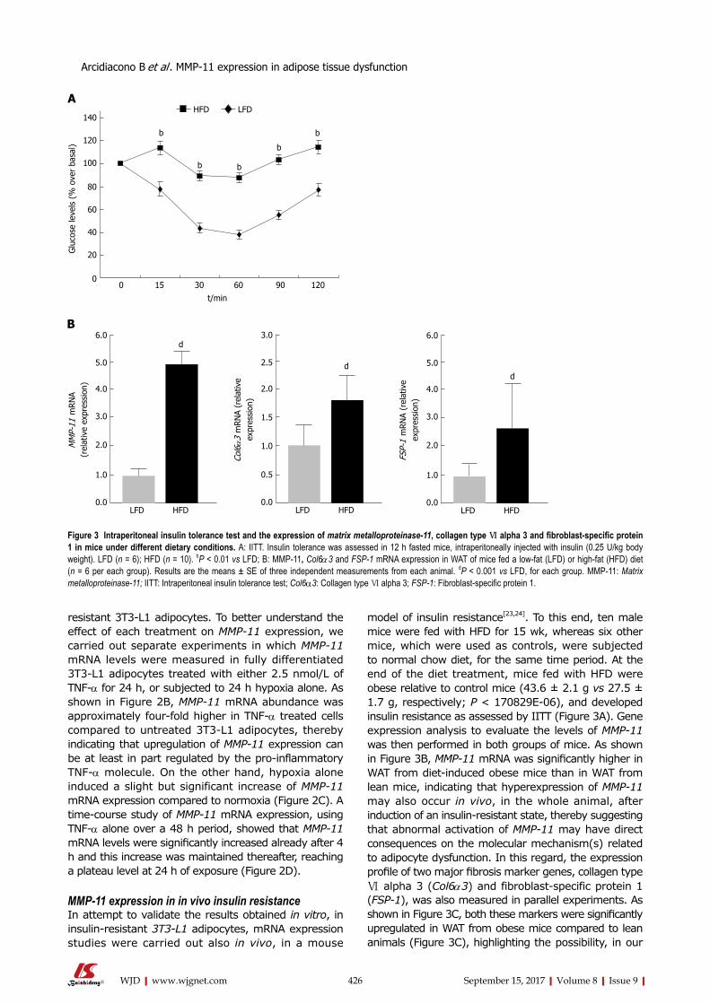
model of insulin resistance[23,24]. To this end, ten male mice were fed with HFD for 15 wk, whereas six other mice, which were used as controls, were subjected to normal chow diet, for the same time period. At the end of the diet treatment, mice fed with HFD were obese relative to control mice (43.6 ± 2.1 g vs 27.5 ± 1.7 g, respectively; P < 170829E-06), and developed insulin resistance as assessed by IITT (Figure 3A). Gene expression analysis to evaluate the levels of MMP-11 was then performed in both groups of mice. As shown in Figure 3B, MMP-11 mRNA was significantly higher in WAT from diet-induced obese mice than in WAT from lean mice, indicating that hyperexpression of MMP-11 may also occur in vivo, in the whole animal, after induction of an insulin-resistant state, thereby suggesting that abnormal activation of MMP-11 may have direct consequences on the molecular mechanism(s) related to adipocyte dysfunction. In this regard, the expression profile of two major fibrosis marker genes, collagen type Ⅵ alpha 3 (Col6α3) and fibroblast-specific protein 1 (FSP-1), was also measured in parallel experiments. As shown in Figure 3C, both these markers were significantly upregulated in WAT from obese mice compared to lean animals (Figure 3C), highlighting the possibility, in our
Glu
cose
leve
ls (
% o
ver
basa
l)
140
120
100
80
60
40
20
00 15 30 60 90 120
t/min
HFD LFD
b
b b
b
b
MM
P-11
mRN
A (r
elat
ive
expr
essi
on)
6.0
5.0
4.0
3.0
2.0
1.0
0.0LFD HFD
d
Col6α3
mRN
A (r
elat
ive
expr
essi
on)
3.0
2.5
2.0
1.5
1.0
0.5
0.0LFD HFD
d
FSP-
1 m
RNA
(rel
ativ
e ex
pres
sion
)
6.0
5.0
4.0
3.0
2.0
1.0
0.0LFD HFD
d
A
B
Figure 3 Intraperitoneal insulin tolerance test and the expression of matrix metalloproteinase-11, collagen type Ⅵ alpha 3 and fibroblast-specific protein 1 in mice under different dietary conditions. A: IITT. Insulin tolerance was assessed in 12 h fasted mice, intraperitoneally injected with insulin (0.25 U/kg body weight). LFD (n = 6); HFD (n = 10). bP < 0.01 vs LFD; B: MMP-11, Col6α 3 and FSP-1 mRNA expression in WAT of mice fed a low-fat (LFD) or high-fat (HFD) diet (n = 6 per each group). Results are the means ± SE of three independent measurements from each animal. dP < 0.001 vs LFD, for each group. MMP-11: Matrix metalloproteinase-11; IITT: Intraperitoneal insulin tolerance test; Col6α3: Collagen type Ⅵ alpha 3; FSP-1: Fibroblast-specific protein 1.
Arcidiacono B et al . MMP-11 expression in adipose tissue dysfunction

427 September 15, 2017|Volume 8|Issue 9|WJD|www.wjgnet.com
obese mouse model, for an ECM dysregulation that would support the hypothesis that this ECM remodeling could indeed exert an adverse effect on adipocyte functions.
DISCUSSIONAdipose tissue is surrounded by ECM elements that provide the right support for adipocyte cell growth and expansion, and maintenance of adipocyte specific functions. Alterations in the organization and flexibility of the ECM as a cause of adipose tissue dysfunction have been reported[25], together with the observation that several MMPs could be involved in these adverse events[26].
In the present work, we focused our attention on the MMP-11 and its activation in conditions of insulin resistance, either in vitro, in 3T3-L1 mouse adipocytes, or in vivo, in obese mice. For the first time, in the present study, we show that MMP-11 was upregulated both in insulin resistant cells treated with TNF-α and/or hypoxia (two elements frequently associated with obesity), and in adipose tissues from insulin-resistant obese mice, suggesting that a direct link may exist between activation of MMP-11 and adipocyte cell dysfunction. Our data are consistent with previous observations that adipokines and hypoxia can alter the expression of MMPs. In this regard, it has been shown that TNF-α upregulated MMP-9 expression in the osteoblast-like MC3T3-E1 cell line[27], while in another study it was found that MMP-2 expression increased in response to hypoxia[28]. Furthermore, an involvement of both MMP-2 and MMP-11 in ECM degradation and collagen accumulation, associated with adipocyte dysfunction, was reported previously[29]. It can be hypothesized that upregulation of MMP-11 in insulin resistance may reflect the increase of nuclear proinflammatory transcription factor(s) whose effective role needs to be investigated.
Fibrosis is considered a new hallmark of the patho-logical dysfunction of WAT[25]. In our study, it is also interesting to note the alteration in the expression of genes related to fibrosis (Col6α3 and FSP-1) in WAT from nutritionally induced obese mice. A link between Col6α3 and MMP-11 has been reported before[29]. Thus, our data in this context well support previous reports that overexpression of MMPs, via degradation of ECM, could be implicated in adipose tissue remodeling[25], and this can play a role in the pathological dysfunction of adipose tissue, which leads to insulin resistance.
Our results appear to challenge findings obtained by studying the MMP-11 knock-in transgenic mouse[30], in which protection from diet-induced obesity was reported, together with a condition of enhanced glucose tolerance and insulin sensitivity due to increased IGF-I bioactivity[30]. The explanation for these divergent results may reside in the fact that overexpression of active MMP-11 in the skin of the transgenic animal may not necessarily reflect the situation in vitro, in 3T3-L1 adipocytes and in vivo, in WAT from diet-induced obese
mice. On the other hand, the existence of compensatory mechanisms/changes that may contribute to counteract genetic manipulation has been proposed[31-35].
Overall, although further studies are still necessary to clarify the role of MMP-11 in insulin resistance, we believe our findings may contribute to shed light on the early process of adipose tissue dysfunction commonly associated with obesity and obesity-related insulin resistance.
COMMENTSBackgroundInsulin resistance is a common metabolic disorder, in which peripheral target tissues fail to respond adequately to insulin, thereby predisposing to type 2 diabetes and other dysmetabolic conditions. More recent discoveries have now strengthened the hypothesis that adipose tissue dysfunction could be the primum movens in the development of insulin resistance. Therefore, studies have been focused on exploring the molecular mechanism(s) underlying adipocyte dysfunction.
Research frontiersMatrix metalloproteinases (MMPs) are a class of endopeptidases that contribute to the degradation of the extracellular matrix components. It has been discovered that they are involved in adipogenesis and remodelling of adipose tissue. A better understanding of the role and function of MMPs in adipose tissue will open new frontiers of investigations.
Innovations and breakthroughsFor the first time, the authors demonstrate that overexpression of MMP-11 occurs in in vitro and in vivo models of insulin resistance.
ApplicationsThis study suggests that MMP-11 could be involved in the early stage of obesity-related insulin resistance. Thus, as a secreted serum protein, MMP-11 could serve as an early biomarker of adipose tissue dysfunction. Research in this area will lead to advancement in understanding the pathophysiology of insulin resistance, as well as advancement in drug development and therapy.
Peer-reviewThe paper is straight forward, well written, and it adds novel information on the topic.
REFERENCES1 Kahn BB, Flier JS. Obesity and insulin resistance. J Clin Invest
2000; 106: 473481 [PMID: 10953022 DOI: 10.1172/JCI10842]2 Chiefari E, Tanyolaç S, Iiritano S, Sciacqua A, Capula C,
Arcidiacono B, Nocera A, Possidente K, Baudi F, Ventura V, Brunetti G, Brunetti FS, Vero R, Maio R, Greco M, Pavia M, Hodoglugil U, Durlach V, Pullinger CR, Goldfine ID, Perticone F, Foti D, Brunetti A. A polymorphism of HMGA1 is associated with increased risk of metabolic syndrome and related components. Sci Rep 2013; 3: 1491 [PMID: 23512162 DOI: 10.1038/srep01491]
3 Saltiel AR. New perspectives into the molecular pathogenesis and treatment of type 2 diabetes. Cell 2001; 104: 517529 [PMID: 11239409]
4 Chiefari E, Iiritano S, Paonessa F, Le Pera I, Arcidiacono B, Filocamo M, Foti D, Liebhaber SA, Brunetti A. Pseudogenemediated posttranscriptional silencing of HMGA1 can result in insulin resistance and type 2 diabetes. Nat Commun 2010; 1: 40 [PMID: 20975707 DOI: 10.1038/ncomms1040]
5 Foti D, Chiefari E, Fedele M, Iuliano R, Brunetti L, Paonessa F, Manfioletti G, Barbetti F, Brunetti A, Croce CM, Fusco A, Brunetti A. Lack of the architectural factor HMGA1 causes insulin resistance and diabetes in humans and mice. Nat Med 2005; 11: 765773 [PMID: 15924147 DOI: 10.1038/nm1254]
Arcidiacono B et al . MMP-11 expression in adipose tissue dysfunction
COMMENTS
428 September 15, 2017|Volume 8|Issue 9|WJD|www.wjgnet.com
6 Chiefari E, Nevolo MT, Arcidiacono B, Maurizio E, Nocera A, Iiritano S, Sgarra R, Possidente K, Palmieri C, Paonessa F, Brunetti G, Manfioletti G, Foti D, Brunetti A. HMGA1 is a novel downstream nuclear target of the insulin receptor signaling pathway. Sci Rep 2012; 2: 251 [PMID: 22355763 DOI: 10.1038/srep00251]
7 Arcidiacono B, Iiritano S, Chiefari E, Brunetti FS, Gu G, Foti DP, Brunetti A. Cooperation between HMGA1, PDX1, and MafA is Essential for GlucoseInduced Insulin Transcription in Pancreatic Beta Cells. Front Endocrinol (Lausanne) 2015; 5: 237 [PMID: 25628604 DOI: 10.3389/fendo.2014.00237]
8 Goossens GH. The role of adipose tissue dysfunction in the pathogenesis of obesityrelated insulin resistance. Physiol Behav 2008; 94: 206218 [PMID: 18037457 DOI: 10.1016/j.physbeh. 2007.10.010]
9 Matziari M, Dive V, Yiotakis A. Matrix metalloproteinase 11 (MMP11; stromelysin3) and synthetic inhibitors. Med Res Rev 2007; 27: 528552 [PMID: 16710861 DOI: 10.1002/med.20066]
10 Rio MC. Stromelysin3, a particular member of the matrix metalloproteinase family. Kluwer Academic Edition. Vol. 4. Dordrecht: Kluwer Academic Publisher, 2002: 81107
11 Motrescu ER, Rio MC. Cancer cells, adipocytes and matrix metalloproteinase 11: a vicious tumor progression cycle. Biol Chem 2008; 389: 10371041 [PMID: 18979628]
12 Yan D, Dai H, Liu JW. Serum levels of MMP11 correlate with clinical outcome in Chinese patients with advanced gastric adenocarcinoma. BMC Cancer 2011; 11: 151 [PMID: 21513571 DOI: 10.1186/14712407111151]
13 Pei D, Majmudar G, Weiss SJ. Hydrolytic inactivation of a breast carcinoma cellderived serpin by human stromelysin3. J Biol Chem 1994; 269: 2584925855 [PMID: 7523394]
14 Mañes S, Mira E, Barbacid MM, Ciprés A, FernándezResa P, Buesa JM, Mérida I, Aracil M, Márquez G, MartínezA C. Identification of insulinlike growth factorbinding protein1 as a potential physiological substrate for human stromelysin3. J Biol Chem 1997; 272: 2570625712 [PMID: 9325295]
15 Messineo S, Laria AE, Arcidiacono B, Chiefari E, Luque Huertas RM, Foti DP, Brunetti A. Cooperation between HMGA1 and HIF1 Contributes to HypoxiaInduced VEGF and Visfatin Gene Expression in 3T3L1 Adipocytes. Front Endocrinol (Lausanne) 2016; 7: 73 [PMID: 27445976 DOI: 10.3389/fendo.2016.00073]
16 Costa V, Foti D, Paonessa F, Chiefari E, Palaia L, Brunetti G, Gulletta E, Fusco A, Brunetti A. The insulin receptor: a new anticancer target for peroxisome proliferatoractivated receptorgamma (PPARgamma) and thiazolidinedionePPARgamma agonists. Endocr Relat Cancer 2008; 15: 325335 [PMID: 18310298 DOI: 10.1677/ERC070226]
17 Lo KA, Labadorf A, Kennedy NJ, Han MS, Yap YS, Matthews B, Xin X, Sun L, Davis RJ, Lodish HF, Fraenkel E. Analysis of in vitro insulinresistance models and their physiological relevance to in vivo dietinduced adipose insulin resistance. Cell Rep 2013; 5: 259270 [PMID: 24095730 DOI: 10.1016/j.celrep.2013.08.039]
18 Bianconcini A, Lupo A, Capone S, Quadro L, Monti M, Zurlo D, Fucci A, Sabatino L, Brunetti A, Chiefari E, Gottesman ME, Blaner WS, Colantuoni V. Transcriptional activity of the murine retinolbinding protein gene is regulated by a multiprotein complex containing HMGA1, p54 nrb/NonO, proteinassociated splicing factor (PSF) and steroidogenic factor 1 (SF1)/liver receptor homologue 1 (LRH1). Int J Biochem Cell Biol 2009; 41: 21892203 [PMID: 19389484 DOI: 10.1016/j.biocel.2009.04.011]
19 Untergasser A, Cutcutache I, Koressaar T, Ye J, Faircloth BC, Remm M, Rozen SG. Primer3new capabilities and interfaces. Nucleic Acids Res 2012; 40: e115 [PMID: 22730293 DOI: 10.1093/nar/gks596]
20 Koressaar T, Remm M. Enhancements and modifications of primer design program Primer3. Bioinformatics 2007; 23: 12891291 [PMID: 17379693 DOI: 10.1093/bioinformatics/btm091]
21 Arnoldo L, Sgarra R, Chiefari E, Iiritano S, Arcidiacono B, Pegoraro S, Pellarin I, Brunetti A, Manfioletti G. A novel mechanism of posttranslational modulation of HMGA functions by the histone chaperone nucleophosmin. Sci Rep 2015; 5: 8552 [PMID: 25711412 DOI: 10.1038/srep08552]
22 Foryst-Ludwig A, Hartge M, Clemenz M, Sprang C, Hess K, Marx N, Unger T, Kintscher U. PPARgamma activation attenuates Tlymphocytedependent inflammation of adipose tissue and development of insulin resistance in obese mice. Cardiovasc Diabetol 2010; 9: 64 [PMID: 20955583 DOI: 10.1186/14752840964]
23 Böhm C, Benz V, Clemenz M, Sprang C, Höft B, Kintscher U, ForystLudwig A. Sexual dimorphism in obesitymediated left ventricular hypertrophy. Am J Physiol Heart Circ Physiol 2013; 305: H211H218 [PMID: 23666673 DOI: 10.1152/ajpheart.00593.2012]
24 Lombardo GE, Arcidiacono B, De Rose RF, Lepore SM, Costa N, Montalcini T, Brunetti A, Russo D, De Sarro G, Celano M. Normocaloric Diet Restores Weight Gain and Insulin Sensitivity in Obese Mice. Front Endocrinol (Lausanne) 2016; 7: 49 [PMID: 27303363 DOI: 10.3389/fendo.2016.00049]
25 Sun K, Tordjman J, Clément K, Scherer PE. Fibrosis and adipose tissue dysfunction. Cell Metab 2013; 18: 470477 [PMID: 23954640 DOI: 10.1016/j.cmet.2013.06.016]
26 Lu P, Takai K, Weaver VM, Werb Z. Extracellular matrix degradation and remodeling in development and disease. Cold Spring Harb Perspect Biol 2011; 3: a005058 [PMID: 21917992 DOI: 10.1101/cshperspect.a005058]
27 Tsai CL, Chen WC, Hsieh HL, Chi PL, Hsiao LD, Yang CM. TNF-α induces matrix metalloproteinase9dependent soluble intercellular adhesion molecule1 release via TRAF2mediated MAPKs and NFκB activation in osteoblast-like MC3T3-E1 cells. J Biomed Sci 2014; 21: 12 [PMID: 24502696 DOI: 10.1186/142301272112]
28 Trayhurn P. Hypoxia and adipocyte physiology: implications for adipose tissue dysfunction in obesity. Annu Rev Nutr 2014; 34: 207236 [PMID: 24819450 DOI: 10.1146/annurevnutr071812161156]
29 Stamenkovic I. Extracellular matrix remodelling: the role of matrix metalloproteinases. J Pathol 2003; 200: 448464 [PMID: 12845612 DOI: 10.1002/path.1400]
30 Motrescu ER, Blaise S, Etique N, Messaddeq N, Chenard MP, Stoll I, Tomasetto C, Rio MC. Matrix metalloproteinase11/stromelysin3 exhibits collagenolytic function against collagen VI under normal and malignant conditions. Oncogene 2008; 27: 63476355 [PMID: 18622425 DOI: 10.1038/onc.2008.218]
31 Dali-Youcef N, Hnia K, Blaise S, Messaddeq N, Blanc S, Postic C, Valet P, Tomasetto C, Rio MC. Matrix metalloproteinase 11 protects from diabesity and promotes metabolic switch. Sci Rep 2016; 6: 25140 [PMID: 27126782 DOI: 10.1038/srep25140]
32 Speakman J, Hambly C, Mitchell S, Król E. The contribution of animal models to the study of obesity. Lab Anim 2008; 42: 413432 [PMID: 18782824 DOI: 10.1258/la.2007.006067]
33 Chiefari E, Paonessa F, Iiritano S, Le Pera I, Palmieri D, Brunetti G, Lupo A, Colantuoni V, Foti D, Gulletta E, De Sarro G, Fusco A, Brunetti A. The cAMPHMGA1RBP4 system: a novel biochemical pathway for modulating glucose homeostasis. BMC Biol 2009; 7: 24 [PMID: 19460132 DOI: 10.1186/17417007724]
34 Iiritano S, Chiefari E, Ventura V, Arcidiacono B, Possidente K, Nocera A, Nevolo MT, Fedele M, Greco A, Greco M, Brunetti G, Fusco A, Foti D, Brunetti A. The HMGA1IGFI/IGFBP system: a novel pathway for modulating glucose uptake. Mol Endocrinol 2012; 26: 15781589 [PMID: 22745191 DOI: 10.1210/me.20111379]
35 Pullinger CR, Goldfine ID, Tanyolaç S, Movsesyan I, Faynboym M, Durlach V, Chiefari E, Foti DP, Frost PH, Malloy MJ, Brunetti A, Kane JP. Evidence that an HMGA1 gene variant associates with type 2 diabetes, body mass Index, and highdensity lipoprotein cholesterol in a HispanicAmerican population. Metab Syndr Relat Disord 2014; 12: 2530 [PMID: 24148075 DOI: 10.1089/met.2013.0086]
P- Reviewer: Beltowski J, Roncucci L S- Editor: Ji FF L- Editor: A E- Editor: Li D
Arcidiacono B et al . MMP-11 expression in adipose tissue dysfunction
Emmanuel Ameyaw, Serwah B Asafo-Agyei, Sumithira Thavapalan, Angela C Middlehurst, Graham D Ogle
ORIGINAL ARTICLE
429 September 15, 2017|Volume 8|Issue 9|WJD|www.wjgnet.com
Clinical profile of diabetes at diagnosis among children and adolescents at an endocrine clinic in Ghana
Retrospective Cohort Study
Emmanuel Ameyaw, Serwah B Asafo-Agyei, Department of Child Health, Komfo Anokye Teaching Hospital, P.O. Box 1934, Kumasi, Ghana
Sumithira Thavapalan, Angela C Middlehurst, Graham D Ogle, International Diabetes Federation Life for a Child Program, Glebe, NSW 2037, Australia
Sumithira Thavapalan, Angela C Middlehurst, Graham D Ogle, Diabetes NSW, Glebe, NSW 2037, Australia
Author contributions: Ameyaw E contributed to study design, conducted the study, and contributed to the manuscript; Asafo-Agyei SB contributed to concept and assisted in conduct of the study; Thavapalan S analysed the data, prepared the figures, and reviewed the manuscript; Middlehurst AC assisted with study design and review of data; Ogle GD designed the study, interpreted the results, and was the lead writer on the manuscript.
Institutional review board statement: The study was reviewed and approved by the Committee on Human Research Publication and Ethics, School of Medical Sciences/Komfo Anokye Teaching Hospital, College of Health Sciences, Kwame Nkrumah Univer-sity of Science and Technology.
Informed consent statement: All subjects gave informed consent.
Conflict-of-interest statement: None of the authors have any conflicts of interest in regards to this study.
Data sharing statement: Not relevant.
Open-Access: This article is an open-access article which was selected by an in-house editor and fully peer-reviewed by external reviewers. It is distributed in accordance with the Creative Commons Attribution Non Commercial (CC BY-NC 4.0) license, which permits others to distribute, remix, adapt, build upon this work non-commercially, and license their derivative works on different terms, provided the original work is properly cited and the use is non-commercial. See: http://creativecommons.org/licenses/by-nc/4.0/
Manuscript source: Invited manuscript
Correspondence to: Dr.Graham D Ogle, International Diabetes Federation Life for a Child Program, 26 Arundel St., Glebe, NSW 2037, Australia. [email protected]: +61-95-529922
Received: January 25, 2017 Peer-review started: January 28, 2017 First decision: May 11, 2017Revised: May 31, 2017 Accepted: June 19, 2017Article in press: June 20, 2017Published online: September 15, 2017
AbstractAIMTo determine the clinical features of diabetes in children and adolescents in Ghana.
METHODSRetrospective review of clinical features of all children and adolescents with new-onset diabetes seen at the paediatric endocrinology clinic of Komfo Anokye Teaching Hospital in Kumasi, from February 2012 to Auguest 2016.
RESULTSOne hundred and six subjects presented with diabetes. Ninety (84.9%) were diagnosed by clinical features and family history as type 1, and 16 (15.1%) type 2. For type 1 subjects, age range at diagnosis was 0.9-19.9 year (y), peak age of onset 12-13 year, and 3.3% were < 5 year, 21.1% 5- < 10 year, 45.6% 10- < 15 year and 30.0% 15- < 20 year. Seventy-one point one percent were female. Common clinical features were polyuria (100%), polydipsia (98.9%), and weight loss (82.2%). Mean BMI SD was -0.54, range -3.84 to 2.47. 60.0% presented in diabetic ketoacidosis (DKA). Nine had infections at onset (skin, abscess, leg ulcer). Mean
Submit a Manuscript: http://www.f6publishing.com
DOI: 10.4239/wjd.v8.i9.429
World J Diabetes 2017 September 15; 8(9): 429-435
ISSN 1948-9358 (online)
430 September 15, 2017|Volume 8|Issue 9|WJD|www.wjgnet.com
Ameyaw E et al . Diabetes in children and adolescents in Ghana
± SD HbA1c at diagnosis was 12.7% ± 1.9% (115 ± 21 mmol/mol). Four have since died: Hypoglycaemia (2), recurrent DKA (1), osteosarcoma (1). Two other type 1 cases died of DKA at presentation in emergency before being seen by the paediatric endocrinologist. Crude mortality rate including these 2 cases was 32.2/1000 patient years. Type 2 cases were 81% female, age of onset 9-19 year. Mean BMI SD was 1.49, range -0.87 to 2.61. Forty-three point eight percent presented in DKA. All type 2 cases had acanthosis nigricans. Overall, 9.8% did not have home refrigeration, most using clay pot evaporative cooling for insulin storage.
CONCLUSIONType 1 occurs with a female preponderance and high DKA rates. Type 2 also occurs. Typology based on clinical features is difficult. Community and professional awareness is warranted.
Key words: Children; Diabetes; Developing countries; Ghana; Mortality
© The Author(s) 2017. Published by Baishideng Publishing Group Inc. All rights reserved.
Core tip: In this study of 106 consecutive new diagnoses of diabetes in young people < 20 years in a tertiary referral centre in Ghana, type 1 predominated (85%) with the remaining cases clinically diagnosed as type 2. Both types had a female preponderance. Type 1 peak age of onset was 12-13 years. All type 2 subjects had acanthosis nigricans. Most presented in ketoacidosis signifying a lack of awareness of presentation features. Clinic numbers quickly rose due to availability of supplies and expertise. Further typology studies are indicated to further define diabetes type.
Ameyaw E, Asafo-Agyei SB, Thavapalan S, Middlehurst AC, Ogle GD. Clinical profile of diabetes at diagnosis among children and adolescents at an endocrine clinic in Ghana. World J Diabetes 2017; 8(9): 429-435 Available from: URL: http://www.wjgnet.com/1948-9358/full/v8/i9/429.htm DOI: http://dx.doi.org/10.4239/wjd.v8.i9.429
INTRODUCTIONUnderstanding the presentation and types of diabetes in children and youth in any particular country is essential in improving awareness and care. Ghana is a less-resourced country in West Africa. There is no published data on clinical features of young Ghanaians with diabetes and, as with many low-income countries[1], there is little public health sector support and also lack of awareness amongst both health workers and the general society[2]. Insulin is only intermittently available from the government health service, and blood glucose meters and strips and HbA1c testing are not provided
by the Ghana National Health Insurance Scheme. The families must often buy these supplies, often at premium prices[3], which many cannot afford to do[2].
The lack of awareness leads to misdiagnosis and mismanagement. Ketoacidosis is very common at initial presentation in Africa[2,4-6], and can mimic infections and acute medical conditions[7-10].
This study determined the clinical features of children and adolescents presenting with diabetes at the Paediatric Endocrine Clinic, Komfo Anokye Teaching Hospital (KATH) at Kumasi, a tertiary referral centre for northern Ghana. This clinic has been supported since 2012 by the International Diabetes Federation (IDF) Life for a Child Program[11] with provision of insulin, blood glucose meters and strips, insulin syringes, HbA1c testing, education materials, and mentoring.
MATERIALS AND METHODSStudy subjectsA total of 106 subjects were enrolled, all < 20 years of age at diagnosis. They included all subjects being followed at the Paediatric Endocrine Clinic on 24/02/2012 as well as all new diagnoses until 31/08/2016. During this period, two other subjects < 20 years old (both female, aged 12 and 15 years old respectively) presented with diabetic ketoacidosis (DKA) and died in the emergency department. They were not seen by the pediatric endocrinologist or in the clinic, and no further information is available. Therefore, they were included in the mortality rate calculation, but excluded from the remainder of the analysis. The study was approved by the institutional ethics board and subjects gave informed consent.
Demographic data Date of birth and sex was recorded, as well as date of diagnosis.
Clinical parametersDiabetes was diagnosed according to standard World Health Organization criteria[12]. Determination of the type of diabetes was made by the local investigators according to available clinical features and history. Type 1 patients generally had lower body mass index (BMI), more rapid symptom onset, and were more sensitive to insulin. Type 2 patients had higher BMI, acanthosis nigricans, and needed more insulin with time, with insulin requirements falling sharply in those started on metformin. The presence of polyuria, polydipsia, weight loss, malnutrition and ketoacidosis at the time of diagnosis were recorded. Body weight and height were measured by electronic scales and stadiometer respectively with subjects wearing light-weight clothing and without shoes. BMI was then calculated. BMI SD scores were calculated using World Health Organization standards[13,14].
Ketoacidosis was defined by clinical features along
431 September 15, 2017|Volume 8|Issue 9|WJD|www.wjgnet.com
with an elevated blood glucose and ketonuria (blood gas measurements are generally not available). Family history of type 1 diabetes, and history of other medical conditions were also recorded.
Biochemical parametersBlood glucose was measured in a laboratory via venous sample. HbA1c was measured using a Clover analyzer (Infopia, Anyang, South Korea).
Socioeconomic parametersThe following information was collected for each sub-ject: Whether the mother or father was living with the subject, mother’s and father’s educational level, who was the primary caregiver, whether the primary caregiver was literate, time spent travelling to clinic, and average weekly household income. It was also recorded whether the subject was at school, whether diabetes was limiting school attendance, and whether they were in the appropriate grade for age, and how well overall the young person was psychologically coping with their diabetes (rated as poor, average or good). Finally, the method of insulin storage was recorded.
Crude mortality rate was calculated as the total number of deaths divided by the sum of the periods from the commencement of the study, or from the date of diagnosis if they were diagnosed after the study commenced. It is expressed as mortality per 1000 patient years.
Statistical analysis Data and descriptive statistics were managed in Excel. Unpaired t-test and χ 2 tests were done using the Social Science Statistics on-line calculators[14]. Significance was set as < 0.05.
RESULTSOne hundred and six subjects with diabetes were seen at the paediatric endocrine clinic. Ninety (84.9%) were diagnosed by clinical features and family history as type 1, and 16 (15.1%) type 2.
Type 1 subjectsTable 1 shows age of onset and gender of the 90 type 1 subjects, as well as BMI, BMI SD score, presence of DKA at diagnosis, and blood glucose and HbA1c at diagnosis. Figure 1A shows the distribution of age of onset. Three point three percent were < 5 years, 21.1% 5- < 10 years, 45.6% 10- < 15 years and 30.0% 15- < 20 years. Common clinical features at diagnosis were polyuria (100.0%), polydipsia (98.9%), and weight loss (82.2%). Nine (10%) had infections at onset (tinea capitis, abscess, leg ulcer, vaginal candidiasis).
Nine type 1 subjects had a first-degree relative with type 1: Sister (two subjects), brother (three), sister and brother (two), two brothers (one), mother (one), with one other subject having a grandmother with type
1. The number of insulin injections each day was two for 17 (18.9%) subjects, three for 24 (26.7%), five for 47 (52.2%) and unknown for two (2.2%). The type of insulin was pre-mixed for 11 (12.2%) subjects, and short-acting combined with long-acting for 79 (87.8%).
Four of the 106 patients have since died: One from metastatic osteosarcoma (diagnosed well after onset of type 1), two from hypoglycemia at home (2 years after diagnosis), and one from a recurrent episode of DKA (2 years after diagnosis). Two others died in emergency department during treatment of DKA at diagnosis, and were not seen by the paediatric endocrinologist (see Methods). Crude mortality rate for the type 1 patients was six deaths per 186 patient years (i.e., 32.2 deaths per 1000 patient years).
Type 2 subjectsFor the 16 type 2 cases, Table 1 shows age of onset and gender, as well as BMI, BMI SD score, presence of DKA at diagnosis and blood glucose and HbA1c at diagnosis. Figure 1B shows age of onset. Six point three percent were 5- < 10 years, 68.7% 10- < 15 years and 25.0% 15- < 20 years. Common clinical features at diagnosis were polyuria (100.0%), polydipsia (100.0%), and weight loss (93.8%). All type 2 subjects had acanthosis nigricans. None had infections at onset. One had substantial visual loss at diagnosis, of uncertain aetiology. Three subjects had first degree relatives with type 2, and two others had a second-degree relative. Four subjects (25.0%) were treated with metformin only, six (37.5%) with insulin only, five (31.3%) with metformin together with insulin and one (6.3%) also with glibenclamide. No subject with type 2 died.
Increase in clinic numbersFigure 2 shows the rapid increase in clinic numbers in the 4 years from June 2012 to June 2016 - clinic numbers were censused at the end of every half-year.
Socioeconomic factorsThe mother was living with the subject in 83 (78.3%) cases and the father in 78 (73.6%). The mother’s educational level was primary school in 31 (29.2%) cases, high school in 26 (24.5%), tertiary in 10 (9.4%), no schooling in 38 (35.8%) and unknown in 1 (0.9%). The father’s educational level was primary school in 26 (24.5%) cases, high school in 31 (29.2%), tertiary in 23 (21.7%), no schooling in 21 (19.8%) and unknown in 5 (4.7%). The primary caregiver was the mother in 75 (70.8%) cases, father in 15 (14.2%), sister in 3 (2.8%), brother in 2 (1.9%), grandmother in 4 (3.8%), aunt in 6 (5.7%) and self in 1 (0.9%). The primary caregiver was literate in 79 (74.5%) cases. Twenty-four (22.6%) families had to travel long distances (> 2 h travelling time each way) for supplies and review. The average weekly household income was 63 USD and the range was 5-625 USD. Ninety-six (90.6%) subjects were attending school. Diabetes was limiting attendance at
Ameyaw E et al . Diabetes in children and adolescents in Ghana

432 September 15, 2017|Volume 8|Issue 9|WJD|www.wjgnet.com
school for 44 (45.8%) subjects, not limiting attendance for 51 (53.1%) and unknown for 1 (1.0%). In addition, 18 (18.8%) were not in the appropriate grade for their age, 76 (79.2%) were in the appropriate grade, and 2 (2.1%) unknown. Diabetes coping abilities were assessed as poor for 12 (11.3%) subjects, average for 37 (34.9%), good for 55 (51.9%) and unknown for 2 (1.9%). Ninety-five (89.6%) subjects were literate or learning at school, 8 (7.5%) were not literate and 3 (2.8%) unknown. Insulin storage method was a refri-gerator at the family home for 92 subjects (90.2%), for two a refrigerator outside the home (2.0%) and for eight clay pot evaporative cooling (7.8%).
DISCUSSIONThere are very limited published data on diabetes in
young people in Ghana. The International Diabetes Federation Diabetes Atlas estimates an incidence of type 1 diabetes of 2.9 per 100000 children < 15 years per annum and a prevalence of 18.0 per 100000 children < 15 years: An estimated 1800 children in the country[15,16]. This is however based on a small study in Nigeria in 1992[17]. It is possible that the current Ghanaian incidence is different from this estimate, and the prevalence/incidence ratio is likely to be substantially lower as the Atlas estimates do not assume any mortality[16]. In Ghana, it is likely that many children and young adults with diabetes die before they are diagnosed, or die during the first episode of DKA or early in ongoing management. DKA is frequently misdiagnosed at first as another condition - with a legion of alternatives in-cluding pneumonia, gastroenteritis, malaria, typhoid, appendicitis and a number of other conditions[1,7-10]. At a training workshop organised by Ghana Society of Pediatric Endocrinology and Diabetes (GSPED) in August 2016, some participants from district and regional hospitals admitted that most of their patients with DKA die. Indeed, two centres admitted that all such patients have died during management. The rate of DKA at onset in type 1 subjects was high at 60.0%, consistent with rates of 69.8% reported from South Africa[4], 75% from Tanzania[5], and 77.1% from Nigeria[6]. Community and health professional awareness on the presentation of diabetes in young people is warranted given this late presentation and the likely substantial numbers of
Type 1 Type 2 Difference
Number (%) 90 (84.9) 16 (15.1) P < 0.001Male: Female ratio 1:2.5 1:4.3 Not significantAge at diagnosis (range), yr 0.9-19.9 9.0-18.7 -Age at diagnosis (mean ± SD), yr 12.6 ± 3.8 13.6 ± 2.3 Not significantPeak age at diagnosis, yr 12-13 13-14 -Diabetic ketoacidosis at onset (%) 54 (60.0) 7 (43.8) Not significantBMI at onset (mean; range) 18.1; 12.5-34.7 27.8; 17.6-38.2 -BMI SD score at onset (mean; range) -0.54, -3.84-2.47 1.49, -0.87-2.61 P < 0.001HbA1c at diagnosis (mean ± SD) (%) (mmol/mol) 12.7 ± 1.9 (115 ± 21) 12.8 ± 1.5 (116 ± 16) Not significant
Table 1 Characteristics of type 1 and type 2 subjects at diagnosis
BMI: Body mass index; HbA1c: Glycosylated haemoglobin.
No.
of
case
s
16
12
8
4
00 1 2 3 4 5 6 7 8 9 10 11 12 13 14 15 16 17 18 19 20
Age (years)
No.
of
case
s
16
12
8
4
00 1 2 3 4 5 6 7 8 9 10 11 12 13 14 15 16 17 18 19 20
Age (years)
A
B
Figure 1 Age at diagnosis of subjects < 20 years of age with diabetes in Kumasi, Ghana. A: Type 1 diabetes: Age at diagnosis; B: Type 2 diabetes: Age at diagnosis.
No.
of
patie
nts
120
100
80
60
40
20
0
t/yr
Jun-12 Dec-12 Jun-13 Dec-13 Jun-14 Dec-14 Jun-15 Dec-15 Jun-16
Figure 2 Numbers of patients with diabetes being seen at the paediatric endocrine clinic in Kumasi, Ghana.
Ameyaw E et al . Diabetes in children and adolescents in Ghana

433 September 15, 2017|Volume 8|Issue 9|WJD|www.wjgnet.com
deaths where the correct diagnosis is not made at all. Type 1 patients were generally lean or underweight at diagnosis, and presented with classic symptoms. There was a female preponderance as is often observed in low-incidence countries[18].
There was also a female preponderance in the type 2 population, consistent with data in adults in Kumasi[19]. Type 2 subjects were often overweight. This is of concern, as overweight is now not uncommon in Ghanaian children and youth[20,21]. All had acanthosis nigricans - a physical marker suggestive of insulin resistance[22]. Interestingly, seven of 16 type 2 subjects presented in DKA, suggesting a diagnosis of ketosis-prone type 2 diabetes, which is well-reported in popu-lations in Africa and of African descent[23,24].
The youngest child was 10 mo of age at diagnosis. Development of diabetes at a young age can indicate a monogenic cause, and genetic testing is indicated if the onset is < 6 mo of age or if there are syndromal features of known single-gene defects (which were not present in the infant in this series)[25]. In some of these cases, alternate non-insulin therapy may be possible[25].
Even with accurate diagnosis, mortality has been high in studies in sub-Saharan Africa[26-28], but there are indications it is falling - for instance in Rwanda it was found to be between 13.9-40.2 per 1000 patient years, depending on the fate of those lost to follow-up[29]. The figure of 32/1000 from this study is in this range - and in Rwanda like in Ghana, care is improving as supplies are made available[30]. This improvement in survival is seen in the dramatic increase in the clinic population from 23 to 102 cases over the five years - “if you build it they will come” - and not just come but survive and thrive. Such rapid increases in numbers in clinics that are able to provide standard care (also seen in Tanzania[31]) indicate the strain that will be on resources as survival improves as insulin and other critical supplies are provided by programs such as IDF Life for a Child, and paediatric endocrinologists, trained in Kenya[32] and elsewhere, return to their home countries to establish clinics.
Patient education is critical in improving care. At this study clinic, all patients are called on the telephone to come in for education every fortnight. They are taught at an appropriate educational level about the pathophysiology of diabetes, how to appropriately store and administer insulin, injection sites, adjust doses, manage diet and exercise, detect acute complications, etc.
The study results demonstrate the socio-economic challenges faced by many subjects, and the necessity for support with supplies, consistent with past reports[1-3,28]. A number of young people were also facing challenges with continuing their education, as demonstrated in the study by Kratzer[2].
Some families do not have access to home refri-geration for insulin storage, and so place the insulin in a clay pot using evaporative cooling. Such methods do substantially reduce storage temperatures unless
humidity is very high[33].
LimitationsThe major limitation of this study is the lack of ability and resources to measure autoantibodies and C-peptide to confirm the diagnosis of type 1 or type 2, or an atypical form. Such assistance with typology would not only be interesting scientifically, but would be helpful to individualise management. However, the presence or absence of autoantibodies alone may not be categorical in this population. Agyei-Frempong et al[34] in a study of autoimmunity in a population of adults with diabetes in Kumasi found that glutamic acid decarboxylase (GAD) antibody and/or insulinoma antibody (IA2) were present in 35% of those on insulin and 16.5% of those not requiring insulin.
Summary and recommendationsIn summary, both type 1 and type 2 diabetes occur in young people in northern Ghana, with high rates of DKA at onset, and a female preponderance. Deaths in the first few years are still not uncommon. Community and health professional awareness is indicated to achieve prompt and accurate diagnosis and prevent deaths at onset. Although not assessed in this study, it is reasonable to conclude that further health professional and patient education is needed to continue to improve management, and therefore reduce the risk of long-term complications. Improvements in the availability of diagnostic technology (particularly blood glucose meters and strips) is also indicated. A patient support group would also be very beneficial.
ACKNOWLEDGMENTSWe thank the staff at the statistics department of KATH and Jean-Pierre Chanoine for helpful comments on the manuscript, and Ms Jane Aquaye for data entry.
COMMENTSBackgroundLimited information is available on types of diabetes in young people in Africa, nor on prognosis.
Research frontiersThe epidemiology and prognosis of diabetes in young people in sub-Saharan Africa is of importance as services are developed to look after these young people.
Innovations and breakthroughsThis study shows that both type 1 and type 2 diabetes are occurring in young people in Ghana, with some phenotypic overlap. Mortality was found to be 32.2 per 1000 patient years.
ApplicationsThe study shows how numbers of children and young people in a clinic in a less-resourced country quickly grow as care is given in a paediatric endocrine clinic.
COMMENTS
Ameyaw E et al . Diabetes in children and adolescents in Ghana

434 September 15, 2017|Volume 8|Issue 9|WJD|www.wjgnet.com
Peer-reviewThis study offers a valuable insight in the clinical profile of diabetes in population of children and adolescents in Ghana. The subject is interesting and worth investigating, since the data regarding diabetes burden in Africa are still scarce and the study population is particularly vulnerable.
REFERENCES1 Ogle GD, Middlehurst AC, Silink M. The IDF Life for a Child
Program Index of diabetes care for children and youth. Pediatr Diabetes 2016; 17: 374-384 [PMID: 26153340 DOI: 10.1111/pedi.12296]
2 Kratzer J. Structural barriers to coping with type 1 diabetes mellitus in Ghana: experiences of diabetic youth and their families. Ghana Med J 2012; 46: 39-45 [PMID: 23661816]
3 Ogle GD, Kim H, Middlehurst AC, Silink M, Jenkins AJ. Financial costs for families of children with Type 1 diabetes in lower-income countries. Diabet Med 2016; 33: 820-826 [PMID: 26482333 DOI: 10.1111/dme.12997]
4 Reddy Y, Ganie Y, Pillay K. Characteristics of children presenting with newly diagnosed type 1 diabetes. S Afr J Child Health 2013; 7: 46-48 [DOI: 10.7196/SAJCH.500]
5 Majaliwa ES, Munubhi E, Ramaiya K, Mpembeni R, Sanyiwa A, Mohn A, Chiarelli F. Survey on acute and chronic complications in children and adolescents with type 1 diabetes at Muhimbili National Hospital in Dar es Salaam, Tanzania. Diabetes Care 2007; 30: 2187-2192 [PMID: 17563337 DOI: 10.2337/dc07-0594]
6 Onyiriuka AN, Ifebi E. Ketoacidosis at diagnosis of type 1 diabetes in children and adolescents: frequency and clinical chara-cteristics. J Diabetes Metab Disord 2013; 12: 47 [PMID: 24355514 DOI: 10.1186/2251-6581-12-47]
7 Rwiza HT, Swai AB, McLarty DG. Failure to diagnose diabetic ketoacidosis in Tanzania. Diabet Med 1986; 3: 181-183 [PMID: 2951164 DOI: 10.1111/j.1464-5491.1986.tb00738.x]
8 Durai R, Hoque H, Ng P. The Acute Abdomen - Commonly missed and mis-diagnosed conditions: Review. Webmed Central Surgery 2010; 1: 1-14
9 Murunga AN, Owira PM. Diabetic ketoacidosis: an overlooked child killer in sub-Saharan Africa? Trop Med Int Health 2013; 18: 1357-1364 [PMID: 24112393 DOI: 10.1111/tmi.12195]
10 Makani J, Matuja W, Liyombo E, Snow RW, Marsh K, Warrell DA. Admission diagnosis of cerebral malaria in adults in an endemic area of Tanzania: implications and clinical description. QJM 2003; 96: 355-362 [PMID: 12702784 DOI: 10.1093/qjmed/hcg059]
11 International Diabetes Federation Life for a Child Program. [accessed 2017 Jan 21]. Available from: URL: https://www.lifeforachild.org/
12 WHO child growth standards: length/height-for-age, weight-for-age, weight-for-length, weight-for-height and body mass index-for-age: methods and development. Geneva: WHO, 2006
13 de Onis M, Onyango AW, Borghi E, Siyam A, Nishida C, Siekmann J. Development of a WHO growth reference for school-aged children and adolescents. Bull World Health Organ 2007; 85: 660-667 [PMID: 18026621 DOI: 10.2471/BLT.07.043497]
14 Social Science Statistics. Statistical Calculators. [accessed 2017 May 20]. Available from: URL: http://www.socscistatistics.com
15 International Diabetes Federation. IDF Diabetes Atlas, 7th ed. Brussels, Belgium, 2015
16 Patterson C, Guariguata L, Dahlquist G, Soltész G, Ogle G, Silink M. Diabetes in the young - a global view and worldwide estimates of numbers of children with type 1 diabetes. Diabetes Res Clin Pract 2014; 103: 161-175 [PMID: 24331235 DOI: 10.1016/j.diabres.2013.11.005]
17 Afoke AO, Ejeh NM, Nwonu EN, Okafor CO, Udeh NJ, Ludvigsson J. Prevalence and clinical picture of IDDM in Nigerian Igbo schoolchildren. Diabetes Care 1992; 15: 1310-1312 [PMID: 1425094 DOI: 10.2337/diacare.15.10.1310]
18 Karvonen M, Pitkäniemi M, Pitkäniemi J, Kohtamäki K, Tajima N,
Tuomilehto J. Sex difference in the incidence of insulin-dependent diabetes mellitus: an analysis of the recent epidemiological data. World Health Organization DIAMOND Project Group. Diabetes Metab Rev 1997; 13: 275-291 [PMID: 9509279 DOI: 10.1002/(SICI)1099-0895(199712)13]
19 Danquah I, Bedu-Addo G, Terpe KJ, Micah F, Amoako YA, Awuku YA, Dietz E, van der Giet M, Spranger J, Mockenhaupt FP. Diabetes mellitus type 2 in urban Ghana: characteristics and associated factors. BMC Public Health 2012; 12: 210 [PMID: 22429713 DOI: 10.1186/1471-2458-12-210]
20 Kumah DB, Akuffo KO, Abaka-Cann JE, Affram DE, Osae EA. Prevalence of Overweight and Obesity among Students in the Kumasi Metropolis. J Nutr Metab 2015; 2015: 613207 [PMID: 25763282 DOI: 10.1155/2015/613207]
21 Mohammed H, Vuvor F. Prevalence of childhood overweight/obesity in basic school in Accra. Ghana Med J 2012; 46: 124-127 [PMID: 23661824]
22 Guran T, Turan S, Akcay T, Bereket A. Significance of acanthosis nigricans in childhood obesity. J Paediatr Child Health 2008; 44: 338-341 [PMID: 18476925 DOI: 10.1111/j.1440-1754.2007.01272.x]
23 Smiley D, Chandra P, Umpierrez GE. Update on diagnosis, pathogenesis and management of ketosis-prone Type 2 diabetes mellitus. Diabetes Manag (Lond) 2011; 1: 589-600 [PMID: 22611441 DOI: 10.2217/DMT.11.57]
24 Lontchi-Yimagou E, Nguewa JL, Assah F, Noubiap JJ, Boudou P, Djahmeni E, Balti EV, Atogho-Tiedeu B, Gautier JF, Mbanya JC, Sobngwi E. Ketosis-prone atypical diabetes in Cameroonian people with hyperglycaemic crisis: frequency, clinical and metabolic phenotypes. Diabet Med 2017; 34: 426-431 [PMID: 27657549 DOI: 10.1111/dme.13264]
25 Rubio-Cabezas O, Hattersley AT, Njølstad PR, Mlynarski W, Ellard S, White N, Chi DV, Craig ME; International Society for Pediatric and Adolescent Diabetes. ISPAD Clinical Practice Consensus Guidelines 2014. The diagnosis and management of monogenic diabetes in children and adolescents. Pediatr Diabetes 2014; 15 Suppl 20: 47-64 [PMID: 25182307 DOI: 10.1111/pedi.12192]
26 Sidibé AT, Traoré HA, Litman-Ali IT, Dembélé M, Traoré AK, Cissé I. Le diabète juvénile au Mali. Rev Franç Endocrinol Clin 1999; 40: 514-520
27 Beran D, Yudkin JS, de Courten M. Access to care for patients with insulin-requiring diabetes in developing countries: case studies of Mozambique and Zambia. Diabetes Care 2005; 28: 2136-2140 [PMID: 16123479 DOI: 10.2337/diacare.28.9.2136]
28 Piloya-Were T, Sunni M, Ogle GD, Moran A. Childhood diabetes in Africa. Curr Opin Endocrinol Diabetes Obes 2016; 23: 306-311 [PMID: 27228228 DOI: 10.1097/MED.0000000000000262]
29 Marshall SL, Edidin D, Arena VC, Becker DJ, Bunker CH, Gishoma C, Gishoma F, LaPorte RE, Kaberuka V, Ogle G, Rubanzana W, Sibomana L, Orchard TJ. Mortality and Natural Progression of Type 1 Diabetes Patients Enrolled in the Rwanda LFAC Program from 2004-2012. Int J Diabetes Dev Countries 2016 [DOI: 10.1007/s13410-016-0536-z]
30 Marshall SL, Edidin DV, Arena VC, Becker DJ, Bunker CH, Gishoma C, Gishoma F, LaPorte RE, Kaberuka V, Ogle G, Sibomana L, Orchard TJ. Glucose control in Rwandan youth with type 1 diabetes following establishment of systematic, HbA1c based, care and education. Diabetes Res Clin Pract 2015; 107: 113-122 [PMID: 25458328 DOI: 10.1016/j.diabres.2014.09.045]
31 Muze KC, Majaliwa ES. Type 1 diabetes care updates: Tanzania. Indian J Endocrinol Metab 2015; 19: S12-S13 [PMID: 25941637 DOI: 10.4103/2230-8210.155348]
32 Odundo GO, Ngwiri T, Otuoma O, Laigong P, Mukhwana R, Limbe MS, Chanzu NM. The Impact and Successes of a Paediatric Endocrinology Fellowship Program in Africa. Int J Endocrinol 2016; 2016: 1560248 [PMID: 26904118 DOI: 10.1155/2016/1560248]
33 Ogle GD, Abdullah M, Mason D, Januszewski AS, Besançon S. Insulin storage in hot climates without refrigeration: temperature
Ameyaw E et al . Diabetes in children and adolescents in Ghana

435 September 15, 2017|Volume 8|Issue 9|WJD|www.wjgnet.com
reduction efficacy of clay pots and other techniques. Diabet Med 2016; 33: 1544-1553 [PMID: 27472257 DOI: 10.1111/dme.13194]
34 Agyei-Frempong MT, Titty FV, Owiredu WK, Eghan BA. The
prevalence of autoimmune diabetes among diabetes mellitus patients in Kumasi, Ghana. Pak J Biol Sci 2008; 11: 2320-2325 [PMID: 19137864 DOI: 10.3923/pjbs.2008.2320.2325]
P- Reviewer: Alamgir MA, Guerrero-Romero F, Lovrencic MV S- Editor: Ji FF L- Editor: A E- Editor: Li D
Ameyaw E et al . Diabetes in children and adolescents in Ghana
Nadira Bibi Mothojakan, Shazia Hussain, Kieran McCafferty, Mohammed Magdi Yaqoob, Tahseen Ahmad Chowdhury
ORIGINAL ARTICLE
436 September 15, 2017|Volume 8|Issue 9|WJD|www.wjgnet.com
Eye and foot checks in patients with diabetes on haemodialysis: Are they done, and who does them?
Retrospective Study
Nadira Bibi Mothojakan, Shazia Hussain, Tahseen Ahmad Chowdhury, Department of Diabetes and Metabolism, Barts Health NHS Trust, the Royal London Hospital, Whitechapel, London E1 1BB, United Kingdom
Kieran McCafferty, Mohammed Magdi Yaqoob, Department of Nephrology, Barts Health NHS Trust, the Royal London Hospital, Whitechapel, London E1 1BB, United Kingdom
Author contributions: Mothojakan NB and Hussain S undertook the research and data analysis; Mothojakan NB wrote the first draft of the manuscript; McCafferty K and Yaqoob MM instigated the research and reviewed the manuscript; Chowdhury TA reviewed the manuscript and undertook all revisions and is the guarantor.
Institutional review board statement: The study was reviewed and approved by the Barts Health Department of Renal Medicine Clinical Governance Board.
Informed consent statement: All patients involved in the survey gave full verbal consent.
Conflict-of-interest statement: All authors declare no conflicts of interest.
Data sharing statement: All authors agree to data sharing.
Open-Access: This article is an open-access article which was selected by an in-house editor and fully peer-reviewed by external reviewers. It is distributed in accordance with the Creative Commons Attribution Non Commercial (CC BY-NC 4.0) license, which permits others to distribute, remix, adapt, build upon this work non-commercially, and license their derivative works on different terms, provided the original work is properly cited and the use is non-commercial. See: http://creativecommons.org/licenses/by-nc/4.0/
Manuscript source: Invited manuscript
Correspondence to: Tahseen Ahmad Chowdhury, Professor, Consultant in Diabetes, Department of Diabetes and Metabolism, Barts Health NHS Trust, the Royal London Hospital, 7th Floor, John Harrison House, Whitechapel, London E1 1BB,
United Kingdom. [email protected]: +44-20-82238384Fax: +44-20-82238806
Received: January 22, 2017Peer-review started: January 23, 2017First decision: April 14, 2017Revised: May 9, 2017Accepted: May 30, 2017 Article in press: May 31, 2017Published online: September 15, 2017
AbstractAIMTo determine if retinal and foot checks are carried out on patients with diabetes receiving haemodialysis.
METHODSEighty-four patients with diabetes receiving haemo-dialysis were asked if they recalled having eye and foot screening in the last year, and if so, by whom was the check done.
RESULTSSeventy-seven (91.7%) patients recalled having an eye check in the preceding 12 mo. Of these, 52 (67.5%) did so in an ophthalmology clinic, 17 (22%) in retinal screening, three (3.9%) in an optician clinic. Three patients (3.9%) went to both ophthalmology and retinal screening, and two (2.6%) attended an ophthalmology and optician. Seventy (83.3%) patients recalled having a foot check in the preceding 12 mo. Of these, 33 (47.1%) were done by practice nurse, 14 (20%) by a diabetes nurse, 11 (15.7%) by a general practitioner, eight (11.4%) by a chiropodist, and four (5.7%) were each checked by renal nurse, diabetes consultant, junior doctor, or unknown person at a foot clinic.
Submit a Manuscript: http://www.f6publishing.com
DOI: 10.4239/wjd.v8.i9.436
World J Diabetes 2017 September 15; 8(9): 436-439
ISSN 1948-9358 (online)
437 September 15, 2017|Volume 8|Issue 9|WJD|www.wjgnet.com
Mothojakan NB et al . Eye and foot checks on haemodialysis
CONCLUSIONMost patients with diabetes on haemodialysis are able to recall having an eye check in the last year, although 8.3% could not. A significant proportion of patients could not recall having a foot check (16.7%) in the last year. This baseline audit suggests that an improvement in the rate of foot screening is important to achieve in patients with diabetes on haemodialysis in our unit.
Key words: Diabetes; Haemodialysis; Foot screening; Retinal screening
© The Author(s) 2017. Published by Baishideng Publishing Group Inc. All rights reserved.
Core tip: Diabetes is the commonest cause of end stage renal failure in many countries. Patients with diabetes on haemodialysis are at high risk of retinal and foot problems, and need regular screening to ensure they do not develop problems related to these complications. Our survey suggests that most patients are getting eye checks, but a significant number are not getting foot checks. This is an important area for all dialysis units to consider. We recommend that patients have foot screening whilst on dialysis, which may require further training for dialysis nurses.
Mothojakan NB, Hussain S, McCafferty K, Yaqoob MM, Chowdhury TA. Eye and foot checks in patients with diabetes on haemodialysis: Are they done, and who does them? World J Diabetes 2017; 8(9): 436-439 Available from: URL: http://www.wjgnet.com/1948-9358/full/v8/i9/436.htm DOI: http://dx.doi.org/10.4239/wjd.v8.i9.436
INTRODUCTIONDiabetic nephropathy is the leading cause of end-stage renal failure in the United Kingdom[1]. Screening programmes enable detection of early changes associated with microvascular complications of dia-betes, including diabetic retinopathy and peripheral neuropathy. Many national guidelines recommend that all patients with diabetes have yearly screening of feet and eyes to reduce the risk of blindness and avoidable limb amputations[2,3]. With the increasing prevalence of diabetes, attendance at screening programmes is of the utmost importance in order to reduce the risk of complications.
Patients with diabetes who have end stage renal failure (ESRF) on regular haemodialysis attend hospital for dialysis very frequently, and as a result find it difficult to attend other appointments. We have previously noted poor attendance to other clinics and appointments in patients on haemodialysis. East London is an area of high social deprivation, and many patients are elderly, with multiple co-morbidities, whose first language is often not English, and these factors
may affect their ability to access healthcare[4]. Patients with diabetes on haemodialysis are at particular risk of foot and eye problems[5]. Microvascular complications of diabetes arise due to poor glycaemic control, and indeed haemodialysis patients with poor glycaemic control have been found to have poorer survival compared to those with good glycaemic control[6].
Recent United Kingdom guidelines highlight the need for annual foot and eye screening for patients with diabetes on haemodialysis[4]. The aim of this study was to determine if patients with diabetes on our haemodialysis unit could recall having retinal screening and foot surveillance in the past year, and to find out who had performed this.
MATERIALS AND METHODSThis retrospective study was carried out on the dialysis unit of the Royal London Hospital, a tertiary centre which serves a large cohort of renal patients in East London, United Kingdom. A brief questionnaire was designed for patients with diabetes receiving dialysis, asking whether patients recalled having “a diabetes eye check” or “diabetes foot check” in the past 12 mo. Patients who had received a diabetes eye check were asked where this had taken place: At an optician, eye clinic, retinal screening service or elsewhere. Patients who had a diabetes foot check where asked who had performed the procedure; a doctor, diabetes nurse, renal nurse or podiatrist.
Participants were recruited to the study from August to September 2015, whilst receiving haemodialysis on the renal unit. Inclusion criteria for the study included: Patient currently receiving haemodialysis, patient was diagnosed with diabetes for at least a year and able to receive care in the community. Patients were excluded from the study if they had communication difficulties.
All statistical analysis and graphs were performed using GraphPad Prism 7 (GraphPad software inc, Cali-fornia, United States) software. Quantitative data were expressed as frequencies or mean ± SD as appropriate. Qualitative data were expressed as frequencies.
RESULTSPatient characteristicsEighty-four patients met the inclusion criteria and agreed to participate in the study. Patient characteristics are shown in Table 1. Sixty point seven percent of the participants were male and 39.3% were female. The mean age of the cohort was 63.9 ± 10.35 years. Insulin only therapy was used by 53.6% of the participants. The remaining participants were diet-controlled (11.9%), on medication only (19%) or medication and insulin (15.5%).
Eye checksFigure 1 shows eye check uptake in the patients

438 September 15, 2017|Volume 8|Issue 9|WJD|www.wjgnet.com
surveyed. Seventy-seven (91.7%) of patients reported having an eye check in the last 12 mo. Of these, 52 (67.5%) did so in an ophthalmology clinic, 17 (22%) in retinal screening, three (3.9%) in an optician, three (3.9%) went to both ophthalmology and retinal screening, and two (2.6%) attended an ophthalmology and optician.
Diabetic foot screeningFigure 2 shows foot check uptake in the patients surveyed. Seventy (83.3%) patients recalled having a foot check in the previous 12 mo. Of these, 33 (47.1%) were carried out by a practice nurse, 14 (20%) by a diabetes specialist nurse, 11 (15.7%) by the general practitioner (GP), eight (11.4%) by a chiropodist, and four (5.7%) had been a renal nurse, a diabetes consultant, a junior doctor, or unknown person at a foot clinic.
DISCUSSIONScreening programmes have an important role in the prevention and early detection of retinopathy and neuropathy. We are unaware of any previous studies investigating the uptake of screening programmes in haemodialysis patients. In the United Kingdom in 2012-2013, 80.2% of patients offered diabetic eye screening attended. Recent recommendations suggest that is should be possible for a minimum of 85% of those offered digital retinal screening to attend. Screening uptake in 2012-2013 was lower than the results of our study, where we found that 91.7% of patients attended an eye check in the previous last year, suggesting that patients with diabetes on haemodialysis are aware of the need to undertake eye checks on a regular basis.
The United Kingdom National Diabetes Audit 2015-2016, found that 86.7% of patients with diabetes had foot surveillance that year[7], which was slightly
higher than in our patient survey (83.3%). This is of some concern, particularly as patients with diabetes on haemodialysis are at high risk of foot problems. Recent guidelines recommend that patients have their feet screened every 3 mo with a locally agreed tool, and by staff on the dialysis unit[4].
In the United Kingdom, co-ordination of screening programmes for eyes and feet are led in the community by primary care health professionals. Retinal screening programmes are locally commissioned within Clinical Commissioning Group, and call and recall is organised by review of primary care records. Most retinal screening occurs via the retinal screening programme, although patients with established significant retinopathy may attend a medical retinal clinic as well. In the present study, it was found that the ophthalmology clinic was the most common place for eye checks, accounting for 67.5% of all patients. This is unsurprising as many patients on haemodialysis also have other microvascular complications such as retinopathy. A small proportion of patients had eye checks carried out by an optician, which, whilst useful, means that such patients may not be accessing a formal retinopathy screening programme. Interestingly, 7.1% of patients had an eye check carried out more than once in the past year, suggesting some duplication.
Foot checks for people with diabetes are generally performed by trained clinical staff in the primary care centre, which is often the practice nurse. Our study confirmed that nurses in the community were the most common group to carry out diabetic foot checks, with 67.1% of foot checks carried out by the nurses in primary care. Very few patients stated that their feet had ever been examined on the dialysis unit during
Variables Patients
Gender Male 51 (60.7) Female 33 (39.3)Age (mean ± SD) 63.9 ± 10.35 Ethnicity African - Caribbean 38 (45.2) Asian - Bangladeshi 22 (26.2) Asian - Indian 4 (4.8) Asian - Pakistani 2 (2.4) Asian - Other 4 (4.8) White - British 9 (10.7) White - Other 2 (2.4) Other 3 (3.6) Treatment regimen Diet only treated 10 (11.9) Oral hypoglycaemic only treated 16 (19.0) Insulin + oral hypoglycaemic treated 13 (15.5) Insulin only treated 45 (53.6)
Table 1 Demographic characteristics of patients surveyed n (%) Opthalmology clinicRetinal screeningOpticianOpthalmology and retinal screeningOpthalmology and opticianNo retinal screening
n = 84
Figure 1 Retinal screening in the cohort.
n = 84
GP practice nurseDiabetes specialist nurseGPChiropodistOtherFeet not examined
Figure 2 Diabetic foot checks in the cohort. GP: General practitioner.
Mothojakan NB et al . Eye and foot checks on haemodialysis
439 September 15, 2017|Volume 8|Issue 9|WJD|www.wjgnet.com
dialysis. Patients on haemodialysis have logistical difficulties that make it difficult for them to attend appointments elsewhere. Perhaps this may account for patients missing screening appointments. A lack of co-ordination between the health care professionals caring for the patients may have also resulted in missed screening opportunities, as it is assumed that they have been carried out elsewhere. Patients spend significant amounts of time on dialysis, and this may provide an excellent opportunity for screening of feet and eyes to be undertaken opportunistically, as well as reducing the need for patients to attend hospital in between dialysis sessions. This is specifically mentioned as an important aim in recent United Kingdom guidelines, and clearly needs to be addressed in our haemodialysis unit[4]. These guidelines recommend that annual checks are documented, and made available to all those involved in the care of these patients.
The introduction of a robust system of documen-tation, would ensure that individuals involved are aware of recent checks and when they last took place, avoiding unnecessary duplication. Furthermore, access to a named link worker on the dialysis unit who would ensure that screening is carried out, which could ensure that patients have received eye and foot screening, and are also educated in looking for early signs of significant foot problems, and highlight these to health professionals at an early stage. Inter-professional learning between diabetes and renal specialists may facilitate improvements in care.
There are some limitations to this study, including a small patient cohort and the fact that it was carried out at a single tertiary centre. Patients with communication difficulties were excluded from the study, and it is possible that this group of patients may have had difficulty accessing healthcare, and may also be more likely to miss screening appointments. The study did not examine the barriers to patients attending screening appointments.
Patients with diabetes on dialysis are at risk of microvascular complications, and due to logistical issues have difficulties attending other appointments. Most patients had an eye check in the last year, with a lower percentage of recalling a foot check in the last year. It is hoped that the introduction of recent guidelines will improve the uptake of screening.
COMMENTSBackgroundPatients with diabetes on haemodialysis are at high risk of diabetes
complications including foot and eye problems. It is not known whether patients with diabetes on haemodialysis attend regular screening appointments for foot and eye checks. This survey aimed to determine this information.
Research frontiersIt is increasingly recognised that prevention of diabetic complications in patients on haemodialysis is important. At the moment, it is unknown whether improving glucose control or other risk factors will reduce morbidity and mortality in such patients.
Innovations and breakthroughsRecent United Kingdom guidelines suggest a more proactive approach to managing patients with diabetes on haemodialysis. It is hoped that with more structured care, better outcomes will be seen.
ApplicationsThe authors show that most patients with diabetes on haemodialysis attend for eye checks, but that foot checks may be neglected. The authors propose that foot checks on dialysis would be an effective way to ensure proactive management of foot problems in patients on dialysis.
Peer-reviewMothojakan et al report the findings of a retrospective study of whether foot and eye screening is being done on diabetic patients undergoing hemodialysis. The paper has been revised in light of a previous review and is well written, easy to follow and without any obvious errors or unfounded claims.
REFERENCES1 Caskey F, Cullen R. UK Renal Registry 18th Annual Report:
Introduction. Nephron 2016; 132 Suppl 1: 1-8 [PMID: 27088327 DOI: 10.1159/000444814]
2 National Institute for Health and Care Excellence. Diabetic foot problems: prevention and management. NICE guideline NG19. Available from: URL: http://www.nice.org.uk/guidance/ng19; Accessed 04.05.17
3 Standards of Medical Care in Diabetes-2017: Summary of Revisions. Diabetes Care 2017; 40: S4-S5 [PMID: 27979887 DOI: 10.2337/dc17-S003]
4 Frankel A, Kazempour-Ardebili S, Bedi R, Chowdhury TA, De P, El-Sherbini N, Game F, Gray S, Hardy D, James J, Kong MF, Ramlan G, Southcott E, Winocour P. Management of adults with diabetes on the haemodialysis unit: summary of new guidance from the Joint British Diabetes Societies and Renal Association. British J Diabetes 2016; 16: 69-77 [DOI: 10.15277/bjd.2016.073]
5 Ndip A, Rutter MK, Vileikyte L, Vardhan A, Asari A, Jameel M, Tahir HA, Lavery LA, Boulton AJ. Dialysis treatment is an independent risk factor for foot ulceration in patients with diabetes and stage 4 or 5 chronic kidney disease. Diabetes Care 2010; 33: 1811-1816 [PMID: 20484126 DOI: 10.2337/dc10-0255]
6 Morioka T, Emoto M, Tabata T, Shoji T, Tahara H, Kishimoto H, Ishimura E, Nishizawa Y. Glycemic control is a predictor of survival for diabetic patients on hemodialysis. Diabetes Care 2001; 24: 909-913 [PMID: 11347753 DOI: 10.2337/diacare.24.5.909]
7 National Diabetes Audit 2015-6. 1. Care processes and treatment targets. Available from: URL: http://www.content.digital.nhs.uk/catalogue/PUB23241/nati-diab-rep1-audi-2015-16.pdf Accessed 04.05.17
P- Reviewer: Ali O, Miller S, Tamemoto H S- Editor: Song XX L- Editor: A E- Editor: Li D
COMMENTS
Mothojakan NB et al . Eye and foot checks on haemodialysis

© 2017 Baishideng Publishing Group Inc. All rights reserved.
Published by Baishideng Publishing Group Inc7901 Stoneridge Drive, Suite 501, Pleasanton, CA 94588, USA
Telephone: +1-925-223-8242Fax: +1-925-223-8243
E-mail: [email protected] Desk: http://www.f6publishing.com/helpdesk
http://www.wjgnet.com