workshop #11 - society of forensic …soft-tox.org/files/annual_meeting/workshops/w11...william...
TRANSCRIPT

WORKSHOP #11
Postmortem Cannabinoids:
Issues of Analysis and Interpretation
SOFT 2016
Dallas, TX
Tuesday, October 18, 2016
Chairs:
Rod McCutcheon and Philip Kemp

Workshop #11 Postmortem Cannabinoids: Issues of Analysis and Interpretation
October 18, 2016
Agenda:
1:30 pm - 1:35 pm Welcome/Introductions Rod McCutcheon
1:35 pm - 2:15 pm Pharmacology of Marijuana Nathalie Desrosiers
2:15 pm - 3:00 pm Analysis of Cannabinoids in Postmortem Specimens Brad Hall
3:00 pm - 3:30 pm Postmortem Distribution/Redistribution of Cannabinoids William Anderson
3:30 pm - 4:00 pm Afternoon Break
4:00 pm - 4:30 pm Interpretation Issues of Cannabinoid Data in Postmortem Cases Nikolas Lemos
4:30 pm - 5:30 pm Panel Discussion/Audience Q & A Speaker Panel

Nathalie A. Desrosiers, Ph.D.
Dr. Desrosiers completed her doctorate in toxicology at the University of Maryland, Baltimore and conducted her research in the Chemistry and Drug Metabolism Section of the National Institute on Drug Abuse (NIDA), National Institutes of Health.
Dr. Desrosiers has a B.Sc. in Forensic Science and Analytical Chemistry and Instrumentation and an M.Sc. in Chemical Sciences from Laurentian University, in Sudbury, Ontario, Canada. Her work focused on the detection of drugs in skeletal remains, including documenting relative drug distribution in skeletal remains, developing a microwave-assisted extraction to pull drugs from bones, and examining the impact of burial on drug concentrations in skeletal remains. At NIDA, she was the lead associate investigator in a study evaluating the pharmacokinetic and pharmacodynamic differences between occasional and frequent cannabis smokers. She was awarded the Society of Forensic Toxicologist’s Education Research Award in 2011 for her research on cannabis and 3,4-methylenedioxymethamphetamine (MDMA, “Ecstasy”). Dr. Desrosiers has presented her work at various national and international conferences and has published 26 peer-reviewed scientific journal articles. She has worked as a forensic toxicologist since 2014 in New York State then in Ontario, Canada.
Dr. Desrosiers is a member multiple professional societies and organizations, including the Society of Forensic Toxicologists (SOFT), SOFT’s Young Forensic Toxicologists (YFT) committee, The International Association of Forensic Toxicologists (TIAFT), and the American Academy of Forensic Sciences (AAFS).

Nathalie Desrosiers, Ph.D.October 18, 2016
Dallas, TX
No conflicts of interest to declare
Any information presented today reflects my own personal scientific opinion
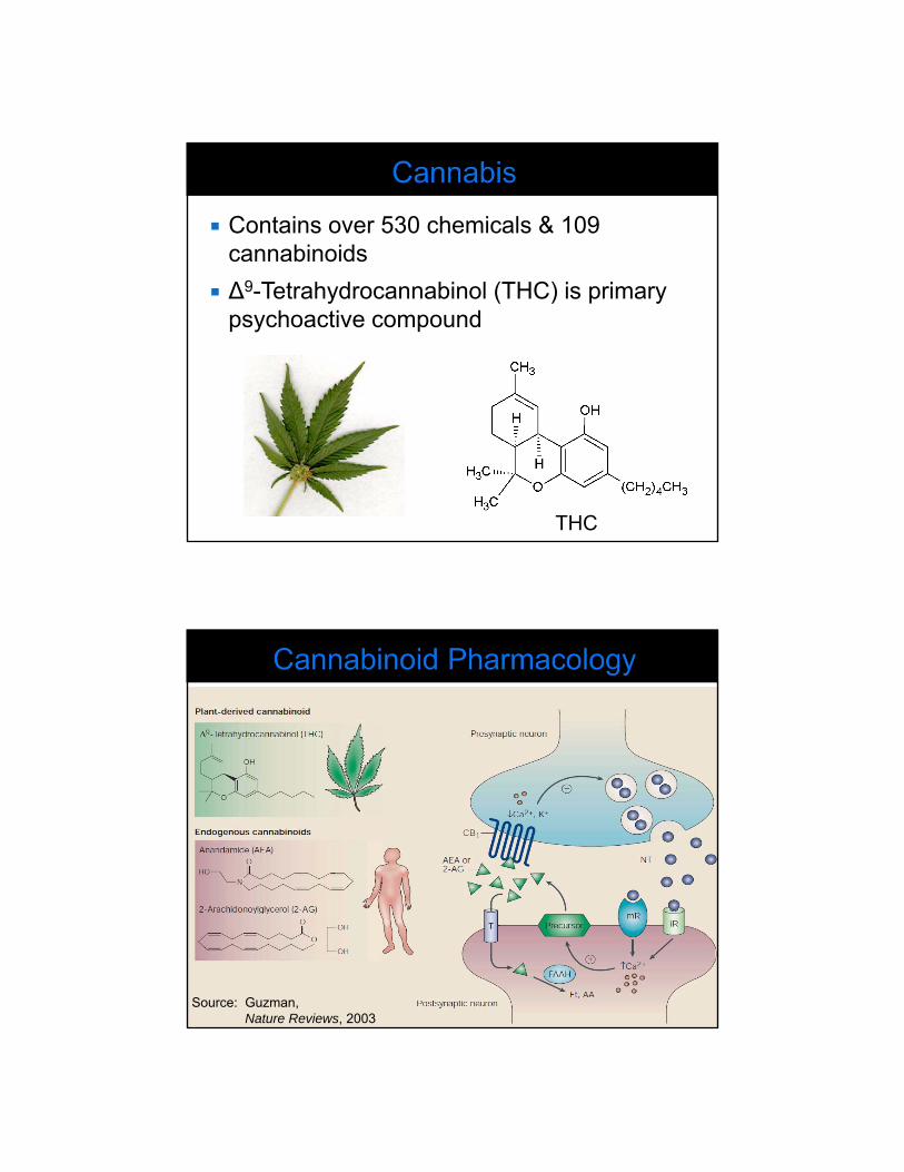
Contains over 530 chemicals & 109 cannabinoids
∆9-Tetrahydrocannabinol (THC) is primary psychoactive compound
THC
Source: Guzman,Nature Reviews, 2003

Two main cannabinoid receptors: CB1 and CB2 CB1 mainly in the brain Basal Ganglia (motor activity) Cerebellum (motor coordination) Hippocampus (short-term memory) Neocortex (thinking, sensory perception, spatial
reasoning) Hypothalamus (appetite, sedation) Periaqueductal gray dorsal horn (pain) Immune cells
CB2 mainly on immune cells, but also in the brain
Subjective feeling of “high”
Euphoria
Sedation
Analgesia
Altered perception
Increased heart rate
Peripheral vasodilation
Motor control impairment
Cognition impairment
Memory impairment

Cannabis impairs psychomotor function, especially in occasional smokers Suggesting some tolerance to psychomotor
impairment in frequent users
More frequent smokers needed to see impairment in frequent smokers (higher power) (Van Wel, Br J Pharmacol, 2013)
Ramaekers, J Psycopharmacology, 2009
Impairment No Impairment
CT
T-La
mbd
a-C
(% O
bser
vatio
ns)
Sto
p R
eact
ion
Tim
e(%
Obs
erva
tions
)
DA
T –
Tra
ckin
ger
ror
(% O
bser
vatio
ns)
DA
T –
Hits
(% O
bser
vatio
ns)
Note: serum concentrations
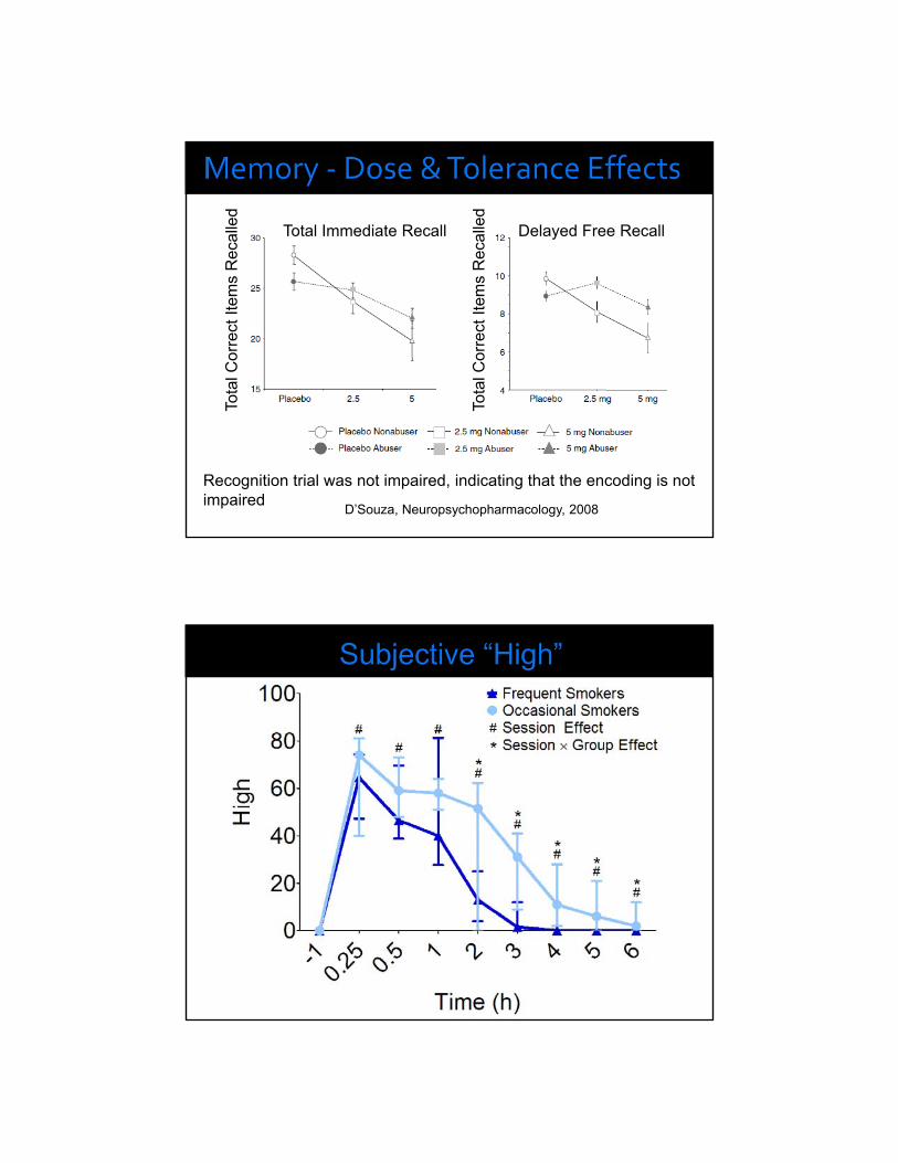
D’Souza, Neuropsychopharmacology, 2008
Total Immediate Recall Delayed Free RecallTo
tal C
orre
ct I
tem
s R
ecal
led
Tota
l Cor
rect
Ite
ms
Rec
alle
dRecognition trial was not impaired, indicating that the encoding is not impaired

CYP 2C9CYP 2C19CYP 2D6
Alcohol dehydrogenase ormicrosomal alcohol oxygenase
& aldehyde oxygenase

CYP 2C9CYP 2C19CYP 2D6
Alcohol dehydrogenase ormicrosomal alcohol oxygenase
& aldehyde oxygenase
UGT 1A9UGT 1A10
UGT 1A9UGT 1A10
UGT 1A3UGT 1A1
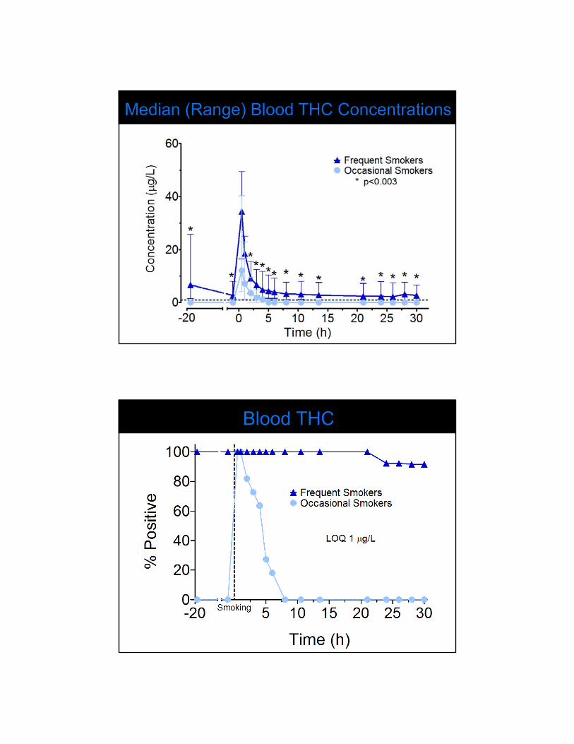
THC (µg/L)
Source: Huestis, 2005
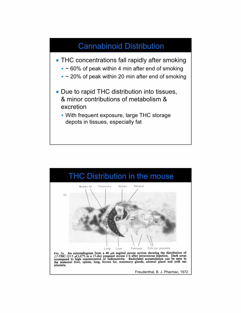
THC concentrations fall rapidly after smoking ~ 60% of peak within 4 min after end of smoking
~ 20% of peak within 20 min after end of smoking
Due to rapid THC distribution into tissues, & minor contributions of metabolism & excretion With frequent exposure, large THC storage
depots in tissues, especially fat
Freudenthal, B. J. Pharmac, 1972

Brunet, Forensic Sci Int, 2006
Blood: Bergamaschi et al. Clin Chem, 2013
THC: up to 30 days, LOQ 0.25 µg/L
11-OH-THC: up to 3 days, LOQ 0.5 µg/L
THCCOOH: at least 33 days, LOQ 0.25 µg/L
Urine: Lowe et al. Drug Alcohol Depend, 2009
THC: up to 24 days, LOQ 2.5 µg/L
11-OH-THC & THCCOOH: at least 24 days,LOQ 2.5 µg/L
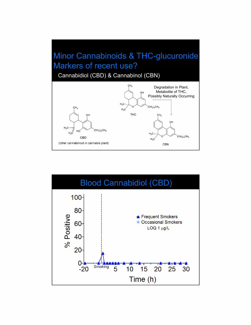
Cannabidiol (CBD) & Cannabinol (CBN)
Degradation in Plant,Metabolite of THC,
Possibly Naturally Occurring

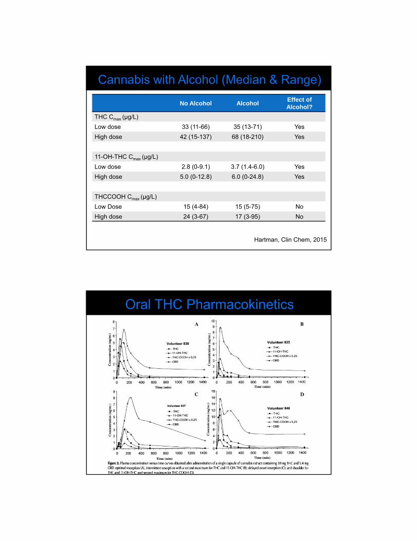
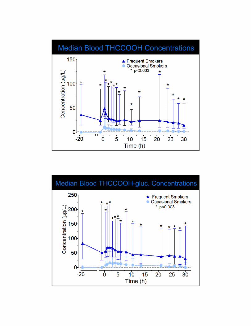
No Alcohol Alcohol Effect of Alcohol?
THC Cmax (µg/L)
Low dose 33 (11-66) 35 (13-71) Yes
High dose 42 (15-137) 68 (18-210) Yes
11-OH-THC Cmax (µg/L)
Low dose 2.8 (0-9.1) 3.7 (1.4-6.0) Yes
High dose 5.0 (0-12.8) 6.0 (0-24.8) Yes
THCCOOH Cmax (µg/L)
Low Dose 15 (4-84) 15 (5-75) No
High dose 24 (3-67) 17 (3-95) No
Hartman, Clin Chem, 2015

THC distributes rapidly smoking
Large differences in concentrations between occasional and frequent smokers
More research needed with high THC or CBD cannabis strains and “newer” routes of administration


Brad J. Hall, Ph.D., F-ABFT Chief Forensic Toxicologist
Travis County Medical Examiner’s Office 1213 Sabine Street Austin, TX 78701
Dr. Brad J. Hall received a B.S. degree in Chemistry in 1994 from Midwestern State University in Wichita Falls, TX, and a Ph.D. in Analytical Chemistry from the University of Texas at Austin, Texas in 1998. His doctoral research focused on analytical applications of quadrupole ion trap mass spectrometry using a variety of ionization techniques including electrospray ionization (ESI) and matrix assisted laser desorption ionization (MALDI). Dr. Hall was awarded an Educational Research Award from the Society of Forensic Toxicologists (SOFT) during his last year in graduate school for application of solid phase microextration and GC/MS towards the analysis of cannabinoids.
Directly after graduate school, Dr. Hall joined the Travis County Office of the Medical Examiner (Austin, TX) toxicology laboratory where he served for approximately six years as a staff Forensic Toxicologist being mentored under J. Rod McCutcheon, BS, F-ABFT. Dr. Hall currently serves as Chief Forensic Toxicologist since 2004. The laboratory is accredited by the American Board of Forensic Toxicology since 2005.
Dr. Hall is a Fellow of the American Board of Forensic Toxicology (F-ABFT), currently serves on the Board of Directors for ABFT, and is an active laboratory inspector for ABFT. He is a member of the Society of Forensic Toxicologists (SOFT), the American Academy of Forensic Sciences (AAFS), The International Association of Forensic Toxicologists (TIAFT), and the Southwestern Association of Toxicologists (SAT). He has presented several research projects/case studies at the national and local forensic toxicology meetings as well as has spoken to local students about pursuing a career in forensic toxicology.
Postmortem Cannabinoid Analysis and Interpretation
Brad J. Hall, Ph.D., F‐ABFTTravis County Medical Examiner, Austin, TX
Special Acknowledgement to: Kayla Ellefsen, Ph.D. and Paul Simmons, B.S.
Disclosure. I have no actual or potential conflict of interest in relation to this program/presentation.
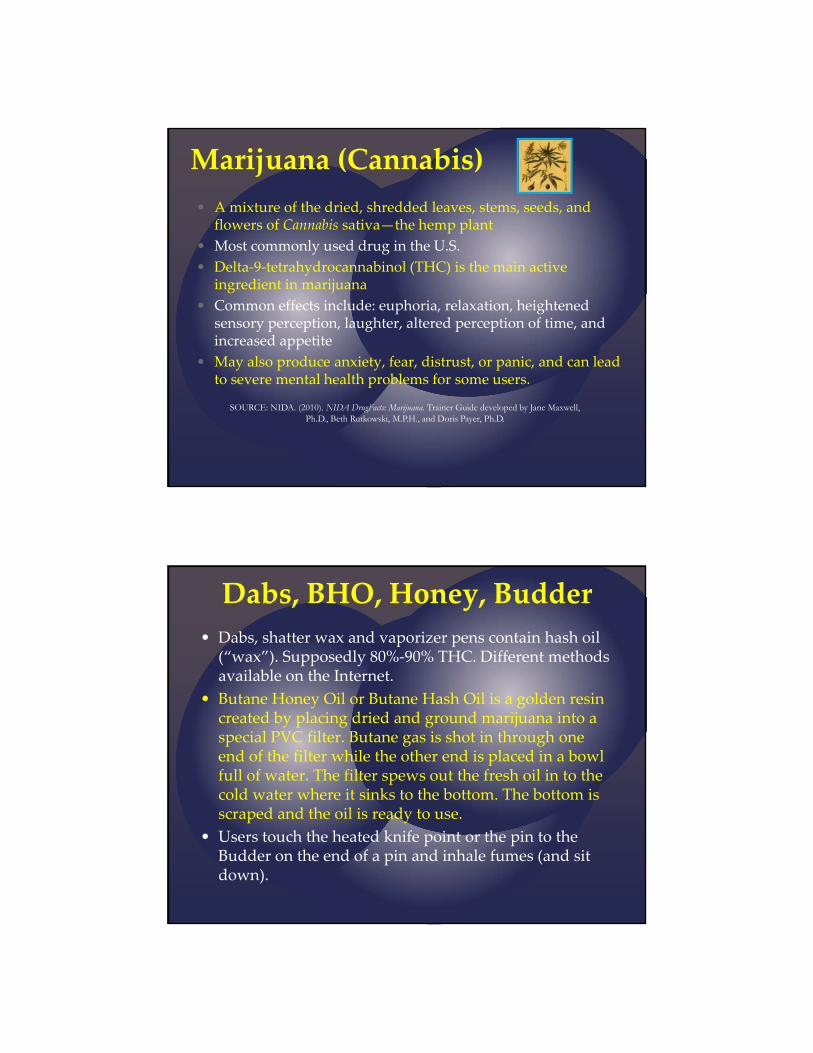
Marijuana (Cannabis)
• A mixture of the dried, shredded leaves, stems, seeds, and flowers of Cannabis sativa—the hemp plant
• Most commonly used drug in the U.S.
• Delta‐9‐tetrahydrocannabinol (THC) is the main active ingredient in marijuana
• Common effects include: euphoria, relaxation, heightened sensory perception, laughter, altered perception of time, and increased appetite
• May also produce anxiety, fear, distrust, or panic, and can lead to severe mental health problems for some users.
SOURCE: NIDA. (2010). NIDA DrugFacts: Marijuana. Trainer Guide developed by Jane Maxwell, Ph.D., Beth Rutkowski, M.P.H., and Doris Payer, Ph.D.
Dabs, BHO, Honey, Budder
• Dabs, shatter wax and vaporizer pens contain hash oil (“wax”). Supposedly 80%‐90% THC. Different methods available on the Internet.
• Butane Honey Oil or Butane Hash Oil is a golden resin created by placing dried and ground marijuana into a special PVC filter. Butane gas is shot in through one end of the filter while the other end is placed in a bowl full of water. The filter spews out the fresh oil in to the cold water where it sinks to the bottom. The bottom is scraped and the oil is ready to use.
• Users touch the heated knife point or the pin to the Budder on the end of a pin and inhale fumes (and sit down).

High speed MVA– severe head trauma – femoral blood tested
THC 73 ng/mL9‐COOH‐THC 197 ng/mL
Blood ethanol 0.15 g/dLVitreous ethanol 0.12 g/dLBlood alprazolam0.039
Vodka bottle in vehicle back seat. Known to abuse butane honey oil.
MVA – Vehicle travelling at a high rate of speed, exit paved roadway for unknown reasons and struck a tree
Witnesses that arrived on scene, believed at least one of the victims was responsive prior to spread of the fire
Drug paraphernalia pipe found under back seat passenger
Femoral Blood THC 25 ng/mL9‐COOH‐THC 68 ng/mLCO 34 % of saturation
Femoral Blood THC 14 ng/mL9‐COOH‐THC 45 ng/mLCO 17 % of saturation
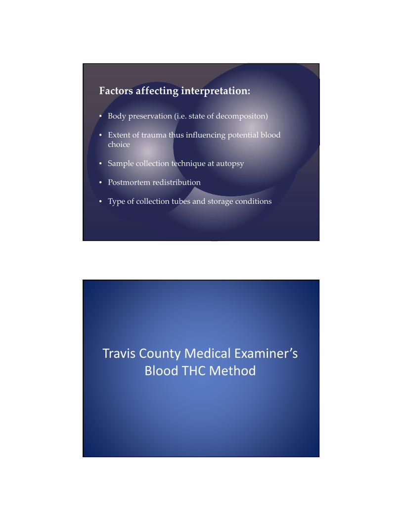
Factors affecting interpretation:
• Body preservation (i.e. state of decompositon)
• Extent of trauma thus influencing potential blood choice
• Sample collection technique at autopsy
• Postmortem redistribution
• Type of collection tubes and storage conditions
Travis County Medical Examiner’s Blood THC Method
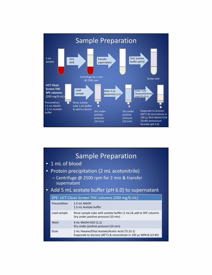
Sample Preparation
2mL ACN
5mL acetate buffer (pH 6)
Transfer supernatant
1 mL sample
Centrifuge for 2 min @ 2500 rpm
Precondition:1.5 mL MeOH1.5 mL Acetate buffer
Load sample
Dry under positive pressure (10 min)
Rinse sample tube 1 mL buffer & add to column
Wash: 4 mL
MeOH:H2O (1:1)
Dry under positive pressure (10 min)
Elute: 2 mL Hexane/EA/Acetic Acid (75:25:1)
Evaporate to dryness (40°C) & reconstitute in 100 μL 85% MeOH/15% 25mM ammonium formate (pH 4.0)
Vortex well
UCT Clean Screen THC SPE columns (200 mg/6 mL)
Sample Preparation • 1 mL of blood
• Protein precipitation (2 mL acetonitrile)– Centrifuge @ 2500 rpm for 2 min & transfer supernatant
• Add 5 mL acetate buffer (pH 6.0) to supernatant
SPE: UCT Clean Screen THC columns (200 mg/6 mL)
Precondition 1.5 mL MeOH1.5 mL Acetate buffer
Load sample Rinse sample tube with acetate buffer (1 mL) & add to SPE columnsDry under positive pressure (10 min)
Wash 4 mL MeOH:H2O (1:1)Dry under positive pressure (10 min)
Elute 2 mL Hexane/Ethyl Acetate/Acetic Acid (75:25:1)Evaporate to dryness (40°C) & reconstitute in 100 μL MPA:B (15:85)
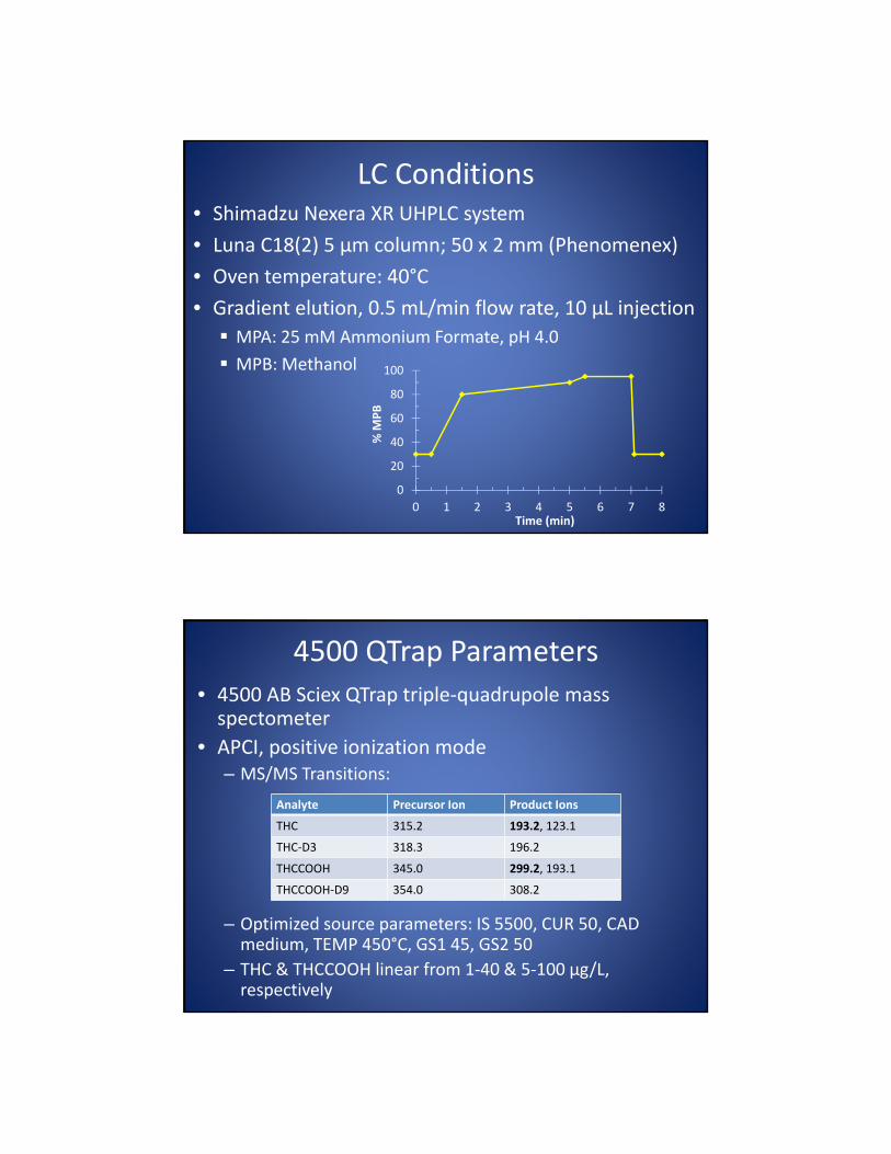
LC Conditions• Shimadzu Nexera XR UHPLC system
• Luna C18(2) 5 μm column; 50 x 2 mm (Phenomenex)
• Oven temperature: 40°C
• Gradient elution, 0.5 mL/min flow rate, 10 μL injection
MPA: 25 mM Ammonium Formate, pH 4.0
MPB: Methanol
0
20
40
60
80
100
0 1 2 3 4 5 6 7 8
% M
PB
Time (min)
4500 QTrap Parameters
• 4500 AB Sciex QTrap triple‐quadrupole mass spectometer
• APCI, positive ionization mode– MS/MS Transitions:
– Optimized source parameters: IS 5500, CUR 50, CAD medium, TEMP 450°C, GS1 45, GS2 50
– THC & THCCOOH linear from 1‐40 & 5‐100 μg/L, respectively
Analyte Precursor Ion Product Ions
THC 315.2 193.2, 123.1
THC‐D3 318.3 196.2
THCCOOH 345.0 299.2, 193.1
THCCOOH‐D9 354.0 308.2

Method Development• ESI, POS/NEG switching
– MPA: 0.1% formic acid in water
– MPB: 0.1% formic acid in acetonitrile• Low signal intensity for THC, especially qualifier
• ESI, NEG only– MPA: 0.1% formic acid in water
– MPB: 0.1% formic acid in acetonitrile• No peaks were observed for THC; great THCCOOH sensitivity
• ESI, POS only– MPA: 0.1% formic acid in water
– MPB: 0.1% formic acid in acetonitrile• Same results as POS/NEG switching for THC; THCCOOH good sensitivity
– MPB: 0.1% formic acid in methanol• Improvement in THC signal intensity, however, higher background noise compared to 0.1% formic acid in acetonitrile
Method Development• APCI, POS only
– MPA: 0.1% formic acid in water
– MPB: 0.1% formic acid in acetonitrile
• Similar results compared to ESI, POS only method
• APCI, POS only
– MPA: 25mM ammonium formate in water, pH 4.0
– MPB: Methanol
• Same constituents as laboratory’s previous isocratic method
• Great peak shape and signal intensity for THC & THCCOOH
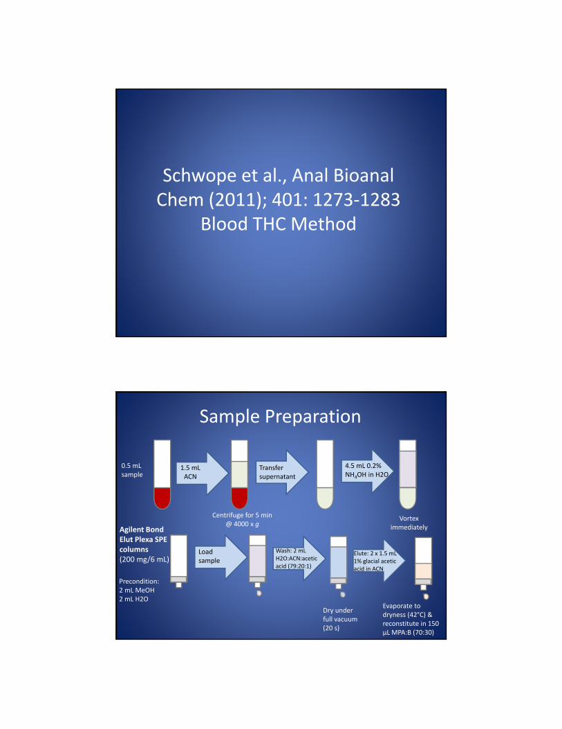
Schwope et al., Anal BioanalChem (2011); 401: 1273‐1283
Blood THC Method
Sample Preparation
1.5 mL ACN
4.5 mL 0.2% NH4OH in H2O
Transfer supernatant
0.5 mL sample
Centrifuge for 5 min @ 4000 x g
Precondition:2 mL MeOH2 mL H2O
Load sample
Wash: 2 mL H2O:ACN:acetic acid (79:20:1)
Dry under full vacuum (20 s)
Elute: 2 x 1.5 mL 1% glacial acetic acid in ACN
Evaporate to dryness (42°C) & reconstitute in 150 μL MPA:B (70:30)
Vortex immediatelyAgilent Bond
Elut Plexa SPE columns (200 mg/6 mL)
Sample Preparation • 0.5 mL of blood
• Protein precipitation (1.5 mL acetonitrile)
– Centrifuge @ 4000 x g for 5 min & transfer supernatant
• Add 4.5 mL 0.2% NH4OH in water to supernatant
SPE: Agilent Bond Elut Plexa Columns (200 mg/6 mL)
Precondition 2 mL MeOH2 mL H2O
Load sample Allow to pass through by gravity
Wash 2 mL H2O/acetonitrile/acetic acid (79:20:1)Dry under full vacuum for 20 s
Elute 2 x 1.5 mL 1% glacial acetic acid in acetonitrile Evaporate to dryness (42°C) & reconstitute in 150 μL MPA:B (70:30)
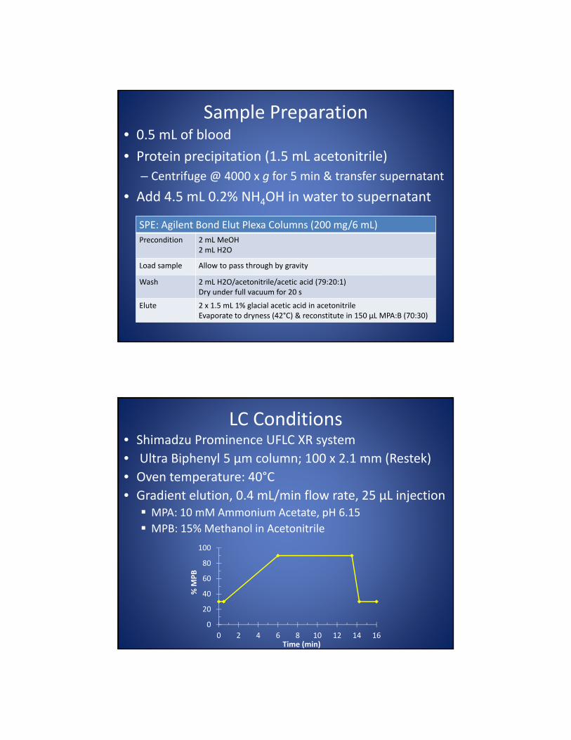
LC Conditions• Shimadzu Prominence UFLC XR system
• Ultra Biphenyl 5 μm column; 100 x 2.1 mm (Restek)
• Oven temperature: 40°C
• Gradient elution, 0.4 mL/min flow rate, 25 μL injection MPA: 10 mM Ammonium Acetate, pH 6.15
MPB: 15% Methanol in Acetonitrile
0
20
40
60
80
100
0 2 4 6 8 10 12 14 16
% M
PB
Time (min)
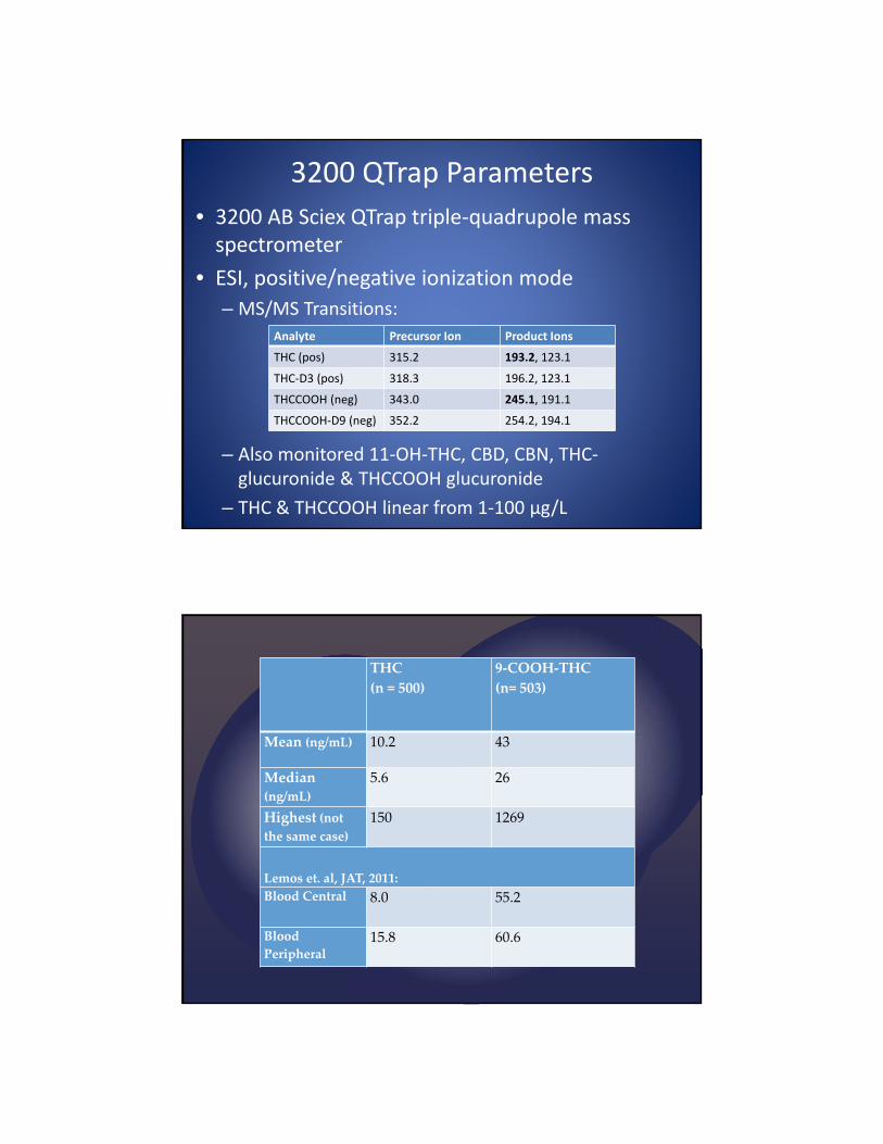
3200 QTrap Parameters
• 3200 AB Sciex QTrap triple‐quadrupole mass spectrometer
• ESI, positive/negative ionization mode
– MS/MS Transitions:
– Also monitored 11‐OH‐THC, CBD, CBN, THC‐glucuronide & THCCOOH glucuronide
– THC & THCCOOH linear from 1‐100 μg/L
Analyte Precursor Ion Product Ions
THC (pos) 315.2 193.2, 123.1
THC‐D3 (pos) 318.3 196.2, 123.1
THCCOOH (neg) 343.0 245.1, 191.1
THCCOOH‐D9 (neg) 352.2 254.2, 194.1
THC
(n = 500)
9‐COOH‐THC
(n= 503)
Mean (ng/mL) 10.2 43
Median
(ng/mL)
5.6 26
Highest (not
the same case)
150 1269
Lemos et. al, JAT, 2011:
Blood Central 8.0 55.2
Blood
Peripheral15.8 60.6

Toxicology (mixed heart blood)
THC 112 ng/mL9‐COOH‐THC 1260 ng/mLDiazepam/Nor 230/328 ng/mLHydrocodone 99 ng/mLHydromorphone 31 ng/mL
CO 31 % of saturation
0
10
20
30
40
50
60
70
80
90
100
110
120
130
140
150
160
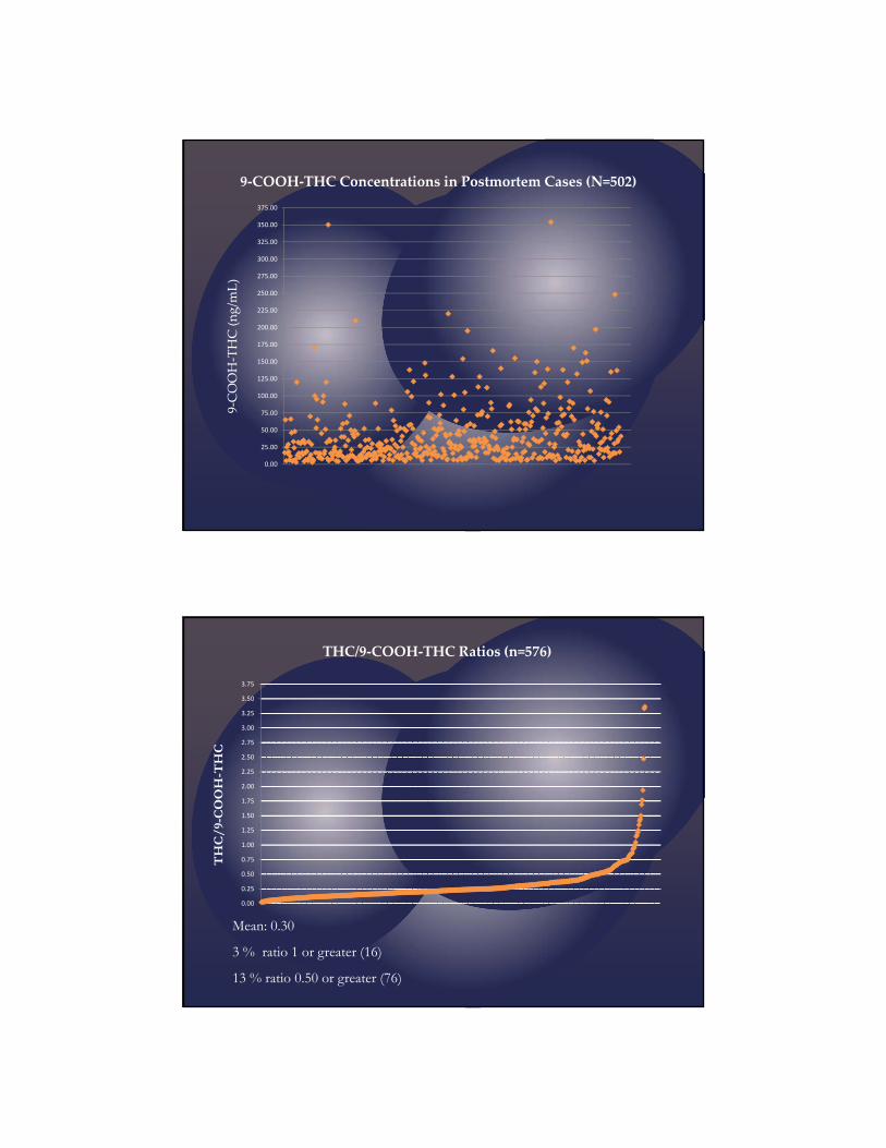
THC Concentrations in Postmortem Cases (N=500)
THC (ng/m
L)
45 % < 5.0 ng/mL 72% <10.0 ng/mL
0.00
25.00
50.00
75.00
100.00
125.00
150.00
175.00
200.00
225.00
250.00
275.00
300.00
325.00
350.00
375.00
9‐COOH‐THC Concentrations in Postmortem Cases (N=502)
9‐COOH‐THC (ng/m
L)
THC/9-COOH-THC
Mean: 0.30
3 % ratio 1 or greater (16)
13 % ratio 0.50 or greater (76)
0.00
0.25
0.50
0.75
1.00
1.25
1.50
1.75
2.00
2.25
2.50
2.75
3.00
3.25
3.50
3.75
THC/9‐COOH‐THC Ratios (n=576)

•20y/o Hispanic male rear drivers side passenger ‐ auto vs. train
accident
•EMS and DPS responded to the scene and found the decedent
approximately 30 feet away from the vehicle face down in a
puddle of water and obvious DOS.
•Strong odor of "marijuana" on the passengers that were
transported to the hospital. No ETOH or illicit drugs were
reported to be found inside the vehicle.
Blood, heart Tetrahydrocannabinol 150 ng/mL
Blood, heart 9‐Carboxy‐THC 120 ng/mL
• 30 y/o Black male the decedent was the unrestrained driver who was involved in a two vehicle collision.
• The decedent was traveling on SH 130 (frontage road), when he reportedly ran a red light and was subsequently T-boned by another vehicle.
• Although no illicit drugs were found, the interior of the decedents vehicle smelled of marijuana.
Blood, hospital THC 102 ng/mL
Blood, hospital 9‐Carboxy‐THC 76 ng/mL
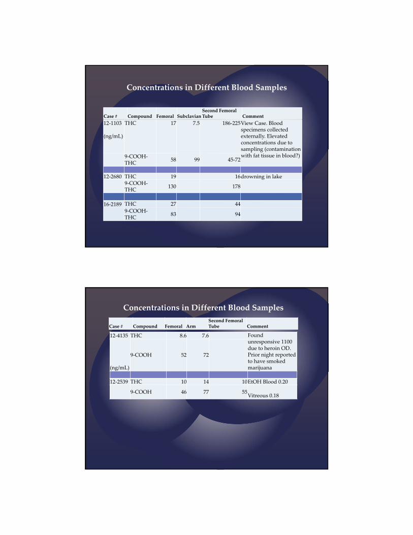
Case # Compound Femoral SubclavianSecond Femoral Tube Comment
12‐1103
(ng/mL)
THC 17 7.5 186‐225View Case. Blood specimens collected externally. Elevated concentrations due to sampling (contamination with fat tissue in blood?)9‐COOH‐
THC58 99 45‐72
12‐2680 THC 19 16drowning in lake9‐COOH‐THC
130 178
16‐2189 THC 27 44
9‐COOH‐THC
83 94
Concentrations in Different Blood Samples
12‐4135 THC 8.6 7.6 Foundunresponsive 1100 due to heroin OD. Prior night reported to have smoked marijuana(ng/mL)
9‐COOH 52 72
12‐2539 THC 10 14 10EtOH Blood 0.20
9‐COOH 46 77 55Vitreous 0.18
Case # Compound Femoral ArmSecond Femoral Tube Comment
Concentrations in Different Blood Samples
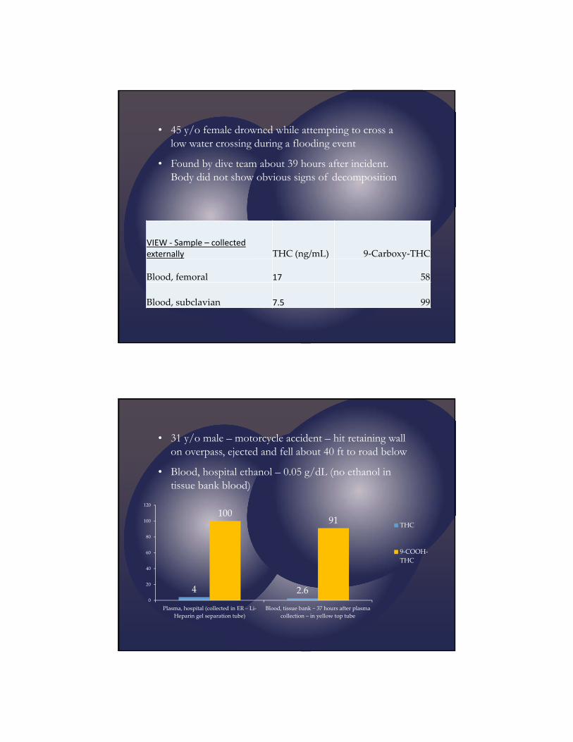
VIEW ‐ Sample – collected externally THC (ng/mL) 9‐Carboxy‐THC
Blood, femoral 17 58
Blood, subclavian 7.5 99
• 45 y/o female drowned while attempting to cross a low water crossing during a flooding event
• Found by dive team about 39 hours after incident. Body did not show obvious signs of decomposition
• 31 y/o male – motorcycle accident – hit retaining wall on overpass, ejected and fell about 40 ft to road below
• Blood, hospital ethanol – 0.05 g/dL (no ethanol in tissue bank blood)
4 2.6
10091
0
20
40
60
80
100
120
Plasma, hospital (collected in ER – Li‐
Heparin gel separation tube)
Blood, tissue bank ~ 37 hours after plasma
collection – in yellow top tube
THC
9‐COOH‐
THC

• 48 y/o male – low-speed MVA– COD intracerebral hemorrhage
2.6
4.1
15
8.3
0
2
4
6
8
10
12
14
16
Blood, hospital‐ (collected in ER –
lavender tube)
Blood, femoral ~ 46 hours after
hospital blood collection – grey top
THC
9‐COOH‐
THC
• 31 y/o male – pedestrian hit by automobile – late into hospital course had surgery for spleen laceraction –COD: ASCVD following surgery
9.9
1.1
44
5.4
0
5
10
15
20
25
30
35
40
45
50
Serum, hospital, (collected in ER –
gold top gel separation tube)
Blood, femoral, ~ 44 hours after
serum collection – grey top
THC
9‐
COOH‐
THC

• 19 y/o – accidental fall apparently from malfunction of bicycle chain
• CT scan revealed acute subdural hematoma and skull base fractures. COD ruled blunt head trauma
2 1.3
5
18
24
15
0
5
10
15
20
25
30
Hospital blood 7/2 1800 Hospital blood 7/3 0600 Postmortem femoral
THC
9‐COOH‐
THC
ng/m
L
12 hr difference
5 hr later pronounced
ER blood ~19 hrs later autopsy performed
9.4 4.5 4.313
107
190
121
84
0
20
40
60
80
100
120
140
160
180
200
Blood, hospital 2/11 2147 Serum, hospital 2/11 2147 Blood, hospital 2/12 0722 Blood, femoral
THC
9‐COOH‐
THC
ng/m
L
~ 9 hr difference ~ 12 hr difference
69y/o Caucasian male, Unwitnessed Fall; the decedent found at the bottom of a stairwell, ʺwith a joint in his mouth“. Radiology revealed Multiple Left Sided Skull Fractures and Bilateral Subdural Hemorrhage.
Hospital blood ethanol: 0.20 g/dL
~ ER blood&serum
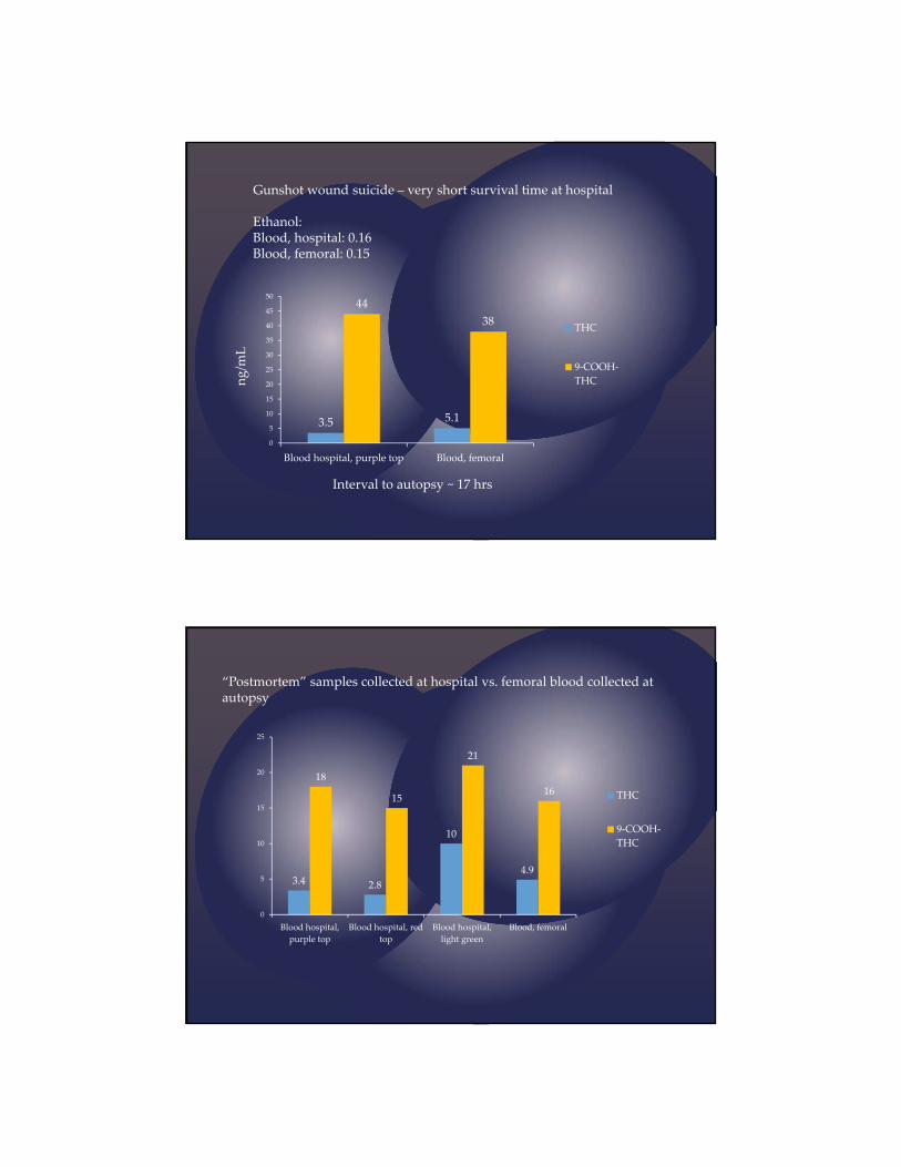
3.5 5.1
44
38
0
5
10
15
20
25
30
35
40
45
50
Blood hospital, purple top Blood, femoral
THC
9‐COOH‐
THC
Gunshot wound suicide – very short survival time at hospital
Ethanol:Blood, hospital: 0.16Blood, femoral: 0.15
ng/m
L
Interval to autopsy ~ 17 hrs
“Postmortem” samples collected at hospital vs. femoral blood collected at autopsy
3.4 2.8
10
4.9
18
15
21
16
0
5
10
15
20
25
Blood hospital,
purple top
Blood hospital, red
top
Blood hospital,
light green
Blood, femoral
THC
9‐COOH‐
THC
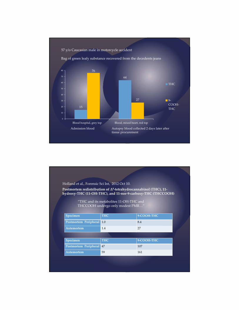
57 y/o Caucasian male in motorcycle accident
Bag of green leafy substance recovered from the decedents jeans
15
64
76
27
0
10
20
30
40
50
60
70
80
Blood hospital, grey top Blood, mixed heart, red top
THC
9‐
COOH‐
THC
Admission blood Autopsy blood collected 2 days later after tissue procurement
“THC and its metabolites 11‐OH‐THC and THCCOOH undergo only modest PMR…”
Holland et al., Forensic Sci Int, 2012 Oct 10.
Postmortem redistribution of Δ9‐tetrahydrocannabinol (THC), 11‐hydroxy‐THC (11‐OH‐THC), and 11‐nor‐9‐carboxy‐THC (THCCOOH)
Specimen THC 9‐COOH‐THC
Postmortem Peripheral 1.0 8.4
Antemortem 1.4 27
Specimen THC 9‐COOH‐THC
Postmortem Peripheral 47 107
Antemortem 59 161
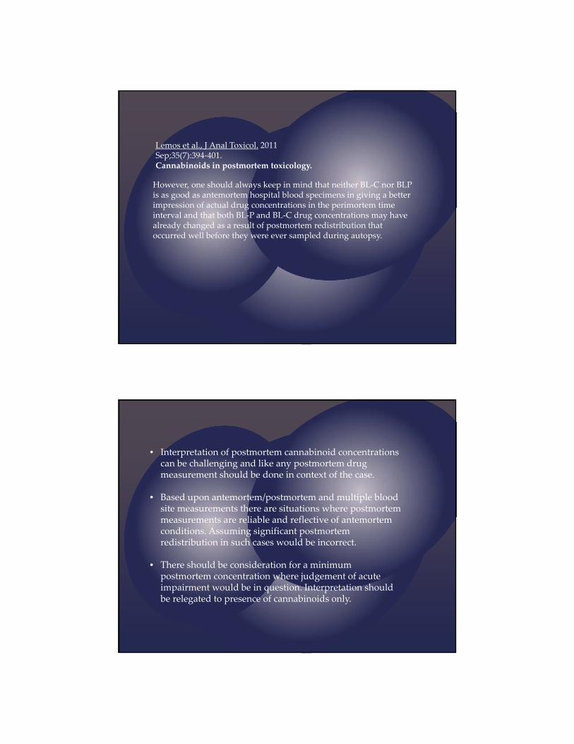
However, one should always keep in mind that neither BL‐C nor BLPis as good as antemortem hospital blood specimens in giving a better impression of actual drug concentrations in the perimortem time interval and that both BL‐P and BL‐C drug concentrations may have already changed as a result of postmortem redistribution that occurred well before they were ever sampled during autopsy.
Lemos et al., J Anal Toxicol. 2011 Sep;35(7):394‐401.Cannabinoids in postmortem toxicology.
• Interpretation of postmortem cannabinoid concentrations can be challenging and like any postmortem drug measurement should be done in context of the case.
• Based upon antemortem/postmortem and multiple blood site measurements there are situations where postmortem measurements are reliable and reflective of antemortemconditions. Assuming significant postmortem redistribution in such cases would be incorrect.
• There should be consideration for a minimum postmortem concentration where judgement of acute impairment would be in question. Interpretation should be relegated to presence of cannabinoids only.
William H. Anderson, Ph.D., F-ABFT Dr. Anderson is a forensic toxicologist employed by NMS Labs. His duties include toxicological investigations of law enforcement, medical examiner/coroner, and clinical cases. He provides expert witness testimony in support of analyses performed at NMS Labs and for private attorneys while providing expert services support. He frequently testifies concerning human performance and postmortem toxicology. Dr. Anderson has previously served as Chief Toxicologist for the Forensic Science Division of the Washoe County Sheriff's Office in Reno, Nevada, Chief Toxicologist for the State of North Carolina Office of the Chief Medical Examiner, Chief Toxicologist for Sierra Nevada Laboratories, Inc., Reno, NV, Deputy Chief Toxicologist for the State of Oklahoma Office of the Chief Medical Examiner, and the Chief Toxicologist for the Tennessee State Crime Laboratory. He has held academic positions as an Assistant Professor of Clinical Laboratory Sciences at the University of Tennessee Center of the Health Sciences, Assistant Clinical Professor of Clinical Laboratory Sciences at the Oklahoma School of Medicine, and Assistant professor of Pathology at the School of Medicine at the University of Nevada, Reno, and at the University of North Carolina School of Medicine. He received his B.S. and M.S. degrees in chemistry from Tennessee Technological University and his Ph.D. degree in Pathology/Toxicology from the University of Tennessee Center for the Health Sciences. His research interests include analytical methodology and the study of postmortem redistribution of drugs. Dr. Anderson is certified as a Fellow by the American Board of Forensic Toxicology. In 2008, he was awarded the Alexander O. Gettler Award by the Toxicology Section of the American Academy of Forensic Sciences. He is active in both the American Academy of Forensic Sciences and the Society of Forensic Toxicologists (SOFT). He has served as President of SOFT and has recently served on the Board of Directors. He is also a Past President of SAT. Dr. Anderson is a frequent presenter at scientific symposia and has numerous publications in the peer reviewed literature.

William H. Anderson, PhD, F-ABFTSOFT WS #11, 2016
The Potential for The Postmortem Redistribution of THC
Conflicts of Interest
None
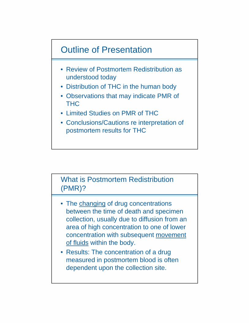
Outline of Presentation
• Review of Postmortem Redistribution as understood today
• Distribution of THC in the human body• Observations that may indicate PMR of
THC • Limited Studies on PMR of THC• Conclusions/Cautions re interpretation of
postmortem results for THC
What is Postmortem Redistribution (PMR)?
• The changing of drug concentrations between the time of death and specimen collection, usually due to diffusion from an area of high concentration to one of lower concentration with subsequent movement of fluids within the body.
• Results: The concentration of a drug measured in postmortem blood is often dependent upon the collection site.

Postmortem Redistribution
• Net change is a function of multiple factors:• Pharmacokinetic properties of the drug• Chemical characteristics of the drug• Orientation of the body • Putrefaction• Drug dosage• Interval between drug ingestion and death• Interval between death and specimen collection
Other Factors That May Contribute to Site Dependence
• Differential distribution at the time of death• Loss of drug through chemical or enzymatic
processes• Cocaine, olanzapine, alcohol, some bath salts
• Increase in drug concentration• Alcohol, potassium, succinylmonocholine
• Trauma Artifacts• Rupture of stomach and diaphragm
• Hydrolysis of Conjugates
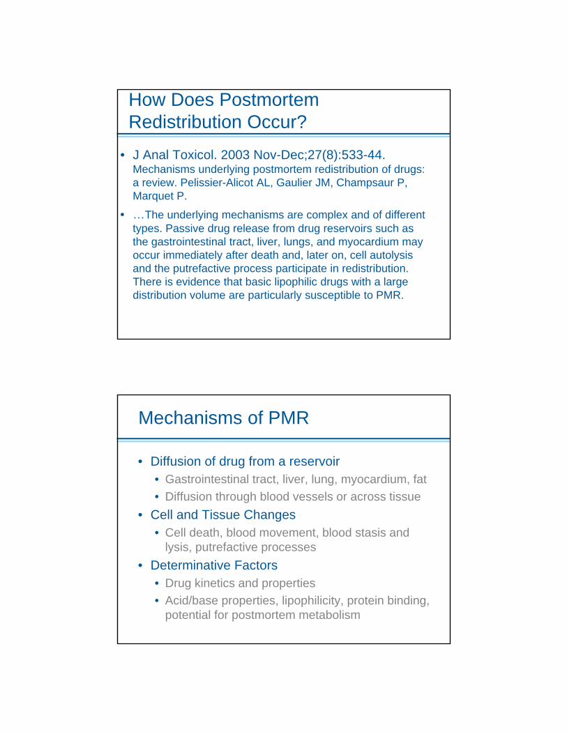
How Does PostmortemRedistribution Occur?
• J Anal Toxicol. 2003 Nov-Dec;27(8):533-44. Mechanisms underlying postmortem redistribution of drugs: a review. Pelissier-Alicot AL, Gaulier JM, Champsaur P, Marquet P.
• …The underlying mechanisms are complex and of different types. Passive drug release from drug reservoirs such as the gastrointestinal tract, liver, lungs, and myocardium may occur immediately after death and, later on, cell autolysis and the putrefactive process participate in redistribution. There is evidence that basic lipophilic drugs with a large distribution volume are particularly susceptible to PMR.
Mechanisms of PMR
• Diffusion of drug from a reservoir• Gastrointestinal tract, liver, lung, myocardium, fat• Diffusion through blood vessels or across tissue
• Cell and Tissue Changes• Cell death, blood movement, blood stasis and
lysis, putrefactive processes• Determinative Factors
• Drug kinetics and properties• Acid/base properties, lipophilicity, protein binding,
potential for postmortem metabolism
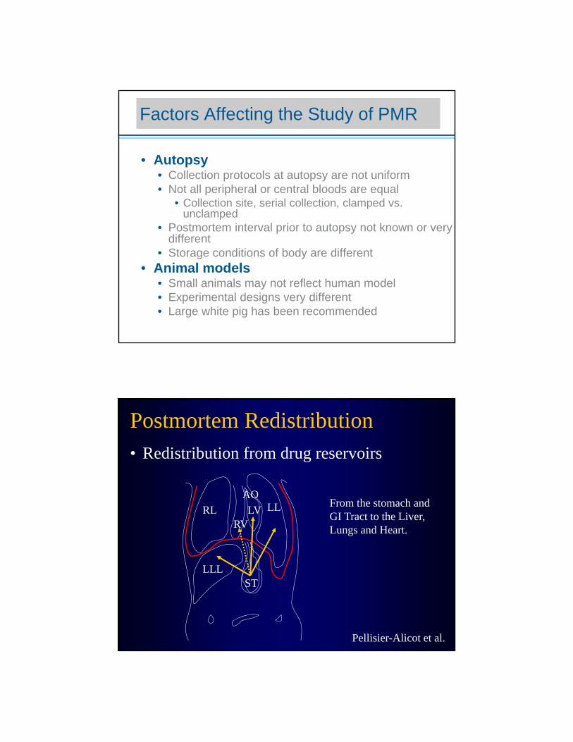
Factors Affecting the Study of PMR
• Autopsy• Collection protocols at autopsy are not uniform• Not all peripheral or central bloods are equal
• Collection site, serial collection, clamped vs. unclamped
• Postmortem interval prior to autopsy not known or very different
• Storage conditions of body are different• Animal models
• Small animals may not reflect human model• Experimental designs very different• Large white pig has been recommended
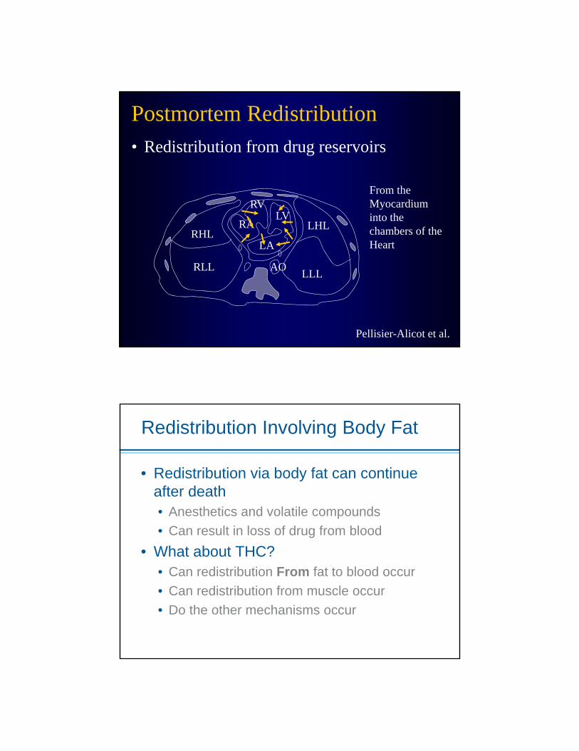
Postmortem Redistribution• Redistribution from drug reservoirs
RL LLAO
RVLV
ST
Pellisier-Alicot et al.
LLL
From the stomach and GI Tract to the Liver, Lungs and Heart.
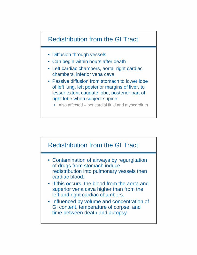
Redistribution from the GI Tract
• Diffusion through vessels• Can begin within hours after death• Left cardiac chambers, aorta, right cardiac
chambers, inferior vena cava• Passive diffusion from stomach to lower lobe
of left lung, left posterior margins of liver, to lesser extent caudate lobe, posterior part of right lobe when subject supine• Also affected – pericardial fluid and myocardium
Redistribution from the GI Tract
• Contamination of airways by regurgitation of drugs from stomach induce redistribution into pulmonary vessels then cardiac blood.
• If this occurs, the blood from the aorta and superior vena cava higher than from the left and right cardiac chambers.
• Influenced by volume and concentration of GI content, temperature of corpse, and time between death and autopsy.

Postmortem Redistribution• Redistribution from drug reservoirs
Pellisier-Alicot et al.
RLL
RHLLHL
LLL
LA
LVRA
RV
AO
From the Lungs to the Heart and Aorta
Redistribution From the Lungs
• Begins within the first two hours after death• Raises concentration in cardiac chambers
and thoracic vessels• Via direct diffusion and via blood vessels
• It is an intense and important mechanism• Especially important for weak bases with high
Vd• Methadone, TCA’s, propoxyphene• It may also be important to THC• Lungs to liver via diaphragm or pleural and
peritoneal fluid
Postmortem Redistribution• Redistribution from drug reservoirs
Pellisier-Alicot et al.
RLL
RHLLHL
LLL
LA
LVRA
RV
AO
From the Myocardium into the chambers of the Heart
Redistribution Involving Body Fat
• Redistribution via body fat can continue after death• Anesthetics and volatile compounds• Can result in loss of drug from blood
• What about THC?• Can redistribution From fat to blood occur• Can redistribution from muscle occur• Do the other mechanisms occur

An Old Friend Huestis et al, Journal of Analytical Toxicology, Vol. 16, September/October 1992
THC Distribution In the Body
Kreutz and Axelrod, Science, 1973
Distribution of THC in Man
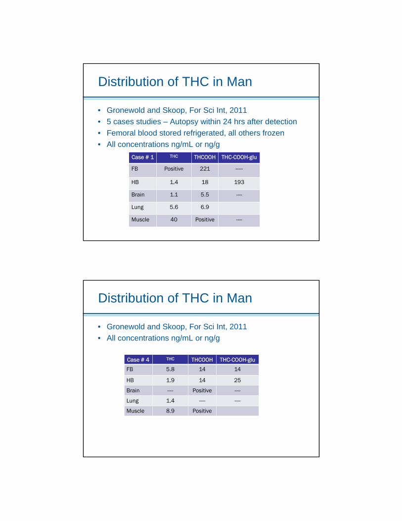
• Gronewold and Skoop, For Sci Int, 2011• 5 cases studies – Autopsy within 24 hrs after detection• Femoral blood stored refrigerated, all others frozen• All concentrations ng/mL or ng/g
Case # 1 THC THCOOH THC-COOH-glu
FB Positive 221 -----
HB 1.4 18 193
Brain 1.1 5.5 ----
Lung 5.6 6.9
Muscle 40 Positive ----
Distribution of THC in Man
• Gronewold and Skoop, For Sci Int, 2011• All concentrations ng/mL or ng/g
Case # 4 THC THCOOH THC-COOH-glu
FB 5.8 14 14
HB 1.9 14 25
Brain ---- Positive ----
Lung 1.4 ---- ----
Muscle 8.9 Positive
Distribution of THC in Man - Cont
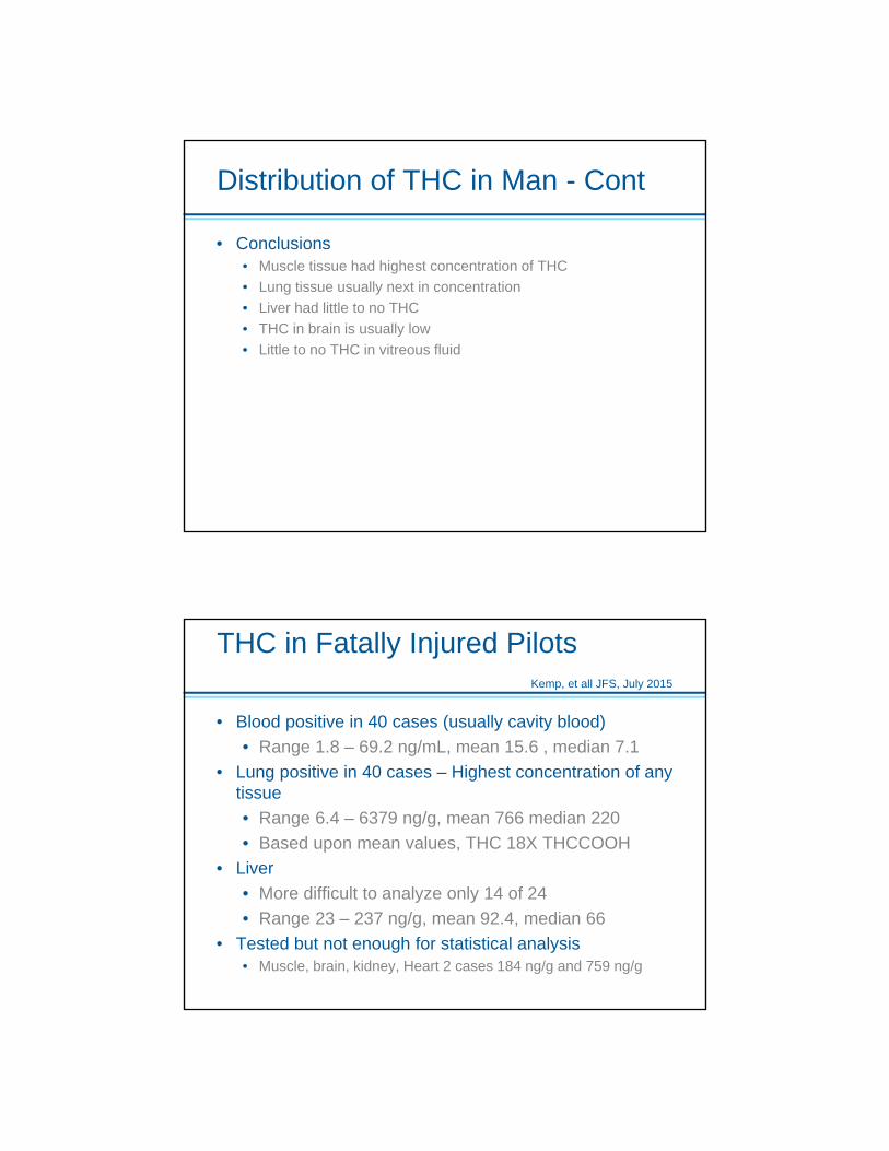
• Conclusions• Muscle tissue had highest concentration of THC• Lung tissue usually next in concentration• Liver had little to no THC• THC in brain is usually low• Little to no THC in vitreous fluid
THC in Fatally Injured Pilots Kemp, et all JFS, July 2015
• Blood positive in 40 cases (usually cavity blood)• Range 1.8 – 69.2 ng/mL, mean 15.6 , median 7.1
• Lung positive in 40 cases – Highest concentration of any tissue • Range 6.4 – 6379 ng/g, mean 766 median 220 • Based upon mean values, THC 18X THCCOOH
• Liver• More difficult to analyze only 14 of 24• Range 23 – 237 ng/g, mean 92.4, median 66
• Tested but not enough for statistical analysis• Muscle, brain, kidney, Heart 2 cases 184 ng/g and 759 ng/g
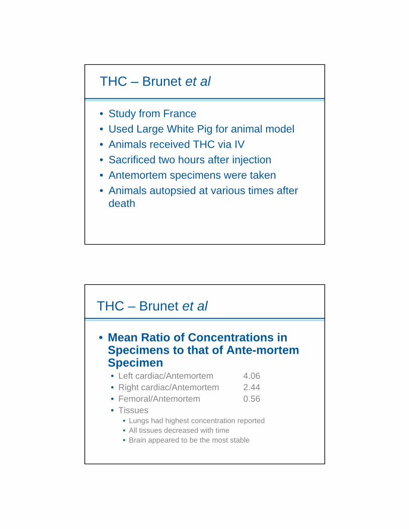
THC – Brunet et al
• Study from France• Used Large White Pig for animal model• Animals received THC via IV• Sacrificed two hours after injection• Antemortem specimens were taken• Animals autopsied at various times after
death
THC – Brunet et al
• Mean Ratio of Concentrations in Specimens to that of Ante-mortem Specimen• Left cardiac/Antemortem 4.06• Right cardiac/Antemortem 2.44• Femoral/Antemortem 0.56• Tissues
• Lungs had highest concentration reported• All tissues decreased with time• Brain appeared to be the most stable
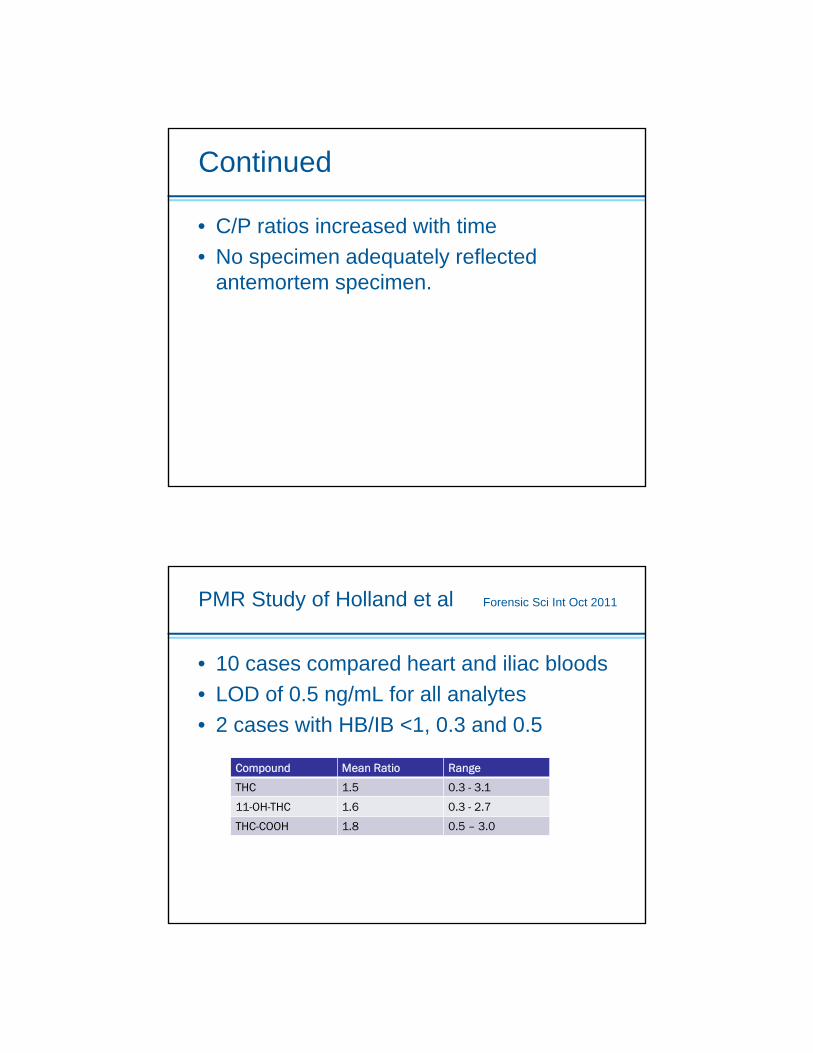
Continued
• C/P ratios increased with time• No specimen adequately reflected
antemortem specimen.
PMR Study of Holland et al Forensic Sci Int Oct 2011
• 10 cases compared heart and iliac bloods• LOD of 0.5 ng/mL for all analytes• 2 cases with HB/IB <1, 0.3 and 0.5
Compound Mean Ratio Range
THC 1.5 0.3 - 3.1
11-OH-THC 1.6 0.3 - 2.7
THC-COOH 1.8 0.5 – 3.0

Case in Reno, NV
• Decedent killed while riding motorcycle 60 mph on 4-lane hwy
• Car traveling in opposite direction made left hand turn in front of decedent
• Motorcycle impacted right front passenger side of car
• No response to resuscitative efforts• Specimens sent for routine toxicology
Toxicology for Reno Case
AnalyteCentral Bloodmg/L
Peripheral Bloodmg/L
Urinemg/L
6-AM 0.016 <0.002 0.42
Morphine - Free 0.10 0.0211.1
THC 0.002 0.036
Carboxy THC 0.013 0.008

THC – Lemos and EngleJAT Vol 35, 2011
• 30 cases compared heart and femoral blood concentration
• Mean HB/PB ratio = 0.62• Range = 0.00-2.60
Facts That Surprise
• At NMS we compared THCCOOH/THC ratio in positive DUI Cases and ME Cases• Data for 2013• 3432 DUI cases have THCCOOH/THC
ranging from 0.72 to 98; Median ratio = 8.1• 8 cases ratio <1• 3472 ME cases have THCCOOH/THC
ranging from 0.110 to 89; median ratio = 3.75
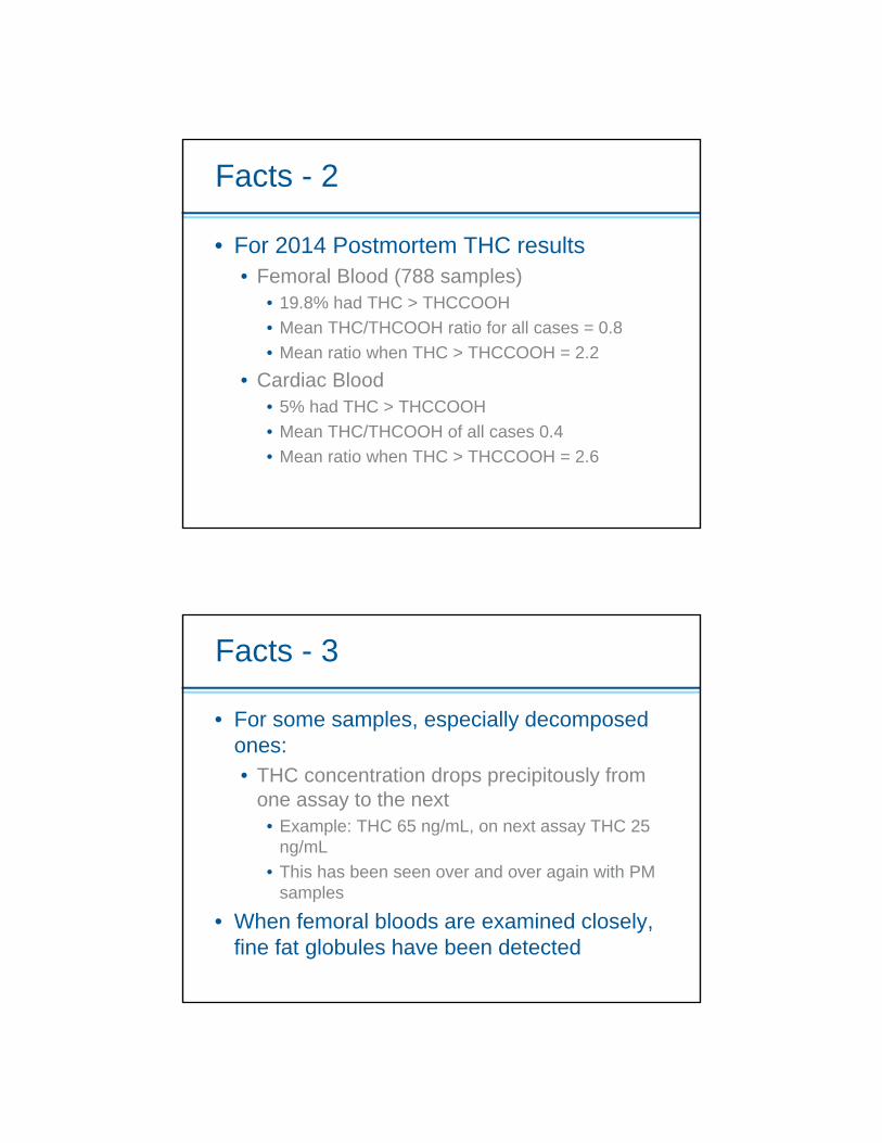
Facts - 2
• For 2014 Postmortem THC results• Femoral Blood (788 samples)
• 19.8% had THC > THCCOOH• Mean THC/THCOOH ratio for all cases = 0.8• Mean ratio when THC > THCCOOH = 2.2
• Cardiac Blood• 5% had THC > THCCOOH• Mean THC/THCOOH of all cases 0.4• Mean ratio when THC > THCCOOH = 2.6
Facts - 3
• For some samples, especially decomposed ones:• THC concentration drops precipitously from
one assay to the next• Example: THC 65 ng/mL, on next assay THC 25
ng/mL• This has been seen over and over again with PM
samples
• When femoral bloods are examined closely, fine fat globules have been detected
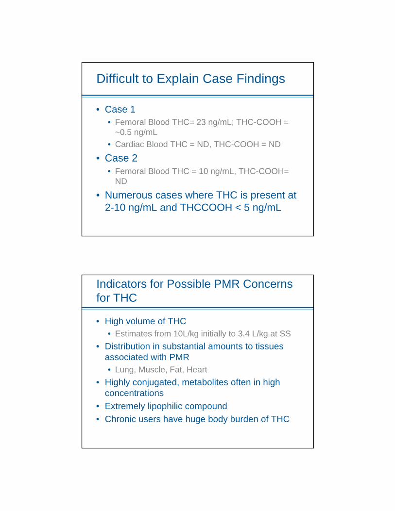
Difficult to Explain Case Findings
• Case 1• Femoral Blood THC= 23 ng/mL; THC-COOH =
~0.5 ng/mL• Cardiac Blood THC = ND, THC-COOH = ND
• Case 2• Femoral Blood THC = 10 ng/mL, THC-COOH=
ND
• Numerous cases where THC is present at 2-10 ng/mL and THCCOOH < 5 ng/mL
Indicators for Possible PMR Concerns for THC
• High volume of THC• Estimates from 10L/kg initially to 3.4 L/kg at SS
• Distribution in substantial amounts to tissues associated with PMR• Lung, Muscle, Fat, Heart
• Highly conjugated, metabolites often in high concentrations
• Extremely lipophilic compound• Chronic users have huge body burden of THC
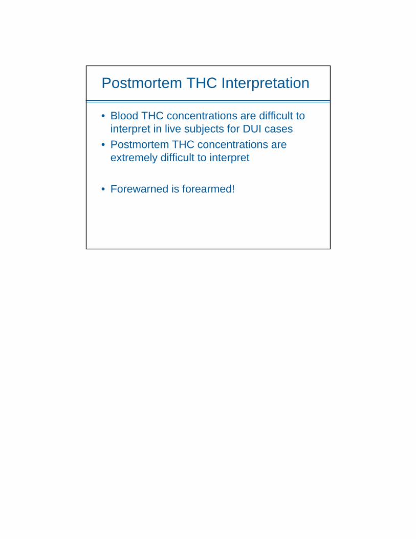
Postmortem THC Interpretation
• Blood THC concentrations are difficult to interpret in live subjects for DUI cases
• Postmortem THC concentrations are extremely difficult to interpret
• Forewarned is forearmed!

Nikolas P. Lemos, PhD, FRSC, F-ABFT Clinical Professor (Full), Department of Laboratory Medicine, School of Medicine The University of California, San Francisco Nikolas P. Lemos, a native of Greece, earned his Bachelor of Science in Criminalistics/Forensic Chemistry from the State University of New York – College at Buffalo and his Master of Science in Forensic Science specializing in Criminalistics and Forensic Toxicology at John Jay College of Criminal Justice of the City University of New York after completing his thesis on “Biliary Excretion of Alcohols” under the supervision of the late Professor Arvind A. Agarwal. In 1999 he was awarded his Doctorate in Forensic Medicine & Science from the University of Glasgow (Scotland, UK) specializing in Analytical and Forensic Toxicology after completing research on “Analysis of Bile and Nail as Alternative Biological Specimens in Forensic Toxicology.” Until 2003, he worked in London (England, UK) as Senior Lecturer at London’s South Bank University and as Head of the Forensic Toxicology Service of the Analytical Unit of St George’s Hospital Medical School of the University of London providing forensic toxicology services to HM Coroners, Home Office Pathologists and Police Forces in Greater London. In October 2003, he became San Francisco’s Chief Forensic Toxicologist where he worked in both postmortem as well human performance forensic toxicology cases for 13 years and where he led the San Francisco Office of the Chief Medical Examiner to ABFT-Accreditation in March 2015. He has a very strong academic presence with positions in Universities in San Francisco (USA), London (UK) and Patra (Greece). He has authored or co-authored over 90 abstracts, peer-reviewed papers and book chapters including the first ever study of Cannabinoids in Fingernails (JAT, May 1999) and the first ever study of Cannabinoids in Postmortem Toxicology (JAT, September 2011). He has previously served as Science Editor of Medicine, Science and the Law, and he is the holder of many scholarships, research and equipment grants and awards. In 2005, while still not a citizen of the USA, he was honored by the 109th Congress of the United States of America “for dedicating his life and career to community safety and social awareness.” He is Fellow of the American Board of Forensic Toxicology, Fellow of the Royal Society of Chemistry, and Fellow of the American Academy of Forensic Sciences where he spearheads their Diversity and Outreach efforts working to promote equal opportunities in knowledge, employment, pay and promotion for all and to eliminate discrimination based on age, race, sexual orientation, gender, faith or belief and/or disability. He has served on numerous scientific meeting organizing committees and co-hosted the 2011 Joint SOFT-TIAFT Meeting in San Francisco with over 1,500 colleagues in attendance. He is the 2016-2017 Scientific Program Chair of the AAFS’s Toxicology Section. He is also actively engaged in several other professional organisations: American Society of Crime Laboratory Directors, American Chemical Society, California Association of Criminalists, California Association of Crime Laboratory Directors, California Association of Toxicologists (Chair: Ethics Committee), Society of Forensic Toxicologists, London Toxicology Group, Society of Hair Testing, The International Association of Forensic Toxicologists (Chair: Internet & Social Media Committee), British Academy of Forensic Sciences and Affiliate Member of the National Association of Medical Examiners (Chair: Forensic Toxicology Committee). His email is [email protected] and his twitter handle is SF_Toxicologist
Postmortem Cannabis:
Ready to Interpret theUninterpretable?
18 October 2016
Nikolas P. Lemos, PhD, FRSC, F-ABFTClinical Professor in Forensic ToxicologyDepartment of Laboratory MedicineSchool of MedicineUniversity of California – San Francisco
Disclosures
• No potential bias or conflict of interest
2
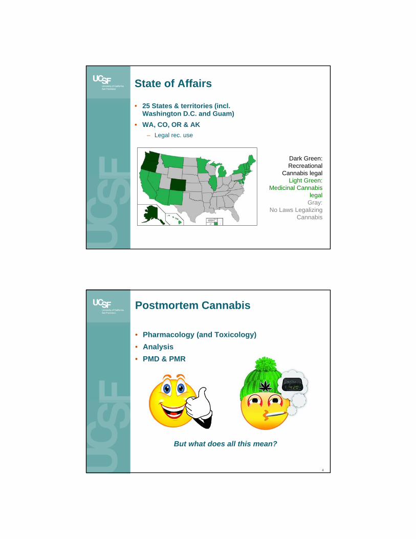
State of Affairs
• 25 States & territories (incl. Washington D.C. and Guam)
• WA, CO, OR & AK– Legal rec. use
Dark Green: Recreational
Cannabis legalLight Green:
Medicinal Cannabis legal
Gray: No Laws Legalizing
Cannabis
Postmortem Cannabis
4
• Pharmacology (and Toxicology)• Analysis• PMD & PMR
But what does all this mean?
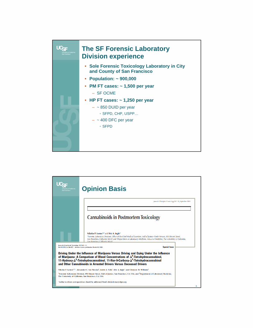
The SF Forensic Laboratory Division experience
• Sole Forensic Toxicology Laboratory in City and County of San Francisco
• Population: ~ 900,000• PM FT cases: ~ 1,500 per year
– SF OCME
• HP FT cases: ~ 1,250 per year– ~ 850 DUID per year
• SFPD, CHP, USPP…
– ~ 400 DFC per year• SFPD
Opinion Basis
6
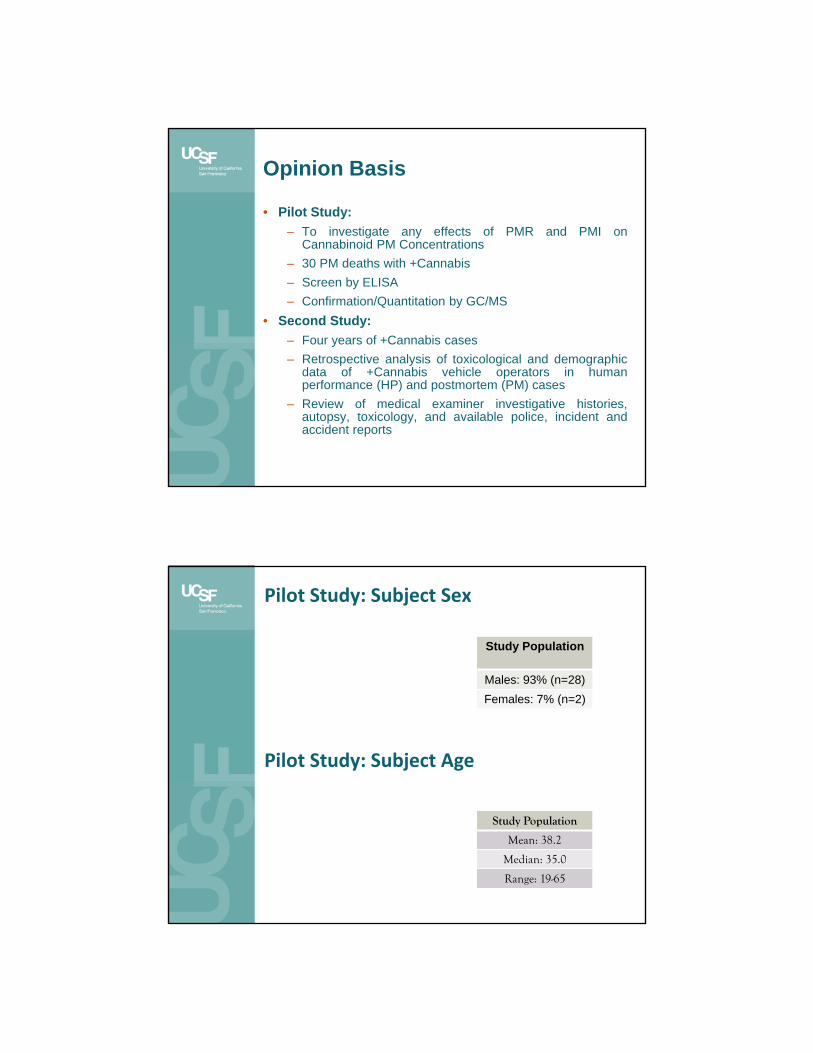
Opinion Basis
• Pilot Study:– To investigate any effects of PMR and PMI on
Cannabinoid PM Concentrations– 30 PM deaths with +Cannabis– Screen by ELISA– Confirmation/Quantitation by GC/MS
• Second Study:– Four years of +Cannabis cases– Retrospective analysis of toxicological and demographic
data of +Cannabis vehicle operators in humanperformance (HP) and postmortem (PM) cases
– Review of medical examiner investigative histories,autopsy, toxicology, and available police, incident andaccident reports
Pilot Study: Subject Sex
Study Population
Males: 93% (n=28)Females: 7% (n=2)
Pilot Study: Subject Age
Study Population
Mean: 38.2
Median: 35.0
Range: 19-65
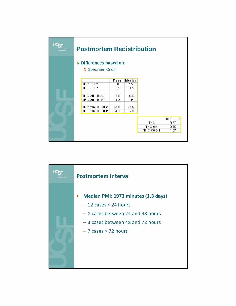
Postmortem Redistribution
Differences based on: Specimen Origin
Postmortem Interval
• Median PMI: 1973 minutes (1.3 days)
– 12 cases < 24 hours
– 8 cases between 24 and 48 hours
– 3 cases between 48 and 72 hours
– 7 cases > 72 hours
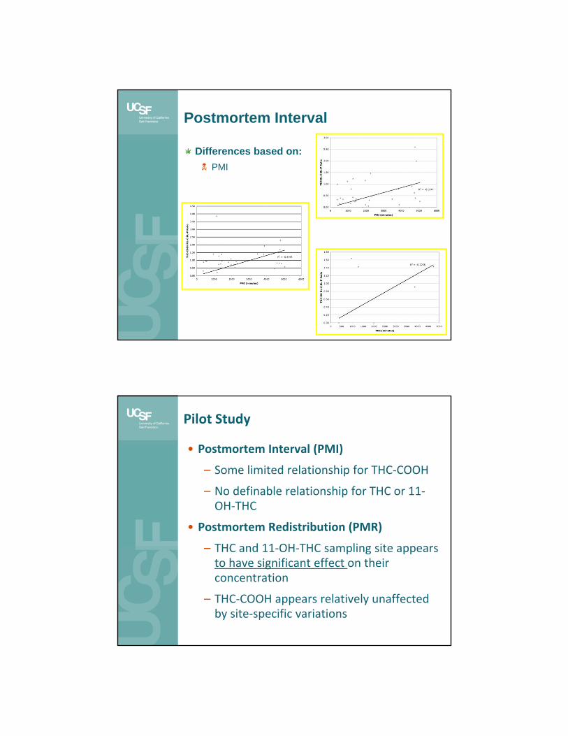
Postmortem Interval
Differences based on: PMI
Pilot Study
• Postmortem Interval (PMI)
– Some limited relationship for THC‐COOH
– No definable relationship for THC or 11‐OH‐THC
• Postmortem Redistribution (PMR)
– THC and 11‐OH‐THC sampling site appears to have significant effect on their concentration
– THC‐COOH appears relatively unaffected by site‐specific variations

Second Study: Subject Selection
Group 1: – JAN 2010 - DEC 2013– Arrested vehicle
operators (all motor vehicle drivers) for allegedly driving while impaired
– SF OCME determined that cannabinoids were present in their venous blood (BL-V)
Group 2:– JAN 2010 - DEC 2013– Vehicle operators who
died while operatingvehicle on publicroadways
– SF OCME determinedthat cannabinoids werepresent in their peripheralblood (BL-P), and/orcardiac/central blood(BL-C)
Blood Specimens
• Group 1: Arrested Drivers– Venipuncture within 3
hours of driving– BL-V
• NaF (100 mg)• C2K2O4 (20 mg)
– Stored at 4°C fromtime of accessioning andanalysis (few hours-fewweeks), until disposal (1year after analysis)
• Group 2: Deceased Drivers– BL-P and/or BL-C
collected at autopsy– Stored overnight in morgue
at 4°C– Specimens stored at 4°C
in the FLD from time ofaccessioning and analysis(few hours-few weeks),until disposal (1 year afteranalysis)
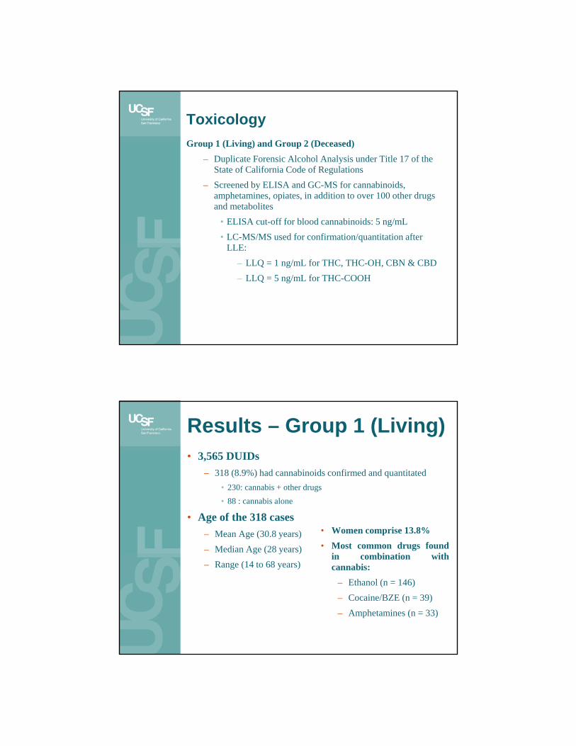
ToxicologyGroup 1 (Living) and Group 2 (Deceased)
– Duplicate Forensic Alcohol Analysis under Title 17 of the State of California Code of Regulations
– Screened by ELISA and GC-MS for cannabinoids, amphetamines, opiates, in addition to over 100 other drugs and metabolites
• ELISA cut-off for blood cannabinoids: 5 ng/mL• LC-MS/MS used for confirmation/quantitation after
LLE:– LLQ = 1 ng/mL for THC, THC-OH, CBN & CBD– LLQ = 5 ng/mL for THC-COOH
Results – Group 1 (Living)• 3,565 DUIDs
– 318 (8.9%) had cannabinoids confirmed and quantitated• 230: cannabis + other drugs• 88 : cannabis alone
• Age of the 318 cases– Mean Age (30.8 years)– Median Age (28 years)– Range (14 to 68 years)
• Women comprise 13.8%• Most common drugs found
in combination withcannabis:
– Ethanol (n = 146)– Cocaine/BZE (n = 39)– Amphetamines (n = 33)

Group 1 [Cannabinoid] in ng/mL; n=318 DUID drivers.Cannabis w/ or w/o other psychoactive compounds
THC THCCOOH
THCOH CBN
Mean 4.9 64.0 4.7 1.3
Median 3 41 3 1
Standard Deviation 4.9 79.7 4.1 0.5
Minimum 1 2 1 1
Maximum 33 720 22 2
Count 253 315 96 6
Group 1 [Cannabinoid] in ng/mL; n=88 DUIDdrivers. Cannabis only
THC THCCOOH
THCOH CBN
Mean 5.8 77.1 4.6 1.5
Median 4 50.5 3 1.5
Standard Deviation 5.0 100.3 4.6 0.7
Minimum 1 5 1 1
Maximum 26 720 22 2
Count 76 88 33 2

Results – Group 2 (Deceased)
• 5,190 PM Cases– 194 cases (3.7%) of these involved vehicular
deaths• 81 of the 194 (41.7%) were vehicle operators• 67 of the 81 Operators had positive toxicology
(82.7%)• 23 of the 67 Operators (28.4%) had blood cannabis
confirmed/quantitated• Women: 13.0%• Mean Age: 31.6; Median: 30; Range: 17-60 years
– Bicyclists: Highest Blood THC• Mean: 31.3 ng/mL; Median: 24 ng/mL
Group 2 [Cannabinoid] in ng/mL; n=19 or 23 PM operators with C/Q in BL-P. Cannabis w/ or w/o other psychoactive compounds
THC THCCOOH
THCOH
Mean 11.7 79.2 7.7
Median 4.5 41 4.5
Standard Deviation 15.4 136.0 12.4
Minimum 1 7 1
Maximum 50 552 43
Count 18 15 10

Group 2 [Cannabinoid] in ng/mL; n=8 or 9 PM operators with C/Q in BL-P. Cannabis only
THC THCCOOH
THCOH
Mean 20.3 114.7 11.2
Median 19.5 44 4
Standard Deviation 20 195 18
Minimum 1 7 1
Maximum 50 552 43
Count 8 7 5
Analysis of Variance
• 230 Group 1 DUID drivers with cannabis and other drugs to 88 Group 1 DUID drivers with cannabis-only
– No Statistically Significant Differences for THC, THC-OH or THC-COOH
• 11 Group 2 PM drivers with cannabis and other drugs to 8 Group 2 PM drivers with cannabis-only
– Statistically Significant Difference for THC (P-Value: 0.02)
– No Statistically Significant Differences for THC-OH or THC-COOH
• 318 Group 1 DUID drivers (with cannabis and other dugs) to 19 Group 2 PM drivers (with cannabis and other drugs)
– Statistically Significant Difference for THC (P-Value: 0.0000009)
– No Statistically Significant Differences for THC-OH or THC-COOH

Results
• Median THC in DUID arrested drivers withcannabis and other drugs is 3 ng/mL
• Median THC in DUID arrested drivers withcannabis only is 4 ng/mL
• Median THC in deceased drivers with cannabisand other drugs is 4.5 ng/mL
• Median THC in deceased drivers with cannabisonly is 19.5 ng/mL
• THC-OH and THC-COOH blood concentrationsappear to be comparable between DUID and PMcases
“The amount of THC, one of the activecomponents of marijuana, in Bland'ssystem was 18 micrograms per liter,according to the report released Monday.That's more than three times the legallimit for drivers in Colorado andWashington, states that permit therecreational use of marijuana.”
• THC blood concentrations between DUID and PMcases cannot be compared to each other as they seem toexhibit statistically-significant differences

Sometimes we get lucky: Hospital (AM) specimens
• ATV Rollover Accident• Middle-aged man involved in ATV rollover• Transported to hospital with “relatively minor
injuries” including broken clavicle• Dies from lacerated subclavian vessel and
massive bleed• Postmortem toxicology negative except +
cannabis– AM Blood (2 hours post accident):
• THC 14 ng/mL; THC-COOH: 110 ng/mL
– PM Blood (60 hours post accident):• THC 31 ng/mL; THC-COOH: 19 ng/mL
25
Sometimes we get lucky: Hospital (AM) specimens
• Young man injured in single MVA• 16.5 hours survival time in hospital• Toxicology
– AM Blood EtOH: 0.16 % (w/v)– AM Blood THC: < 1 ng/mL– AM Blood THC-COOH: 7 ng/mL
– PM Blood (Femoral) THC: 54 ng/mL– PM Blood (Femoral) THC-COOH: 8 ng/mL
(possible contamination from groin area’s fat?)
26
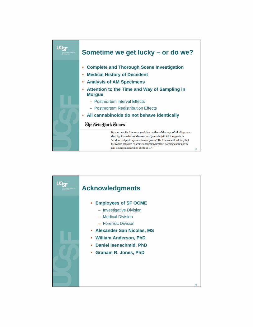
Sometime we get lucky – or do we?
• Complete and Thorough Scene Investigation• Medical History of Decedent• Analysis of AM Specimens• Attention to the Time and Way of Sampling in
Morgue– Postmortem interval Effects– Postmortem Redistribution Effects
• All cannabinoids do not behave identically
27
Acknowledgments
• Employees of SF OCME– Investigative Division– Medical Division– Forensic Division
• Alexander San Nicolas, MS• William Anderson, PhD• Daniel Isenschmid, PhD• Graham R. Jones, PhD
28