workbook to accompany understanding anato ph...
TRANSCRIPT

SECOND EDITION
WORKBOOK TO ACCOMPANY
UNDERSTANDING
ANATO &
PH SIOLOG

Workbook to Accompany
UNDERSTANDING
ANATOMY &
PHYSIOLOGYA Visual, Auditory, Interactive Approach2nd edition
4374_FM_i-x 12/2/14 10:00 AM Page i

4374_FM_i-x 12/2/14 10:00 AM Page ii
Workbook to Accompany
UNDERSTANDING
ANATOMY &
PHYSIOLOGYA Visual, Auditory, Interactive Approach
Gale Sloan Thompson, RN
2nd Edition
4374_FM_i-x 12/2/14 10:00 AM Page iii

F. A. Davis Company1915 Arch StreetPhiladelphia, PA 19103www.fadavis.com
Copyright © 2015 by F. A. Davis Company
Copyright © 2015 by F. A. Davis Company. All rights reserved. This book is protected by copyright. No part of it may bereproduced, stored in a retrieval system, or transmitted in any form or by any means, electronic, mechanical, photocopying,recording, or otherwise, without written permission from the publisher.
Printed in the United States of America
Last digit indicates print number: 10 9 8 7 6 5 4 3 2 1
Publisher, Nursing: Lisa B. HouckDirector of Content Strategy: Darlene D. PedersenContent Project Manager II: Victoria WhiteDesign and Illustration Manager: Carolyn O’Brien
As new scientific information becomes available through basic and clinical research, recommended treatments and drugtherapies undergo changes. The author(s) and publisher have done everything possible to make this book accurate, up to date,and in accord with accepted standards at the time of publication. The author(s), editors, and publisher are not responsible forerrors or omissions or for consequences from application of the book, and make no warranty, expressed or implied, in regardto the contents of the book. Any practice described in this book should be applied by the reader in accordance withprofessional standards of care used in regard to the unique circumstances that may apply in each situation. The reader isadvised always to check product information (package inserts) for changes and new information regarding dose andcontraindications before administering any drug. Caution is especially urged when using new or infrequently ordered drugs.
Authorization to photocopy items for internal or personal use, or the internal or personal use of specific clients, is granted byF. A. Davis Company for users registered with the Copyright Clearance Center (CCC) Transactional Reporting Service,provided that the fee of $.25 per copy is paid directly to CCC, 222 Rosewood Drive, Danvers, MA 01923. For thoseorganizations that have been granted a photocopy license by CCC, a separate system of payment has been arranged. The feecode for users of the Transactional Reporting Service is: 8036-1169-2/04 0 + $.25.
4374_FM_i-x 12/2/14 10:00 AM Page iv

REVIEWERS
Tetteh Abbeyquaye, PhDAssistant ProfessorQuinsigamond Community CollegeWorcester, MA
Janice Ankenmann, RN, MSN, CCRN,FNP-C
ProfessorNapa Valley CollegeNapa, CA
Dan Bickerton, MSInstructorOgeechee Technical CollegeStatesboro, GA
Anne L. Brown, RN, BSNNursing Instructor Broome-Tioga BOCESBinghamton, NY
Susan E. Brown, MS, RNFacultyRiverside School of Health CareersNewport News, VA
Henry Steven Carter, MS, CRC, CVE
Coordinator of Continuing and Workforce Education/Instructor
El Centro CollegeDallas, TX
Thea L. Clark, RN, BS, MSCoordinator Practical NursingTulsa Technology CenterTulsa, OK
Ginny Cohrs, RN, BSNNursing FacultyAlexandria Technical CollegeAlexandria, MN
Tamera Crosswhite, RN, MSNNursing InstructorGreat Plains Technology CenterFrederick, OK
Fleurdeliza Cuyco, BS, MDDean of EducationPreferred College of Nursing, Los AngelesLos Angeles, CA
Judith L. Davis, RN, MSN, FNPPractical Nursing InstructorDelta-Montrose Technical CollegeDelta, CO
Carita Dickson, RNLVN InstructorSan Bernardino Adult School LVN
ProgramSan Bernardino, CA
Teddy Dupre, MSNInstructorCapital Area Technical CollegeBaton Rouge, LA
Hisham S. Elbatarny, MB BCh, MSc, MD
ProfessorSt. Lawrence College—Queen’s
UniversityKingston, Ontario
Alexander EvangelistaAdjunct FacultyThe Community College of Baltimore
CountyBaltimore, MD
John Fakunding, PhDAdjunct InstructorUniversity of South Carolina, BeaufortBeaufort, SC
Kelly Fleming, RN, BN, MSNPractical Nurse FacilitatorColumbia CollegeCalgary, Alberta
Ruby Fogg, MAProfessorManchester Community CollegeManchester, NH
Cheryl S. Fontenot, RNProfessorAcadiana Technical CollegeAbbeville, LA
Shena Borders Gazaway, RN, BSN, MSN
Lead Nursing/Allied Health InstructorLanier Technical CollegeCommerce, GA
Daniel G. Graetzer, PhDProfessorNorthwest UniversityKirkland, WA
Dianne Hacker, RN, MSNNursing InstructorCapital Area School of Practical
NursingSpringfield, IL
Leslie K. Hughes, RN, BSNPractical Nursing InstructorIndian Capital Technology CenterTahlequah, OK
Constance Lieseke, CMA (AAMA),MLT, PBT (ASCP)
Medical Assisting Faculty ProgramCoordinator
Olympic CollegeBremerton, WA
v
4374_FM_i-x 12/2/14 10:00 AM Page v

Julie S. Little, MSNAssociate ProfessorVirginia Highlands Community
CollegeAbingdon, VA
C. Kay Lucas, MEd, BS, ASNurse EducatorCommonwealth of Virginia
Department of Health Professions
Henrico, VA
Barbara Marchelletta, CMA (AAMA),CPC, CPT
Program Director, Allied HealthBeal College Bangor, ME
Nikki A. Marhefka, EdM, MT (ASCP),CMA (AAMA)
Medical Assisting Program Director
Central Penn CollegeSummerdale, PA
Jean L. Mosley, CMA (AAMA), AAS, BS
Program Director/InstructorSurry Community CollegeDobson, NC
Elaine M. Rissel Muscarella, RN, BSN
LPN InstructorJamestown, NY
Brigitte Niedzwiecki, RN, MSNMedical Assistant Program Director
and InstructorChippewa Valley Technical CollegeEau Claire, WI
Jill M. Pawluk, RN, MSNNursing InstructorThe School of Nursing at Cuyahoga
Valley Career CenterBrecksville, OH
Kathleen Hope Rash, MSN, RNCurriculum & Instructional Resource
CoordinatorRiverside Schools of NursingNewport News, VA
Amy Fenech Sandy, MS, MSDean, School of SciencesColumbus Technical CollegeColumbus, GA
Marianne Servis, RN, MSNNurse Educator/Clinical CoordinatorCareer Training SolutionsFredericksburg, VA
Glynda Renee Sherrill, RN, MSPractical Nursing InstructorIndian Capital Technology CenterTahlequah, OK
Cathy Soto, PhD, MBA, CMAEl Paso Community CollegeEl Paso, TX
Joanne St. John, CMAAdjunct Instructor—Health
ScienceIndian River State CollegeFort Pierce, FL
Diana A. Sunday, RN, BSN, MSN/ED
Nurse Educator—Practical NursingProgram
York County School of TechnologyYork, PA
Joyce B. Thomas, CMA (AAMA)InstructorCentral Carolina Community
CollegePittsboro, NC
Marianne Van Deursen, MS Ed, CMA(AAMA)
Medical Assisting ProgramDirector/Instructor
Warren County Community CollegeWashington, NJ
Monna L. Walters, MSN, RNDirector of Vocational Nursing
ProgramLassen Community CollegeSusanville, CA
Amy Weaver, MSN, RN, ACNS-BCInstructorYoungstown State UniversityYoungstown, OH
vi
4374_FM_i-x 12/2/14 10:00 AM Page vi

CONTENTS
PART I ORGANIZATION OF THE BODY
chapter 1 Orientation to the Human Body 1chapter 2 Chemistry of Life 11chapter 3 Cells 23
PAR T II COVERING, SUPPORT, AND MOVEMENT OF
THE BODY
chapter 4 Tissues 35chapter 5 Integumentary System 45chapter 6 Bones & Bone Tissue 51chapter 7 Skeletal System 61chapter 8 Joints 81chapter 9 Muscular System 93
PART III REGULATION AND INTEGRATION OF THE BODY
chapter 10 Nervous System 109chapter 11 Sense Organs 135chapter 12 Endocrine System 151
PART IV MAINTENANCE OF THE BODY
chapter 13 Blood 163chapter 14 Heart 175chapter 15 Vascular System 187chapter 16 Lymphatic & Immune Systems 205chapter 17 Respiratory System 221chapter 18 Urinary System 239chapter 19 Fluid, Electrolyte, and Acid-Base Balance 249chapter 20 Digestive System 257chapter 21 Nutrition & Metabolism 277chapter 22 Human Microbiome 287
PART V CONTINUITY
chapter 23 Reproductive Systems 295chapter 24 Pregnancy & Human Development 307chapter 25 Heredity 315
vii
4374_FM_i-x 12/2/14 10:00 AM Page vii

4374_FM_i-x 12/2/14 10:00 AM Page viii

INTRODUCTION
Most of us have one predominant learning style. Visuallearners learn best when they can see a figure or an image;auditory learners prefer verbal explanations; kinestheticlearners need to incorporate movement into their learningtime. Regardless, everyone can facilitate learning byemploying a variety of techniques when studying, no mattertheir particular learning style.
Unlike any other study guide, the Understanding Anatomy& Physiology study guide is packed with unique activitiesinvolving drawing, coloring, and highlighting in addition tomore traditional activities such as fill-in-the-blank andcrossword puzzles. The drawing, coloring, highlighting, andwriting activities give kinesthetic learners a chance to move,something they long to do when learning. Even if you’re notprimarily a kinesthetic learner, using your muscles will breakup the monotony of studying and, by stimulating differentparts of your brain, improve your learning experience. What’smore, the colorful results of your drawing, coloring, andhighlighting will provide quick, visual cues regardingimportant topics when you review.
To make the most of this study guide, you’ll need at least10 different colored pencils. Colored pencils will allow youto color various shades of each color, making them a betterchoice than colored pens. You’ll also need an assortment ofcolorful highlighters, such as yellow, green, blue, and pink.
The following list summarizes the activities you’ll find inthis study guide:
● Conceptualize in Color: This activity involves coloringanatomical structures different colors. Kinestheticlearners will benefit from the movement associated withcoloring as they focus on the various features of thehuman body, whereas visual learners will appreciate thevisual aspect of coloring. Auditory learners can enhancetheir learning experience by saying the names of variousstructures out loud as they color. And, because coloringforces you to slow down and focus on one structure at atime, all learners will benefit.
● Drawing Conclusions: Combining drawing or coloringwith some other activity, such as fill-in-the-blanksentences, “Drawing Conclusions” will hone yourreasoning skills while allowing you to link written words
to something visual. The physical activity of drawingimproves learning that much more.
● Just the Highlights: In this activity, you’ll placesentences describing various structures, physiologicalprocesses, or disorders into separate groups byhighlighting sentences in distinct colors. For example,in Chapter 5, Integumentary System, a “Just theHighlights” activity contains sentences describingfeatures of first-, second-, and third-degree burns. You’llbe asked to highlight sentences pertaining to first-degreeburns in yellow, second-degree burns in orange, andthird-degree burns in pink. Once complete, each groupwill be visually apparent, making reviewing easy.
● Illuminate the Truth: A variation of fill-in-the blank,you’ll use a highlighter to identify the correct word orphrase that completes each sentence.
● Fill in the Gaps: In this fill-in-the-blank activity, youwill write the correct word or phrase to complete eachsentence. A Word Bank is provided.
● Sequence of Events: This activity will challenge you toplace statements about a physiological process—such asthe formation of cerebrospinal fluid—in propersequence by inserting numbers in the blank line beforeeach sentence.
● Make a Connection: This two-part activity involves firstunscrambling words to reveal the names of certainstructures or processes. Once identified, you’ll draw aline from the word to a statement or description,linking the two together. For example, in Chapter 10,Nervous System, you’ll unscramble words to discover thenames of nervous system cells. You’ll then draw lines tolink the name of a cell to sentences describingcharacteristics of that type of cell.
● List for Learning: This activity will test your recall byasking you to make a list of certain things, such as thefive functions of skin.
● Puzzle It Out: This traditional crossword puzzle is afun way to test your knowledge of key termsrelated to anatomy and physiology.
ix
4374_FM_i-x 12/2/14 10:00 AM Page ix

● Describe the Process: Using figures as visual cues,you’ll test your recall of physiological processes bydescribing the steps in a particular process, such asthat of endochondral ossification. Successfullycompleting this activity will assure you that youhave committed the process to memory.
Each activity in this book focuses on a specific topic. Thatway, if you are struggling in a particular area, you can choosethe activities relating to that topic. After completing all the
exercises for a chapter, consult the answer guide in the backof the book to check your answers.
Mastering the topic of anatomy and physiology requiresstudy and repetition. There is no other way to construct thefoundation of knowledge upon which you’ll build yourfuture career in health care. The unique activities in theUnderstanding Anatomy & Physiology study guide will aid youtoward that end: They will break up the monotony ofstudying as you use color and movement to help you commitkey facts to memory.
x
4374_FM_i-x 12/2/14 10:00 AM Page x

chap
ter 1ORIENTATION TO
THE HUMAN BODY
As you begin to study anatomy and physiology, you first need to understand how the body is organized. What’smore, you need to learn the terms used to describe the various regions of the body. Doing the activities in thischapter will help.
Chapter 1 Orientation to the Human Body 1
List for Learning: Organization of the Body
The various elements of the human body are organized in a hierarchy ranging from the very simple to the verycomplex. Use the spaces below to list the major structures in this hierarchy, beginning with the atom and endingwith the human organism.
1. Atoms
2.
3.
4.
5.
6.
7.
8. A human organism
4374_Ch01_001-010 10/6/14 1:14 PM Page 1

2 Chapter 1 Orientation to the Human Body
Make a Connection: Types of Tissue
Unscramble the words below to discover the names of the four types of tissue found in the human body. Then drawa line to link each type of tissue with its particular characteristics.
1. LEAPILEHIT
2. VICECENTON
3. CLESUM
4. ENREV
A. Contracts to produce movement
B. Examples include the brain and nerves
C. Covers or lines body surfaces
D. Generates and transmits impulses to regulate bodyfunction
E. Connects and supports parts of the body; may alsotransport and store materials
F. Examples include the outer layer of the skin, thewalls of capillaries, and kidney tubules
G. Examples include bone, cartilage, and adiposetissue
H. Examples include skeletal muscles and the heart
4374_Ch01_001-010 10/6/14 1:14 PM Page 2

Chapter 1 Orientation to the Human Body 3
Drawing Conclusions: Directional Terms
Use the figure above to complete the following instructions.
A. Draw a midline incision on the patient’s abdomen, superior to the navel.
B. Draw a deep wound on the lateral portion of the right knee.
C. Draw a superficial wound on the medial right ankle.
D. Place a bandage on the proximal right arm.
E. Place an adhesive bandage on the left leg, distal to the knee.
F. Place a small bandage on the patient’s abdomen, inferior to the navel.
4374_Ch01_001-010 10/6/14 1:14 PM Page 3

4 Chapter 1 Orientation to the Human Body
Puzzle It Out: Organ Systems
The human body consists of 11 organ systems, with each contributing to a particular function. Test your knowledgeabout these systems by completing the following crossword puzzle.
ACROSS
1. System consisting of skin, hair, and nails3. System consisting of the heart, arteries, veins, and capillaries6. System that participates in heat production7. System involved in the breakdown and absorption of nutrients9. System that helps regulate blood volume and pressure
10. System charged with the control, regulation, and coordination of other systems as well as sensation and memory11. System that produces immune cells
DOWN
2. System consisting of the testes, vas deferens, prostate, seminal vesicles, and penis in males and the ovaries,fallopian tubes, uterus, vagina, and breasts in females
4. System consisting of the nose, pharynx, larynx, trachea, bronchi, and lungs5. System involved in hormone production8. System that plays a key role in blood formation
4374_Ch01_001-010 10/6/14 1:14 PM Page 4

Chapter 1 Orientation to the Human Body 5
Drawing Conclusions: Body Planes
Test your knowledge of body planes by drawing the planes as instructed on the figure below. Then fill in the blanksto correctly describe each plane.
1. Draw a green square through the figure to illustrate a sagittal plane. Also called a
plane, this plane divides the body into and sides.
2. Draw an orange square to divide the body into two halves using a transverse plane. Also called a
plane, this plane divides the body into and
portions.
3. Draw a purple square through the body to illustrate a frontal plane. Also called a
plane, this plane divides the body into and portions.
4374_Ch01_001-010 10/6/14 1:14 PM Page 5

6 Chapter 1 Orientation to the Human Body
Conceptualize in Color: Body Regions
Various terms are used to describe different regions in the body. These terms are used extensively when performingclinical examinations and medical procedures. To help solidify your knowledge of the locations of these regions,color the figures below as described.
● Patellar area: Green
● Femoral area: Pink
● Pedal area: Yellow
● Gluteal area: Orange
● Palmar area: Orange
● Digital area: Green
● Scapular area: Pink
● Inguinal area: Green
● Axillary area: Blue
● Sacral area: Red
● Brachial area: Pink
● Deltoid area: Green
● Popliteal area: Green
● Sternal area: Red
● Pelvic area: Blue
● Pubic area: Yellow
● Antecubital area: Blue
● Buccal area: Yellow
● Occipital area: Red
● Lumbar area: Yellow
● Otic area: Blue
● Carpal area: Purple
4374_Ch01_001-010 10/6/14 1:14 PM Page 6

Chapter 1 Orientation to the Human Body 7
Drawing Conclusions: Body Cavities
The body’s internal organs are contained in spaces called cavities. Color each cavity a color of your own choosing;then write the name of the cavity in the space provided.
Diaphragm
�
�
�
�
�
�
�
1.
2.
3.
4.
5.
6.
7.
8.
4374_Ch01_001-010 10/6/14 1:14 PM Page 7

8 Chapter 1 Orientation to the Human Body
Drawing Conclusions: Abdominal Regions
Because the abdominopelvic cavity is so large and because it contains numerous organs, it is divided into regions.Using the figure below, draw lines to divide this cavity into nine regions. Identify the names of the regions bycoloring each region as described.
● Right hypochondriac region: Orange
● Left iliac region: Green
● Umbilical region: Yellow
● Right iliac region: Blue
● Left lumbar region: Brown
● Left hypochondriac region: Pink
● Right lumbar region: Purple
● Hypogastric region: Red
● Epigastric region: Gray
Next, identify the organs in each region by placing the correct letter in the proper square on the figure above. Forexample, if the small intestines, descending colon, and sigmoid colon are found in the right hypochondriac region,you would insert the letter A in that region on the figure.
A. Small intestines, descending colon, sigmoid colon
B. Small intestines, appendix, cecum, ascending colon
C. Pancreas, small intestines, transverse colon
D. Liver, gallbladder, right kidney
E. Stomach, liver (tip), left kidney, spleen
F. Stomach, liver, pancreas, right and left kidneys
G. Small intestines, descending colon, left kidney
H. Liver (tip), small intestines, ascending colon, right kidney
I. Small intestines, sigmoid colon, bladder
4374_Ch01_001-010 10/6/14 1:14 PM Page 8

Chapter 1 Orientation to the Human Body 9
Illuminate the Truth: The Basics
Review some key concepts from this chapter by highlighting the correct word or words in each of the followingsentences.
1. The study of the structure of the body is called (physiology)(anatomy).
2. The smallest living units that make up the body’s structure are called (organelles)(cells).
3. Structures of two or more tissue types that work together are called (organs)(organ systems).
4. Anatomical position is when the body is standing erect, arms at the sides, with palms facing(backward)(forward).
5. A disruption in one organ system usually has (no effect on) (consequences in) other systems.
Fill in the Gaps: Homeostasis
To maintain a stable environment, the body must constantly monitor conditions and make adjustments as conditionschange. This process is called homeostatic regulation. Review this crucial process by filling in the blanks to complete eachof the following sentences. Choose from the words listed in the Word Bank below. (Hint: Not all the words will be used.)
1. Homeostasis is the state of dynamic of the internal environment of the body.
2. To maintain homeostasis, a system must have three components: (1) a , which
detects external changes that could influence the environment; (2) a , which
receives and processes the information from the first component; and (3) a , which responds to signals from the second component by either opposing or enhancing the stimulus.
3. Negative feedback is when the effector the stimulus and the direction of change.
4. Positive feedback is when the effector the stimulus and the direction of change.
5. Most systems supporting homeostasis operate by feedback.
AMPLIFIES
CONTROL CENTER
EFFECTOR
EQUILIBRIUM
NEGATIVE
OPPOSES
POSITIVE
RECEPTOR
REINFORCES
REVERSES
SUPPRESSES
4374_Ch01_001-010 10/22/14 8:27 AM Page 9

4374_Ch01_001-010 10/6/14 1:14 PM Page 10

chap
ter 2CHEMISTRY OF LIFE
The human body is made of chemicals. What’s more, life depends upon a precise balance between all thosechemicals. So before you can understand how the body functions, you must have a firm grasp on how thechemicals in the body interact. The exercises in this chapter should help you do just that.
Chapter 2 Chemistry of Life 11
Illuminate the Truth: Basic Structures
Highlight the word or phrase that correctly completes each sentence.
1. An element’s atomic number is determined by the (number of protons)(number of neutrons) in the nucleus.
2. An element’s atomic weight is determined by adding the number of (protons, neutrons, and electrons)(protonsand neutrons).
3. The number of electrons equals the number of (protons)(protons and neutrons added together).
4. Atoms are electrically (neutral)(positive).
5. The number of electron rings, or shells, (varies)(is the same) between atoms.
6. (All)(Some) isotopes of an element are unstable and emit radiation as they decay.
7. An element’s unique chemical properties result from (the various combinations of protons, neutrons, andelectrons)(the various types of protons, neutrons, and electrons making up the atoms of the element).
4374_Ch02_011-022 10/6/14 1:12 PM Page 11

12 Chapter 2 Chemistry of Life
Puzzle It Out: Chemistry Terms
Test your knowledge of general chemistry terms by completing the crossword puzzle below.
ACROSS
4. Anything that has mass and occupies space5. An atom of an element that contains a different
number of neutrons7. Center of an atom9. Element represented by the letter O
10. A pure substance that can’t be broken down ordecomposed into two or more substances
11. Particles in the center of an atom that carry anegative charge
13. Tiny particles that whirl around the center of anatom
DOWN
1. Particles in the center of an atom that carry apositive charge
2. Element represented by the letter C3. One of the four elements that comprise the body:
carbon, hydrogen, oxygen, and 6. Element represented by the letter H8. Chemical combinations of two or more elements
12. Smallest part of an element
4374_Ch02_011-022 10/6/14 1:12 PM Page 12

Chapter 2 Chemistry of Life 13
Drawing Conclusions: Chemical Bonds
First identify the key characteristics of ionic, covalent, and hydrogen bonds by filling in the blanks in the followingsentences. Then, in the space provided, draw an example of each type of bond.
1. Ionic Bonds
Ionic bonds are formed when one atom (a) an electron from its outer shell to
another atom. The electrical charge of the atom then changes from (b) to either
(c) or (d) .
Draw an example of an ionic bond between sodium and chlorine atoms in the space provided. An illustration of asodium and chlorine atom has been provided to get you started.
Na Cl
2. Covalent Bonds
In covalent bonds, two atoms (a) one or more pairs of electrons. Covalent bonds are
(b) than ionic bonds.
Draw an example of a double covalent bond between carbon and oxygen atoms to form CO2. An illustration of acarbon and oxygen atom has been provided to get you started.
Oxygen atom
8p+
8n0
Carbon atom
6p+
6n0
3. Hydrogen Bonds
A hydrogen bond is a weak (a) between a slightly positive (b) atom in
one molecule and a slightly negative (c) or (d) atom in another.
Using the illustration of a water molecule (shown here), draw an example of a hydrogen bond between two watermolecules.
O
H
H
–
+
+
4374_Ch02_011-022 10/6/14 1:12 PM Page 13

14 Chapter 2 Chemistry of Life
Make a Connection: Chemical Reactions
Unscramble the words on the left to reveal the three types of chemical reactions. Then draw lines to link each typewith its particular characteristics.
1. HENSISSTY
2. OPTICDOMINOES
3. HANGEXEC
a. Involves the breakdown of a complex substanceinto two or more simpler substances
b. Requires energy input
c. Represented by the equation AB + CD →AC + BD
d. Occurs when two molecules exchange atoms orgroups of atoms, forming two new compounds
e. Involves the combination of two or moresubstances to form a different, more complexsubstance
f. Represented by the equation AB →A + B
g. Releases energy
h. Represented by the equation A + B → AB
4374_Ch02_011-022 10/6/14 1:12 PM Page 14

Chapter 2 Chemistry of Life 15
Puzzle It Out: Chemistry Concepts
Hone your knowledge of chemistry concepts by completing the following crossword puzzle.
ACROSS
2. Particle composed of two or more atoms unitedby a chemical bond
4. The electrons in an atom’s outer shell5. Energy in motion7. Type of molecule with an uneven distribution of
electrons8. Atoms having a positive charge9. Compounds that ionize in water to create a
solution capable of conducting electricity10. Process whereby ionic bonds dissociate in water
to create a solution of positively and negativelycharged particles
12. Involves building larger and more complexchemical molecules, which requires energy input
13. The capacity to do work
DOWN
1. Reactions that can go in either direction underdifferent circumstances
3. All the chemical reactions in the body6. Involves breaking down complex compounds into
simpler ones, releasing energy in the process10. Electrically charged atoms11. Atoms having a negative charge
4374_Ch02_011-022 10/6/14 1:12 PM Page 15

16 Chapter 2 Chemistry of Life
Just the Highlights: Characteristics of Water
Unlike any other fluid, water has a number of characteristics that make it essential for life. Link the characteristicsof water with its performance in the body by highlighting each of the following sentences as suggested.
● Highlight in pink the sentences describing water’saction as a solvent.
● Highlight in yellow the sentences describing water’saction as a lubricant.
● Highlight in blue the sentences describing water’sability to absorb and release heat.
1. Cells receive the chemicals they need to function.
2. The heart beats freely without encounteringfriction.
3. Joints operate smoothly, allowing the body tomove.
4. The body can maintain a stable temperaturedespite changes in activity level or environmentaltemperature.
5. Large chemical compounds are broken down intocomponents cells can use.
6. The lungs expand and contract freely for effortlessbreathing.
7. Sweat cools the body.
Make a Connection: Body Fluids
Unscramble the words on the left to discover two types of fluid. Then draw lines linking each fluid type to itscharacteristics as well as to examples of each.
1. DOCUMPON
2. REMITUX
a. Results when two or more mixtures blendtogether, with each retaining its own chemicalproperties
b. Results when two or more elements combine tomake a new substance that has its own chemicalproperties
c. Substances involved can be separated
d. Example: Table salt
e. Example: Water
f. Example: Salt in scrambled eggs
g. Example: Lemonade
4374_Ch02_011-022 10/6/14 1:12 PM Page 16

Chapter 2 Chemistry of Life 17
Just the Highlights: Types of Mixtures
Mixtures of substances in water can be solutions, colloids, or suspensions. Identify the characteristics of each byhighlighting each of the following sentences as suggested.
● Highlight in yellow the sentences describingsolutions.
● Highlight in orange the sentences describingcolloids.
● Highlight in blue the sentences describingsuspensions.
1. Usually consist of a mixture of protein and water
2. Contain large particles, causing them to becloudy or opaque
3. May be gas, solid, or liquid
4. Can change from a liquid to a gel
5. Are clear, with no visible particles; particles don’tseparate when the mixture is allowed to stand
6. Have particles that separate if the solution isallowed to stand
7. Contain particles small enough to staypermanently mixed but large enough to make themixture cloudy
8. Example in the body: Glucose in the blood
9. Example in the body: Blood cells in plasma
10. Example in the body: Albumin in blood plasma
Fill in the Gaps: The pH Scale
Fill in the blanks with the appropriate words to complete each sentence. Choose from the words listed in the WordBank below.
1. Solutions with a pH less than 7 are .
2. Solutions with a pH greater than 7 are or .
3. When acidic solutions are dissolved in water, they release ions.
4. Alkaline solutions release ions when dissolved in water.
5. Acidic solutions are called proton .
6. Alkaline solutions are called proton .
ACID
ACIDIC
ACCEPTORS
ALKALINE
BASE
BASIC
DONORS
HYDROGEN (H+)
HYDROXIDE (OH−)
NEUTRAL
4374_Ch02_011-022 10/6/14 1:12 PM Page 17

18 Chapter 2 Chemistry of Life
Illuminate the Truth: Organic Compounds
Highlight the word or phrase that correctly completes each sentence.
1. The term organic is used to describe compounds (containing carbon)(without carbon).
2. (Proteins)(Carbohydrates) are the body’s main energy source.
3. Monosaccharides, disaccharides, and polysaccharides are all types of (carbohydrates)(fats).
4. The primary form of sugar found in the body is (glycogen)(glucose).
5. The stored form of glucose is (galactose)(glycogen).
6. Triglycerides, the most abundant lipid in the body, (serve no known purpose and are a major contributor toheart disease)(function as a concentrated source of energy).
7. Saturated fatty acids (form a solid mass)(are liquid) at room temperature.
8. Cholesterol is the body’s chief (steroid)(triglyceride).
9. (Proteins)(Fats) are the most abundant, and most important, organic compound in the body.
10. Nonessential amino acids are so named because they (are not necessary for normal body function)(can bemanufactured by the body).
11. Proteins consist of (electrolytes)(amino acids) linked together by peptide bonds.
12. Unsaturated fatty acids are derived mostly from (animal)(plant) sources.
13. A protein’s function is determined by (the number of amino acids it contains)(its shape).
List for Learning: Lipids
List five major roles that lipids fulfill in the body.
1.
2.
3.
4.
5.
7. The greater the concentration of hydrogen (H+) ions, the stronger the .
8. The greater the concentration of hydroxide (OH–) ions, the stronger the .
9. A solution containing equal numbers of H+ and OH– ions is known as a solution.
4374_Ch02_011-022 10/6/14 1:12 PM Page 18

Chapter 2 Chemistry of Life 19
Make a Connection: Proteins
Unscramble the words below to reveal the names of some of the body’s proteins. Then draw a line linking each ofthe proteins to its function.
1. TEARINK
2. BIASEDINTO
3. NILISNU
4. NIMBLEGOHO
5. CLEANLOG
6. SNEZEMY
a. Defend the body against bacteria
b. Carries oxygen in the blood
c. Strengthens nails, hair, and skin
d. Gives structure to bones, cartilage, and teeth
e. Serves as a chemical messenger to cells throughoutthe body
f. Act as catalysts for crucial chemical reactions
List for Learning: Cholesterol
List four key roles cholesterol fulfills in the body.
1.
2.
3.
4.
4374_Ch02_011-022 10/6/14 1:12 PM Page 19

20 Chapter 2 Chemistry of Life
Fill in the Gaps: Glucose and Glycogen
Fill in the blanks in the following paragraph to clarify how the body uses its prime energy source. Choose from thewords listed in the Word Bank below. (Hint: Some words are used multiple times.)
GLUCOSE GLYCOGEN LIVER MUSCLES
When blood (1) levels are high (such as after eating), the (2)
converts excess (3) into (4) , which it then stores. When blood
(5) levels drop (such as between meals), the (6) converts
(7) back into (8) and releases it into the blood. This keeps blood
(9) levels within normal limits and provides cells with a constant supply of energy. The
(10) also store (11) to meet its energy needs during physical exercise.
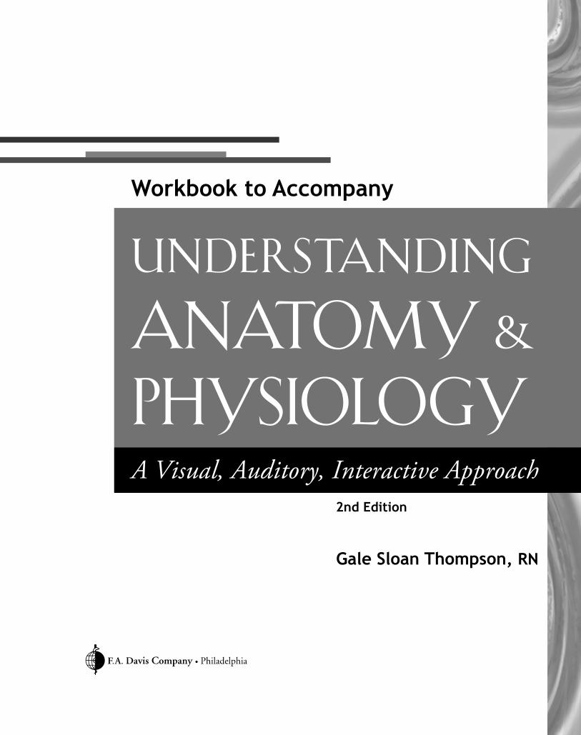
Describe the Process: ATP
Cells use energy in the form of ATP. Test your understanding of how the body’s cells obtain and then restore theirsupply of energy by describing the process in the spaces provided. Use the illustrations as a guide. To get you started,the first part of each sentence is provided for each illustration.
P P P
1. ATP consists of
.
4374_Ch02_011-022 10/6/14 1:12 PM Page 20
Chapter 2 Chemistry of Life 21
P P P
P P P
Energy
P P P
Energy
2. When one of the bonds is broken through a chemical reaction,
.
3. After the bond is broken,
.
4. Meanwhile, the cell uses
.
4374_Ch02_011-022 10/6/14 1:13 PM Page 21

4374_Ch02_011-022 10/6/14 1:13 PM Page 22

chap
ter 3CELLS
Cells may be microscopic, but they are far from simple. In fact, each cell is packed with specialized structures thatallow it to perform various tasks crucial to human life. Use this chapter to enhance your understanding of cellularstructure and function. Doing so is the key to understanding the inner workings of the body, in both health anddisease.
Chapter 3 Cells 23
Conceptualize in Color: The Cell
Identify the various structures in a human cell by coloring the figure below. Use the suggested colors or choose colorsof your own.
● Plasma membrane: Blue
● Nucleus and nuclearenvelope: Purple
● Nucleolus: Dark blue
● Centriole: Green
● Mitochondria: Pink
● Golgi apparatus:Orange
● Smooth endoplasmicreticulum: Brown
● Rough endoplasmicreticulum: Tan
● Lysosomes: Red
● Ribosomes: Black
● Cytoplasm: Yellow
4374_Ch03_023-034 10/6/14 1:17 PM Page 23

24 Chapter 3 Cells
Fill in the Gaps: Plasma Membrane
Test your knowledge of the plasma membrane by filling in the blanks in the following sentences. Choose fromthe words listed in the Word Bank below. (Hint: Words may be used more than once; also, not all the wordswill be used.)
1. Besides defining the boundaries of the cell, the plasma membrane regulates the of
into and out of the cell.
2. The plasma membrane consists of , , and
.
3. The heads of the phospholipids are water , causing them to face
the fluid in the cell’s interior and exterior.
4. The tails of the phospholipids are water , causing them to face
the fluid in the cell’s interior and exterior.
5. molecules stiffen and strengthen the plasma membrane.
6. are embedded in various spots in the membrane and fulfill a number of roles.
7. Because some substances can easily pass through the membrane, while others cannot, the plasma membrane is
called .
AWAY FROM
CHOLESTEROL
FEARING
GLUCOSE
LOVING
PASSAGE
PHOSPHOLIPIDS
POROUS
PROTEIN(S)
SELECTIVELY PERMEABLE
SUBSTANCES
TOWARD
List for Learning: Proteins and the Plasma Membrane
Using the spaces provided, describe the three roles fulfilled by proteins in the plasma membrane.
1.
2.
3.
4374_Ch03_023-034 10/6/14 1:17 PM Page 24

Chapter 3 Cells 25
Puzzle It Out: Cellular Structures
Complete the following crossword puzzle to enhance your learning of cellular structures and their functions.
ACROSS
1. Thread-like structures composed of DNA andprotein extending throughout the nucleus
3. Supporting framework of the cell5. Apparatus that prepares and packages proteins for
export to other parts of the body6. Rod-like structures consisting of tightly coiled
DNA10. Bundles of microtubules that participate in cell
division11. Cellular garbage disposals12. The cell’s control center13. Whip-like projection that helps move a cell
DOWN
1. Hair-like processes that propel substances along acell’s surface
2. The cell’s powerhouses4. Structures filling the cytoplasm that perform
specific tasks in cellular metabolism7. Folds of the cell membrane that greatly increase a
cell’s surface area8. Gel-like substance that fills the space between the
plasma membrane and nucleus9. Cell’s protein-producing structures
4374_Ch03_023-034 10/6/14 1:17 PM Page 25

26 Chapter 3 Cells
Drawing Conclusions: Diffusion
Fill in the blanks in the following sentences to demonstrate your knowledge of diffusion. Then use theaccompanying figure to draw the process.
1. Diffusion is a transport mechanism, meaning that it doesn’t require energy.
2. Diffusion involves the movement of particles from an area of concentration to
an area of concentration.
3. The point at which no further diffusion occurs is called .
4. A difference in concentration of a substance from one point to another is called a
.
Dye tablet
Time
4374_Ch03_023-034 10/6/14 1:17 PM Page 26

Chapter 3 Cells 27
Drawing Conclusions: Osmosis
Fill in the blanks in the following sentences to demonstrate your knowledge of osmosis. Then draw the process ofosmosis in the empty container shown here.
1. Osmosis involves the diffusion of across a selectively permeable membrane
from an area of to concentration.
2. In the body, osmosis occurs when a particular substance cross the cell membrane.
3. This process helps make the concentration of solutes on both sides of the plasma membrane.
4. As water diffuses by osmosis into a solution, the of that solution increases.
5. Water pressure that develops in a solution as a result of osmosis is called
.
5% Albumin 10% Albumin
Side A Side B
4374_Ch03_023-034 10/6/14 1:17 PM Page 27

28 Chapter 3 Cells
Illuminate the Truth: Tonicity
Highlight the word or phrase that correctly completes each sentence.
1. Tonicity is the ability of a solution to affect the (fluid volume)(solute concentration) and pressure in a cellthrough osmosis.
2. An isotonic solution contains a concentration of solutes that is (greater than)(the same as) the concentrationof solutes inside the cell.
3. A hypertonic solution contains a (higher)(lower) concentration of solutes compared with the fluid in the cell.
4. A hypotonic solution contains a (higher)(lower) concentration of solutes compared with the fluid in the cell.
5. When a blood cell is placed in an isotonic solution, fluid moves into the cell (faster than)(at the same rate as)it moves out of the cell.
6. When a red blood cell is placed in a hypertonic solution, it (swells)(shrivels).
7. When a red blood cell is placed in a hypotonic solution, it (swells)(shrivels).
8. If infused into the human body, distilled water acts as a (hypotonic)(hypertonic) solution.
9. If infused into the human body, a concentrated salt solution would act as a (hypotonic)(hypertonic) solution.
10. Normal saline is considered (a hypotonic)(an isotonic) intravenous solution.
CAPILLARIES
CHANNEL PROTEIN
CONCENTRATION
MITOCHONDRION
PRESSURE
VEINS
Fill in the Gaps: Filtration and Facilitated Diffusion
Fill in the blanks to complete each sentence. Choose from the words listed in the Word Bank below. (Hint: Not allof the words will be used.)
1. In filtration, water and dissolved particles move from an area of higher to lower .
2. Filtration is the method used by to deliver water and nutrients to the body’s
tissues.
3. In facilitated diffusion, molecules move from an area of higher to lower .
4. In facilitated diffusion, a solute enters a to pass through the plasma membrane.
4374_Ch03_023-034 10/6/14 1:17 PM Page 28
Chapter 3 Cells 29
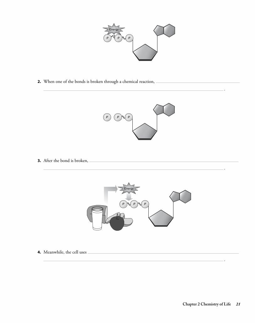
Drawing Conclusions: Sodium–Potassium Pump
Identify the following structures by coloring them as described in the figures below:
Extracellular
A.
Intracellular
1.
2.
3.
ATP
B.
C.
● Channel proteins:Purple
● Sodium molecules:Orange
● Potassium molecules:Green
● Extracellular area: Blue
Insert arrows by the molecules to show the direction of flow. Then, using each illustration as a guide, describe thefour steps in the sodium–potassium pump in the spaces provided.
4374_Ch03_023-034 10/6/14 1:17 PM Page 29

30 Chapter 3 Cells
Make a Connection: Vesicular Transport
Unscramble the following words to reveal terms associated with transport by vesicles. Then draw lines to link eachterm to its characteristics.
1. SCOOTEDYINS
2. ACHOOSPIGSTY
3. OPTICSNOISY
4. SOCIETYSOX
a. Occurs when vesicles release substances outside the cell
b. Occurs when the cell engulfs a solid particle andbrings it into the cell
c. General type of vesicular transport that bringssubstances into the cell
d. Occurs when vacuoles bring droplets ofextracellular fluid containing dissolved substancesinto the cell
e. An example is when mammary glands secretebreast milk
f. Method used by white blood cells
List for Learning: DNA
List the four nitrogenous bases found in DNA. Then draw circles around the names of two bases that pair togetherand a square around the names of the other two bases that pair together.
1.
2.
3.
4.
4.D.
4374_Ch03_023-034 10/6/14 1:17 PM Page 30

Chapter 3 Cells 31
Illuminate the Truth: DNA
Highlight the correct word or phrase to complete each sentence.
1. DNA molecules (store all of a cell’s genetic information)(contribute to energy production in the cell).
2. DNA forms a dense coil that resembles an X (at all times)(when the cell is preparing to divide).
3. The sugar in DNA is (ribonucleic acid)(deoxyribose).
4. The sequence of bases in DNA is (predetermined)(varied); it’s this sequence of bases that (provides the geneticcode)(allows DNA to replicate).
5. One of DNA’s main functions is to provide information for building (lipids)(proteins).
List for Learning: RNA
List three ways RNA differs from DNA.
1.
2.
3.
Sequence of Events: Transcription
Following are the various steps in the transcription process. Place the steps in the proper sequence by insertingnumbers in the spaces provided: Insert number 1 by the first step, number 2 by the second step, etc.
A. The mRNA begins the process of translation.
B. The nucleus receives a chemical message to make a new protein.
C. The mRNA separates from the DNA molecule and moves through a nuclear pore and into the cytoplasm.
D. An RNA enzyme assembles RNA nucleotides that would be complementary to the exposed bases.
E. The segment of DNA with the relevant gene unwinds.
F. RNA nucleotides attach to the exposed DNA and then bind to each other to form messenger RNA(mRNA).
4374_Ch03_023-034 10/6/14 1:17 PM Page 31

32 Chapter 3 Cells
Illuminate the Truth: Translation
1. After the mRNA enters the cytoplasm, it attaches to (the Golgi apparatus)(a ribosome), which begins theprocess of translating the protein.
2. The mRNA carries the code for each amino acid in a series of three bases, called a (codon)(chromosome).
3. Each tRNA consists of (one base)(three bases).
4. The tRNA deposits (three amino acids)(a single amino acid) at the complementary site.
5. The (ribosome/mRNA) uses enzymes to attach the chain of amino acids together with (cholesterol)(peptide)bonds.
Make a Connection: Cell Cycle
Unscramble the following words to discover the names of the four phases of the cell cycle. Then use lines to link eachphase to its particular characteristics.
1. NESTISSHY
2. CODENS PAG
3. TICOMIT
4. RIFTS PAG
a. The cell is busy performing the tasks it was createdto do.
b. Cell division occurs.
c. The cell synthesizes enzymes necessary for division.
d. The cell accumulates materials necessary for DNAreplication.
e. The cell makes an extra set of DNA.
4374_Ch03_023-034 10/6/14 1:17 PM Page 32

Chapter 3 Cells 33
Describe the Process: Mitosis
Following are illustrations of four cells in various stages of mitosis. In the blank to the right of each cell, write thename of each phase and describe what is occurring.
A.
B.
C.
D.
4374_Ch03_023-034 10/6/14 1:17 PM Page 33

4374_Ch03_023-034 10/6/14 1:17 PM Page 34

chap
ter 4TISSUES
All the trillions of cells of the body can be categorized as belonging to one of four distinct groups of tissue.Remember: Tissues are simply groups of similar cells that perform a common function. Complete the exercises inthis chapter to strengthen your knowledge of the four categories of tissue: epithelial, connective, nervous, andmuscular.
Chapter 4 Tissues 35
Drawing Conclusions: Epithelial Tissue (Single Layer)
Fill in the blanks to complete the following sentences about the different types of epithelial tissue having one layer.(Hint: Some blanks require more than one word.) Next, color and then label each of the sample epithelial drawings.
1. Epithelial tissue having only one layer is called .
2. epithelium consists of a single layer of flat, scale-like cells.
3. epithelium consists of a single layer of cube-like cells.
4. epithelium consists of a single layer of irregularly shaped columnar cells; the different cell heights make the tissue appear to be stratified.
5. epithelium consists of a single layer of columnar cells.
A.
B.
C.
D.
4374_Ch04_035-044 10/22/14 8:28 AM Page 35

36 Chapter 4 Tissues
ACROSS
1. Dense connective tissue band or sheet that bindsorgans and muscles together
3. Type of cell that can differentiate into a numberof different cells
5. Gland that secretes its product directly into thebloodstream
6. Tissue that lacks blood vessels and depends onunderlying connective tissue for oxygen andnutrients
9. The body’s most abundant protein12. Gland that secretes its product into ducts13. Tissue dominated by fat cells14. Cell shape that is flat and plate-like
DOWN
2. The most widespread and most varied of all thetissues
3. Tissue that has multiple layers in which somecells don’t touch the basement membrane
4. Modified cells containing secretory vesicles thatproduce large quantities of mucus
7. Cord-like tissues that attach bones to bones8. Key component of connective tissue9. Cell shape that is tall and cylindrical
10. Dense, cord-like tissues that attach muscles tobones
11. Groups of cells that perform a common function
Puzzle It Out: Tissues
Stretch your knowledge of tissues by completing this crossword puzzle.
4374_Ch04_035-044 10/6/14 1:19 PM Page 36

Chapter 4 Tissues 37
Just the Highlights: Epithelial Function
In the following lists, link each type of epithelial tissue to its location in the body by highlighting each with the samecolor. For example, highlight “simple squamous epithelium” in blue; then highlight the body location where simplesquamous epithelium is found in blue. Choose a different color for each type of epithelial tissue.
1. Simple squamous epithelium
2. Simple cuboidal epithelium
3. Simple columnar epithelium
4. Pseudostratified columnar epithelium
a. Lines the intestines
b. Lines the trachea, large bronchi, and nasal mucosa
c. Lines the alveoli
d. Lines the ducts and tubules of many organs,including the kidneys
Drawing Conclusions: Epithelial Tissue (Several Layers)
Color each of the following tissue samples. Beneath each figure, identify the tissue type and list at least one locationwhere the tissue can be found.
1.
Tissue Type
Location in the Body
2.
Tissue Type
Location in the Body
4374_Ch04_035-044 10/6/14 1:19 PM Page 37

38 Chapter 4 Tissues
Drawing Conclusions: Connective Tissue
Color and then label the following tissue samples. (Hint: Use your textbook if necessary to help you choose colors.)
A.
B.
C.
D.
E.
F.
G.
4374_Ch04_035-044 10/6/14 1:19 PM Page 38

Chapter 4 Tissues 39
Just the Highlights: Tissue Traits
Differentiate between the traits of various tissues by highlighting the sentences different colors, as suggested:
● The sentences relating to epithelial tissues: Pink
● The sentences relating to connective tissue: Blue
● The sentences relating to muscle tissue: Orange
● The sentences relating to nervous tissue: Yellow
1. Glands are made of this tissue.
2. This tissue consists of elongated cells that contractin response to stimulation.
3. The cells of this tissue are embedded in anextracellular matrix.
4. This tissue contains no blood vessels and dependsupon underlying connective tissue to supply itsneeds for oxygen and nutrients.
5. Types of this tissue include blood, bone, cartilage,and fat.
6. This tissue is found in the brain, spinal cord, andnerves.
7. This tissue forms the epidermis of the skin.
8. This tissue contains various types of fibers.
9. Tendons and ligaments consist of this type oftissue.
10. This type of tissue has a high degree of excitabilityand conductivity.
11. The key functions of this tissue involveprotection, absorption, filtration, and secretion.
12. Three types of this tissue include skeletal, cardiac,and smooth.
13. This tissue serves to connect the body togetherand to support, bind, or protect organs.
4374_Ch04_035-044 10/6/14 1:19 PM Page 39

40 Chapter 4 Tissues
Illuminate the Truth: Connective Tissue
Highlight the word or phrase that correctly completes each sentence.
1. (Collagenous)(Reticular) fibers occur in networks and support small structures, such as capillaries and nerves.
2. (Collagenous)(Elastic) fibers are the most abundant fibers in connective tissue.
3. Connective tissue is classified according to its (cell layers)(structural characteristics).
4. (Areolar)(Adipose) is a type of loose connective tissue found underneath almost all epithelia.
5. (Adipose)(Reticular) tissue forms the framework of the spleen, lymph nodes, and bone marrow.
6. (Cartilage)(Bone) is a type of dense connective tissue that has no blood supply, making healing slow or absentfollowing an injury.
7. (Elastic cartilage)(Fibrocartilage) resists compression and absorbs shock, making it ideal to form the discsbetween the vertebrae.
8. The matrix of (bone)(cartilage) serves as a storage site for calcium.
9. Unlike other connective tissues, (bone)(blood) doesn’t contain any fibers.
10. Most connective tissue has a (rich)(limited) blood supply.
Drawing Conclusions: Tissue Repair
Demonstrate your knowledge of tissue repair by coloring the illustration (which depicts the various stages in tissuerepair) according to the following instructions. Then use the spaces below each figure to describe what’s occurring ineach step.
Use different colors as suggested to color the following figure:
● Wound: Dark red
● Scab: Brown
● Granulation tissue: Pink
● White blood cells: Purple
● New cells: Tan
● Scar tissue: White
● Fibroblasts: Orange
4374_Ch04_035-044 10/6/14 1:19 PM Page 40

Chapter 4 Tissues 41
3
4
12
1.
2.
3.
4.
4374_Ch04_035-044 10/6/14 1:19 PM Page 41

42 Chapter 4 Tissues
Making a Connection: Membranes
Unscramble the following words to reveal the names of the three types of epithelial membranes. Then draw lines tolink each membrane with its characteristics.
1. CUMSOU
2. SAUCEUNTO
3. ROSEUS
a. Composed of simple squamous epithelium restingon a thin layer of areolar connective tissue
b. Lines body surfaces that open directly to thebody’s exterior
c. Lines some of the closed body cavities and coversmany of the organs in those cavities
d. The body’s largest membrane
e. Secretes a fluid to help prevent friction as theorgans move
f. Consists of different types of epithelium,depending on the location and function of themembrane
g. Consists of a layer of epithelium resting on a layerof connective tissue
h. Secretes mucus
i. Consists of three types: pleural, pericardial, andperitoneal
4374_Ch04_035-044 10/6/14 1:19 PM Page 42

Chapter 4 Tissues 43
Conceptualize in Color: Epithelial Membranes
Firm up your knowledge of epithelial membranes by coloring the structures in the figure as suggested:
● Outline the mucous membranes in pink.
● Outline the cutaneous membrane in yellow.
● Outline the visceral pleura in blue.
● Outline the parietal pleura in purple.
● Outline the peritoneal membrane in green.
● Outline the pericardial membrane in red.
4374_Ch04_035-044 10/6/14 1:19 PM Page 43

4374_Ch04_035-044 10/6/14 1:19 PM Page 44

chap
ter 5INTEGUMENTARY SYSTEM
The skin—which, together with the hair and nails, forms the integumentary system—is one of the largest organsin the body. Despite its thinness, it consists of multiple layers and many cell types. It also performs manyfunctions crucial for homeostasis and even survival. Use the activities in this chapter to test yourself about thisvital body system.
Chapter 5 Integumentary System 45
Conceptualize in Color: Skin Structures
Color these skin structures in the figure shown here. Use the colors suggested or choose your own.
● Hair follicle: Light beige
● Hair bulb: Blue
● Hair shaft: Black
● Dermal papilla: Dark pink
● Arrector pili muscle: Red
● Epidermis: Brown
● Dermis: Pink
● Hypodermis: Yellow
● Eccrine sweat gland: Orange
● Apocrine sweat gland: Purple
● Sebaceous gland: Green
4374_Ch05_045-050 10/6/14 1:21 PM Page 45

46 Chapter 5 Integumentary System
Puzzle It Out: Skin Structures
Complete the following crossword puzzle to strengthen your knowledge of skin structures and terms.
ACROSS
3. The skin is also called themembrane.
4. Excessive hair loss5. The inner, deeper layer of the skin8. Outermost layer of the epidermis is called the
stratum .9. Inflammation of the skin characterized by itching
and redness10. Oily substance produced by sebaceous glands11. Dead tissue resulting from a burn
DOWN
1. Flattened cells making up the skin’s outermostlayer
2. Substance that gives hair and skin its color6. The outermost layer of the skin7. Ear wax
4374_Ch05_045-050 10/6/14 1:21 PM Page 46

Chapter 5 Integumentary System 47
Sequence of Events: Formation of New Skin Cells
Following are the various events in the formation of new skin cells. Using the spaces provided, order the events inthe proper sequence by inserting numbers.
A. Flattened, dead cells called keratinocytes arrive at the stratum corneum.
B. Cells stop dividing and produce keratin, which replaces the cytoplasm and nucleus of the cell.
C. Stem cells in the stratum basale undergo mitosis.
D. The cells flatten and die.
E. As new cells are produced, they push older cells upward.
Drawing Conclusions: Skin Color
Fill in the blanks to test your knowledge about skin color. Then color the figure below using the instructionsprovided.
1. Melanocytes are scattered through the layer of the epidermis.
2. These cells produce a substance called .
3. A person’s skin color is determined by the amount, and type, of .
4. The forms a cap over the top of the cell to protect it from the
rays of the sun.
Color each of the structures in the figure shown here. Use the colors suggested or choose your own.
● Melanocyte: Green
● Keratinocytes: Tan
● Melanin granules: Red
● Cell nucleus: Orange
4374_Ch05_045-050 10/6/14 1:21 PM Page 47

48 Chapter 5 Integumentary System
Fill in the Gaps: Skin Changes
Fill in the blanks to complete each of the following sentences. Choose from the words listed in the Word Bank.
ADRENAL
ALBINISM
BLUE
BRONZING
CYANOSIS
JAUNDICE
MELANIN
PALLOR
YELLOW
RED
ERYTHEMA
1. A deficiency of oxygen in circulating blood gives skin a tint, a condition called
.
2. Impaired liver function may cause the skin to take on a tone, a condition called
.
3. A deficiency of hormones from the gland causes the skin to become golden-brown in
color, which is called .
4. A genetic lack of results in extremely pale skin, white hair, and pink eyes, a condition
called .
5. Decreased blood flow, such as from exposure to cold, emotional stress, or low blood pressure, produces
.
6. Increased blood flow in dilated vessels close to the skin’s surface makes the skin appear more
in color, which is called .
List for Learning: Functions of the Skin
Using the spaces provided, list five key functions of the skin.
1.
2.
3.
4.
5.
4374_Ch05_045-050 10/6/14 1:21 PM Page 48

Chapter 5 Integumentary System 49
Fill in the Gaps: Thermoregulation
Complete the following sentences to test your understanding of how the skin helps the body maintain a stabletemperature. Choose from the words listed in the Word Bank.
CONSTRICT
DILATE
EVAPORATION
INCREASES
NERVES
REDUCES
SWEATING
1. The skin contains to cause blood vessels in the skin to dilate or constrict to regulate heat loss.
2. When chilled, the blood vessels ; this blood flow through the skin and conserves heat.
3. When overheated, the blood vessels in the skin ; this the flow of blood and increases heat loss.
4. If the body is still overheated, the brain stimulates ; this results in and cooling occurs.
Illuminate the Truth: Nail Changes
Highlight the correct word in each of the following sentences.
1. A condition in which the distal ends of the fingers enlarge, making them look like a drumstick, is called(clubbing)(breaking); it typically results from long-term (iron)(oxygen) deficiency.
2. One of the first signs of oxygen deficiency is when the nail beds appear (dark red)(blue).
3. Flattened or concave nail beds may indicate a deficiency of (protein)(iron).
4. Anemia may cause the nail beds to look (pale)(cyanotic).
5. Dark lines beneath the nail may indicate (melanoma)(basal cell carcinoma).
4374_Ch05_045-050 10/6/14 1:21 PM Page 49

50 Chapter 5 Integumentary System
Make a Connection: Skin Glands
Unscramble the following words to discover the names of four types of skin glands. Then draw a line to link eachgland with its particular characteristics.
1. RICEECN
2. CANOERIP
3. BECAUSESO
4. CURIOUSMEN
a. Contains a duct that empties onto the skin’s surface
b. Produces ear wax
c. Contains a duct that empties sweat onto a hairfollicle
d. Begin to function at puberty
e. Secretes an oily substance onto the hair follicle
f. Located mainly in the axillary and anogenitalregion
g. Secretion has a mild antifungal effect
h. Are widespread throughout the body but areespecially abundant on the palms, soles, forehead,and upper torso
i. Excess secretions may accumulate in gland ducts,leading to pimples and blackheads
j. Produce sweat to help the body maintain aconstant core temperature
k. Are scent glands that respond to stress and sexualstimulation
Just the Highlights: Burns
Highlight in yellow the sentences that pertain to first-degree burns, the sentences that pertain to second-degreeburns in orange, and the sentences that pertain to third-degree burns in pink.
1. Known as a partial-thickness burn, this burn only involves the epidermis.
2. This burn results in blisters, severe pain, and swelling.
3. This burn often results from sunlight.
4. Because nerve endings have been destroyed, this burn may not be painful initially.
5. Known as a full-thickness burn, this burn extends through the epidermis and dermis and into thesubcutaneous tissue.
6. This burn, known as a partial-thickness burn, involves the epidermis and part of the dermis.
7. This burn results in redness, slight swelling, and pain.
8. This burn often requires skin grafts.
4374_Ch05_045-050 10/6/14 1:21 PM Page 50

chap
ter 6BONES & BONE TISSUE
The bones forming the human skeleton are dynamic living tissue. Besides providing the framework of the body,bones generate blood cells, regulate blood calcium levels, and join with the muscular system to allow us to move.The activities in this chapter should help you refine your knowledge of both the form and function of this vitaltissue.
Chapter 6 Bones and Bone Tissue 51
Make a Connection: Bone Classifications
Unscramble the following words to discover the four categories of bones. Then draw a line to link each category toits specific characteristics.
1. GLON
2. STROH
3. TLAF
4. ARULERRIG
a. Serve to protect organs
b. Tend to be shaped like cubes
c. Are longer than they are wide
d. Come in various sizes and shapes
e. Work like levers to move limbs
f. Are thin and often curved
g. Are often clustered in groups
h. Examples include the femur of the thigh and thehumerus of the arm
i. Are about as broad as they are long
j. Examples include the carpal bones of the wrist
k. Examples include the skull and the ribs
l. Examples include the vertebrae and facial bones
4374_Ch06_051-060 10/8/14 8:16 AM Page 51
52 Chapter 6 Bones and Bone Tissue
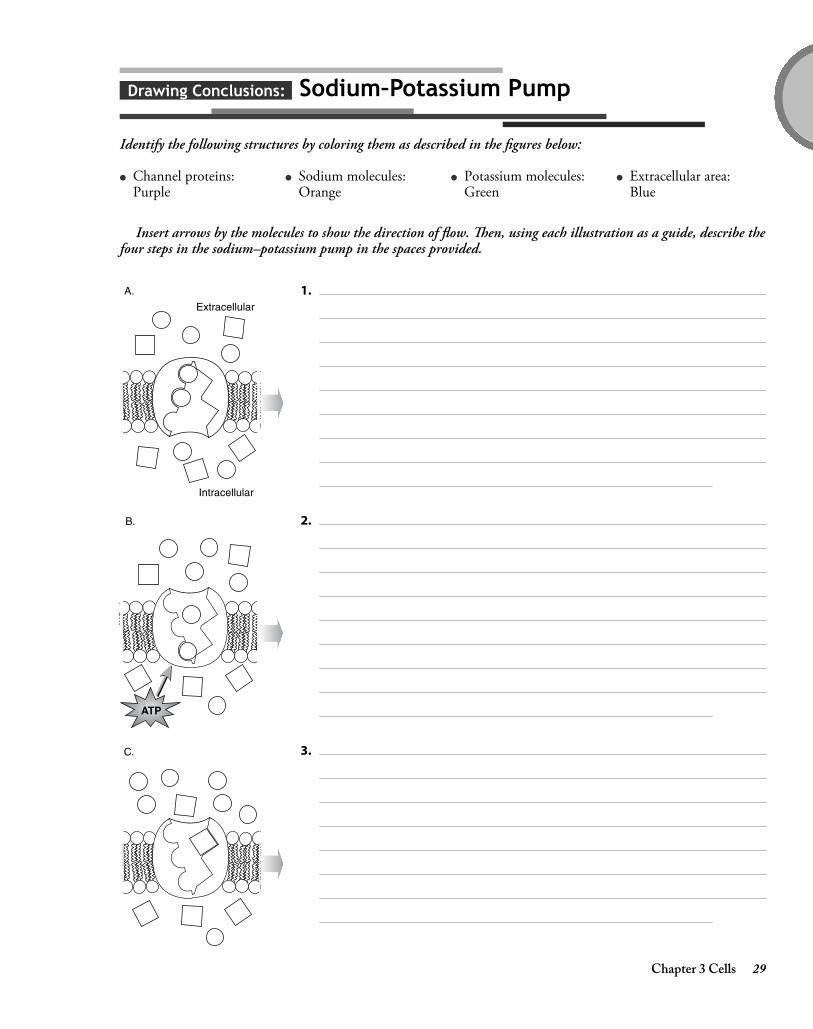
Puzzle It Out: Bone Terms
ACROSS
4. Disease meaning “porous bones”6. Process by which bone cells destroy old bone and
deposit new bone7. Bone cells that dissolve unwanted or unhealthy
bone9. Bone found in the ends of long bones and the
centers of most other bones10. Mature bone cells that have become entrapped in
the hardened bone matrix11. Name for bone tissue12. Break in a bone
DOWN
1. Type of marrow that produces blood cells2. Process whereby fetal skeleton becomes bone3. Substance from which most bones evolve4. Bone cells that help form bone5. Type of bone that forms the shafts of long bones
and surrounds outer surfaces of other bones8. Latticework of bone that makes up spongy bone
4374_Ch06_051-060 10/8/14 8:16 AM Page 52

Chapter 6 Bones and Bone Tissue 53
Conceptualize in Color: Parts of a Long Bone
Color these parts of the long bone in the following figure. Use the colors suggested or choose your own.
1. Articular cartilage: Blue
2. Bone marrow: Yellow
3. Medullary cavity: Gold
4. Periosteum: Tan
5. Epiphyseal line: Brown
6. Draw a red line along the endosteum
7. Draw a bracket around each epiphysis
8. Draw a bracket around the diaphysis
4374_Ch06_051-060 10/8/14 8:16 AM Page 53

54 Chapter 6 Bones and Bone Tissue
Conceptualize in Color: Cancellous Bone
Color the following parts of cancellous (spongy) bone. Use the colors suggested or choose your own.
● Periosteum: Brown
● Compact bone: Gold
● Trabeculae: Yellow
Fill in the Gaps: Compact Bone
Fill in the blanks to complete the following sentences. Choose from the words listed in the Word Bank.
BLOOD VESSELS
HAVERSIAN
LACUNAE
LAMELLAE
NERVES
OSTEOCYTES
OSTEON
VOLKMANN’S
1. In compact bone, layers of matrix are arranged in concentric rings called around a
central canal. This basic structural unit is called an .
2. and run through the center of the canal.
3. Tiny gaps between the rings, called , contain .
4. Transverse passageways called canals transport blood and nutrients from the bone’s exterior to the living cells inside.
4374_Ch06_051-060 10/8/14 8:16 AM Page 54

Chapter 6 Bones and Bone Tissue 55
Conceptualize in Color: Bone Marrow
Using the following figure, color red all the bones, or portions of bones, containing red bone marrow.
4374_Ch06_051-060 10/8/14 8:17 AM Page 55

56 Chapter 6 Bones and Bone Tissue
List for Learning: Bone Functions
Using the blank spaces, list seven functions of bone.
1.
2.
3.
4.
5.
6.
7.
Cartilage model
Step 1
Bone formation
Step 2
Diaphysis
EpiphysisOssifying cartilage
Primary ossification center
Blood vessel
Step 3
Marrow cavity
Blood vessel
Step 4
Marrow cavity
Step 1:
Step 2:
Step 3:
Step 4:
Describe the Process: Ossification
Using the following figures as a guide, describe each step in the process of endochondral ossification.
4374_Ch06_051-060 10/8/14 8:17 AM Page 56

Chapter 6 Bones and Bone Tissue 57
Illuminate the Truth: Bone Growth
Highlight the correct word or phrase that makes each of the following statements true.
1. After birth, bone lengthening occurs at the (epiphyseal plate)(epiphyseal line).
2. (Articular)(Hyaline) cartilage forms the (epiphyseal plate)(epiphyseal line).
3. When bone lengthening stops, (articular cartilage)(the epiphyseal plate) is replaced with (compact bone)(spongy bone).
4. Bone thickening and widening occurs (for a fixed number of years)(throughout the life span).
5. The process by which old bone is destroyed and then replaced with new bone is called(remodeling)(ossification).
6. Physical exercise causes bones to (increase)(decrease) in density.
List for Learning: Factors Affecting Bone Growth
Using the spaces provided, list four factors that affect bone growth and maintenance.
1.
2.
3.
4.
4374_Ch06_051-060 10/8/14 8:17 AM Page 57

58 Chapter 6 Bones and Bone Tissue
Drawing Conclusions: Fractures
Using the spaces provided, draw a bone that illustrates the types of fractures listed below. Then fill in the blanks ineach sentence to correctly describe each fracture. Hint: Some blanks require more than one word.
A. Simple fractureIn this type of fracture, the skin and tissue near the fracture is .
B. Compound fractureIn this type of fracture, the skin near the site has been .
4374_Ch06_051-060 10/8/14 8:17 AM Page 58

Chapter 6 Bones and Bone Tissue 59
C. Greenstick fractureThis type of fracture usually occurs in .
D. Comminuted fractureThis type of fracture is often the result of a .
E. Spiral fractureThis type of fracture results from a .
4374_Ch06_051-060 10/8/14 8:17 AM Page 59

60 Chapter 6 Bones and Bone Tissue
Drawing Conclusions: Fracture Repair
In the following figures, color the stages in fracture repair. Use the colors suggested or choose your own.
● Hematoma: Red
● Soft callus: Pink
● Hard callus: Yellow
● Remodeled bone: Tan
Then, using the figures as a guide, describe each step in the fracture repair process.
1.
2.
3.
4.
4374_Ch06_051-060 10/8/14 8:17 AM Page 60

chap
ter 7SKELETAL SYSTEM
Learning the names of the body’s 206 bones, not to mention their locations and landmarks, can provechallenging. Completing the activities in this chapter, however, should help you do just that.
Chapter 7 Skeletal System 61
6. Radius
7. Os coxae
8. Sacrum
9. Shoulder girdle
10. Mandible
Just the Highlights: The Skeleton
In the following list, highlight the bones of the axial skeleton in one color (such as yellow) and the bones of theappendicular system in another color (such as blue).
1. Skull
2. Femur
3. Ribs
4. Clavicles
5. Vertebrae
4374_Ch07_061-080 10/8/14 8:19 AM Page 61

62 Chapter 7 Skeletal System
Puzzle It Out: Bone Surface Markings
Test your knowledge of the common terms used to describe bone surface markings by completing the followingcrossword puzzle.
ACROSS
2. A projection or raised area5. A round opening; usually a passageway for vessels
and nerves6. A rough, raised bump, usually for muscle
attachment7. A cavity within a bone8. A moderately raised ridge
10. A flat surface
DOWN
1. A large process found only on the femur3. A rounded knob; usually fits into a fossa on
another bone to form a joint 4. A tube-like opening8. A tunnel through a bone9. The prominent, expanded end of a bone
4374_Ch07_061-080 10/8/14 8:19 AM Page 62

Chapter 7 Skeletal System 63
Conceptualize in Color: The Skull
Color these bones of the skull in the figure below. Use the colors suggested or choose your own.
● Frontal bone: Orange
● Parietal bone: Blue
● Temporal bone: Green
● Occipital bone: Yellow
● Sphenoid bone: Red
● Ethmoid bone: Purple
4374_Ch07_061-080 10/8/14 8:19 AM Page 63
64 Chapter 7 Skeletal System
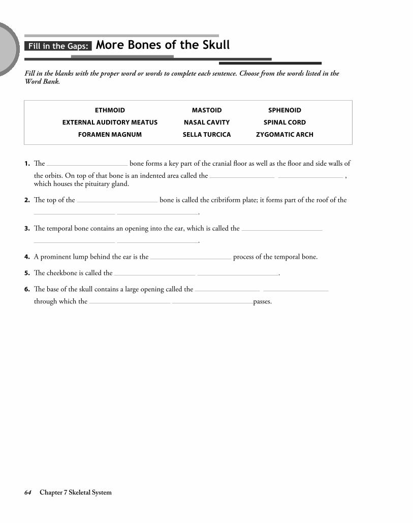
Fill in the Gaps: More Bones of the Skull
Fill in the blanks with the proper word or words to complete each sentence. Choose from the words listed in theWord Bank.
ETHMOID
EXTERNAL AUDITORY MEATUS
FORAMEN MAGNUM
MASTOID
NASAL CAVITY
SELLA TURCICA
SPHENOID
SPINAL CORD
ZYGOMATIC ARCH
1. The bone forms a key part of the cranial floor as well as the floor and side walls of
the orbits. On top of that bone is an indented area called the ,which houses the pituitary gland.
2. The top of the bone is called the cribriform plate; it forms part of the roof of the
.
3. The temporal bone contains an opening into the ear, which is called the
.
4. A prominent lump behind the ear is the process of the temporal bone.
5. The cheekbone is called the .
6. The base of the skull contains a large opening called the
through which the passes.
4374_Ch07_061-080 10/8/14 8:19 AM Page 64

Chapter 7 Skeletal System 65
a. The line of articulation between the parietal bonesand the occipital bone
b. The joint between the parietal bones and thefrontal bone
c. The joint between the right and left parietal bones
d. The line of articulation that runs along the topedge of the temporal bone
1. RANLOCO
2. ATLASGIT
3. QUASUMSO
4. ABALDLIMOD
Make a Connection: Cranial Sutures
Unscramble the words in the left column below to discover the names of the four cranial sutures. Then draw a lineto link each suture to its location.
4374_Ch07_061-080 10/8/14 8:19 AM Page 65

66 Chapter 7 Skeletal System
Conceptualize in Color: Facial Bones
Test your knowledge of the facial bones by coloring the following bones in the figure below. Use the colors suggestedor choose your own.
● Nasal bones: Yellow
● Inferior nasal conchae: Orange
● Vomer: Brown
● Mandible: Blue
● Maxillae: Green
● Zygomatic bones: Purple
● Lacrimal bones: Red
4374_Ch07_061-080 10/8/14 8:19 AM Page 66

Chapter 7 Skeletal System 67
Conceptualize in Color: Sinuses
Identify the various paranasal sinuses by coloring them in the following figure. Use the colors suggested or chooseyour own.
● Frontal sinuses: Yellow
● Maxillary sinuses: Blue
● Ethmoid sinuses: Pink
● Sphenoid sinus: Orange
4374_Ch07_061-080 10/8/14 8:19 AM Page 67

68 Chapter 7 Skeletal System
Drawing Conclusions: Infant Skull
Solidify your knowledge of the infant skull by coloring the features of the skull. Then fill in the blanks of thesentences to identify some conditions that can cause changes in the fontanels.Color these features in the following figure. Use the colors suggested or choose your own.
● Anterior fontanel: Pink
● Posterior fontanel: Blue
● Sagittal suture: Yellow
1. A bulging anterior fontanel signals increased .
2. A sunken anterior fontanel suggests .
3. causes the suture lines to widen abnormally.
● Lambdoid suture: Green
● Squamous suture: Purple
● Coronal suture: Red
4374_Ch07_061-080 10/8/14 8:19 AM Page 68

Chapter 7 Skeletal System 69
Drawing Conclusions: The Vertebral Column
In the figure below, color each of the following structures a different color: the cervical vertebrae, the thoracicvertebrae, the lumbar vertebrae, the sacrum, and the coccyx. Draw lines indicating the cervical curve, the thoraciccurve, the lumbar curve, and the sacral curve.
4374_Ch07_061-080 10/8/14 8:19 AM Page 69

70 Chapter 7 Skeletal System
Conceptualize in Color: Vertebrae
Hone your knowledge of vertebrae by coloring each of these structures in the following figure. Use the colorssuggested or choose your own.
● Body: Purple
● Spinous process: Yellow
● Transverse process: Tan
● Lamina: Brown
● Vertebral foramen: Outline in blue
4374_Ch07_061-080 10/8/14 8:19 AM Page 70

Chapter 7 Skeletal System 71
Conceptualize in Color: Thoracic Cage
Color each of these structures of the thoracic cage in the following figure. Use the colors suggested or choose yourown.
● Manubrium: Brown
● Body of the sternum: Tan
● Xiphoid process: Pink
● True ribs: Blue
● False ribs: Yellow
● Floating ribs: Green
● Draw a red line at the suprasternal notch
● Draw a purple line at the costal margins
4374_Ch07_061-080 10/8/14 8:19 AM Page 71

72 Chapter 7 Skeletal System
Drawing Conclusions: Pectoral Girdle
Test your knowledge of the bones of the pectoral girdle by first coloring the structures listed below. Use the colorssuggested or choose your own. Then fill in the blanks in the sentences to describe their function.
● Scapula: Light tan
● Clavicle: Yellow
● Acromion process: Brown
1. The acromion process articulates with the ; it is the only point where the
and attach to the rest of the skeleton.
2. The coracoid process provides a point of attachment for .
3. The glenoid cavity articulates with the head of the .
● Coracoid process: Dark tan
● Glenoid cavity: Outline in pink
4374_Ch07_061-080 10/8/14 8:19 AM Page 72

Chapter 7 Skeletal System 73
Drawing Conclusions: Upper Limb
Color the bones of the arm as described below. Use the colors suggested or choose your own. Then identify keymarkings of those bones by writing the name of each marking in the appropriate space.
● Humerus: Brown
● Radius: Yellow
● Ulna: Pink
4
3
2
1
4374_Ch07_061-080 10/8/14 8:19 AM Page 73

74 Chapter 7 Skeletal System
Drawing Conclusions: The Hand
Improve your knowledge of the bones of the hand by coloring the following structures in the figure below. Use thecolors suggested or choose your own. Then label each digit with the correct Roman numeral.
● Carpal bones: Blue
● Metacarpal bones: Pink
● Proximal Phalanges: Yellow
● Middle Phalanges: Orange
● Distal Phalanges: Green
4374_Ch07_061-080 10/8/14 8:19 AM Page 74

Chapter 7 Skeletal System 75
Conceptualize in Color: Pelvic Girdle
Color the following structures in each os coxae, which together form the pelvic girdle. Use the suggested colors orchoose your own.
● Ilium: Yellow
● Ischium: Orange
● Pubis: Pink
● Symphysis pubis: Blue
● Acetabulum: Green
● Iliac crest: Outline in Red
● Obturator foramen: Outline in Purple
Illuminate the Truth: The Pelvis
Highlight the word or phrase that makes each statement correct.
1. The (false pelvis)(true pelvis) extends between the outer, flaring edges of the iliac bones.
2. The (false pelvis)(true pelvis) extends between the pelvic brim.
3. The lower edge of the true pelvis is known as the (lesser pelvis)(pelvic outlet).
4. The distance between the two (ischial)(iliac) bones determines the pelvic outlet.
5. In males, the true pelvis is (deep and narrow)(wide and shallow).
6. Females have a pubic arch that is (narrower)(wider) than in males.
7. Females also have a (larger)(smaller) pelvic outlet.
4374_Ch07_061-080 10/8/14 8:19 AM Page 75

76 Chapter 7 Skeletal System
Drawing Conclusions: Femur
Color the femur shown here using a color or colors of your choosing. Then identify key markings on the femur bywriting the name of each marking in the appropriate space.
6
5
4
31
2
7
4374_Ch07_061-080 10/8/14 8:19 AM Page 76

Chapter 7 Skeletal System 77
Drawing Conclusions: Lower Leg
Color the bones of the lower leg listed below. Use the colors suggested or choose your own. Then identify keylandmarks by writing the name of each landmark in the appropriate space.
● Patella: Brown
● Tibia: Yellow
● Fibula: Pink
2
1
3
4374_Ch07_061-080 10/8/14 8:19 AM Page 77

78 Chapter 7 Skeletal System
Drawing Conclusions: Foot
Identify the bones of the foot by coloring each one listed below. Use the colors suggested or choose your own. Thennumber the metatarsals by labeling each with the proper Roman numeral.
● Calcaneus: Green
● Talus: Pink
● Navicular: Orange
● Cuneiforms: Yellow
● Metatarsals: Blue
● Proximal phalanges: Purple
● Middle phalanges: Red
● Distal phalanges: Tan
● Cuboid: Light blue
4374_Ch07_061-080 10/8/14 8:19 AM Page 78
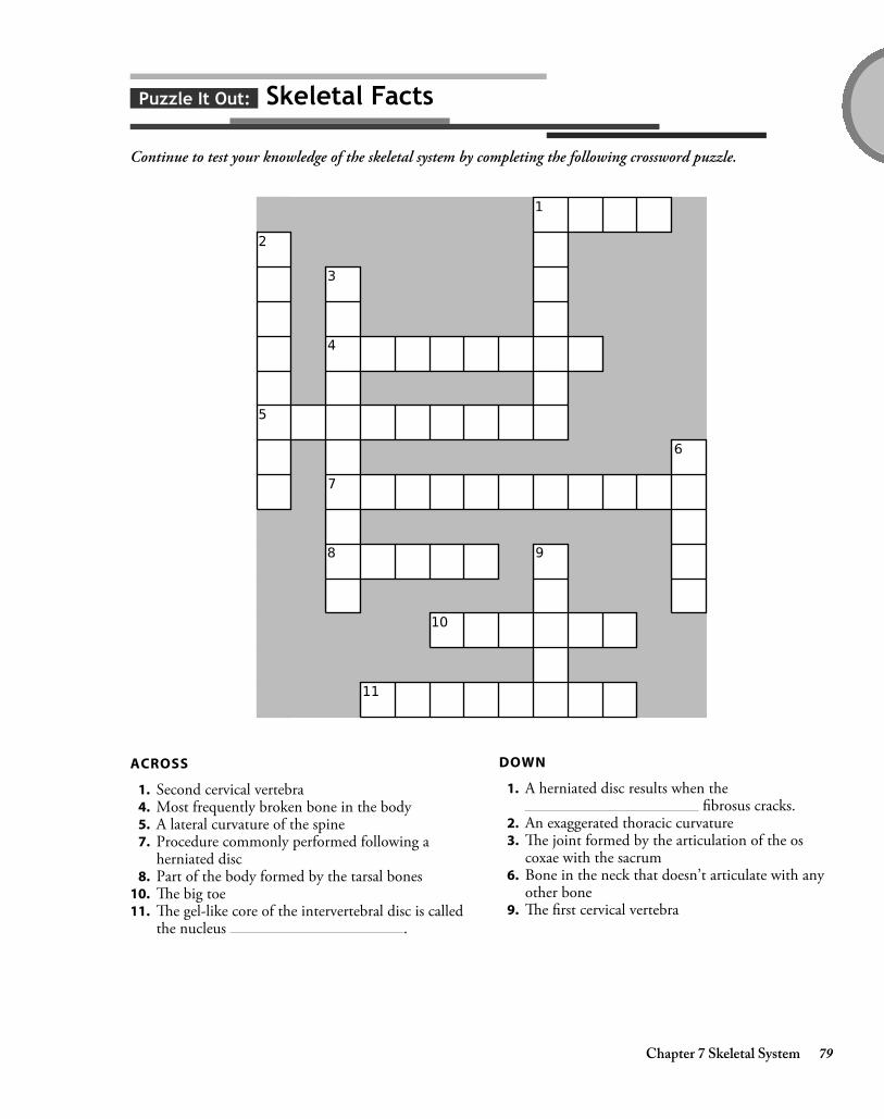
Chapter 7 Skeletal System 79
Puzzle It Out: Skeletal Facts
Continue to test your knowledge of the skeletal system by completing the following crossword puzzle.
ACROSS
1. Second cervical vertebra4. Most frequently broken bone in the body5. A lateral curvature of the spine7. Procedure commonly performed following a
herniated disc8. Part of the body formed by the tarsal bones
10. The big toe11. The gel-like core of the intervertebral disc is called
the nucleus .
DOWN
1. A herniated disc results when thefibrosus cracks.
2. An exaggerated thoracic curvature3. The joint formed by the articulation of the os
coxae with the sacrum6. Bone in the neck that doesn’t articulate with any
other bone9. The first cervical vertebra
4374_Ch07_061-080 10/8/14 8:19 AM Page 79

4374_Ch07_061-080 10/8/14 8:19 AM Page 80

chap
ter 8JOINTS
Joints, or articulations, are the points where two bones meet. The body contains a number of differentarticulations, varying in both size and shape. It’s these differences that allow us to perform a wide variety ofmovements, ranging from walking and running to writing or playing the piano.
Chapter 8 Joints 81
a. Are freely movable
b. Result when bones are joined by cartilage
c. Result when collagen fibers from one bonepenetrate the adjacent bone
d. The most numerous of the body’s joints
e. Are slightly movable
f. Also called diarthroses
g. Are fixed joints
h. Also called amphiarthroses
i. An example is the symphysis pubis
j. Examples include the knee, hip, and elbow
k. An example is the suture joints in the adult skull
l. Also called synarthroses
Make a Connection: Classifications of Joints
Unscramble the following words to discover three classifications of joints. Then draw lines to the list on the right tolink each joint classification to its characteristics.
1. VISAONLY
2. BIOSURF
3. ANTISOCIALRUG
4374_Ch08_081-092 10/8/14 8:20 AM Page 81

82 Chapter 8 Joints
Conceptualize in Color: Synovial Joints
Identify the key structures found in synovial joints by coloring the following figure. Use the colors suggested orchoose your own colors.
● Joint capsule: Green
● Synovial membrane: Purple
● Articular cartilage: Dark blue
● Ligaments: Gray
● Joint cavity: Light blue
4374_Ch08_081-092 10/8/14 8:20 AM Page 82

Chapter 8 Joints 83
Drawing Conclusions: Types of Synovial Joints
Identify the type of synovial joint represented by each of the “hardware” joints. First, fill in the blanks to identifythe type of joint, the movement the joint allows, and at least one location in the body where the joint can be found.
1.
● Type of joint:
● Movement:
● Location in the body:
2.
● Type of joint:
● Movement:
● Location in the body:
3.
● Type of joint:
● Movement:
● Location in the body:
4.
● Type of joint:
● Movement:
● Location in the body:
5.
● Type of joint:
● Movement:
● Location in the body:
6.
● Type of joint:
● Movement:
● Location in the body:
4374_Ch08_081-092 10/8/14 8:20 AM Page 83

84 Chapter 8 Joints
Drawing Conclusions: Movements of Synovial Joints
Describing the great variety of movements performed by synovial joints depends upon a specific vocabulary.Demonstrate your knowledge of these terms by writing the name of the movement being demonstrated by each ofthe following figures. Color the arrow signifying the direction of movement.
1.
2.
4374_Ch08_081-092 10/8/14 8:20 AM Page 84

Chapter 8 Joints 85
3.
4.
4374_Ch08_081-092 10/8/14 8:20 AM Page 85

86 Chapter 8 Joints
5.
6.
4374_Ch08_081-092 10/22/14 8:31 AM Page 86

Chapter 8 Joints 87
7.
8.
9.
4374_Ch08_081-092 10/8/14 8:20 AM Page 87

88 Chapter 8 Joints
10.
11.
4374_Ch08_081-092 10/8/14 8:20 AM Page 88

Chapter 8 Joints 89
12.
13.
14.
4374_Ch08_081-092 10/8/14 8:20 AM Page 89
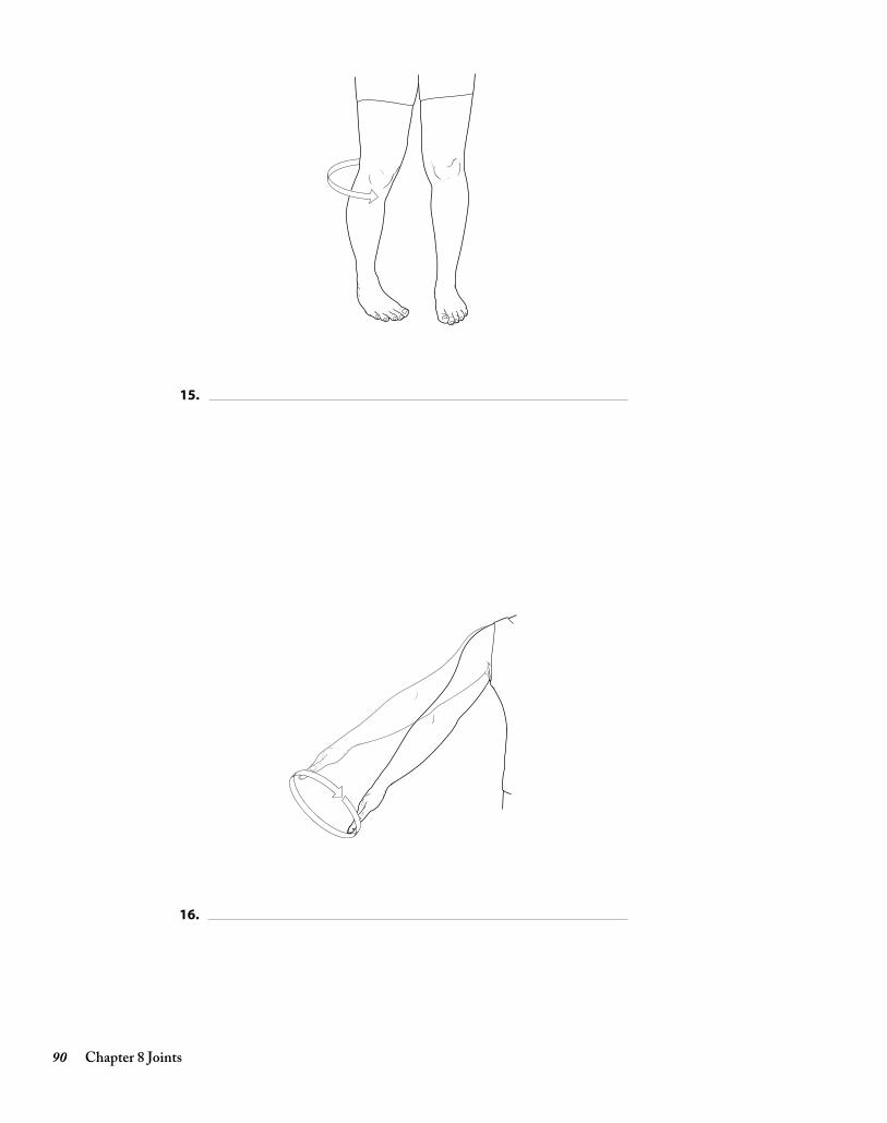
90 Chapter 8 Joints
15.
16.
4374_Ch08_081-092 10/8/14 8:20 AM Page 90

Chapter 8 Joints 91
Conceptualize in Color: The Knee
Identify the key structures of the knee by coloring the following figure. Use the colors suggested or choose your owncolors.
● Femoral condyles: Gray
● Medial meniscus: Light blue
● Lateral meniscus: Dark blue
● Posterior cruciate ligament: Orange
● Anterior cruciate ligament: Green
● Fibular collateral ligament: Tan
● Tibial collateral ligament: Brown
4374_Ch08_081-092 10/8/14 8:20 AM Page 91

92 Chapter 8 Joints
Puzzle It Out: Joints
Test your general knowledge concerning joints by completing the following crossword puzzle.
ACROSS
3. Type of arthritis that’s an autoimmune disease7. Type of arthritis attributed to the wear and tear of
aging11. Another name for a joint12. Body joint consisting of two articulations
DOWN
1. Fluid-filled sac that resides in some joints2. Type of surgical procedure often performed on
the knee4. The anterior cruciate ligament keeps the knee
from doing this5. Type of fluid that fills the joint capsule and
lubricates the joint6. Joint replacement8. Meniscus is made of this material9. Another name for the humeroscapular joint
10. Another name for the tibiofemoral joint
4374_Ch08_081-092 10/8/14 8:20 AM Page 92

chap
ter 9MUSCULAR SYSTEM
The body’s more than 600 skeletal muscles give the body its shape and also allow it to move. Attached to bones,muscles produce movement through their ability to contract. Learning the names of key muscles and understandingthe physiology behind movement can prove challenging. The activities in this chapter should help you master thisimportant body system.
Chapter 9 Muscular System 93
Make a Connection: Types of Muscles
Unscramble the words on the left to discover the three types of muscle. Then draw a line linking each muscle type toits particular characteristics. (Note: Answers may be used more than once.)
a. Found only in the heart
b. Does not appear striped, so it is called nonstriated
c. Consists of short, branching fibers that fit togetherat intercalated discs
d. Found in the digestive tract, blood vessels, andairways
e. Appears striped, or striated, when viewed under amicroscope
f. Attached to bone and causes movement of thebody
g. Known as involuntary muscle
h. Known as voluntary muscle
1. ACIDCAR
2. MOSHTO
3. TALESELK
4374_Ch09_093-108 10/8/14 8:23 AM Page 93

94 Chapter 9 Muscular System
Conceptualize in Color: Skeletal Muscle Structure
Test your knowledge of the various parts of a skeletal muscle by coloring the following structures in the figurebelow. Use the colors suggested or choose your own.
● Endomysium: Blue
● Perimysium: Yellow
● Epimysium: Red
● Fascia: Brown
● Draw a red circle around a single muscle fiber
● Draw a green circle around a fascicle
4374_Ch09_093-108 10/8/14 8:23 AM Page 94

Chapter 9 Muscular System 95
Drawing Conclusions: Muscle Fiber Structure
To reinforce your knowledge of the inner workings of a muscle, first fill in the blanks with the correct word orwords. Then color each structure in the following figure; use the colors suggested or choose your own.
1. The membrane surrounding each fiber is called a .In the figure, color this structure pink.
2. Long protein bundles called fill the sarcoplasm. These structures store
(which is used for energy) as well as oxygen.Color this structure yellow.
3. The smooth endoplasmic reticulum of a muscle fiber is called the ;
this is where are stored.Color this structure blue.
4. A system of tubules called allows to travel deep into the cell.Color these structures green.
5. Myofibrils consist of even finer fibers called .Color these structures brown.
4374_Ch09_093-108 10/8/14 8:23 AM Page 95

96 Chapter 9 Muscular System
Drawing Conclusions: Myofilaments
Review the sliding-filament model of contraction by filling in the blanks in the following sentences. Then, in thespace provided, draw two myofilaments: one displaying muscle relaxation and the second, muscle contraction.
1. Myofibrils consist of thin myofilaments (made of a protein called ) and thick
myofilaments (made of a protein called ).
2. Myofibrils are arranged to form units called .
3. A plate or disc called a is the anchor point for the thin myofilaments.
4. When a muscle begins to contract, the heads of the myofilament latch onto the
myofilament, forming a .
5. The heads of the first myofilament then pull the second myofilament forward, shortening the
and pulling the closer together.
4374_Ch09_093-108 10/8/14 8:23 AM Page 96

Chapter 9 Muscular System 97
Sequence of Events: Muscle Contraction
See if you can place the events that occur during muscle contraction and relaxation in their proper order. Insertnumbers in the spaces provided to order the events in the proper sequence.
A. An electrical impulse travels over the sarcolemma and inward along the T tubules, causing sacs in thesarcoplasmic reticulum to release calcium.
B. The release of ACh stops and acetylcholinesterase breaks down any remaining ACh.
C. Troponin and tropomyosin prevent the myosin heads from grasping the thin filament, and the musclefiber relaxes.
D. An electrical impulse causes small vesicles at the end of a motor neuron to bind with the cell wall andrelease the neurotransmitter acetylcholine (ACh).
E. Calcium binds with the troponin on the actin filament, exposing attachment points.
F. ACh diffuses across the synaptic cleft, where it stimulates receptors in the sarcolemma.
G. Calcium ions are pumped back into the sarcoplasmic reticulum.
H. The myosin heads of the thick filaments grab onto the thin filaments and muscle contraction occurs.
4374_Ch09_093-108 10/8/14 8:23 AM Page 97

98 Chapter 9 Muscular System
Puzzle It Out: Muscle Terms
Test your knowledge of some of the terms associated with muscles by completing the following crossword puzzle.
ACROSS
1. Muscle that assists another muscle3. A strong, fibrous cord that attaches muscle to bone4. In attachment, muscle
fibers merge with the periosteum of the bone6. The continuous state of partial muscle
contraction is called muscle 8. A flat sheet of connective tissue that attaches a
muscle to another muscle10. A lack of ATP, such as occurs following death,
causes muscles to become 11. Contraction in which the muscle changes length
while the tension remains the same12. The condition of rapid contraction with
only partial relaxation is called incomplete
13. Contraction in which the tension within a muscleincreases while its length remains the same
DOWN
1. The cytoplasm of a muscle cell2. The connection between a motor neuron and a
muscle fiber is called a junction5. Muscle that opposes the action of a prime mover7. The process by which an increasing number of
motor units are called into action, increasing theforce of contraction
9. A single, brief contraction resulting when amuscle fiber receives a stimulus at or abovethreshold
4374_Ch09_093-108 10/8/14 8:23 AM Page 98

Chapter 9 Muscular System 99
List for Learning: How Muscles Are Named
Using the spaces provided, list the six characteristics of muscles that form the basis for muscle names.
1.
2.
3.
4.
5.
6.
4374_Ch09_093-108 10/8/14 8:23 AM Page 99

100 Chapter 9 Muscular System
Puzzle It Out: More Muscle Terms
Strengthen your knowledge of the muscular system by completing the following crossword puzzle.
ACROSS
2. Thick midsection of a muscle3. The end of a muscle that attaches to the more
stationary bone4. Type of muscle respiration that takes place
without oxygen8. Disuse causes a muscle to do this
10. At rest, muscles obtain most of their energy bymetabolizing acids.
11. Muscle term meaning shortest12. Muscle term meaning longest
DOWN
1. Strength training causes a muscle to do this5. Type of muscle respiration that uses oxygen6. The muscle attachment that’s more mobile7. Muscle term meaning largest9. Muscle term meaning small
4374_Ch09_093-108 10/8/14 8:23 AM Page 100

Chapter 9 Muscular System 101
Drawing Conclusions: Muscles of the Head and Neck
Color the muscles in the figure shown here as described below; use the suggested colors or choose your own. Then link eachmuscle to its function by using the same color to highlight the sentence describing that muscle’s action. For example, colorthe frontalis muscle brown; then, locate the sentence describing the action of the frontalis muscle and highlight it in brown.
● Frontalis: Brown
● Orbicularis oculi: Pink
● Zygomaticus: Yellow
● Orbicularis oris: Orange
● Buccinator: Green
● Temporalis: Purple
● Masseter: Blue
● Sternocleidomastoid: Tan
● Trapezius: Red
Color the following muscles: Muscle function:
● Extends head when looking upward; elevates shoulder
● Closes eye when blinking or squinting
● Assists in smiling or blowing, such as when playing trumpet
● Aids in closing jaw
● Raises eyebrows when glancing upward or when showing surprise
● Closes mouth and purses lips, such as when kissing
● Closes jaw
● Flexes head downward; called “praying muscle”
● Draws mouth upward when laughing
4374_Ch09_093-108 10/8/14 8:23 AM Page 101

102 Chapter 9 Muscular System
Drawing Conclusions: Muscles of the Trunk
Color the muscles in the figure shown here as described below; use the colors suggested or choose your own. Next,link each muscle to its function by using the same color to highlight the sentence describing the muscle’s action. Forexample, color the internal intercostals pink; then, locate the action of the internal intercostals in the list below andhighlight it in pink.
Color the following muscles:
● External intercostals: Pink
● Internal intercostals: Red
● Diaphragm: Brown
Muscle function:
● Enlarges the thorax to trigger inspiration
● Elevate the ribs during inspiration
● Depress the ribs during forced exhalation
4374_Ch09_093-108 10/8/14 8:23 AM Page 102

Chapter 9 Muscular System 103
Drawing Conclusions: Muscles Forming the Abdominal Wall
Color the muscles in the following figure as described below; use the colors suggested or choose your own. Next, linkeach muscle to its function by using the same color to highlight the sentence describing the muscle’s action. Forexample, color the rectus abdominis pink; then, locate the sentence describing the action of the rectus abdominis and highlight it in pink.
Color the following muscles:
● Rectus abdominis: Pink
● Transverse abdominal: Yellow
● Internal oblique: Green
● External oblique: Purple
● Linea alba: Red
Muscle function:
● Compresses the contents of the abdomen
● Tough band of connective tissue; where theaponeuroses of muscles that form the abdominalwall meet
● Flexes the lumbar region of the spinal column toallow bending forward at the waist
● Stabilizes the spine and permits rotation at thewaist
● Stabilizes the spine and aids in forceful expiration
4374_Ch09_093-108 10/8/14 8:23 AM Page 103

104 Chapter 9 Muscular System
Drawing Conclusions: Muscles of the Shoulder and Upper Arm
Color the muscles in the following figures as described below; use the colors suggested or choose your own. Next, link each muscle to its function by using the same color to highlight the sentence describing the muscle’s action. For example, color the deltoid muscle yellow; then, locate the sentence describing the action of the deltoid muscleand highlight it in yellow.
Anterior Posterior
Color the following muscles:
● Deltoid: Yellow
● Pectoralis major: Green
● Serratus anterior: Pink
● Trapezius: Purple
● Latissimus dorsi: Blue
Muscle function:
● Raises and lowers shoulder
● Flexes and adducts upper arm, such as whenhugging
● Abducts, flexes, and rotates arm, such as whenswinging the arm (walking); also raises arm toperform tasks, such as writing on an elevatedsurface
● Pulls shoulder down and forward to drive forward-reaching and pushing movements
● Involved in activities such as swimming to adductthe humerus and extend upper arm backward
4374_Ch09_093-108 10/8/14 8:23 AM Page 104

Chapter 9 Muscular System 105
Drawing Conclusions: Muscles of the Arm
Color the muscles as instructed. Next, link each muscle to its function by using the same color to highlight the sentencedescribing the muscle’s action . For example, color the brachialis pink; then, locate sentence describing the action of thebrachialis muscle and highlight it in pink.
Color the following muscles:
● Brachialis: Pink
● Biceps brachii: Yellow
● Triceps brachii: Blue
● Brachioradialis: Green
● Pronator muscle: Purple
● Flexors: Orange
Muscle function:
● Prime mover when extending forearm
● Allows arm to pronate (palms down)
● Flexes wrist
● Prime mover when flexing forearm
● Assists with flexion of forearm
● Also assists with flexing forearm
4374_Ch09_093-108 10/8/14 8:23 AM Page 105

106 Chapter 9 Muscular System
Drawing Conclusions: Muscles Acting on the Hip and Thigh
Color the muscles in the following figures as described at the top of the facing page; use the colors suggested or chooseyour own. Next, link each muscle to its function by using the same color to highlight the sentence describing themuscle’s action.
4374_Ch09_093-108 10/8/14 8:23 AM Page 106

Chapter 9 Muscular System 107
Color the following muscles:
● Iliacus: Red
● Psoas major: Green
● Sartorius: Dark blue
● Gracilis: Pink
● Adductor muscle group: Orange
● Gluteus maximus: Brown
● Gluteus medius: Yellow
● Hamstring group: Purple
● Quadriceps femoris: Light blue
Muscle function:
● Adducts thigh (two answers)
● Extends hip at thigh and flexes knee
● Prime mover for knee extension
● Flexes thigh (two muscles)
● Produces backswing of leg when walking; powersclimbing up stairs
● Abducts and rotates thigh outward
● Aids in sitting cross-legged
List for Learning: Quadriceps Femoris
List the four muscles comprising the quadriceps femoris.
1.
2.
3.
4.
List for Learning: Hamstring Group
List the three muscles comprising the hamstrings.
1.
2.
3.
4374_Ch09_093-108 10/8/14 8:23 AM Page 107

108 Chapter 9 Muscular System
Drawing Conclusions: Muscles of the Lower Leg
Color the muscles in the following figure as described below; use the colors suggested or choose your own. Next, linkeach muscle to its function by using the same color to highlight the sentence describing the muscle’s action. (Hint:One muscle is used more than once.)
Color the following muscles:
● Gastrocnemius: Green
● Soleus: Pink
● Calcaneal (Achilles) tendon: Yellow
● Tibialis anterior: Purple
● Extensor digitorum longus: Blue
Muscle function:
● Causes dorsiflexion of foot
● Extends toes and turns foot outward; also causesdorsiflexion of foot
● Cause plantar flexion (two muscles)
4374_Ch09_093-108 10/8/14 8:23 AM Page 108

chap
ter 10NERVOUS SYSTEM
The nervous system oversees and coordinates the activities of all of the body’s systems. It receives millions ofsignals each day about changes within the body as well as the external environment. It processes that information,decides what actions need to occur, and then sends messages telling the various cells and organs how to respond.The nervous system is as complex as it is fascinating, and mastering its intricacies will require extra study. Doingthe activities in this chapter will help.
Chapter 10 Nervous System 109
Make a Connection: Cells of the Nervous System
Unscramble the words on the left to discover the names of nervous system cells. Then draw a line to link each cell toits characteristics. (Note: Some cells have more than one characteristic listed.)
1. DECODELYINGROOT
2. DEPLANEMY LECL
3. CIGARLIMO
4. WASHCNN CLEL
5. CASTERTOY
a. Provide structural support in the central nervoussystem
b. Some secrete cerebrospinal fluid
c. Form myelin sheath in the brain and spinal cord
d. Engulf microorganisms and cellular debris
e. Form myelin sheath around nerves in theperipheral nervous system
f. Line spinal cord and cavities of the brain
g. Help form blood–brain barrier
4374_Ch10_109-134 10/8/14 8:32 AM Page 109

110 Chapter 10 Nervous System
Puzzle It Out: Overview of the Nervous System
Complete the following crossword puzzle to review some of the basics about the nervous system.
ACROSS
5. Another name for sensory neurons6. Type of neuron that detects stimuli and transmits
impulses to the brain and spinal cord7. Essential for an injured nerve to regenerate8. Division of the nervous system that provides for
the body’s “automatic” activities10. Type of neuron that relays messages from the
brain to muscles or gland cells12. Impulse conducting cells of the nervous system
DOWN
1. Another name for motor neurons
2. Connect incoming sensory pathways withoutgoing motor pathways in the CNS
3. The nervous system consists ofthe network of nerves throughout the body
4. Division of the nervous system that carries signalsfrom the organs
6. Division of the nervous system that carriesimpulses to and from skeletal muscles
7. Supportive cells of the nervous system9. The nervous system consists of
the brain and spinal cord11. Myelinated nerve fibers transmit impulses
than unmyelinated fibers
4374_Ch10_109-134 10/8/14 8:32 AM Page 110

Chapter 10 Nervous System 111
Drawing Conclusions: Neuron Structure
Improve your knowledge of neuron structure and function by filling in the blanks in the following sentences. Next,color the neuron using the colors suggested, or use your own colors.
1. The cell body, also called the ,
is the control center of the neuron and contains
the .(Color the cell body blue.)
2. Dendrites receive signals from other
and transmit the information the cell body.(Color the dendrites green.)
3. The axon carries signals
the cell body.(Color the axon yellow.)
4. Gaps in the myelin sheath are called
.(Color these areas red.)
5. The synaptic knobs, which lie at the terminal
point on each branch of the axon, secrete a
.(Color the synaptic knobs brown.)
4374_Ch10_109-134 10/8/14 8:33 AM Page 111

112 Chapter 10 Nervous System
Drawing Conclusions: Nerve Impulse Conduction
Describe the process of neural transmission by filling in the blanks in the following sentences. As you do, use thefigures provided beneath each sentence to illustrate the process.
1. The phase in which the nerve is resting but has the potential to react if a stimulus comes along is called
. In this phase, the inside of the cell has a
charge while the outside has a charge. The interior is rich in ions
while the exterior is rich in ions.
1
2
3
2. In depolarization, a stimulus causes ions to enter the cell, and the interior changes from
to .
3. In potential, the nerve becomes active and conducts an impulse along the axon. When
this happens, more ions enter the cell, and the nerve impulse continues down the axon.
4374_Ch10_109-134 10/8/14 8:33 AM Page 112

Chapter 10 Nervous System 113
4
5
4. In repolarization, ions flow out of the cell. When this happens, the interior assumes a
charge while the exterior has a charge.
5. During the period, the neuron is polarized but won’t respond to a stimulus until the
sodium–potassium pump restores ions to the outside of the membrane and
ions to the inside.
4374_Ch10_109-134 10/8/14 8:33 AM Page 113

114 Chapter 10 Nervous System
Drawing Conclusions: Synapses
The transmission of an impulse from one neuron to the next follows a precise sequence of events. Use the spacesbelow to describe the numbered events in the illustration. Color the portion of the figure illustrating each step asyou go along.
Ca2+
Action potentialAxon Dendrite
432
1
5
Action potential+–
+–
+–
+–
+–
+–
Na+
1.
2.
3.
4.
5.
4374_Ch10_109-134 10/8/14 8:33 AM Page 114

Chapter 10 Nervous System 115
Illuminate the Truth: The Spinal Cord
Highlight the word or phrase that correctly completes each sentence.
1. The spinal cord extends from the base of the brain until about the (first lumbar vertebra)(last lumbar vertebra).
2. Extending from the end of the spinal cord is a bundle of nerve roots called the (cauda equina)(ganglion).
3. Gray matter appears gray because of its (abundance of myelin)(lack of myelin).
4. The (white matter)(gray matter) contains bundles of axons called tracts that carry impulses from one part ofthe nervous system to another.
5. The small space between the outer covering of the spinal cord and the vertebrae is called the (subduralspace)(epidural space).
List for Learning: Meninges
List the three layers of the meninges, starting with the innermost layer.
1.
2.
3.
4374_Ch10_109-134 10/8/14 8:33 AM Page 115

116 Chapter 10 Nervous System
Conceptualize in Color: Spinal Nerves and Meninges
Color the structures in the following figure. Use the colors suggested or choose your own.
● Dorsal nerve root: Green; insert a red arrow to depict the direction the impulse is traveling
● Ventral nerve root: Blue; insert a red arrow to depict the direction the impulse is traveling
● Ganglia: Yellow
● Spinal nerve: Orange
● Pia mater: Beige
● Dura mater: Purple
● Arachnoid mater: Pink
● Subarachnoid space: Dark blue
● Gray matter: Gray
● White matter: White
4374_Ch10_109-134 10/8/14 8:33 AM Page 116

Chapter 10 Nervous System 117
Just the Highlights: Spinal Tracts
Differentiate between sensory and motor tracts by highlighting all the names and characteristics of sensory tracts inyellow and the names and characteristics of motor tracts in blue.
1. Ascending tracts
2. Descending tracts
3. Impulses travel down the spinal cord to skeletal muscles
4. Impulses travel up the spinal cord to the brain
5. Dorsal column (which relays sensations of deep pressure, proprioception, and vibration)
6. Extrapyramidal tracts (associated with balance and muscle tone)
7. Spinocerebellar tract (responsible for proprioception)
8. Corticospinal (responsible for fine movements of hands, feet, fingers, and toes)
9. Spinothalamic tract (relays sensations of temperature, pressure, pain, and touch)
10. Pyramidal tracts
4374_Ch10_109-134 10/8/14 8:33 AM Page 117

118 Chapter 10 Nervous System
Puzzle It Out: Spinal Nerves
Complete the following crossword puzzle to test your knowledge of some of the key terms related to spinal nerves.
ACROSS
2. Paralysis of the legs resulting from a spinal cordinjury between levels T1 and L1
4. Bundles of nerve fibers7. Area innervated by a spinal nerve9. Largest nerve in the body
10. Fibrous connective tissue that covers the spinalcord
11. Nerve susceptible to damage from improper useof crutches
DOWN
1. The crossing of spinal cord tracts from one side ofthe body to the other in the brainstem
2. A network of spinal nerves3. Term used for nerves that carry both sensory and
motor fibers4. Key nerve of the lumbar plexus5. Nerve that stimulates the diaphragm6. Plexus that innervates the lower part of the
shoulder and arm8. Plexus formed from fibers from nerves L4, L5, and
S1 through S4
4374_Ch10_109-134 10/8/14 8:33 AM Page 118

Chapter 10 Nervous System 119
Conceptualize in Color: Dermatomes
Color the area innervated by cervical nerves blue, by thoracic nerves pink, by lumbar nerves orange, and by sacralnerves green.
Sequence of Events: Somatic Reflex
Insert the numbers 1 through 5 in the spaces provided to identify the sequence of events that occur in a somaticreflex.
A. The motor neuron sends an impulse to a muscle.
B. Somatic receptors detect a sensation.
C. The impulse immediately passes to a motor neuron.
D. Afferent fibers send a signal to the spinal cord.
E. The muscle contracts.
4374_Ch10_109-134 10/8/14 8:33 AM Page 119

120 Chapter 10 Nervous System
Illuminate the Truth: Overview of the Brain
Highlight the correct word or phrase in the following sentences.
1. The largest portion of the brain is the (cerebrum)(cerebellum).
2. The structure residing deep inside the brain that contains the thalamus and hypothalamus is the (corpuscallosum)(diencephalon).
3. The area of the brain that contains more neurons that the rest of the brain combined is the(cerebrum)(cerebellum).
4. The deep groove that divides the cerebrum into right and left hemispheres is the (corpuscallosum)(longitudinal fissure).
5. The thick ridges covering the surface of the brain are called (sulci)(gyri).
6. The shallow grooves along the surface of the brain are called (sulci)(gyri).
7. (Gray matter)(White matter) covers the surface of the brain while (gray matter)(white matter) fills theinterior.
8. Chambers inside the brain that are filled with cerebrospinal fluid are called (sinuses)(ventricles).
9. The bundle of nerves that connects the brain’s two hemispheres is the (longitudinal fissure)(corpus callosum).
10. Patches of gray matter inside the brain are called (ganglia)(nuclei).
List for Learning: The Brainstem
List the three structures that make up the brainstem.
1.
2.
3.
4374_Ch10_109-134 10/8/14 8:33 AM Page 120

Chapter 10 Nervous System 121
Conceptualize in Color: Meninges
Identify key structures associated with the meninges by coloring each structure in the following figure. Use the colorssuggested or choose your own.
● Dura mater: Pink
● Arachnoid mater: Orange
● Pia mater: Tan
● Dural sinus: Blue
● Arachnoid villi: Yellow
● Falx cerebri: Purple
● Draw a red line to identify the subdural space
● Draw a green line to identify the subarachnoid space
● Color the gray matter gray and the white matter either white or light gray
4374_Ch10_109-134 10/8/14 8:33 AM Page 121

122 Chapter 10 Nervous System
Sequence of Events: Cerebrospinal Fluid
Trace the formation of cerebrospinal fluid (CSF) by placing the following events in the proper sequence: Insert thenumber 1 in the space by the first event, 2 by the second event, and so on. Then insert those same numbers in theaccompanying figure to trace the flow of CSF. Color the ventricles and insert arrows to show the direction of flow.
A. Some of the CSF flows through two tiny openings (foramina).
B. The CSF is reabsorbed into the venous bloodstream by arachnoid villi.
C. The choroid plexus in each lateral ventricle secretes CSF.
D. The CSF flows through the subarachnoid space, up the back of the brain, down around the spinal cord,and up the front of the brain.
E. The CSF flows into the third ventricle, where the choroid plexus adds more fluid.
F. The CSF flows into the fourth ventricle, where a choroid plexus adds more CSF.
4374_Ch10_109-134 10/8/14 8:33 AM Page 122

Chapter 10 Nervous System 123
Make a Connection: Brainstem
Unscramble the words on the left to discover the three components of the brainstem. Then draw lines to link eachstructure to its characteristics.
1. BRADMINI
2. SNOP
3. LADLEMU BOLOGNATA
a. Attaches brain to spinal cord
b. Contains centers for auditory and visual reflexes
c. Cranial nerves V (trigeminal), VI (abducens), VII (facial), and VIII (vestibulocochlear) arise here
d. Contains cardiac center (which regulates heartrate) as well as vasomotor center (which controlsblood vessel diameter)
e. Contains two respiratory centers, which regulatebreathing
f. Contains clusters of neurons integral to musclecontrol
g. Contains tracts that convey signals to and fromdifferent parts of the brain
Illuminate the Truth: Brain Structures
Test your knowledge of the various brain structures and their functions by highlighting the correct word or phrasein each of the following sentences.
1. The (cerebrum)(cerebellum) contains more neurons than any other portion of the brain.
2. The (hippocampus)(diencephalon) lies in the central portion of the brain and consists of several structures,including the thalamus and hypothalamus.
3. The (reticular activating system)(limbic system) is charged with maintaining a state of wakefulness.
4. The ability to think and use judgment is attributed to the (cerebellum)(cerebrum).
5. The thalamus is a gateway for nearly every (motor)(sensory) impulse traveling to the cerebral cortex.
6. Sometimes called the “emotional brain,” the (hypothalamus)(limbic system) is the seat of emotion andlearning.
7. The (amygdala) (hippocampus) converts short-term to long-term memory, making it crucial for memory andlearning.
8. Cerebellar dysfunction often causes (stiffness and muscle pain)(poor balance and a spastic gait).
9. The (cerebral cortex)(hypothalamus) contains control centers for hunger, thirst, and temperature regulation.
10. The cerebrum is divided into five lobes by obvious (sulci)(gyri).
11. Two almond-shaped masses of neurons on either side of the thalamus, the (amygdala)(hippocampus) storesand can recall emotions from past events.
4374_Ch10_109-134 10/8/14 8:33 AM Page 123

124 Chapter 10 Nervous System
Drawing Conclusions: Cerebrum
Color the lobes of the cerebrum using the colors suggested below, or choose your own colors. Then, in the spacesprovided, list some of the key functions of each lobe.
12. The hippocampus and amygdala are components of the (thalamus)(limbic system).
13. The surface of the cerebrum is called the (corpus callosum)(cerebral cortex).
14. The (hypothalamus)(hippocampus) controls the autonomic nervous system.
● Frontal lobe: GreenFrontal lobe functions:
● Occipital lobe: PinkOccipital lobe functions:
● Parietal lobe: BlueParietal lobe functions:
● Temporal lobe: YellowTemporal lobe functions:
● Draw a red line along the central sulcus
● Draw a purple line along the lateral sulcus
4374_Ch10_109-134 10/8/14 8:33 AM Page 124

Chapter 10 Nervous System 125
Conceptualize in Color: Inside the Cerebrum
Color the structures in the figure below. Use the colors suggested or choose your own.
● Cerebral cortex: Gray
● Basal nuclei: Gray
● Corpus callosum: Blue
● Tracts in the brainstem: Green
4374_Ch10_109-134 10/8/14 8:33 AM Page 125

126 Chapter 10 Nervous System
Conceptualize in Color: Functional Areas of the CerebralCortex
Color the following areas of the cerebral cortex. Use the colors suggested or choose your own.
● Primary motor cortex: Dark blue
● Motor association area: Light blue
● Primary somatic sensory area: Dark green
● Somatic sensory association area: Light green
● Olfactory association area: Orange
● Primary auditory area: Red
● Auditory association area: Pink
● Primary visual cortex: Dark purple
● Visual association area: Light purple
● Primary gustatory complex: Yellow
● Wernicke’s area: Brown
● Broca’s area: Fuchsia
4374_Ch10_109-134 10/8/14 8:33 AM Page 126

Chapter 10 Nervous System 127
Fill in the Gaps: Functions of the Cerebral Cortex
Fill in the blanks with the correct word or phrase to identify the functions of specific areas in the cerebral cortex.Choose from the words listed in the Word Bank. (Hint: Not all the words will be used.)
ANGULAR GYRUS
AUDITORY ASSOCIATION AREA
BROCA’S AREA
LEFT
MOTOR ASSOCIATION AREA
POSTCENTRAL GYRUS
PRECENTRAL GYRUS
PRIMARY AUDITORY COMPLEX
PRIMARY GUSTATORY COMPLEX
PRIMARY VISUAL CORTEX
RIGHT
SOMATIC SENSORY ASSOCIATION AREA
VISUAL ASSOCIATION AREA
WERNICKE’S AREA
1. The is the primary somatic sensory area, receiving impulses of heat, cold, and touch from all over the body.
2. The is the area that allows us to pinpoint the location of pain, identify a texture, and be aware of how our limbs are positioned.
3. Neurons in the determine which movements are required to perform a specific task.
4. Neurons in the send impulses through the motor tracts in the brainstem and spinal cord to skeletal muscles to trigger movement.
5. The is responsible for hearing, while the
gives us the ability to recognize familiar sounds.
6. The is responsible for sight while the
interprets the visual information and allows us to recognize familiar objects.
7. is responsible for language comprehension; an injury here affects one’s ability to understand what others are saying.
8. controls the muscle movements required for speech; an injury here will impair a person’s ability to form words.
9. Because of decussation, the right side of the cerebral cortex receives signals from the side of the body.
4374_Ch10_109-134 10/8/14 8:33 AM Page 127

128 Chapter 10 Nervous System
Make a Connection: Cranial Nerves
Write the name of each cranial nerve next to the Roman numeral representative of that nerve. Then draw a line tolink each nerve to its characteristics. Some characteristics may link to more than one nerve, while other nerves mayhave more than one characteristic.
I
II
III
IV
V
VI
VII
VIII
IX
X
XI
XII
a. Mixed branch controls chewing and detectssensations in lower jaw
b. Links the retina to the brain’s visual cortex;damage causes blindness in part or all of a visualfield
c. Regulates voluntary movements of the eyelid andeyeball
d. Damage to sensory branch causes loss of sensationin upper face
e. Governs tongue movements, swallowing, andgagging
f. Damage causes tongue to deviate toward injuredside
g. Damage here can cause a drooping eyelid anddilated pupil
h. Damage causes sagging facial muscles anddistorted sense of taste
i. Longest and most widely distributed cranial nerve
j. Concerned with hearing and balance
k. Plays a key role in many heart, lung, digestive, and urinary functions
l. Controls movement in the head, neck, andshoulders
m. Ophthalmic branch triggers corneal reflex
n. Governs sense of smell
o. Damage causes deafness, dizziness, nausea, andloss of balance
4374_Ch10_109-134 10/8/14 8:33 AM Page 128

Chapter 10 Nervous System 129
Fill in the Gaps: Comparing Somatic and Autonomic NervousSystems
Fill in the blanks in the following chart to compare these two divisions of the nervous system.
SOMATIC AUTONOMIC
Innervates
muscle
Consists of
nerve fiber leading from CNS to target (Hint: A
number)
Secretes the neurotransmitter
Has an
effect on target cells
Operates under
control
Innervates ,
muscle, and cardiac muscle
Consists of
nerve fibers that synapse at a
before reaching target
Secretes and
as neurotransmitters
Has an or
effect on target cells
Operates
4374_Ch10_109-134 10/8/14 8:33 AM Page 129

130 Chapter 10 Nervous System
Just the Highlights: Actions of Sympathetic andParasympathetic Divisions
In the following list, highlight the effects of the sympathetic division in pink and those of the parasympatheticdivision in yellow.
1. Increases alertness
2. Increases heart rate
3. Constricts bronchial tubes to decrease air flow in lungs
4. Dilates bronchial tubes to increase air flow in lungs
5. Stimulates secretion of thick salivary mucus
6. Has a calming effect
7. Has no effect on blood vessels of skeletal muscles
8. Stimulates sweat glands
9. Stimulates intestinal motility and secretion to promote digestion
10. Causes “fight or flight” response
11. Has no effect on sweat glands
12. Has no effect on the urinary bladder or internal sphincter
13. Stimulates the bladder wall to contract and the internal sphincter to relax to cause urination
14. Stimulates adrenal medulla to secrete epinephrine
15. Causes the “resting and digesting” state
16. Dilates blood vessels of skeletal muscles to increase blood flow
17. Inhibits intestinal motility
4374_Ch10_109-134 10/8/14 8:33 AM Page 130

Chapter 10 Nervous System 131
Make a Connection: Divisions of the Autonomic NervousSystem
Unscramble the words on the left to discover the names of two divisions of the autonomic nervous system. Thendraw lines to link each division with its characteristics.
1. ABATHROOMCURL
2. RASCALAIRCON
a. Another name for the sympathetic division
b. Employs mostly norepinephrine as aneurotransmitter
c. Originates in the brain and sacral region
d. Fibers leave brainstem by joining with cranialnerve III, VIII, IX, or X
e. Produces widespread, generalized effects
f. Ganglia lie in or near target organ
g. Has long preganglionic and short postganglionicfibers
h. Employs acetylcholine as a neurotransmitter
i. Arises from the thoracic and lumbar regions of thespinal cord
j. Ganglia lie in a chain alongside spinal cord
k. Produces local effects
l. Has short preganglionic and long postganglionicfibers
m. Another name for the parasympathetic division
4374_Ch10_109-134 10/8/14 8:33 AM Page 131

132 Chapter 10 Nervous System
Puzzle It Out: Autonomic Nervous System Terms
Complete the following crossword puzzle to test your knowledge of some key terms associated with the autonomicnervous system.
ACROSS
1. These receptors have a variable response to acetylcholine3. These receptors are excited by acetylcholine4. Fibers that secrete norepinephrine5. The adrenal medulla secretes hormones that helps prolong the effects of this division of the autonomic nervous
system8. Beta-adrenergic receptors are by norepinephrine9. Alpha-adrenergic receptors are by norepinephrine
DOWN
2. Fibers that secrete acetylcholine6. The autonomic nervous system is sometimes called the motor system7. The effect of a neurotransmitter is determined mainly by the
4374_Ch10_109-134 10/8/14 10:06 AM Page 132

Chapter 10 Nervous System 133
Fill in the Gaps: Sympathetic and Parasympathetic Pathways
The two divisions of the autonomic nervous system employ two different neurotransmitters. After being released,neurotransmitters bind to different types of receptors. Test your knowledge of the neurotransmitters and receptors ofthe sympathetic and parasympathetic pathways by filling in the blanks in the following sentences. Choose from thewords listed in the Word Bank. (Hint: Words may be used multiple times; also, some words may not be used at all.)
ACETYLCHOLINE (ACH)
ADRENERGIC
ALPHA-ADRENERGIC
BETA-ADRENERGIC
CHOLINERGIC
MUSCARINIC
NICOTINIC
NOREPINEPHRINE (NE)
RECEPTOR
TARGET ORGAN
1. The autonomic nervous system employs the neurotransmitters and
.
2. Fibers that secrete ACh are called fibers; fibers that secrete NE are called
fibers.
3. The preganglionic fibers of both the sympathetic and parasympathetic divisions as well as the postganglionic
fibers of the parasympathetic division are fibers. They secrete
the neurotransmitter .
4. Most of the postganglionic fibers of the sympathetic division are fibers. They
secrete the neurotransmitter .
5. ACh binds to receptors and NE binds to receptors.
6. The effect produced by a neurotransmitter is determined by the type of ______________________.
7. ACh may bind to either receptors or receptors.
8. All cells with receptors are excited by ACh.
9. All cells with receptors exhibit a variable response to ACh.
10. Cells with receptors are excited by NE.
11. Cells with receptors are inhibited by NE.
4374_Ch10_109-134 10/8/14 8:33 AM Page 133

4374_Ch10_109-134 10/8/14 8:33 AM Page 134

chap
ter 11SENSE ORGANS
The sense organs allow us to see, hear, taste, and feel. These sensations not only allow us to derive pleasure fromthe environment, they also protect us from danger and contribute to our mental well-being. Complete theactivities in this chapter to review this system.
Chapter 11 Sense Organs 135
List for Learning: Sensations
List the three kinds of information sensory receptors transmit about each sensation.
1.
2.
3.
List for Learning: Taste
See if you can list all five primary taste sensations.
1.
2.
3.
4.
5.
4374_Ch11_135-150 10/14/14 1:06 PM Page 135

136 Chapter 11 Sense Organs
Puzzle It Out: Terms of the Sensory System
Test your knowledge of some of the terms of the sensory system by completing the following crossword puzzle.
ACROSS
1. Protrusions of the tongue on which taste buds arelocated
5. Pain receptors8. Pain originating in a deep organ that’s sensed on
the body’s surface9. Receptors that respond to stretch
11. Specialized nerve cells that detect physical orchemical events outside the cell membrane
12. Receptor that allows you to orient your body inspace
13. Drug used to relieve pain
DOWN
2. Receptors found only in the eyes3. Type of pain fiber that produces sharp, localized
pain4. Receptors activated by a change in temperature6. When a stimulus is continuous, the firing
frequency of the nerve begins to slow, causing the sensation to diminish; this is known as
.7. Receptors that react to odors
10. Type of pain fiber found on deep body organs
4374_Ch11_135-150 10/14/14 1:06 PM Page 136

Chapter 11 Sense Organs 137
Drawing Conclusions: Pain Pathway
Review each step in the body’s main pain pathway by filling in the blanks in the following sentences. As you goalong, insert arrows in the figure to identify the pathway. Then color the key structures in the figure as desired.
1. Injured tissues release several chemicals that stimulate the and trigger pain.
2. A neuron conducts a pain signal to the horn of the spinal cord and
then up the tract to the .
3. At the same time, the tract carries pain signals to the reticular
formation of the .
4. The thalamus relays the signal from the tract to the
of the . At that point, the person becomes aware of the pain.
5. The impulse from the spinoreticular tract bypasses the and travels to
the and system. These areas trigger
and responses to pain, such as fear and nausea.
Thalamus
Spinal cord
Brainstem
Spinothalamic tract
Dorsal ganglion
Reticularformation
Spinal cord
1
2
34
5
4374_Ch11_135-150 10/14/14 1:06 PM Page 137

138 Chapter 11 Sense Organs
Fill in the Gaps: Sense of Smell
Fill in the blanks to describe how the sense of smell occurs. Choose from the words listed in the Word Bank. (Hint:Not all the words will be used.)
CILIA
CRANIAL
ETHMOID
NASAL
OLFACTORY
OLFACTORY BULBS
PAPILLAE
PRIMARY OLFACTORY
SPHENOID
1. Receptors for the sense of smell, called receptors, are buried in the roof of the
cavity.
2. Incoming odor molecules bind to projecting from the ends of the receptor cells.
3. This triggers a nerve impulse along nerve fibers leaving the nasal cavity through pores in the
bone.
4. The fibers synapse with other neurons in , a pair of structures underneath the brain’s frontal lobe.
5. After being partially processed here, the signals continue to the cortex in the brain.
4374_Ch11_135-150 10/14/14 1:06 PM Page 138

Chapter 11 Sense Organs 139
Just the Highlights: Sections of the Ear
The ear has three sections: the outer ear, the middle ear, and the inner ear. In the following list, highlight thestructures of the outer ear pink, the middle ear blue, and the inner ear yellow.
1. Eustachian tube
2. Bony labyrinth
3. Auricle
4. Vestibule
5. Auditory canal
6. Cochlea
7. Auditory ossicles
8. Tympanic membrane
9. Semicircular canals
4374_Ch11_135-150 10/14/14 1:06 PM Page 139

140 Chapter 11 Sense Organs
1. The is the visible part of the ear. Its main function is to funnel
into the ear. (Color this part of the ear tan.)
2. The leads through the temporal bone to the
eardrum. Glands in this part of the ear secrete . (Outline this structure in red.)
3. The malleus, incus, and stapes are called the ; they connect the
to the ear. (Color the malleus pink, the incus blue, and the stapes green.)
4. The stapes fits in the window of the . (Outline this window in dark blue.)
5. The separates the outer ear from the middle ear. It vibrates
in response to . (Color this structure orange.)
6. The tube is a passageway from the middle ear to the nasopharynx. Its purpose is to
equalize on both sides of the tympanic membrane. (Color this structure purple.)
7. The lie at right angles to each other and are crucial for the maintenance of balance. (Color this structure green.)
8. The marks the entrance to the labyrinths and contains organs necessary for the sense of balance. (Color this structure light blue.)
9. The is a snail-like structure that contains the organ of Corti. (Color this structure brown.)
10. The two nerves leading from the ear are the nerve, which is linked to the vestibule, and the
nerve, which leaves from the cochlea. (Color these two nerves pink and yellow, respectively.)
Drawing Conclusions: The Ear
Improve your understanding of ear structures and functions by filling in the blanks in the following sentences.Color the structures as described in the figure below.
Outer ear Middle ear Inner ear
4374_Ch11_135-150 10/14/14 1:06 PM Page 140

Chapter 11 Sense Organs 141
Drawing Conclusions: How Hearing Occurs
The structures of the inner ear are essential for hearing. Test your knowledge of these structures by coloring them asdescribed in the figure.
● Cochlear duct: Green
● Tectorial membrane: Orange
● Basilar membrane: Purple
1. Sound waves enter the ear and travel down the .
The waves strike the , causing it to vibrate.
2. The vibration spreads through the , the , and the .
3. The movement of the stapes against the shakes the
on either side of the .
4. The ripples in the are transmitted through the roof of the
to the . This causes nerve impulses to be transmitted along the
nerve. The impulses reach the in the
brain’s lobe, where it’s interpreted as sound.
5. The ripples continue through the and dissipate by striking the window.
● Hair cells on the organ of Corti: Yellow
● Cochlear nerve: Tan
Next, describe the process of hearing by completing the following sentences. Insert arrows into the figure to show theprogression of sound waves.
4374_Ch11_135-150 10/14/14 1:06 PM Page 141

142 Chapter 11 Sense Organs
Drawing Conclusions: Accessory Eye Structures
Fill in the blanks in the following sentences to complete the information about key accessory structures of the eye.Then follow the instructions to color the structures in the figure.
1. The most significant role of the is to enhance facial expression.(Color this structure black.)
2. The protect the eye from foreign bodies and block light to allow for sleeping.(Color this structure pink.)
3. The thickened edge along the lower eyelid is called the ; it
secretes to slow the evaporation of tears.(Draw a black line along this structure.)
4. The gland secretes that flow onto the surface of the conjunctiva.(Color this structure blue.)
5. Tears drain through a tiny pore called the .(Draw red dots at each of these locations.)
6. The passageway that carries tears into the nasal cavity is called the duct.(Color this structure yellow.)
4374_Ch11_135-150 10/14/14 1:06 PM Page 142

Chapter 11 Sense Organs 143
Conceptualize in Color: Eye Anatomy
To enhance your knowledge of the anatomy of the eye, color the structures of the eye as instructed.
● Sclera: Tan
● Cornea: Light blue
● Iris: Green
● Ciliary body: Red
● Choroid: Brown
● Retina: Gold
● Optic nerve: Purple
● Part of the eye containing vitreous humor: Orange
● Part of the eye containing aqueous humor: Pink
● Lens: Gray
4374_Ch11_135-150 10/14/14 1:06 PM Page 143

144 Chapter 11 Sense Organs
Illuminate the Truth: Eye Structures
In each of the following sentences, highlight the word or phrase that correctly completes each sentence.
1. The opening between the eyelids is called the (palpebral fissure)(palpebrae).
2. The transparent mucous membrane lining the inner surface of the eyelid and most of the anterior surface ofthe eyeball is the (sclera)(conjunctiva).
3. Most of the muscles charged with moving the eyeball are innervated by the (facial nerve)(oculomotor nerve).
4. The (cornea)(sclera) is transparent tissue that sits at the anterior portion of the eye and admits light into the eye.
5. The (choroid)(ciliary body) secretes aqueous humor.
6. The (choroid)(sclera) supplies oxygen and nutrients to the retina and sclera.
7. The (oculomotor)(optic) nerve exits from the posterior portion of the eyeball.
8. Photoreceptors in the eye are located in the (cornea)(retina).
9. The center point of the retina when viewed through an ophthalmoscope is the (macula lutea)(fovea centralis).
10. The area that produces the sharpest vision is the (macula lutea)(fovea centralis).
11. Nerve fibers leave the retina at the (optic disc)(fovea centralis).
4374_Ch11_135-150 10/14/14 1:06 PM Page 144

Chapter 11 Sense Organs 145
Conceptualize in Color: Extrinsic Eye Muscles
In the following figure, color the muscles as described.
● Superior oblique: Yellow
● Superior rectus: Blue
● Inferior oblique: Purple
● Inferior rectus: Green
● Medial rectus: Orange
● Lateral rectus: Pink
4374_Ch11_135-150 10/14/14 1:06 PM Page 145

146 Chapter 11 Sense Organs
Drawing Conclusions: The Process of Vision
Describe how vision occurs by filling in the blanks in the sentences below and then carrying out the drawingactivity described for each step.
1. Light rays entering the eye must be so they focus on the retina. This process is
called . The curved surface of the allows much of this to happen.
(In the figure provided, insert lines and arrows to symbolize light rays entering the eye and focusing on the retina asdescribed in the sentence above.)
2. The eyes also must be aligned so that the light rays from an object fall on the
of each retina. This is called .
(In the space provided below the figures, draw a set of eyeballs focusing on a distant object [the Statue of Liberty]and a second set of eyeballs focusing on a nearby object [the book]. Insert lines to signify light rays from the objectsentering the eye and focusing on the retina.)
4374_Ch11_135-150 10/15/14 12:12 PM Page 146

Chapter 11 Sense Organs 147
3. When focusing on a nearby object, the pupil . To accomplish this, the
muscle constricts.
(Insert arrows into the figure below to illustrate this process.)
6. When focusing on a nearby object, the muscle and the lens .
(Illustrate this process using the figures provided. Insert light rays; also insert arrows to show the actions occurringwithin the eye.)
4. The opposite of this reaction is pupillary , which is accomplished when the
muscle contracts.
(In the space beside the iris above, draw a second iris. Insert arrows to illustrate the process described here.)
5. To fine-tune for sharper focus, the lens also changes shape, a process called . When focusing
on a distant object, the muscle and the lens .
(Illustrate this process using the figures provided. Insert light rays; also insert arrows to show the actions occurringwithin the eye.)
4374_Ch11_135-150 10/14/14 1:06 PM Page 147

148 Chapter 11 Sense Organs
Make a Connection: Photoreceptors
Unscramble the words on the left to discover the names of the photoreceptors of the eye. Then draw lines to connecteach receptor to its characteristics.
1. DORS
2. NOSEC
a. Concentrated in the center of the retina
b. Concentrated at the periphery of the retina
c. Active in dim light
d. Active in bright light
e. Responsible for color vision
f. Responsible for night vision
g. Cannot distinguish colors from each other
h. Primarily responsible for sharp vision
Just the Highlights: Vision Neural Pathway
The final step in the visual process involves relaying nervous impulses to the brain, where images are interpreted assight. Highlight the correct word or phrase in each of the following sentences to identify the steps in the visionneural pathway.
1. Nerve impulses generated by the rods and cones leave the eye via the (optic)(oculomotor) nerve.
2. Nerve fibers from the nasal side (remain on the same side)(cross to the opposite side) of the brain at the(foramen magnum)(optic chiasm).
3. Nerve fibers on the temporal side (remain on the same side)(cross to the opposite side) of the brain at the(foramen magnum)(optic chiasm).
4. The impulses travel to the (primary visual cortex)(accessory visual cortex) in the (frontal)(occipital) lobe forinterpretation.
4374_Ch11_135-150 10/14/14 1:06 PM Page 148

Chapter 11 Sense Organs 149
Puzzle It Out: Eye Terms
Test your knowledge of terms relating to the eye by completing the following crossword puzzle.
ACROSS
1. The jelly-like substance filling the posterior cavityis called humor.
4. Clouding of the lens of the eye6. Farsightedness9. Eye structure that changes shape for near and far
vision12. Sharpness of vision is called visual
.13. Condition resulting from uneven curvature of the
cornea
DOWN
2. Group of eye muscles arising from within the eye3. Aqueous humor drains into the canal of
.5. Nearsightedness7. Difficulty focusing on near objects due to a loss of
lens flexibility as a result of aging8. Normal vision
10. Group of eye muscles that move the eyeball11. Condition caused by increased intraocular
pressure
4374_Ch11_135-150 10/14/14 1:06 PM Page 149

4374_Ch11_135-150 10/14/14 1:06 PM Page 150

chap
ter 12ENDOCRINE SYSTEM
Like the nervous system, the endocrine system promotes communication between cells, allowing them tocoordinate and integrate their activities. Complete the exercises in this chapter to improve your knowledge of thisimportant system.
Chapter 12 Endocrine System 151
Drawing Conclusions: Organs of the Endocrine System
Color each of the organs in the following figure. Write the name of the organ next to each number.
2
1
5
6
7
8
4
3
10
9
4374_Ch12_151-162 10/15/14 12:14 PM Page 151

152 Chapter 12 Endocrine System
Puzzle It Out: Endocrine System Overview
Review some general information about the endocrine system by completing the following crossword puzzle.
ACROSS
3. Endocrine glands are known as glands.4. Name for the posterior pituitary8. Field of medicine that diagnoses and treats disorders of the endocrine system9. Nonsteroid hormones are based on this substance.
10. Gland that exerts more influence over body processes than any other gland11. Type of tissue making up the posterior pituitary12. Type of tissue making up the anterior pituitary
DOWN
1. Cells having receptors for a particular hormone are called cells.2. Name for the anterior pituitary 5. Chemical messengers secreted by the endocrine system6. Substance from which steroid hormones are made7. Cells with more of these are more sensitive to a hormone’s influence
4374_Ch12_151-162 10/14/14 1:08 PM Page 152

Chapter 12 Endocrine System 153
Just the Highlights: Comparison of Endocrine and NervousSystems
While both the endocrine and nervous systems work toward the same goal—homeostasis—their methods differ. In the following list, highlight the actions of the endocrine system in blue and the actions of the nervous system inyellow.
1. Employs hormones to relay messages
2. Secretes neurotransmitters into tiny space of a synapse
3. Adapts slowly to continual stimulation
4. Responds to stimuli quickly (milliseconds)
5. Exerts short-lived effects
6. Adapts quickly to continual stimulation
7. Responds slowly to stimuli (seconds to days)
8. Distributes hormones throughout the body via the bloodstream
9. Exerts long-lasting effects
10. Employs neurotransmitters to relay messages
4374_Ch12_151-162 10/14/14 1:08 PM Page 153

154 Chapter 12 Endocrine System
Conceptualize in Color: Pituitary Gland Anatomy
Refine your knowledge of the anatomy of the pituitary gland by coloring the structures in the following figure. Usethe colors suggested or choose colors of your own.
● Hypothalamus: Orange
● Anterior pituitary: Blue
● Posterior pituitary: Yellow
● Sphenoid bone: Brown
● Optic chiasm: Red
● Infundibulum: Pink
4374_Ch12_151-162 10/14/14 1:08 PM Page 154

Chapter 12 Endocrine System 155
Drawing Conclusions: Anterior Pituitary
To understand the function of the anterior pituitary, fill in the blanks in the sentences, which are numbered tocoordinate with the numbered structures in the illustration. Then follow the instructions to color the figure asdescribed.
4
2
3
1
1. Neurons within the synthesize various hormones that act on the anterior pituitary. Some of these hormones stimulate the anterior pituitary to secrete its hormones; these
hormones are called hormones. Others suppress hormone
secretion by the anterior pituitary; these hormones are called hormones. (In the figure, color the neurons yellow.)
2. These neurons release their hormones into a system of blood vessels called the
. (Color the structure described in this sentence light blue.)
3. From there, the blood travels straight to the , where the hormones act on target cells. (Insert a series of small red dots to show the path traveled by the hormones. Insert small black arrows to show where the hormones are acting on target cells.)
4. This stimulates the to release certain hormones into
. (Insert red arrows to show the release of the hormones.)
4374_Ch12_151-162 10/14/14 1:08 PM Page 155

156 Chapter 12 Endocrine System
Fill in the Gaps: Hormones of the Anterior Pituitary
Test your knowledge about the hormones of the anterior pituitary by filling in the blanks in the following sentences.Choose from the list of words in the Word Bank.
ADRENOCORTICOTROPIC HORMONE (ACTH)
CARBOHYDRATES
CORTICOSTEROIDS
ESTROGEN
FOLLICLE-STIMULATING HORMONE (FSH)
GROWTH HORMONE (GH)
LIPIDS
LUTEINIZING HORMONE (LH)
MILK
OVULATION
PROGESTERONE
PROLACTIN
SOMATOTROPIN
SPERM
TESTOSTERONE
THYROID HORMONE
THYROID-STIMULATING HORMONE
THYROTROPIN
1. The thyroid gland is stimulated by (also called
) prompting it to secrete .
2. The mammary glands are stimulated by to secrete
.
3. The adrenal cortex is stimulated by to secrete
.
4. (also called ) acts on the entire body
to promote growth in bones and muscles and to metabolize and
.
5. The secretion of the female hormones estrogen and progesterone is triggered by the release of
; this same hormone stimulates in
females and the synthesis of the hormone by the testes in males.
6. stimulates the production of eggs in the ovaries of females and
in the testes of males.
4374_Ch12_151-162 10/15/14 12:15 PM Page 156

Chapter 12 Endocrine System 157
Illuminate the Truth: Posterior Pituitary
Reinforce your knowledge of the function and hormones of the posterior pituitary by highlighting the word thatcorrectly completes each sentence.
1. Unlike the anterior pituitary, which is composed of (glandular)(neural) tissue, the posterior pituitary is madeof (glandular)(neural) tissue.
2. Nerve fibers forming the posterior pituitary originate in the (anterior pituitary)(hypothalamus).
3. Hypothalamic neurons (store)(synthesize) hormones, which they send down to the posterior pituitary to be(refined)(stored).
4. The posterior pituitary releases the hormones (continually)(when stimulated by the nervous system).
5. The hormones associated with the posterior pituitary are (somatotropin)(oxytocin) and (ACTH)(ADH).
Illuminate the Truth: Thyroid, Parathyroid, and Pineal Glands
Use a highlighter to illuminate the correct word or phrase in each of the following sentences.
1. The pineal gland produces (melatonin)(calcitonin), a hormone that rises (at night)(during the day) to trigger(alertness)(sleepiness).
2. The thyroid gland secretes (thyroid-stimulating hormone)(thyroid hormone), which (suppresses)(boosts) thebody’s metabolic rate.
3. The two lobes of the thyroid are connected by the (isthmus)(infundibulum).
4. The parathyroid glands are located on the (anterior)(posterior) side of the thyroid.
5. The parathyroid glands secrete parathyroid hormone in response to low levels of (thyroid hormone)(calcium).
6. An excess of thyroid hormone would cause (increased heart and respiratory rate and an increasedappetite)(weight gain, hair loss, and fatigue).
7. Parathyroid hormone (PTH) activates (vitamin A)(vitamin D) to promote the absorption of calcium by theintestines.
4374_Ch12_151-162 10/14/14 1:08 PM Page 157

158 Chapter 12 Endocrine System
Sequence of Events: Regulation of Blood Calcium Levels
Calcium balance is crucial to homeostasis. Review how the body achieves calcium balance by drawing arrowsbetween the boxes to place the events in the proper sequence. (Note: There are two separate chains of events. One isfor a blood calcium excess, and one is for a blood calcium deficiency.)
Blood calciumDEFICIENCY
Blood calciumEXCESS
Parathyroidreleases PTH
Calcium moves from bloodto bone
Blood calciumlevels decrease
Blood calciumlevels increase
Calcium moves from bones,kidneys, and intestines tothe bloodThyroid releases
calcitonin
Adrenal gland
Kidney
Drawing Conclusions: Adrenal Glands
Identify the two key structures of the adrenal cortex by coloring the adrenal medulla yellow and the adrenal cortexbrown. Then complete the sentences to identify the actions of this endocrine gland. Circle the sentences related tothe adrenal cortex in brown and those related to the adrenal medulla in yellow.
1. The adrenal medulla is part of the nervous system; it secretes hormones called
.
2. The adrenal cortex secretes hormones known as .
3. The adrenal cortex secretes a hormone that causes the kidneys to retain sodium (and excrete potassium), which
leads to water retention. The name of this hormone is .
4. The adrenal cortex also secretes a hormone that helps the body adapt to stress and repair damaged tissue; the
name of this hormone is .
4374_Ch12_151-162 10/14/14 1:08 PM Page 158

Chapter 12 Endocrine System 159
Drawing Conclusions: Blood Glucose Regulation
The body must constantly work to keep blood glucose levels steady so as to provide cells with a steady supply of fuel.To improve your understanding of this process, fill in the blanks in the following sentences. Then enhance thefigures next to each sentence by inserting arrows and hormone “dots” to illustrate what’s occurring.
1
2
3
4
5
1. After eating, blood glucose levels .
2. This stimulates the cells of
the pancreas to secrete .
3. Insulin triggers two key reactions:
● It stimulates cells to take up .
● It causes the liver to take up
and store it as .
4. When blood glucose levels drop, the
cells of the pancreas
release into the blood.
5. This stimulates the liver to break down
into , which it then releases into the bloodstream. This
causes blood glucose levels to .
4374_Ch12_151-162 10/14/14 1:08 PM Page 159

160 Chapter 12 Endocrine System
Illuminate the Truth: Disorders of the Endocrine System
Use a highlighter to highlight the correct word or phrase in each sentence.
1. The secretion of too much growth hormone after epiphyseal plates have fused causes (gigantism)(acromegaly).
2. Low levels of (thyroid hormone)(melatonin) have been linked to mood disorders, particularly seasonalaffective disorder.
3. Hypersecretion of thyroid hormone causes (simple goiter)(Graves’ disease).
4. (Hypocalcemia)(Hypercalcemia) can lead to tetany.
5. Cushing syndrome results from (hyposecretion)(hypersecretion) of cortisol.
6. Hyposecretion of mineralocorticoids and glucocorticoids causes a disorder called (Addison’s disease)(Graves’disease).
7. A lack of iodine results in (simple goiter)(hypoparathyroidism).
8. A diminished number of normal insulin receptors results in (hypoglycemia)(hyperglycemia).
9. In diabetes mellitus, the cells suffer from a(n) (insufficient)(surplus) amount of glucose.
10. If a child is born without a thyroid, he will develop a disorder called (dwarfism)(cretinism), which ischaracterized by retarded growth and sexual development, a low metabolic rate, and mental retardation.
4374_Ch12_151-162 10/14/14 1:08 PM Page 160

Chapter 12 Endocrine System 161
Puzzle It Out: More Endocrine Information
Further test your knowledge of the endocrine system by completing the following crossword puzzle.
ACROSS
1. Primary sex organs5. Hormone secreted by the corpus luteum after
ovulation6. Hormone that stimulates the development of
secondary sexual characteristics in males10. Antidiuretic hormone urine
volume.
DOWN
2. Hormone secretion is often controlled byfeedback.
3. Chemical messengers released within the tissueswhere they are produced
4. Hormone secreted by the cells of the ovarianfollicle
5. Term for excessive thirst (often a symptom ofdiabetes)
7. Gland that helps in the development of theimmune system
8. Endocrine gland containing cell clusters called theislets of Langerhans
9. Term for excessive urination (often a symptom ofdiabetes)
4374_Ch12_151-162 10/14/14 1:08 PM Page 161

4374_Ch12_151-162 10/14/14 1:08 PM Page 162

chap
ter 13BLOOD
Blood is actually a connective tissue. Unique because of its fluid matrix, blood serves as the body’s transportmedium: It delivers oxygen and removes waste products from the body’s cells; it transports nutrients, hormones,and enzymes; it helps protect the body against infection; and it helps stabilize body temperature. What’s more,analyzing blood components gives clues about the body’s state of health. Use the exercises in this chapter toimprove your understanding of this vital fluid.
Chapter 13 Blood 163
Illuminate the Truth: Red Blood Cells
Highlight the correct word or phrase to complete each sentence about red blood cells.
1. Immature red blood cells—as well as white blood cells and platelet-producing cells—arise from (pluripotentstem cells)(lymphatic tissue).
2. Red blood cells (have a fixed shape)(are flexible).
3. Red blood cells (have a large nucleus)(have no nucleus), which means they (cannot replicate)(replicate easily).
4. How much oxygen the blood can carry depends on the quantity of (plasma)(red blood cells).
5. Red blood cells affected by sickle cell disease are (overly flexible)(overly stiff), causing them to elongate whenthey enter narrow vessels.
6. Each red blood cell contains (millions of hemoglobin molecules)(a single molecule of hemoglobin).
7. Red blood cells have a life span of about (365 days)(120 days).
4374_Ch13_163-174 10/14/14 1:14 PM Page 163

164 Chapter 13 Blood
Puzzle It Out: Blood Basics
Fill in the following crossword puzzle to test your knowledge of some key terms related to the blood.
DOWN
2. An increased number of these cells reflects anincrease in the production of RBCs
4. The main protein in plasma5. Most blood cells are created in
bone marrow6. Red pigment that gives blood its color7. Concentration of RBCs in a sample of blood8. Excessive destruction of red blood cells
ACROSS
1. Blood contains more of this formed element thanany other
3. Main component of plasma9. Production of blood
10. White blood cell11. Plasma without the clotting proteins12. Necessary in the diet for hemoglobin synthesis13. Property of blood determined by the
combination of plasma and blood cells
4374_Ch13_163-174 10/14/14 1:14 PM Page 164

Chapter 13 Blood 165
Drawing Conclusions: Hemoglobin
Over one-third of the interior of a red blood cell is filled with hemoglobin. Fill in the blanks in the followingsentences to answer key questions about hemoglobin. Then color the figure as described.
Sequence of Events: The Formation of Red Blood Cells
The body must constantly produce new red blood cells to maintain homeostasis. Demonstrate your understanding ofthe life cycle of red blood cells by placing the following events in the proper sequence. For example, place a number1 in the blank line before the first event in the sequence, a number 2 by the second event, and so on.
A. An immature form of an erythrocyte, called a reticulocyte, is released into the circulation.
B. EPO stimulates stem cells in red bone marrow to begin creating new erythrocytes.
C. After one to two days, the reticulocyte becomes a mature erythrocyte.
D. The kidneys detect the declining levels of oxygen and respond by secreting a hormone callederythropoietin (EPO).
E. As the number of RBCs increases, oxygen levels rise. Less EPO is produced, and RBC productiondeclines.
F. As damaged RBCs are removed from circulation, oxygen levels fall.
1. Globin is made up of . (Color the globin pink.)
2. Heme contains . (Color the heme green.)
3. One hemoglobin molecule can bind with molecules of oxygen. (Draw blue circlesaround the oxygen-binding sites.)
4. When hemoglobin is saturated with oxygen, it is called .
4374_Ch13_163-174 10/14/14 1:14 PM Page 165

166 Chapter 13 Blood
Describe the Process: The Breakdown of Red Blood Cells
Just as new red blood cells are continually formed, old blood cells are recycled. Describe this process, using theillustrations provided as clues. The first step in the process has been provided to get you started.
5
1
4
63
Heme
Bilirubin
2
Macrophages in theliver and spleen ingest and
destroy old RBCs.
Globin
IronFe+
1.
2.
3.
4.
5.
6.
4374_Ch13_163-174 10/14/14 1:14 PM Page 166

Chapter 13 Blood 167
Fill in the Gaps: Red Blood Cell Disorders
Fill in each of the blanks to correctly complete the following sentences about common blood disorders. Choose fromthe list of words in the Word Bank. (Hint: Not all the words will be used.)
ANEMIA
HEMOLYTIC ANEMIA
JAUNDICE
PERNICIOUS ANEMIA
POLYCYTHEMIA
PROTEINURIA
SICKLE CELL ANEMIA
IRON DEFICIENCY ANEMIA
1. The disease caused by an excess of RBCs is called .
2. A deficiency of RBCs is called .
3. A disorder that causes the excessive destruction of RBCs is .
4. A dietary deficiency of iron will cause .
5. A lack of vitamin B12 causes .
6. A disorder common to African Americans that causes RBCs to elongate and clump together is called
.
Just the Highlights: Granulocytes
Granulocytes—one of the two classifications of white blood cells—can be one of three types. Color each type of granulocytea different color, such as the neutrophil yellow, the eosinophil blue, and the basophil green. Then link each cell to itscharacteristics by highlighting the sentences that describe neutrophils yellow, eosinophils blue, and basophils green.
Neutrophil Eosinophil Basophil
1. The fewest of the WBCs, making up 0.5% to 1% of the WBC count2. Numerous in the lining of the respiratory and digestive tract3. Most abundant of the WBCs, making up 60% to 70% of all the WBCs in circulation4. Kill parasites5. Account for 2% to 5% of circulating WBCs6. Are sometimes called band cells or stab cells7. Secrete heparin 8. Quickly move out of blood vessels into tissue spaces to engulf and digest foreign materials9. Possess little to no phagocytic ability
10. Form the main component of pus11. Involved in allergic reactions12. Secrete histamine
4374_Ch13_163-174 10/14/14 1:14 PM Page 167

168 Chapter 13 Blood
Make a Connection: Agranulocytes
Agranulocytes—the second classification of white blood cells—can be one of two types. Unscramble the words onthe left to discover the names of those two types. Then draw a line to link each cell to its particular characteristics.
1. CLOTHEMYSPY
2. TYCOONSME
a. Are the smallest WBCs
b. Comprise 3% to 8% of the WBC count
c. Responsible for long-term immunity
d. May mature in the bone marrow or may migrateto the thymus to finish developing
e. Are largest WBCs
f. Are highly phagocytic; can engulf large bacteria as well as virus-infected cells
g. Colonize the organs and tissues of the lymphsystem when mature
h. May survive a few weeks to decades
i. Transform into macrophages in the tissues, wherethey ingest bacteria, cellular debris, and cancerouscells
j. May survive for several years
k. Are the second most numerous WBCs, making up25% to 33% of the WBC count
4374_Ch13_163-174 10/14/14 1:14 PM Page 168

Chapter 13 Blood 169
Sequence of Events: Formation of a Blood Clot
Blood clotting involves a complex series of events. Test your knowledge of these events by placing the followingreactions in the proper order. Place a number 1 in the blank by the first reaction, a 2 in the blank by the secondreaction, and so on.
A. Sticky fibrin threads form a web at the site of the injury.
B. Both the intrinsic and extrinsic pathways result in the production of the enzyme prothrombin activator.
C. Thrombin transforms the soluble plasma protein fibrinogen into insoluble fibrin.
D. Red blood cells and platelets become ensnared in the fibrin web to create a clot of fibrin, blood cells, andplatelets.
E. Prothrombin is converted to the enzyme thrombin.
F. Prothrombin activator acts on the globulin prothrombin (factor II).
List for Learning: Preventing Clot Formation
List three factors that discourage blood clot formation.
1.
2.
3.
4374_Ch13_163-174 10/14/14 1:14 PM Page 169

170 Chapter 13 Blood
Puzzle It Out: Blood Clotting
Complete the following crossword puzzle to strengthen your knowledge of key concepts in blood clotting.
ACROSS
3. Rare disorder resulting from a deficiency of oneof the clotting factors
5. Term for blood clotting7. Another name for platelets8. Stopping bleeding
10. A rough spot inside a blood vessel makes plateletsbecome this
DOWN
1. Process of breaking up a blood clot2. Adequate blood level of this mineral is required
for clotting4. A mass of platelets that forms a temporary seal on
a vessel wall is called a platelet .5. Fibers that are exposed when a vessel is injured6. When a piece of a clot breaks off and circulates
through the bloodstream7. Unwanted blood clot inside a vessel9. The first thing a blood vessel does when cut.
11. Vitamin necessary for clotting
4374_Ch13_163-174 10/14/14 1:14 PM Page 170

Chapter 13 Blood 171
Fill in the Gaps: Blood Types
Fill in the blanks to complete each of the following sentences. Choose from the words listed in the Word Bank.(Hint: Not all the words will be used; also, words may be used more than once.)
AGGLUTINATION
ANTIBODIES
ANTIGEN
HEMOLYSIS
NEGATIVE
PLASMA
POSITIVE
RED BLOOD CELLS
A
B
O
AB
1. Each red blood cell carries a protein called an , of which there are two types (A and B).
2. Plasma carries against the of the other blood types.
3. A transfusion reaction occurs when the antibodies of one blood type attack the cells of the other blood
type, causing the cells to clump together; the process of producing large clumps of cells is called
.
4. During a reaction, red blood cells may burst; this is called .
5. Blood type is sometimes called the universal donor, although the term is not completely
accurate. Reactions usually don’t occur because only the are being transfused.
6. Blood type is sometimes called the universal recipient, although this is not completely accurate either.
7. Many people also carry another antigen, called the Rh antigen; when this is the case, they are said to have
Rh- blood.
4374_Ch13_163-174 10/14/14 1:14 PM Page 171

172 Chapter 13 Blood
Drawing Conclusions: Rh Factor
Improve your understanding of what happens when a woman with Rh-negative blood becomes pregnant with anRh-positive fetus by doing the following:
● First, highlight the correct word or phrase in the sentences underneath each of the following illustrations.
● Next, illustrate what is occurring by inserting symbols for Rh-negative blood, Rh-positive blood, and anti-Rhantibodies. Use arrows to indicate the movement of the blood and antibodies. Finally, draw any reaction thatmay occur.
A.The first pregnancy of an Rh-negative mother with an Rh-positive fetus is (normal)(complicated).
B. During delivery, or miscarriage, the fetus’ blood often (separates from)(mixes with) that of the mother, thusintroducing (Rh antibodies)(Rh antigens) into the mother’s bloodstream.
4374_Ch13_163-174 10/14/14 1:14 PM Page 172

Chapter 13 Blood 173
C. The mother’s body responds by forming (anti-Rh antibodies)(Rh antigens).
D. If the mother becomes pregnant with another Rh-positive fetus, the (anti-Rh antibodies)(Rh antigens) in herbody can pass through the placenta and attack the fetal RBCs, causing (hemophilia)(agglutination).
4374_Ch13_163-174 10/14/14 1:14 PM Page 173

4374_Ch13_163-174 10/14/14 1:14 PM Page 174

chap
ter 14HEART
The structure of the heart allows it to serve as two distinct pumps. One side of the heart pumps blood to the lungswhile the other side propels oxygenated blood throughout the body. The heart is unique in other ways as well. Forexample, it consists of muscle found nowhere else in the body. What’s more, cardiac cells can generate andtransmit electrical impulses spontaneously. Learn more about the characteristics of this vital organ by completingthe activities in this chapter.
Chapter 14 Heart 175
Conceptualize in Color: Heart Layers
Test your knowledge of the layers of the heart’s wall and the pericardium by coloring the figure as suggested.
● Endocardium: Light pink
● Myocardium: Dark pink
● Fibrous pericardium: Orange
● Serous pericardium: Light blue
● Place green Xs in the pericardial space.
● Draw a bracket linking the layers that make up theserous pericardium.
4374_Ch14_175-186 10/14/14 2:14 PM Page 175

176 Chapter 14 Heart
Puzzle It Out: Cardiac Terms
Complete the following crossword puzzle to test your knowledge of terms used in the study of the heart.
ACROSS
1. The heart’s inner layer4. The study of the heart and the treatment of
related disorders5. Thin layer of squamous epithelial cells covering
the heart’s surface7. Space between the lungs and beneath the sternum
where the heart resides9. Pointed end of the heart
10. Two lower chambers of the heart12. Phase of the cardiac cycle when the ventricles
contract13. Heart’s middle layer
DOWN
2. Unique ability of the cardiac muscle to contractwithout nervous stimulation
3. Where the great vessels enter and leave the heart6. Double-walled sac surrounding the heart and root
of the great vessels8. The period of cardiac muscle relaxation
11. Two upper chambers of the heart
4374_Ch14_175-186 10/14/14 2:14 PM Page 176

Chapter 14 Heart 177
Drawing Conclusions: Heart Structures
Color the heart structures in the following figure; use the colors suggested or choose your own. Then identify each of the heart’s four valves (noted by the letters A through D in the figure) and describe the function of each; use thespaces provided.
● Right atrium: Dark blue
● Right ventricle: Light blue
● Left atrium: Light red
● Left ventricle: Dark red
● Interatrial septum: Orange
● Interventricular septum: Yellow
● Tendinous cords (chordae tendineae): Black
● Papillary muscle: Brown
Heart valves and functions:
A.
B.
C.
D.
C
D
A
B
4374_Ch14_175-186 10/14/14 2:14 PM Page 177

178 Chapter 14 Heart
Illuminate the Truth: Heart Valves
Highlight the correct word or phrase in each of the following sentences.
1. (Nerve impulses) (Pressure changes within the heart) trigger the opening and closing of the heart’s valves.
2. A heart valve that fails to prevent the backflow of blood during contraction is called (incompetent) (stenotic),and the condition it causes is called valvular insufficiency.
3. (Valvular stenosis) (Regurgitation) is a condition resulting when a heart valve becomes narrowed, such as fromscar tissue.
4. The mitral valve consists of (three) (two) cusps or leaflets.
5. The pulmonary and aortic valves are also called (semilunar) (tricuspid) valves.
6. The valves regulating flow between the atria and the ventricles are called (atrioventricular) (semilunar) valves.
7. The semi-rigid, fibrous connective tissue encircling each valve is called the (skeleton) (epicardium) of the heart.
8. Tendinous cords called chordae tendineae work to (pull the valves open when the heart contracts) (keep thetricuspid valve from inverting during ventricular contraction).
Drawing Conclusions: Heart Sounds
When heart valves close, they produce vibrations that can be heard with a stethoscope on the body’s surface. In thefollowing figure, identify the areas where each valve can be heard by coloring the circles as suggested; then fill in thename of each valve.
2
3
4
1
● Pulmonic area: Blue ● Mitral area: Yellow ● Tricuspid area: Green ● Aortic area: Orange
4374_Ch14_175-186 10/14/14 2:14 PM Page 178

Chapter 14 Heart 179
Sequence of Events: Blood Flow Through the Heart
Test your knowledge of the flow of blood through the heart by placing the following cardiac events in the properorder. The first step has been provided to get you started. Arrange the subsequent events by inserting the numbers 2 through 18 in the spaces provided.
1. Deoxygenated blood flows through the superior and inferior vena cava and into the right atrium.
A. Blood fills the left atrium.
B. The right atrium contracts.
C. The pulmonary valve closes.
D. Blood leaves the lungs via the pulmonary veins.
E. The left atrium contracts.
F. The pulmonary valve opens.
G. Blood flows into the aorta for distribution throughout the body.
H. The mitral valve closes.
I. The left ventricle contracts.
J. The aortic valve opens.
K. The right ventricle contracts.
L. The tricuspid valve opens.
M. Blood is pumped into the right and left pulmonary arteries and into the lungs.
N. Blood fills the left ventricle.
O. The mitral valve opens.
P. Blood flows into the right ventricle.
Q. The tricuspid valve snaps closed.
4374_Ch14_175-186 10/14/14 2:14 PM Page 179

180 Chapter 14 Heart
Fill in the Gaps: Coronary Circulation
Fill in the blanks to complete the sentences. Choose from the words provided in the Word Bank. (Hint: Not all thewords will be used.)
ANTERIOR DESCENDING
ASCENDING AORTA
CIRCUMFLEX
CONTRACTION
CORONARY SINUS
INTERVENTRICULAR SEPTUM
LEFT
LEFT VENTRICLE
1. The right and left coronary arteries arise from the .
2. The coronary artery supplies blood to the right atrium, part of the left atrium, most of the right ventricle, and the inferior part of the left ventricle.
3. The left coronary artery branches into the and
arteries.
4. The left coronary artery supplies blood to the left atrium, most of the left ventricle, and most of the
.
5. Most cardiac veins empty into the , a large transverse vein on the heart’s posterior, which returns blood to the right atrium.
6. The coronary arteries receive their supply of blood during ventricular .
7. The most abundant blood supply goes to the myocardium of the .
RELAXATION
RIGHT
SUPERIOR VENA CAVA
List for Learning: Pacemakers of the Heart
List the heart’s three possible pacemakers (the primary pacemaker as well as the “backup” pacemakers) along withtheir firing rates.
1.
2.
3.
4374_Ch14_175-186 10/14/14 2:14 PM Page 180

Chapter 14 Heart 181
Drawing Conclusions: Cardiac Conduction System
In the following illustration, color and then label the structures of the cardiac conduction system. Use the colorssuggested or choose your own.
● SA node: Red
● AV node: Green
● Bundle of His: Purple
● Right and left bundle branches: Blue
● Purkinje fibers: Pink
● Interatrial tracts: Brown
● Internodal tracts: Orange
Next, indicate the conduction pathway by placing a number by each of the labels, beginning by placing the number1 by the name of the structure where the impulse begins and the number 7 by the structure where the impulse ends.
4374_Ch14_175-186 10/14/14 2:14 PM Page 181

182 Chapter 14 Heart
Conceptualize in Color: Electrocardiogram
Color each part of the ECG waveform shown here as suggested.
● P wave: Green
● QRS complex: Yellow
● PR interval: Purple
● ST segment: Brown
● T wave: Blue
Next, link each part of the waveform to the cardiac activity it represents by underlining each particular statementwith the color you used in the waveform. For example, if you colored the P wave green, underline in green thestatement describing what the P wave represents.
1. This part of the waveform represents ventricular repolarization.
2. This part of the waveform represents atrial depolarization.
3. This part of the waveform represents the time it takes for the cardiac impulse to travel from the atria to theventricles.
4. This part of the waveform represents ventricular depolarization.
5. This part of the waveform represents the end of ventricular depolarization and the beginning of ventricularrepolarization.
4374_Ch14_175-186 10/14/14 2:14 PM Page 182

Chapter 14 Heart 183
Describe the Process: Cardiac Cycle
The following illustrations show the five events of the cardiac cycle. Identify each event by filling in the blanksbelow each figure. Circle the name of the event when the first heart sound can be heard. Underline the name of the event when the second heart sound can be heard.
1 2 3 4 5
1.
2.
3.
4.
5.
4374_Ch14_175-186 10/14/14 2:14 PM Page 183

184 Chapter 14 Heart
Fill in the Gaps: Cardiac Output
Place one word in each blank line to complete the following sentences. Choose from the words listed in the WordBank. (Hint: Not all the words will be used; also, one word will be used more than once.)
CARDIAC OUTPUT
DECREASES
EIGHT
FIVE
HEART RATE
INCREASES
PARASYMPATHETIC
SIX
STROKE VOLUME
SYMPATHETIC
TEN
1. The amount of blood ejected with each heartbeat is called .
2. The amount of blood the heart pumps in 1 minute is called .
3. To determine cardiac output, you would multiply by
.
4. The average resting cardiac output is between and liters per minute.
5. When heart rate increases, cardiac output .
6. The nervous system can cause the heart rate to increase by sending impulses via the nervous system.
7. The nervous system sends signals to slow the heart rate.
Illuminate the Truth: Stroke Volume
Highlight the correct word in each of the following sentences.
1. (Preload)(Afterload) is the amount of tension, or stretch, in the ventricular muscle just before it contracts.
2. (Contractility)(Stroke volume) is the force with which ventricular ejection occurs.
3. Starling’s law of the heart states that the more the ventricle is stretched—within limits—the (less)(more)forcefully it will contract.
4. The force against which the heart must work to eject its volume of blood is called the (cardiacoutput)(afterload).
5. Factors that affect contractility are called (chronotropic)(inotropic) agents.
6. Factors that affect heart rate are called (diastolic)(chronotropic) agents.
4374_Ch14_175-186 10/14/14 2:14 PM Page 184

Chapter 14 Heart 185
Make a Connection: Congestive Heart Failure
Unscramble the following words to discover the two types of heart failure. Then draw lines to link each type to itssymptoms.
1. GIRTH CURRANTVEIL AREFLUI
2. FELT CAVALIERRUNT AIREFUL
a. Shortness of breath
b. Swelling of ankles, feet, and fingers
c. Enlargement of the liver and spleen
d. Pulmonary edema
e. Distention of jugular veins
f. Generalized swelling
g. Coughing
h. Pooling of fluid in the abdomen
4374_Ch14_175-186 10/14/14 2:14 PM Page 185

186 Chapter 14 Heart
Puzzle It Out: More Heart Facts
Hone your knowledge of the heart by completing the following crossword puzzle.
ACROSS
4. A persistent pulse rate slower than 60 beats per minute
6. An ECG that appears normal is called normalrhythm.
7. Pacemakers other than the SA node are called this9. An irregular heartbeat
10. The ventricle with the thickest walls12. Pooling of fluid in the abdomen13. The blood remaining in the ventricles at the end of
the ejection period is thevolume.
14. The gender most likely to die from a first heartattack
DOWN
1. A persistent, resting heart rate greater than 100 beats per minute
2. Pressure sensors in the aorta and internal carotidarteries that detect changes in blood pressure
3. The right and left ventricles receive 70% of theirblood .
5. Results from an interruption of blood to themyocardium
8. Cell death11. Heart sound that results from turbulent blood
flow through a stenotic valve
4374_Ch14_175-186 10/14/14 2:14 PM Page 186

chap
ter 15VASCULAR SYSTEM
The vascular system consists of an elaborate network of vessels designed to deliver oxygen and nutrients to thebody’s cells, remove waste products, and carry hormones from one part of the body to another. Varying in sizeand structure according to their purpose, many of these vessels have names that are important to remember. Usethis chapter to learn the names of the various vessels and also to review their structure and function.
Chapter 15 Vascular System 187
Drawing Conclusions: Vessel Structure
The walls of arteries and veins consist of three layers. Color and label each of these layers in the following figure.Then, in the corresponding space below the figure, identify the type of tissue making up each layer as well as themechanism allowing the vessel to change diameter (if applicable).
1
2
3
1.
2.
3.
4374_Ch15_187-204 10/14/14 1:43 PM Page 187

188 Chapter 15 Vascular System
Make a Connection: Arteries
The body contains three types of arteries. Unscramble the following words to discover the names of these types. Then draw lines to link each artery to its characteristics.
1. DUCTCOGINN
2. TIDBITSRUING
3. EARLIESTOR
a. These are the body’s largest arteries.
b. Also called muscular arteries, these arteries havenames.
c. These are the smallest arteries.
d. These are also called resistance vessels.
e. Also called elastic arteries, these arteries havenames.
f. These vessels are too numerous to be named.
g. Examples include the aorta, common carotid, andsubclavian arteries.
h. Examples include the brachial, femoral, and renalarteries.
i. These are connected to capillaries by shortconnecting vessels called metarterioles.
j. These arteries expand as blood surges into themand recoil when ventricles relax.
k. These arteries carry blood to specific organs andareas of the body.
Just the Highlights: Veins
Highlight in orange the sentences that pertain to large veins, the sentences that pertain to medium-sized veins inblue, and the sentences that pertain to venules in pink.
1. Contain one-way valves
2. Include the vena cavae, pulmonary veins, and internal jugular veins
3. Have a tunica media that is poorly developed, resulting in very thin vessel walls
4. Include the radial and ulnar veins of the forearm and the saphenous veins in the legs
5. Collect blood from the capillaries
6. Formed by the convergence of medium-sized veins
7. Formed by the convergence of venules on their route toward the heart
8. Are porous and can exchange fluid with surrounding tissue
4374_Ch15_187-204 10/14/14 1:43 PM Page 188

Chapter 15 Vascular System 189
Puzzle It Out: Vascular Terms
Test your knowledge of vascular terms by completing the following crossword puzzle.
ACROSS
2. Results from the pressure of blood against aweakened area in the wall of an artery
6. Veins are sometimes called ______________vessels because of their capacity for storing blood.
9. Results when fluid filters out of the capillariesfaster than it’s reabsorbed and accumulates in thetissues
10. The most important mechanism of capillaryexchange
11. The circulatory system that begins at the leftventricle and involves the circulation of bloodthrough the body
DOWN
1. The circulatory system that begins at the rightventricle and involves the circulation of bloodthrough the lungs
3. Vessels that carry blood away from the heart4. Unique capillary found in liver and bone marrow
that allows for passage of large substances, such asproteins
5. Where nutrients, wastes, and hormones aretransferred between the blood and tissues
7. Capillaries are called the ____________________vessels of the circulatory system.
8. Vessels that return blood to the heart
4374_Ch15_187-204 10/14/14 1:43 PM Page 189

190 Chapter 15 Vascular System
Illuminate the Truth: Capillaries
Highlight the word or phrase that makes each of the following statements true.
1. Capillaries (are)(are not) evenly distributed throughout the body.
2. The majority of the capillaries in skeletal muscles are (constantly filled with blood)(shut down during periodsof rest).
3. Capillaries (take up)(release) wastes, such as carbon dioxide and ammonia.
4. Water (flows into)(moves into and out of) capillaries.
5. In diffusion, substances move from areas of (greater)(lesser) to (greater)(lesser) concentration.
6. The concentration of oxygen within capillaries is (greater than)(less than) the concentration of oxygen in thesurrounding tissue fluid; therefore, oxygen diffuses (into)(out of) the capillaries.
7. (Osmosis) (Filtration) occurs close to the arterial side of the capillary bed, while (colloid osmoticpressure)(diffusion) operates toward the venous side.
8. Of all the fluid filtered at the arterial end of the capillary bed, about (85%)(15%) is reabsorbed at the venous end.
4374_Ch15_187-204 10/14/14 1:43 PM Page 190

Chapter 15 Vascular System 191
2
3
4
1
Arterial end
Tissue fluid pressure 2 mm Hg
Arterial blood Pressure 30 mm HG
Blood enters the capillary through the metarteriole. The pressure here is about 30–35 mm Hg, while the pressure in the surrounding tissue fluid isabout 2 mm Hg.
1.
The higher pressure in the capillary pushes
and
through the capillary wall and
into the surrounding fluid. This is called
.
2.
Meanwhile, as the blood moves toward the venous
end of the capillary, blood pressure inside the
capillary .
3.
This allows the proteins in the blood, such as
albumin, to exert
, in which the albumin
in the blood pulls
along with
into the capillaries.
4.
Drawing Conclusions: Filtration and Osmotic Pressure
Fill in the blanks in the following sentences to correctly describe two methods of capillary exchange. As you proceed,use the space provided to illustrate the process described by each sentence. The first illustration and sentence arecompleted for you.
4374_Ch15_187-204 10/14/14 1:43 PM Page 191

192 Chapter 15 Vascular System
List for Learning: Edema
List three main causes of edema.
1.
2.
3.
Sequence of Events: Pulmonary Circulation
Blood follows a specific route as it circulates through the lungs. Identify this path by placing the numbers 1 through10 in the spaces provided. (Hint: Begin with the right ventricle.)
A. Left atrium
B. Venules
C. Arterioles
D. Lobar arteries in the lungs
E. Pulmonary vein
F. Veins
G. Right ventricle
H. Pulmonary arteries
I. Lung capillaries
4374_Ch15_187-204 10/14/14 1:44 PM Page 192

Chapter 15 Vascular System 193
Conceptualize in Color: The Aorta
Color the regions of the aorta and its branches using the colors suggested, or choose your own colors.
● Ascending aorta: Blue
● Aortic arch: Green
● Descending aorta: Pink
● Abdominal aorta: Red
● Right common carotid: Orange
● Left common carotid: Orange
● Left subclavian artery: Purple
● Right subclavian artery: Purple
● Brachiocephalic artery: Brown
● Right and left common iliac arteries: Yellow
4374_Ch15_187-204 10/14/14 1:44 PM Page 193

194 Chapter 15 Vascular System
Conceptualize in Color: Principal Arteries
Color these principal arteries in the following figure using the colors suggested, or choose your own colors.
● Subclavian: Red
● Axillary: Green
● Brachial: Orange
● Radial: Purple
● Celiac trunk: Red
● Superior mesenteric: Blue
● Inferior mesenteric: Yellow
● Renal: Brown
● Common iliac: Pink
● Internal iliac: Green
● External iliac: Orange
● Femoral: Purple
● Popliteal: Green
● Anterior tibial: Blue
● Posterior tibial: Orange
● Dorsalis pedis: Red
4374_Ch15_187-204 10/14/14 1:44 PM Page 194

Chapter 15 Vascular System 195
Conceptualize in Color: Arteries of the Head and Neck
Identify and color the key arteries in the head and neck; use the colors suggested, or choose your own.
● Subclavian artery: Red
● Brachiocephalic: Orange
● Vertebral artery: Blue
● Common carotid: Yellow
● Internal carotid: Green
● External carotid: Purple
4374_Ch15_187-204 10/14/14 1:44 PM Page 195

196 Chapter 15 Vascular System
Conceptualize in Color: The Circle of Willis
Identify the circle of Willis in the figure by coloring these arteries. Use the colors suggested, or choose your own.
● Anterior cerebral arteries: Red
● Anterior communicating artery: Green
● Posterior communicating arteries: Orange
● Posterior cerebral arteries: Purple
● Basilar artery: Yellow
● Internal carotids: Dark blue
● Vertebral arteries: Brown
● Common carotid arteries: Pink
● Right and left subclavian arteries: Gray
● Brachiocephalic artery: Light blue
● Aortic arch: Dark red
4374_Ch15_187-204 10/14/14 1:44 PM Page 196

Chapter 15 Vascular System 197
Conceptualize in Color: Principal Veins
Identify the principal veins in the following figure by coloring them as suggested, or choose your own colors.
● Basilic: Purple
● Cephalic: Orange
● Axillary: Pink
● Subclavian: Blue
● Brachiocephalic: Green
● Superior vena cava: Dark red
● Internal jugular: Orange
● External jugular: Purple
● Median cubital: Green
● Inferior vena cava: Tan
● External iliac: Red
● Internal iliac: Dark blue
● Common iliac: Yellow
● Hepatic: Light red
● Great saphenous: Brown
● Posterior tibial: Orange
● Anterior tibial: Gold
● Popliteal: Pink
● Femoral: Purple
4374_Ch15_187-204 10/14/14 1:44 PM Page 197

198 Chapter 15 Vascular System
Conceptualize in Color: Veins of the Head and Neck
Identify the key veins of the head and neck in the figure by coloring them as suggested, or choose your own colors.
● Internal jugular: Red
● Right subclavian: Yellow
● Brachiocephalic: Brown
● External jugular: Purple
● Vertebral: Green
4374_Ch15_187-204 10/14/14 1:44 PM Page 198

Chapter 15 Vascular System 199
Conceptualize in Color: Hepatic Portal Circulation
Identify the key veins of the hepatic portal circulation in the following figure by coloring them as suggested, orchoose your own colors.
● Inferior vena cava: Red
● Hepatic veins: Orange
● Portal vein: Purple
● Superior mesenteric vein: Green
● Splenic vein: Yellow
● Inferior mesenteric vein: Pink
4374_Ch15_187-204 10/14/14 1:44 PM Page 199

200 Chapter 15 Vascular System
Illuminate the Truth: Circulation and Blood Pressure
Highlight the word or phrase to correctly complete each sentence.
1. A (portal)(venous) network is one in which blood flows through two sets of capillaries.
2. Blood always flows from an area of (higher)(lower) pressure to an area of (higher)(lower) pressure.
3. This difference in pressure is called a pressure (resistance)(gradient).
4. When the left ventricle contracts, it ejects blood into the aorta, producing a typical, normal blood pressure of(140 mm Hg)(110 mm Hg); this is called the (diastolic)(systolic) pressure.
5. When the ventricle relaxes, the pressure drops to an average of (70 mm Hg)(90 mm Hg); this is called the(diastolic)(systolic) pressure.
6. As blood moves away from the heart, blood pressure (remains constant)(continues to decline).
7. The greater the (pressure difference)(resistance) between two points, the greater the flow.
8. A lower than normal blood pressure is called (hypotension)(hypertension), while higher than normal bloodpressure is called (hypotension)(hypertension).
9. A blood pressure of 128/84 mm Hg would be considered (normal)(prehypertension).
10. A blood pressure of 152/91 mm Hg would be considered (stage I hypertension)(stage II hypertension).
11. The opposition to flow resulting from the friction of moving blood against the vessel walls is called (highblood pressure)(peripheral resistance).
Describe the Process: Maintenance of Blood Pressure
The maintenance of blood pressure is crucial for normal body functioning. The following chart lists three factorsthat determine blood pressure. In the spaces provided, describe how each factor influences blood pressure andprovide formulas for each. Formulas for cardiac output have been provided to get you started.
FACTOR HOW FACTOR INFLUENCES BLOOD PRESSURE FORMULA
1. Cardiac output ↑CO = ↑BP↓CO = ↓BP
2. Blood volume
3. Resistance
4374_Ch15_187-204 10/14/14 1:44 PM Page 200

Chapter 15 Vascular System 201
Just the Highlights: High and Low Blood Pressure
In the following list, highlight in yellow the factors that would cause blood pressure to rise; highlight in blue thefactors that would cause a drop in blood pressure.
1. Increased blood viscosity
2. Vasodilation
3. Vasoconstriction
4. Hemorrhage
5. Atherosclerosis
6. Decreased number of red blood cells
7. Dehydration
8. Exercise
List for Learning: Capillary Blood Flow
List four reasons why blood flow is slower in capillaries than in other parts of the vascular system.
1.
2.
3.
4.
4374_Ch15_187-204 10/14/14 1:44 PM Page 201

202 Chapter 15 Vascular System
Fill in the Gaps: Neural Regulation of Blood Pressure
The vascular system can quickly adjust blood pressure and alter the flow of blood to respond to the body’s changingneeds. Test your understanding of how the nervous system affects blood pressure by filling in the blanks in thefollowing sentences. Choose from the words listed in the Word Bank.
Just the Highlights: Hormonal Regulation of Blood Pressure
In the following list, highlight in pink the names of the hormone(s) that cause blood pressure to rise and highlightin blue the names of the hormone(s) that cause blood pressure to fall. (Hint: Not all the hormones listed will behighlighted.)
1. Epinephrine
2. Aldosterone
3. Insulin
4. Antidiuretic hormone (ADH)
5. Atrial natriuretic peptide (ANP)
AORTIC ARCH
CAROTID SINUS
DROPS
GLOSSOPHARYNGEAL
MEDULLA
PARASYMPATHETIC
RISES
SYMPATHETIC
VASOCONSTRICTION
VASODILATION
VAGUS
1. Baroreceptors in the and detect changes in blood
pressure and transmit signals along the and
nerves to the cardiac control center and the vasomotor center in the .
2. If the pressure is too high:
a. The medulla increases its output of impulses.
b. occurs; heart rate and stroke volume decrease.
c. Blood pressure .
3. If the blood pressure is too low:
a. The medulla increases its output of impulses.
b. occurs; heart rate and stroke volume increase.
c. Blood pressure .
6. Gastrin
7. Renin, angiotensin I, and angiotensin II
8. Norepinephrine
9. Estrogen
4374_Ch15_187-204 10/14/14 1:44 PM Page 202

Chapter 15 Vascular System 203
Describe the Process: Venous Return
Using the following illustrations as a guide, identify and describe the two key mechanisms that aid in venousreturn.
1
1. What mechanism is illustrated by the figure below?
How does this mechanism function?
4374_Ch15_187-204 10/14/14 1:44 PM Page 203

204 Chapter 15 Vascular System
2. What mechanism is illustrated by the figure below?
How does this mechanism function?
2
4374_Ch15_187-204 10/14/14 1:44 PM Page 204

chap
ter 16LYMPHATIC & IMMUNE
SYSTEMS
The lymphatic and immune systems work together to protect the body against disease. The immune system consistsprimarily of a population of cells that defend the body against invasion by viruses, bacteria, and other disease-causingagents. These cells inhabit the lymphatic system: a network of organs and vessels that extend throughout the body.
Chapter 16 Lymphatic & Immune Systems 205
Make a Connection: Lymphatic Organs
Patches of specialized tissue containing lymphocytes exist throughout the body. Lymphatic organs, however, are welldefined. Unscramble the words on the left to discover the names of the lymphatic organs. Then draw lines to linkeach organ to its description or function. (Hint: Some organs have more than one correct answer.)
1. MYTHUS
2. MYPHL SNEDO
3. LISTSNO
4. PLESNE
a. Consists of masses of lymphoid tissue that form aprotective circle at the back of the throat
b. Analyzed to determine whether cancer hasmetastasized
c. The body’s largest lymphatic organ
d. Large in children, this organ shrinks to a fractionof its former size by adulthood.
e. Besides cleansing lymph, also serve as sites for finalmaturation of some types of lymphocytes andmonocytes
f. Provides a location for B and T lymphocytes tomature
g. Prone to injury, rupture of this organ could resultin a fatal hemorrhage
h. Removes 99% of the impurities in lymph before itreturns the fluid to the bloodstream
4374_Ch16_205-220 10/22/14 10:27 AM Page 205

206 Chapter 16 Lymphatic & Immune Systems
List for Learning: Functions of Lymph
Lymphatic vessels are filled with lymph: a clear, colorless fluid that originates in the tissues as the fluid left behindfollowing capillary exchange. Using the spaces provided, list the three functions of lymph.
1.
2.
3.
Drawing Conclusions: Lymphatic Vessels
Although lymphatic vessels are similar to veins, they have some key differences. Review your knowledge of lymphaticvessels by first filling in the blanks with the correct word or words. Then follow the instructions after each sentenceto color the figure as described.
1. Lymphatic vessels originate in as microscopic, blind-ended
sacs within a bed of .(Color the lymphatic vessel shown in the figure green.)
2. flows into lymphatic vessels through gaps between cells. ,
, and flow in with the fluid.(Insert arrows in the figure to illustrate how fluid enters the vessel.)
3. Valves prevent backflow, ensuring that lymph moves from tissues and
the heart.(Color the valves in the figure dark blue; insert arrows to show the direction of flow.)
4. anchor the lymphatic vessels to surrounding cells, which helps to
keep the vessels from collapsing.(Color these structures in the figure purple.)
4374_Ch16_205-220 10/22/14 10:27 AM Page 206

Chapter 16 Lymphatic & Immune Systems 207
Conceptualize in Color: Overview of the Lymphatic System
The lymphatic system consists of lymphatic vessels, lymph, lymphatic tissue, and lymphatic organs. Identify thestructures of the lymphatic system by coloring them as suggested or choose your own colors.
● Spleen: Red
● Red bone marrow: Pink
● Cervical lymph nodes: Green
● Axillary lymph nodes: Purple
● Inguinal lymph nodes: Orange
● Tonsils: Brown
● Thymus gland: Tan
● Trace the lymphatic vessels in blue
● Shade the area drained by the right lymphatic ductyellow
● Shade the area drained by the thoracic duct lightpurple
4374_Ch16_205-220 10/22/14 10:27 AM Page 207

208 Chapter 16 Lymphatic & Immune Systems
Conceptualize in Color: Lymph Node
As lymph flows through lymphatic vessels, it passes through multiple lymph nodes. Identify the structures of thelymph node using the colors suggested or choose your own colors.
List for Learning: Functions of the Spleen
The spleen is the body’s largest lymphatic organ. Using the spaces provided, list four functions of this majorlymphatic organ.
1.
2.
3.
4.
Artery and vein
● Fibrous capsule: Tan
● Trabeculae: Green
● Sinuses: Light blue
● Cortical nodules: Purple
● Germinal centers: Pink
● Afferent vessels: Brown
● Efferent vessel: Orange
● Insert arrows showing directionof lymph flow
4374_Ch16_205-220 10/22/14 10:27 AM Page 208

Chapter 16 Lymphatic & Immune Systems 209
Drawing Conclusions: The Spleen
Build your knowledge about the spleen by filling in the blanks in the sentences with the correct word or words.Then color the splenic structures using the colors suggested or choose your own colors.
1. The spleen lies in the upper quadrant of the abdomen.
2. The spleen is structurally similar to the .
3. Lymphocytes and macrophages in the pulp screen the passing blood for foreign antigens.
4. Macrophages in the remove and digest worn-out blood cells.
5. The spleen stores 20% to 30% of the body’s .
6. The spleen produces in the fetus.
Color the following structures:
● Spleen: Brown
● Venous sinuses: Purple
● White pulp: Yellow
● Red pulp: Pink
● Capsule: Tan
● Artery: Red
● Veins: Blue
● Splenic artery: Red
● Splenic vein: Blue
4374_Ch16_205-220 10/22/14 10:27 AM Page 209

210 Chapter 16 Lymphatic & Immune Systems
Puzzle It Out: The Lymphatic System
Complete the crossword puzzle to review some key facts about the lymphatic system.
ACROSS
1. Red bone marrow and the thymus are known aslymphatic organs
because they provide a location for B and T lymphocytes to mature.
5. An acute inflammation of the tonsils7. The tonsils most likely to become infected8. Location of lymph nodes that monitor lymph
coming from the head and neck
DOWN
1. Lymph has a lowercontent than does plasma.
2. Term for pharyngeal tonsils3. Lymph nodes through which most breast cancers
metastasize4. Tonsils concentrated in patches on either side of
the tongue6. Swelling that results when the lymphatic vessel is
obstructed
4374_Ch16_205-220 10/22/14 10:27 AM Page 210

Chapter 16 Lymphatic & Immune Systems 211
List for Learning: Nonspecific Immunity
Nonspecific immunity uses a variety of mechanisms to protect the body against a broad range of pathogens. In thespaces provided, list six mechanisms of nonspecific immunity.
1.
2.
3.
4.
5.
6.
Drawing Conclusions: Phagocytosis
Phagocytes are cells whose sole job is to ingest and destroy microorganisms. Review the process of phagocytosis byfilling in the blanks in the sentences. Then follow the instructions to illustrate the process in the space provided.
1. The most important phagocytes are and . When a
phagocyte encounters a microorganism, it sends out membrane projections called .(In the space below, draw a cell sending out projections as described. Be sure to include a nucleus and lysosomes insidethe cell. Also draw structures symbolizing bacteria outside the cell.)
4374_Ch16_205-220 10/22/14 10:27 AM Page 211

3. The travels to the interior of the cell and fuses with a .(Draw a third cell showing the process of phagocytosis; illustrate what is being described in sentence number 3.)
212 Chapter 16 Lymphatic & Immune Systems
2. The envelops the organism, forming a complete sac called a .(Draw the second step in phagocytosis as described in sentence 2. Be sure to include a bacterium inside the second cell.)
4374_Ch16_205-220 10/22/14 10:27 AM Page 212

Chapter 16 Lymphatic & Immune Systems 213
4. from the destroy the microorganism.(Draw the final step in phagocytosis as described.)
Illuminate the Truth: Process of Inflammation
Tissue injury produces inflammation, which stimulates the immune system. Review the process of inflammation byhighlighting the correct word or phrase in the following sentences.
1. Injured cells secrete chemicals, such as (histamine)(complement) that (constrict)(dilate) blood vessels in thearea. Blood rushes in, bringing necessary (platelets)(leukocytes) and flushing out toxins and wastes.
2. The same chemicals cause the cells in the capillary walls to (constrict)(separate) slightly. Fluid, leukocytes, andplasma proteins—as well as antibodies, clotting factors, and complement—(flow into the capillary)(leak intothe tissue).
3. (Neutrophils)(Platelets), which have been drawn to the area by chemicals released from damaged cells, (form aclot)(phagocytize the pathogens).
4374_Ch16_205-220 10/22/14 10:27 AM Page 213

214 Chapter 16 Lymphatic & Immune Systems
Puzzle It Out: Immune System
Review some of the aspects of the immune system by completing the following crossword puzzle.
ACROSS
3. Antimicrobial protein secreted by a virus-infected cell
7. First line of defense against microorganisms9. Area of increased blood flow
10. Immunoglobulins11. Phagocytic white blood cells that congregate in
areas where invasion is likely to occur
DOWN
1. An accumulation of pus within a cavity2. Fever4. Type of immunity aimed at a broad range of
attackers5. Phagocytic white blood cells that roam the body,
seeking bacteria6. Process whereby neutrophils are attracted to sites
of infection as a result of a chemical released frominflamed cells
8. Enzyme found in mucus, tears, and saliva thatdestroys bacteria
4374_Ch16_205-220 10/22/14 10:27 AM Page 214

Chapter 16 Lymphatic & Immune Systems 215
Drawing Conclusions: Complement System
Proteins called complement circulate in the bloodstream in inactive form. Once activated, a complement reactionbegins. Test your understanding of this process by first filling in the blanks in each of the following sentences. Thenfollow the instructions to draw the process in the space provided.
1. or activate complement, triggering a cascade of
chemical reactions. The final group of proteins, called the
, embed themselves into the bacterium’s
membrane in ring-like circles. (On the left-hand side of the space below, draw a large oval to symbolize a bacterium. Draw small triangles outsidethe bacterium to symbolize complement. Draw the complement embedding in the wall of the bacterium in ring-likecircles.)
2. and rush into the bacterium through the openings.(Draw another bacterium with the ring-like circles of complement embedded in the wall. Draw arrows and symbolsto represent the process being described in sentence number 2.)
3. The bacterium and .(Draw what is being described in sentence number 3.)
4374_Ch16_205-220 10/22/14 10:27 AM Page 215

216 Chapter 16 Lymphatic & Immune Systems
List for Learning: Inflammation
List the four classic signs of inflammation.
1.
2.
3.
4.
Sequence of Events: Fever
Insert the numbers 1 through 7 in the spaces provided to place the events that occur during a fever in their proper order.
A. The temperature rises until it reaches its new set point, where it remains as long as the pathogen ispresent.
B. Neutrophils and macrophages secrete a fever-producing substance called a pyrogen.
C. When the pathogen is no longer a threat, the phagocytes stop producing the pyrogen, and the body’s setpoint for temperature returns to normal.
D. The body loses excess heat through sweating and dilating the blood vessels in the skin, resulting in warm,flushed skin.
E. PGE resets the body’s set point for temperature.
F. The body shivers and constricts the blood vessels in the skin, resulting in chills and cold, clammy skin.
G. The pyrogen stimulates the anterior hypothalamus to secrete prostaglandin E (PGE).
4374_Ch16_205-220 10/22/14 10:27 AM Page 216

Chapter 16 Lymphatic & Immune Systems 217
Make a Connection: Classes of Immunity
Unscramble the words on the left to discover the four classes of immunity. Then draw lines to link each class ofimmunity with its characteristics.
1. ULTRAAN TEICAV
2. ACILIARIFT VATICE
3. LAATURN VASESPI
4. ITALICFARI PAVESIS
Illuminate the Truth: Specific Immunity
Review some of the key concepts about specific immunity by highlighting the correct word or phrase in eachsentence.
1. (Cellular immunity)(Humoral immunity) destroys foreign cells or host cells that have become infected with apathogen.
2. B cells mature in the (thymus)(bone marrow) while T cells mature in the (thymus)(bone marrow).
3. Also known as immunoglobulins, (antibodies)(antigens) are formed by B cells.
4. An (antigen)(allergen) is any molecule that triggers an immune response.
5. A secondary immune response (takes longer)(happens more quickly) than the primary immune response.
a. Results following an injection of serum from aperson or animal that has produced antibodiesagainst a pathogen
b. Occurs when the body produces antibodies or T cells after being exposed to a particular antigen
c. Follows a tetanus shot
d. Follows an infection with measles
e. Occurs through breastfeeding
f. Results when the body makes T cells andantibodies following a vaccination
g. Follows an injection for rabies
h. Results when a fetus acquires antibodies from themother through the placenta
4374_Ch16_205-220 10/22/14 10:28 AM Page 217

218 Chapter 16 Lymphatic & Immune Systems
Fill in the Gaps: Cellular Immunity
Cellular immunity destroys pathogens that exist within a cell. Review key facts about cellular immunity by fillingin the blanks to complete the following sentences. Choose from the words listed in the Word Bank. (Hint: Not allthe words will be used; also, words may be used more than once.)
ANTIGEN PRESENTATION
ANTIGEN-PRESENTING CELL (APC)
B
COMPLEMENT
CYTOTOXIC T
EFFECTOR CELLS
HELPER T
INTERLEUKINS
LEUKOCYTE
MEMORY T
PHAGOCYTE
T
T CELL
1. The immune process begins when a ingests an antigen.
2. Called an , this cell displays fragments of the antigen on its
surface—a process called , which alerts the immune system to the
presence of a foreign antigen. When a spots the foreign antigen, it binds to it.
3. This activates (or sensitizes) the , which begins dividing repeatedly to form clones.
Some of these cells become (such as
cells and cells), which will carry out the attack, while others become
cells.
4. The cell binds to the surface of the antigen and delivers a toxic dose of chemicals that will kill it.
5. cells support the attack by secreting the chemical , which attracts neutrophils, natural killer cells, and macrophages. It also stimulates the production of
and cells.
4374_Ch16_205-220 10/22/14 10:28 AM Page 218

Chapter 16 Lymphatic & Immune Systems 219
Fill in the Gaps: Humoral Immunity
In contrast to cellular immunity, humoral immunity focuses on pathogens outside the cell. Review the process ofhumoral immunity by filling in the blanks in the following sentences. Choose from the words in the Word Bank.(Hint: Not all the words will be used. Also, words may be used more than once.)
Illuminate the Truth: Hypersensitivity
Highlight the correct word or phrase in each sentence.
1. When someone with a genetic disposition to an allergy is first exposed to the (allergen)(antigen), the bodyproduces large amounts of the (antibody)(antigen) (IgE)(IgG).
2. This reaction (does)(does not) produce an allergic reaction, and the body is now (sensitized)(immune) to theoffending substance.
3. When the person encounters the same substance or material at a later date, the (allergen)(antigen) binds to the(antibodies)(antigens) already in the body.
4. (B cells)(Mast cells) release (interleukins)(histamine) and other inflammatory chemicals that produce thesymptoms of an allergy, such as runny nose, watery eyes, congestion, and hives.
ANTIBODIES
ANTIGENS
B CELL
EFFECTOR B
HELPER T
INTERLEUKINS
MEMORY B
PLASMA
T CELL
1. The surface of a contains thousands of receptors for a specific antigen. When the
antigen specific to that receptor comes along, it binds to the .
2. The then engulfs the antigen, digests it, and displays some of the antigen’s
fragments on its surface. A cell binds to the presented antigen and secretes
, which activates the .
3. The begins to rapidly reproduce, creating a clone, or family, of identical cells that are programmed against the same antigen.
4. Some of these cloned cells become cells or cells;
most, though, become cells.
5. The cells secrete large numbers of .
4374_Ch16_205-220 10/22/14 10:28 AM Page 219

4374_Ch16_205-220 10/22/14 10:28 AM Page 220

chap
ter 17RESPIRATORY
SYSTEM
The respiratory and cardiovascular systems work closely together to provide the body with essential oxygen and toremove carbon dioxide. What’s more, the respiratory system also influences your ability to speak, smell, and taste.Use this chapter to review the structures and function of the respiratory system.
Chapter 17 Respiratory System 221
List for Learning: Upper and Lower Respiratory Tract
List the structures of the upper and lower respiratory tracts.Upper respiratory tract:
1.
2.
3.
4.
5.
Lower respiratory tract:
1.
2.
3.
4374_Ch17_221-238 10/23/14 11:57 AM Page 221

222 Chapter 17 Respiratory System
Puzzle It Out: Respiratory System Terms
ACROSS
4. Portion of the respiratory tract that carries airthrough the neck and upper chest
6. Area of the brain that contains the centers forcontrolling breathing
8. Primary structure for gas exchange 9. Bony structure that separates the mouth from the
nasal cavity10. Type of pressure that drives respiration12. During times of forced breathing, these muscles
join in to aid respiratory effort.16. The top of the lung17. The opening between the vocal cords
DOWN
1. Three bones projecting from the lateral wall of thenasal cavity
2. A collapsed lung3. Another name for throat5. Closes over the top of the larynx during
swallowing to direct food and liquids into theesophagus
7. Lipoprotein secreted by alveolar cells thatdecreases surface tension of the fluid lining thealveoli, permitting expansion
11. Respiratory structure where sound is produced13. Vertical plate of bone and cartilage that separates
the nasal cavity into two halves14. Serous membrane covering the lungs and the
thoracic cavity15. Most common chronic illness in children
4374_Ch17_221-238 10/23/14 11:57 AM Page 222

Chapter 17 Respiratory System 223
Conceptualize in Color: Nasal Cavity
Test your knowledge of the structures of the nasal cavity by coloring the following structures in the figure below:
Make a Connection: The Pharynx
Unscramble the following words to discover the names of the three regions of the pharynx. Then draw a line to linkeach region with its particular characteristics.
1. ANAXHORNSPY
2. HARRYNOPOX
3. GRAYORPHANLYNX
a. Space between the soft palate and the base of thetongue
b. Contains openings for the right and left auditory(Eustachian) tubes
c. Lies just behind the soft palate
d. Ends at the inferior end of the larynx (thebeginning of the esophagus)
e. Contains the palatine and lingual tonsils
● Frontal sinus: Orange
● Sphenoid sinus: Blue
● Olfactory receptors: Green
● Nasal conchae: Pink
● Hard palate: Red
● Soft palate: Yellow
4374_Ch17_221-238 10/23/14 11:57 AM Page 223

224 Chapter 17 Respiratory System
Illuminate the Truth: The Larynx
Highlight the correct word or words in each of the following sentences.
1. The larynx prevents (food and liquids)(dust) from entering the trachea.
2. The larynx acts as a passageway for (food)(air) between the pharynx and trachea.
3. The (vestibular folds)(vocal cords) found in the larynx produce sound.
4. The larynx is formed by nine (bones)(pieces of cartilage) that keep it from collapsing.
5. The opening between the vocal cords is called the (glottis)(epiglottis).
6. The vestibular folds, or false vocal cords, function to (close the glottis during swallowing)(form high-pitchedsounds).
7. The “Adam’s apple” is actually (the anterior portion of the trachea)(a large piece of cartilage called the thyroidcartilage).
4374_Ch17_221-238 10/23/14 11:57 AM Page 224

Chapter 17 Respiratory System 225
Conceptualize in Color: The Larynx and Bronchial Tree
Color the structures of the larynx and bronchial tree in the following figure; use the suggested colors or choose your own.
● Larynx: Blue
● Trachea: Green
● Carina: Orange
● Primary bronchi: Yellow
● Secondary bronchus: Purple
● Tertiary bronchi: Pink
● Bronchioles: Tan
4374_Ch17_221-238 11/7/14 9:52 AM Page 225

226 Chapter 17 Respiratory System
Fill in the Gaps: Alveoli
Fill in the blanks to correctly complete the following sentences. Choose from the words listed in the Word Bank.(Hint: Not all the words will be used.)
Drawing Conclusions: The Lungs
Fissures divide the lungs into lobes. In the following figure, draw lines to show the locations of the fissures. Writethe name of each fissure by the appropriate line. Then write the name of each lobe within the appropriate spaceinside the lungs.
BLOOD CAPILLARIES LIQUID MEMBRANE MUCUS SURFACTANT VEINS
1. The alveoli are wrapped in a fine mesh of .
2. Gas exchange occurs through the respiratory .
3. For gas to enter or leave a cell, it must be dissolved in .
4. is a substance that helps reduce surface tension inside the alveolus to keep it from collapsing.
4374_Ch17_221-238 10/23/14 11:57 AM Page 226

Chapter 17 Respiratory System 227
Drawing Conclusions: Pleura
In the following illustration, color the structures as suggested. Then, in the spaces provided, state the two purposes ofpleural fluid.
● Visceral pleura: Green
● Parietal pleura: Orange
● Pleural space: Light blue
Posterior
Aorta
Left lung
Sternum
Vertebra
Heart
Anterior
Right lung
Esophagus
Ribs
TracheaBronchi
Pleural fluid serves two purposes:
1.
2.
4374_Ch17_221-238 10/23/14 11:57 AM Page 227

228 Chapter 17 Respiratory System
Conceptualize in Color: Respiratory Muscles
Identify the muscles used for inspiration andexpiration in the figure below by coloring them as suggested:
● External intercostal muscles: Pink
● Internal intercostal muscles: Green
● Diaphragm: Yellow
Identify the accessory muscles used for deep inspirationby coloring the following muscles as suggested:
● Sternocleidomastoids: Orange
● Scalenes: Purple
● Pectoralis minor: Light blue
Identify the accessory muscles used during forcedexpiration by coloring these muscles as suggested:
● Rectus abdominis: Brown
● External abdominal obliques: Gray
Next, identify the action of the muscles duringinspiration. Use a black pen to insert arrows on the rightside of the chest (the left side of the figure), showing thedirection of pull for each group of muscles used.
Then identify the action of the muscles duringexpiration. Use a blue pen to insert arrows on the leftside of the chest (the right side of the figure), showingthe direction of pull for each group of muscles used.
Lastly, identify the action of accessory muscles. Usea red pen to insert arrows, showing the direction ofpull for the muscles used in deep inspiration andforced expiration.
4374_Ch17_221-238 11/6/14 8:17 AM Page 228

Chapter 17 Respiratory System 229
Illuminate the Truth: Neural Control of Breathing
Highlight the word or words that correctly completes each of the following sentences.
1. The skeletal muscles used for breathing (contract spontaneously)(require neural input to contract).
2. The centers for controlling breathing in the medulla (are interconnected)(operate independently).
3. The inspiratory center sends impulses to the intercostal muscles via the (vagus)(intercostal) nerves and to thediaphragm via the (phrenic)(glossopharyngeal) nerve to cause muscle contraction.
4. Exhalation results when (the expiratory center sends impulses to cause muscle relaxation)(nerve output ceasesand the inspiratory muscles relax).
5. In the pons, the (apneustic)(pneumotaxic) center stimulates the inspiratory center to increase the length anddepth of inspiration.
6. Also in the pons, the (apneustic)(pneumotaxic) center helps prevent overinflation of the lungs.
7. The (medulla)(cerebral cortex) allows for voluntary control of breath for activities such as singing.
8. (Oxygen)(Carbon dioxide) levels are the primary regulator of respiration.
Describe the Process: Factors Influencing Breathing
The respiratory center can alter breathing patterns in response to a number of factors. (Five such factors are shownin a table in the textbook entitled “Factors Influencing Breathing.”) In the table below, identify the sensoryreceptor for each factor by filling in the blanks. Then describe what happens when each of these sensory receptors isactivated, using the spaces provided. Some information has been provided to get you started.
FACTOR SENSORY RECEPTOR ACTION
Oxygen
chemoreceptors
(located in the
and
bodies)
Low blood levels of oxygen cause:
As a result:
Continued
4374_Ch17_221-238 11/6/14 8:17 AM Page 229

230 Chapter 17 Respiratory System
FACTOR SENSORY RECEPTOR ACTION
Hydrogen ions (pH)
Stretch
Pain and emotion
Irritants
(such as smoke, dust,
pollen, noxious
chemicals, and mucus)
chemoreceptors
(located in the
)
Receptors in the
and
and
system
in the airway
Declining pH levels indicate:
When pH falls, these
chemoreceptors signal the
respiratory centers to:
Receptors detect stretching during
inspiration and:
This is called the:
In response to pain and emotion:
These receptors respond to
irritants by:
This helps to:
4374_Ch17_221-238 10/23/14 11:57 AM Page 230

Chapter 17 Respiratory System 231
Illuminate the Truth: Pressure and Airflow
Highlight the correct words or phrases in each of the following sentences.
1. Air moves in and out of lungs because of a (pressure)(oxygen) gradient.
2. When the pressure within the lungs drops lower than atmospheric pressure, (inspiration)(expiration) occurs;when it rises above atmospheric pressure, (inspiration)(expiration) occurs.
3. The thin film of fluid between the visceral and parietal pleura (causes them to cling together)(keeps themseparated), aiding lung expansion.
4. The potential space between the two pleura maintains a pressure slightly (less)(greater) than atmosphericpressure; this is called (negative)(positive) pressure.
4374_Ch17_221-238 10/23/14 11:57 AM Page 231

232 Chapter 17 Respiratory System
Drawing Conclusions: The Respiratory Cycle
Review the process of inspiration and expiration in two ways. First, insert the correct word or phrase in the blanksin each sentence. Then illustrate the process by inserting arrows into the figure as described.
Inspiration:
1. The intercostal muscles , pulling the ribs up and out; the diaphragm
contracts and moves . (Insert red arrows in the drawing above to illustrate this part of the process.)
2. The lungs along with the chest. (Insert green arrows to illustrate what’s happening.)
3. The pressure within the bronchi and alveoli . (Insert a black arrow inside the lung next to a letter P to indicate the change in pressure.)
4. When intrapulmonic pressure than atmospheric pressure, air flows
the lungs. (Insert blue arrows to indicate the direction of air flow.)
4374_Ch17_221-238 10/23/14 11:57 AM Page 232

Chapter 17 Respiratory System 233
Expiration:
1. The diaphragm and external intercostals and the thoracic cage
to its original size. (Insert red arrows in the drawing above to illustrate this part of the process.)
2. The lungs are by the thoracic cage. (Insert green arrows to illustrate what is happening.)
3. Intrapulmonary pressure . (Insert a black arrow inside the lung next to a letter P toindicate the change in pressure.)
4. Air flows of the lungs. (Insert blue arrows to indicate the direction of air flow.)
4374_Ch17_221-238 10/23/14 11:57 AM Page 233

234 Chapter 17 Respiratory System
Drawing Conclusions: Measurements of Ventilation
Fill in the blanks to complete the following sentences about measuring lung capacity. Then follow the instructions tocreate a spirographic record. Use a pencil to create the waveform so that you can alter it as needed to complete each step.
1. The amount of air inhaled and exhaled during quiet breathing is known as . In a
healthy young adult, this measurement is typically mL. (Create a spirographic record in the rectangle below. Begin by inserting a waveform showing the measurementdescribed in sentence 1. Use the numbers along the left of the rectangle as a guide. Color the area on the spirographicrecord indicative of this measurement blue. In other words, highlight the area behind the waveform blue. Write thename of the measurement in the blue area.)
2. The amount of air inhaled using maximum effort after a normal inspiration is called the
volume. This amount is typically mL. (Alter the waveform to include this reading. Color the area on the spirographic record indicative of this measurementgreen; write the name of the measurement in the green area.)
3. The amount of air that can be exhaled after a normal expiration using maximum effort is the
volume. This amount is typically mL. (Alter the waveform to include this reading. Color the area on the spirographic record indicative of this measurementorange and write the name of the measurement in the orange area.)
4. The amount of air remaining in the lungs after a forced expiration is called the
. This amount is about mL. (Color the area on the spirographic record indicative of this measurement pink; write the name of the measurementin this pink area.)
5. The amount of air that can be inhaled and exhaled with the deepest possible breath is the
. This number can be obtained by adding the tidal volume to the
and volumes.(Draw a bracket around the part or parts of the waveform that indicates this measurement. Label the bracket withthe name of the measurement.)
6. The maximum amount of air that the lungs can contain is called the . (Draw a second bracket around the part or parts of the waveform that indicate this measurement. Label the bracketwith the name of the measurement.)
Lung
vol
ume
(mL)
6000
5000
4000
3000
2000
1000
0
4374_Ch17_221-238 11/6/14 8:18 AM Page 234

Chapter 17 Respiratory System 235
Drawing Conclusions: Gas Exchange
Fill in the blanks to complete the following sentences about gas exchange in the alveoli. Then follow the instructionsto illustrate the process.
1. Gas diffuses from an area of pressure to pressure until the pressures are equalized.
2. Air flowing into the alveoli has a partial pressure of oxygen that is than the partialpressure of oxygen in the pulmonary capillaries.(In the following figure, insert the symbol for oxygen within the alveoli with an arrow showing whether the level ofoxygen is high or low.)
3. The pulmonary capillaries surrounding the alveoli contain venous blood that contains a partial pressure of
carbon dioxide that is than the partial pressure of carbon dioxide in the alveoli.(Near the capillary, insert the symbol for carbon dioxide with an arrow showing whether the level is high or low.)
4. These differences in partial pressures cause oxygen to flow the alveoli and
the capillaries. (Between the capillary and the alveoli, illustrate the direction of gas exchange by inserting red arrows to signify themovement of oxygen.)
5. Simultaneously, carbon dioxide flows the alveoli and the capillaries.(Insert blue arrows to show the movement of carbon dioxide across the capillary membrane. Next, color the bloodcells with low levels of oxygen blue, color those with high levels of oxygen red, and those undergoing gas exchangepurple.)
4374_Ch17_221-238 10/23/14 11:57 AM Page 235

236 Chapter 17 Respiratory System
Puzzle It Out: Gas Transport
Complete the following crossword puzzle to test your knowledge of gas transport. Also included are some terms usedto describe variations in breathing.
ACROSS
1. Labored breathing that occurs when a person islying flat but improves when standing or sitting up
3. Labored or difficult breathing7. Most carbon dioxide is transported to the lungs
in this form.8. The contribution of a single gas in a mixture of
gases is called pressure.10. Reduced rate and depth of respirations
DOWN
2. Increased rate and depth of respirations4. Temporary cessation of breathing5. In the lungs, oxygen forms a weak bond with the
iron portion of hemoglobin to form.
6. The process of carrying gases from the alveoli tothe and back isknown as gas transport.
9. Hemoglobin releases oxygen in response todifferences inbetween arterial and venous blood.
4374_Ch17_221-238 10/23/14 11:57 AM Page 236

Chapter 17 Respiratory System 237
List for Learning: Optimum Gas Exchange
List the three main factors necessary for optimum gas exchange.
1.
2.
3.
4374_Ch17_221-238 10/23/14 11:57 AM Page 237

4374_Ch17_221-238 10/23/14 11:57 AM Page 238

chap
ter 18URINARY SYSTEM
Cells throughout the body continually perform a variety of metabolic processes, each of which results in wasteproducts. The urinary system filters the blood to remove these products. But that’s not all the kidneys do. Theyalso regulate fluid volume, alter the levels of sodium and potassium, adjust the pH level, regulate blood pressure,and even contribute to the production of red blood cells. Complete the activities in this chapter to make sure youare well versed in all the functions of this body system.
Chapter 18 Urinary System 239
Conceptualize in Color: The Kidney
Color the structures of the kidney in the following figure. Use the suggested colors or choose your own.
● Renal cortex: Brown
● Renal columns: Pink
● Renal pyramids: Green
● Renal papilla: Orange
● Minor calyces: Yellow
● Major calyces: Gold
● Renal pelvis: Red
● Ureter: Blue
4374_Ch18_239-248 10/23/14 11:58 AM Page 239

240 Chapter 18 Urinary System
Puzzle It Out: Urinary System
Review your knowledge of the urinary system by completing the following crossword puzzle.
ACROSS
3. The renal funnels urinefrom the calyces to the ureter.
5. Cluster of capillaries inside the renal corpuscle6. Location in the kidney where renal corpuscles
reside8. Term for urination9. Functional unit of the kidney
10. The part of the nephron where urine formationbegins is the renal .
11. The process of eliminating waste from the body13. Branch of medicine concerned with the urinary
tract15. Blood leaves the kidney through the renal
.16. This kidney sits lower than its mate
DOWN
1. Process by which substances pass from theglomerulus into Bowman’s capsule
2. Common cause of kidney damage4. The part of the nephron that filters blood plasma
is the renal .7. Blood vessels, the ureters, and nerves enter and leave
the kidney through a slit called the.
12. Cup-like structures that collect urine leaving therenal papilla
14. Secreted by the kidney in response to low bloodpressure
4374_Ch18_239-248 10/23/14 11:58 AM Page 240

Chapter 18 Urinary System 241
Drawing Conclusions: Nephron Blood Supply
To more clearly understand the blood supply to nephrons, color the structures in the following figure. Use thesuggested colors or choose your own.
● Afferent arteriole: Pink
● Efferent arteriole: Light red
● Glomerulus capillaries: Orange
● Peritubular capillaries: Dark red
● Veins that feed into renal vein: Blue
Next, draw arrows to show the direction of blood flow. Also, label the following structures:
● Proximal convoluted tubule
● Distal convoluted tubule
● Loop of Henle
● Collecting duct
4374_Ch18_239-248 10/23/14 11:58 AM Page 241

242 Chapter 18 Urinary System
Conceptualize in Color: The Nephron
Identify the structures of the nephron by coloring them as described here. Insert arrows to show the direction of flow.
● Afferent arteriole: Dark red
● Efferent arteriole: Blue
● Proximal convoluted tubule: Pink
● Descending limb of loop of Henle: Green
● Ascending limb of loop of Henle: Purple
● Distal convoluted tubule: Orange
● Collecting duct: Yellow
● Outline Bowman’s capsule in brown
● Glomerulus: Light red
List for Learning: Urine Formation
List the three processes involved in urine formation.
1.
2.
3.
4374_Ch18_239-248 10/23/14 11:58 AM Page 242

Chapter 18 Urinary System 243
Fill in the Gaps: Glomerular Filtration
Review your knowledge of glomerular filtration by filling in the blanks in the following sentences. Choose from thewords listed in the Word Bank. (Hint: Not all the words will be used.)
AFFERENT ARTERIOLE
BLOOD
EFFERENT ARTERIOLE
FILTRATION
GLOMERULAR FILTRATION RATE (GFR)
LOOP OF HENLE
OSMOSIS
PLASMA PROTEINS
PORES
RENAL TUBULES
WATER
SMALL SOLUTES
1. Blood flows into the glomerulus through the .
2. The walls of the glomerular capillaries are dotted with (a) ,
allowing (b) and (c)
to pass into Bowman’s capsule through the process of (d) .
Cells that are too large to pass through include (e) cells and
(f) .
3. Fluid in the Bowman’s capsule flows into the (a) . The
amount of fluid filtered by the kidneys is called the (b) .
4374_Ch18_239-248 10/23/14 11:58 AM Page 243

244 Chapter 18 Urinary System
Describe the Process: Renin-Angiotensin-Aldosterone System
A key mechanism for maintaining blood pressure and a steady glomerular filtration rate is the renin-angiotensin-aldosterone system. Describe the events, numbered 1 to 5 in the illustration, that occur in this system by writing inthe numbered blanks. Then insert the name of the key hormone for each step in the boxes labeled A to D in theillustration.
Juxtaglomerular cells
Liver
LungKidney
Adrenal gland
Afferent arteriole
Efferent arteriole
Glomerulus
Proximal convolutedtubule
Distal convolutedtubule
1
2
3
4
5
A
C
D
B
1. A drop in blood pressure leads to decreased flow to the kidneys. This causes
2.
3.
4.
5.
4374_Ch18_239-248 10/23/14 11:58 AM Page 244

Chapter 18 Urinary System 245
Drawing Conclusions: Tubular Reabsorption and Secretion
Clarify your understanding of tubular reabsorption and secretion by first coloring the structures of the renal tubule;use the colors suggested or choose your own. Then identify key substances reabsorbed or secreted along the varioussections of the tubule. Write the symbol for each substance along the pertinent section with an arrow pointing outof the tubule to signify reabsorption and arrows pointing into the tubule to signify secretion. (Hint: Substancesinclude H2O, NaCl, K+, H+, glucose, urea, uric acid, NH3, and drugs.) Insert arrows to depict the direction of flowwithin the tubule.
● Proximal convoluted tubule: Green
● Descending limb of the loop of Henle: Blue
● Ascending limb of the loop of Henle: Pink
● Distal convoluted tubule: Purple
● Collecting duct: Yellow
4374_Ch18_239-248 10/23/14 11:58 AM Page 245

246 Chapter 18 Urinary System
Make a Connection: Hormones Affecting Urinary System
Unscramble the words on the left to discover the names of hormones acting on the renal tubule. Then draw lines tolink the name of each hormone to its characteristics.
1. RELATEDSOON
2. LARIAT RACERINTUIT DIETPEP
3. DRAINCUTIEIT EMHONOR
4. HAIRDOPARTY HOMERNO
a. Inhibits the secretion of aldosterone andantidiuretic hormone
b. Causes the cells of the distal and collecting tubulesto become more permeable to water; water flowsinto the interstitial fluid and urine volumedecreases
c. Called the “salt-retaining hormone”
d. Secreted by the adrenal cortex in response tofalling blood levels of Na+ or rising blood levels of K+
e. Secreted by the parathyroid glands in response tolow blood calcium levels
f. Prompts the distal convoluted tubule to absorbmore Na+ and secrete more K+
g. Secreted by atria of the heart in response to risingblood pressure
h. Causes blood volume to increase and bloodpressure to rise
i. Secreted by the posterior pituitary gland(neurohypophysis)
j. Causes distal convoluted tubule to excrete moreNaCl and water
k. Prompts the renal tubules to reabsorb morecalcium and excrete more phosphate
Illuminate the Truth: Components of Urine
In the following list, highlight the substances not normally found in urine. (The presence of these substances inurine indicates a disease process.)
Urea
Glucose
Blood
Uric acid
Ketones
Albumin
Ammonia
Hemoglobin
Creatinine
4374_Ch18_239-248 10/23/14 11:58 AM Page 246

Chapter 18 Urinary System 247
Conceptualize in Color: Urinary Bladder
Color the following structures in the figure below. Use the colors suggested or choose your own.
● Detrusor muscle: Pink
● Ureters: Brown
● Urethra: Green
● External urinary sphincter: Purple
● Internal urethral sphincter: Orange
● Trigone: Blue
● Rugae: Outline in black
Illuminate the Truth: Renal Disorders
Highlight the word or phrase that correctly completes each sentence.
1. Kidney stones often become lodged in the (urethra)(ureter).
2. Women are prone to bladder infections because of their relatively short (urethra)(ureter).
3. Diabetes insipidus results from (hyposecretion)(hypersecretion) of ADH, resulting in (excessive)(scant) urineoutput.
4. In acute renal failure, the kidneys (often resume normal function)(rarely resume normal function) followingtreatment.
5. (Chronic renal insufficiency)(Acute renal failure) often results from diseases such as diabetes or hypertension.
6. In chronic renal insufficiency, nephron damage is (extensive and irreversible)(limited and reversible).
4374_Ch18_239-248 11/6/14 8:20 AM Page 247

4374_Ch18_239-248 10/23/14 11:58 AM Page 248

chap
ter 19FLUID, ELECTROLYTE, &
ACID-BASE BALANCE
About two-thirds of the body consists of water. Dissolved within the water are thousands of substances, such aselectrolytes, that are used in the body’s biochemical reactions. The maintenance of homeostasis, and even life itself,depends upon a precise balance of water, electrolytes, and pH. Complete the activities in this chapter to gain abetter understanding of this complex topic.
Chapter 19 Water, Electrolyte, & Acid-Base Balance 249
List for Learning: Fluid Compartments
Write the names of the four types of fluid that make up extracellular fluid.
1.
2.
3.
4.
List for Learning: Output
List the four ways fluid leaves the body.
1.
2.
3.
4.
4374_Ch19_249-256 10/22/14 10:40 AM Page 249

250 Chapter 19 Water, Electrolyte, & Acid-Base Balance
Describe the Process: Regulation of Intake and Output
Test your knowledge of the mechanisms used by the body to regulate intake and output by completing the followingsentences.
Various factors, including excessive sweating, cause the volume of total body water to decline.
1. Blood pressure , sodium concentration ,
and osmolarity .
2. Physical changes stimulate the thirst center in
the .
3. decreases, causing a
dry mouth and a sensation of
.
4. Consumption of
leads to a in total water volume.
5. Physical changes stimulate the
, which stimulates the posterior pituitary to secrete the hormone
.
6. The hormone prompts the
kidneys to more water and
produce urine.
7. The rate of fluid loss .
Sequence of Events: Dehydration
Test your understanding of the fluid shifts that occur in dehydration by placing the following statements in theproper sequence. Do so by inserting a 1 by the sentence describing what would occur first, a 2 by the sentencedescribing what would occur next, and so on.
A. Fluid shifts from tissues into the bloodstream.
B. Water is lost through sweat.
C. Fluid shifts from cells into tissues.
D. Fluid moves from the bloodstream to the sweat glands.
E. The osmolarity of the blood rises.
F. The osmolarity of tissue fluid rises.
4374_Ch19_249-256 10/22/14 10:40 AM Page 250

Chapter 19 Water, Electrolyte, & Acid-Base Balance 251
Make a Connection: Electrolytes
Unscramble the words on the left to discover the chief electrolytes in blood plasma. Draw a line to link eachelectrolyte to its characteristics.
1. MUDISO
2. AIMSTOUPS
3. LACCUMI
4. COLDHIRE
5. HEATHSPOP
a. The main electrolyte in extracellular fluid
b. Is retained or excreted along with sodium, so thatan imbalance in this ion occurs along with asodium imbalance
c. The terms hyperkalemia and hypokalemia refer toabnormal plasma concentrations of this ion.
d. Levels are primarily regulated by aldosterone andADH
e. Besides strengthening bones, this ion plays a keyrole in muscle contraction, nerve transmission,and blood clotting.
f. The terms hypernatremia and hyponatremia refer toabnormal plasma concentrations of this ion.
g. Aldosterone (which is released in response to risingplasma levels of this ion) causes the kidneys toincrease excretion of this ion.
h. The body can tolerate broad variations in theconcentration of this ion.
i. The chief cation of extracellular fluid
j. The terms hypercalcemia and hypocalcemia refer toabnormal plasma concentrations of this ion.
k. An increased plasma concentration of this ionusually represents a water deficit.
l. High plasma levels of this ion can lead to fatalcardiac arrhythmias.
m. Influences how body water is distributed betweenfluid compartments
n. The most abundant extracellular anion
4374_Ch19_249-256 10/22/14 10:40 AM Page 251

252 Chapter 19 Water, Electrolyte, & Acid-Base Balance
Fill in the Gaps: Regulation of Sodium
Complete the following sentences to clarify the mechanisms the body uses to regulate sodium and normalize serumosmolarity in situations involving water excess as well as water deficit. Choose from the words listed in the WordBank. (Hint: Some words will be used more than once.)
ADH
ALDOSTERONE
CHLORIDE
CONSUMPTION
DECREASE
FALL
INCREASE
INCREASED
REABSORB
RISE
SECRETE
SECRETION
SODIUM
THIRST
WATER
WATER EXCESS
A water excess causes serum sodium levels to (1) . This causes serum osmolarity to
(2) . The hormone (3) then prompts renal tubules to reabsorb
(4) . (5) and (6) are also reabsorbed.
The release of the hormone (7) is suppressed, which causes the kidney to
(8) more water. Reabsorption of (9) , combined with the
(10) of water, causes serum sodium levels to (11) .
WATER DEFICIT
A water deficit causes serum sodium levels to (1) . This causes serum osmolarity to
(2) . The hormone (3) stimulates the kidneys to (4) water.
At the same time, the hormone (5) stimulates (6) to promote water
(7) . (8) renal absorption of water combined
with (9) water intake causes serum sodium levels to (10) .
4374_Ch19_249-256 10/22/14 10:40 AM Page 252

Chapter 19 Water, Electrolyte, & Acid-Base Balance 253
Illuminate the Truth: General Concepts of Fluids andElectrolytes
Highlight the correct word or phrase to complete each sentence.
1. The average daily fluid intake for an adult is (2500 mL)(1800 mL).
2. Water usually follows the movement of (potassium)(sodium).
3. pH reflects the concentration of (hydrogen ions)(bicarbonate ions) in the blood.
4. The body’s normal pH range is (7.45 to 7.55)(7.35 to 7.45).
5. An acid (releases)(accepts) H+ ions.
6. As the concentration of H+ ions increases, the pH (increases)(decreases), and the solution becomes more (acidic)(alkaline).
7. (Physiological buffers)(Chemical buffers) are the first to respond to an acid-base imbalance.
8. When the body’s pH drops, chemical buffers work by (binding)(releasing) H+ ions.
9. A rising CO2 level causes the concentration of H+ ions to (decrease)(increase).
10. The (respiratory system)(renal system) is the slowest system to respond to an acid-base imbalance.
4374_Ch19_249-256 10/22/14 10:40 AM Page 253

254 Chapter 19 Water, Electrolyte, & Acid-Base Balance
Drawing Conclusions: Respiratory Control of pH
The respiratory system helps the body maintain an appropriate pH. Review this process by completing the followingsentences. Then follow the instructions to illustrate the process in the accompanying figures.
1. Central chemoreceptors in the detect a fall in
pH resulting from an accumulation of . (Insert symbols inside the small boxes in the accompanying figure to illustratewhat is being described.)
2. The central chemoreceptors signal the
centers to the rate and depth of
breathing. This results in the expulsion of .(Insert symbols in the small boxes in the accompanying figure to illustratewhat is being described. Also, insert other arrows and symbols as appropriateto complete the illustration.)
3. Since less is available to combine with water to
form , the concentration of
falls and pH .(Insert symbols in the small boxes in the accompanying figure to illustratewhat is being described.)
A
B
C
4374_Ch19_249-256 10/22/14 10:40 AM Page 254

Chapter 19 Water, Electrolyte, & Acid-Base Balance 255
Puzzle It Out: Fluid and Electrolytes
Test your knowledge of fluid and electrolyte principals by completing the following crossword puzzle.
ACROSS
2. When CO2 levels are high, the kidneys reabsorbthis to compensate.
7. Substances that break up into electrically chargedparticles when dissolved in water
11. Fluid deficiency that results when the bodyeliminates more water than sodium
12. Skin elasticity
DOWN
1. Cerebrospinal fluid and synovial fluid arecategorized as fluid.
3. Fluid between cells inside tissue4. Volume depletion5. Water produced as a byproduct of metabolic
reactions is called water.6. This fluid makes up one component of
extracellular fluid.8. Persistence of pinched skin after release; a sign of
dehydration9. The kidneys are the only buffer system that expels
this.10. Occurs when fluid accumulates in interstitial
spaces
4374_Ch19_249-256 10/22/14 10:40 AM Page 255

256 Chapter 19 Water, Electrolyte, & Acid-Base Balance
Just the Highlights: Acid-Base Imbalances
One of the most important factors influencing homeostasis is the body’s balance between acids and bases. Animbalance between acids and bases may result in acidosis or alkalosis. Test your ability to differentiate betweenthese two conditions by highlighting the sentences pertaining to acidosis in pink and those pertaining to alkalosis in yellow.
1. May result from hypoventilation
2. May cause breathing to slow, so as to allow CO2 to accumulate
3. May result from a loss of gastric juices, such as through vomiting or suctioning
4. Results from an excess concentration of H+ ions in the plasma
5. Causes hypokalemia
6. Causes hyperkalemia
7. Produces symptoms of CNS depression, such as disorientation, confusion, and coma
8. Would result from a loss of CO2
9. May trigger an increased rate of breathing so as to “blow off” CO2
10. May result from diabetes mellitus
11. Caused by a loss of acid
12. Caused by a gain in acid
4374_Ch19_249-256 10/22/14 10:40 AM Page 256

chap
ter 20DIGESTIVE SYSTEM
Transforming food into fuel that the body can use is no simple task; it requires the use of a dozen organs, multipleenzymes, and thousands of chemical reactions. Use the activities in this chapter to help make sense of the digestiveprocess.
Chapter 20 Digestive System 257
Conceptualize in Color: Organs of the Digestive System
Color the organs of the digestive system in the figure below as follows:
● Esophagus: Pink
● Pharynx: Red
● Mouth: Tan
● Stomach: Yellow
● Liver: Brown
● Large intestine: Purple
● Small intestine: Orange
● Gallbladder: Green
● Salivary gland: Blue
● Pancreas: Outline in brown
● Rectum: Blue
● Anus: Pink
4374_Ch20_257-276 10/22/14 10:47 AM Page 257

258 Chapter 20 Digestive System
Conceptualize in Color: Tissue Layers of the Digestive Tract
Color the layers of the digestive tract in the figure below as follows:
● Mucosa: Red
● Submucosa: Pink
● Muscularis: Orange
● Serosa: Yellow
4374_Ch20_257-276 10/22/14 10:47 AM Page 258

Chapter 20 Digestive System 259
Drawing Conclusions: The Mouth
Review the structures of the mouth, particularly as they relate to the digestive process, by filling in the blanks in thefollowing sentences with the correct words or phrases. Then color each structure as described.
1. A key role of the tongue in digestion is to . (Color the tongue pink.)
2. A fold of mucous membrane called the anchors the tongue to the floor of the mouth.(Color this structure red.)
3. The separates the mouth from the nasal cavity.(Color this structure tan.)
4. The forms an arch between the mouth and the
nasopharynx.(Color this structure orange.)
5. The hangs down from the soft palate.(Color this structure yellow.)
4374_Ch20_257-276 10/22/14 10:47 AM Page 259

260 Chapter 20 Digestive System
Drawing Conclusions: Salivary Glands
Fill in the blanks in the following sentences to link each of the main salivary glands with the region into which theydrain. Then color each gland as described.
1. The gland drains saliva to an area near the second upper molar.(Color this salivary gland purple.)
2. The gland empties into the mouth on either side of the lingual frenulum.(Color this salivary gland orange.)
3. The gland drains through multiple ducts onto the floor of the mouth.(Color this salivary gland green.)
4374_Ch20_257-276 10/22/14 10:47 AM Page 260

Chapter 20 Digestive System 261
Puzzle It Out: Digestive System Basics
Fill in the following crossword puzzle to review some basic facts about the digestive system.
ACROSS
4. Chewing5. Tissue layer of the digestive tract that contains
glands, blood vessels, lymphatic vessels, and nerves6. Chisel-like front teeth with sharp edges for
cutting food10. Early teeth that fall out and are replaced by
permanent teeth11. Mass of moistened food that can be swallowed
easily12. Pointed teeth designed to tear food
DOWN
1. The area of medicine concerned with the study ofthe digestive tract and the diagnosis and treatmentof its diseases
2. Clear fluid secreted by the mouth that containsdigestive enzymes
3. Process of breaking down food into a form thatcells can use
4. Holds the intestines loosely in place7. The mouth is also called the
cavity.8. Hardest substance in the body9. Teeth with flat surfaces for crushing or grinding
food
4374_Ch20_257-276 10/22/14 10:47 AM Page 261

262 Chapter 20 Digestive System
List for Learning: Organs of the Digestive Tract
List the six organs of the digestive tract.
1.
2.
3.
4.
5.
6.
List for Learning: Accessory Organs of the Digestive Tract
List the six accessory organs of the digestive tract.
1.
2.
3.
4.
5.
6.
4374_Ch20_257-276 10/22/14 10:47 AM Page 262

Chapter 20 Digestive System 263
Conceptualize in Color: Teeth
Color the structures of the tooth as described:
● Enamel: Light tan
● Dentin: Gold
● Pulp cavity: Red
● Cementum: Gray
● Periodontal ligament: Purple
● Gingiva: Pink
● Draw a red bracket around the crown
● Draw a green bracket around the root
● Draw an orange bracket around the neck
● Outline the root canal in black
4374_Ch20_257-276 10/22/14 10:47 AM Page 263

264 Chapter 20 Digestive System
Fill in the Gaps: Esophagus and Stomach
Fill in the blanks in the following sentences to describe the esophagus and stomach. Choose from the words listed inthe Word Bank. (Hint: Not all the words will be used.)
CHIEF
CHYME
LOWER ESOPHAGEAL SPHINCTER (LES)
OBLIQUE
PARIETAL
PHARYNX
POSTERIOR
PYLORIC
RUGAE
STOMACH
TRACHEA
VITAMIN B12
VITAMIN D
1. The esophagus extends from the to the .
2. A muscular sphincter called the prevents the backflow of stomach acid into the esophagus.
3. The esophagus lies to the trachea.
4. The stomach contains folds in its lining called .
5. The muscularis of the stomach contains an additional layer of muscle.
6. The stomach mixes particles of food with gastric juice to create a semifluid mixture called .
7. The sphincter controls the opening between the stomach and duodenum.
8. cells secrete hydrochloric acid as well as intrinsic factor, which is necessary for the
absorption of .
9. cells secrete digestive enzymes, such as pepsinogen.
4374_Ch20_257-276 11/6/14 8:23 AM Page 264
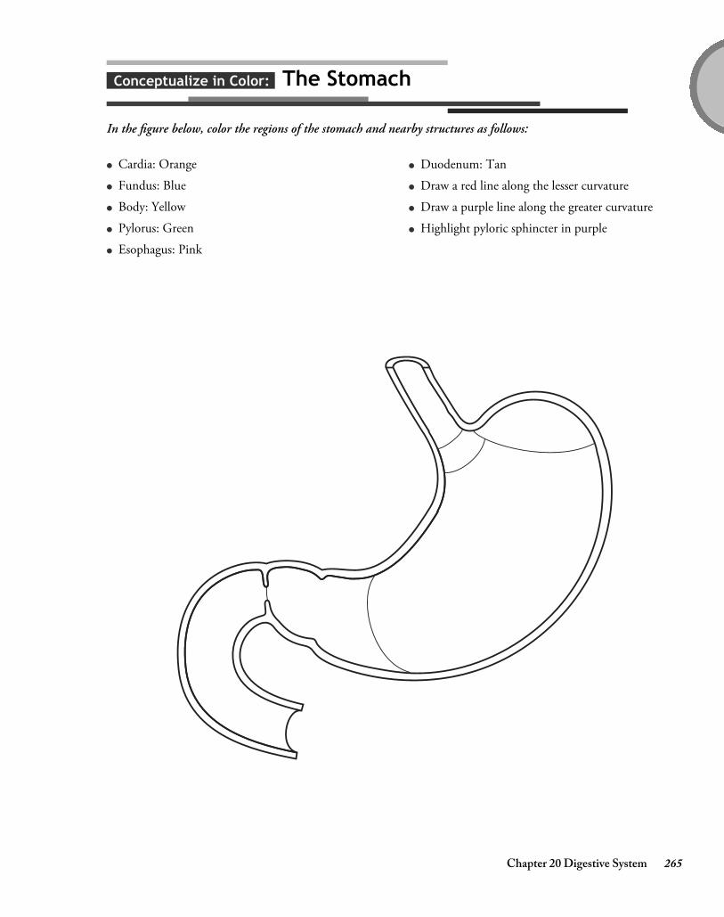
Chapter 20 Digestive System 265
Conceptualize in Color: The Stomach
In the figure below, color the regions of the stomach and nearby structures as follows:
● Cardia: Orange
● Fundus: Blue
● Body: Yellow
● Pylorus: Green
● Esophagus: Pink
● Duodenum: Tan
● Draw a red line along the lesser curvature
● Draw a purple line along the greater curvature
● Highlight pyloric sphincter in purple
4374_Ch20_257-276 10/22/14 10:47 AM Page 265

266 Chapter 20 Digestive System
Fill in the Gaps: Gastric Secretion
Fill in the blanks in the following sentences to describe the three phases of gastric secretion. Choose from the wordslisted in the Word Bank. (Hint: Words may be used more than once.)
Conceptualize in Color: The Liver
The figure below shows a posterior view of the liver. Color the structures of the liver as described.
● Right lobe: Light brown
● Left lobe: Dark brown
● Caudate lobe: Tan
● Quadrate lobe: Gold
● Falciform ligament: Gray
Posterior view
● Portal vein: Blue
● Hepatic artery: Red
● Inferior vena cava: Purple
● Gallbladder: Dark green
● Common hepatic duct: Light green
CEPHALIC
GASTRIC
GASTRIN
INCREASE
INHIBIT
INTESTINAL
NEURAL
PARASYMPATHETIC
1. The sight, smell, taste, or thought of food sends (a) impulses to the brain. The
(b) nervous system then signals the stomach to secrete (c) juice
as well as (d) . This is called the (e) phase of gastric secretion.
2. When food enters the stomach, the (a) phase begins. The stretching of the stomach
triggers nervous impulses that (b) the secretion of (c)
juice and (d) .
3. As chyme moves into the duodenum, the (a) phase begins. At this point, nervous
impulses and hormones secreted by the duodenum (b) gastric secretion.
4374_Ch20_257-276 11/6/14 8:25 AM Page 266

Chapter 20 Digestive System 267
Drawing Conclusions: Hepatic Lobules
Fill in the blanks in the following sentences to describe the structure and function of hepatic lobules. Then color thefigure as described.
1. Nutrient-rich blood from the stomach and intestine enters the lobule through small branches of the
.(Color this structure dark blue.)
2. Oxygen-rich blood enters the lobule through small branches of the .(Color this structure red.)
3. The blood filters through the , allowing the cells to remove nutrients, hormones, toxins, and drugs. (Color these structures light blue. Insert arrow to show direction of flow.)
4. (a) cells called (b) remove bacteria, worn-out red blood cells, and debris from the bloodstream.(Color these structures yellow.)
5. The carries the processed blood out of the liver.(Color this structure purple.)
6. (a) carry the bile secreted by hepatic cells and ultimately drain into the
(b) .
4374_Ch20_257-276 10/22/14 1:52 PM Page 267

268 Chapter 20 Digestive System
Conceptualize in Color: The Pancreas
Color the structures in the figure below as instructed.
● Right and left hepatic ducts: Blue
● Common hepatic duct: Purple
● Common bile duct: Light green
● Cystic duct: Yellow
● Gallbladder: Dark green
● Duodenum: Pink
● Pancreas: Gold
● Pancreatic duct: Orange
● Hepatopancreatic ampulla (ampulla of Vater):Mark with red dot
● Major duodenal papilla: Brown
● Hepatopancreatic sphincter (sphincter of Oddi):Black
4374_Ch20_257-276 10/22/14 10:47 AM Page 268

Chapter 20 Digestive System 269
Illuminate the Truth: The Pancreas and Duodenum
Highlight the correct word or phrase to complete each sentence.
1. Most of the pancreas contains (endocrine)(exocrine) tissue.
2. (Acinar)(Duct) cells in the pancreas secrete digestive enzymes in an inactive form.
3. The enzymes are activated in the (stomach)(duodenum), after which they help break down lipids, proteins, andcarbohydrates.
4. (Acinar)(Duct) cells in the pancreas secrete sodium bicarbonate, which buffers the acidic chyme entering theduodenum.
5. The arrival of chyme in the duodenum prompts the duodenum to secrete (cholecystokinin)(insulin), which(stimulates)(inhibits) the release of bile and pancreatic enzymes.
6. The hormone (gastrin)(secretin) also triggers gallbladder contraction and pancreatic enzyme secretion.
7. The hormone (gastrin)(secretin) causes the pancreatic ducts to release bicarbonate to neutralize the stomach acid.
4374_Ch20_257-276 10/22/14 10:47 AM Page 269

270 Chapter 20 Digestive System
Drawing Conclusions: The Small Intestine
In the figure below, color the regions of small intestine as instructed. Then use the same color to highlight thecharacteristics of each region. (For example, after coloring the duodenum green, highlight the sentences describingcharacteristics of the duodenum in green.)
● Duodenum: Green
● Ileum: Yellow
● Jejunum: Blue
1. Begins at the pyloric valve and ends where the intestine turns abruptly downward
2. Forms the last 12 feet of intestine
3. Receives chyme from the stomach as well as pancreatic juice and bile
4. Is where stomach acid is neutralized and pancreatic enzymes begin the task of chemical digestion
5. Provides an ideal location for nutrient absorption
6. Forms the first 10 inches of small intestine
7. Wall of this part of the intestine is thick and muscular with a rich blood supply
8. Contains clusters of lymphatic nodules called Peyer’s patches
9. Is where more digestive processes occur than in any other part of the intestine
4374_Ch20_257-276 10/22/14 10:47 AM Page 270

Chapter 20 Digestive System 271
Fill in the Gaps: Intestinal Wall
Fill in the blanks with the correct word or phrase to complete each sentence. Choose from the words in the WordBank. (Hint: Not all of the words will be used. Also, some words may be used more than once.)
1. The intestinal lining contains circular folds that the progress of
and its contact with the mucosa.
2. On top of the circular folds are projections called .
3. These projections are covered by absorptive cells as well as mucus-secreting
cells.
4. An arteriole, a venule, and a lymph vessel called a fill the core of each villus.
5. The villi have a brush border of ultrafine ; these projections serve to
the absorptive area and produce .
ACCELERATE
BILE
CHYME
DECREASE
DIGESTIVE ENZYMES
EPITHELIAL
GOBLET
HORMONES
INCREASE
LACTEAL
MICROVILLI
MUCUS
SLOW
VILLI
4374_Ch20_257-276 11/6/14 8:26 AM Page 271

272 Chapter 20 Digestive System
Puzzle It Out: Digestive System Basics
Further your knowledge of key facts about the digestive system by completing the following crossword puzzle.
ACROSS
3. Term referring to the liver5. Wave-like contractions that propel food through
the digestive tract7. Type of digestion that breaks food into smaller
particles9. Secretes bile
10. Sheets of hepatic cells that fan out from thecenter of the hepatic lobule
11. The portion of the mesentery that hangs over the small intestine like an apron is called thegreater .
12. Main bile pigment
DOWN
1. Yellow-green fluid that aids in the digestion andabsorption of fat
2. Serves to store and concentrate bile4. Type of digestion that uses digestive enzymes to
break down food particles into nutrients6. The most important component of bile is bile
.8. Another name for the digestive tract is the
canal.
4374_Ch20_257-276 10/22/14 10:47 AM Page 272

Chapter 20 Digestive System 273
Describe the Process: Carbohydrate Digestion
Describe the process of carbohydrate digestion by filling in the blanks in the following sentences with the correctword or phrase.
1. Salivary glands in the mouth secrete the enzyme , which works to hydrolyze
to .
2. The low pH of the stomach the enzyme .
3. In the intestine, chyme mixes with and the process of
digestion .
4. The membranes of the cells covering the villi contain the enzymes ,
, and .
5. As chyme slides along the brush border, . This is
called digestion.
6. The final step in the digestion process produces , which is immediately absorbed.
Illuminate the Truth: Protein Digestion
Highlight the correct word or phrase to complete each sentence.
1. Enzymes called (proteases)(sucrases) work in the (mouth)(stomach) and (esophagus)(small intestine) to breakpeptide bonds.
2. In the stomach, the enzyme (pepsin)(amylase) hydrolyzes the peptide bonds between certain amino acids.
3. In the duodenum, the pancreatic enzymes (lipase and amylase)(trypsin and chymotrypsin) assume the task ofbreaking peptide bonds.
4. Brush border enzymes called (pepsin)(peptidases) break the remaining chains into individual amino acids,which are then absorbed into the bloodstream.
4374_Ch20_257-276 10/22/14 10:47 AM Page 273

274 Chapter 20 Digestive System
Sequence of Events: Fat Digestion
Test your knowledge of fat digestion by placing the following events in the proper sequence. Arrange the events byinserting numbers in the spaces provided. The first step has been provided to get you started.
A. A fat globule enters the duodenum.
B. Pancreatic lipase begins to digest fat.
C. Triglycerides travel through the lymphatic system and enter the bloodstream at the left subclavian vein.
D. Glycerol and short-chain fatty acids are absorbed into the bloodstream of villi, and long-chain fatty acidsare absorbed into the walls of the villi.
E. Two substances in bile (lecithin and bile salts) emulsify fat.
F. Fats are broken down into a mixture of glycerol, short-chain fatty acids, long-chain fatty acids, andmonoglycerides.
G. The gallbladder secretes bile.
H. Triglycerides enter the lacteal of the villi.
1
4374_Ch20_257-276 10/22/14 10:47 AM Page 274

Chapter 20 Digestive System 275
Conceptualize in Color: Large Intestine
Color the regions of the large intestine as instructed.
● Cecum: Pink
● Appendix: Green
● Ileocecal valve: Brown
● Ascending colon: Blue
● Transverse colon: Red
● Descending colon: Orange
● Sigmoid colon: Yellow
● Rectum: Tan
● Anal canal: Light blue
● Label the right colic (hepatic) flexure and the left colic (splenic) flexure
4374_Ch20_257-276 10/22/14 10:47 AM Page 275

276 Chapter 20 Digestive System
Fill in the Gaps: Large Intestine
Fill in the blanks with the correct word or phrase to complete each sentence. Choose from the words in the WordBank. (Hint: Not all the words will be used.)
APPENDIX
BACTERIAL FLORA
CHYME
FECES
FLATUS
HAUSTRA
NUTRIENTS
WATER
1. The large intestine acts to absorb a large amount of from food residue.
2. Pouches called exist all along the large intestine.
3. The contains masses of lymphoid tissue and serves as a source for immune cells.
4. The large intestine houses hundreds of bacteria called .
5. One of the products of bacterial action is intestinal gas, called .
6. Waste material that exits the large intestine is called .
4374_Ch20_257-276 11/6/14 8:27 AM Page 276

chap
ter 21NUTRITION &
METABOLISM
The body’s source of energy is food. However, before the body can access that energy, food must be broken downinto usable components. In turn, those components must undergo chemical reactions within cells before thatenergy is unleashed. To ensure that you have a working knowledge of nutrition and metabolism, complete theactivities in this chapter.
Chapter 21 Nutrition & Metabolism 277
Make a Connection: Carbohydrates
Unscramble the words on the left to discover the names of the three forms of dietary carbohydrates. Then draw aline to link each form with its particular characteristics. (Hint: One characteristic is used more than once.)
1. AMECHANICSDOORS
2. CARDIACDISHES
3. ACHILDCARESPOSY
a. Known as complex carbohydrates
b. Known as simple sugars
c. Broken down into monosaccharides during thedigestive process
d. Consist of the starches found in vegetables, grains,potatoes, rice, and legumes
e. Taste sweet
f. Examples include fruit sugar as well as glucose andgalactose
g. Examples include table sugar as well as the sugarfound in milk
h. Are absorbed through the small intestine withoutbeing broken down
i. Includes cellulose
4374_Ch21_277-286 10/28/14 12:21 PM Page 277

278 Chapter 21 Nutrition & Metabolism
ACROSS
1. Chemical reactions occurring within cells thattransform nutrients into energy
7. An important contributor of fiber in the diet 8. The amount of energy the body needs at rest
(abbreviation)10. Energy-dense nutrient containing 9 calories/gram11. The digestive system breaks food into usable
components called .12. The body’s primary energy source13. One factor affecting basal metabolic rate
DOWN
2. Group of vitamins and minerals needed by thebody in small quantities
3. Nutrient used in every aspect of cellularmetabolism
4. Location of the control centers for hunger andsatiety
5. Hormone that stimulates appetite6. Hormone secreted by adipose tissue that
suppresses appetite9. The amount of heat needed to raise the
temperature of 1 g of water by 1°C
Puzzle It Out: Nutrition Concepts
Improve your knowledge of some of the concepts of nutrition by completing the following crossword puzzle.
4374_Ch21_277-286 10/28/14 12:21 PM Page 278

Chapter 21 Nutrition & Metabolism 279
Illuminate the Truth: Nutrition Basics
Highlight the words or phrase that correctly completes each sentence about some basic nutrition concepts.
1. (Protein)(Carbohydrates) should make up the greatest percentage of the diet.
2. Most of the carbohydrates consumed should be in the form of (simple sugars)(complex carbohydrates).
3. Excess carbohydrates are (stored in the body as fat)(converted into protein).
4. Carbohydrates, lipids, proteins, and water are called (macronutrients)(essential nutrients).
5. Saturated fats are derived mainly from (plants)(animals).
6. The most energy-dense nutrient is (protein)(fat).
7. (Unsaturated)(Saturated) fats tend to be solid at room temperature.
8. Hydrogenated oils, such as margarine, are (saturated)(unsaturated) fats.
9. All carbohydrates are eventually broken down into (glucose)(glycogen), which is then used as fuel for energy.
10. Fats that must be obtained through the diet—such as linoleic acid, which is a necessary part of the cellmembrane—are called (essential fatty acids)(essential lipids).
Fill in the Gaps: Proteins
Proteins fulfill a vast array of functions in the body. Fill in the blanks with the correct word or phrase thatcompletes each sentence about this vital nutrient. Choose from the words listed in the Word Bank. (Hint: Not allthe words will be used.)
AMINO ACIDS
ANIMAL
CAN
CANNOT
COMPLETE PROTEINS
ESSENTIAL AMINO ACIDS
INCOMPLETE PROTEINS
MOLECULES
NONESSENTIAL AMINO ACIDS
PLANT
1. During the process of digestion, proteins are broken down into their individual .
2. Foods that supply all the essential amino acids are called ;
these foods mainly come from sources.
3. Foods that lack one or more essential amino acids are called ;
these foods mainly come from sources.
4374_Ch21_277-286 10/28/14 12:21 PM Page 279

280 Chapter 21 Nutrition & Metabolism
Illuminate the Truth: Vitamins and Minerals
The body requires a number of vitamins and minerals to function properly. Highlight the word or phrase thatcorrectly completes each sentence about some of these essential nutrients.
1. (Vitamin C)(Vitamin A) aids in wound healing.
2. (Vitamin K)(Vitamin E) is necessary for normal blood clotting.
3. A deficiency of (vitamin A)(vitamin D) results in night blindness.
4. A deficiency of (folic acid)(vitamin D) results in rickets.
5. Pernicious anemia results from a deficiency of (vitamin B12)(vitamin B6).
6. Besides being necessary for the formation of bones and teeth, calcium is also necessary for (carbohydratedigestion)(muscle contraction and relaxation).
7. (Iodine)(Iron) is necessary for hemoglobin production.
8. Folic acid is needed for (bone growth and development)(synthesis of DNA).
9. A potassium deficiency can lead to (softening of the bones)(abnormal cardiac rhythm).
List for Learning: Fat-Soluble Vitamins
Fat-soluble vitamins are absorbed with dietary fat, after which they are stored in the liver and fat tissues of thebody. List the four fat-soluble vitamins.
1.
2.
3.
4.
4. The body store extra amino acids.
5. Amino acids that can be synthesized by the body and, therefore, do not need to be consumed in the diet are
called .
6. Amino acids that cannot be synthesized by the body and, therefore, must be consumed in the diet are called
.
4374_Ch21_277-286 10/28/14 12:21 PM Page 280

Chapter 21 Nutrition & Metabolism 281
Fill in the Gaps: Carbohydrate Metabolism
Test your knowledge of carbohydrate metabolism by filling in the blanks in the following sentences with the correctword or phrase. Choose from the words listed in the Word Bank. (Hint: Not all the words will be used.)
Make a Connection: Metabolism
Nutrients are transformed through metabolism into energy the body can either use immediately or store for lateruse. Unscramble the words on the left to discover the two types of metabolic processes. Then draw lines to the list onthe right to link each process to its particular characteristics.
1. ANIMALSOB
2. BOATSCLAIM
AEROBIC
AEROBIC RESPIRATION
A FRACTION
ALL
ANAEROBIC
ANAEROBIC FERMENTATION
BURNED AS ENERGY
GLUCOSE
GLYCOGEN
GLYCOLYSIS
STORED
1. All ingested carbohydrates are converted to , most of which is immediately
.
2. The first phase of carbohydrate metabolism is called ; because it does not use
oxygen, this is called an process.
3. This first phase releases of the available energy.
4. During a period of strenuous exercise, the next second phase of metabolism would be
.
5. During a time of rest, the next phase of metabolism would be
.
a. Used in protein metabolism
b. Breaks down complex substances into simpler onesor into energy
c. Forms complex substances out of simpler ones
d. Used in the metabolism of carbohydrates and lipids
4374_Ch21_277-286 10/28/14 12:21 PM Page 281

282 Chapter 21 Nutrition & Metabolism
Drawing Conclusions: Anaerobic Fermentation
The process the body uses to metabolize carbohydrates varies according to the availability of oxygen. Test yourknowledge of how the body metabolizes carbohydrates when oxygen is in short supply by filling in the blanks in thefollowing sentences. Then illustrate the process by filling in the symbols to the left of each sentence with the name ofthe substances produced along the way
1. When oxygen is in short supply,
is converted into
.
2. The then travels
through the bloodstream to the
, where it is stored.
3. Later, during a period of rest when oxygen
becomes available,
is converted into ,
which may then enter the series of reactions in
.
4374_Ch21_277-286 10/28/14 12:21 PM Page 282

Chapter 21 Nutrition & Metabolism 283
Drawing Conclusions: Aerobic Respiration
Now test your knowledge of how the body metabolizes carbohydrates when oxygen is available by filling in theblanks in the following sentences. Then illustrate the process by filling in the symbols to the left of each sentence withthe name of the substances produced along the way.
CO2 + H2O+
1. When oxygen is available,
enters the
and is
converted into .
2. This begins a series of reactions known as the
.
3. This is followed by still another series of reactions
called the
.
4. The end result is the complete breakdown of
into carbon
dioxide, water, and, most importantly, large
amounts of .
4374_Ch21_277-286 10/28/14 12:21 PM Page 283

284 Chapter 21 Nutrition & Metabolism
Fill in the Gaps: Lipid Metabolism
Review the process of lipid metabolism by filling in the blanks in the following sentences. Choose from the wordslisted in the Word Bank. (Hint: Not all the words will be used. Also, words may be used more than once.)
Illuminate the Truth: Protein Metabolism
While carbohydrates and fat are mainly used to supply energy, proteins have a different role. Review some of thefacts about protein metabolism by highlighting the word or phrase that correctly completes each sentence.
1. Proteins are mainly used to (supply energy)(build tissue); this is called protein (anabolism)(catabolism).
2. Protein (anabolism)(catabolism) is the process whereby proteins may be converted to glucose or fat or useddirectly as fuel.
3. During the digestion process, proteins are broken down into (glucose)(individual amino acids), which are then(stored as fat)(recombined to form new proteins).
4. Before proteins are used for energy, they are altered in the liver to produce (ammonia and keto acid)(urea andcitric acid).
5. A nitrogenous waste product resulting from protein catabolism is (urea)(ketones).
ACETYL CoA
ADIPOSE TISSUE
CITRIC ACID
ELECTRON TRANSPORT
FATTY ACID(S)
GLUCOSE
GLYCOLYSIS
LARGE
SMALL
THE LIVER
1. When needed for energy, fat molecules stored within are
hydrolyzed into glycerol and three molecules of .
2. Glycerol enters the pathway and yields a amount of energy.
3. At the same time, are broken down to form .
4. In turn, proceeds through the cycle and
chain to yield amounts of energy.
4374_Ch21_277-286 10/28/14 12:21 PM Page 284

Chapter 21 Nutrition & Metabolism 285
Make a Connection: Thermoregulation
Unscramble the words on the left to discover three ways the body loses heat. Then draw lines to link each to itsparticular characteristics.
1. ARADIOTIN
2. TICCOONDUN
3. AVIATORNOPE
Illuminate the Truth: Regulation of Body Temperature
Several negative feedback mechanisms regulate body temperature. Refine your knowledge of these processes byhighlighting the word or phrase that correctly completes each sentence.
1. The (pituitary gland)(hypothalamus) acts as the body’s thermostat: it monitors the temperature of the(blood)(skin) and sends signals to blood vessels and sweat glands.
2. When the body temperature rises too high, the (hypothalamus)(pituitary gland) signals cutaneous blood vesselsto (constrict)(dilate). More warm blood flows (close to the body’s surface)(deep into the body) and heat is(lost)(conserved) through the skin.
3. If the temperature remains high, sweat glands produce sweat; in turn, the (production)(evaporation) of sweatproduces (cooling)(warming).
4. When the body temperature falls below normal, the (pituitary gland)(hypothalamus) signals cutaneous bloodvessels to (constrict)(dilate). More warm blood (flows to the body’s surface)(remains confined deep in thebody). As a result, (less)(more) heat is lost through the skin.
5. If the temperature remains below normal, the body begins to (shiver)(grow rigid). As a result, muscles(retain)(release) heat and the body’s temperature (falls)(rises).
a. Involves the transfer of heat between two materialsthat touch each other
b. Involves the transfer of heat through the air byelectromagnetic heat waves
c. Uses heat to change water into a gas
d. How our bodies tend to lose heat
e. An example is standing in bright sunlight
f. An example is perspiration
g. An example is a warm chair after we stand fromsitting
4374_Ch21_277-286 10/28/14 12:21 PM Page 285

4374_Ch21_277-286 10/28/14 12:21 PM Page 286

chap
ter 22HUMAN
MICROBIOME
The human body houses a community of microbes that outnumber human cells 10 to 1. Discovery of this vastecosystem is transforming how practitioners view both health and disease. Use the activities in this chapter tobuild upon your knowledge of the microbes that exist on, and in, the human body.
Chapter 22 Human Microbiome 287
Illuminate the Truth: Facts about the Human Microbiome
Highlight the word or phrase that most accurately completes each sentence.
1. The human microbiome (is present at birth)(builds over time).
2. Scientists have learned that the inhabitants of specific bacterial communities—such as those in the mouth orintestines—(tend to be consistent)(often vary widely) between individuals.
3. (Bacteria)(Viruses) make up the bulk of the human microbiome.
4. The sinus passages of a person with sinusitis tend to contain (more)(fewer) species of bacteria as compared tothe sinus passages of a person without sinusitis.
5. Children acquire significant components of their microbiomes (through their diet)(from their mothers).
4374_Ch22_287-294 11/7/14 12:46 PM Page 287

288 Chapter 22 Human Microbiome
Conceptualize in Color: Bacterial Structures
Identify the structures of bacteria by coloring the structures in the figure as suggested:
● Capsule: Green
● Cell wall: Orange
● Cytoplasmic membrane: Blue
● DNA: Pink
● Ribosomes: Red
● Plasmids: Brown
● Pilli: Purple
● Flagellum: Dark blue
4374_Ch22_287-294 11/7/14 10:04 AM Page 288

Fill in the Gaps: Bacteria
Improve upon your knowledge of bacterial structures by completing the sentences below. Choose the correct word orphrase from the Word Bank. (Hint: Some words may be used more than once; also, not all words or phrases will be used.)
Chapter 22 Human Microbiome 289
CAPSULE
CELL WALL
CYTOPLASMIC MEMBRANE
FLAGELLA
GROUPED IN THE CELL’S CENTRAL REGION
PILI
PLASMIDS
RESIST
RESPOND TO
THICK
THIN
WITHIN THE CELL’S NUCLEUS
1. Some species of bacteria contain a gelatinous covering called a __________________ that keeps the
bacterium from drying out.
2. A rigid _______________________ encloses the bacterium and gives the cell its shape.
3. The __________________________________ regulates the flow of materials into and out of the cell.
4. Because the composition of the _______________________ varies between species, it is an important
distinguishing factor between bacterial types.
5. Many species of bacteria have small, hair-like projections called _________________ that allow bacteria to
attach to other cells and surfaces.
6. Small loops of bacterial DNA are called __________________________.
7. Some bacteria have ______________________, which aid in motility.
8. Bacterial DNA is found ____________________________________________________________.
9. Bacterial structures that allow bacteria to trade genes are called ________________________.
10. Bacteria containing a ________________________ are more likely to cause disease.
11. Gram-positive bacteria have a _________________ layer of peptidoglycan and tend to
_____________________ antibiotic treatment.
4374_Ch22_287-294 11/12/14 11:03 AM Page 289

290 Chapter 22 Human Microbiome
Conceptualize in Color: Viruses
Review the structures of viruses by coloring the structures in the figure below as suggested.
● Capsid: Blue
● Envelope: Orange
● Nucleic acid: Yellow
● Spikes: Brown
4374_Ch22_287-294 11/7/14 10:04 AM Page 290

Make a Connection: Viral Shapes
Unscramble the following words to discover the three basic viral shapes. Then draw a line to link each shape to itsparticular characteristics.
1. LCIHAEL
2. YHLLRDOPAE
3. CPLMXOE
Chapter 22 Human Microbiome 291
a. Adenovirus has this shape
b. Rabies virus has this shape
c. Is a protein cylinder
d. A bacteriophage has this shape
e. Consists of many triangular faces
4374_Ch22_287-294 11/7/14 10:04 AM Page 291

292 Chapter 22 Human Microbiome
Describe the Process: Viral Replication
Viral replication occurs rapidly: a single cell may produce from 10,000 to 50,000 new viruses in as little as 48 hours. Test your knowledge of how this occurs by describing the process in the spaces below. Use the figureprovided for clues. To further solidify your knowledge, color the figures using colors of your own choosing.
1.
2.
3.
4.
1
1
1
1
Nucleic acid (DNA or RNA)
Virus
Nucleus
2
3
4
1
4374_Ch22_287-294 11/12/14 11:04 AM Page 292

Puzzle it Out: Microbiome Facts
Complete the following crossword puzzle to review key facts about the human microbiome.
Chapter 22 Human Microbiome 293
9
2
5
8
4
7
11
6
10
1
13
12
3
ACROSS
5. Interferes with a bacterium’s ability tomanufacture peptidoglycan
7. Bacteria having a rod shape9. Complex virus that attaches to the surface of a
cell before injecting its nucleic acid into the target cell
11. Structure that allows bacteria to ward off whiteblood cells
13. Molds and yeasts
DOWN
1. Round bacteria2. Frequent culprit in microbiome disruption3. Place on the body where fungal infections often
begin4. Spiral-shaped bacteria6. Bacteria lack this8. Microorganism known to cause disease
10. Bacterial structure that allows bacteria to beextremely adaptable
12. Gram-negative bacteria stain this color
4374_Ch22_287-294 11/7/14 10:04 AM Page 293

4374_Ch22_287-294 11/7/14 10:04 AM Page 294

chap
ter 23REPRODUCTIVE SYSTEMS
In human reproduction, sex cells from the male and the female fuse to create a genetically unique individual.Structurally, the male and female reproductive systems differ significantly. Build upon your knowledge of these two systems by completing the activities in this chapter.
Chapter 23 Reproductive Systems 295
Make a Connection: Accessory Glands
The male reproductive system includes three sets of accessory glands. Unscramble the words on the left to discoverthe names of these glands. Then draw lines to link each gland to its particular characteristics.
1. LANESIM SECVILE
2. SPARETOT DALNG
3. HARBOURBULLET LAGND
a. Secretes a clear fluid into the penile portion of theurethra during sexual arousal
b. Encircles urethra and ejaculatory duct
c. Secretes a thick, yellowish fluid into theejaculatory duct
d. Fluid secreted comprises about 30% of the fluidportion of semen
e. Fluid secreted comprises about 60% of semen
f. Fluid secreted serves as a lubricant duringintercourse and also neutralizes the acidity ofresidual urine in the urethra
g. Secreted fluid contains fructose as well as othersubstances that nourish sperm and help ensuremotility
h. Secretes a thin, milky, alkaline fluid into the urethra
4374_Ch23_295-306 10/28/14 12:26 PM Page 295

296 Chapter 23 Reproductive Systems
ACROSS
2. Term for the primary sex organ4. Male structure that serves both the reproductive
and urinary systems6. Foreskin7. Hormone necessary for the development of
female secondary sex characteristics10. Process by which gametes divide12. Whitish fluid emitted during ejaculation that
contains both sperm and the secretions of theaccessory glands
14. Male hormone necessary for sperm productionand the development of male secondary sexcharacteristics
DOWN
1. Undescended testes3. Primary sex organs in females5. Sac of tissue in which the testes lie8. Sex cells9. Muscle that contracts in cold weather to pull the
testes closer to the body11. Primary sex organs in males13. Secretion of this hormone by hypothalamus
marks the onset of puberty
Puzzle It Out: Reproductive Terms
Test your knowledge of some terms specific to the reproductive system by completing the following crossword puzzle.
4374_Ch23_295-306 10/28/14 12:26 PM Page 296

Drawing Conclusions: The Testes
Fill in the blanks in the following sentences to correctly describe the structure and function of the testes. Then colorthe figure as suggested to enhance your learning.
Chapter 23 Reproductive Systems 297
1. A strand of connective tissue called the extends from the abdomen to each testicle; housed inside are blood, lymphatic vessels, and nerves.(Color this structure gray.)
2. Fibrous tissue separates each testis into more than 200 . (Outline these areas in brown.)
3. Coiled inside each of these structures are one to three tightly coiled ducts called ,
which is where sperm are . (Color these structures yellow.)
4. A network of vessels called the lead away from this area and provide a location in which sperm partially mature.(Color these vessels blue.)
5. ductules conduct immature sperm away from the testes.(Color these vessels green.)
6. Sperm pass into the , where they remain fertile for 40 to 60 days.(Color this structure orange.)
7. Sperm leave the previous structure and pass into the .(Color this structure purple.)
4374_Ch23_295-306 10/28/14 12:26 PM Page 297

298 Chapter 23 Reproductive Systems
Conceptualize in Color: Male Reproductive Organs
Color the structures of the male reproductive system in the following figure. Use the colors suggested or choose your own.
● Vas deferens: Dark blue
● Ejaculatory duct: Fuschia
● Bulbourethral gland: Brown
● Seminal vesicle: Yellow
● Prostate gland: Light green
● Urethra: Purple
● Testis: Gray
● Epididymis: Gold
● Scrotum: Tan
● Corpus spongiosum: Red
● Corpus cavernosum: Pink
● Glans penis: Orange
● Prepuce: Dark green
● Urinary bladder: Light blue
4374_Ch23_295-306 10/28/14 12:26 PM Page 298

Chapter 23 Reproductive Systems 299
Sequence of Events: Spermatogenesis
Thousands of sperm are produced each second. Insert numbers in the spaces provided to arrange the events ofspermatogenesis in the proper order.
A. Daughter cells of spermatogonia differentiate into slightly larger cells called primary spermatocytes,which move toward the lumen of the seminiferous tubule.
B. Spermatids differentiate to form heads and tails and eventually transform into mature spermatozoa(sperm), each with 23 chromosomes.
C. Each secondary spermatocyte divides to form two spermatids.
D. Spermatogonia in the walls of the seminiferous tubules divide by mitosis to produce two daughter cells,each with 46 chromosomes.
E. Through meiosis, the primary spermatocyte yields two genetically unique secondary spermatocytes, eachwith 23 chromosomes.
4374_Ch23_295-306 10/28/14 12:26 PM Page 299

300 Chapter 23 Reproductive Systems
Illuminate the Truth: More Reproductive Facts
Highlight the word or phrase that correctly completes each sentence.
1. The most common cancer in American men is (prostate)(testicular) cancer.
2. The body of the penis is called the (shaft)(prepuce).
3. Through the process of meiosis, each daughter cell has (46)(23) chromosomes.
4. Each daughter cell of meiosis is (genetically identical to)(genetically unique from) the parent cell.
5. An important quality of semen is its (acidity)(alkalinity).
6. The portion of sperm that contains genetic information is the (acrosome)(head).
7. The portion of the sperm that contains an enzyme that helps it penetrate an egg is the (acrosome)(middlepiece).
8. The testes are located outside the body to (protect them from infection)(keep them cooler).
List for Learning: Sexual Response
Using the spaces provided, list the four phases of both the male and female sexual response.
1.
2.
3.
4.
4374_Ch23_295-306 10/28/14 12:26 PM Page 300

Conceptualize in Color: Female Reproductive System
Color the structures of the female reproductive system in the following figure. Use the colors suggested or choose your own.
Chapter 23 Reproductive Systems 301
● Uterus: Red
● Fallopian tube: Pink
● Ovary: Gray
● Cervix: Orange
● Rectum: Brown
● Vagina: Purple
● Bladder: Blue
● Labium majus: Tan
● Labium minus: Gold
● Urethra: Light blue
4374_Ch23_295-306 10/28/14 12:26 PM Page 301

302 Chapter 23 Reproductive Systems
Drawing Conclusions: Internal Genitalia
Unlike the male, the organs of the female reproductive system are housed within the abdominal cavity. Reviewthese structures by filling in the blanks in the following sentences. Then follow the instructions to color the structuresin the figure below.
1. The narrow portion of the fallopian tube closest to the uterus is called the ; the
middle portion is called the , while the funnel-shaped distal portion is called the
.(Color the fallopian tubes pink.)
2. Finger-like projections called fan over the ovary.(Color these structures orange.)
3. The uterus is held in place by the ligament.(Color this structure tan.)
4. The curved, upper portion of the uterus is called the . (Outline this portion in blue.)
5. The inferior end of the uterus is called the .(Color this structure brown.)
6. The serves as a receptacle for the penis and sperm and as a route for the discharge of menstrual blood.(Color this structure purple.)
7. The smooth muscle layer of the uterus is called the .(Color this structure light red.)
8. The innermost layer of the uterus, called the , is where an embryo attaches.(Color this structure dark red.)
4374_Ch23_295-306 10/28/14 12:26 PM Page 302

Conceptualize in Color: Breasts
Color the structures of the breasts. Use the colors suggested or choose your own.
Chapter 23 Reproductive Systems 303
● Pectoralis muscle: Pink● Acini: Red● Lobules: Outline in purple● Lactiferous ducts: Tan
● Lactiferous sinuses: Orange● Areola: Brown● Adipose tissue: Yellow● Suspensory ligaments: Blue
4374_Ch23_295-306 10/28/14 12:26 PM Page 303

304 Chapter 23 Reproductive Systems
Puzzle It Out: Female Reproductive System
Enhance your knowledge of some of the terms specific to the female reproductive system by completing the followingcrossword puzzle.
ACROSS
2. Thick folds of skin and adipose tissue on eitherside of the vaginal opening are called the
majora.3. Collective term for female external genitalia5. Process through which a mature ovum is formed6. Cells surrounding the egg7. The central region of the uterus9. First menstrual period
10. Fold of mucous membrane partially covering theentrance to the vagina
DOWN
1. Small mound of erectile tissue in females thatresembles a penis
3. Area inside the labia4. State of life when menstruation ceases
permanently8. Another name for egg9. Mound of hair-covered adipose tissue overlying
the symphysis pubis is called thepubis.
4374_Ch23_295-306 10/28/14 12:26 PM Page 304

Fill in the Gaps: Ovarian Cycle
A woman’s reproductive system undergoes cyclical changes each month as it prepares for the possibility ofpregnancy. These changes, called the reproductive cycle, consist of two interrelated cycles: the ovarian cycle and themenstrual cycle. Test your knowledge of the sequence of events of the ovarian cycle by filling in the blanks in thefollowing sentences. Choose from the words listed in the Word Bank. (Hint: Words may be used more than once.)
Chapter 23 Reproductive Systems 305
ANTERIOR
CORPUS ALBICANS
CORPUS LUTEUM
ESTROGEN
FOLLICULAR
FSH
GONADOTROPIN-RELEASING HORMONE (GnRH)
POSTERIOR
PROGESTERONE
LH
LUTEAL
1. Low levels of and stimulate the hypothalamus to
release .
2. The hormone ____________ stimulates the pituitary to release the hormones
and .
3. The hormone triggers several follicles in the ovary to resume development, beginning
what is known as the phase.
4. As the follicle develops, it secretes , which stimulates the thickening of the endometrium,
as well as small amounts of .
5. At the mid-point of the cycle, levels peak, triggering a spike in , which causes ovulation.
6. The remnants of the follicle remain on the ovary and form the , which marks
the beginning of the phase.
7. The secretes large amounts of the hormone and
small amounts of .
8. If fertilization doesn’t occur, the degenerates into inactive scar tissue called
the .
9. Levels of and plummet, causing the endometrium to slough off, resulting in menstruation.
4374_Ch23_295-306 10/28/14 12:26 PM Page 305

306 Chapter 23 Reproductive Systems
Make a Connection: Menstrual Cycle
Unscramble the words on the left to discover the names of the four phases of the menstrual cycle. Then draw a lineto link each phase to its particular characteristics.
1. LAMETURNS
2. FIRETRAPOLIVE
3. CRYTOERES
4. MANTLEPURSER
a. Involves the atrophy of the corpus luteum
b. Blood vessels grow as the base layer is repaired
c. Time when menstruation occurs
d. Involves ischemia and necrosis of theendometrium
e. Dominated by increased levels of progesterone
f. Involves the shedding of the endometrium’sfunctional layer
g. Encompasses days 26 to 28 of the cycle
h. Encompasses days 6 to 14 of the cycle
i. Rising levels of estrogen dominate
j. Encompasses days 15 to 26 of the cycle
k. Encompasses days 1 to 5 of the cycle
l. Involves increased thickening of the endometriumas it becomes suitable for implantation of afertilized egg
m. Instituted by plummeting levels of progesterone
4374_Ch23_295-306 10/28/14 12:26 PM Page 306

chap
ter 24PREGNANCY & HUMAN
DEVELOPMENT
The creation of a new life—beginning with the fusion of an egg and a sperm and culminating with the entrance ofa neonate into the world—involves numerous body processes. Review some of these processes by completing theactivities in this chapter.
Chapter 24 Pregnancy & Human Development 307
Drawing Conclusions: Fertilization
Conception occurs when an egg and a sperm fuse. Strengthen your understanding of the fertilization process byfilling in the blanks to complete the sentences. Then follow the instructions to color and label the figure on the next page.
1. As hundreds of sperm swarm the egg, the on their heads release
that break down the cells and a gel-like layer called the
surrounding the egg. (Color the body of the sperm gray; the heads of the sperm yellow; the covering over the heads of the sperm green; theganulosa cells tan; the gel-like coating of the egg blue; and the substance released from the heads of the sperm pink.)
2. A path is created that allows (Hint: a number) sperm to penetrate the egg.(Label the sperm being blocked, the two sperm that are helping to clear a path into the egg, and the sperm that haspenetrated the egg.)
3. As fertilization occurs, the of the sperm is released into the ovum as its
degenerates and falls away. (Label the sperm that is fertilizing the egg.)
4374_Ch24_307-314 10/28/14 12:33 PM Page 307

308 Chapter 24 Pregnancy & Human Development
4. The of the sperm, which has chromosomes, fuses
with the of the egg, which also has chromosomes,
creating a single cell with chromosomes.
5. The fertilized egg is called a .
4374_Ch24_307-314 10/28/14 12:33 PM Page 308

Chapter 24 Pregnancy & Human Development 309
1. The stage begins when fertilization forms a zygote with 23 chromosomes.(By the number 1 in the illustration, draw the egg being fertilized.)
2. Within 24 to 36 hours, the zygote divides by the process of to form two daughter
cells called .(By the number 2 in the illustration, draw this process using two separate figures. Label these figures.)
3. The divisions continue, with the cells with each division. Finally, a blackberry-like
cluster of (Hint: a number) cells called a results. (Draw this process by the number 3 in the illustration, using three separate figures. Label the final figure.)
4. Three to four days after fertilization, the morula enters the uterine cavity, where it
for 2 or 3 days. As the cell cluster continues to divide, a hollow cavity forms; the cell cluster is now called a
. This differentiates into two layers: an outer layer of cells called the
, which eventually forms the , and an inner cell
mass that becomes the .(Draw and label this structure by the number 4 in the illustration.)
5. About 6 days after ovulation, the lands on and attaches to the endometrium—a
process called .(Draw what is being described, using the space by the number 5 in the illustration.)
Drawing Conclusions: Fertilization to Implantation
Fill in the blanks in the sentences to describe what occurs between fertilization and implantation. Then follow theinstructions to draw the product of conception in each stage of development as it proceeds through the fallopian tube.
Ovum
Ovary
1
2 3
4
5
4374_Ch24_307-314 10/28/14 12:33 PM Page 309
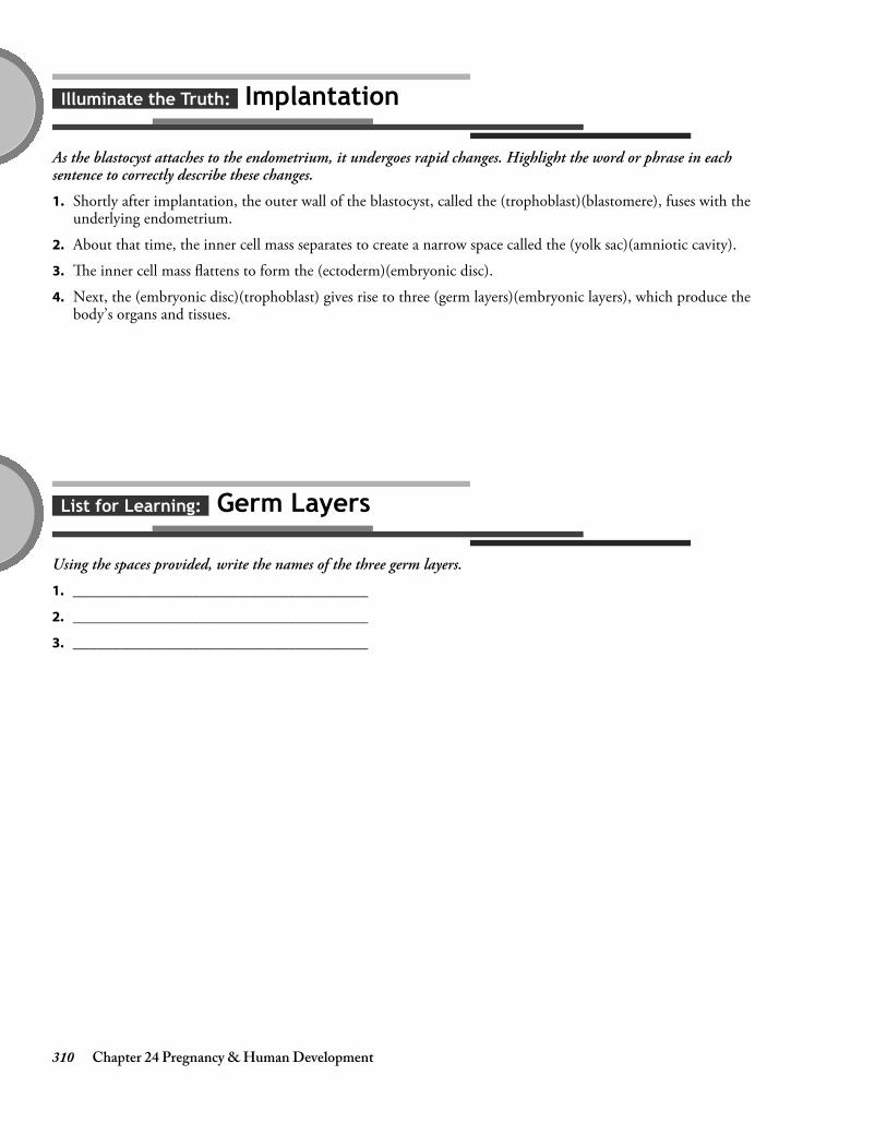
310 Chapter 24 Pregnancy & Human Development
List for Learning: Germ Layers
Using the spaces provided, write the names of the three germ layers.
1. _____________________________________
2. _____________________________________
3. _____________________________________
Illuminate the Truth: Implantation
As the blastocyst attaches to the endometrium, it undergoes rapid changes. Highlight the word or phrase in eachsentence to correctly describe these changes.
1. Shortly after implantation, the outer wall of the blastocyst, called the (trophoblast)(blastomere), fuses with theunderlying endometrium.
2. About that time, the inner cell mass separates to create a narrow space called the (yolk sac)(amniotic cavity).
3. The inner cell mass flattens to form the (ectoderm)(embryonic disc).
4. Next, the (embryonic disc)(trophoblast) gives rise to three (germ layers)(embryonic layers), which produce thebody’s organs and tissues.
4374_Ch24_307-314 10/28/14 12:33 PM Page 310

Chapter 24 Pregnancy & Human Development 311
Puzzle It Out: Human Growth and Development Terms
Complete the crossword puzzle to test your understanding of terms used in human growth and development.
ACROSS
4. Stage of development beginning at fertilization and lasting for 16 days5. The 3-month periods dividing the term of pregnancy9. Transparent sac filled with fluid that surrounds the embryo and fetus
13. Extraembryonic membrane that serves as the foundation for the umbilical cord14. Supplies the fetus with oxygen and nutrients during pregnancy15. Fertilized egg
DOWN
1. Outermost membrane that surrounds other extraembryonic membranes2. The union of the nuclei of the egg and sperm3. Hormone that prompts the corpus luteum to secrete estrogen and progesterone and also forms the basis for
pregnancy tests (Hint: use initials)6. Stage of development lasting from the third until the eighth week of pregnancy7. Fine hair covering the fetus’ body8. The time of growth before birth is called the period.
10. Length of time from conception until birth11. Process by which the fertilized cell divides by mitosis12. Stage of development beginning the eighth week and lasting until birth
4374_Ch24_307-314 10/28/14 12:33 PM Page 311

312 Chapter 24 Pregnancy & Human Development
Just the Highlights: Stages of Prenatal Development
The approximately 9-month period between fertilization and birth transforms a single cell into a complete humanbeing. Test your knowledge of the events that occur in each stage by highlighting the events of the pre-embryonicstage in pink, the events of the embryonic stage in blue, and the events of the fetal stage in yellow.
1. Formation of the amnion, yolk sac, allantois, andchorion
2. Begins at fertilization
3. Involves rapid growth
4. Differentiation of germ layers into organs andorgan systems
5. Fusion of the trophoblast with the endometrium
6. Movement that can be felt by the mother occurs
7. Formation of the amniotic cavity
8. Begins sixteen days after conception and lasts for 6 weeks
9. Formation of the embryonic disc
10. Begins the eighth week and lasts until birth
11. Development of the placenta and umbilical cord
12. Formation of the three germ layers
13. Encompasses the time of the zygote
14. The heart begins to beat
Conceptualize in Color: Embryonic and Fetal Membranes
Learn to differentiate between the various membranes surrounding the embryo and fetus by coloring them as described.
● Uterus: Pink
● Chorion: Green
● Amnion: Orange
● Amniotic fluid: Blue
● Umbilical cord: Gray
● Yolk sac: Yellow
● Chorionic villi: Red
4374_Ch24_307-314 10/28/14 12:33 PM Page 312

Chapter 24 Pregnancy & Human Development 313
Conceptualize in Color: Fetal Circulation
Because the fetus depends on the placenta for oxygen and nutrients as well as for the removal of waste products, thecirculatory system of the fetus differs significantly from that of a newborn infant. Trace the pathway of fetalcirculation by coloring the structures in the following figure as described.
1. Color the umbilical vein red to depict the oxygen-rich blood flowing in from the placenta.
2. Color the ductus venosus red as it bypasses the liver; change the color to purple as blood merges into theinferior vena cava. Draw a black arrow depicting the movement of blood from the umbilical vein, through theductus venosus, and into the inferior vena cava.
3. Color blood in the right atrium purple; continue coloring into the left atrium and through the foramen ovale.Insert a black arrow depicting the movement of blood through foramen ovale.
4. Color the right ventricle purple; extend the purple color into the pulmonary trunk. Insert black arrowsshowing the movement of blood into the right ventricle and into the pulmonary trunk. Then show the passageof blood into the ductus arteriosus and the descending aorta by coloring these areas purple and insertingarrows.
5. Continue the purple color into the iliac and umbilical arteries. Insert black arrows to depict the flow of bloodinto the umbilical arteries and back to the placenta.
Ductus arteriosus
Pulmonary trunk
Common iliac artery
Ascending aorta
Inferior vena cava
Placenta
Fetal umbilicus
Ductus venosus
Umbilical vein
Umbilical cord
Umbilical arteries
Foramen ovale
4374_Ch24_307-314 10/28/14 12:33 PM Page 313

314 Chapter 24 Pregnancy & Human Development
Fill in the Gaps: Childbirth
Test your knowledge of the events occurring during childbirth by filling in the blanks to complete each sentencecorrectly. Choose from the words listed in Word Bank. (Hint: Not all the words will be used. Also, some words maybe used more than once.)
Illuminate the Truth: Mammary Glands
After childbirth, the mammary glands produce and secrete milk to nourish the neonate. Review this process byhighlighting the correct word or phrase in each of the following sentences.
1. The process by which the mammary glands produce and secrete milk is called (parturition)(lactation).
2. The growth of ducts throughout the mammary glands is stimulated by the high levels of(progesterone)(estrogen) during pregnancy.
3. The development of acini at the ends of the ducts is stimulated by the high levels of (estrogen)(progesterone)during pregnancy.
4. The production of milk is initiated by the secretion of (prolactin)(progesterone).
5. The secretion of the hormone (estrogen)(oxytocin) causes milk to be released into the ducts.
6. Before milk is secreted, the mammary glands secrete a yellowish fluid called (vernix caseosa)(colostrum).
AFTERBIRTH
BRAXTON-HICKS
DILATION
EFFACEMENT
ESTROGEN
FIRST
OXYTOCIN
PROGESTERONE
PSEUDO-CONTRACTIONS
SECOND
THIRD
1. The onset of labor is thought to result from a decline in the hormone and an
increase in the hormones and .
2. The contractions sometimes known as “false labor” are called contractions.
3. The stage of labor is the longest.
4. The first stage of labor involves thinning of the cervical walls, called cervical , as
well as progressive widening of the cervix, called cervical .
5. The baby is born during the stage of labor.
6. The amniotic sac usually breaks during the stage of labor.
7. The final stage of labor involves delivery of the .
4374_Ch24_307-314 10/28/14 12:33 PM Page 314

chap
ter 25HEREDITY
The process of passing traits from biological parents to children is called heredity, while the study of heredity iscalled genetics. Test your knowledge of this fascinating topic by completing the activities in this chapter.
Illuminate the Truth: Basic Heredity
Review some basic heredity concepts by highlighting the correct word or phrase in each of the following sentences.
1. Gametes contain (23)(46) chromosomes.
2. Chromosomes consist of long strands of tightly coiled (DNA)(genes).
3. Both males and females need (one normal Y chromosome)(one normal X chromosome).
4. X-linked recessive traits typically express themselves in (females)(males).
5. Trisomy 21 results from (nondisjunction)(mutation) of a chromosome.
6. The 23rd pair of chromosomes are (autosomes)(sex chromosomes).
7. Many common diseases result from an interaction between genetic mutations and (hormonal)(environmental)influences.
8. When homologous chromosomes have different alleles at a particular site, the person is said to be(homozygous)(heterozygous) for that trait.
Chapter 25 Heredity 315
4374_Ch25_315-326 12/3/14 7:59 AM Page 315

316 Chapter 25 Heredity
ACROSS
4. An alternative form of a gene9. Non-sex chromosomes
10. The location of a gene on a specific chromosome11. When homologous chromosomes fail to separate
during meiosis12. When an individual has two alleles that are the
same, he is said to be for the trait.
DOWN
1. These are the only cells that contain a single set of23 chromosomes.
2. A complete set of genetic information for oneperson
3. Chart showing all the chromosomes arranged inorder by size and structure
5. Contain the traits that each person inherits6. DNA is spiraled into packages called
.7. A permanent change in genetic material8. Name for similar-looking matched chromosomes
Puzzle It Out: Heredity Terms
Complete the following crossword puzzle to review some of the key terms associated with the study of genetics andheredity.
4374_Ch25_315-326 12/3/14 7:59 AM Page 316

Chapter 25 Heredity 317
Drawing Conclusions: Gender
Insert the symbols for X and Y chromosomes by each of the male and female figures to symbolize their gender. Thendraw arrows from each parent to each offspring to show how those chromosomes are transmitted. (One arrow andchromosome is inserted to get you started.)
Female child
Malechild
X
4374_Ch25_315-326 12/3/14 7:59 AM Page 317

318 Chapter 25 Heredity
Fill in the Gaps: Dominant and Recessive Genes
Review the transmission of dominant and recessive traits by filling in the blanks in the following sentences. Choosefrom the words listed in the Word Bank. (Hint: Words may be used more than once.)
BLUE
BOTH
BROWN
CODOMINANT
DOMINANT
HETEROZYGOUS
HOMOZYGOUS
NEITHER
RECESSIVE
WILL
WILL NOT
1. A allele overshadows the effect of a allele.
2. If only one chromosome carries a dominant allele, the offspring express the trait.
3. If only one chromosome carries a recessive allele, the offspring express the trait.
4. When an allele for brown eyes is paired with an allele for blue eyes, the offspring (who is
for the trait) will have eyes.
5. When an allele with brown eyes is paired with another allele for brown eyes, the offspring (who is
for that trait) will have eyes.
6. When an allele with blue eyes is paired with another allele for blue eyes, the offspring (who is
for that trait) will have eyes.
7. Alleles that are equally dominant are called ; in this instance,
alleles are expressed.
4374_Ch25_315-326 12/3/14 7:59 AM Page 318

Illuminate the Truth: Sex-linked Inheritance
Highlight the correct word or phrase to complete each sentence.
1. Almost all sex-linked traits are (dominant)(recessive).
2. Sex-linked traits are carried on the (X)(Y) chromosome.
3. A common sex-linked disorder is (Down syndrome)(red-green color blindness).
4. If a female inherits the allele for a sex-linked disorder, she will (develop the disorder)(be a carrier of thedisorder).
5. If a male inherits the allele for a sex-linked disorder, he will (develop the disorder)(be a carrier of the disorder).
Chapter 25 Heredity 319
Conceptualize in Color: Autosomal Dominant Inheritance
Review the process of autosomal dominant inheritance by coloring the following figures. In this instance, the fatheris affected and the mother is unaffected. To illustrate the transmission of the disorder from the father to hischildren, color the figures as described.
● Dominant, affected gene: Red
● Recessive gene: Blue
● Everyone affected by the disorder: Red
● Unaffected people: Blue
● Carriers of the disorder: Purple
4374_Ch25_315-326 12/3/14 7:59 AM Page 319

320 Chapter 25 Heredity
Conceptualize in Color: Autosomal Recessive Inheritance
Review the process of autosomal recessive inheritance by coloring the following figures. In this instance, bothparents are carriers of a trait. To illustrate the transmission of the disorder from the parents to their children, color the figures as described.
● Dominant, affected gene: Red
● Recessive gene: Blue
● Everyone affected by the disorder: Red
● Unaffected people: Blue
● Carriers of the disorder: Purple
(Hint: Since both parents are carriers, each will have one red and one blue gene.)
4374_Ch25_315-326 12/3/14 7:59 AM Page 320

Chapter 25 Heredity 321
Drawing Conclusions: Nondisjunction
Nondisjunction, which can affect sex chromosomes as well as autosomal chromosomes, is a common cause of geneticdisorders. Use the space provided to illustrate the process of nondisjunction, with the end result being that one cellexhibits monosomy while the other one exhibits trisomy. (Hint: A sperm will fertilize the “egg” and add itschromosome in the last step.)
Nondisjunction
4374_Ch25_315-326 12/3/14 7:59 AM Page 321

4374_Ch25_315-326 12/3/14 7:59 AM Page 322

4374_Ch25_315-326 12/3/14 7:59 AM Page 323

4374_Ch25_315-326 12/3/14 7:59 AM Page 324

4374_Ch25_315-326 12/3/14 7:59 AM Page 325

4374_Ch25_315-326 12/3/14 7:59 AM Page 326