wnt/wingless pathway activation is promoted by a critical ... · pdf filewnt/wingless pathway...
TRANSCRIPT

Wnt/Wingless pathway activation is promoted by a critical threshold of Axin maintained by the tumor suppressor APC and the ADP-ribose
polymerase Tankyrase
Zhenghan Wang1,6, Ofelia Tacchelly-Benites1,6, Eungi Yang1, Curtis A. Thorne2,3, Hisashi
Nojima4, Ethan Lee2,5, Yashi Ahmed1,*
1Department of Genetics and the Norris Cotton Cancer Center, Geisel School of Medicine
at Dartmouth College, Hanover, NH 03755, USA
2Department of Cell and Developmental Biology, Vanderbilt University Medical Center,
Nashville, TN 37232, USA
3Current affiliation: Department of Pharmacology, Green Center for Systems Biology,
Simmons Cancer Center, University of Texas Southwestern Medical Center, Dallas, TX
75390, USA
4The Francis Crick Institute, Mill Hill Laboratory, London NW7 1AA, UK
5Vanderbilt Ingram Cancer Center and Vanderbilt Institute of Chemical Biology, Vanderbilt
University Medical Center, Nashville, TN 37232, USA
6equal contributions
*Corresponding author: [email protected]
Genetics: Early Online, published on March 14, 2016 as 10.1534/genetics.115.183244
Copyright 2016.

2
ABSTRACT
Wnt/β–catenin signal transduction directs metazoan development and is deregulated in
numerous human congenital disorders and cancers. In the absence of Wnt stimulation, a
multi-protein “destruction complex”, assembled by the scaffold protein Axin, targets the key
transcriptional activator β-catenin for proteolysis. Axin is maintained at very low levels that
limit destruction complex activity, a property that is currently being exploited in the
development of novel therapeutics for Wnt-driven cancers. Here, we use an in vivo
approach in Drosophila to determine how tightly basal Axin levels must be controlled for
Wnt/Wingless pathway activation, and how Axin stability is regulated. We find that for
nearly all Wingless-driven developmental processes, a three- to four-fold increase in Axin
was insufficient to inhibit signaling, setting a lower-limit for the threshold level of Axin in the
majority of in vivo contexts. Further, we find that both the tumor suppressor Adenomatous
polyposis coli (APC) and the ADP-ribose polymerase Tankyrase (Tnks) have evolutionarily
conserved roles in maintaining basal Axin levels below this in vivo threshold, and we define
separable domains in Axin that are important for APC- or Tnks-dependent destabilization.
Together, these findings reveal that both APC and Tnks maintain basal Axin levels below a
critical in vivo threshold to promote robust pathway activation following Wnt stimulation.
INTRODUCTION
The Wnt/Wingless signal transduction pathway directs fundamental processes in
metazoans, whereas Wnt pathway deregulation underlies numerous human congenital
disorders and cancers (Clevers and Nusse, 2012; MacDonald et al., 2009). The

3
development of more than 80% of colorectal cancers is triggered by inactivation of the
tumor suppressor Adenomatous polyposis coli (APC), which results in the aberrant
activation of Wnt signaling. APC is part of a “destruction complex” that includes the scaffold
protein Axin, and two kinases: glycogen synthase kinase 3 and casein kinase 1α. Under
basal conditions, the destruction complex targets the key transcriptional activator β-catenin
for proteasomal degradation. Following Wnt stimulation, destruction complex activity is
inhibited, resulting in increased concentrations of cytoplasmic and nuclear β-catenin and
the transcriptional regulation of Wnt target genes (Clevers and Nusse, 2012; MacDonald et
al., 2009).
Biochemical studies in Xenopus egg extracts revealed that the concentration of Axin is
several magnitudes lower than that of other destruction complex components (Lee et al.,
2003; Salic et al., 2000). Because Axin is an essential scaffold for destruction complex
assembly, its limiting concentration was proposed to dictate the amount of β-catenin that is
targeted for degradation. Supporting this model, Axin overexpression inhibits Wnt/Wingless
signaling (Hamada et al., 1999; Willert et al., 1999; Zeng et al., 1997), whereas Axin
inactivation results in the constitutive activation of the Wnt pathway in vivo (Hamada et al.,
1999; Willert et al., 1999).
The mechanisms controlling Axin stability are not fully understood, but previous studies
have implicated roles for APC (Takacs et al., 2008), Protein Phosphatase 1 (Luo et al.,
2007) and the Wnt co-receptor LRP6 (Cselenyi et al., 2008; Tolwinski et al., 2003) in
regulating Axin proteolysis. More recently, the ADP-ribose polymerase Tankyrase (Tnks)
was found to target Axin for proteasomal degradation (Huang et al., 2009). Small molecule

4
inhibitors of Tnks disrupt Wnt signaling in cultured colon carcinoma cells by stabilizing Axin
(Chen et al., 2009; Huang et al., 2009) and impede the growth of Wnt pathway-dependent
intestinal adenomas in mice (Lau et al., 2013; Waaler et al., 2012). These findings have
suggested a promising new therapeutic strategy based on agents that increase Axin
concentration to target Wnt-driven cancers.
Here, we investigate how tightly Axin levels must be controlled to permit the activation of
signaling following Wingless stimulation, and we examine the factors that regulate Axin
stability. We find that for nearly all Wingless-driven developmental processes, a three- to
four-fold increase in Axin was insufficient to inhibit signaling, setting a lower-limit for the
threshold level of Axin in the majority of in vivo contexts. Further, inactivation of Tnks
increases Axin levels by two-fold, which remains below the threshold at which signaling is
inhibited in nearly all in vivo contexts. We find, however, that increases in Axin transcription
that do not disrupt Wingless signaling in wild-type flies are sufficient to inhibit Wingless-
dependent developmental processes in Tnks mutants. These results highlight the critical
function of Tnks in buffering Axin activity. Moreover, we demonstrate that like Tnks, APC
also has an evolutionarily conserved role in promoting Axin destabilization, and that
separable proteolysis pathways requiring APC or Tnks function through distinct Axin
domains to promote Axin degradation. Together, these findings define the in vivo threshold
for Axin, and reveal the important roles of APC and Tnks in maintaining Axin below this
critical threshold to promote robust Wingless/Wnt pathway activation.
RESULTS

5
An in vivo threshold for Axin in Wingless signaling
To define the upper threshold for Axin levels that is compatible with Wingless pathway
activation in physiological contexts, we sought to increase Axin levels in vivo to different
extents. However, the quantification of Axin levels has been challenging, as endogenous
Drosophila Axin had not been detectable by immunoblotting in previous studies, which was
thought to result from the very low Axin levels (Tolwinski et al., 2003; Willert et al., 1999).
Thus, detection of endogenous Axin had been dependent on a combination of
immunoprecipitation followed by immunoblotting, which made accurate quantification
difficult (Peterson-Nedry et al., 2008; Tolwinski et al., 2003; Willert et al., 1999). Recently,
we overcame this obstacle by generating a new polyclonal Axin antibody with greatly
improved sensitivity that permitted the detection endogenous Axin (Wang et al., 2016). The
specificity of this Axin antibody was demonstrated by loss of Axin signal in lysates from
cultured embryonic S2R+ cells subjected to RNAi-mediated Axin knockdown (Figure 1, A).
Having established conditions that permitted detection of endogenous Axin, we examined
the effect of increasing Axin levels on Wingless-dependent developmental processes. To
increase Axin ubiquitously in vivo, we generated transgenic flies with a BAC clone that
contained 110 kilobases surrounding the Axin locus, such that Axin was expressed under
the control of its endogenous promoter/enhancers (Gerlach et al., 2014). A V5 epitope tag
was inserted at the carboxy-terminus of the Axin coding sequence (Venken et al., 2009).
BAC Axin-V5 was integrated at a single genomic site on either the second or the third
chromosome using site-specific recombination (Markstein et al., 2008). Whereas Axin
inactivation is known to result in fully penetrant embryonic lethality resulting from the

6
aberrant activation of the Wingless pathway (Hamada et al., 1999), expression of BAC
Axin-V5 rescued Axin null mutants to viability. The rescued adults appeared
morphologically wild-type, indicating complete recovery of the many Wingless-dependent
developmental processes required for normal development, and importantly, no Wingless
loss-of-function phenotypes were observed. These findings indicated that the BAC Axin-V5
protein was fully functional and also expressed at levels subject to physiological regulation.
We next examined how increases in Axin to different levels affected Wingless-dependent
processes in vivo. In otherwise wild-type flies that were homozygous for the BAC Axin-V5
transgene on the second or the third chromosome, the Axin protein levels were increased
by approximately two-fold, as revealed by immunoblotting of larval extracts (Figure 1, B and
C). Further, in flies homozygous for the BAC Axin-V5 transgene on both the second and
the third chromosomes, the Axin protein levels were increased by three- to four-fold (Figure
1, B and C). Despite the increased Axin levels, no defects in Wingless-dependent
epidermal cell fate specification were observed in embryos, as revealed by the expression
of Wingless, the Wingless pathway target gene engrailed (Bejsovec and Martinez Arias,
1991) (Figure 1, D-I), and by the embryonic hatch rate (Figure 1, J). Further, nearly all
external structures in adults were morphologically indistinguishable from wild-type,
indicating that Wingless-dependent developmental processes had not been disrupted. The
only process for which we observed a defect was in the patterning of the adult ventral
abdomen. During pupation, Wingless signaling specifies the fate of cells that generate
sternites, the bristle-bearing cuticular plates in the ventral abdomen, as well as specifying
the cells that generate sensory bristles emanating from the sternites (Baker, 1988). 2% of

7
the flies carrying BAC Axin-V5 on the second or third chromosome, and 23% of the flies
carrying BAC Axin-V5 on both the second and third chromosome displayed loss of one or
more sternal bristles, which is indicative of Wingless pathway inhibition (Figure 1, K-M).
Together, these findings revealed that increases of three- to four-fold above the
endogenous Axin level reach the threshold at which signaling is disrupted in one
developmental context; however, the Axin threshold is higher than this for most Wingless-
dependent processes during development, consistent with previous work (Peterson-Nedry
et al., 2008).
Tankyrase promotes Wingless signaling by buffering Axin activity
Axin stability is regulated in part by an ADP-ribose polymerase, Tankyrase (Tnks), which
targets Axin for proteasomal degradation (Huang et al., 2009). We sought to determine the
extent to which the control of basal Axin levels is dependent on Tnks in vivo. We isolated
two Tnks null alleles (Wang et al., 2016) and confirmed that Axin protein levels were
increased in lysates from Tnks mutants by immunoblotting with our Axin antibody (Figure 2,
A). Quantification of immunoblots revealed a two- to three-fold increase in Axin levels in
Tnks mutants, which is below the physiological threshold at which Axin levels inhibit
Wingless pathway activation. This conclusion may explain the observations from recent
work, which revealed that Tnks inactivation had no effect on Wingless-dependent
developmental processes unless Axin was concomitantly overexpressed at levels high
enough to inhibit Wingless signaling (Feng et al., 2014). In that background, Tnks loss
further exacerbated the phenotypes that resulted from Axin overexpression.

8
However, we reasoned that overexpression of Axin in this prior study, at levels that were
high enough to abrogate Wingless signaling, would likely have masked its physiological
regulation; thus we sought to examine the in vivo role of Tnks in regulating Axin at levels
that remained within physiological range. We examined Axin regulation in the larval wing
imaginal disc, where Wingless signaling is critical for growth and patterning, and where
inhibition of the Wingless pathway results in well-characterized defects in cell fate
specification (Couso et al., 1994). We used the UAS/Gal4 system (Brand and Perrimon,
1993), which facilitated the conditional expression of Axin tagged with a V5 epitope (Yang
et al., 2016), or mutant forms of Axin with deletions, for structure-function analysis. To
express Axin at near-physiological levels in the wing disc, we screened for Gal4 drivers that
permitted Axin expression at levels that did not disrupt Wingless-dependent wing
development, and identified two drivers that met these criteria: 71B and C765. To compare
the relative level of Axin-V5 driven by 71B-Gal4 with that of endogenous Axin, we took
advantage of our Axin antibody, which permits sensitive detection of endogenous Axin by
immunostaining of imaginal discs (Figure S1). Wing discs expressing the Axin-V5
transgene were stained with both anti-V5 and anti-Axin antibodies. Quantification of the
Axin and V5 signals revealed a three-fold increase in Axin levels in wing disc cells
expressing Axin-V5, by comparison with neighboring wild-type cells that did not express
Axin-V5 (Figure 2, C). As expected, under these conditions, both the expression of the
Wingless target gene senseless at the dorso-ventral boundary of the third instar larval wing
imaginal disc, and the morphology of the adult wing margin were indistinguishable from
wild-type (Figure 2, D-G). These findings indicated that under these conditions, Axin was
expressed at levels that were subject to physiological regulation.

9
Having established conditions at which Axin was expressed within physiological range, we
sought to determine the effects of Tnks inactivation on Axin activity. We found that
expression of Axin-V5 in Tnks null mutants under the same conditions resulted in loss of
senseless at the dorso-ventral boundary of the larval wing imaginal discs, loss of sensory
bristles at the adult wing margin, notched wings, and ectopic bristles in the wing blade
(Figure 2, H-K); each of these defects is indicative of inhibition of Wingless signaling.
Similar results were observed with the C765-Gal4 driver (see below, Figure 5, D-G), and
thus we used these two drivers interchangeably in subsequent studies. These findings
reveal that increases in Axin levels that are compatible with normal signaling in wild-type
flies markedly inhibited signaling upon Tnks inactivation.
To identify the extent to which Axin levels were increased in Tnks mutant wing discs cells,
we compared Axin-V5 levels in wild-type cells that were juxtaposed with Tnks null mutant
cells. As expected, we found the levels of Axin-V5 were higher in Tnks mutant clones
compared with the surrounding tissue (Figure 2, L-N), consistent with the known role of
Tnks in promoting Axin degradation. Quantification of the Axin immunofluorescence
intensity revealed a three-fold increase in Axin-V5 levels in Tnks null mutant cells (Figure 2,
O). As expression of Axin-V5 in wild-type cells resulted in a three-fold increase above
endogenous Axin levels, and elimination of Tnks resulted in an additional three-fold
increase, we postulated that the threshold at which Axin levels inhibited Wingless signaling
in wing discs is between three and nine-fold above the endogenous Axin level. These
findings provided additional evidence to support the hypothesis that Tnks-dependent Axin
proteolysis buffers Axin activity.

10
To determine whether Tnks has the same function in other physiological contexts, we
examined a different developmental stage and tissue. When transgenic Axin was
expressed in the pupal abdomen with the 71B-Gal4 driver, Wingless-dependent cell fate
specification was indistinguishable from wild-type, as revealed by the size, morphology,
spacing, and number of sternites and sternal bristles in adults (compare Figure 1, K and
Figure 2, P). These findings indicated that the levels of Axin expressed under these
conditions remained within the range that is subject to physiological regulation. In contrast,
under the same conditions, the abdomens of Tnks null mutant adults displayed a reduced
number of sternites and sternal bristles, a decreased size in the sternites that remained,
and expansion of the pleura (Figure 2, Q). These phenotypes are indicative of loss of
Wingless signaling, as observed previously in wingless mutants, or upon inactivation of the
Wingless pathway transcriptional activators dTCF/Pangolin and Legless/BCL9 (Baker,
1988; Brunner et al., 1997; Kramps et al., 2002). Thus, as observed in the wing, increases
in Axin transcription that are compatible with normal development in the wild-type abdomen
inhibit Wingless-dependent developmental processes following Tnks inactivation. These
findings provided evidence that Tnks-dependent Axin proteolysis serves to buffer Axin
activity in multiple in vivo contexts.
APC-dependent Axin proteolysis controls basal Axin levels in vivo
As we had found that Axin levels increased by only two- to three-fold following Tnks loss,
we hypothesized that other degradation pathways also function to maintain Axin below the
in vivo threshold compatible with Wingless signaling. Therefore, we further investigated our
previous discovery that the two fly homologs of the APC tumor suppressor facilitate the

11
control of basal Axin levels (Takacs et al., 2008). Consistent with our previous results, we
found that in larval wing discs expressing Axin-V5, the levels of Axin-V5 were higher in
Apc1, Apc2 mutant clones as compared with surrounding tissues (Figure 3, A-C).
Importantly, the increased Axin staining intensity in Apc mutant clones was detected
throughout the cell, revealing that the increased staining was not secondary to a change in
the subcellular localization of Axin. These findings suggested that APC destabilizes Axin.
To further test this conclusion, we utilized our new Axin antibodies, which allowed analysis
of the effect of Apc inactivation on endogenous Axin levels with immunoblots, and thus
provided an independent test of the regulation of Axin by Apc. Axin immunoblots revealed
that by comparison with lysates from wild-type larvae, Axin levels were increased in lysates
from Apc2 mutant larvae to an extent similar to that in Tnks null mutants (Figure 3, D). We
also sought to determine the effect of simultaneous elimination of Apc1 and Apc2 on Axin
levels; however, since loss of Apc1 and Apc2 results in lethality during larval development
(Ahmed et al., 2002; Akong et al., 2002), we examined Axin levels in lysates from Apc2
Apc1 double null mutant embryos. By comparison with wild-type embryos, Axin levels were
increased in Apc2 Apc1 double mutants (Figure 3, E), consistent with previous findings of
increased Axin levels in Apc2 Apc1 double mutant clones in imaginal discs revealed by
immunostaining (Takacs et al., 2008). Of note, one-half of these mutant embryos have wild-
type Apc supplied paternally; therefore, the level to which Axin is increased following Apc
inactivation is likely higher. Nonetheless, the increased Axin levels in Apc2 Apc1 double
mutant and Tnks mutant embryos were present by 2 hours of development, which is prior

12
to the onset of Wingless expression. Together, these findings indicate that like Tnks, Apc
also negatively regulates the basal levels of Axin, independently of Wingless exposure.
APC has an evolutionarily conserved role in regulating Axin stability
To determine whether the regulation of Axin by APC is an evolutionarily conserved
process, we reconstituted cytoplasmic degradation of β-catenin and Axin in a cell-free
system using Xenopus egg extracts as previously described (Lee et al., 2003; Salic et al.,
2000). To determine if APC regulates Axin turnover in vertebrates, Xenopus egg extracts
were immunodepleted of APC. APC-immunodepleted extracts resulted in a slower rate of
Axin degradation compared to mock-depleted extracts (Figure 4, A-B). As expected, the
rate of β-catenin degradation was also reduced in extracts depleted of APC (Lee et al.,
2003) (Figure 4, B). Addition of immunoprecipitated APC complexes to APC-depleted
extracts, however, stimulated the rate of Axin turnover in a dose-dependent manner (Figure
4, C). Adding back APC to levels approaching that of the endogenous protein (100nM)
(Salic et al., 2000) maximally stimulated Axin degradation.
To examine the in vivo role of APC in Axin degradation in vertebrates, we first tested the
effects of perturbing the interaction between Axin and APC (Figure 4, D). mRNA encoding
Myc-tagged Axin (MT-Axin) was injected either alone or together with mRNA encoding
AxinRGS (a protein encoding only the APC binding domain of Axin) into two-cell stage
Xenopus embryos, and Axin levels were determined by immunoblotting with Myc antibody.
Embryos that were co-injected with AxinRGS mRNA displayed increased levels of MT-Axin
relative to control, suggesting that inhibition of its interaction with APC increases the

13
stability of MT-Axin in vivo. We next tested whether negative regulation of endogenous
APC by morpholino oligonucleotide (MO) injection would similarly increase steady-state
levels of Axin. Embryos were co-injected with MT-Axin mRNA and either an APC MO or a
control MO. As shown in Figure 4, D, injection of the APC MO decreased levels of APC but
elevated levels of MT-Axin relative to the control MO. We conclude that in both flies and
vertebrates, APC stimulates Axin destabilization, and that as found for Tnks, APC has an
evolutionarily conserved role in regulating the basal concentration of Axin.
The Tankyrase and APC binding domains in Axin are important for control of basal
Axin levels
To analyze the mechanisms by which Tnks and APC regulate Axin levels, we investigated
the roles of the conserved Tnks binding domain (TBD; (Huang et al., 2009)) and APC
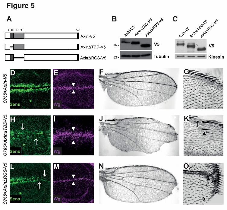
binding domain (RGS; (Fagotto et al., 1999)) of Axin. We generated Axin transgenes with
deletions in these domains (AxinΔTBD-V5 and AxinΔRGS-V5 respectively) (Figure 5, A),
and examined the effects of these deletions on Axin levels in the Wingless-responsive
embryonic cell line S2R+. Probing of cell lysates with V5 antibody revealed that steady
state levels of both AxinΔTBD-V5 and AxinΔRGS-V5 were increased by comparison to
Axin-V5 (Figure 5, B). To test this conclusion in vivo, we generated transgenic flies
expressing AxinΔTBD-V5 or AxinΔRGS-V5. To allow for their direct comparison in the
absence of transcriptional position effects, the UAS-AxinΔTBD-V5 or UAS-AxinΔRGS-V5
transgenes were inserted at the same site in the genome as UAS-Axin-V5 using site-
specific integration (Markstein et al., 2008). Each transgene was expressed in third instar
wing disc using the C765-Gal4 driver. We probed larval lysates with V5 antibody and found

14
that the levels of both AxinΔTBD-V5 and AxinΔRGS-V5 were higher than Axin-V5 (Figure
5, C). Together, these results indicate that the Tnks and APC binding domains of Axin are
important for the negative regulation of Axin levels.
Based on these findings, we sought to determine whether the Tnks and APC binding
domains of Axin promote Wingless signaling in vivo. As noted above, Wingless signaling is
critical for the growth and patterning of larval wing imaginal discs (Couso et al., 1994).
Expression of Axin-V5 using the C765-Gal4 driver resulted in no developmental defects;
Wingless-dependent cell fate specification was the same as in wild-type, as revealed by
expression of the Wingless target gene senseless (Nolo et al., 2000), and by the wild-type
morphology and size of the adult wing (Figure 5, D-G). In contrast, expression of
AxinΔTBD-V5 under the same conditions resulted in decreased senseless expression at
the dorso-ventral boundary of wing discs, loss of sensory bristles and blade tissue at the
margin of adult wings, an increase of the number of cells near the boundary that express
Wingless, and ectopic sensory bristles away from the margin; each of these phenotypes is
diagnostic for the inhibition of Wingless signaling (Figure 5, H-K). Similarly, expression of
AxinΔRGS-V5 under the same conditions also resulted in diminished senseless expression
at the dorso-ventral boundary, loss of sensory bristles at the margin, and loss of wing blade
tissue, although the phenotypes were not as severe as those resulting from expression of
AxinΔTBD-V5 (Figure 5, L-O). Similar results were observed when we performed the same
experiments using the Gal4 driver 71B (data not shown). Taken together, these findings
provide in vivo evidence that the Tnks binding domain and the APC binding domain
facilitate the control of basal Axin levels and the response to Wingless stimulation.

15
Tankyrase and APC promote Axin destabilization through distinct mechanisms
Next, we sought to compare the mechanisms by which Tnks and APC regulate Axin
degradation. As our mutant clonal analysis had revealed that Axin-V5 levels are regulated
by both Tnks and Apc in vivo (Figure 2, L-N and Figure 3, A-C), this provided a sensitive in
vivo assay for investigating the regulation of Axin by Apc and Tnks. To determine whether
Tnks and APC function in the same Axin proteolysis pathway in vivo, we determined
whether the domains in Axin required for Tnks- and APC-dependent Axin destabilization
were shared. To examine the role of the APC binding domain in Axin (RGS), we expressed
AxinΔRGS-V5 in wing imaginal discs. In contrast with Axin-V5 (Figure 3, A-C), the levels of
AxinΔRGS-V5 did not increase in Apc1 Apc2 double mutant clones; instead, equivalent
AxinΔRGS-V5 levels were detected inside and outside the clones (Figure 6, A-C). Thus as
expected, deletion of the APC binding domain prevents the ability of APC to negatively
regulate Axin. We next tested whether the RGS domain is also important for Tnks-
dependent Axin regulation. In contrast with Apc1 Apc2 double mutant clones, the level of
AxinΔRGS-V5 increased markedly in Tnks null mutant clones as compared to the
surrounding tissue (Figure 6, D-F). These results indicated that Tnks-mediated regulation of
Axin does not require Axin’s Apc binding domain. We conclude that the destabilization of
Axin mediated by Tnks does not require Apc-dependent Axin degradation.
We next examined the importance of the Tnks binding domain (TBD) in Tnks- or APC-
mediated Axin destabilization. As expected, deletion of the TBD abolished the ability of
Tnks to promote Axin degradation; in contrast with Axin-V5 (Figure 2, L-N), the levels of
AxinΔTBD-V5 were indistinguishable inside and outside of Tnks null mutant clones (Figure
16
6, J-L). This finding confirmed that Tnks-mediated Axin degradation requires the TBD. We
next examined whether the TBD is also important for APC-dependent Axin degradation. In
Apc1 Apc2 double mutant null clones, the level of AxinΔTBD-V5 was the same as that
found in the surrounding tissue (Figure 6, G-I). These findings indicate, unexpectedly, that
not only Tnks-mediated degradation, but also Apc-mediated degradation of Axin requires
the Tnks binding domain in Axin.
The Axin TBD and RGS domains are juxtaposed (Figure 5, A). Thus, deletion of the Axin
TBD might result in a conformational change in the Axin RGS domain that indirectly inhibits
the interaction between Axin and Apc. To address this possibility, we determined whether
deletion of the Axin TBD disrupts the interaction between Axin and Apc. We expressed
Axin-V5, AxinΔTBD-V5, and AxinΔRGS-V5 in Drosophila S2R+ cells. Axin was
immunoprecipitated with a V5 antibody and the immunoprecipitates were probed with Apc2
antibody. We detected Apc2 in immunoprecipitates of lysates from cells expressing Axin-V5
and AxinΔTBD-V5. In contrast, we did not detect Apc2 in immunoprecipitates of lysates
from cells expressing AxinΔRGS-V5 (Figure 6, M). These results indicate that the Axin TBD
is not required for the physical interaction between Axin and APC, and thus that the Axin-
APC interaction is important, but not sufficient for APC-mediated Axin degradation. Taken
together, these findings indicate that APC and Tnks promote Axin destabilization through
partially distinct mechanisms.
DISCUSSION

17
Our results demonstrate that regulation of the basal levels of Axin is a dynamic process
that requires the activity of the ADP-ribose polymerase Tnks and the tumor suppressor
APC. By increasing the gene dosage of Axin, we found that endogenous Axin levels can
increase by three- to four-fold without reaching the minimal threshold at which Wingless
signaling is disrupted in nearly all developing tissues, consistent with previous work
(Peterson-Nedry et al., 2008). These findings support the hypothesis that Axin levels
regulate destruction complex activity, but also reveal that there exists a physiological range
of at least three- to four-fold within which Axin levels may fluctuate and yet remain
compatible with the activation of the pathway following Wnt stimulation in all cells. This
narrow range perhaps serves two essential functions of Axin: (1) degradation of β-catenin
to maintain its low levels in the absence of Wnt ligands, and (2) robust responsiveness to
Wnt stimulation.
These results also provide evidence that Tnks promotes Wingless signaling by maintaining
Axin below this in vivo threshold. Axin levels increase in the absence of Tnks, but remain
below this threshold in nearly all developmental contexts. However, a relatively small
increase in Axin expression, which in itself has no effect on Wingless-dependent
developmental process in wild-type flies, is sufficient to result in classic Wingless loss-of-
function phenotypes in Tnks mutants. These findings suggest that Tnks-dependent
regulation buffers Axin activity and thus is likely important in specific in vivo contexts for
promoting Wingless signaling.
Our previous work revealed that whereas complete loss of Apc results in the constitutive
activation of Wingless signaling, partial reduction in Apc levels resulted in Wingless loss-of-

18
function phenotypes in multiple in vivo contexts, indicating that Apc has dual negative and
positive roles in Wingless signaling (Takacs et al., 2008). Our new ability to detect
endogenous Axin by immunoblotting provided an independent approach to test the
hypothesis that negative regulation of Axin levels contributes to the positive role of Apc in
Wingless signaling (Takacs et al., 2008). Supporting this hypothesis, Axin levels were
aberrantly increased in lysates from Apc mutant embryos and larvae. In addition, our
studies in frog egg extracts and frog embryos suggest that the function of APC in promoting
Axin degradation is evolutionarily conserved. Importantly, the role of APC in Axin
degradation, like that of Tnks, is independent of Wingless stimulation. These results
suggest that several pathways likely contribute to maintaining basal Axin levels below a
critical concentration, above which Wingless signaling is inhibited.
Our findings reveal that Tnks- and Apc-dependent proteolysis of Axin are achieved through
partly separable mechanisms. Tnks-mediated Axin destabilization requires the Tnks
binding domain of Axin, and thus their physical interaction. Apc-mediated Axin regulation is
dispensable for the Tnks-dependent proteolysis of Axin (Figure 6). Conversely, Apc-
mediated Axin regulation requires both the Tnks and APC binding domains in Axin (Figure
6). Our analysis reveals that the interaction between Axin and Apc is important, but not
sufficient, for APC-mediated Axin degradation (Figure 6). Together, these results suggest
that the APC-mediated regulation of Axin involves several distinct domains in Axin and/or a
specific Axin conformation.
Given the essential role of Wnt signaling in many fundamental processes, and the
requirement that Axin concentrations are maintained below a threshold level for the

19
activation of signaling (Lee et al., 2003; Salic et al., 2000), it is not surprising that several
degradation pathways have evolved to ensure precise control of Axin levels (Cselenyi et
al., 2008; Huang et al., 2009; Luo et al., 2007; Takacs et al., 2008). Redundancy in Axin
degradation pathways would provide a compensatory fail-safe mechanism to prevent an
increase in Axin above its threshold, and the resultant inhibition of signaling. Functional
redundancy in Axin degradation pathways may also explain why no defects in Wnt-
dependent embryonic development were observed upon disruption of fish or fly Tnks (Feng
et al., 2014; Huang et al., 2009; Wang et al., 2016). Nonetheless, the high degree of
sequence conservation present in Tnks homologs suggests that Tnks loss cannot be fully
compensated by other pathways in all in vivo contexts. Indeed, small molecule inhibitor
studies have indicated that Tnks is important for the Wnt-dependent regeneration of fins
following injury in adult fish (Chen et al., 2009; Huang et al., 2009). Moreover, our recent
work has revealed that regulation of Axin by Drosophila Tnks is required for Wingless target
gene activation and the Wingless-dependent control of intestinal stem cell proliferation in
the adult midgut (Tian et al., 2016; Wang et al., 2016). Importantly, in the midgut, Tnks is
essential for target gene activation in regions where the Wingless pathway is activated at
relatively low levels, but dispensable at high levels (Wang et al., 2016), suggesting an
critical role for Tnks in the amplification of signaling. Furthermore, we have found that Tnks
not only targets Axin for proteolysis, but also promotes the central role of Axin in rapid Wnt
pathway activation (Yang et al., 2016).
If the basal concentration of human Axin, like that of fly Axin, is determined by several
degradation pathways, then small molecule Tnks inhibitors will likely have the greatest

20
therapeutic efficacy in contexts for which Tnks-mediated Axin proteolysis has a
predominant role in controlling Axin levels. For example, as APC activity is disrupted in the
majority of colorectal carcinomas, APC-dependent Axin degradation is likely compromised
in these cells; thus colon carcinoma cells might be particularly sensitive to treatment with
Tnks inhibitors, whereas the untransformed neighboring cells that contain wild-type APC
levels would be less susceptible. Indeed, in mice for which APC activity has been disrupted
by conditional targeting, daily treatment with a small molecule Tnks inhibitor for three
weeks markedly reduced the proliferation of colonic adenoma cells, but resulted in little
change in the proliferation rate or morphology of cells in the juxtaposed healthy intestinal
mucosa (Waaler et al., 2012). These studies indicate that small molecule inhibitors of Axin
degradation are promising agents for the targeted therapy of Wnt pathway-dependent
diseases, and coupled with the work presented here, suggest that the conceptual
framework needed to identify new therapeutic agents in this category relies on our ability to
elucidate the distinct pathways that control endogenous Axin concentrations in vivo.
MATERIALS AND METHODS
Fly stocks and transgenes
The BAC Axin-V5 was constructed using an Axin BAC clone (CH321-39B08) containing
110 kb surrounding the Axin locus (Gerlach et al., 2014). A V5 tag was inserted at the
carboxy-terminus of the Axin coding region using recombineering as described previously
(Venken et al., 2009), and verified by sequencing. The modified BAC was introduced using
21
φC31-mediated integration at the VK30 (PBac{y[+]-attP-9A}VK00030) or VK33 (PBac{y[+]-
attP-3B}VK00033) docking sites.
To generate the pUASTattB-AxinΔTBD-V5 transgene, residues D-12 through K-32 were
deleted by PCR-based mutagenesis of pUASTattB-Axin-V5 (Yang et al., 2016) using the
oligonucleotide: 5’-GGT ATC TGC TAC CCC TTC GGT CAT ATG TTT CCG GAT TCC-3’.
The resulting AxinΔTBD-V5 fragment was digested with KpnI and XbaI, and then inserted
into the pUASTattB vector at the KpnI and XbaI sites. To generate the pUASTattB-
AxinΔRGS-V5 transgene, residues T-54 through Y-168 were deleted by PCR-based
mutagenesis of pUASTattB-Axin-V5. The resulting AxinΔRGS-V5 fragment was digested
with KpnI and XbaI, and then inserted into the pUASTattB vector at the KpnI and XbaI
sites. Transgenic flies were generated using site-specific integration at the attP33 site using
φC31-based integration (Bischof et al., 2007).
A complete deletion of the Axin gene, Axin18, was isolated by FLP-mediated trans-
recombination between FRT sites (Parks et al., 2004) in PBac{RB}Mgat2e01270 and
PBac{WH}Axnf01654 (Exelixis collection, Harvard Medical School). Potential deletions were
identified by lethal complementation tests with the mutant allele Axins044230.
Other stocks: Tnks19 (Wang et al., 2016), Tnks503 (Wang et al., 2016), C765-Gal4
(Bloomington Drosophila Stock Center (BDSC)) (Brand and Perrimon, 1993), 71B-Gal4
(BDSC) (Brand and Perrimon, 1993), Axins044230 (Hamada et al., 1999), Apc233 (Takacs et
al., 2008), Apc219.3 (Takacs et al., 2008), Apc1Q8 (Ahmed et al., 1998), hsFLP1 (Golic and
Lindquist, 1989), FRT82B arm-lacZ (Vincent et al., 1994) (provided by J. Treisman, Skirball

22
Institute, New York), hsFLP1 (Golic and Lindquist, 1989), vestigial-Gal4 UAS-FLP (Chen
and Struhl, 1999), FRT82B ovoD1 (Chou and Perrimon, 1992).UAS attB Axin-V5 (Yang et
al., 2016). Canton S flies were used as wild-type controls. All crosses were performed at
25°C unless otherwise indicated.
Generation of somatic mitotic clones
Somatic mitotic mutant clones were generated by FLP-mediated recombination (Xu and
Rubin, 1993) using hsFLP1 or vestigial-Gal4 UAS-FLP. When using hsFLP1, clones were
induced by subjecting first and second instar larvae to a 37°C heat shock for 2 hours, and
were detected by the loss of expression of an arm-lacZ transgene in third instar larval wing
imaginal discs.
Genotypes for generating mitotic clones were as follows:
Tnks mutant clones expressing Axin-V5 with the 71B driver: hsFLP1/+; UAS-Axin-V5/+;
FRT82B Tnks19/71B-Gal4 FRT82B arm-lacZ.
Tnks mutant clones expressing Axin-V5 with the C765 driver: hsFLP1/+; UAS-Axin-V5/+;
FRT82B Tnks19/C765-Gal4 FRT82B arm-lacZ.
Apc1 Apc2 double mutant clones expressing Axin-V5 with the vestigial driver: vestigial-
Gal4 UAS-FLP /+; UAS-Axin-V5/+; FRT82B Apc233Apc1Q8/FRT82B arm-lacZ.
Apc1 Apc2 double mutant clones mutant clones expressing Axin-V5 with the C765 driver:
hsFLP1/+; UAS-Axin-V5/+; FRT82B Apc233Apc1Q8/C765-Gal4 FRT82B arm-lacZ.

23
Generation of germline clones
Apc1 Apc2 double null mutant germline clones were generated using the FLP-DFS
technique (Chou and Perrimon, 1992). First and second instar larvae of the genotype
hsFLP1/+; FRT82B ovoD1/ FRT82B Apc219.3 Apc1Q8 were heat shocked for 2 hours, and
subsequently as adults were crossed to Apc219.3 Apc1Q8/TM6B males. Lysates for
immunoblots were made from their embryos at 0-2 hours of development.
Antibodies
The primary antibodies used were guinea pig anti-Axin (1:1000) (Wang et al., 2016), mouse
anti-V5 (1:5000 for immunoblots; Invitrogen), rabbit anti-V5 (1:2000 for immunostaining;
Abcam), mouse anti-Wingless (1:20, 4D4; Developmental Studies Hybridoma Bank
(DSHB)); mouse anti-Engrailed (1:100, 4D9; DSHB); guinea pig anti-Senseless (1:1000)
(Nolo et al., 2000); rabbit anti-β-gal (1:1000; MP Biomedicals); mouse anti-β-gal (1:1000;
Promega), rabbit anti-GFP (1:200; Invitrogen), mouse anti-α-tubulin (1:4000; Sigma), and
rabbit anti-Kinesin Heavy Chain (1:10000; Cytoskeleton).
Secondary antibodies used for immunostaining were goat or donkey Alexa Fluor 488, 555,
or 568 conjugates (1:400; Invitrogen), and goat Cy5 conjugates (1:200; Jackson
ImmunoResearch). Secondary antibodies used in immunoblots were guinea pig and rat
peroxidase-conjugates (1:5000; Jackson ImmunoResearch) or mouse and rabbit
peroxidase-conjugates (1:10000; Biorad).
Immunostaining, immunoblotting and quantification

24
For immunostaining, third instar larval wing imaginal discs were dissected in PBS, fixed in
4% paraformaldehyde in PBS for 20 minutes, and washed with PBS with 0.1% Triton X-
100, followed by incubation in PBS with 0.5% Triton X-100 and 10% BSA for 1 hour at
room temperature. Incubation with primary antibodies was performed at 4°C overnight in
PBS with 0.5% Triton X-100. Incubation with secondary antibodies was for 2 hours at room
temperature. Fluorescent images were obtained on a Nikon A1RSi confocal microscope
and processed using Adobe Photoshop software. Quantification of immnuostaining was
performed with NIS Elements (Nikon). The same region of interest (ROI, with area of 5-
10µm2) was placed in the adjacent cells with different genotypes (wild-type and 71B>Axin-
V5 for Fig.2C; Axin-V5 and Axin-V5; Tnks for Fig. 2O). Mean intensity was obtained for
each ROI and the relative intensity was calculated for the two correlated ROIs. 20-30
measurements were done for each experiment.
For immunostaining of embryos, embryos were dechorionated and then fixed in 3.7%
formaldehyde and rehydrated in PBT (PBS with 0.1% Tween-20, and 1% BSA). Following
incubation for one hour in blocking solution (PBS with 0.1% Tween-20 and 10% BSA),
embryos were incubated overnight at 4°C with primary antibodies in PBT. After washing
with PTW (PBS with 0.1% Tween-20), embryos were incubated with secondary antibodies
for one hour at room temperature. Embryos were then washed with PTW and mounted in
Prolong Gold (Invitrogen).
For larval lysates used in immunoblots, third instar larvae were dissected to remove
salivary glands, fat body, and gut tissues in cold PBS. After removal of PBS, 4X Laemmli
loading buffer was added and the lysates were vortexed briefly and incubated for 5 minutes

25
at 100°C before SDS-PAGE analysis. Embryos were homogenized in 4X Laemmli loading
buffer, and lysates were incubated at 100°C for 5 minutes. Quantification of immunoblots
was performed with Image J (Wayne Rasband, NIH, USA).
Immunoprecipitation
For immunoprecipitation experiments, S2R+ cells were harvested 48 hours after
transfection, washed with 1X PBS, then lysed in lysis buffer (50mM TrisHCl [pH 8.0],
100mM NaCl, 1% NP-40, 10% glycerol, 1.5mM EDTA [pH 8.0]) supplemented with 1µM
ADP-HPD (Enzo Life Sciences) and phosphatase and protease inhibitor cocktail (1:100,
Thermo Scientific). Lysates were incubated with mouse anti-V5 antibody (Invitrogen)
overnight at 4°C, followed by addition of protein A/G-sepharose beads (Santa Cruz) for 1
hour at 4°C. Beads were washed three times with wash buffer (50mM TrisHCl [pH 8.0],
150mM NaCl, 1% NP-40, 10% glycerol, 1.5mM EDTA [pH 8.0]) supplemented with 1µM
ADP-HPD and phosphatase and protease inhibitor cocktail (1:100), and boiled with 4X
sample buffer supplemented with 1M DTT. Samples were resolved by SDS-PAGE and
immunoblotted with the indicated antibodies.
Cell culture and transfection
S2R+ cells (Drosophila Genomics Resource Center) were maintained at 25°C in
Schneider’s complete medium: Schneider’s Drosophila medium with L-glutamine (Gibco)
supplemented with 10% FBS (Gibco) and 0.1 mg/mL penicillin/streptomycin (Invitrogen).
Cells were transiently transfected using calcium-phosphate DNA precipitation (Graham and
van der Eb, 1973).

26
Plasmids
Plasmids used for transfection of Drosophila cells were pAc5.1-Axin-V5, pAc5.1-AxinΔTBD-
V5, and pAc5.1-AxinΔRGS-V5. To generate the plasmids pAc5.1-Axin-V5, pAc5.1-
AxinΔTBD-V5 and pAc5.1-AxinΔRGS-V5, fragments encoding Axin-V5, AxinΔTBD-V5 and
AxinΔRGS-V5 from pUASTattB-Axin-V5, pUASTattB-AxinΔTBD-V5 and pUASTattB-
AxinΔRGS-V5 respectively, were digested using KpnI and XbaI. The resulting fragments
were inserted into the pAc5.1 A vector (Invitrogen) at the KpnI and XbaI sites.
dsRNA generation and RNAi-mediated knockdown
Generation of double-stranded RNAs (dsRNAs) and dsRNA-mediated knockdown were
performed as described previously (Rogers and Rogers, 2008). Briefly, DNA templates of
200-900 nucleotides in length targeting Axin or the white negative control (Zhang et al.,
2011) were generated by PCR from genomic DNA extracted from S2R+ cells. PCR
templates contained T7 promoter sequences on both ends. The DNA templates were
amplified using the following primer pairs:
white: forward 5’-T7- ACCTGTGGACGCCAAGG-3’ and reverse 5’-T7-
AAAAGAAGTCGACGGCTTC-3’
Axin: forward 5’-T7-CACAAAATAAAGAAGCAGCAGACGG-3’ and reverse 5’-T7-
ATTTGATTGTAGCTTTAACGGCTGG-3’
dsRNAs were transcribed from PCR generated templates using the T7 Megascript kit
(Ambion) according to manufacturer’s instructions. For RNAi-mediated knockdown, S2R+

27
cells were plated in 10 cm2 plates with 2.5 mL of serum-free, antibiotic-free Schneider’s
medium with L-glutamine. 25 µg of each dsRNA was added to the medium and cells were
incubated with gentle rotation at room temperature for 1 hour. Following incubation, 2.5 mL
of complete medium were added and cells were incubated at 25°C. After 24 hours, the
medium was removed from the cells. This procedure was repeated once every 24 hours
for a total of 96 hours.
Xenopus assays
Preparation of Xenopus egg extract and degradation assays, as well as immunodepletion
and reconstitution of APC in Xenopus egg extracts were performed as previously described
(Salic et al., 2000). For Axin degradation, egg extracts were supplemented with lithium
chloride (25 mM) to enhance turnover. APC antibodies were raised against recombinant
MBP-APCm3 (amino acids 1342-2075 of Xenopus APC) expressed and purified using the
baculovirus/Sf9 system, and the amount of APC added back to Xenopus egg extracts was
quantified by immunoblotting and compared to a standard curve of MBP-APCm3. MT-Axin
and MT-AxinΔRGS were a gift from Dr. Frank Costantini (Columbia University, New York).
AxinRGS encoding amino acids 1-216 of mouse Axin was cloned into the CS2+ plasmid.
Labeled [35S] Axin and β-catenin for degradation assays was synthesized in vitro using the
TNT system (Promega). Capped mRNAs for Xenopus embryo injections were synthesized
from linearized plasmid DNA templates using mMessage mMachine (Ambion).
ACKNOWLEGDEMENTS

28
We thank the investigators listed in Methods for generously sharing reagents and V. Marlar
for technical assistance. We thank Claudio Pikielny, Ai Tian and Hassina Benchabane for
critical reading and thoughtful comments on this manuscript. This work was funded by
grants from the NIH (RO1CA105038 to YA, R01GM081635 and R01GM103926 to EL,
P40OD018537 to the BDSC), the Emerald Foundation (to YA), the Norris Cotton Cancer
Center (to YA) and the National Science Foundation (DBI-1039423 for purchase of a Nikon
A1RSi confocal microscope).
FIGURE LEGENDS
Figure 1. A threshold above which Axin disrupts Wingless signaling.
(A) Lysates from S2R+ cells treated with mock, white (negative control), or Axin dsRNAs
were subjected to immunoblot with Axin antibody. Axin antibody specifically detected
endogenous Axin in lysates treated with mock or white dsRNA. Kinesin was used as a
loading control. (B) Lysates from third instar larvae of the indicated genotypes were
subjected to immunoblot with Axin antibody. (C) Quantification of Axin protein levels by
immunoblot. Results represent three independent experiments. Values indicate mean±SD.
(D-I) Stage 9 or 10 wild-type embryos (D-F) and embryos expressing BAC Axin-V5 on both
the second and third chromosomes (G-I) stained with antibodies against Wingless (Wg)
and Engrailed (En). Images in (F) and (I) are higher magnification views of embryos in (E)
and (H) respectively. Wg and En expression patterns appeared indistinguishable in wild-
type embryos and embryos expressing BAC Axin-V5. (J) The hatch rate of wild-type
embryos and embryos expressing BAC Axin-V5. 100 wild-type embryos and 80 BAC Axin-

29
V5 embryos were analyzed. (K) Ventral abdomen of wild-type females exhibited normal
organization of pleura (Pl), sternites, and sternal bristles (St, arrow). (L) Loss of sternal
bristles and expansion of the pleura in flies expressing BAC Axin-V5 on both the second
and third chromosomes. This phenotype was present with varying severity, shown is a
representative example. (M) Percentage of flies exhibiting loss of sternal abdominal
bristles.
Figure 2. Tnks promotes Wingless signaling by promoting Axin degradation.
(A) Lysates from third instar larvae of the indicated genotypes were subjected to
immunoblot with Axin antibody. Kinesin was used as a loading control. (B) Quantification of
Axin protein levels by immunoblots with wild-type or Tnks19 mutant larvae. Result
represents four independent experiments. Values indicate mean ± SD. (C) Quantification of
Axin levels by immunostaining. Wing discs expressing Axin-V5 were stained with anti-V5
and anti-Axin antibodies. Axin protein levels were measured by the intensity of Axin
antibody staining. Cells that did not express Axin-V5 were identified by the absence of V5
staining. Values indicate mean±SD (n=30). (D-E) Confocal images of third instar larval wing
discs expressing Axin-V5 with the 71B-Gal4 driver. Expression of Axin-V5 does not disrupt
the Wingless target gene senseless (D), Wingless expression (E), or cell fate in the adult
wing (F-G; 100% of flies had wild-type appearing wings, n=20). (H-I) Expression of Axin-V5
with the 71B-Gal4 driver in Tnks null mutants (Tnks19/ Tnks503) results in attenuation of
Senseless expression (H, arrows), and a slight increase in the number of cells expressing
Wingless (I, arrowheads). In the adult wing, expression of Axin-V5 in Tnks null mutants
causes loss of wing blade tissue (J), loss of sensory bristles at the margin (K, arrow), and

30
extra bristles in the wing blade (K, arrowhead); 100% of Tnks mutant flies displayed wing
margin defects (n= 35). (L-N) Wing disc expressing Axin-V5 labeled with α-V5 (green), α-β-
gal (magenta), and merge. Absence of β-gal staining marks Tnks19 mutant clones. The
levels of Axin-V5 inside Tnks19 mutant clones are increased compared to that of the
surrounding wild-type tissue. Patchy expression from the 71B driver likely accounts for the
few cells within Tnks mutant clones in which the Axin-V5 level is not increased. (O)
Quantification of Axin protein levels by immunostaining with V5 antibody. Tnks mutant
clones were induced in wing discs expressing the Axin-V5 transgene. Intensity of V5
staining was measured inside and outside Tnks mutant clones. Values indicate mean±SD
(n=18). (P) Ventral abdomen of wild-type adult female expressing Axin-V5 with the 71B-
Gal4 driver. Pleura (Pl), sternites (St), and sternal bristles (arrow) display normal
organization. Some sternites and sternal bristles are lost in Tnks19/Tnks503 mutants (Q),
indicating that Tnks promotes Wingless-dependent cell fate specification.
Figure 3. Apc promotes Axin proteolysis
(A-C) Wing disc expressing Axin-V5 labeled with α-V5 (green), α-β-gal (magenta), and
merge. Absence of β-gal staining marks Apc1Q8Apc233 mutant clones. The levels of Axin-
V5 inside Apc1Q8Apc233 mutant clones were increased compared to that of the surrounding
wild-type tissue. (D) Larval lysates from indicated genotypes were analyzed by immunoblot
using Axin antibody. Axin protein levels were increased in lysates from Tnks and Apc2
mutant larvae. Kinesin was used as a loading control. (E) Lysates from embryos, 0-2.5
hours old, were analyzed by immunoblot using Axin antibody. Axin protein levels were
increased in lysates from Tnks and Apc1 Apc2 double mutant embryos. One-half of the

31
Apc1 Apc2 double mutant embryos expressed wild-type Apc1 and Apc2 from the paternal
alleles. Kinesin was used as a loading control.
Figure 4. The destabilization of Axin by APC is conserved in vertebrates
(A) Immunoblotting revealed that the majority of endogenous APC was removed in APC-
depleted Xenopus egg extracts. (B) Radiolabeled [35S] Axin or β-catenin was added to
Xenopus egg extracts. At the indicated times, samples were withdrawn and subjected to
SDS-PAGE/autoradiography. The rate of Axin degradation is reduced in APC-depleted
Xenopus egg extracts. (C) Apc immunoprecipitated from Xenopus egg extracts was titrated
into Apc-depleted egg extracts. Radiolabeled Axin was added, samples were removed after
3 hrs for SDS-PAGE/autoradiography, and the amount of Axin remaining was quantified.
The rate of Axin degradation is regulated by APC in a dose-dependent manner.
Concentrations of APC in non-depleted extracts (~ 100 nM) and amount of APC added
back was calculated as described in (Lee et al., 2003). (D) APC regulates Axin turnover in
Xenopus embryos. Top panel: mRNAs encoding AxinRGS or control (empty vector) plus
MT-Axin mRNA (200 pg each) were co-injected into the dorsal blastomeres of 4-cell
embryos. Embryos were lysed in RIPA buffer at embryonic stage 7 and analyzed by Myc
immunoblotting. Bottom panel: Control or Apc MO (25 ng) plus mRNA encoding MT-Axin
mRNAs (200 pg) were co-injected into the dorsal blastomeres of 4-cell stage embryos.
Embryos were then lysed at stage 7 and analyzed by Myc immunoblotting.
Figure 5. The Tnks and APC binding domains of Axin are important for promoting
Wingless signaling.

32
(A) Schematic representation of Axin-V5, AxinΔTBD-V5, and AxinΔRGS-V5. (B) Lysates of
S2R+ cells transfected with the indicated plasmids (250 ng) were analyzed by
immunoblotting using a V5 antibody. Deletion of the Tnks binding domain of Axin
(AxinΔTBD-V5) or the APC binding domain of Axin (AxinΔRGS-V5) resulted in aberrant
stabilization of Axin compared to wild-type controls (Axin-V5). (C) Lysates of third instar
larvae expressing the indicated transgenes with C765-Gal4 driver were analyzed by
immunoblotting. AxinΔTBD-V5 and AxinΔRGS-V5 were stabilized compared with Axin-V5
protein. Confocal images of third instar larval wing discs expressing Axin-V5 (D-E),
AxinΔTBD-V5 (H-I), or AxinΔRGS-V5 (L-M) with the C765-Gal4 driver. Staining with
Wingless and Senseless antibodies shows that expression of Axin-V5 did not disrupt
expression of the Wingless pathway target Senseless (D) or of the number of cells
expressing Wingless (E, arrowheads), indicating that Axin-V5 was expressed at a level that
is compatible with physiological regulation. In contrast, expression of AxinΔTBD-V5
resulted in loss of Senseless (H, arrows), and expansion in the number of cells expressing
Wingless (I, arrowheads), indicating that Wingless signaling is inhibited by AxinΔTBD-V5.
Expression of AxinΔRGS-V5 results in loss of Senseless (L, arrows), indicating that
AxinΔRGS-V5 inhibits Wnt signaling. Adult wings expressing Axin-V5 (F-G), AxinΔTBD-V5
(J-K), or AxinΔRGS-V5 (N-O) using the C765-Gal4 driver. 95% of wings expressing Axin-
V5 (F-G) have normal morphology (n=136), whereas 92% of wings expressing AxinΔTBD-
V5 (J-K) (n= 127) and 30% of wings expressing AxinΔRGS-V5 (N-O) (n=135) display loss
of sensory bristles and tissue at the wing margin (K and O, arrow), as well as extra bristles
in the wing blade (K and O, arrowhead), which indicate inhibition of Wingless signaling.

33
Figure 6. APC- and Tnks-mediated regulation of Axin are achieved through partially
separable mechanisms
Confocal images of third instar larval wing imaginal discs stained with antibodies indicated
at bottom left; genotypes are indicated on the right. (A-L) Wing disc labeled with α-V5
(green), α-β-gal (magenta), and merge. Absence of β-gal staining marked Apc1Q8Apc233
mutant clones (A-C and G-I), and Tnks19 mutant clones (D-F and J-L). In contrast with
Axin-V5 (Figure 3, A-C), the levels of AxinΔRGS-V5 did not increase inside Apc1Q8Apc233
mutant clones compared to the surrounding wild-type tissue (A-C). The levels of
AxinΔRGS-V5 inside Tnks19 mutant clones were increased compared to that of the
surrounding wild-type tissue (D-F). In contrast with Axin-V5 (Figure 3, A-C and Figure 2, L-
N), the levels of AxinΔTDB-V5 did not increase in Apc1Q8Apc233 mutant clones (G-I) or in
Tnks19 mutant clones (J-L) compared to the surrounding wild-type tissue. (M)
Immunoprecipitation with V5 antibody from S2R+ cell lysates transfected with Axin-V5,
AxinΔTBD-V5, and AxinΔRGS-V5. Apc2 was pulled-down with Axin-V5. Deletion of the
Tnks binding domain of Axin (AxinΔTBD-V5) had no effect in the interaction between Axin
and Apc2. In contrast, deletion of the APC binding domain of Axin (AxinΔRGS-V5) inhibited
the interaction with Apc2.
RREFERENCES
Ahmed, Y., Hayashi, S., Levine, A., and Wieschaus, E. (1998). Regulation of armadillo by a Drosophila APC inhibits neuronal apoptosis during retinal development. Cell 93, 1171-‐1182.
Ahmed, Y., Nouri, A., and Wieschaus, E. (2002). Drosophila Apc1 and Apc2 regulate Wingless transduction throughout development. Development 129, 1751-‐1762.

34
Akong, K., Grevengoed, E.E., Price, M.H., McCartney, B.M., Hayden, M.A., DeNofrio, J.C., and Peifer, M. (2002). Drosophila APC2 and APC1 play overlapping roles in wingless signaling in the embryo and imaginal discs. Developmental biology 250, 91-‐100.
Baker, N.E. (1988). Transcription of the segment-‐polarity gene wingless in the imaginal discs of Drosophila, and the phenotype of a pupal-‐lethal wg mutation. Development 102, 489-‐497.
Bejsovec, A., and Martinez Arias, A. (1991). Roles of wingless in patterning the larval epidermis of Drosophila. Development 113, 471-‐485.
Bischof, J., Maeda, R.K., Hediger, M., Karch, F., and Basler, K. (2007). An optimized transgenesis system for Drosophila using germ-‐line-‐specific phiC31 integrases. Proceedings of the National Academy of Sciences of the United States of America 104, 3312-‐3317. Brand, A.H., and Perrimon, N. (1993). Targeted gene expression as a means of altering cell fates and generating dominant phenotypes. Development 118, 401-‐415.
Brunner, E., Peter, O., Schweizer, L., and Basler, K. (1997). pangolin encodes a Lef-‐1 homologue that acts downstream of Armadillo to transduce the Wingless signal in Drosophila. Nature 385, 829-‐833.
Chen, B., Dodge, M.E., Tang, W., Lu, J., Ma, Z., Fan, C.W., Wei, S., Hao, W., Kilgore, J., Williams, N.S., et al. (2009). Small molecule-‐mediated disruption of Wnt-‐dependent signaling in tissue regeneration and cancer. Nature chemical biology 5, 100-‐107. Chen, C.M., and Struhl, G. (1999). Wingless transduction by the Frizzled and Frizzled2 proteins of Drosophila. Development 126, 5441-‐5452.
Chou, T.B., and Perrimon, N. (1992). Use of a yeast site-‐specific recombinase to produce female germline chimeras in Drosophila. Genetics 131, 643-‐653.
Clevers, H., and Nusse, R. (2012). Wnt/beta-‐catenin signaling and disease. Cell 149, 1192-‐1205.
Couso, J.P., Bishop, S.A., and Martinez Arias, A. (1994). The wingless signalling pathway and the patterning of the wing margin in Drosophila. Development 120, 621-‐636. Cselenyi, C.S., Jernigan, K.K., Tahinci, E., Thorne, C.A., Lee, L.A., and Lee, E. (2008). LRP6 transduces a canonical Wnt signal independently of Axin degradation by inhibiting GSK3's phosphorylation of beta-‐catenin. Proceedings of the National Academy of Sciences of the United States of America 105, 8032-‐8037.
Fagotto, F., Jho, E., Zeng, L., Kurth, T., Joos, T., Kaufmann, C., and Costantini, F. (1999). Domains of axin involved in protein-‐protein interactions, Wnt pathway inhibition, and intracellular localization. The Journal of cell biology 145, 741-‐756.
Feng, Y., Li, X., Ray, L., Song, H., Qu, J., Lin, S., and Lin, X. (2014). The Drosophila tankyrase regulates Wg signaling depending on the concentration of Daxin. Cellular signalling 26, 1717-‐1724.

35
Gerlach, J.P., Emmink, B.L., Nojima, H., Kranenburg, O., and Maurice, M.M. (2014). Wnt signalling induces accumulation of phosphorylated beta-‐catenin in two distinct cytosolic complexes. Open biology 4, 140120.
Golic, K.G., and Lindquist, S. (1989). The FLP recombinase of yeast catalyzes site-‐specific recombination in the Drosophila genome. Cell 59, 499-‐509.
Graham, F.L., and van der Eb, A.J. (1973). A new technique for the assay of infectivity of human adenovirus 5 DNA. Virology 52, 456-‐467. Hamada, F., Tomoyasu, Y., Takatsu, Y., Nakamura, M., Nagai, S., Suzuki, A., Fujita, F., Shibuya, H., Toyoshima, K., Ueno, N., et al. (1999). Negative regulation of Wingless signaling by D-‐axin, a Drosophila homolog of axin. Science 283, 1739-‐1742. Huang, S.M., Mishina, Y.M., Liu, S., Cheung, A., Stegmeier, F., Michaud, G.A., Charlat, O., Wiellette, E., Zhang, Y., Wiessner, S., et al. (2009). Tankyrase inhibition stabilizes axin and antagonizes Wnt signalling. Nature 461, 614-‐620.
Kramps, T., Peter, O., Brunner, E., Nellen, D., Froesch, B., Chatterjee, S., Murone, M., Zullig, S., and Basler, K. (2002). Wnt/wingless signaling requires BCL9/legless-‐mediated recruitment of pygopus to the nuclear beta-‐catenin-‐TCF complex. Cell 109, 47-‐60.
Lau, T., Chan, E., Callow, M., Waaler, J., Boggs, J., Blake, R.A., Magnuson, S., Sambrone, A., Schutten, M., Firestein, R., et al. (2013). A novel tankyrase small-‐molecule inhibitor suppresses APC mutation-‐driven colorectal tumor growth. Cancer research 73, 3132-‐3144.
Lee, E., Salic, A., Kruger, R., Heinrich, R., and Kirschner, M.W. (2003). The roles of APC and Axin derived from experimental and theoretical analysis of the Wnt pathway. PLoS biology 1, E10.
Luo, W., Peterson, A., Garcia, B.A., Coombs, G., Kofahl, B., Heinrich, R., Shabanowitz, J., Hunt, D.F., Yost, H.J., and Virshup, D.M. (2007). Protein phosphatase 1 regulates assembly and function of the beta-‐catenin degradation complex. The EMBO journal 26, 1511-‐1521. MacDonald, B.T., Tamai, K., and He, X. (2009). Wnt/beta-‐catenin signaling: components, mechanisms, and diseases. Developmental cell 17, 9-‐26.
Markstein, M., Pitsouli, C., Villalta, C., Celniker, S.E., and Perrimon, N. (2008). Exploiting position effects and the gypsy retrovirus insulator to engineer precisely expressed transgenes. Nature genetics 40, 476-‐483.
Nolo, R., Abbott, L.A., and Bellen, H.J. (2000). Senseless, a Zn finger transcription factor, is necessary and sufficient for sensory organ development in Drosophila. Cell 102, 349-‐362.
Parks, A.L., Cook, K.R., Belvin, M., Dompe, N.A., Fawcett, R., Huppert, K., Tan, L.R., Winter, C.G., Bogart, K.P., Deal, J.E., et al. (2004). Systematic generation of high-‐resolution deletion coverage of the Drosophila melanogaster genome. Nature genetics 36, 288-‐292.
Peterson-‐Nedry, W., Erdeniz, N., Kremer, S., Yu, J., Baig-‐Lewis, S., and Wehrli, M. (2008). Unexpectedly robust assembly of the Axin destruction complex regulates Wnt/Wg signaling in Drosophila as revealed by analysis in vivo. Developmental biology 320, 226-‐241.

36
Rogers, S.L., and Rogers, G.C. (2008). Culture of Drosophila S2 cells and their use for RNAi-‐mediated loss-‐of-‐function studies and immunofluorescence microscopy. Nature protocols 3, 606-‐611.
Salic, A., Lee, E., Mayer, L., and Kirschner, M.W. (2000). Control of beta-‐catenin stability: reconstitution of the cytoplasmic steps of the wnt pathway in Xenopus egg extracts. Molecular cell 5, 523-‐532.
Takacs, C.M., Baird, J.R., Hughes, E.G., Kent, S.S., Benchabane, H., Paik, R., and Ahmed, Y. (2008). Dual positive and negative regulation of wingless signaling by adenomatous polyposis coli. Science 319, 333-‐336.
Tian, A., Benchabane, H., Wang, Z., and Ahmed, Y. (2016). Regulation of Stem Cell Proliferation and Cell Fate Specification by Wingless/Wnt Signaling Gradients Enriched at Adult Intestinal Compartment Boundaries. PLoS genetics 12, e1005822. Tolwinski, N.S., Wehrli, M., Rives, A., Erdeniz, N., DiNardo, S., and Wieschaus, E. (2003). Wg/Wnt signal can be transmitted through arrow/LRP5,6 and Axin independently of Zw3/Gsk3beta activity. Developmental cell 4, 407-‐418. Venken, K.J., Carlson, J.W., Schulze, K.L., Pan, H., He, Y., Spokony, R., Wan, K.H., Koriabine, M., de Jong, P.J., White, K.P., et al. (2009). Versatile P[acman] BAC libraries for transgenesis studies in Drosophila melanogaster. Nature methods 6, 431-‐434. Vincent, J.P., Girdham, C.H., and O'Farrell, P.H. (1994). A cell-‐autonomous, ubiquitous marker for the analysis of Drosophila genetic mosaics. Developmental biology 164, 328-‐331.
Waaler, J., Machon, O., Tumova, L., Dinh, H., Korinek, V., Wilson, S.R., Paulsen, J.E., Pedersen, N.M., Eide, T.J., Machonova, O., et al. (2012). A novel tankyrase inhibitor decreases canonical Wnt signaling in colon carcinoma cells and reduces tumor growth in conditional APC mutant mice. Cancer research 72, 2822-‐2832. Wang, Z., Tian, A., Benchabane, H., Tacchelly-‐Benites, O., Yang, E., Nojima, H., and Ahmed, Y. (2016). The ADP-‐ribose polymerase Tankyrase regulates adult intestinal stem cell proliferation during homeostasis in Drosophila. Development , in press. Willert, K., Logan, C.Y., Arora, A., Fish, M., and Nusse, R. (1999). A Drosophila Axin homolog, Daxin, inhibits Wnt signaling. Development 126, 4165-‐4173.
Xu, T., and Rubin, G.M. (1993). Analysis of genetic mosaics in developing and adult Drosophila tissues. Development 117, 1223-‐1237.
Yang, E., Tacchelly-‐Benites, O., Wang, Z., Randall, M.P., Tian, A., Benchabane, H., Freemantle, S., Pikielny, C., Tolwinski, N.S., Lee, E., et al. (2016). Wnt pathway activation by ADP-‐ribosylation. Nature Communications , in press.
Zeng, L., Fagotto, F., Zhang, T., Hsu, W., Vasicek, T.J., Perry, W.L., 3rd, Lee, J.J., Tilghman, S.M., Gumbiner, B.M., and Costantini, F. (1997). The mouse Fused locus encodes Axin, an inhibitor of the Wnt signaling pathway that regulates embryonic axis formation. Cell 90, 181-‐192.

37
Zhang, Y., Liu, S., Mickanin, C., Feng, Y., Charlat, O., Michaud, G.A., Schirle, M., Shi, X., Hild, M., Bauer, A., et al. (2011). RNF146 is a poly(ADP-‐ribose)-‐directed E3 ligase that regulates axin degradation and Wnt signalling. Nature cell biology 13, 623-‐629.