windows on the mind
TRANSCRIPT

A s you read this, your eyes are rapidly fl icking from left to right in small hops, bringing each word sequentially into
focus. When you stare at a person’s face, your eyes will similarly dart here and there, resting momentarily on one eye, the other eye, nose, mouth and other features. With a little introspec-tion, you can detect this frequent fl exing of your eye muscles as you scan a page, face or scene.
But these large voluntary eye movements, called saccades, turn out to be just a small part of the daily workout your eye muscles get. Your eyes never stop moving, even when they are ap-parently settled, say, on a person’s nose or a sail-boat bobbing on the horizon. When the eyes fi x-ate on something, as they do for 80 percent of your waking hours, they still jump and jiggle imperceptibly in ways that turn out to be essen-tial for seeing. If you could somehow halt these miniature motions while fi xing your gaze, a static scene would simply fade from view.
And yet only recently have researchers come to appreciate the profound importance of such
“fi xational” eye movements. For fi ve decades, a debate has raged about whether the largest of these involuntary movements, the so-called mi-crosaccades, serve any purpose at all. Some sci-
KEY CONCEPTS■ When the eyes fi x on some-
thing, they still jump impercep-tibly in ways that turn out to be essential for seeing.
■ For decades, scientists have debated the purpose, if any, of these so-called fi xational eye movements, the largest of which are called microsaccades. Now the authors have demonstrated that microsaccades engender visibility when a person’s gaze is fi xed and that bigger and faster microsaccades work best.
■ Microsaccades may also shed light on subliminal thoughts. Recent research suggests that the direction of microsaccades is biased toward objects to which people are unconsciously attracted, no matter where they are actually looking.
—The Editors
NEUROSCIENCE
Once scorned as nervous
tics, certain tiny,
unconscious flicks of the
eyes now turn out to
underpin much of our
ability to see. These
movements may even
re veal subliminal thoughts
By Susana Martinez-Conde and Stephen L. Macknik
Windows on the JE
NS
NIE
TH ze
fa/C
orbi
s (ph
otog
raph
); JE
N C
HRIS
TIA
NSE
N (p
hoto
illus
tratio
n)
56 SC IE NTIF IC AMERIC AN
CRED
IT
w w w. Sc iAm.com SC IE NTIF IC AMERIC AN 57
Mind
SC IE NTIF IC AME RIC AN 57

58 SC IE NTIF IC AME RIC AN August 20 07
entists have opined that microsaccades might even impair eyesight by blurring it. But recent work in the laboratory of one of us (Martinez-Conde) at the Barrow Neurological Institute in Phoenix has made the strongest case yet that these minuscule ocular meanderings separate vision from blindness when a person looks out at a stationary world.
Meanwhile microsaccades are also helping neuroscientists crack the brain’s code for creat-ing conscious perceptions of the visual world. In the past few years, we and others have detected telltale patterns of neural activity that correlate with these little movements, which we now be-lieve drive most of what people perceive. What is more, microsaccades may form a window into your mind. Instead of being random, these little ocular shifts may point to where your mind is secretly focusing—even if your gaze is directed elsewhere—revealing hidden thoughts and desires.
Fatigued by SamenessThat the eyes move constantly has been known for centuries. For example, in 1860 German
doctor and physicist Hermann von Helmholtz pointed out that keeping one’s eyes motionless was a diffi cult proposition and suggested that
“wandering of the gaze” prevented the retina, several layers of cells at the back of the eye, from becoming tired.
Indeed, animal nervous systems have evolved to detect changes in the environment, because spotting differences promotes survival. Motion in the visual fi eld may indicate that a predator is approaching or that prey is escaping. Such changes prompt visual neurons to respond with electrochemical impulses. Unchanging objects do not generally pose a threat, so animal brains—and visual systems—did not evolve to notice them. Frogs are an extreme case. A fl y sit-ting still on the wall is invisible to a frog, as are all static objects. But once the fl y is aloft, the frog will immediately detect it and capture it with its tongue.
Frogs cannot see unmoving objects because, as Helmholtz hypothesized, an unchanging stimulus leads to neural adaptation, in which visual neurons adjust their output such that they gradually stop responding. Neural adaptation
With these three illusions, you can observe various visual effects of your fi xational eye movements, which are typically beneath your awareness.
MINIATURE EYE MOVEMENTS REVEALED
SOU
RCES
: IG
NA
Z PA
UL
VITA
L TR
OXL
ER (l
eft)
; “A
SIM
PLE
AFT
ER IM
AGE
MET
HOD
DEM
ON
STRA
TIN
G T
HE IN
VOLU
NTA
RY M
ULT
IDIR
ECTI
ON
AL
EYE
MO
VEM
ENTS
DU
RIN
G F
IXAT
ION
,”
BY F.
J. V
ERHE
IJEN
, IN
JOU
RNAL
OF
MO
DER
N O
PTIC
S, V
OL.
8, N
O. 4
, PAG
ES 3
09–3
12; O
CTO
BER
1961
; © T
AYLO
R A
ND
FRA
NCI
S LT
D. (r
ight
)
TROXLER TEST: In 1804 Swiss philosopher Ignaz Paul Vital Troxler discovered that deliberately focusing on something causes surrounding stationary images to fade away. To elicit this experience, stare at the red spot while paying attention to the pale blue circle. The circle soon vanishes, and the red spot appears set against a white background. Move your eyes, and it pops back into view.
SEEING THE EYES MOVE: Here is a way to “see” your fi xational eye movements. Look at the central black dot for about a minute, then look at the white dot in the adjacent dark square. Notice that the dark afterimage of the white cross-hatching is in constant motion. That is a result of your fi xational eye movements.
Tiny subcon-scious eye move-ments are help-ing neuroscien-tists crack the
brain’s code for conscious visual
perceptions.

w w w. Sc iAm.com SC IE NTIF IC AMERIC AN 59
saves energy but also limits sensory perception. Human neurons also adapt to sameness. But the human visual system does much better than a frog’s at detecting unmoving objects, because human eyes create their own motion. Fixation-al eye movements shift the entire visual scene across the retina, prodding visual neurons into action and counteracting neural adaptation. They thus prevent stationary objects from fad-ing away.
In 1804 Swiss philosopher Ignaz Paul Vital Troxler reported the fi rst fading phenomenon in humans related to a decrease in fi xational eye movements. Troxler noted that deliberately fo-cusing on something causes stationary images in the surrounding region to gradually fade away [see left illustration in box on opposite page]. This fading happens to you every day, be-cause deliberately focusing on something can briefl y slow or reduce fi xational eye movements, which are also less effective outside your area of focus. Thus, even a small reduction in the rate and size of your eye movements greatly impairs your vision. You do not notice the impairment, because you are not paying attention to invisible
portions of your view, focusing on what is di-rectly in front of you instead.
Totally ceasing all eye movements, however, can only be done in a laboratory. In the early 1950s some research teams achieved this stilling effect by mounting a tiny slide projector onto a contact lens and affi xing the lens to a person’s eye with a suction device. In this setup, a subject views the projected image through this lens, which moves with the eye. Using such a retinal stabilization technique, the image remains still with respect to the eye, causing the visual neu-rons to adapt and the image to fade away. Now-adays researchers create this same result by measuring the eye’s movements with a camera pointed at it. They transmit the eye-position data to a projection system that moves the im-age with the eye.
In the late 1950s researchers were able to isolate a role for microsaccades: after suppress-ing all eye movements in the lab, including the larger voluntary saccades, they superimposed micro saccadelike motions and found that do-ing so restored perception. Other research teams, however, found otherwise: adding back JE
N C
HRIS
TIA
NSE
N (d
iagr
ams a
t rig
ht);
SOU
RCE:
AKI
YOSH
I KIT
AOKA
, © 2
004
(bel
ow)
MOVING TARGETS Fixational eye movements, including microsaccades (straight lines), drifts (wavy lines) and tremor (zigzags superimposed on drifts), transport the visual image over a mosaic of photo re-ceptors on the retina.
ILLUSORY MOTION: Let your eyes wander around the pattern above, and the three “rollers” will appear to spin. But if you hold your gaze steady on one of the green spots in the center of the image, the illusory motion will slow down or even stop. Because holding the eyes still stops the illusory motion, the authors speculate that the fi xational eye movements may be required to see it, although they do not yet know exactly how.
MICROSACCADE
DRIFT
TREMOR
Activated photoreceptor

60 SC IE NTIF IC AMERIC AN August 20 07
Microsaccades elicit neuronal
rejoinders in every part of
the visual system
we have examined.
microsaccades after freezing eye movements had no effect at all in these experiments. The truth was hard to discern because none of the techniques for stabilizing the retina was perfect; for instance, a contact lens attached to the eye can slip, leaving some residual eye move-ments. In the end, no one could tell whether an experimental result was caused by those residual movements or the super imposed microsaccades.
Nervous Tics?Around the same time, investigators identifi ed two other fl avors of fi xational eye movements: drifts and tremor. Drifts are slow, meandering motions that occur between the fast, linear mi -cro saccades. Tremor is a tiny, rapid oscillation superimposed on drifts. Microsaccades are the largest of the fi xational eye movements, carry-ing an image across dozens to several hundred of the eye’s photoreceptor (light-detecting) cells, including cones for detail and color vision and rods for low-light and peripheral vision. Tremor is the smallest of the fi xational eye movements, its motion no bigger than the size of one of these cells. We do not yet understand the relative roles of these various fi xational eye movements in vision, however.
In fact, for decades, many vision scientists doubted whether any of these fi xational eye movements—especially microsaccades, which were the most studied—had a role in maintain-ing vision. Critics noted that some individuals could suppress microsaccades for a couple of
seconds without their central vision fading away. (You can see this in the Troxler test; as you briefl y suppress your microsaccades, the ring fades, but you can still see the red dot in the center of your view.) And people naturally hold microsaccades at bay momentarily when they perform precision tasks such as shooting a rifl e or threading a needle. In 1980 University of Maryland psychologists Eileen Kowler and Robert M. Steinman concluded that microsac-cades were useless, supposing that they might be “merely a kind of nervous tic.”
There the fi eld stood until the late 1990s, when researchers began to investigate which neuronal responses, if any, fi xational eye move-ments might be generating in the eye and brain. Starting in 1997, along with Nobel laureate Da-vid Hubel of Harvard Medical School, we trained monkeys to fi xate on a small spot pre-sented on a computer monitor, which also dis-played a stationary bar of light elsewhere on the screen. As the monkeys stared, we recorded their eye movements and the electrical activity from neurons in the lateral geniculate nucleus (LGN) in their midbrain and in the primary vi-sual cortex at the back of their brain [see box on opposite page]. In each experiment the bar was placed in a location that would elicit an optimal electrical response—in the form of impulses called spikes—from the recorded neurons.
The results of these experiments, published in 2000 and 2002, showed that microsaccades increased the rate of neural impulses generated by both LGN and visual cortex neurons by ush-
[THE AUTHORS]
Susana Martinez-Conde is director of the Laboratory of Visual Neuroscience at the Barrow Neurological Institute in Phoenix. She holds a Ph.D. in medicine and surgery from the University of Santiago de Compostela in Spain. Stephen L. Macknik is director of the Laboratory of Behavioral Neurophysiology at the Barrow Neurological Institute and earned a Ph.D. in neurobiology from Harvard University.
Neuroscientists David Coppola and Dale Purves of Duke University showed that the blood vessels in the retina, which are stationary with respect
to each eye, can fade from a person’s view in a mere 80 milliseconds (thousandths of a second). You can see this for
yourself. Close your eyes while lightly holding a small fl ashlight (not too bright!) up to the side of one of your eyes. If you move the fl ashlight around rapidly, you might catch a glimpse of your retinal blood vessels in your peripheral vision. But notice how quickly they fade from view.
Neural adaptation takes place in all of the senses, including touch. For instance, you may feel your shoes when you fi rst put them on in the morning, but the feeling goes
away after a while. You probably do not want to be aware of your shoes 16 hours a day, after all. If you wiggle your toes,
however, you can feel your shoes again. Similarly, fi xational eye movements constantly “wiggle” the images on the retina so that
your vision never goes away. —S.M.-C. and S.L.M.
VANISHING VESSELS[DO-IT-YOURSELF]
STEV
E A
LLEN
Bra
nd X
/Cor
bis (
top)
; JEF
F N
OBL
E (M
artin
ez-C
onde
and
Mac
knik
)

w w w. Sc iAm.com SC IE NTIF IC AMERIC AN 61
ering stationary stimuli, such as the bar of light, in and out of a neuron’s receptive fi eld, the re-gion of visual space that activates it. This fi nd-ing bolstered the case that microsaccades have an important role in preventing visual fading and maintaining a visible image. And assuming such a role for microsaccades, our neuronal studies of microsaccades also began to crack the visual system’s code for visibility. In our mon-key studies we found that microsaccades were more closely associated with rapid bursts of spikes than single spikes from brain neurons,
suggesting that bursts of spikes are a signal in the brain that something is visible.
Cracking the CaseOther researchers also found that microsac-cades elicit neuronal rejoinders in every part of the visual system that they examined. Neverthe-less, the fi eld was still haunted by the confl ict-ing results in the retinal stabilization experi-ments, casting lingering doubt over the impor-tance of microsaccades in vision. So a few years ago, at the Barrow Neurological Institute, we
Retina
Optic nerve
Lateralgeniculatenucleus
Optic nerve
Opticradiation
Activated photoreceptor
Microsaccade
Light
Primary visual cortex
PHOTORECEPTO
R
MOSAIC
Ganglion cellAmacrine cell
Bipolar cell
Horizontal cell
RodCone
Photoreceptor cells
Light Vision begins when light refl ects off an object and hits the retina, several layers of cells at the back of both eyes.
Light travels to the rear of the retina, where pho-toreceptor cells transform the light energy into neural signals. Fixational eye movements such as microsaccades refresh the neural activity several times a second; that is, each movement causes any given group of photoreceptors to intercept the light from a different part of the visual scene, thus changing its responses. Microsaccades similarly alter the responses of other cells in the visual system. Without these movements, visual neurons would adapt to the unchanging stimulus by decreasing their activity, and vision would fade.
The neural impulses from the retina zip along a cable of a mil-lion fi bers—the optic nerve—to the brain. There visual signals stop fi rst at the later-al geniculate nucleus
in the thalamus, and then neuronal cords
called the optic radiations carry them to the primary visual cortex at the back of the brain.
JEN
CHR
ISTI
AN
SEN
[THE BASICS]
REFRESHING THE PICTURE
62 SC IE NTIF IC AMERIC AN August 20 07
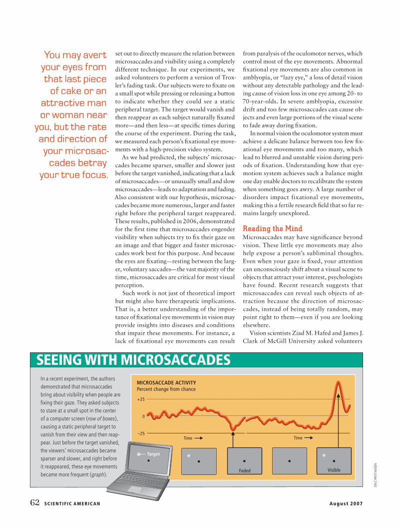
set out to directly measure the relation between microsaccades and visibility using a completely different technique. In our experiments, we asked volunteers to perform a version of Trox-ler’s fading task. Our subjects were to fi xate on a small spot while pressing or releasing a button to indicate whether they could see a static peripheral target. The target would vanish and then reappear as each subject naturally fi xated more—and then less—at specifi c times during the course of the experiment. During the task, we measured each person’s fi xational eye move-ments with a high-precision video system.
As we had predicted, the subjects’ microsac-cades became sparser, smaller and slower just before the target vanished, indicating that a lack of microsaccades—or unusually small and slow microsaccades—leads to adaptation and fading. Also consistent with our hypothesis, microsac-cades became more numerous, larger and faster right before the peripheral target reappeared. These results, published in 2006, demonstrated for the fi rst time that microsaccades engender visibility when subjects try to fi x their gaze on an image and that bigger and faster microsac-cades work best for this purpose. And because the eyes are fi xating—resting between the larg-er, voluntary saccades—the vast majority of the time, microsaccades are critical for most visual perception.
Such work is not just of theoretical import but might also have therapeutic implications. That is, a better understanding of the impor-tance of fi xational eye movements in vision may provide insights into diseases and conditions that impair these movements. For instance, a lack of fixational eye movements can result
from paralysis of the oculomotor nerves, which control most of the eye movements. Abnormal fi xational eye movements are also common in amblyopia, or “lazy eye,” a loss of detail vision without any detectable pathology and the lead-ing cause of vision loss in one eye among 20- to 70-year-olds. In severe amblyopia, excessive drift and too few microsaccades can cause ob-jects and even large portions of the visual scene to fade away during fi xation.
In normal vision the oculomotor system must achieve a delicate balance between too few fi x-ational eye movements and too many, which lead to blurred and unstable vision during peri-ods of fi xation. Understanding how that eye-motion system achieves such a balance might one day enable doctors to recalibrate the system when something goes awry. A large number of disorders impact fixational eye movements, making this a fertile research fi eld that so far re-mains largely unexplored.
Reading the MindMicrosaccades may have signifi cance beyond vision. These little eye movements may also help expose a person’s subliminal thoughts. Even when your gaze is fi xed, your attention can unconsciously shift about a visual scene to objects that attract your interest, psychologists have found. Recent research suggests that micro saccades can reveal such objects of at-traction because the direction of microsac -cades, instead of being totally random, may point right to them—even if you are looking elsewhere.
Vision scientists Ziad M. Hafed and James J. Clark of McGill University asked volunteers
In a recent experiment, the authors demonstrated that microsaccades bring about visibility when people are fi xing their gaze. They asked subjects to stare at a small spot in the center of a computer screen (row of boxes), causing a static peripheral target to vanish from their view and then reap-pear. Just before the target vanished, the viewers’ microsaccades became sparser and slower, and right before it reappeared, these eye movements became more frequent (graph).
SEEING WITH MICROSACCADES
You may avert your eyes from that last piece
of cake or an attractive man or woman near
you, but the rate and direction of your microsac-
cades betray your true focus.
MICROSACCADE ACTIVITY Percent change from chance
+25
0
–25Time Time
Target
Faded Visible
JEN
CHR
ISTI
AN
SEN

w w w. Sc iAm.com SC IE NTIF IC AMERIC AN 63
to direct their eyes to a central spot on a com-puter monitor while paying attention to a pe-ripheral spot that changed color at the end of each trial. The volunteers were supposed to in-dicate this color change. In 2002 Hafed and Clark reported that the direction of the subjects’ microsaccades was biased toward their true point of focus, even though they were looking elsewhere. This fi nding indicated not only that microsaccades may point to people’s covert thoughts but also, the authors noted, that covert shifts of attention actually control the direction of microsaccades.
In another experiment, computational neu-roscientist Ralf Engbert and cognitive psychol-ogist Reinhold Kliegl of the University of Pots-dam in Germany found that the frequency of microsaccades also conveys the presence of something that secretly attracts a person’s at-tention. The abrupt appearance of a visual cue in the periphery of a person’s fi eld of view, they stated in 2003, causes fi rst a brief drop in the rate of microsaccades, followed by a rapid re-
bound in which microsaccade frequency ex-ceeds normal. Furthermore, the microsaccades they detected were biased in the direction of the cue. The study suggests that microsaccade frequency and direction can signal sudden changes in the environment that attract a per-son’s attention when he or she does not look di-rectly at them.
Thus, no matter how hard you might avert your eyes from the last piece of cake on the table or the attractive male or female standing across the room, the rate and direction of your micro-saccades betray your attentional spotlight. This betrayal is not a practical concern, however. In the laboratory, scientists can detect and mea-sure these minuscule eye movements to reveal the hidden brain mechanisms of attention, but people around you cannot easily use them to read your mind—yet. g
ATTENTION MONITOR: Scientists can track microsaccades to determine if something is secretly attracting a person’s attention—such as a slice of chocolate cake—even when that person is looking elsewhere. But don’t worry. Ordinary people cannot easily use these eye movements to read your mind.
➥ MORE TO EXPLORE
Microsaccades as an Overt Measure of Covert Attention Shifts. Z. M. Hafed and J. J. Clark in Vision Research, Vol. 42, pages 2533–2545; 2002.
Microsaccades Uncover the Orientation of Covert Attention. R. Engbert and R. Kliegl in Vision Research, Vol. 43, pages 1035–1045; 2003.
The Role of Fixational Eye Movements in Visual Perception. S. Martinez-Conde, S. L. Macknik and D. H. Hubel in Nature Reviews Neuroscience, Vol. 5, pages 229–240; 2004.
Fixational Eye Movements in Normal and Pathological Vision. S. Martinez-Conde in Progress in Brain Research, Vol. 154, pages 151–176; 2006.
Microsaccades Counteract Visual Fading during Fixation. S. Martinez-Conde, S. L. Macknik, X. G. Troncoso and T. A. Dyar in Neuron, Vol. 49, pages 297–305; 2006.
Akiyoshi Kitaoka’s illusion pages: www.ritsumei.ac.jp/
~akitaoka/index-e.html
Martinez-Conde Laboratory: www.neuralcorrelate.com/smc_lab
For further reading on this subject, and for other related articles, log on to www.SciAm.com/ontheweb
SCIAM
BRIA
N M
ARA
NA
N P
INED
A