:william h. calvin washington
TRANSCRIPT
:William H . CalvinWCalvin*U.Washington.edu
U N I V E R S I T Y O F
WASHINGTONSEATTLE, WASHINGTON 98195-1800 USA
97
Epilepsy: A Window to Brain Mechanisms,edited by Joan S . Lockard and Arthur A. Ward, Jr.Raven Press, New York © 1980.
6
Normal Repetitive Firing and Its Pathophysiology
William H. Calvin
Department of Neurological Surgery, University of Washington, Seattle, Washington 98195
A neuron communicates over long distances (more than a few millimeters)by generating a train of impulses which propagates down the axon to releasea series of prepackaged quanta of neurotransmitter molecules . The rate, or per-haps the patterning, of the impulse train carries the information . One of thehallmarks of an interictal epileptogenic focus is that many of its neurons areobserved to cluster their impulses into bursts, with the intervals between impulsesbeing unusually short (several milliseconds) . Is the bursting neuron some sortof pacemaker, driving other normal neurons into synchronous activity and thusspreading the trouble? Or is the bursting one observes just one of those recruitedneurons, having nothing more wrong with it than an oversized synaptic input?Or perhaps there are no pacemaker neurons ; the trouble could be subtly distrib-uted over many neurons, changing the balance of excitation and inhibition sothat the whole circuit tends to go into a bursting-type oscillation .
There are many other ways of stating the "epileptic neuron" versus "epilepticaggregate" dichotomy . As presented above, the argument bears a strong resem-blance to an argument that has occurred in the more general field of patterngeneration : How are actions with alternating activity and silence, such as walkingor breathing, generated by groups of neurons?
While there could simply be pacemaker bursting neurons, there could alsobe a steady level of excitatory drive onto two neurons (or groups of neurons)which mutually inhibit each other (33) . When A is firing, it inhibits B intosilence . If one allows for inhibition which fatigues (antifacilitation, depression),soon the declining inhibition from A will allow B to begin firing, which willthen inhibit A into silence, and so on, back and forth . Hartline (33) has reviewedthe emerging data on a number of pattern-generating circuits . Even when thereare mutually inhibitory synaptic connections which aid reciprocal bursting, theneurons themselves often have intrinsic bursting properties too . In other words,there are redundant means of enforcing bursting ; it is not a matter of cell orcircuit bursting but of both cell and circuit . While it would be more convenientfor neurophysiologists if nature would use only one bursting mechanism at atime, it would appear that if something is worth doing, it may be done usingredundant mechanisms .
In this chapter, the individual neuron is examined for its ability to exaggerate
98
NORMAL AND ECTOPIC REPETITIVE FIRING
its normal output by overproducing impulses . This emphasis on overproductionat the various stages of computation and data transmission within the individualneuron is not to deny that underproduction (e .g., in inhibitory neurons) couldalso be important . As in considering the origins of forest fires, one can eitheremphasize who started the forest fire or consider the factors enhancing theflammability of the forest . Tracing through one of the trees, i.e., the dendriticand axonal arborization of a neuron, is useful for evaluating flammability pro-spects even if no one neuron is a pacemaker .
THE PROCESSING PATH THROUGH A NEURON
Some neurons are much simpler than the neurons that we examine here .There are spikeless neurons, such as those in the retina, where tonic transmitterrelease rates are controlled by the size of a graded depolarization . Such neuronsare short enough so that electrotonic current spread suffices for communicationbetween the postsynaptic regions, collecting information from upstream neurons,and the presynaptic regions that output the processed result (30,31) . Thus thespikeless processing path is particularly simple .
Repetitive firing is the mechanism that allows the postsynaptic and presynapticregions of the neuron to be separated by more than a few millimeters ; transmitterrelease rate is now controlled by impulse production rate . Firing rates are inturn controlled by synaptic depolarizations, as seen by the, spike trigger zone(usually at the initial segment of the axon) . Thus impulse trains can be seenas an intermediate coding form, allowing transmitter release rate to remainproportional to the sum of synaptic depolarizing and hyperpolarizing currents .A more detailed comparison of spikeless and spiking modes of operation canbe found in Calvin and Graubard (13) .
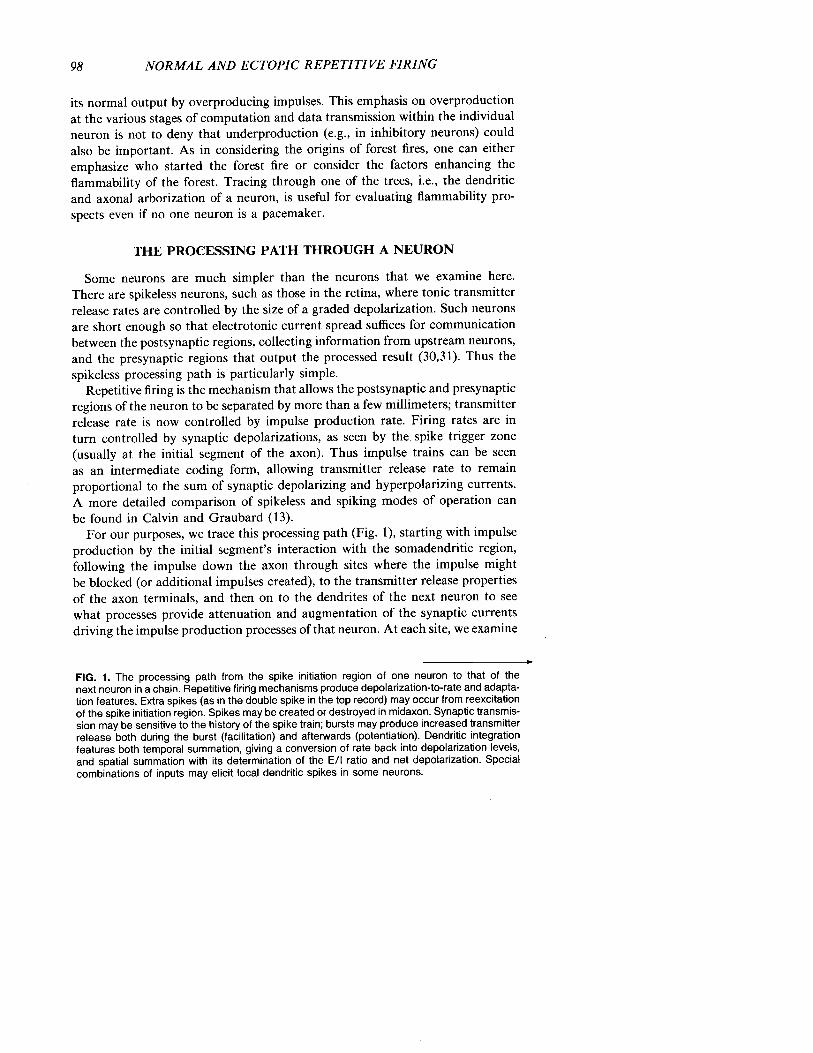
For our purposes, we trace this processing path (Fig . 1), starting with impulseproduction by the initial segment's interaction with the somadendritic region,following the impulse down the axon through sites where the impulse mightbe blocked (or additional impulses created), to the transmitter release propertiesof the axon terminals, and then on to the dendrites of the next neuron to seewhat processes provide attenuation and augmentation of the synaptic currentsdriving the impulse production processes of that neuron . At each site, we examine
FIG . 1 . The processing path from the spike initiation region of one neuron to that of thenext neuron in a chain . Repetitive firing mechanisms produce depolarization-to-rate and adapta-tion features. Extra spikes (as in the double spike in the top record) may occur from reexcitationof the spike initiation region . Spikes may be created or destroyed in midaxon . Synaptic transmis-sion may be sensitive to the history of the spike train ; bursts may produce increased transmitterrelease both during the burst (facilitation) and afterwards (potentiation) . Dendritic integrationfeatures both temporal summation, giving a conversion of rate back into depolarization levels,and spatial summation with its determination of the E/I ratio and net depolarization . Specialcombinations of inputs may elicit local dendritic spikes in some neurons .

NORMAL AND ECTOPIC REPETITIVE FIRING
99
II . SPIKE CONDUCTION IN AXONintermittent conduction .channeling at bifurcationsectopic spike initiation sites
III . SYNAPTIC TRANSFERfacilitation by burstspost-tetanic potentiation
SPIKE INITIATIONdepolarization-to-rate encodingadaptation and rebounds
retrograde soma-dendritic invasionreexcitation bursts
J
iHHL
IV . DENDRITIC INTEGRATIONdecoding rate-to-depolarizationspatial summation . E/I ratiodendritic spike boosters
100
NORMAL AND ECTOPIC REPETITIVE FIRING
the features capable of overproduction, particularly those relevant to epilepticbursting .
Depolarization-to-Rate Conversion : Normal Rhythmic Firingfrom the Trigger Zone
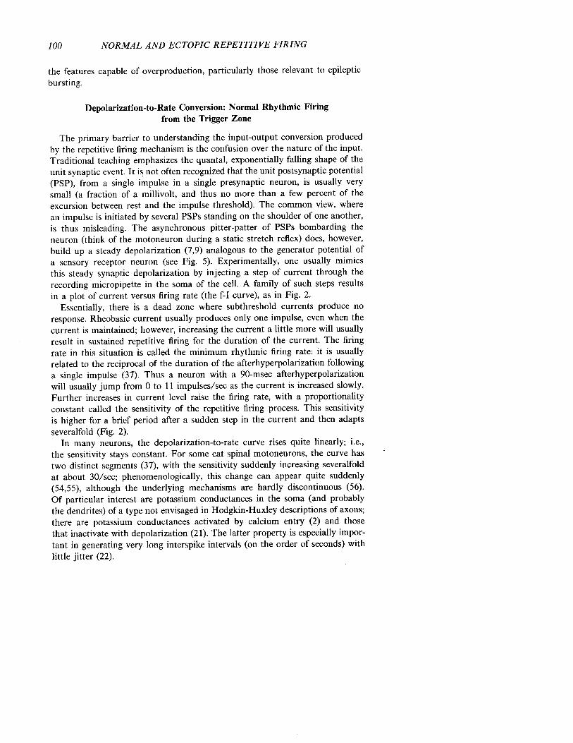
The primary barrier to understanding the input-output conversion producedby the repetitive firing mechanism is the confusion over the nature of the input .Traditional teaching emphasizes the quantal, exponentially falling shape of theunit synaptic event . It is not often recognized that the unit postsynaptic potential(PSP), from a single impulse in a single presynaptic neuron, is usually verysmall (a fraction of a millivolt, and thus no more than a few percent of theexcursion between rest and the impulse threshold) . The common view, wherean impulse is initiated by several PSPs standing on the shoulder of one another,is thus misleading. The asynchronous pitter-patter of PSPs bombarding theneuron (think of the motoneuron during a static stretch reflex) does, however,build up a steady depolarization (7,9) analogous to the generator potential ofa sensory receptor neuron (see Fig . 5). Experimentally, one usually mimicsthis steady synaptic depolarization by injecting a step of current through therecording micropipette in the soma of the cell . A family of such steps resultsin a plot of current versus firing rate (the f-I curve), as in Fig . 2 .
Essentially, there is a dead zone where subthreshold currents produce noresponse . Rheobasic current usually produces only one impulse, even when thecurrent is maintained ; however, increasing the current a little more will usuallyresult in sustained repetitive firing for the duration of the current. The firingrate in this situation is called the minimum rhythmic firing rate : it is usuallyrelated to the reciprocal of the duration of the afterhyperpolarization followinga single impulse (37) . Thus a neuron with a 90-msec afterhyperpolarizationwill usually jump from 0 to 11 impulses/sec as the current is increased slowly .Further increases in current level raise the firing rate, with a proportionalityconstant called the sensitivity of the repetitive firing process. This sensitivityis higher for a brief period after a sudden step in the current and then adaptsseveralfold (Fig. 2) .
In many neurons, the depolarization-to-rate curve rises quite linearly ; i .e .,the sensitivity stays constant . For some cat spinal motoneurons, the curve hastwo distinct segments (37), with the sensitivity suddenly increasing severalfoldat about 30/sec ; phenomenologically, this change can appear quite suddenly(54,55), although the underlying mechanisms are hardly discontinuous (56) .Of particular interest are potassium conductances in the soma (and probablythe dendrites) of a type not envisaged in Hodgkin-Huxley descriptions of axons ;there are potassium conductances activated by calcium entry (2) and thosethat inactivate with depolarization (21) . The latter property is especially impor-tant in generating very long interspike intervals (on the order of seconds) withlittle jitter (22) .

100-
11-
NORMAL AND ECTOPIC REPETITIVE FIRING
DEPOLARIZING CURRENT
C
20 m V140 nA20 msec
J
I
eJ
JB
c
.1, .
By tee dI • ~
SUSta ~~
0 -
0
- •-.-.nt
i
I19
40 nA J
11
101
FIG. 2. Rhythmic firing in a cat spinal motoneuron, responding to steps of injected current(lower traces) injected through the recording microelectrode into the neuron's soma, resultsin this depolarization-to-rate relationship (often called the f-I curve) . Light upper line connectsthe points representing the initial firing rate (reciprocal of the interval between the first andsecond spikes of the train) . Heavy lower line is the plot of the average firing rate followingadaptation . At A, two responses are superimposed ; a rheobasic current gives only one spike,but a slightly larger current gives a repetitive response with an interspike interval equal tothe duration of the afterhyperpolarization (90 msec) in this neuron . (Reprinted with permissionfrom Federation Proceedings, ref. 10 .)
Rhythmic firing does not require these specialized ionic mechanisms; evensquid axon will fire rhythmically, although the dynamic range of its f-I curvemay be small (58) . The trigger zone at the axon initial segment may, by itself,have such axon-like repetitive firing properties . However, the impulse retro-gradely invades the somadendritic region at the same time that the newly mintedimpulse is propagating down the axon . This retrograde invasion activates thespecialized currents of the somadendritic region that, in turn, subtract fromthe synaptic currents and determine the time taken to reach the threshold andinitiate the next impulse . It has been hypothesized that the extent of retrogradeinvasion is important (9,17,62) . Certainly, it is likely that fairly extensive retro-grade invasion of the dendritic tree occurs in chromatolytic motoneurons (34)

102
NORMAL AND ECTOPIC REPETITIVE FIRING
because of the excitability evidenced by their orthograde dendritic spikes ; theirf-I curves lack the sensitivity alterations of normal spinal motoneurons .
Extra Spikes From Normal Trigger Zones : Another Sensitivity-ChangingMechanism
Some motoneurons, while firing a spike with great regularity every 100 msec,may occasionally produce an extra spike only a few milliseconds after a priorspike (Fig . 1, top), despite holding the synaptic and injected current inputs tothe motoneuron constant. Lowering the current slightly may cause extra spikesto occur more often, perhaps after almost every rhythmic spike, so that thefiring is mostly in doublets. Raising the current sufficiently will eliminate theextra spikes and restore pure rhythmic firing at the appropriate firing rate forthat current. Is the doublet a junior-sized version of an epileptic burst? Arethere automatic sensitivity controls, augmenting extra spikes if the neuron re-ceives little input? Such questions have led to a comparative survey of repetitivefiring and extra spikes in cat spinal motoneurons (8,11,17), in neurons of catexternal cuneate nucleus and human dorsal column nuclei (15), in primaryvestibular neurons of cat (52), in cat pyramidal tract neurons (PTNs) (18,19),and in crustacean stretch receptor neurons (14,25,29) .
Extra spikes are thought to occur by reexcitation near the end of the refractoryperiod of the prior impulse . This requires a source of depolarizing current atthe same time as the threshold drops; usually the refractory period lasts untilthe membrane potential has returned to rest ; i .e ., there are various wavefrontspropagating down the axon, one after the other . The refractory period wavefrontnever catches up with the impulse waveform . Extra spikes are thought to repre-sent an exception to this rule ; it is not a matter of the refractory period wavefrontspeeding up, but rather of the impulse waveform lasting longer so that its fallingphase lags behind .
Perhaps the clearest demonstrations of reexcitation are the theoretical studieson flaring-diameter axons (28) and the experimental investigations of lobsterstretch receptor neurons using multiple recording sites (14) . In the Goldsteinand Rall (28) study, a uniform axon was presumed to change its diameter,flaring 2 to 3 diameters into a larger axon with the same membrane properties .If there was too much flare, conduction from small-to-large axon would fail .Lesser amounts of flare caused the impulse to broaden in duration severalfoldat the junction, as the impulse of the small axon was capacitively loaded bythe increased surface area of the larger axon . Under these circumstances, onemay observe a new impulse propagating backwards from the junction : the longduration of the impulse at the junction reexcited the small axon after its refrac-tory period. Thus, while the original impulse continues forwards, an extra im-pulse is "reflected" backwards .
Impulses initiated at the initial segment of the axon, or antidromically propa-

NORMAL AND ECTOPIC REPETITIVE FIRING
103
gating up the axon, retrogradely invade the somadendritic region . Sometimesthis invasion is slowed, as evidenced by a distinct notch developing on therising phase of the intrasomatic spike (the IS-SD break becomes more prominent) .With another recording electrode downstream on the axon, as Calvin and Hart-line (14) used with lobster stretch receptors, one may see an extra spike (Fig .3); evidently, the slowed invasion of the somadendritic region has allowed depo-larization there to persist long enough to reexcite the axon at the end of theaxon's refractory period . The extra spike cannot itself invade the soma retro-gradely as the soma is refractory; thus, for this particular situation, a self-perpetu-ating cycle cannot start .
There are other reexcitation phenomena not as readily explained by a simpletwo-compartment interaction . In the typical case, extra spikes are seen to arisefrom the top of a depolarizing afterpotential, which follows the prior spike .The extra spike may or may not itself retrogradely invade the somadendriticregion; if it does, it too may have a depolarizing afterpotential. In some cases,this extra spike's aftermath may trigger still another extra spike . Such burstsof extra spikes have been intracellularly observed in cat spinal motoneurons(8), cat PTNs (18,19), and in various crustacean stretch receptor neurons (25,29) ;many of the burst discharges of hippocampal pyramidal neurons (59) probablyqualify, although their depolarizing afterpotentials are more complex .
The postspike hump, the most prominent form of the depolarizing afterpoten-tial, is probably of dendritic origin . The theory of reexcitation is merely a three-compartment version of the previous story (14) . As the impulse propagatesretrogradely from the initial segment trigger zone, it should slow down as itinvades the dendritic tree. Intradendritic recordings, (e.g ., ref. 29), indeed showa delayed beginning of the dendritic impulse and a broader duration (bothaspects could arise just from the cable properties of the dendritic tree, even ifthe spread were passive rather than active) . As the axon and soma repolarize,the dendritic spike may lag behind . This source of depolarizing current is impor-tant. As the resistance of the soma and axon rise after their spikes, the depolariz-ing current from the dendrites may cause an increasing I •R product, even ifthe current itself is not rising . This theory for the postspike hump (9,45) containsthe necessary ingredients for a self-reexciting process ; an extra spike can triggersuch a sequence as well as the first spike . This spatial aspect of the depolarizingafterpotential is not its only aspect . The ionic mechanisms may change fromaxon to soma to dendrite . One reason that the spike of the dendrite has alonger duration may be a slow calcium current (68) .
Cat PTNs are much richer in extra spike phenomena than motoneurons,probably because most fast-conducting (> 20 m/sec) PTNs have prominentpostspike humps (19) . More than 25% of the fast PTNs in the Calvin andSypert (19) intracellular series also exhibited extra spikes during sustained rhyth-mic firing driven by a step of injected current (Fig . 4) . The extra spikes arosefrom the top of the postspike hump (as observed from the microelectrode site,which was probably intrasomatic) .

z
150 -1
100
0 -1
r-25
4
1-2- 3-j
56
DEPOLARIZING CURRENT
If.
9e/
20mV
Lf-~5msec
25nA
.j~,~LLL
axon'-4-

NORMAL AND ECTOPIC REPETITIVE FIRING
105
Historical Factors Affecting Extra Spikes
It was noted that motoneurons tend to produce extra spikes only at lowrhythmic firing rates ; this was also true of primary vestibular neurons (52)and of external cuneate nucleus neurons (15) . Many lobster stretch receptorneurons fall into this category ; their f-I curves may have a negative sensitivityregion where average firing rate falls as current ascends above the extra spikingregion. Other lobster stretch receptor neurons, namely those with delayed retro-grade invasion (Fig . 3), tend to produce extra spikes only at high firing rates,but this may be secondary to the fatigue aspect in these neurons .
PTNs produce extra spikes from postspike humps only at intermediate andhigher rhythmic firing rates, but there is no suggestion of fatigue . Why theydiffer from most other neurons in this aspect is unknown . The effect of extraspikes on the f-I curve is not always simple. Sometimes, double spike firingpatterns may double the sensitivity of the f-I curve ; in other cases, the intervalbetween rhythmic spikes is lengthened by an extra spike that produces a compen-satory pause. In some cases, f-I curves may appear perfectly compensated asthe firing pattern progresses from rhythmic to doublets to triplet bursts withno change in f-I curve sensitivity. PTNs with pronounced bursting tendenciesmay exhibit extra spikes atop large postspike humps even at minimum rhythmicfiring rates .
Many neurons exhibit a tendency to produce shorter interspike intervals atthe beginning of a spike train ; this adaptation in firing rate seems to have anumber of mechanisms in different neurons (see list in ref. 13). About 50%of fast PTNs in the Calvin and Sypert series exhibited a very short interspikeinterval of the extra spike variety after the first evoked spike, such that extraspikes serve to augment the initial response to a sustained input. Even moreinteresting is the tendency of other PTNs (11,19) and some motoneurons (8,9,17)to increase the size of the postspike hump with successive spikes of the rhythmicresponse ; thus an extra spike may first occur after the second rhythmic spikeof the train. Does the first spike serve to "prime" the postspike hump mechanismso that the second rhythmic spike evokes it?
In a limited series of cells, a single spike (evoked by a very brief pulse ofinjected current) was located at various times prior to a standard-sized currentstep evoking a repetitive response. In these cells, the unconditioned responsewas a spike train with clustered extra spikes following the second rhythmic
FIG. 3 . Reexcitation of the axon's spike trigger zone results in extra spikes (seen only atthe axon recording electrode downstream ; tower traces) . Slowly increasing current was injectedthrough a double-barreled microelectrode into the soma of this lobster stretch receptor neuron .With moderate heating, the retrograde invasion of the somadendritic region is more susceptibleto fatigue, slowing the invasion from the axon trigger zone (note IS-SD notch developing inspike rising phase in soma recordings) . When the soma spike lasts longer than the axon'srefractory period, the axon is reexcited. [From the Calvin and Hartline study (14), reprintedwith permission from Federation Proceedings (10) .]

200-
too-
0- i
5110
DEPOLARIZING CURRENT
I
I. . . . . . . . . . .
. . . . . . . . . . . . . . . . . . . . . . . . . . . . . . . .1
15nA
J

NORMAL AND ECTOPIC REPETITIVE FIRING
107
spike. The conditioning spike shortened the interval between first and secondrhythmic spikes; at any conditioning interval shorter than several hundred milli-seconds, extra spikes would cluster after the first rhythmic spike rather thanthe second (11) . This conditioning effect, much longer than the duration ofthe afterhyperpolarization in fast PTNs, suggests that the extra spike mecha-nism(s) may be primed by antecedent activity .
Extra Spikes in Pathophysiology: Augmentation of a Normal Mechanism?
The priming phenomenon noted above produces firing patterns with a clusterof extra spikes after the second spike of the response ; the first interspike intervalvaries with the current strength in the usual way, but the following interspikeintervals are short and relatively fixed in the extra spike manner. This longfirst interval (LFI) or stereotyped afterburst pattern was first noted in chronicmonkey epileptogenic foci (20) and again in human epileptic neurons (16), sug-gesting that some epileptic bursts may be clusters of extra spikes .
The short interspike intervals of epileptic bursts are indeed analogous to thetypical 2-msec interspike intervals of normal extra spike firing . The primingphenomenon also has analogous features, helping fulfill the original hopes ex-pressed (20) that the structured nature of the LFI burst would place a consider-able number of constraints on possible explanations . One interpretation of LFIepileptic bursts would thus be that a moderate-sized synaptic wave sets it off,the first interspike interval being that predicted from the f-I curve, and theafterburst being the extra spikes that tend to appear after the second rhythmicspike . The main problem with this interpretation is that monkey LFI burstsare seen following antidromic stimulation as well (see Wyler and Ward, thisvolume), something that the cat PTNs did not exhibit . This, together with someunusual properties of the LFI itself (20), keeps the question open as to theorigins of the epileptic LFI burst .
Assuming extra spike involvement, the high firing rates in epileptic neuronscould be produced by relatively low levels of synaptic input . The high sensitivityof extra spike repetitive firing suggests an alternative concept to the "pacemaker"epileptic neuron . Pacemaker suggests autogenic firing, requiring no synapticinput, but high sensitivity merely says that a small input could give a largeoutput.
FIG. 4 . Extra spikes in a cat fast PTN arise from postspike humps, not delayed axon-to-soma invasion, as in Fig . 3. The large postspike humps characteristic of fast PTNs are seenin the bottom trace, evoked by a near-rheobasic current ; note that the second spike's humpis much larger than that after the first spike . At intermediate current levels, an extra spikearises from the second spike's hump (middle traces); similar extra spikes are seen intermittentlyfor as long as the current step is maintained . At even higher currents, extra spikes are oftenseen; indeed, the sensitivity of the f-I curve doubles in this case . Calibration bars: 20 mV,10 nA, 20 msec. [From the Calvin and Sypert (19) study . (Modified from ref. 11 ; copyright1976, Raven press) .]

108
NORMAL AND ECTOPIC REPETITIVE FIRING
As was noted earlier, the sensitivity of the ordinary rhythmic firing processmay be controlled by the extent of retrograde invasion, i .e ., by the excitabilityof the dendritic tree . Extra spike production also seems likely to involve dendriticexcitability, although the duration of the dendritic spikes and the time courseof threshold recovery (8) are other important factors .
One of the major short-term ways of altering the reexcitation phenomenahas been anoxia . Niechaj and Van Harreveld (46) found that the motoneuronshad increased postspike humps and occasionally extra spikes within minutesafter clamping the circulation. There are a variety of drugs affecting crustaceanstretch receptor reexcitation-type firing (65) .
On a longer time scale, deafferentation is thought to affect reexcitation-typefiring . In the cat external cuneate nucleus, neurons can be partially deafferentedby extensive dorsal rhizotomies or by dorsal column sections (38) . These neuronsnormally exhibit extra spike firing patterns with stereotyped interspike intervalsof 0.8 to 2.0 msec (15) . This spontaneous activity is largely secondary to anextensive tonic bombardment from forelimb proprioceptors . When deafferented,the spontaneous activity disappears (38) . Within a few days, spontaneous activityreturns, although it is no longer driven by forelimb receptors . The 80% ofthe synapses that are large and contain round vesicles disappear ; the 20% thatare small, containing flattened vesicles, remain . The extra spike-type short stereo-typed interspike intervals are again prominent ; in some cases, the burst containsa dozen spikes . The bursts thus have a stereotyped appearance rather like epilep-tic bursts, except that they never exhibit LFIs . This deafferentation experiment,like others in the dorsal column nuclei (44) and elsewhere, suggests changesin the repetitive firing properties : bursts are seen, despite presumably small(and perhaps inhibitory) synaptic inputs .
Before considering abnormal sites of repetitive spike initiation, one must ask :are extra spikes initiated at the usual trigger zone (e .g., the axon's initial segment)or elsewhere (e .g ., perhaps starting in the dendrites and propagating downthrough the soma to the axon)? So far, studies of trigger zone localizationduring extra spiking have been limited to crustacean stretch receptor neurons .Extra spikes are initiated in the general vicinity of the normal trigger zone(14,25, 29) .
For most central nervous system (CNS) neurons, localization studies havebeen more difficult. Generally, one can say that the trigger zone is downstreamfrom the soma ; but one cannot be specific about whether it is at the initialsegment, first node, and so on . Yet one can still ask whether the sequence ofretrograde invasion of the somadendritic region is the same for normal spikesand for extra spikes . Differentiation of the spike waveforms of fast PTNs showsat least three distinct components; all are the same in normal spikes and inextra spikes (11) . This makes it unlikely that the extra spike is beginning inthe dendrites and sweeping through the soma, opposite to the sequence of thenormal retrogradely invading spikes .

NORMAL AND ECTOPIC REPETITIVE FIRING
109
Do Axons Conserve Spikes? The Creation and Destruction of Impulsesat Midaxon
It is tempting to think of the axon as a rather uninteresting but reliableconduit for getting impulses from the trigger zone to the presynaptic regionsin the axon terminals . In reality, spikes sometimes get lost along the way (48) ;occasionally, impulses are initiated ectopically, as in neuralgias .
The creation of impulses at midaxon, or at other ectopic sites, is a majorproblem in neuralgias . Normal dorsal root ganglion (DRG) is, unlike normaldorsal roots or peripheral nerves, tonically mechanosensitive (36) . In root ornerve, only quick distortions of an axon are capable of initiating spikes ; slowerdistortions may eventually block conduction without ever having initiated animpulse (36) . Yet DRG will initiate spikes tonically for many minutes afterlaying a light weight atop the exposed DRG ; this would appear to provide aphysiological basis for the radicular pains of herniated intervertebral discs. Fo-cally injured roots and nerves also develop mechanosensitivity after some daysof irritation by chronic suture material ; this may play an important role inscarred nerves (such as when radicular pain to leg lifting persists after a decom-pression) .
There appear to be two ectopic impulse initiation processes at work . Reexcita-tion can occur at the focally demyelinated regions as well as at normal DRG(35) . Second, a tonic repetitive firing mechanism develops, capable of producingsustained spike trains whenever tonic depolarizations are present . While oneordinarily thinks of mechanosensitive generator potentials, there may also bechemosensitivity (64). The afterdischarge seen following a priming train of im-pulses conducted through a focally demyelinated region (35) is suggestive of agenerator potential too, perhaps secondary to extracellular potassium accumula-tion. What is different in the cases exhibiting tonic ectopic spiking (normalDRG, demyelinated axons)?
It is interesting to consider this problem from the standpoint of specializationsof the axon for reliable conduction (12) . Neural structures have a problemanalogous to the impedance matching problem in transmission lines . The activenodes must drive the capacitive and resistive load presented by the yet-silentnodes downstream. When that load changes, as when approaching an axonbifurcation or the unmyelinated terminal area, the requirements on the drivingnodes may be substantially increased . One finds nodes more closely spaced ina number of such situations (12). Focal demyelination presents the midaxonwith a large capacitive load, and simulations (66) suggest that conductionmay often fail unless compensatory steps are taken . Those presumed compensa-tory steps may have, as a byproduct, effects on impulse initiation (as opposedto replication) by the injured region . Could too much source conductance (toohigh a sodium channel density, too little potassium conductance or leakage)make impulse origination easier, as well as facilitating impulse conduction in
110
NORMAL AND ECTOPIC REPETITIVE FIRING
the face of a capacitive load? This is the thesis advanced elsewhere (12);central to it is the presumption that axons specialize not only for conductionbut also to avoid initiation, by positioning their source conductance in a middleground .
From other studies of abnormal repetitive firing, several ionic mechanismscan be mentioned. Lower extracellular cation concentrations may initiate tonicfiring (47), perhaps via shifting the activation curve of the sodium conductance(27). Another important factor is the leakage current ; the lack of chloride cur-rents in muscle (attributable to either lowered external Cl - or to congenitallyreduced Cl - conductances in myotonic goats) may also convert a faithful followercell into a cell with afterdischarge (1). In some situations, one may be dealingwith sprouting nerves ; the sprouts are thought to be mechanosensitive, as isregenerating nerve more generally .
Ectopic Initiation in Epileptic Foci
One of our early postulates to help explain the LFI epileptic burst was thatthe first spike was initiated ectopically in the axon (20) . Subsequent evidencehas suggested more promising explanations for the structured bursts, but thereis good evidence also for impulses initiated in axon terminals ending withinan epileptic focus. Gutnick and Prince (32) showed backfiring from the axonterminals of thalamocortical projection neurons in a penicillin focus, and therehas been much subsequent investigation along this line (57) . This suggests thatantidromic impulses could also help spread the bursting activity via axon reflexfrom a focus to other nondisrupted areas .
Another exception to the initiation-resistant midaxon property would appearto be axon terminals. Normal axon terminals sometimes initiate impulses, asin the dorsal root reflex (61) . For axons in the epileptic focus, the issue becomesone of the strength of the initiating currents (e.g., those associated with theextracellular fields of the EEG spike), the excitability of the axon terminalsfor single spike initiation (as in the accommodation problem), and the repetitivefiring capability of the terminals .
Transmitter Release : Are Bursts Especially Effective?
When impulses are separated by long times, e.g ., > 40 msec, the secondimpulse of a pair may produce a smaller excitatory PSP (EPSP) than the first.For closer spacings, it may be larger . In the la pathway to cat spinal motoneurons(24), the second one may be 15% larger than the first at optimal separations .
There are, however, other synapses with more impressive facilitation proper-ties. The corticospinal pathway onto cervical motoneurons (49) may exhibitsubstantial facilitation, with the second PSP doubling or tripling in size at optimalspacings; this is also true for the corticorubral pathway (63) . The optimal spacingis several milliseconds, much the same interval that extra spikes prefer. This

NORMAL AND ECTOPIC REPETITIVE FIRING
111
suggests that an epileptic burst might be a rather imperative stimulus to somedownstream synapses. There are longer-term effects of bursting stimuli. Thebest studied is the long-term posttetanic potentiation in hippocampus, wherethe pathway may remain potentiated for hours, days, and so on .
Another example of the sensitivity of a postsynaptic cell to patterning ofthe spike train occurs in mammalian muscle . In single motor units of cat gastroc-nemius, for example, just one short interspike interval in an otherwise rhythmictrain may double the plateau tension produced by the train for seconds thereafter(6) .
Spatial and Temporal Summation in Dendrites
Denervation supersensitivity has been one model for hyperexcitable neurons .There is some evidence in various chronic CNS diseases for increased levelsof receptors for certain neurotransmitters (3). The ionic channels associated withextrajunctional acetylcholine (ACh) supersensitivity in muscle are also differentfrom junctional channels (53), and one must consider the possibility that chronicepileptogenic foci pathology will include such altered features of the synapticmechanism.
An epileptic burst is effective in producing temporal summation of PSPs inthe postsynaptic neuron. Figure 5 (top) contrasts the temporal summation ex-pected from an irregular spike train from a normal cortical neuron with thatexpected from an epileptic bursting neuron . The mean depolarization causedby a single input, assuming small unit PSP sizes so that the driving potentialcorrection may be omitted, is simply the product of the average firing rateand the area beneath the unit PSP (9) . The highest peaks of the membranepotential will correspond to the shortest interspike intervals ; since the meandepolarization level is attained in the time that it takes a unit PSP to decay(7), epileptic bursts are easily long enough to cause depolarizations which corre-spond to 500/sec average firing rates . If one averages over a longer time thanone burst, e .g ., the whole sweep in Fig . 5, the mean depolarization also refersto that rate averaged over that time .
Spatial summation of many irregular inputs (Fig . 5, lower left) gives a meandepolarization which, assuming linear summation, is just the sum of the individ-ual inputs' rate-area products . When bursting inputs are not synchronized (Fig .5, center), the spatial summation gives a sustained noisy depolarization, as inthe irregular spatial summation case . When bursting inputs are roughly syn-chronized (Fig . 5, right), the peaks become much higher, being predictablefrom the rate-area products of the individual inputs using the rates within thebursts. Nonlinear summation effects (facilitation, driving potential decreases)will increase or reduce the net depolarization predicted by the linear summation ;however, the point still remains that bursting in inputs producing small PSPsmay not be significant until the bursting neurons are synchronized .
The effectiveness of a synaptic input depends not only on the synaptic mecha-

IRREGULAR FIRING OF NORMAL NEURONS
ASYNCHRONOUS BURSTING NEURONS
SYNCHRONIZED BURSTING NEURONSC:0m
EE
CD00-Ed
1 1IL Wlillll Illlll
IIIII11IIL lI ;IIIIIIIII
o iI11I I11II1( WIIIII IIl
I111 1 11 1i ~1_
_1W 11 I MaJ
llllllll lII11 lBill 1 l .
l1IIII_Ilull l llll
a ~ I1I11W1 11_.Lju l ill l_
11111I11111 Llili li II Illllll IlWll I lit
_V *--
J
slid 111911_ A 111 L
911 If 1111. II__
Ill 1 lW I'_
IW 111 Wllll l
_WW1_ All It I
IIIIIIG 1
r
\
FIG. 5. Temporal summation of simulated PSPs for an irregularly firing input (top left) and an input with an epileptic bursting firing pattern (top center).Clustering of spikes into bursts results in peak depolarizations about three times as high as a single PSP . Spatial summation of many asynchronousinputs (bottom left and center) is little different for epileptic bursting inputs than for normal irregular inputs ; only when the various bursting inputs areroughly synchronized (bottom right) is a large depolarizing wave produced . Simulated on a cable model with current injected (hence linear summation)using the methods of Calvin (7) and tape-recorded firing patterns from one normal and two epileptic neurons (20).

NORMAL AND ECTOPIC REPETITIVE FIRING
113
nisms but on its location relative to spike trigger zones or presynaptic regionsin the dendrites . This aspect is often quantified by the voltage attenuation betweensites in the somadendritic region ; but this alone can be misleading . Moving asynaptic input from the proximal to the distal dendrites of a model neuronmay cause only minor (10%) changes in the area beneath the EPSP `recorded'in the soma . The many-fold voltage attenuation between distal dendrites andsoma is largely compensated by the increase in the local size of the EPSPwhen situated on the high input resistance of distal dendritic structures (31) .While synaptic loci may not be especially important, in this model, for theinitiation of spikes at the initial segment trigger zone, synaptic loci may beimportant when the relevant variable is the voltage generated within the dendritictree itself. A presynaptic region in the dendritic tree, as in dendrodendriticsynapses, may be more strongly influenced by the local synaptic inputs thanby those located more proximally or on another dendrite (31) . While dendroden-dritic synapses are not common in cerebral cortex, their occurrence in abnormalcortex remains to be determined .
Dendritic Spikes
Large intradendritic voltage may also trigger dendritic spikes . By this termone does not usually imply a propagating spike that travels down through thesoma and continues past the normal trigger zone ; as noted earlier, dendritic"hot spots" usually provide a booster mechanism for regional synaptic potentialswhich results in a sharp transient of several millivolts at the soma and initialsegment . Whether or not an axon spike is initiated depends on the overallintegration of many inputs, as in ordinary synaptic potential summation.
The best examples of orthograde dendritic spikes are from Purkinje cells(41) and from chromatolytic spinal motoneurons where even one impulse in asingle afferent fiber may set off a dendritic spike (40). There is evidence thatdendritic spikes have substantial calcium components (but see ref . 51), suggestinglonger duration in dendrites than soma . This has implications for transmitterrelease (13) from presynaptic dendrites, for reexcitation possibilities, and forcontrolling afterhyperpolarization magnitudes via calcium-activated potassiumconductances (2). In addition, calcium spikes in dendrites could have a directeffect on transmitter release from presynaptic dendrites, e .g., by local increasesin intracellular calcium concentration, as well as by the indirect effect via mem-brane potential .
Besides their presynaptic effects, bursting firing patterns in the synaptic inputscould have postsynaptic effects too; e .g ., Fifkova and Van Harreveld (26) showdendritic spines that swell following tetanic stimulation, although it is not yetknown whether this is firing pattern-sensitive or a mass action effect. If thereare dendritic spikes, then input bursts might produce enough temporal summa-tion in the finer dendrites to cross threshold for the booster spike phenomena.

114
NORMAL AND ECTOPIC REPETITIVE FIRING
EPILEPTIC FOCI: CELL MALFUNCTION OR CIRCUIT PROBLEM?
By tracing through the mechanisms in the axonal and dendritic arborizationsof an individual neuron, the trees in the forest have been examined for theirflammability prospects . In this section, the circuit aspects are stressed . Thistakes two forms : the recruitment problem and the unstable circuit problem.
Recruitment by Bursting Neurons
A bursting firing pattern in only one input to a normal neuron should haveminimal effects; many such inputs, if they were not synchronized, might produceonly a steady background depolarization (Fig . 5). A group of endogenouslybursting neurons, if synchronized so that their bursts overlap (not necessarilysynchronized spike-for-spike), may recruit other normal neurons to burst alongwith them by providing a large depolarizing wave of synaptic input which brieflyreaches a high level on the f-I curve of that neuron . Given typical values forunit PSP sizes, shapes, and neuron f-I curves, it was calculated that fewerthan 1 % of the input of a neuron would be required to burst synchronouslyto evoke a burst response (7) . The other inputs could affect the outcome bybiasing the synaptic current up or down . The result merely says that turningon such a burst pattern in 1 % of the thousands of synaptic inputs could besufficient to cause bursting . With augmentation from facilitation or dendriticspikes, the number required would decrease ; with concomitant inhibitory bursts,more inputs would be required .
Excitatory/Inhibitory Ratios
Converting the numbers of cytologically characterized synaptic endings (e .g .,round versus flattened vesicle types) into percentages of types on an individualneuron has been done for the cat spinal motoneuron (39) using Conradi's (23)electron microscope data . Adopting, for illustrative convenience, the round-flatinterpretation as excitatory-inhibitory (E/I), one can say that about half ofthe synaptic endings on the motoneuron are excitatory (the E/I ratios are 40 :60on the soma, grading distally to 60 :40 at the dendritic tips) . Abnormal develop-ment can give rise to considerable alterations in such ratios ; for example, Lundand Lund (42) showed that a normal 61 :39 ratio changed to 26 :74 in the superiorcolliculus after enucleation at birth. Thus the relative amounts of excitatoryand inhibitory synaptic potentials might change with time . Ribak et al. (50)have shown that there is a decrease in GABAergic terminals in monkey epilepticfoci. Examples of decreased inhibition exist in other chronic CNS diseases,e.g ., the loss of dopaminergic terminals in the striatum in Parkinson's disease(69) .
One conclusion is that there may be cases where the flammability cannotbe assessed by examining individual trees but only by describing their admixture

NORMAL AND ECTOPIC REPETITIVE FIRING
115
and specific connectivity . The tendency of a neural circuit to go into oscillationcan be described in some simple cases . In clonus, the control systems aspectsof the fusimotor bias on the stretch reflex can be elucidated, and there arethalamocortical circuits that may exhibit similar oscillatory tendencies . Thecentral questions are likely to be : What determines relative strengths of inputs?Is there an automatic gain control at a synaptic level (e.g., feedback from post-to pre-) or at a circuit level (e .g ., turning up the level of inhibition to producesynchronizing influences)?
FROM ANTECEDENTS TO ICTAL EVENTS
There are few theories for how a neuron changes its properties to becomean epileptic neuron; similarly, there is no comprehensive theory for how aninterictal focus evolves to initiate a seizure . In this concluding section, examplesare given for both levels of theory . The purpose of this exercise is to demonstratethe need for such theories and what ground they should be expected to cover,not to offer serious answers .
The "Sprouting" Theory
Denervation supersensitivity stands as one of the few fundamental theoriesfor the origin of neuronal hyperactivity in an epileptic focus . Its virtue liesnot in its congruence with experimental facts but rather in its attempt to relatethe pathophysiology back to a more fundamental process presumably involvedwith cellular development and regulation .
While the depopulation of epileptic foci and loss of dendritic spines (67)suggests denervation, depopulation might also give rise to collateral sprouting .This may seem a paradoxical proposal, since the most obvious feature of afocus is the truncated, weathered-looking dendritic tree . Such shapes are alsoprominent in aged brains ; yet careful measurements of terminal apical dendriticbranches in aged brain show sprouting (5), presumably collateral sprouting inresponse to the loss or shrinkage of neighboring neurons .
If sprouting should occur in epileptic foci, what might its physiological conse-quences be? While there are currently no data on alterations in the physiologyof collaterally sprouting neurons, there is information on both regenerating neu-rons and those undergoing normal developmental stages . In normal developmentin various cell lines, originally inexcitable neurons first acquire a calcium (Ca)spike, then a mixed sodium-calcium (Na-Ca) spike, and then most parts ofthe neuron make the final transition to a sodium-only (Na) spike (60) . Dendrites(see Schwartzkroin, this volume) and axon terminals may retain mixed Na-Caspikes . In the regeneration of a severed axon, Meiri et al . (43) show that thecut end first seals; then, perhaps 12 hr later, the Na spike gains a Ca componentnear the terminal end . This is transient, becoming undetectable with microelec-trodes in the main axon after 60 hr ; by then the terminal end is bulging out,

116
NORMAL AND ECTOPIC REPETITIVE FIRING
and obvious sprouting can be seen in later days . Bray and Bunge (4) postulatea role of calcium entry in elongating the growth cone .
This suggestion that collaterally sprouting neurons might have enhanced Caspikes in their dendritic trees leads one to ask what effect this might have onbursting . The most obvious difference between Na spikes and Ca spikes is theirduration, with mixed Na-Ca spikes being intermediate in duration between thefast Na spikes and the slower Ca spikes . Thus the retrograde invasion of thedendritic tree following spike initiation in the initial segment of the axon mightbe prolonged . Because of the enhanced dendritic excitability that might occurwith additional Ca channels, the retrograde dendritic spike might be both longerand larger than in normal neurons (see schematic spikes in Fig. 6) .
It is the duration of this retrograde dendritic spike, seen at the soma orinitial segment, that creates the postspike hump that intersects the falling thresh-old and gives rise to extra spikes . Thus an ordinary event initiating a spikemight set off a regenerative sequence of many extra spikes .
This theory for how bursting neurons arise begins with cell loss (secondaryto anoxia, aging, etc .), postulates collateral sprouting of adjacent dendrites,augmented calcium spikes in those dendrites, an increased duration of retrogradedendritic spikes as a' consequence, allowing the initiation of a single spike togive rise to a regenerative burst . Since seizures give rise to continuing degenera-tion in a focus (see Harris, this volume), the process might be expected to continuein other neurons even if each neuron only went through a brief phase (as inthe regenerating axons) of sprouting and augmented Ca spikes .
While its congruence with the experimental facts may not be any more exten-sive at present than the original denervation supersensitivity theory, the sproutingtheory better illustrates the need to specify each of the steps between a morefundamental cellular principle and the end product of the pathophysiology, inthis case the interictal bursting neurons . If the end product is a seizure, thesubject is more complicated (circuits of neurons, extracellular ion changes, andso on), but a similar sequence can be proposed to help organize the facts .
The "Epileptic Sequence" Theory
An interictal epileptic focus is sometimes thought of as a localized seizure ;considerations of extracellular potassium and calcium alterations arise alongwith possibilities of spreading depression, depletion of inhibitory transmitterstores, and so forth . Yet the foregoing examination of cell and circuit aspects
FIG . 6 . Impulse initiation typically occurs at the beginning of the axon ; spikes propagate bothforward (filled arrows) and backward (open arrows) into the soma and dendrites. One explana-tion for the postspike hump seen intrasomatically is that the dendritic spike is of longer duration .Ca spikes seen in dendrites might be augmented during collateral sprouting in response toloss of neighboring neurons, resulting in a larger and longer dendritic spike upon retrogradeinvasion . This could cause repeated firing of the axon trigger zone in the pattern characteristicof epileptic bursts .

RETROGRADE INVASION OF NORMAL DENDRITIC TREE
may produce slow Ca spike in dendrites . seen in the somaas a postspike hump If hump were larger . the axons tr q-per zone would be reexcited after the refractory period
Onespikestarts at theaxon's trigger zone,propagates backwards Qas well as forwards .
41W
DOES DENDRITIC SPROUTING CAUSE EPILEPTIC BURSTS?
A larger and longer Ca spike following the first retrogradeinvasion of the dendrites could cause the axons triggerzone to fire repeatedly after each refractory period
I
l
J
n
The aftermath of a single spikecould now be sufficient tocause a high-frequency burst .

118
NORMAL AND ECTOPIC REPETITIVE FIRING
of bursting suggests that collections of bursting neurons could exist withoutthe more elaborate environmental aspects of seizures ; i .e ., the focus need notbe a "little seizure ." Certainly, the areas around a focus, which are recruitedinto a seizure, undergo a different evolution than the focus itself. In a sense,it is like the distinction between impulse initiation and impulse propagation ;although both are impulses, the antecedent of the trigger zone impulse is asummed synaptic potential, while the antecedent of the midaxon impulse issimply another nearby impulse .
One can propose an epileptic sequence, a set of stages through which a particu-lar patch of cortex evolves before and during a seizure . The items listed beloware intentionally simplistic, as the intent here is merely to illustrate the conceptof an epileptic sequence rather than to propose a particular one :
(-7) Antecedent causes (anoxia? aging? toxicity?)(-6) Fundamental cellular mechanism responses (denervation supersensitiv-
ity? disuse responses? sprouting?)(-5) Dendritic excitability changes (augmented Ca spikes?) and prolonged
retrograde invasion .(-4) Reexcitation bursting triggered by normal synaptic inputs .(-3) A progressive synchronization of previously asynchronous bursting neu-
rons, due to synaptic mechanisms (sleep spindles or recurrent inhibition)or field effects, leading to
(-2) Large synaptic depolarizations in normal neurons, whose depolarization-to-rate mechanism responds with a burst . Enough synchronized neuronscould now give rise to a surface EEG spike .
(-1) A decline in the afterinhibition following EEG spikes, perhaps due toinhibitory transmitter depletion or potentiated excitation (E/I ratio in-creases) ; increases in extracellular potassium ; such declines in repolar-ization mechanisms could lead to a
(0) Sustained depolarization of many neurons (seizure tonic phase), followedby
(+1) Interactions between pumping mechanisms and synaptic mechanismsto produce the instability of the clonic phase of the seizure .
(+2) Rundown of ionic gradients, depletion of transmitter stores, and theirreestablishment during postictal depression .
In the case of an afterdischarge seizure evoked by stimulating contralateralcortex, the local epileptic sequence might start at level -2 ; if the seizure werespreading from the cortex next door, it might enter the local sequence throughboth synaptic bombardment and diffusion of extracellular potassium, for exam-ple. Drug-induced seizures might start the sequence by reducing inhibition (level-1) . Although it would be convenient for experimenters, it is unlikely thatthe epileptic sequence actually works through a set of mechanisms seriatum ;parallel actions and interactions back and forth between levels are more likely .
Whether the paths will turn out to funnel through certain essential levels,

NORMAL AND ECTOPIC REPETITIVE FIRING
119
e.g ., requiring increases in physiological E/I ratios before a seizure can start,remains to be seen. Candidates for the more chronic aspects of epileptogeniccortex are placed nearer the top of the list; yet one could also have a changein E/I ratios occur chronically (on the model of Parkinson's disease), whichmight bypass earlier levels and have its primary effect at -1 . Whether epilepsyis primarily a disorder of a cellular mechanism or of a circuit is still unanswered ;given the diversity of the epilepsies, the answer is likely to be both . Unlessthere turns out to be an essential level in the epileptic sequence which can bedisabled by a specific treatment, the understanding and control of epilepsy willdepend on the elucidation of how the neuron controls its sensitivity all alongthe processing path .
ACKNOWLEDGMENTS
Susan M. Johnston provided much assistance . This work was supported bythe National Institutes of Health research grants NS 04053 and NS 09677 .
REFERENCES
1 . Adrian, R. H., and Bryant, S . H . (1974): On the repetitive discharge in myotonic musclefibres. J. Physiol., 240 :505-515 .
2 . Barrett, E . F., and Barrett, J . N . (1976) : Separation of two voltage-sensitive potassium currents,and demonstration of a tetrodotoxin-resistant calcium current in frog motoneurones . J. Physiol.,255 :737-774 .
3 . Bird, E. D ., Spokes, E. G ., and Iverson, L . L. (1979) : Brain norepinephrine and dopamine inschizophrenia. Science, 204:93-94 .
4 . Bray, D ., and Bunge, M . B . (1973) : The growth cone in neurite extension . In : Locomotion ofTissue Cells, Vol . 14, pp . 195-203 . Ciba Foundation Symposium, London .
5 . Buell, S. J., and Coleman, P. D. (1979) : Dendritic growth in the aged human brain and failureof growth in senile dementia . Science, 206 :854-856 .
6 . Burke, R. E ., Rudomin, P., and Zajac, F. D ., III (1976): The effect of activation history ontension production by individual muscle units. Brain Res., 109 :515-529 .
7 . Calvin, W . H . (1972): Synaptic potential summation and repetitive firing mechanisms : Input-output theory for the recruitment of neurons into epileptic firing patterns . Brain Res., 39:71-94 .
8 . Calvin, W . H . (1974) : Three modes of repetitive firing and the role of threshold time coursebetween spikes . Brain Res., 69:341-346 .
9. Calvin, W . H . (1975): Generation of spike trains in CNS neurons . Brain Res., 84:1-22.10. Calvin, W . H . (1978) : Setting the pace and pattern of discharge : Do CNS neurons vary their
sensitivity to external inputs via their repetitive firing processes? Fed. Proc., 37:2165-2170 .11 . Calvin, W . H . (1978) : Re-excitation in normal and abnormal repetitive firing of CNS neurons .
In : Abnormal Neuronal Discharges, edited by N. Chalazonitis and M . Boisson, pp . 49-61 .Raven Press, New York .
12. Calvin, W . H . (1980) : To spike or not to spike? Controlling the neuron's rhythm, preventingthe ectopic beat . In: Abnormal Nerves and Muscles as Impulse Generators, edited by W. Culpand J . Ochoa. Oxford University Press .
13. Calvin, W . H ., and Graubard, K . (1979) : Styles of neuronal computation . In : The Neurosciences,Fourth Study Program, edited by F . O . Schmitt and F . G . Worden, pp. 513-524. MIT Press,Cambridge .
14. Calvin, W. H ., and Hartline, D . K . (1977) : Retrograde invasion of lobster stretch receptorsomata in the control of firing rate and extra spike patterning . J. Neurophysiol., 40:106-118 .
15 . Calvin, W . H., and Loeser, J . D . (1975) : Doublet and burst firing patterns within the dorsalcolumn nuclei of cat and man. Exp. Neurol., 48:406-426.

120
NORMAL AND ECTOPIC REPETITIVE FIRING
16. Calvin, W. H., Ojemann, G. A., and Ward, A . A ., Jr. (1973) : Human cortical neurons inepileptogenic foci : Comparison of interictal firing patterns to those of "epileptic" neurons inanimals . Electroencephalogr. Clin. NeurophysioL, 34:337-351 .
17 . Calvin, W . H ., and Schwindt, P . C . (1972): Steps in production of motoneuron spikes duringrhythmic firing. J. Neurophysiol., 35:297-310 .
18 . Calvin, W . H ., and Sypert, G . W . (1975) : Cerebral cortex neurons with extra spikes: A normalsubstrate for epileptic discharges? Brain Res., 83 :498-503 .
19 . Calvin, W . H ., and Sypert, G . W . (1976) : Fast and slow pyramidal tract neurons : An intracellularanalysis of their contrasting repetitive firing properties in the cat . J. Neurophysiof, 39:420-434 .
20. Calvin, W. H ., Sypert, G . W., and Ward, A . A., Jr . (1968) : Structured timing patterns withinbursts from epileptic neurons in undrugged monkey cortex . Exp. Neurol., 21 :535-549.
21 . Connor, J. A ., and Stevens, C. F . (1971) : Prediction of repetitive firing behaviour from voltageclamp data on an isolated neurone soma . J. Physiol., 213 :31-53 .
22 . Connor, J . A ., Walter, D ., and McKown, R. (1977) : Neural repetitive firing . Modifications ofthe Hodgkin-Huxley axon suggested by experimental results from crustacean axons . Biophys.J, 18 :81-102 .
23 . Conradi, S . (1969) : On motoneuron synaptology in adult cats. An electron microscopic studyof the structure and location of neuronal and glial elements on cat lumbosacral motoneuronsin the normal state and after dorsal root section . Acta Physiol. Scand. [Suppl.], 332 :1-115 .
24. Curtis, D. R ., and Eccles, J . C . (1960) : Synaptic action during and after repetitive stimulation .J. Physiol., 150 :374-398 .
25 . Edwards, C ., and Ottoson, D . (1958): The site of impulse initiation in a nerve cell of a crustaceanstretch receptor. J. Physiol., 143 :138-148 .
26. Fifkova, E ., and Van Harreveld, A . (1977) : Long-lasting morphological changes in dendriticspines of dentate granular cells following stimulation of the entorhinal area. J. Neurocytol.,6:211-230.
27. Frankenhauser, B ., and Hodgkin, A . L . (1957) : The action of calcium on the electrical propertiesof squid axon . J Physiol., 137 :218-244 .
28 . Goldstein, S . S ., and Rall, W . (1974) : Changes of action potential shape and velocity for changingcore conductor geometry. Biophys. J., 14:731-757 .
29. Grampp, W . (1966) : The impulse activity in different parts of the slowly adapting stretchreceptor neuron of the lobster . Acta Physiol. Scand. [Suppl], 262 :3-36 .
30. Graubard, K . (1978) : Synaptic transmission without action potentials: Input-output propertiesof a nonspiking presynaptic neuron . J. Neurophysiol., 41 :1014-1025 .
31 . Graubard, K ., and Calvin, W . H . (1979) : Presynaptic dendrites : Implications of spikeless synaptictransmission and dendritic geometry . In : The Neurosciences, Fourth Study Program, edited byF. O . Schmitt and F . G . Worden, pp. 317-331 . MIT Press, Cambridge .
32. Gutnick, M . J ., and Prince, D . A. (1974) : Effects of projected cortical epileptiform dischargeson neuronal activities in cat VPL . I . Interictal discharge . J. Neurophysiol., 37:1310-1327 .
33 . Hartline, D . K . (1978) : Nervous oscillations . Conference Report 48 of the UCLA Brain Informa-tion Service.
34 . Heyer, C . B ., and Llinas, R . (1977) : Control of rhythmic firing in normal and axotomizedcat spinal motoneurons. J Neurophysiol., 40:480-488 .
35. Howe, J . F., Calvin, W. H ., and Loeser, J. D. (1976) : Impulses reflected from dorsal rootganglia and from focal nerve injuries . Brain Res ., 116 :139-144.
36 . Howe, J . F ., Loeser, J . D ., and Calvin, W . H . (1977) : Mechanosensitivity of dorsal root gangliaand chronically injured axons : A physiological basis for the radicular pain of nerve root compres-sion . Pain, 3 :25-41 .
37 . Kernell, D . (1965) : The limits of firing frequency in cat lumbosacral motoneurones possessingdifferent time course of afterhyperpolarization . Acta PhysioL Scand., 65 :87-100 .
38 . Kjerulf, T. D ., O'Neal, J . T ., Calvin, W . H ., Loeser, J. D ., and Westrum, L . E. (1973) : Deafferen-tation effects in lateral cuneate nucleus of the cat : Correlation of structural alterations withfiring pattern changes . Exp. Neurol., 39:86-102 .
39. Koziol, J . A ., and Tuckwell, H . C . (1978) : Analysis and estimation of synaptic densities andtheir spatial variation on the motoneuron surface . Brain Res., 150 :617-624 .
40. Kuno, M., and Llinas, R . (1970) : Enhancement of synaptic transmission by dendritic potentialsin chromatolysed motoneurones of the cat. J. Physiol, 210 :807-821 .

NORMAL AND ECTOPIC REPETITIVE FIRING
121
41 . Llinas, R., and Hess, R. (1976) : Tetrodotoxin-resistant dendritic spikes in avian Purkinje cells .Proc. Natl. Acad. Sci. USA, 73:2520-2523 .
42. Lund, R . D ., and Lund, J . S . (1971) : Synaptic adjustment after deafferentation of the superiorcolliculus of the rat. Science, 171 :804.
43. Meiri, H ., Spira, M . E., and Parnas, I . (1980) : Electrical membrane properties of a regeneratingcockroach axon . (In preparation .)
44. Millar, J ., Basbaum, A . I ., and Wall, P . D . (1976) : Restructuring of the somatotopic mapand appearance of abnormal neuronal activity in the gracile nucleus after partial deafferentation .Exp. Neurol., 50:658-672.
45. Nelson, P . G ., and Burke, R. E. (1967) : Delayed depolarization in cat spinal motoneurons .Exp. Neurol., 17 :16-26 .
46. Niechaj, A., and Van Harreveld, A . (1968) : Effect of asphyxia on the delayed depolarizationin cat spinal motoneurons . Brain Res., 7 :463-464 .
47. Orchardson, R . (1978) : The generation of nerve impulses in mammalian axons by changingthe concentration of the normal constituents of extracellular fluid . J. Physiol., 275:177-189 .
48. Parnas, I . (1979) : Propagation in nonuniform neurites : Form and function in axons. In : TheNeurosciences, Fourth Study Program, edited by F . O . Schmitt and F . G . Worden, pp . 479-512 . MIT Press, Cambridge.
49. Porter, R . (1970) : Early facilitation at corticomotoneuronal synapses . J. Physiol., 207 :733-746 .50 . Ribak, C . E ., Harris, A. B ., Vaughn, J . E ., and Roberts, E . (1979) : Inhibitory, GABAergic
nerve terminals decrease at sites of focal epilepsy . Science, 205 :211-214.51 . Roberts, W . M. (1979) : Crayfish stretch receptor neurons have different sodium channels in
axons and dendrites . Neurosci. Abstr., 5 :295 .52. Rupert, A., Moushegian, G ., and Galambos, R . (1962) : Microelectrode studies of primary
vestibular neurons in cat . Exp. Neurol., 5 :100-109 .53. Sakmann, B . (1978) : Acetylcholine-induced ionic channels in rat skeletal muscle . Fed. Proc.,
37:2654-2659 .54 . Schwindt, P. C., and Calvin, W . H . (1972) : Membrane-potential trajectories between spikes
underlying motoneuron firing rates . J. Neurophysiol., 35:311-325 .55 . Schwindt, P . C ., and Calvin, W. H. (1973) : Equivalence of synaptic and injected current in
determining the membrane potential trajectory during motoneuron rhythmic firing . Brain Res.,59:389-394.
56. Schwindt, P. C ., and Crill, W . (1980) : Role of a persistant inward current in motoneuronbursting during spinal seizures . J. Neurophysiol., 43:1296-1318 .
57 . Scobey, R . P., and Gabor, A . J . (1975) : Ectopic action-potential generation in epileptogeniccortex. J. Neurophysiot, 38 :383-394.
58 . Shapiro, B . I ., and Lenheer, F . K . (1972) : Hodgkin-Huxley axon. Increased modulation andlinearity of response to constant current stimulus . Biophys. J., 12 :1145-1158 .
59 . Spencer, W . A ., and Kandel, E. R. (1961) : Electrophysiology of hippocampal neurons . IV .Fast prepotentials. J. Neurophysiol., 24:272-285 .
60. Spitzer, N . C. (1979) : Ion channels in development.'Ann . Rev. Neurosci., 2:363-397 .61 . Toennies, J . F . (1938) : Reflex discharge from the spinal cord over the dorsal roots . J. Neuro-
physiol., 1 :378-390.62. Traub, R . D ., and Llinas, R . (1977) : The spatial distribution of ionic conductances in normal
and axotimized motorneurons . Neuroscience, 2 :829-849 .63 . Tsukahara, N . (1978) : Synaptic plasticity in the red nucleus neurons . J. Physiol. (Paris), 74:339-
345 .64. Wall, D ., and Gutnick, M . (1974) : Ongoing activity in peripheral nerves . The physiology and
pharmacology of impulses originating from a neuroma . Exp. Neurol., 43:580-593 .65 . Washizu, Y . (1965) : Grouped discharges of the crayfish stretch receptor neuron under intracellu-
lar injections of drugs and ions . Comp. Biochem. Physiol., 15 :535-545 .66. Waxman, S. G ., and Brill, M . H . (1978) : Conduction through demyelinated plaques in multiple
sclerosis : Computer simulations of facilitation by short internodes . J. Neurol. Neurosurg. Psychia-try, 41:408-416 .
67. Westrum, L. E ., White, L . E., Jr., and Ward, A . A ., Jr . (1964) : Morphology of the experimentalepileptic focus . J. Neurosurg., 21:1033-1046 .
68. Wong, R . K . S ., and Prince, D. A. (1978) : Participation of calcium spikes during intrinsicburst firing in hippocampal neurons . Brain Res., 159 :385-390 .
69. Yahr, M . D . (1976) : The Basal Ganglia. Raven Press, New York .