widespread occurrence of antisense transcription in the human genome
TRANSCRIPT

www.nature.com/naturebiotechnology • APRIL 2003 • VOLUME 21 • nature biotechnology
Widespread occurrence of antisense transcriptionin the human genome
Rodrigo Yelin1*, Dvir Dahary1*, Rotem Sorek1*, Erez Y. Levanon1*, Orly Goldstein1*, Avi Shoshan2*, Alex Diber1,Sharon Biton1, Yael Tamir1, Rami Khosravi1, Sergey Nemzer1, Elhanan Pinner1, Shira Walach1, Jeanne Bernstein1,
Kinneret Savitsky1, and Galit Rotman1†
Published online 17 March 2003; doi:10.1038/nbt808
An increasing number of eukaryotic genes are being found to have naturally occurring antisense transcripts.Here we study the extent of antisense transcription in the human genome by analyzing the public databases ofexpressed sequences using a set of computational tools designed to identify sense-antisense transcriptionalunits on opposite DNA strands of the same genomic locus. The resulting data set of 2,667 sense-antisensepairs was evaluated by microarrays containing strand-specific oligonucleotide probes derived from the regionof overlap. Verification of specific cases by northern blot analysis with strand-specific riboprobes proved tran-scription from both DNA strands.We conclude that ≥60% of this data set, or ∼ 1,600 predicted sense-antisensetranscriptional units, are transcribed from both DNA strands. This indicates that the occurrence of antisensetranscription, usually regarded as infrequent, is a very common phenomenon in the human genome.Therefore, antisense modulation of gene expression in human cells may be a common regulatory mechanism.
RESEARCH ARTICLE
Numerous examples of naturally occurring antisense transcriptshave been documented in prokaryotes and viruses, where they arefound to regulate gene expression by affecting mRNA transcription,processing, and translation1. A growing number of endogenousantisense RNA transcripts have also been reported during the lastseveral years in a variety of eukaryotic organisms2–5. Antisense tran-scripts often code for proteins involved in diverse biological func-tions. Noncoding antisense transcripts have also been identified.Their role appears to be mainly regulatory6 and their transcriptionis often associated with genomic imprinting7. Although the effectsof eukaryotic antisense RNAs on the corresponding sense RNAshave not been clearly established, a number of documented exam-ples indicate that they may exert control at various levels of geneexpression, such as transcription, mRNA processing, splicing, sta-bility, transport, and translation2,8,9.
Whatever the mechanism by which antisense RNAs alter senseexpression, it is clear that the presence of double-stranded (ds) RNA(corresponding to annealed sense and antisense sequences) is apotent trigger of posttranscriptional gene regulation10. Eukaryoticcells contain specialized enzymatic machineries for processingdsRNA, such as dsRNA-specific nucleases11 and dsRNA adenosinedeaminase12. Of major relevance is the recent discovery that dsRNAcan trigger posttranscriptional gene silencing through a phenome-non called RNA interference13. This evolutionarily conservedprocess involves the excision of small interfering RNAs (siRNAs)from dsRNA precursors by a multidomain ribonuclease III proteinnamed ‘Dicer’.
Here we set out to study the extent of overlapping transcriptionin the human genome. Using a set of computational tools designedfor identification and assembly of sense-antisense transcripts, we
analyzed human expressed sequences in public databases and iden-tified 2,667 genomic loci with evidence of transcriptional units onboth DNA strands. Approximately one-tenth of this hypotheticaldata set of sense-antisense pairs was evaluated using microarrayscontaining strand-specific oligonucleotide probes derived from theregion of overlap. Subsequent verification of specific cases by north-ern blot analysis using strand-specific RNA probes, confirmed over-lapping expression.
ResultsIn silico identification of sense-antisense gene pairs. To identifytranscripts that derive from both strands of the same genomiclocus, we used the output of LEADS, a software platform that cleansexpressed sequences, omitting repeats, vectors, and highly abundantgenes such as immunoglobulins, and then aligns them to thegenome, taking into account alternative splicing (described inSupplementary Note online). Overlapping expressed sequences areassembled and combined into ‘clusters’ that represent genes or par-tial genes. Analysis of the August 2001 draft human genomesequence and the human expressed sequences (82,289 mRNAs and3,733,145 expressed-sequence tags (ESTs)) from GenBank (version125) with this software yielded 61,048 clusters, excluding singletonsand doubletons (i.e., clusters with only one or two ESTs). Of these,20,301 clusters contained at least one mRNA sequence—a resultthat in general correlates with UniGene build no. 148, which con-tains 20,876 such clusters. These 20,301 clusters contained 2.4 mil-lion ESTs. The remaining 40,747 clusters contained 0.34 millionESTs. The rest of the EST sequences were either discarded in thecleaning process, or found in singleton or doubleton clusters thatwere not analyzed.
1Compugen Ltd., 72 Pinchas Rosen St., Tel Aviv 69512, Israel. 2Compugen Inc., 7 Centre Drive, Jamesburg, NJ 08831, USA.*These authors contributed equally to this work. †Corresponding author ([email protected]).
379
©20
03 N
atu
re P
ub
lish
ing
Gro
up
h
ttp
://w
ww
.nat
ure
.co
m/n
atu
reb
iote
chn
olo
gy
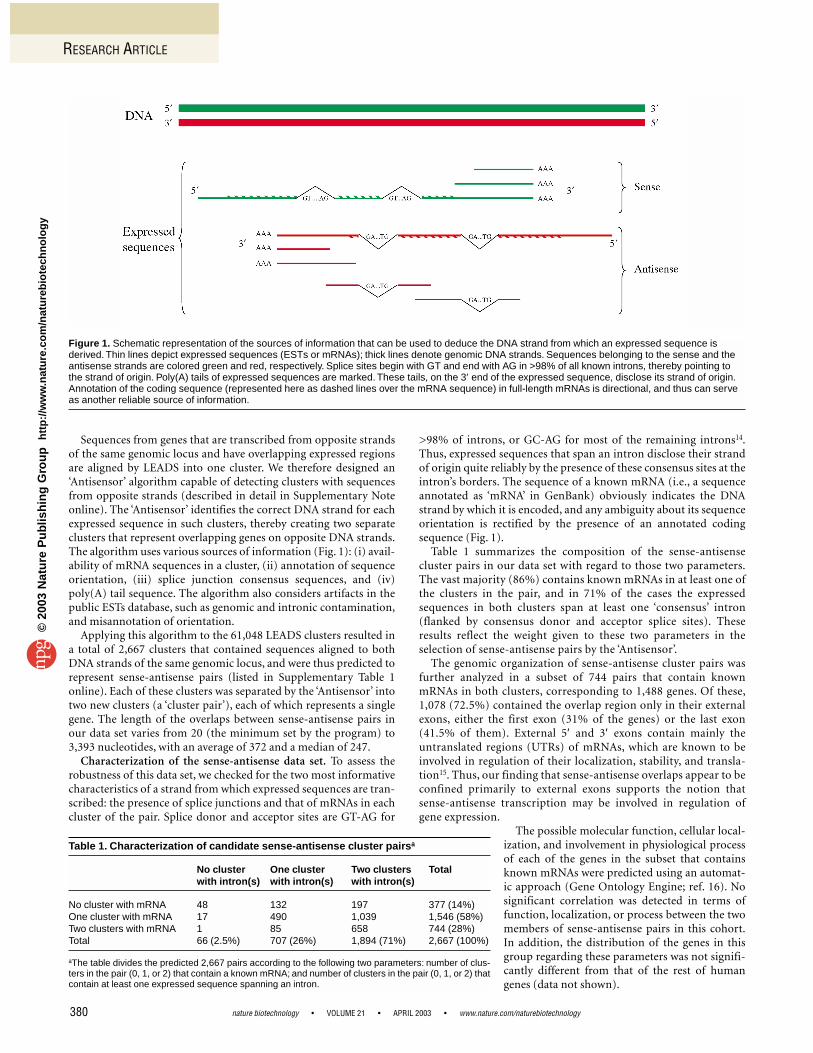
Sequences from genes that are transcribed from opposite strandsof the same genomic locus and have overlapping expressed regionsare aligned by LEADS into one cluster. We therefore designed an‘Antisensor’ algorithm capable of detecting clusters with sequencesfrom opposite strands (described in detail in Supplementary Noteonline). The ‘Antisensor’ identifies the correct DNA strand for eachexpressed sequence in such clusters, thereby creating two separateclusters that represent overlapping genes on opposite DNA strands.The algorithm uses various sources of information (Fig. 1): (i) avail-ability of mRNA sequences in a cluster, (ii) annotation of sequenceorientation, (iii) splice junction consensus sequences, and (iv)poly(A) tail sequence. The algorithm also considers artifacts in thepublic ESTs database, such as genomic and intronic contamination,and misannotation of orientation.
Applying this algorithm to the 61,048 LEADS clusters resulted ina total of 2,667 clusters that contained sequences aligned to bothDNA strands of the same genomic locus, and were thus predicted torepresent sense-antisense pairs (listed in Supplementary Table 1online). Each of these clusters was separated by the ‘Antisensor’ intotwo new clusters (a ‘cluster pair’), each of which represents a singlegene. The length of the overlaps between sense-antisense pairs inour data set varies from 20 (the minimum set by the program) to3,393 nucleotides, with an average of 372 and a median of 247.
Characterization of the sense-antisense data set. To assess therobustness of this data set, we checked for the two most informativecharacteristics of a strand from which expressed sequences are tran-scribed: the presence of splice junctions and that of mRNAs in eachcluster of the pair. Splice donor and acceptor sites are GT-AG for
>98% of introns, or GC-AG for most of the remaining introns14.Thus, expressed sequences that span an intron disclose their strandof origin quite reliably by the presence of these consensus sites at theintron’s borders. The sequence of a known mRNA (i.e., a sequenceannotated as ‘mRNA’ in GenBank) obviously indicates the DNAstrand by which it is encoded, and any ambiguity about its sequenceorientation is rectified by the presence of an annotated codingsequence (Fig. 1).
Table 1 summarizes the composition of the sense-antisensecluster pairs in our data set with regard to those two parameters.The vast majority (86%) contains known mRNAs in at least one ofthe clusters in the pair, and in 71% of the cases the expressedsequences in both clusters span at least one ‘consensus’ intron(flanked by consensus donor and acceptor splice sites). Theseresults reflect the weight given to these two parameters in theselection of sense-antisense pairs by the ‘Antisensor’.
The genomic organization of sense-antisense cluster pairs wasfurther analyzed in a subset of 744 pairs that contain knownmRNAs in both clusters, corresponding to 1,488 genes. Of these,1,078 (72.5%) contained the overlap region only in their externalexons, either the first exon (31% of the genes) or the last exon(41.5% of them). External 5′ and 3′ exons contain mainly theuntranslated regions (UTRs) of mRNAs, which are known to beinvolved in regulation of their localization, stability, and transla-tion15. Thus, our finding that sense-antisense overlaps appear to beconfined primarily to external exons supports the notion thatsense-antisense transcription may be involved in regulation ofgene expression.
The possible molecular function, cellular local-ization, and involvement in physiological processof each of the genes in the subset that containsknown mRNAs were predicted using an automat-ic approach (Gene Ontology Engine; ref. 16). Nosignificant correlation was detected in terms offunction, localization, or process between the twomembers of sense-antisense pairs in this cohort.In addition, the distribution of the genes in thisgroup regarding these parameters was not signifi-cantly different from that of the rest of humangenes (data not shown).
RESEARCH ARTICLE
nature biotechnology • VOLUME 21 • APRIL 2003 • www.nature.com/naturebiotechnology380
Table 1. Characterization of candidate sense-antisense cluster pairsa
No cluster One cluster Two clusters Total with intron(s) with intron(s) with intron(s)
No cluster with mRNA 48 132 197 377 (14%)One cluster with mRNA 17 490 1,039 1,546 (58%)Two clusters with mRNA 1 85 658 744 (28%)Total 66 (2.5%) 707 (26%) 1,894 (71%) 2,667 (100%)
aThe table divides the predicted 2,667 pairs according to the following two parameters: number of clus-ters in the pair (0, 1, or 2) that contain a known mRNA; and number of clusters in the pair (0, 1, or 2) thatcontain at least one expressed sequence spanning an intron.
Figure 1. Schematic representation of the sources of information that can be used to deduce the DNA strand from which an expressed sequence isderived. Thin lines depict expressed sequences (ESTs or mRNAs); thick lines denote genomic DNA strands. Sequences belonging to the sense and theantisense strands are colored green and red, respectively. Splice sites begin with GT and end with AG in >98% of all known introns, thereby pointing tothe strand of origin. Poly(A) tails of expressed sequences are marked. These tails, on the 3′ end of the expressed sequence, disclose its strand of origin.Annotation of the coding sequence (represented here as dashed lines over the mRNA sequence) in full-length mRNAs is directional, and thus can serveas another reliable source of information.
©20
03 N
atu
re P
ub
lish
ing
Gro
up
h
ttp
://w
ww
.nat
ure
.co
m/n
atu
reb
iote
chn
olo
gy

RESEARCH ARTICLE
Microarray-based experimental evaluation. For experimental vali-dation of the predicted sense-antisense pairs in our original data set (of2,667 pairs), we carried out a microarray-based analysis using oligonu-cleotide probes that hybridize to the target in a strand-specific manner.For this purpose we designed two complementary 60-mer oligonu-cleotide probes derived from the predicted overlap regions of the sense-antisense pairs. Single 60-mer oligonucleotides were previously shownto offer reliability and sensitivity for detecting specific transcripts17.
To select the sense-antisense pairs to be evaluated by microarrays,we first chose only those with an overlap >60 bases (2,464 pairs sat-isfied this restriction). The overlap region of each antisense pair wasthen checked for the presence of 60-mer oligonucleotides thatmatched a set of standards, such as minimal sequence similarityelsewhere in the human genome, uniform G+C content and Tm
(melting temperature), and absence of palindromic sequences (see‘Oligonucleotide Design’ in Supplementary Note online), to maxi-
www.nature.com/naturebiotechnology • APRIL 2003 • VOLUME 21 • nature biotechnology 381
Figure 2. Validation of sense-antisense pairs by northern blot analysis. Ten pairs were selected, eight (A–H) that showed positive signals for both senseand antisense genes on the microarray analysis, and two (I–J) that showed positive signals for only one of the genes, whereas the counterpart is a knownRefSeq mRNA. The diagrams describe the genomic organization of the relevant region for each of the sense-antisense genes. Exons are numbered onlywhen an mRNA sequence is available for the cluster. Also shown is the region of overlap from which the strand-specific riboprobes were derived. TheGenBank accession number of the overlapping sequence is given for each gene. Below are the autoradiograms after hybridization with the appropriateriboprobe. Northern blots contained poly(A) mRNAs from the different human tissues or cell lines. Equal loading was evaluated by ethidium bromidestaining. Size markers (in kb) are indicated. PBL, peripheral blood lymphocytes.
A B C
D E
F
H I
G
J
©20
03 N
atu
re P
ub
lish
ing
Gro
up
h
ttp
://w
ww
.nat
ure
.co
m/n
atu
reb
iote
chn
olo
gy

mize the hybridization specificity. Optimal oligonucleotide probeswere found for 1,211 sense-antisense pairs. From these, a randomsample of 264 pairs was selected for analysis by microarrays (listedin Supplementary Table 2 online), roughly one-tenth of the originaldata set of 2,667 sense-antisense pairs. In this sample, the propor-tion of each of the nine subgroups depicted in Table 1 is similar tothat of the original data set, indicating a good representation of thevarious subgroups.
Microarrays were constructed by spotting each of the aforemen-tioned oligonucleotide probes onto treated glass slides in quadrupli-cate. The two counterpart oligonucleotide probes, derived from theoverlap region of a sense-antisense pair, were spotted next to each
other to ensure similar hybridizationconditions. For positive controls weselected probes for four ubiquitouslyexpressed genes (see SupplementaryNote online). For negative controlswe used two random oligonu-cleotides. These computer-createdarbitrary sequences display no sub-stantial alignment to the humangenome (upon BLAST search usingdefault parameters) but have thesame characteristics as the otheroligonucleotide probes. In addition,22 probes for 11 previously docu-mented sense-antisense pairs werealso spotted on these microarrays.
The microarrays were hybridizedwith poly(A)+ RNAs obtained from19 human cell lines representing avariety of tissues and four normalhuman tissues (see ExperimentalProtocol). Each poly(A)+ RNA wasreverse-transcribed by priming witholigo(dT) and random nonamers inthe presence of fluorescently labeleddNTPs. A pooled sample containingan equal mix of the RNAs from all celllines was also transcribed and used as areference target. The resulting fluores-cently labeled cDNAs were combinedand hybridized to the oligonucleotidemicroarrays. The experiments weredone in duplicate with a fluorescentreversal of the cyanin-3 (Cy3)- andCy5-labeled cDNA and pool. Wefirst tested different conditions ofhybridization and chose stringentconditions so as to minimize theappearance of false-positive signals,although this could compromise thedetection of low-abundance tran-scripts. (The raw data from themicroarray experiments is presentedin http://www.labonweb.com/anti-sense/Raw_Data.)
The raw data were normalized atseveral levels: within each slide,between reciprocal slides, and global-ly among slides (see SupplementaryNote online). Nonspecific levels ofhybridization were estimated fromthe negative controls. The threshold
for a significant positive signal resulting from authentic hybridiza-tion was set at 4 standard deviations of the mean normalized signalsof the negative controls. The processed data are presented as normal-ized signal intensity and as normalized signal ratios (SupplementaryTable 3 online). Positive signals were obtained for both sense andantisense transcripts in 65 cluster pairs. In another 47 cases, wedetected significant hybridization signals for antisense sequenceswhose counterpart sense transcripts are known RefSeq(http://www.ncbi.nlm.nih.gov/LocusLink/RSfaq.html) mRNAs thatdid not give clear hybridization signals on our microarrays. Thus, ina total of 112 cases, or 42.5% of the 264 sense-antisense pairs repre-sented on the microarrays, we could detect antisense transcription.
RESEARCH ARTICLE
nature biotechnology • VOLUME 21 • APRIL 2003 • www.nature.com/naturebiotechnology382
Figure 3. Analysis of the overlap region between the transcriptional units of TP53BP1 and 76P. (A) Schematicrepresentation of the known mRNA of each gene (with the respective exons), and their overlapping ESTs(depicted by thick short lines, which represent several ESTs). The thin colored lines depict the predictedextensions of the respective 3′ UTRs through alternative polyadenylation, which were confirmed by theexperimental analysis shown in (B) and (C). The expected sizes of the known and the putative transcriptscontaining these alternative 3′ UTRs are given. Also shown is the location of the primers (depicted asarrows) used in the RT-PCR analysis, and the region from which the riboprobes were derived for northernblot analysis. (B) PCR analysis with the primers shown in (A), using genomic DNA (gDNA) or RT productsfrom the indicated cell lines as template. The presence (+) or absence (–) of enzyme in the RT reaction isindicated. The sizes of the PCR products that were obtained are indicated in kilobases. The sizes of theexpected and observed PCR products are summarized in Table 2. The transcriptional unit from which theRT-PCR products were derived (TP53BP1 or 76P) was confirmed by the spanning of the respectivesplicing event, as detected upon sequencing. (C) Northern blot analysis with poly(A) mRNA from differentcell lines, using the strand-specific riboprobes described in A. The size of transcripts detected fit theprediction of the 3′ UTR extensions, as described in (A).
A
B C
©20
03 N
atu
re P
ub
lish
ing
Gro
up
h
ttp
://w
ww
.nat
ure
.co
m/n
atu
reb
iote
chn
olo
gy
RESEARCH ARTICLE
The sensitivity of our experimental approach, in other words ourability to detect a given transcript, is determined by the stringency ofthe microarray hybridization conditions and the tissue specificityand expression levels of the mRNA. This sensitivity can be estimatedfrom the positive signals that we obtained for 65% of the oligos rep-resenting known RefSeq mRNAs on the microarrays. This level ofdetection is comparable to that obtained in other studies, such as the58% of known exons verified using microarray analysis18. To detect asense-antisense pair we need to detect two genes, each of which has a0.65 probability of detection. Thus, our level of detection (sensitivi-ty) for a sense-antisense pair is 0.65 × 0.65= 0.42. Of the 264 predict-ed sense-antisense pairs that were analyzed with microarrays, the 65cases that showed significant signals for both sense and antisensetranscripts represent the 0.42 fraction that we expect to detect fromthe real sense-antisense pairs on the microarray. Thus, there wereactually 154 (60/0.42) such pairs on the microarray, and therefore atleast 60% (154 of 264) of the clusters we analyzed represented truecases of overlapping antisense transcription. Extrapolating this fig-ure to our predicted antisense data set of 2,667 clusters, we concludethat there are ≥1,600 sense-antisense transcriptional units in thehuman genome.
RNA-based strand-specific verification. From the sense-antisensepairs showing positive signals on the microarrays, we chose ten casesfor further verification by northern blot analysis. Eight of the select-ed pairs had shown positive signals for both the sense and antisensegenes in the microarray analysis, and two pairs were chosen becauseonly one of the genes passed the validation by microarrays and itscounterpart oligo was fully contained within a known RefSeqmRNA. As seen in Figure 2A–J, the presence of transcripts with com-plementary sequences was confirmed for all ten gene pairs throughhybridization with strand-specific riboprobes derived from the pre-dicted region of overlap.
Two additional sense-antisense pairs were selected for detailedstudy of the extent of overlap. Analysis using bioinformatic toolspointed to the presence of neighboring ESTs in the antisense orienta-tion in both cases—suggesting alternative polyadenylation thatresults in long 3′ UTRs and an extensive overlap between the twotranscriptional units. The known mRNAs of the first pair, TP53BP1(the gene encoding 53BP1, p53-binding protein-1, ref. 19) and 76P(the gene encoding 76p, a member of the γ-tubulin-associatedprotein family, ref. 20), do not overlap but are derived from oppo-site strands of the same genomic locus, within 1.9 kilobases fromeach other’s 3′ end (Fig. 3A). The presence of ESTs in the antisenseorientation in both genes suggested the existence of long alterna-tive 3′ UTRs, creating an overlap of several kilobases between thetwo genes (Fig. 3A). This assumption was verified by a RT-PCR-based approach (Fig. 3B). The newly predicted transcripts werefurther confirmed by northern blot analysis using strand-specificriboprobes (Fig. 3C). A similar situation was observed between thetranscriptional units of CCNE2 (the gene encoding cyclin E2, refs.21, 22) and FLJ20530 (RefSeq mRNA no. NM_017864.1, encoding a
hypothetical protein) (Fig. 4A). The apparent overlap between thetwo known mRNAs is only 75 base pairs long. However, the presenceof ESTs in the antisense direction to CCNE2 suggested alternativepolyadenylation, resulting in a much longer 3′ UTR of the FLJ20530,and an overlap of several kilobases between the two genes. This wasagain confirmed by RT-PCR and northern blot analysis (Fig. 4B,C).These results indicate that the true overlap between sense and anti-sense genes may be much longer than that expected from analysis ofmRNAs deposited in GenBank.
DiscussionThe results of this study, which integrates computational tools withexperimental methods, suggest the presence of ≥1,600 sense-anti-sense gene pairs in the human genome. This finding indicates thatthe occurrence of loci transcribed from both DNA strands in thehuman genome is a common phenomenon. We believe that natural-ly occurring antisense transcripts may be even more prevalent thanestimated from this study for the following reasons: (i) Our compu-tational tools enable detection of sense-antisense genes only if bothsense and antisense expressed sequences were deposited in the pub-lic databases, and the transcripts overlap within exonic regions.Antisense genes for which expressed sequences are not yet available,or that are too short to show the overlap, will be missed by ourdetection tools. (ii) Our methods led us to the identification of cis-antisense transcripts in the human transcriptome, in other words,transcripts that are transcribed from opposite DNA strands of thesame locus. Trans-encoded antisense RNAs (those transcribed fromanother locus) cannot be detected by our tools. (iii) Antisense tran-scripts that do not span introns, or do not contain clear poly(A)tails, are more likely to be rejected by our current tools.
In addition, it appears that the overlap between the sense-antisensetranscriptional units is often more extensive than can be estimatedfrom the sequences in the public databases. This is due to under-representation of long 3′ UTRs, particularly in the cDNA databases,and therefore the overlap between genes is frequently established bycomplementary ESTs, even when mRNAs are present in the clusters.This observation is supported by our detailed analysis of two sense-antisense pairs, TP53BP1-76P and CCNE2-FLJ20530.
Three recent papers, one published during the course of our workand two while it was being reviewed, report the computational dis-covery of transcripts with the potential for sense-antisense pair-ing23–25. In two of these reports (refs. 23 and 24) the study was limitedto BLAST analysis of publicly available, full-length human mRNAs(RefSeq database containing ∼ 12,000 human mRNAs). Using thisapproach, Lehner et al. (ref. 23) reported the in silico identification of87 cis-antisense human transcripts, whereas Fahey et al. (ref. 24)identified 56 such pairs. By extrapolation, both groups estimated∼ 800 pairs of genes with sense-antisense transcription in the humangenome, assuming 40,000 genes. Lehner et al. refrained from usingEST databases because of the uncertainties regarding the correct ori-entation of ESTs. Fahey et al. tried to use EST databases but foundthem unreliable for the detection of antisense transcription, again
www.nature.com/naturebiotechnology • APRIL 2003 • VOLUME 21 • nature biotechnology 383
Table 3. Analysis of the overlap between CCNE2 and FLJ20530by PCRa
CCNE2 FLJ0530 gDNA
Primers Exp Obs Exp Obs Exp Obs
1 + 2 1.6 1.6 2.0 2.0 2.0 2.01 + 3 / / 2.1 2.1 5.9 5.9
aSizes (in kb) of expected and observed PCR products (in Fig. 4B) using twoprimer pairs for CCNE2 and FLJ20530, on genomic DNA (gDNA) or RT prod-ucts as template.
Table 2. Analysis of the overlap between TP53BP1 and 76P byPCRa
TP53BP1 76P gDNA
Primers Exp Obs Exp Obs Exp Obs
2 + 15 / / 4.5 4.5 5.2 5.26 + 16 3.9 3.9 3.0 3.0 4.3 4.37 + 17 4.3 4.3 / / 4.3 4.3
aSizes (in kb) of expected and observed PCR products (in Fig. 3B) using differ-ent primer pairs for TP53BP1 and 76P, on genomic DNA (gDNA) or RT productsas template.
©20
03 N
atu
re P
ub
lish
ing
Gro
up
h
ttp
://w
ww
.nat
ure
.co
m/n
atu
reb
iote
chn
olo
gy

because of the problematic assignment of ESTs orientation. Ourdesign of the appropriate computational tools facilitates the use ofthe vast public data on millions of human ESTs and partial cDNAs,permitting their assembly, clustering, and alignment to the correctDNA strand. Thus, our approach provides a more realistic estimateof the extent of antisense transcription. Furthermore, we validateour in silico findings with experimental evidence.
The recent study by Shendure and Church (ref. 25) analyzed data-bases of expressed sequences using publicly available bioinformatictools. To avoid problems related to the correct orientation of ESTs,they used only sequences from high-quality directionally clonedEST libraries. They identified 144 candidate overlapping transcrip-tional units in the human genome. They also carried out experi-mental validation of a subset of their predictions by orientation-specific RT-PCR, and found evidence of antisense transcription in33 out of 39 cases tested. The number of sense-antisense loci predict-ed by our computational tools, and experimentally validated usingoligonucleotide microarrays, exceeds those of Shendure and Churchby roughly an order of magnitude. Furthermore, their experimental
analysis is limited to one tissue,whereas we analyzed >20 human tis-sues by both microarray and north-ern blot analysis, and carried out fur-ther in-depth characterization of twospecific examples. A comparisonbetween our data set of sense-anti-sense pairs and those predicted in thestudies just described showed that>85% of their pairs appear in ourdata set. Manual examination of theirpairs that were missing in our data setshowed most of these to be artifacts.A detailed description of this com-parison is given in the SupplementaryNote online).
Our estimate of ≥1,600 sense-antisense pairs (i.e., 3,200 genes)suggests that >8% of the estimated40,000 human genes have an anti-sense partner. As mentioned byShendure and Church25, further sup-port for this estimate comes fromthe work of Shoemaker et al.18, whoused 60-mer oligonucleotides to val-idate a large number of predictednovel exons by microarray analysis.As negative controls, they used thereverse-complement probes for∼ 78,000 exons annotated as ‘con-firmed’. Using two sources of mRNA,they observed hybridization to ∼ 5%of these supposedly ‘negative con-trols’. In light of our results, webelieve that some of these ‘false posi-tives’ could actually stem from anti-sense transcription.
Widespread regulation of geneexpression by antisense transcriptionoffers an explanation for the puzzlingobservation of ortholog-specific con-servation of 3′ UTRs in >30% of verte-brate mRNAs26. Lipman proposed thatsuch long conserved blocks of uniquenoncoding sequences, previously
shown to be associated with mRNA stability, are involved in forminglong perfect duplexes with antisense transcripts27. Of relevance is ourobservation that ∼ 70% of the sense-antisense genes overlap in their 5′or 3′ external exons, giving further support to the notion that sense-antisense overlap could be involved in gene regulation, as externalexons contain the untranslated regulatory regions of mRNAs.
Knowledge of human antisense transcription can be of help in thestudy of RNA interference, an important biological phenomenon thatis based on the presence of dsRNA and leads to degradation of com-plementary mRNAs and to gene silencing. Such information may alsobe important for the selection of synthetic antisense oligonucleotidesin functional studies and drug design28. The broad occurrence of com-plementary transcripts in the human transcriptome has clear implica-tions for the interpretation of experimental data resulting from the useof dsDNA probes, such as northern blot analysis and cDNA microar-rays. Clearly, both experimental approaches would benefit from theexclusive use of strand-specific probes.
Recent reports in the literature indicate that antisense transcrip-tion in other eukaryotes may be more common than had been
RESEARCH ARTICLE
nature biotechnology • VOLUME 21 • APRIL 2003 • www.nature.com/naturebiotechnology384
Figure 4. Analysis of the overlap region between the transcriptional units of CCNE2 and FLJ20530.(A) Schematic representation showing the known mRNAs (with the respective exons) and the ESTs (shortthick lines) that suggest an extension of the 3′ UTR of FLJ20530 (depicted by the thin pink line), which wasconfirmed by RT-PCR as shown in (B). Also shown are the location of the primers (depicted as arrows) usedin the RT-PCR analysis, and the region from which the riboprobes were derived. (B) PCR analysis using theprimers shown in (A), essentially as described in Figure 3B. The sizes of the expected and observed PCRproducts are summarized in Table 3. (C) Northern blot analysis on poly(A) mRNAs from different cell lines,using the strand-specific riboprobes described in (A).
A
B
C
©20
03 N
atu
re P
ub
lish
ing
Gro
up
h
ttp
://w
ww
.nat
ure
.co
m/n
atu
reb
iote
chn
olo
gy

RESEARCH ARTICLE
thought. Kumar et al. (ref. 29) reported that out of 137 newly discov-ered open reading frames in yeast, 79 are predicted to lie oppositepreviously annotated genes. Antisense transcription in plants hasalso been shown in a growing number of cases5.
Taken together, the above findings and our results suggest thatlarge numbers of antisense transcripts are found throughout theeukaryotic world and may play a role in antisense-mediated generegulation, as is the case in prokaryotes. Our data set forms a basis forfuture studies on the modulation of gene expression by antisensetranscription in eukaryotes.
Experimental protocolHuman cell lines. The following cell lines were used: MCF-7 (breast adeno-carcinoma, American Type Culture Collection (ATCC) no. HTB-22), HeLa(cervical adenocarcinoma, ATCC no. CCL-2), HEK-293 (embryonal kidneycells, ATCC no. CRL-1573), Jurkat (acute T-cell leukemia, ATCC no. TIB-152), K562 (chronic myelogenous leukemia, ATCC no. CCL-243), HepG2(liver carcinoma, ATCC no. HB-8065), T24 (urinary bladder carcinoma,ATCC no. HTB-4), SK-N-DZ (neuroblastoma, ATCC no. CRL-2149), NK-92(non-Hodgkin’s lymphoma, ATCC no. CRL-2407), MG-63 (osteosarcoma,ATCC no. CRL-1427), DU 145 (prostatic carcinoma, ATCC no. HTB-81),G-361 (melanoma, ATCC no. CRL-1424), PANC-1 (pancreatic carcinoma,ATCC no. CRL-1469), ES-2 (ovary clear-cell carcinoma, ATCC no. CRL-1978), Y79 (retinoblastoma, ATCC no. HTB-18), HT-29 (colorectal adeno-carcinoma, ATCC no. HTB-38), DLD-1 (colorectal adenocarcinoma, ATCCno. CCL-221), H1299 (large-cell lung carcinoma, ATCC no. CRL-5803),NL564 (Epstein-Barr virus (EBV)–transformed human lymphoblasts),SNU1 (gastric carcinoma, ATCC no. CRL-5971), LNCaP (prostatic adenocar-cinoma), and MCF10 (benign breast tissue cells).
RNA purification. Total RNA was extracted from the aforementioned humancell lines using TriReagent (Molecular Research Center, Cincinnati, OH).Poly(A)+ mRNA was purified using two cycles of the Dynabeads mRNAPurification Kit (Dynal Biotech ASA, Oslo, Norway). The removal of traces ofribosomal RNA was confirmed by agarose gel electrophoresis. Poly(A)+
mRNA from human testis, placenta, lung, and brain was purchased fromBioChain Institute, Inc. (Hayward, CA).
RT-PCR. Reverse transcription (RT) was carried out in a final volume of 20 µlusing 2 µg of total RNA and 2.5 units of Superscript II Reverse Transcriptase(Invitrogen, Paisley, UK), in the buffer supplied by the manufacturer, supple-mented with 10 pmol of oligo(dT)15 (Promega, Madison, WI) and 30 units ofRNasin (Promega). PCR was carried out using 1 µl of the RT reaction, in thepresence of 2 mM dNTPs, 25 pmol of primers, and 2.5 units of DNA Pol mixof the Expand Long Template PCR System (Roche, Mannheim, Germany), inthe reaction buffer supplied by the manufacturer. RT reactions carried out inthe absence of enzyme were used as negative controls for genomic DNA con-tamination.
The following primers were used for the RT-PCR analysis shown in Figure 3:Primer 2, 5′-ggtgaggagtttctttgtgtaacc-3′; primer 6, 5′-ccagaatctgaag-
gtactcttg-3′; primer 7, 5′-tcacaagagtgggtgatccagtg-3′; primer 17, 5′-ccttaag-gagctcacaatctagttgg-3′; primer 16, 5′-cgctggtcagtcagaacctag-3′; primer 15,5′-cactcctgttcaagattctctcc-3′.
Primers used for the analysis shown in Figure 4 are as follow: Primer 1,5′-ctagtccagtgaagctgaagac-3′ ; primer 2, 5′-tacagctggcagcgcagagaag-3′ ;primer 3, 5′-cctactacgactacatatgggatg-3′ .
Riboprobe synthesis. For each sense-antisense pair analyzed in Figure 2, theoverlapping riboprobes were designed to include the 60-mer oligonu-cleotides used in the microarray analysis. Riboprobes were obtained by RT-PCR on selected RTs. The sequence of the primers used to obtain the various
RT-PCR products are given in Supplementary Table 4 online. RT-PCR prod-ucts were electrophoresed on agarose gels, extracted with Gel Extraction Kit(Qiagen, Hilden, Germany), and ligated to the T7 adapter using theLig’nScribe Kit (Ambion, Austin, TX). The P2 primer (in the T7 adapter) anda forward-specific primer were used to amplify the sense-specific riboprobe,and a reverse-specific primer to amplify the antisense-specific riboprobe.These PCR products were then separated on agarose gels, and extracted asdescribed earlier. Each one was subsequently sequenced with the appropriateprimers for verification. For labeling, 50–100 ng of each PCR product wasused per 500 bp riboprobe, in a reaction containing α-[32P]UTP and T7 poly-merase, using the SP6/T7 Transcription Kit (Roche, Mannheim, Germany),as suggested by the manufacturer. The riboprobe was separated from thenonincorporated ribonucleoside triphosphates using Mini Quick Spin RNAColumns (Roche, Indianapolis, IN).
The following primers were used to obtain riboprobes for the northernblot analysis shown in Figure 3C: Riboprobe 1, primer R1, 5′-ccagaatctgaag-gtactcttg-3′; primer F1, 5′-cacaatctccacgatagcagg-3′. Riboprobe 2, primer R2,5′-gctagaattgcccaatctgtgtag-3′; primer F2, 5′-cgctggtcagtcagaacctag-3′.
The following primers were used to obtain riboprobes for the northernblot analysis shown in Figure 4C: Riboprobe 1, primer F1, 5′-cctactacgacta-catatgggatg-3′; primer R1: 5′-gctctagacagatatgtgaatctcttctcc-3′. Riboprobe2,primer F2, 5′-gacagagttgaatgcaagcaatcc-3′; primer R2, 5′-gctgtggctccttcc-taactgg-3′.
Northern blot analysis. Poly(A)+ RNA (1 µg) of human cells or tissues wasfractionated by electrophoresis on 1.0% (wt/vol) agarose gels containingformaldehyde and blotted onto Nytran Super Charge membranes (Schleicher& Schuell, Dassel, Germany). Equal loading was verified by ethidium bro-mide staining of the gels. In addition, we used commercial northern blotscontaining poly(A)+ RNA samples from a variety of tissues (Human MultipleTissue Northern blots; BD Biosciences, Palo Alto, CA). Northern blots wereprehybridized at 68 °C for 30 min in Ultrahyb Buffer (Ambion, Austin, TX).32P-labeled riboprobe (at 106 c.p.m./ml) was added and the hybridization wascarried out for 16 h at 68 °C. The membranes were rinsed twice with 2× SSC,0.1% (wt/vol) SDS at room temperature, washed twice for 5 min each with2× SSC, 0.1% SDS at 68 °C, and a last wash for 30 min with 0.1× SSC, 0.1%SDS at 68 °C. Autoradiograms were obtained by exposing the membranes toBioMax MS film (Kodak, Rochester, NY) for various times, according to theintensity of the signals.
URL. The LEADS-Antisensor is accessible for academic research through thefollowing website: http://www.labonweb.com/antisense.
Raw data from microarray experiments can be found at:http://www.labonweb.com/antisense/Raw_Data.
Note: Supplementary information is available on the Nature Biotechnologywebsite.
AcknowledgmentsWe want to thank Alon Wasserman and Brian Meloon for their critical help withthe Antisensor design and analysis of microarray results; Mervi Heiskanen, RuthGoldin, and Paul Nisson for their valuable assistance in the setup of the microarraymethodology; Han Xie for the GO engine analysis; Dan Sztybel for technicalhelp; Raveh Gill-More, Gady Cojocaru, Naftali Kaminski, and Gidi Rechavi forcritical reading of the manuscript and helpful discussions; Sarah Pollock for herinnovative ideas, and Salomon Langer for his inspirational support.
Competing interests statementThe authors declare competing financial interests: see the Nature Biotechnologywebsite (http://www.nature.com/naturebiotechnology) for details.
Received 28 August 2002; accepted 7 January 2003
www.nature.com/naturebiotechnology • APRIL 2003 • VOLUME 21 • nature biotechnology 385
©20
03 N
atu
re P
ub
lish
ing
Gro
up
h
ttp
://w
ww
.nat
ure
.co
m/n
atu
reb
iote
chn
olo
gy

RESEARCH ARTICLE
nature biotechnology • VOLUME 21 • APRIL 2003 • www.nature.com/naturebiotechnology386
1. Wagner, E.G. & Simons, R.W. Antisense RNA control in bacteria, phages, andplasmids. Annu. Rev. Microbiol. 48, 713–742 (1994).
2. Knee, R. & Murphy, P.R. Regulation of gene expression by natural antisense RNAtranscripts. Neurochem. Int. 31, 379–392 (1997).
3. Kumar, M. & Carmichael, G.G. Antisense RNA: function and fate of duplex RNA incells of higher eukaryotes. Microbiol. Mol. Biol. Rev. 62, 1415–1434 (1998).
4. Vanhee-Brossollet, C. & Vaquero, C. Do natural antisense transcripts make sensein eukaryotes? Gene 211, 1–9 (1998).
5. Terryn, N. & Rouze, P. The sense of naturally transcribed antisense RNAs inplants. Trends Plant Sci. 5, 394–396 (2000).
6. Kelly, R.L. & Kuroda, M.I. Noncoding RNA genes in dosage compensation andimprinting. Cell 103, 9–12 (2000).
7. Sleutels, F., Barlow, D.P. & Lyle, R. The uniqueness of the imprinting mechanism.Curr. Opin. Genet. Dev. 10, 229–233 (2000).
8. Li, A.W. & Murphy, P.R. Expression of alternatively spliced FGF-2 antisense RNAtranscripts in the central nervous system: regulation of FGF-2 mRNA translation.Mol. Cell. Endocrinol. 162, 69–78 (2000).
9. Hastings, M.L., Ingle, H.A., Lazar, M.A. & Munroe, S.H. Post-transcriptional regu-lation of thyroid hormone receptor expression by cis-acting sequences and a nat-urally occurring antisense RNA. J. Biol. Chem. 275, 11507–11513 (2000).
10. Bass, B.L. Double-stranded RNA as a template for gene silencing. Cell 101,235–238 (2000).
11. Wu, H., MacLeod, A.R., Lima, W.F. & Crooke, S.T. Identification and partial purifi-cation of human double strand RNase activity. A novel terminating mechanism foroligoribonucleotide antisense drugs. J. Biol. Chem. 273, 2532–2542 (1998).
12. Bass, B.L. RNA editing and hypermutation by adenosine deamination. TrendsBiochem. Sci. 22, 157–162 (1997).
13. Zamore, P.D. Ancient pathways programmed by small RNAs. Science 296,1265–1269 (2002).
14. International Human Genome Sequencing Consortium. Initial sequencing andanalysis of the human genome. Nature 409, 860–921 (2001).
15. Pesole, G. et al. Structural and functional features of eukaryotic mRNA untranslat-ed regions. Gene 276, 73–81 (2001).
16. Xie, H. et al. Large-scale protein annotation through gene ontology. Genome Res.12, 785–794 (2002).
17. Hughes, T.R. et al. Expression profiling using microarrays fabricated by an ink-jetoligonucleotides synthesizer. Nat. Biotechnol. 19, 342–347 (2001).
18. Shoemaker, D.D. et al. Experimental annotation of the human genome usingmicroarray technology. Nature 409, 922–927 (2001).
19. Iwabuchi, K., Bartel, P.L., Li, B., Marraccino, R. & Fields, S. Two cellular proteinsthat bind to wild-type but not mutant p53. Proc. Natl. Acad. Sci. USA 91,6098–6102 (1994).
20. Fava, F. et al. Human 76p: a new member of the γ-tubulin-associated protein fami-ly. J. Cell Biol. 147, 857–868 (1999).
21. Lauper, N. et al. Cyclin E2: a novel CDK2 partner in the late G1 and S phases ofthe mammalian cell cycle. Oncogene 17, 2637–2643 (1998).
22. Zariwala, M., Liu, J. & Xiong, Y. Cyclin E2, a novel human G1 cyclin and activatingpartner of CDK2 and CDK3, is induced by viral oncoproteins. Oncogene 17,2787–2798 (1998).
23. Lehner, B., William, G., Campbell, R.D. & Sanderson, C.M. Antisense transcriptsin the human genome. Trends Genet. 18, 63–65 (2002).
24. Fahey, M.E., Moore, T.F. & Higgins, D.G. Overlapping antisense transcription in thehuman genome. Comp. Funct. Genom. 3, 244–253 (2002).
25. Shendure, J. & Church, G.M. Computational discovery of sense-antisense tran-scription in the human and mouse genomes. Genome Biol. 3, 1–14 (2002).
26. Duret, L., Dorkeld, F. & Gautier, C. Strong conservation of non-codingsequences during vertebrates evolution: potential involvement in post-tran-scriptional regulation of gene expression. Nucleic Acids Res. 21, 2315–2322(1993).
27. Lipman, D.J. Making (anti)sense of non-coding sequence conservation. NucleicAcids Res. 25, 3580–3583 (1997).
28. Delihas, N., Rokita, S.E. & Zheng, P. Natural antisense RNA/target RNA interac-tions: possible models for antisense oligonucleotide drug design. Nat. Biotechnol.15, 751–753 (1997).
29. Kumar, A. et al. An integrated approach for finding overlooked genes in yeast. Nat.Biotechnol. 20, 58–63 (2002).
©20
03 N
atu
re P
ub
lish
ing
Gro
up
h
ttp
://w
ww
.nat
ure
.co
m/n
atu
reb
iote
chn
olo
gy