whole-genome sequencing of retinoblastoma reveals the
TRANSCRIPT

cancers
Article
Whole-Genome Sequencing of Retinoblastoma Reveals theDiversity of Rearrangements Disrupting RB1 and Uncovers aTreatment-Related Mutational Signature
Helen R. Davies 1,2,†, Kevin D. Broad 3,†, Zerrin Onadim 4 , Elizabeth A. Price 4, Xueqing Zou 1,2,Ibrahim Sheriff 5, Esin Kotiloglu Karaa 6, Irene Scheimberg 6, M. Ashwin Reddy 5,7, Mandeep S. Sagoo 3,5,7,* ,Shin-ichi Ohnuma 3,* and Serena Nik-Zainal 1,2,*
�����������������
Citation: Davies, H.R.; Broad, K.D.;
Onadim, Z.; Price, E.A.; Zou, X.;
Sheriff, I.; Karaa, E.K.; Scheimberg, I.;
Reddy, M.A.; Sagoo, M.S.; et al.
Whole-Genome Sequencing of
Retinoblastoma Reveals the Diversity
of Rearrangements Disrupting RB1
and Uncovers a Treatment-Related
Mutational Signature. Cancers 2021,
13, 754. https://doi.org/10.3390/
cancers13040754
Academic Editor: David MacPherson
Received: 14 December 2020
Accepted: 9 February 2021
Published: 11 February 2021
Publisher’s Note: MDPI stays neutral
with regard to jurisdictional claims in
published maps and institutional affil-
iations.
Copyright: © 2021 by the authors.
Licensee MDPI, Basel, Switzerland.
This article is an open access article
distributed under the terms and
conditions of the Creative Commons
Attribution (CC BY) license (https://
creativecommons.org/licenses/by/
4.0/).
1 Academic Department of Medical Genetics, University of Cambridge, Addenbrooke’s Treatment Centre,Cambridge Biomedical Campus, Cambridge CB2 0QQ, UK; [email protected] (H.R.D.);[email protected] (X.Z.)
2 MRC Cancer Unit, Hutchison/MRC Research Centre, Cambridge Biomedical Campus,University of Cambridge, Cambridge CB2 0XZ, UK
3 Institute of Ophthalmology, University College London, London EC1V 9EL, UK; [email protected] Retinoblastoma Genetic Screening Unit, The Royal London Hospital, Barts Health NHS Trust,
London E1 1FR, UK; [email protected] (Z.O.); [email protected] (E.A.P.)5 Retinoblastoma Service, Royal London Hospital, Barts Health Trust, London E1 1FR, UK;
[email protected] (I.S.); [email protected] (M.A.R.)6 Pathology Department, Royal London Hospital, Barts Health NHS Trust, London E1 1FR, UK;
[email protected] (E.K.K.); [email protected] (I.S.)7 NIHR Biomedical Research Centre for Ophthalmology, Moorfields Eye Hospital, Institute of Ophthalmology,
University College London, London EC1V 2PD, UK* Correspondence: [email protected] (M.S.S.); [email protected] (S.-i.O.); [email protected] (S.N.Z.)† These authors contributed equally to this paper.
Simple Summary: Retinoblastoma, a childhood cancer of the eye, is thought to be caused byinactivating mutations of both copies of the RB1 gene. The majority of RB1 mutations can be detectedby clinical screening. However, retinoblastoma cases exist where mutations in RB1 have not beendetected. We used whole-genome sequencing to investigate the landscape of mutations in a cohortof sporadic retinoblastomas, including cases where mutations in both copies of RB1 had not beenpreviously identified. We looked for mutations in cancer driver genes and revealed a wide variety ofstructural rearrangements disrupting RB1. In addition, we investigated mutation burden and specificmutation patterns (mutational signatures), uncovering a treatment-related mutational signature in atumour exposed to chemotherapy. The power of whole-genome sequencing to identify RB1 mutationsof all mutation types can have significant relevance to the clinical management of retinoblastomapatients and genetic counselling of their families.
Abstract: The development of retinoblastoma is thought to require pathological genetic changes inboth alleles of the RB1 gene. However, cases exist where RB1 mutations are undetectable, suggestingalternative pathways to malignancy. We used whole-genome sequencing (WGS) and transcriptomicsto investigate the landscape of sporadic retinoblastomas derived from twenty patients, sought RB1and other driver mutations and investigated mutational signatures. At least one RB1 mutation wasidentified in all retinoblastomas, including new mutations in addition to those previously identifiedby clinical screening. Ten tumours carried structural rearrangements involving RB1 ranging fromrelatively simple to extremely complex rearrangement patterns, including a chromothripsis-like pat-tern in one tumour. Bilateral tumours obtained from one patient harboured conserved germline butdivergent somatic RB1 mutations, indicating independent evolution. Mutational signature analysisshowed predominance of signatures associated with cell division, an absence of ultraviolet-relatedDNA damage and a profound platinum-related mutational signature in a chemotherapy-exposedtumour. Most RB1 mutations are identifiable by clinical screening. However, the increased resolutionand ability to detect otherwise elusive rearrangements by WGS have important repercussions onclinical management and advice on recurrence risks.
Cancers 2021, 13, 754. https://doi.org/10.3390/cancers13040754 https://www.mdpi.com/journal/cancers

Cancers 2021, 13, 754 2 of 19
Keywords: retinoblastoma; whole-genome sequencing; structural rearrangement; mutational signature
1. Introduction
The retinoblastoma (Rb) is the most common primary intraocular cancer of childhoodaccounting for approximately 2% of all childhood cancers [1], causing significant long-termsequelae. Loss-of-function mutations in RB transcriptional co-repressor 1 (RB1) are causallyimplicated in Rb development. RB1 was the first tumour suppressor gene to be isolated,in contrast to activating oncogenes, which were characterised earlier [2,3]. In humans, RB1is located on chromosome 13q14.2, spans approximately 180 Kb and contains 27 exonsencoding a 928 amino acid nucleophosphoprotein, known as pRB [4].
pRB is a multifunctional protein involved in a variety of processes regulating cellproliferation at multiple levels including apoptosis, histone methylation and chromatinremodelling [5–8]. Loss of function of pRB in the retina is thought to lead to dysregulationof these events, resulting in uncontrolled cell proliferation and chromosomal instability.
Rb patients present with a wide spectrum of RB1 mutations. The prevailing view isthat mutations in both alleles of the RB1 gene are required to enable tumourigenesis [9].Heritable predisposition is present in ~45% of cases and is transmitted in an autosomaldominant manner. Typically, penetrance is almost complete (>90%) and is characterisedby the development of tumours in both eyes (bilateral retinoblastomas) in most cases.Germline RB1 mutation carriers are also at an increased lifetime risk of non-ocular tu-mours [10]. In 55% of cases, somatically-acquired RB1 mutations underpin tumourigenesis,are unilateral and do not incur predisposition to develop tumours at other sites [11–14].
Although Knudson’s “two-hits hypothesis” suggests that a loss of function of bothRB1 alleles is required to initiate retinoblastoma [15], using standard clinical screeningtechniques, in 3–4% of tumours, only one RB1 mutation can be identified [9,16]. Indeed,in approximately 2% of tumours, RB1 mutations are elusive [17,18]. This suggests thatundetected RB1 mutations possibly involving translocations, intronic mutations or novelpromoter mutations may be harboured by these tumours, or the development of someretinoblastoma tumours may be driven through an RB1-independent mechanism [17,19].Two papers recently highlighted the importance of other potential driver mutations in-cluding focal MYCN amplification, first reported in 1–2 % of tumours with no detectableRB1 mutations [17,19]. Focal amplifications of the orthodenticle homeobox 2 gene (OTX2)were observed in 3% and focal deletions of the BCL6 Corepressor (BCOR) observed in 4%of the same cohort of retinoblastoma tumours [19]. Truncating point mutations in BCORhave been also reported in 10–17% of Rb [18,20–22]. In addition, a number of studies haveidentified consistent large-scale copy number changes including gains of 1q, 2p and 6pand losses of 16q [19,20,23]. The contribution of structural mutational mechanisms to RB1disruption has also been reported [19].
To date, the majority of studies investigating genomic aberrations in Rb have focusedon copy number aberrations [23], whole-exome sequencing [20] or targeted gene pan-els [18,22]. Very limited numbers of whole-genome sequencing (WGS) experiments havebeen performed. WGS of 4 Rb tumours was performed by Zang et al., with RB1 being theonly mutated known cancer gene identified [21]. The data from the same 4 WGS were alsoincluded in a combined analysis of 547 paediatric tumours from 24 cancer types [24]. In aseparate study by McEvoy et al., WGS of 10 tumours identified 3 cases with chromothripsisat the RB1 locus [19]. In this study, we performed WGS on a total of 21 retinoblastomasfrom 20 individuals with no previous family history (3 with bilateral and 17 unilateraltumours). In addition, transcriptomic profiling was performed on a subset of tumours.The Rb tumours comprised two cohorts: Cohort 1, made up of nine tumours, where two dis-tinct RB1 mutations were previously identified in each tumour; Cohort 2 comprised tentumours, where seven had only one RB1 mutation, and in three, RB1 mutations remainedeither elusive (one tumour) or could not be satisfactorily confirmed by clinical screening

Cancers 2021, 13, 754 3 of 19
methods (two tumours). RB1 promoter hypermethylation had previously been excludedin these tumours. Additionally, in one case, tumours from both the left and right eyewere available from a paediatric patient with a germline RB1 mutation providing an op-portunity to determine phylogenetic relatedness between bilateral tumours in germlinemutation carriers.
2. Results2.1. All Retinoblastoma Tumours Harbour At Least One RB1 Mutation
At least one RB1 mutation was identified in each of the 19 tumours interrogated fromCohorts 1 and 2 (Figure 1, Table S1). We confirmed all the mutations identified by clinicalscreening in tumours from nine patients of Cohort 1 (Table 1). One sample from a patientwith a germline essential splice site mutation in RB1 (PD34255) had lost the alternativeparental allele through loss of heterozygosity (LOH). Of the remaining eight tumours,six had small truncating somatic mutations on one allele (new stop codons, frameshiftingindels or essential splice site variants) and lost the other allele through LOH or multiexondeletion (PD34256, PD34260, PD37500, PD37501, PD37518, and PD37519), one case had alarge-scale structural deletion and LOH of the alternative allele (PD34259) and one case hadtwo small truncating variants (PD34257), a somatic essential splice site mutation togetherwith a somatic nonsense mutation.
Cancers 2021, 13, x 5 of 19
Figure 1. Driver gene mutations and copy number aberrations in retinoblastoma. Summary of RB1 mutations, mutations
in driver genes and large copy number changes >3 Mb in size.
2.1.1. WGS Reveals Novel RB1 Disruptions by Structural Variation
Novel RB1 disruptions were successfully identified in three tumours, two of which
had previously characterised LOH of RB1. In PD37495, balanced translocations between
intron 2 of the RB1 gene with chromosomes 16 and 18 confirmed disruption of RB1 and
thus biallelic RB1 loss. In PD37493, biallelic loss was a result of a complex rearrangement
involving a translocation between intron 17 of RB1 and intron 15 of THSD7B on chromo-
some 2. In PD37488, complex intrachromosomal rearrangements resulted in multiple
break points transecting RB1 although the effect on the alternative parental allele could
not be resolved.
2.1.2. Diverse Rearrangements Transect RB1
It was previously reported that complex structural variation, specifically a phenom-
enon called chromothripsis, underpinned some disruptions of RB1 [19]. Chromothripsis
is a compound chromosomal outcome where large numbers of rearrangements are clus-
tered in localised genomic regions, sometimes involving multiple chromosomes, resulting
in an oscillating copy number state. In this study, we identified ten tumours with RB1
structural rearrangements spanning a continuum of complexity (see Figure 2 and Figure
S1 for examples of rearrangements disrupting RB1).
Bilateral Rb
Unilateral Rb Trucating point mutation
R B 1 germline Small deletion/insertion
Large deletion
LOH
M YCN amplification Structural rearrangement
M DM 4 amplification Germline mosaic tandem duplication
B CO R mutation Not detected
CR E B B P mutation Effect on second allele not resolved
EXT 2 mutation Missense1p1q Copy number gain2p Copy number loss (LOH)2q Copy number neutral LOH3p3q4p4q5p5q6p6q7p7q8p8q9p9q
10p10q11p11q12p12q13q14q15q16p16q17p17q18p18q19p19q20p20q21p21q22q
PD34
255
PD34
256
PD34
257
PD34
259
PD34
260
PD37
500
PD37
501
PD37
518
PD37
519
PD37
490
PD37
491
PD37
488
PD37
489
PD37
492
PD37
493
PD37
494
PD37
495
PD37
496
PD37
497
PD34
258
PD37
502
L
AR
GE
COPY
NU
MB
ER C
HA
NG
ES >
3 M
b
R B 1 somatic
COHORT 1 COHORT 2Bilateral
case
LAR
GE
CO
PY
NU
MB
ER C
HA
NG
ES >
3 M
b
Figure 1. Driver gene mutations and copy number aberrations in retinoblastoma. Summary of RB1 mutations, mutations indriver genes and large copy number changes >3 Mb in size.
Cancers 2021, 13, 754 4 of 19
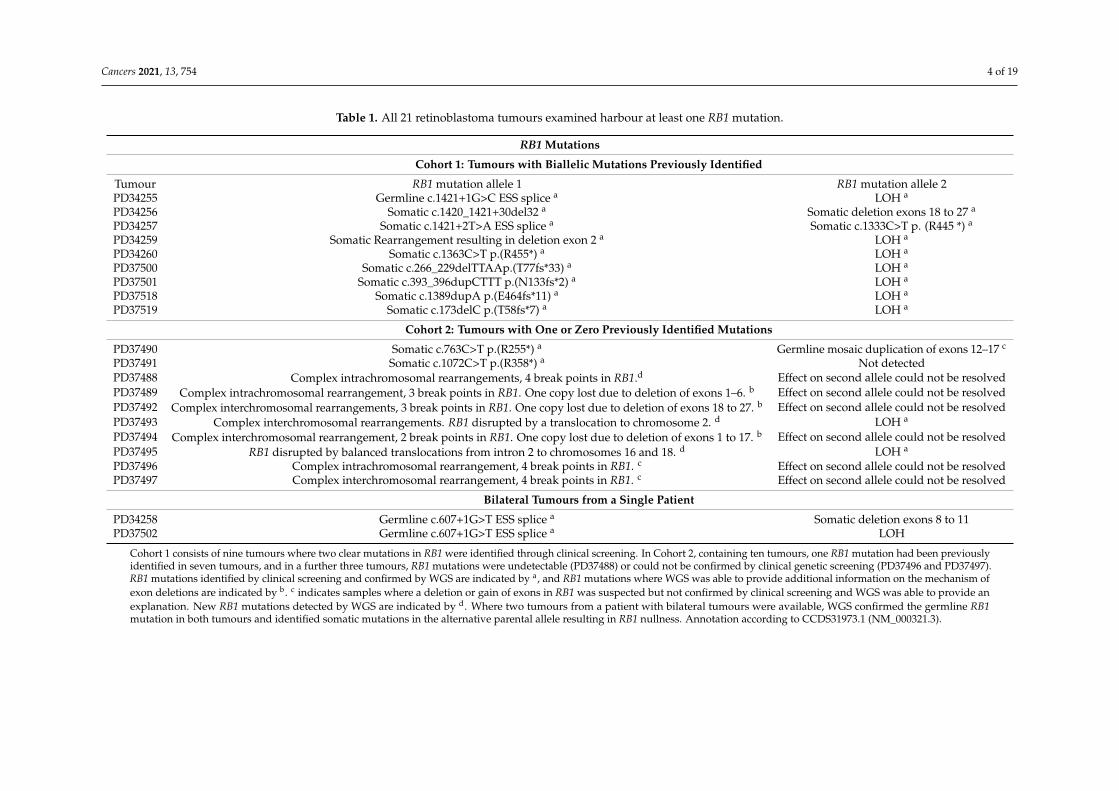
Table 1. All 21 retinoblastoma tumours examined harbour at least one RB1 mutation.
RB1 Mutations
Cohort 1: Tumours with Biallelic Mutations Previously Identified
Tumour RB1 mutation allele 1 RB1 mutation allele 2PD34255 Germline c.1421+1G>C ESS splice a LOH a
PD34256 Somatic c.1420_1421+30del32 a Somatic deletion exons 18 to 27 a
PD34257 Somatic c.1421+2T>A ESS splice a Somatic c.1333C>T p. (R445 *) a
PD34259 Somatic Rearrangement resulting in deletion exon 2 a LOH a
PD34260 Somatic c.1363C>T p.(R455*) a LOH a
PD37500 Somatic c.266_229delTTAAp.(T77fs*33) a LOH a
PD37501 Somatic c.393_396dupCTTT p.(N133fs*2) a LOH a
PD37518 Somatic c.1389dupA p.(E464fs*11) a LOH a
PD37519 Somatic c.173delC p.(T58fs*7) a LOH a
Cohort 2: Tumours with One or Zero Previously Identified Mutations
PD37490 Somatic c.763C>T p.(R255*) a Germline mosaic duplication of exons 12–17 c
PD37491 Somatic c.1072C>T p.(R358*) a Not detectedPD37488 Complex intrachromosomal rearrangements, 4 break points in RB1.d Effect on second allele could not be resolvedPD37489 Complex intrachromosomal rearrangement, 3 break points in RB1. One copy lost due to deletion of exons 1–6. b Effect on second allele could not be resolvedPD37492 Complex interchromosomal rearrangements, 3 break points in RB1. One copy lost due to deletion of exons 18 to 27. b Effect on second allele could not be resolvedPD37493 Complex interchromosomal rearrangements. RB1 disrupted by a translocation to chromosome 2. d LOH a
PD37494 Complex interchromosomal rearrangement, 2 break points in RB1. One copy lost due to deletion of exons 1 to 17. b Effect on second allele could not be resolvedPD37495 RB1 disrupted by balanced translocations from intron 2 to chromosomes 16 and 18. d LOH a
PD37496 Complex intrachromosomal rearrangement, 4 break points in RB1. c Effect on second allele could not be resolvedPD37497 Complex interchromosomal rearrangement, 4 break points in RB1. c Effect on second allele could not be resolved
Bilateral Tumours from a Single Patient
PD34258 Germline c.607+1G>T ESS splice a Somatic deletion exons 8 to 11PD37502 Germline c.607+1G>T ESS splice a LOH
Cohort 1 consists of nine tumours where two clear mutations in RB1 were identified through clinical screening. In Cohort 2, containing ten tumours, one RB1 mutation had been previouslyidentified in seven tumours, and in a further three tumours, RB1 mutations were undetectable (PD37488) or could not be confirmed by clinical genetic screening (PD37496 and PD37497).RB1 mutations identified by clinical screening and confirmed by WGS are indicated by a, and RB1 mutations where WGS was able to provide additional information on the mechanism ofexon deletions are indicated by b. c indicates samples where a deletion or gain of exons in RB1 was suspected but not confirmed by clinical screening and WGS was able to provide anexplanation. New RB1 mutations detected by WGS are indicated by d. Where two tumours from a patient with bilateral tumours were available, WGS confirmed the germline RB1mutation in both tumours and identified somatic mutations in the alternative parental allele resulting in RB1 nullness. Annotation according to CCDS31973.1 (NM_000321.3).
Cancers 2021, 13, 754 5 of 19
In Cohort 2, of twenty disrupted alleles that could possibly be identified across the tenpatients, one was not detected (PD37491) and six could not be definitively resolved. We con-firmed four previously discovered RB1 disruptions (a in Table 1: p.(R255*) in PD37490,p.(R358*) in PD37491, and LOH in PD37493 and PD37495), provided confirmatory evidencefor three possible mutations (c in Table 1), provided clarity on mutational mechanism foranother three (b in Table 1) and identified three new RB1 disruptions caused by structuralvariation (d in Table 1: PD37488, PD37493 and PD37495).
2.1.1. WGS Reveals Novel RB1 Disruptions by Structural Variation
Novel RB1 disruptions were successfully identified in three tumours, two of which hadpreviously characterised LOH of RB1. In PD37495, balanced translocations between intron 2of the RB1 gene with chromosomes 16 and 18 confirmed disruption of RB1 and thus biallelicRB1 loss. In PD37493, biallelic loss was a result of a complex rearrangement involvinga translocation between intron 17 of RB1 and intron 15 of THSD7B on chromosome 2.In PD37488, complex intrachromosomal rearrangements resulted in multiple break pointstransecting RB1 although the effect on the alternative parental allele could not be resolved.
2.1.2. Diverse Rearrangements Transect RB1
It was previously reported that complex structural variation, specifically a phe-nomenon called chromothripsis, underpinned some disruptions of RB1 [19]. Chromoth-ripsis is a compound chromosomal outcome where large numbers of rearrangements areclustered in localised genomic regions, sometimes involving multiple chromosomes, result-ing in an oscillating copy number state. In this study, we identified ten tumours with RB1structural rearrangements spanning a continuum of complexity (see Figure 2 and Figure S1for examples of rearrangements disrupting RB1).
Not all structural variations were complex. We observed simple translocations inPD37495 involving a break in intron 2 of RB1. The N-terminus of RB1 was joined to exon5 of CREBBP on chromosome 16, while the C-terminus was translocated to intergenicchromosome 18. Although the LOH of the alternate copy of RB1 was detected by clinicalscreening, the translocation break points lie within introns which resulted in a transectionof the gene but no discernible change in copy number. Thus, the rearrangement was notdetected by customary clinical testing.
A diverse range of more complex rearrangements were also observed. One relativelycomplicated rearrangement was detected by clinical testing as a deletion involving exon 2(PD34259 in cohort1). WGS revealed a more complicated structural outcome where adeletion may have arisen from multiple inversions resulting in loss of the 5′ region ofthe RB1 gene including exons 1 and 2 and a separate deletion of other copy of exon 2(Figure S1).
In three tumours, PD37488, PD37489 and PD37496, multiple rearrangements tran-sected RB1 as part of a wider collection of complex, intrachromosomal rearrangements(Figure 2a,b). These tumours contained between 7 and 64 break points on chromosome 13,with 3 to 4 break points within RB1 itself. With such complex rearrangement patterns itwas not possible to resolve the order of rearrangements or whether both copies of RB1were affected.

Cancers 2021, 13, 754 6 of 19Cancers 2021, 13, x 6 of 19
Figure 2. Examples of structural variation affecting the RB1 locus (a) Sample PD37489 demonstrat-
ing an intrachromosomal rearrangement, (b) Sample PD37488 showing complex intrachromoso-
mal rearrangements, and (c) Sample PD37494 showing chromothripsis-like interchromosomal
rearrangements between chromosome 13 and chromosomes 8 and 14. Translocation partners for
each interchromosomal breakpoint are shown above. Somatic copy number estimates (Y-axis) are
plotted against genomic coordinates of chromosome 13 encompassing the RB1 locus (X-axis).
Structural variation classes: blue for inversions, red for deletions, green for tandem duplication
and black for translocations.
Not all structural variations were complex. We observed simple translocations in
PD37495 involving a break in intron 2 of RB1. The N-terminus of RB1 was joined to exon
5 of CREBBP on chromosome 16, while the C-terminus was translocated to intergenic
chromosome 18. Although the LOH of the alternate copy of RB1 was detected by clinical
screening, the translocation break points lie within introns which resulted in a transection
of the gene but no discernible change in copy number. Thus, the rearrangement was not
detected by customary clinical testing.
A diverse range of more complex rearrangements were also observed. One relatively
complicated rearrangement was detected by clinical testing as a deletion involving exon 2
(PD34259 in cohort1). WGS revealed a more complicated structural outcome where a deletion
may have arisen from multiple inversions resulting in loss of the 5′ region of the RB1 gene
including exons 1 and 2 and a separate deletion of other copy of exon 2 (Figure S1).
In three tumours, PD37488, PD37489 and PD37496, multiple rearrangements tran-
sected RB1 as part of a wider collection of complex, intrachromosomal rearrangements
(Figure 2a,b). These tumours contained between 7 and 64 break points on chromosome
13, with 3 to 4 break points within RB1 itself. With such complex rearrangement patterns
it was not possible to resolve the order of rearrangements or whether both copies of RB1
were affected.
Five tumours demonstrated complex interchromosomal rearrangement patterns
transecting RB1 through a variety of structural variations including translocations to other
chromosomes (PD37492, PD37493, PD37494, PD37495, and PD37497). PD37492 showed
Figure 2. Examples of structural variation affecting the RB1 locus (a) Sample PD37489 demonstratingan intrachromosomal rearrangement, (b) Sample PD37488 showing complex intrachromosomal rear-rangements, and (c) Sample PD37494 showing chromothripsis-like interchromosomal rearrangementsbetween chromosome 13 and chromosomes 8 and 14. Translocation partners for each interchromoso-mal breakpoint are shown above. Somatic copy number estimates (Y-axis) are plotted against genomiccoordinates of chromosome 13 encompassing the RB1 locus (X-axis). Structural variation classes:blue for inversions, red for deletions, green for tandem duplication and black for translocations.
Five tumours demonstrated complex interchromosomal rearrangement patterns tran-secting RB1 through a variety of structural variations including translocations to otherchromosomes (PD37492, PD37493, PD37494, PD37495, and PD37497). PD37492 showedmultiple interchromosomal exchange between 13q and a region of chromosome 11, re-sulting in three separate break points within intron 17 of RB1 and the loss of one copy ofthe 3′ end of the gene, including exons 18 to 27. Clinical testing of PD37494 had detecteda deletion of the 5′ end of RB1 encompassing exons 1 to 17. However, WGS was ableto clarify that this mutation was part of a complex pattern of rearrangements betweenchromosomes 13, 4 and 8 with a total of over 40 break points. The oscillating copy numberstates observed are characteristic of a chromothripsis-like pattern, in keeping with previousreports of chromothripsis in retinoblastoma [19] (Figure 2c).
2.1.3. WGS Provides Clarification of Ambiguous Clinical Testing Results
Notably, the increased resolution afforded by WGS provided clarification of am-biguous results from clinical testing. Case PD37490 contained a somatic point mutation,c.763C>T p.(R255*) on one allele. Clinical screening using MLPA detected a suspectedamplification of exons 12 to 17 of RB1 in the tumour and possible low-level amplification inthe normal blood sample. However, RNA was not available to confirm this result. WGS ofthe tumour confirmed the presence of a tandem duplication with break points in introns11 and 17, causing gene disruption. Whilst there was no overt increase in read depthin the normal blood sample, reads spanning the tandem duplication break points were

Cancers 2021, 13, 754 7 of 19
detected in the blood DNA sample, albeit at a reduced fraction compared to the tumour.It is therefore possible that the patient is constitutionally mosaic for the tandem duplication.This patient was the only one of three cases with bilateral tumours, without a confirmedgermline mutation and therefore the constitutional mosaic tandem duplication of RB1presumably acquired during embryogenesis would be entirely consistent with the clinicalpicture of bilateral tumours in this patient.
Where structural rearrangements are accompanied by large intragenic deletions ofRB1, the resulting exonic deletions can be detected by clinical screening methods, as seenin PD34259, PD37489, PD37492 and PD37494.
In two additional cases, where intragenic RB1 deletions were suspected by clinicalscreening but could not be confirmed at the transcript level, WGS provided confirmatoryevidence of RB1 disruption. In PD37497, a monoallelic deletion of exons 23 and 24 wassuspected by clinical screening. WGS revealed a complex, interchromosomal rearrangementbetween chromosomes 13, 7, 8 and 14 including 4 break points in RB1. Two of the breakpoints involved in an inversion and tandem duplication, lie in close proximity to the 5′ and3′ intron/exon boundaries of exons 23 and 24 (72 bp and 49 bp, respectively). In PD37496,a monoallelic deletion of exon 4 was suspected by clinical screening. WGS showed anextremely complex intrachromosomal rearrangement pattern including four break pointswithin the RB1 gene. Although no changes in copy number were seen, two break pointsinvolved in a deletion and inversion lie in close proximity to the 5′ and 3′ intron/exonboundaries of exon 4 (170 pb and 302 bp). In both cases, these rearrangements mayhave disrupted the binding or orientation of probes used in clinical screening leadingto the impression of deleted exons, when the gene was in fact disrupted by alternativestructural aberrations.
PD37495 and PD37493, discussed above, had translocations disrupting RB1 in con-junction with LOH and hence biallelic mutation was confirmed. However, in the six othercases, it was not possible to determine whether both copies of the RB1 gene were disruptedby rearrangements. WGS can add increased granularity to the details of RB1 mutationsinvolving structural rearrangements and can provide explanations for suspected intrageniccopy number changes detected by standard clinical methods which cannot be confirmed atthe transcript level. However, with such complex rearrangement patterns present in someretinoblastoma samples it is not possible to resolve the precise order of rearrangementsand whether both copies are affected without also employing alternative approaches suchas long read sequencing.
We thus note that nine of ten cases (90%) where biallelic RB1 mutations were notpreviously identified had simple rearrangements, complex intrachromosomal as well ascomplex interchromosomal rearrangements, and one incidence of chromothripsis-like pat-tern observed. This highlights the contribution of structural variation to the developmentof retinoblastoma. In contrast to a previous report that 30% of cases (n = 3/10) with nodetectable RB1 mutation by exon sequencing had RB1 disruption via chromothripsis [19],our study indicates that, RB1 transection can occur through a wide variety of rearrange-ment mechanisms, and not necessarily via chromothripsis. These results highlight theimportance of searching for RB1 inactivating mutations of all possible types in Rb tumours.
2.2. Retinoblastoma Tumours Harbour a Low Burden of Mutations
The overall tumour mutational burden of retinoblastoma is low. An average count of275 (range 26–931) (density 0.085 per Mb), 70 (range 19–231) (density 0.021 per Mb) and17 (range 1–66) (density 0.005 per Mb) variants were seen for substitutions, small inser-tions/deletions and structural rearrangements, respectively (See Tables S2–S4 for somaticmutations). Structural variation involved in complex rearrangement patterns leading todisruption of RB1 in eight tumours contributed to the majority of the total rearrangements(191 of the total 356 rearrangements detected). Tumours with simple point mutations anddeletions of RB1 harboured a lower incidence of structural rearrangements (average 6,range 1–15). Two large-scale WGS studies of multiple paediatric cancer types have reported

Cancers 2021, 13, 754 8 of 19
very low mutation burden in all paediatric cancers [24,25]. Our reported mutation burdensare in keeping with these previous reports.
2.3. Retinoblastomas Are Not Driven by Ultraviolet Damage
Mutational signatures are characteristic patterns of mutations that provide a recordof the types of DNA damage that have occurred during the development of a cancer [26].Due to the very low mutation burden and small sample size, it was not possible to performde novo mutational signature extraction to identify the mutational signatures presentin retinoblastoma. However, we did perform a “fitting” experiment, asking whethermutational signatures that have been previously reported in cancers were present in thisdataset [26]. Caution should be exercised when performing signature fitting withoutprior knowledge of the mutational signatures contributing to a particular cancer type,since overfitting of inappropriate signatures can occur. Grobner et al. attempted to performsignature fitting in only four retinoblastoma WGS as part of a large study of 547 paediatrictumours from 24 cancer types [24]. However, with very small sample numbers, signaturecontribution estimates are likely to be unreliable.
Of note, ultraviolet (UV)-related DNA damage has been raised as a potential contrib-utor to the development of retinoblastoma [27], although in a subsequent study, it wassuggested that it was a confounding factor [28]. In conjunctival melanoma, an adulteye cancer, a striking contribution of C>T mutations characteristic of UV damage hasbeen demonstrated [29]. By contrast, sequencing studies of another adult eye cancer,uveal melanoma, have not found UV to be a contributory factor to tumorigenesis [30,31].Signature fitting in the 21 WGS retinoblastomas although limited by the low mutationnumbers, showed no evidence of the signature of UV light (Figure 3a), indicating that UVdamage is unlikely to be a major source of DNA damage in this tumour type. Furthermore,features such as transcriptional strand bias and double substitutions, which are additionalcharacteristics of UV-associated damage, were also not observed in the retinoblastomas(Tables S5 and S6).
Somatic mutations are continuously acquired at each cell division during the courseof human life. The mutational processes that contribute to these replicative errors wouldthus be correlated to human age [32]. We assessed the correlation between total number ofsubstitution mutations per sample with the age of enucleation. We find a clear correlationbetween mutation burden and age of enucleation of treatment-naïve tumours (r2 = 0.835,p-value 0.001) (Figure 3b). This is in keeping with mutational processes that are mainlyassociated with cell division and an absence of other mutational processes.

Cancers 2021, 13, 754 9 of 19Cancers 2021, 13, x 9 of 19
Figure 3. Mutational signatures in retinoblastoma (a) 96-trinucleotide mutation profile plots. Single-base pair substitutions
are converted to the pyrimidine context and each of the six substitution subtypes, C>A, C>G, C>T, T>A, T>C, and T>G,
plotted according to the bases immediately 5′ and 3′ to the mutated base. Profiles produced using Signal (https://signal.mu-
tationalsignatures.com/) [33]. Top, profile of the combined substitution mutations from all twenty treatment-naive tu-
mours. Bottom, profile of UV light-associated signature RefSig 7 shows no similarity to that of the retinoblastomas. Refer-
ence signature profiles are the mean of tissue specific signatures [33], with error bars representing 1 standard deviation
from the mean (https://signal.mutationalsignatures.com/). (b) Scatter plot showing the correlation between age of enucle-
ation (x axis) and number of substitution mutations (y axis). Blue dots, treatment-naive samples. Red dot, the bilateral
tumour exposed to platinum-based chemotherapy (PD37502). (c) Top, profile of the bilateral tumour exposed to platinum-
based chemotherapy (PD37502). Bottom, profile of the platinum therapy associated mutational signature shows a striking
similarity to that of PD37502 (Cosine similarity = 0.96).
2.4. Insights from Analysis of Independent Tumours in a Bilateral Retinoblastoma Case
A patient with no prior family history of ocular tumours presented aged 19 months
with bilateral retinoblastomas, International Classification of Retinoblastoma Group E/E.
The right eye had raised intraocular pressure, iris neovascularisation and multiple white
calcified retinal tumours, with lens touch, a total retinal detachment and multiple vitreous
seeds. The left eye had early iris neovascularisation, with normal intraocular pressure and
a large white calcified tumour touching the lens, with multiple subretinal seeds. The right
eye was enucleated immediately and an attempt was made to save the left eye as it was
slightly less advanced. The patient was treated with 6 cycles of systemic JOE chemother-
apy (vincristine (1.5 mg per 10 kg), carboplatin (600 mg per 10 kg), and etoposide (300 mg
per 10 kg)) over a four-month period. Despite an initial reduction in tumour size of the
left eye, there was evidence of localised relapse at fundoscopy and the remaining left eye
was enucleated at 34 months of age.
A germline RB1 c.607+1G>T splice donor site mutation was identified through clini-
cal screening. This mutation is predicted to cause a frameshift (p.lle181Glyfs*8), which
follows skipping of exon 6 [34]. However, diverse somatic mutations of the alternate pa-
rental copy of RB1 were identified in the two tumours (PD34258 (right eye tumour),
PD37502 (left eye tumour)).
R efSig PLA TIN U M
R efSig 7
C om bined profile of treatm ent naive R b tum ours
PD 37502
(a)
(c)
(b)
0
100
200
300
400
500
600
700
800
900
1000
0.0 10.0 20.0 30.0 40.0 50.0 60.0 70.0
Nu
mb
er o
f su
bst
itu
tio
n m
uta
tio
ns
Age at enucleation (months)
Age at enucleation vs number of substitution mutations
Figure 3. Mutational signatures in retinoblastoma (a) 96-trinucleotide mutation profile plots. Single-base pair substitutionsare converted to the pyrimidine context and each of the six substitution subtypes, C>A, C>G, C>T, T>A, T>C, and T>G,plotted according to the bases immediately 5′ and 3′ to the mutated base. Profiles produced using Signal (https://signal.mutationalsignatures.com/ (accessed on 10 December 2020)) [33]. Top, profile of the combined substitution mutations fromall twenty treatment-naive tumours. Bottom, profile of UV light-associated signature RefSig 7 shows no similarity to that ofthe retinoblastomas. Reference signature profiles are the mean of tissue specific signatures [33], with error bars representing1 standard deviation from the mean (https://signal.mutationalsignatures.com/ (accessed on 10 December 2020)). (b) Scatterplot showing the correlation between age of enucleation (x axis) and number of substitution mutations (y axis). Blue dots,treatment-naive samples. Red dot, the bilateral tumour exposed to platinum-based chemotherapy (PD37502). (c) Top,profile of the bilateral tumour exposed to platinum-based chemotherapy (PD37502). Bottom, profile of the platinum therapyassociated mutational signature shows a striking similarity to that of PD37502 (Cosine similarity = 0.96).
2.4. Insights from Analysis of Independent Tumours in a Bilateral Retinoblastoma Case
A patient with no prior family history of ocular tumours presented aged 19 monthswith bilateral retinoblastomas, International Classification of Retinoblastoma Group E/E.The right eye had raised intraocular pressure, iris neovascularisation and multiple whitecalcified retinal tumours, with lens touch, a total retinal detachment and multiple vitreousseeds. The left eye had early iris neovascularisation, with normal intraocular pressure anda large white calcified tumour touching the lens, with multiple subretinal seeds. The righteye was enucleated immediately and an attempt was made to save the left eye as it wasslightly less advanced. The patient was treated with 6 cycles of systemic JOE chemotherapy(vincristine (1.5 mg per 10 kg), carboplatin (600 mg per 10 kg), and etoposide (300 mgper 10 kg)) over a four-month period. Despite an initial reduction in tumour size of theleft eye, there was evidence of localised relapse at fundoscopy and the remaining left eyewas enucleated at 34 months of age.
A germline RB1 c.607+1G>T splice donor site mutation was identified through clinicalscreening. This mutation is predicted to cause a frameshift (p.lle181Glyfs*8), which followsskipping of exon 6 [34]. However, diverse somatic mutations of the alternate parental

Cancers 2021, 13, 754 10 of 19
copy of RB1 were identified in the two tumours (PD34258 (right eye tumour), PD37502(left eye tumour)).
A large somatic deletion of RB1 involving exons 8 to 11 was identified in the tumourof the right eye. It also had 65 substitutions, 28 small indels, 8 rearrangements, gain of6p and deletions of 2q and 22q. In the tumour of the left eye, the remaining copy of RB1was lost via a large somatic deletion encompassing most of chromosome 13q, resultingin LOH of RB1. A substantially greater number of mutations was detected including931 substitutions, 103 small indels, 5 rearrangements, gains on chromosomes 3q, 6p, 14q,12p and 12q and two deletions on chromosomes 17 and X. None of the somatic mutationswere shared between the tumours from the two eyes, confirming that the two tumours hadarisen independently.
The increased burden of mutation could result in part from an increased age asthe second tumour was removed 15 months after the first. However, when assessingmutation burden as a function of age, this tumour was a clear outlier (Figure 3b), suggestingmutational processes in addition to those associated with age were operative in this tumour.Indeed, the substitution mutation profile was consistent with an exposure to platinum,which is a key component of carboplatin (Cosine similarity = 0.96) (Figure 3c). The presenceof this signature suggests that this tumour has been derived from an ancestral clone thatsurvived six exposures to carboplatin. Carboplatin inhibits cancer growth by inducingDNA damage and initiating cell death, which has resulted in the acquisition of additionalmutations consistent with this exposure and a selection pressure for gene mutations thatfacilitate survival of that clone, during its resurgent growth prior to enucleation.
2.5. Large-Scale Copy Number Changes
A number of studies have identified large-scale copy number aberrations (CNAs)(greater than 3 Mb in size) in human retinoblastoma [19,20]. In treatment-naive samplesin this study, gains in 1q (12/20, 60%), 2p (11/20, 55%) and 6p (14/20, 70%) and losses in16q (9/20, 40%) were observed, in keeping with previous reports (Figure 1). Copy numberchanges in chromosome 13, harbouring RB1, included gains, losses and copy numberneutral LOH.
Notably, tumours with gains in 1q, 2p and 6p rarely had them as singular events.A singular gain of 1q (1/20, 5%), 2p (1/20, 5%) or 6p (2/20, 10%) were seen in only fourindividual cases. Paired gains were also infrequent, no examples of 1q+2p were observed,while 2p+6p (1/20, 5%) and 1q+6p (2/20, 10%) were present. In contrast, 45% of thetumours examined had concurrent gains in 1q, 2p and 6p (9/20). This co-occurrence ofCNAs may suggest that either the gains observed in 1q, 2p and 6p are very strongly selectedfor in this tumour type, or that a common mechanism is driving these gains.
2.6. RB1 Mutations and N-MYC Dysregulation Are Not Mutually Exclusive and N-MYCDysregulation Is Universal
It was originally believed that profound amplification of MYCN (≥10 copies) was onlyassociated with a small subset of retinoblastomas [35] that lack detectable RB1 mutations.As a consequence, it was suggested that MYCN amplifications and RB1 mutations weremutually exclusive and that N-MYC dysregulation could provide an alternative pathwayto malignancy in retinoblastomas [17]. Subsequently, a separate report indicated that six ofeight tumours with MYCN amplifications also contained at least one RB1 mutation [19].Whilst these driver events are not mutually exclusive, a small proportion of RB1 wild-type Rb with MYCN amplifications may still exist [19]. Here we identified two tumourswhich were biallelically null for RB1 that also harboured focal MYCN amplifications, in thecase of PD34256 an amplification of 168-fold, while PD37495 was amplified 51-fold (TableS7). Both RB1 mutations were detected by clinical screening in one tumour (PD34256).However, in the other (PD37495), clinical screening only detected LOH across RB1, the al-ternative allele being disrupted by balanced translocations with chromosomes 16 and 18which were only detected by WGS, thus presenting the possibility that the proportionof MYCN amplified Rb tumours perceived to be RB1 wild type or have monoallelic RB1

Cancers 2021, 13, 754 11 of 19
loss maybe smaller than previously thought due to undetected RB1 mutations involvingstructural rearrangements.
Contrary to previous reports [17], we did not see any difference in the age of di-agnosis for the two patients with MYCN amplification compared to rest of our cohort.The age of diagnosis was 40.7 months for PD34256 and 38.3 months for PD37495, comparedwith a mean age of diagnosis of retinoblastoma tumours with somatic RB1 mutations of34.32 ± 16.42 months, (95% CI 41.88, 26.72).
Moreover, we performed RNA sequencing of five retinoblastoma samples and com-pared the gene expression of these tumours to normal retinal tissue to find that all casesof retinoblastoma are associated with an increased expression of N-MYC even in the ab-sence of a MYCN genomic abnormality. We observed a 366-fold increase in MYCN mRNAexpression in the tumour with 168 copies of MYCN (PD34256) and 11- to 21-fold increasein expression in 4 tumours with 2 or 3 copies. (Table S7). This suggests that N-MYCdysregulation is a common pathway towards retinoblastoma formation.
2.7. Other Potential Driver Mutations Identified in Retinoblastoma Tumours
We identified a total of 94 non-synonymous mutations, which were assessed forpotential driver mutations. In addition, we interrogated regions of homozygous deletionsand amplifications as putative copy number drivers (Figure 1 and Table S8).
Mutations of BCOR were identified in 5 out of 21 tumours, an incidence of 24%.Mutations included 4 small indels resulting in frameshift mutations and a large 588 Kbdeletion disrupting the 5′UTR. This frequency is in line with previous reports of loss offunction mutations in BCOR (10–23%) [18,20–22]. Three patients, PD34259, PD37492 andPD37518 were male and since BCOR is located on chromosome X p11.4, these mutationswere hemizygous in nature. The two remaining patients with BCOR mutations were female(PD37501 and PD37489). Although neither demonstrated LOH across the BCOR locus,it is possible that the alternative parental allele in these two tumours may be affected byX-inactivation. BCOR is thought to regulate transcription and is an important potentialdriver mutation in a number of cancers including acute myeloid leukaemia and CYLDcutaneous syndrome [36,37]. A focal amplification of MDM4, resulting in 11 copies of thegene, was observed in one patient (PD37497) consistent with a recent report [18]. MDM4encodes an E3 ubiquitin ligase that functions as a negative regulator of p53 activity and ithas been postulated that the MDM4 gene on chromosome 1q32 is the potential oncogenedriving the frequent gains of chromosome 1q in retinoblastoma [20,38]. We did not identifyany focal amplifications of the OTX2 gene reported previously [19].
Mutations in the Creb-binding protein gene (CREBBP) are associated with follicularneoplasia and lymphoblastic leukaemia [39,40] and occasional focal losses and pointmutations of the CREBBP gene have been recorded in retinoblastoma tumours [19,20].We recorded two tumours with mutations in CREBBP. In PD37495, CREBBP was disruptedas a consequence of a translocation between intron 2 of RB1 and exon 5 of CREBBPlocated on chromosome 16. However, there was no LOH of the remaining wild-typecopy. PD37490a contained a missense mutation, c.3029C>T p.(P1010L), which has notbeen reported previously in cancer. The significance of both of these mutations is unclear.We also identified a solitary tumour (PD34259) containing a c.2113G>T p.(E705*) mutationin EXT2. The exostosin glycotransferase 2 gene (EXT2) is thought to act as a putativetumour suppressor gene with regard to the development of osteochondroma tumours [41].However, since there was no LOH of the wild-type allele, the significance of this mutationis unknown.
3. Discussion
Early theories of retinoblastoma development established that loss-of-function muta-tions in both alleles of the RB1 gene were required to enable its development. However,the failure to identify mutations of both copies of RB1 in a proportion of Rb tumours led tospeculation that other genes may be involved with alternative pathways to malignancy.

Cancers 2021, 13, 754 12 of 19
These theories were further fuelled by the discovery of a subset of Rb tumours with amplifi-cation of MYCN which initially appeared to be mutually exclusive with RB1 mutations [17].Subsequently, Rb tumours with mutation of both RB1 alleles and also amplification ofMYCN were discovered [19].
We show that all Rb tumours in our cohort had at least one RB1 mutation, includingthree tumours where clinical testing was not able to confirm the existence of RB1 mutations.We have demonstrated the power of WGS to identify structural rearrangements disruptingRB1, a class of mutation which otherwise goes undetected by standard clinical screeningapproaches. The wide spectrum of rearrangement patterns capable of disrupting RB1highlight the need for exhaustive searching for genomic aberrations in Rb tumours. WGS isable to complement the results of clinical screening by confirming the mechanisms ofRB1 loss.
However, despite the increased resolution of WGS, with such complicated rearrange-ment patterns, it was not possible to confirm whether both copies of RB1 were affected bythe rearrangements in some tumours. It is possible that long-range sequencing techniquesmay be able to resolve the full complexity of these rearrangements.
Seven tumours remained with unconfirmed biallelic RB1 mutations. Six of the seveninvolved highly complex rearrangements. While we could not confirm that these affectedboth alleles, we could not rule out complex structural variation as the mechanism forinactivation of both copies either. Alternatively, additional RB1 mutations may havegone undetected by both WGS and clinical screening or may exist in as yet unrecognisedregulatory regions outside the coding sequence of the RB1 gene.
We identified two examples of Rb tumours with both biallelic RB1 mutations andMYCN amplification. In one example, standard clinical screening detected LOH of theRB1 locus but was not able to detect the translocation disrupting the remaining alleleidentified by WGS. Alternative mechanisms for RB pathway inactivation such as phos-phorylation of pRb have been proposed in MYCN-amplified Rb with apparently intactRB1 [42]. However, our study suggests a more thorough search for structural rearrange-ments disrupting RB1 may reveal that the proportion of MYCN-amplified Rb which trulyretain intact copies of RB1 is smaller than previously thought. Furthermore, RNAseq datafrom five Rb tumours suggested that increased N-MYC expression is a common feature oftumours with 2 or 3 copies (11- to 21-fold), compared to normal retina, and demonstrateda dramatic increase in expression in the 185-fold MYCN amplified tumour, thus sug-gesting that MYCN expression may be increased in all Rb tumours, further implicatingthe role of N-MYC in the development of Rb tumours in general. Indeed, it has beenshown that proliferation and survival of retinoblastoma cells require expression of N-MYC [43,44]. In a study of 6 retinoblastomas, Ganguly et al. observed a 9-fold increasein MYCN expression, in line with the increases seen in the non-amplified tumours inthis study [45]. The numbers in both studies are small and further expression studies onlarger cohorts of tumours are warranted to confirm whether MYCN over expression is acommon feature of retinoblastoma. Targeting N-MYC may offer a potential therapeuticstrategy for the treatment of retinoblastoma, especially in cases where its copy number isprofoundly increased [46].
It has been postulated that biallelic inactivation of RB1 can result in benign retinomasand that subsequent genetic alterations are required for progression to retinoblastoma [38].A number of studies have focused on the search for additional mutations that may beinvolved in driving Rb. We confirmed the presence of recurrent copy number changespreviously identified in Rb [20] and the existence of mutations in BCOR and MDM4.Our study only interrogated 21 Rb tumours and therefore our ability to detect new potentialdriver mutations is limited. However, WGS allowed us to look at the full compendiumof mutations in our cohort of retinoblastomas and suggests that driver mutations beyondRB1, MYCN BCOR and recurrent copy number changes on 1q, 2p, 6p, and 16q are rare.
The study of mutational signatures in WGS data from large cohorts of adult tumourshas provided exciting insights into the mechanisms of DNA damage and repair operating

Cancers 2021, 13, 754 13 of 19
in cancer and a number of potential therapeutic opportunities [47,48]. However, with thelimited number of WGS available and the low mutation burden in this rare paediatriccancer, it was not feasible to perform mutational signature analysis to the same depth.Nevertheless, we were able to make some observations about mutational signatures in Rb.In contrast to conjunctival melanoma, we were able to rule out UV light as a source of DNAdamage in Rb. In addition, the association of mutation burden with age suggests mutationalprocesses linked to cell division are most likely to be contributing to somatic mutations.Previous studies using comparative genomic hybridisation have shown similar associationsbetween age of enucleation and increased frequency of another class of mutation, large-scale chromosomal aberrations [49–51].
Finally, we describe an interesting case of bilateral retinoblastoma where we had theopportunity to study tumours from both eyes. In this germline RB1 carrier, we showedthat loss of the second allele was different in the two tumours and the lack of sharedsomatic mutations confirmed that the tumours had arisen independently. The tumour fromthe eye which remained in situ during chemotherapy contained the mutational signaturepreviously associated with platinum-based therapies, suggesting the recurrence of thistumour could be due to the development of a treatment resistant clone. Exploration of othersuch cases may provide clues to help identify therapeutics or combinations of therapeuticswhich may be able to reduce the development of resistance.
4. Materials and Methods4.1. Patient Details
Peripheral blood and tumour samples were obtained from patients referred to theRetinoblastoma Service and Retinoblastoma Genetic Screening Unit of the Royal LondonHospital (Barts Health NHS Trust) for enucleation. All procedures were performed inaccordance with the Human Tissue Act 2004. Informed consent was obtained prior toenucleation from all subjects. Ethical approval for the work was obtained from the NationalResearch Ethics Service Committee London—Hampstead (15/LO/0647) under the projecttitled “Identification of new mechanisms and targets in retinoblastoma: a cohort studyusing fresh tissue samples for in vitro studies, leading to novel in vivo imaging techniquesand treatment strategies” and the Barts Health NHS Trust Institutional Review Board andMoorfields Eye Hospital Ethical Committee under the project entitled “Expression analysisof proteoglycans in the retina” (10/H0106/57-17ETR57).
4.2. Tissue Processing and DNA/RNA Extraction
DNA was extracted from peripheral blood samples as described previously [52].Retinoblastoma and retinal tissue samples were dissociated by homogenisation andDNA/RNA isolated using a Qiagen All Prep DNA/RNA Mini Kit in accordance withthe manufacturer’s instructions. Control retinal tissue samples were obtained from theenucleated eyes of 3 paediatric Rb cases, from an area which was noted to be geograph-ically distant from cancerous tissue and normal in appearance. The pieces of retinaltissue were separated from the choroid layer underneath, placed into RNAlater andimmediately frozen.
4.3. mRNA Expression Profiling
mRNA expression was compared in 3 control retinas and 5 retinoblastoma tumours.cDNA was generated using KAPA Stranded RNA-Seq kit with RiboErase (HMR) (Roche,Herts, UK). cDNA synthesis was carried out using Illumina TruSeq RNA sample prep kitversion 2 (RS-122-2001) in accordance with the manufacturer’s instructions, with the follow-ing variations in protocol, 250 ng total RNA was used as a starting material, fragmentationwas carried out for 10 min and 12 cycles of PCR were used. Samples were sequenced in a24 plex pool on a NextSeq 500 (Illumina, Camb, UK) using 43 bp paired-end sequencingand 16 to 20 million read pairs generated per sample. FastQ files have been deposited atNIH Sequence Read Archive, BioProject ID PRJNA693838.

Cancers 2021, 13, 754 14 of 19
4.4. Analysis of mRNAseq Data
Expressional analysis was conducted using DESeq2, where raw data were normalisedby scaling followed by linear modelling and outlier removal. Expression of a transcript wasconsidered to be significantly perturbed in retinoblastoma if its statistical significance wasless than p = 0.05 after a Benjamini–Hochberg multiple testing correction and its expressionwas increased or decreased 2-fold (Table S9).
4.5. Clinical RB1 Mutation Screening
RB1 mutation screening in a clinical context was performed at the Royal LondonHospital as described previously [9]. Briefly, conformation analysis followed by Sangersequencing of any exon displaying a profile different from wild-type controls was used toscreen all exons, splice sites (except splice acceptor for exon 22) and the promoter region,for point mutations and small insertions and deletions. A combination of an in-houseQuantitative Fluorescent PCR (QF-PCR) dosage assay and Multiplex Ligation-dependentProbe Amplification (MLPA RB1) (SALSA P047 RB1) were used to detect large deletions andgains of all exons and splice sites, the promoter region and the 3′Untranslated Region (UTR).Where possible, dosage mutations were confirmed by reverse transcriptase polymerasechain reaction (RT-PCR) and Sanger sequencing of the RB1 gene transcript from 5′RB1 to3′UTR (c.-67 to c.*55). RB1 promoter hypermethylation was investigated using MethylationSpecific PCR of the RB1 promoter [52]. LOH analysis was performed using intragenic andflanking chromosome 13 polymorphic markers. D13S118, STRs 1 Kb 5′ of RB1, RBi.2 (RB1intron 2), RBi.4 (RB1 intron 4), RB1.20 (RB1 intron 20), STR 17 Kb 3′of RB1, D13S1307 (allon 13q14.2).
4.6. Whole-Genome Sequencing
Whole-genome sequencing was performed using standard methods as previouslydescribed [53]. Short-insert 500 bp genomic libraries were constructed in accordance withIllumina library protocols and 150 base paired-end sequencing was performed using anIllumina HiSeq X Ten. Average sequence coverage was 37.6 for both tumour and normalsamples. The resultant reads were aligned to the reference human genome (GRCh37)using a Burrows–Wheeler Aligner, BWA (0.7.16a (r1181)). Single-nucleotide substitutionswere called using CaVEMan (Cancer Variants through Expectation Maximisation, http://cancerit.github.io/CaVEMan (accessed on 10 December 2020)). Insertions and deletions(indel) were called using split-read mapping using a modified Pindel version 2.0 (http://cancerit.github.io/cgpPindel/ (accessed on 10 December 2020)). Structural rearrangementswere identified by grouping discordant read pairs that point to the same break point eventusing the BRASS (break point via assembly) algorithm, (github.com/cancerit/BRASS)followed by de novo local assembly using Velvet to determine the exact co-ordinates andfeatures of a break point junction sequence. All annotation was to Ensembl build 75. Non-synonymous point mutations and small indels were assessed for potential driver mutationsby comparison to the list of genes in the Cancer Gene Census (https://cancer.sanger.ac.uk/census (accessed on 10 December 2020)). Mutations within these genes were considered tobe potential drivers if the same mutation exists multiple times in the COSMIC database.In addition, mutations in genes which are reported in the Cancer Gene Census as tumoursuppressor genes were considered to be potential drivers if the mutation was predictedto result in a premature truncation (nonsense, essential splice, frameshift mutations).The WGS data have been deposited in the European Genome-phenome Archive (EGA)database under the accession code EGAD00001006431.
4.7. ASCAT Copy Number Analysis
Allele-specific copy number analysis of tumours were performed using ASCAT (v2.1.1)applied to whole-genome sequencing data as described previously [53]. ASCAT takes non-neoplastic cellular infiltration and overall tumour ploidy into consideration to generateinteger-based allele-specific copy number profiles for the tumour cells. Copy number
Cancers 2021, 13, 754 15 of 19
values and estimates of aberrant tumour cell provided by ASCAT were then put into theCaVeMan substitution algorithm. In addition, ASCAT segmentation profiles were used toestablish the presence of LOH across the RB1 gene and to search for homozygous deletionand amplification of cancer driver genes. Copy number aberrations were considered asamplifications if the copy number was more than or equal to 5 for diploid tumours (withploidy < 2.7 n) or more than or equal to 9, for tumours with evidence of whole-genomeduplication with ASCAT ploidy >2.7 n. Large-scale copy number changes of greaterthan 3 Mb were considered to be gained if the total copy number exceeded 2 in diploidgenomes and 4 in tumours with whole-genome duplication, and losses if the minor allelecopy number was 0 indicating regions of LOH. Regions restricted to telomeres and thosespanning centromeres where the size of the segment is not reliable were excluded from thelarge-scale copy number analysis.
4.8. Mutational Signature Analysis
Due to the very low mutation burden present in retinoblastomas, any backgroundartefactual noise or contamination with germline SNPs will make up a much higher pro-portion of mutations than observed in typical adult solid tumours with a much highermutation burden. Consequently, even the smallest amount of contamination with germlineSNPs, either resulting from failure to remove personal SNPs by comparison with thematched normal or from contamination with DNA from another individual, will makea significant difference to mutation burden counts and mutational signatures analysis.Therefore, after seeking potenital driver mutations, we performed a stringent filteringprocess on single-nucleotide variant (SNV) substitution mutations to increase the levelof high-confidence somatic mutations for subsequent analysis. This additional filteringinvolved removal of all SNVs present in the 1000 genomes project, as indicated by pop-ulation frequency in 1000G obtained from dbSNP. In addition, all SNVs with a variantallele fraction (VAF) less than 0.2 were also removed. The resulting high-confidence SNVswere used to investigate the correlation of mutation burden with age and for mutationalsignature analysis.
To investigate the contributions of substitution signatures, we used a fitting approachas described by Degasperi et al. [33]. Briefly, the substitution profile is described as a 96-channel vector. For each mutation, of which there are six substitution classes of C>A, C >G,C>T, T>A, T>C, and T>G, the flanking 5′ and 3′ sequence context is taken into accountgiving a total of 96 channels. A given set of mutational signatures were fitted into themutational profile of each sample to estimate the exposure of each of the given signaturesin that sample. The fitting algorithm detects the presence of mutational signatures withconfidence, using a bootstrap approach to calculate the empirical probability of an exposureto be larger or equal to a given threshold (i.e., 5% of mutations of a sample).
5. Conclusions
We have used WGS to comprehensively investigate the mutations driving tumori-genesis in a cohort of 20 sporadic retinoblastomas. WGS has revealed the wide rangeof structural rearrangements capable of disrupting RB1 which may otherwise go unde-tected by standard clinical screening approaches. Mutation of additional driver genesbeyond MYCN and BCOR are rare. Using WGS, we looked at mutational signatures inRb and found no evidence for a role of UV light exposure. However, in a patient treatedwith platinum-based chemotherapy, we were able to demonstrate a contribution from atreatment-related mutational signature.
Establishing the existence of RB1 mutations and determining whether they are somaticor germline have huge implications for patients and their families. Confirming that biallelicRB1 mutations are somatic, resulting in the consequential reduction in risk of bilateraltumours, has the benefit of reducing the frequency of future screening required for thepatient and the reassurance of low risk to their relatives, while confirming the exactgermline mutation present can aid detection of mutation carriers in other family members.

Cancers 2021, 13, 754 16 of 19
Standard clinical screening techniques are able to identify the majority of RB1 mutations.However, in those cases where RB1 mutations remain undetected, extensive search usingWGS may help provide the answers which are so vital to the patient.
Supplementary Materials: The following are available online at https://www.mdpi.com/2072-6694/13/4/754/s1, Figure S1: Examples of structural rearrangements disrupting RB1, Table S1: Muta-tions in RB1, Table S2: High-quality substitution mutations (all SNVs present in 1000 genomes projectand with VAF in tumour less than 0.2 removed), Table S3: Small insertion and deletion mutations,Table S4: Structural rearrangement mutations, Table S5: Proportion of dinuleotide substitutions,Table S6: Estimate of transcriptional strand bias for substitution mutations, Table S7: (a). The rela-tionship between MYCN copy number and mRNA expression in five retinoblastoma tumours. (b)MYCN copy number for all 21 tumours, Table S8: Potential driver mutations in genes other than RB1and MYCN, Table S9: Information relating to RNAseq comparison of three control “normal” retinasand material obtained from five retinoblastoma tumours.
Author Contributions: H.R.D., K.D.B., M.S.S., S.-i.O. and S.N.-Z. contributed to the experiments,scientific hypotheses, data analysis, and compiling of this manuscript. X.Z. contributed to dataanalysis; Z.O. and E.A.P. supplied the archival tumour materials, corresponding blood DNA, clinicallaboratory screening data and contributed to this manuscript; E.K.K. and I.S. (Irene Scheimberg)provided and prepared clinical samples; M.A.R. and M.S.S. provided clinical samples; I.S. (IbrahimSheriff) provided clinical data; K.D.B., H.R.D., M.S.S., S.-i.O., and S.N.-Z designed the experiments;K.D.B., H.R.D., M.S.S., S.-i.O. and S.N.-Z. wrote this manuscript. All authors have read and agreed tothe published version of this manuscript.
Funding: H.R.D. is funded by a CRUK Grand Challenge Award (C60100/A25274) and S.N.-Z.is funded by a CRUK Advanced Clinician Scientist Award (C60100/A23916). S.N.-Z.’s research isalso funded by a Wellcome-Beit Award, Wellcome Strategic Award (101126/Z/13/Z), a CRUK GrandChallenge Award (C60100/A25274) and Dr Josef Steiner Foundation Award 2019. This work wassupported by the Special Trustees of Moorfields Eye Hospital (Grants ST 14 11 D and GR000113).K.B. is funded by a Moorfields Eye Charity grant (ST 14 11 D). M.S.S. and S.O.’s research is fundedby a Moorfields Eye Charity grant (ST 14 11 D and GR000113), the Children Eye Cancer Trust(18/01/Ohnuma/1 and 5081/5082), and Fight for Sight (5081/5082). M.S.S. is funded by RoyalBlind and the Royal College of Surgeons of Edinburgh. This research was supported by the NationalInstitute for Health Research (NIHR) Biomedical Research Centre based at Moorfields Eye HospitalNHS Foundation Trust and the UCL Institute of Ophthalmology, and the National Institute for HealthResearch (NIHR) Biomedical Research Centre in Cambridge. The views expressed are those of theauthor(s) and not necessarily those of the NHS, the NIHR or the Department of Health.
Institutional Review Board Statement: This study was conducted according to the guidelines of theDeclaration of Helsinki. Ethical approval for this work was obtained from the National ResearchEthics Service Committee London—Hampstead (15/LO/0647) and the Barts Health NHS TrustInstitutional Review Board and Moorfields Eye Hospital Ethical Committee (10/H0106/57-17ETR57).
Informed Consent Statement: Informed consent was obtained from all subjects involved in this study.
Data Availability Statement: The WGS data have been deposited in the European Genome-phenomeArchive (EGA) database under the accession code EGAD00001006431. RNASeq FastQ files have beendeposited at NIH Sequence Read Archive, BioProject ID PRJNA693838.
Acknowledgments: We are indebted to the patients and families who took part in this study.We would also like to thank Blue Water Energy, Helena Boas, and Tom Sikorski for their kinddonations supporting this work. We are also grateful to the Retinoblastoma Nurse Specialists, MaxineFraser, Laura Rouse and Charlotte Clifton for supporting this study. We would like to thank theWellcome Sanger Institute core sequencing facility for their assistance.
Conflicts of Interest: H.R.D. and S.N.-Z. are inventors on a number of patents encompassing theuse of mutational signatures. However, these are not relevant to the content of this manuscript.The remaining authors declare no competing interests relevant to this study.
Cancers 2021, 13, 754 17 of 19
References1. Siegel, R.L.; Miller, K.D.; Jemal, A. Cancer statistics, 2020. CA Cancer J. Clin. 2020, 70, 7–30. [CrossRef] [PubMed]2. Friend, S.H.; Bernards, R.; Rogelj, S.; Weinberg, R.A.; Rapaport, J.M.; Albert, D.M.; Dryja, T.P. A human DNA segment with
properties of the gene that predisposes to retinoblastoma and osteosarcoma. Nature 1986, 323, 643–646. [CrossRef]3. Lee, W.H.; Bookstein, R.; Hong, F.; Young, L.J.; Shew, J.Y.; Lee, E.Y. Human retinoblastoma susceptibility gene: Cloning,
identification, and sequence. Science 1987, 235, 1394–1399. [CrossRef]4. Hong, F.D.; Huang, H.J.; To, H.; Young, L.J.; Oro, A.; Bookstein, R.; Lee, E.Y.; Lee, W.H. Structure of the human retinoblastoma
gene. Proc. Natl. Acad. Sci. USA 1989, 86, 5502–5506. [CrossRef] [PubMed]5. Nielsen, S.J.; Schneider, R.; Bauer, U.M.; Bannister, A.J.; Morrison, A.; O’Carroll, D.; Firestein, R.; Cleary, M.; Jenuwein, T.;
Herrera, R.E.; et al. Rb targets histone H3 methylation and HP1 to promoters. Nature 2001, 412, 561–565. [CrossRef]6. Chau, C.M.; Deng, Z.; Kang, H.; Lieberman, P.M. Cell cycle association of the retinoblastoma protein Rb and the histone
demethylase LSD1 with the Epstein-Barr virus latency promoter Cp. J. Virol. 2008, 82, 3428–3437. [CrossRef]7. Collard, T.J.; Urban, B.C.; Patsos, H.A.; Hague, A.; Townsend, P.A.; Paraskeva, C.; Williams, A.C. The retinoblastoma protein (Rb)
as an anti-apoptotic factor: Expression of Rb is required for the anti-apoptotic function of BAG-1 protein in colorectal tumourcells. Cell Death Dis. 2012, 3, e408. [CrossRef] [PubMed]
8. Uchida, C. Roles of pRB in the Regulation of Nucleosome and Chromatin Structures. Biomed. Res. Int. 2016, 2016, 5959721.[CrossRef]
9. Price, E.A.; Price, K.; Kolkiewicz, K.; Hack, S.; Reddy, M.A.; Hungerford, J.L.; Kingston, J.E.; Onadim, Z. Spectrum of RB1mutations identified in 403 retinoblastoma patients. J. Med. Genet. 2014, 51, 208–214. [CrossRef] [PubMed]
10. Schonfeld, S.J.; Kleinerman, R.A.; Abramson, D.H.; Seddon, J.M.; Tucker, M.A.; Morton, L.M. Long-term risk of subsequent cancerincidence among hereditary and nonhereditary retinoblastoma survivors. Br. J. Cancer 2021. [CrossRef]
11. Draper, G.J.; Sanders, B.M.; Kingston, J.E. Second primary neoplasms in patients with retinoblastoma. Br. J. Cancer 1986,53, 661–671. [CrossRef]
12. Onadim, Z.; Hogg, A.; Baird, P.N.; Cowell, J.K. Oncogenic point mutations in exon 20 of the RB1 gene in families showingincomplete penetrance and mild expression of the retinoblastoma phenotype. Proc. Natl. Acad. Sci. USA 1992, 89, 6177–6181.[CrossRef]
13. Lohmann, D.R.; Gerick, M.; Brandt, B.; Oelschlager, U.; Lorenz, B.; Passarge, E.; Horsthemke, B. Constitutional RB1-genemutations in patients with isolated unilateral retinoblastoma. Am. J. Hum. Genet. 1997, 61, 282–294. [CrossRef]
14. Rushlow, D.; Piovesan, B.; Zhang, K.; Prigoda-Lee, N.L.; Marchong, M.N.; Clark, R.D.; Gallie, B.L. Detection of mosaic RB1mutations in families with retinoblastoma. Hum. Mutat. 2009, 30, 842–851. [CrossRef]
15. Knudson, A.G., Jr. Mutation and cancer: Statistical study of retinoblastoma. Proc. Natl. Acad. Sci. USA 1971, 68, 820–823.[CrossRef]
16. Dommering, C.J.; Mol, B.M.; Moll, A.C.; Burton, M.; Cloos, J.; Dorsman, J.C.; Meijers-Heijboer, H.; van der Hout, A.H. RB1mutation spectrum in a comprehensive nationwide cohort of retinoblastoma patients. J. Med. Genet. 2014, 51, 366–374. [CrossRef][PubMed]
17. Rushlow, D.E.; Mol, B.M.; Kennett, J.Y.; Yee, S.; Pajovic, S.; Theriault, B.L.; Prigoda-Lee, N.L.; Spencer, C.; Dimaras, H.;Corson, T.W.; et al. Characterisation of retinoblastomas without RB1 mutations: Genomic, gene expression, and clinical studies.Lancet Oncol. 2013, 14, 327–334. [CrossRef]
18. Afshar, A.R.; Pekmezci, M.; Bloomer, M.M.; Cadenas, N.J.; Stevers, M.; Banerjee, A.; Roy, R.; Olshen, A.B.; Van Ziffle, J.;Onodera, C.; et al. Next-Generation Sequencing of Retinoblastoma Identifies Pathogenic Alterations beyond RB1 InactivationThat Correlate with Aggressive Histopathologic Features. Ophthalmology 2020, 127, 804–813. [CrossRef] [PubMed]
19. McEvoy, J.; Nagahawatte, P.; Finkelstein, D.; Richards-Yutz, J.; Valentine, M.; Ma, J.; Mullighan, C.; Song, G.; Chen, X.;Wilson, M.; et al. RB1 gene inactivation by chromothripsis in human retinoblastoma. Oncotarget 2014, 5, 438–450. [CrossRef]
20. Kooi, I.E.; Mol, B.M.; Massink, M.P.; Ameziane, N.; Meijers-Heijboer, H.; Dommering, C.J.; van Mil, S.E.; de Vries, Y.;van der Hout, A.H.; Kaspers, G.J.; et al. Somatic genomic alterations in retinoblastoma beyond RB1 are rare and limitedto copy number changes. Sci. Rep. 2016, 6, 25264. [CrossRef]
21. Zhang, J.; Benavente, C.A.; McEvoy, J.; Flores-Otero, J.; Ding, L.; Chen, X.; Ulyanov, A.; Wu, G.; Wilson, M.; Wang, J.; et al. A novelretinoblastoma therapy from genomic and epigenetic analyses. Nature 2012, 481, 329–334. [CrossRef] [PubMed]
22. Francis, J.H.; Richards, A.L.; Mandelker, D.L.; Berger, M.F.; Walsh, M.F.; Dunkel, I.J.; Donoghue, M.T.A.; Abramson, D.H.Molecular Changes in Retinoblastoma beyond RB1: Findings from Next-Generation Sequencing. Cancers 2021, 13, 149. [CrossRef][PubMed]
23. Kooi, I.E.; Mol, B.M.; Massink, M.P.; de Jong, M.C.; de Graaf, P.; van der Valk, P.; Meijers-Heijboer, H.; Kaspers, G.J.; Moll, A.C.;Te Riele, H.; et al. A Meta-Analysis of Retinoblastoma Copy Numbers Refines the List of Possible Driver Genes Involved inTumor Progression. PLoS ONE 2016, 11, e0153323. [CrossRef]
24. Grobner, S.N.; Worst, B.C.; Weischenfeldt, J.; Buchhalter, I.; Kleinheinz, K.; Rudneva, V.A.; Johann, P.D.; Balasubramanian, G.P.;Segura-Wang, M.; Brabetz, S.; et al. The landscape of genomic alterations across childhood cancers. Nature 2018, 555, 321–327.[CrossRef] [PubMed]

Cancers 2021, 13, 754 18 of 19
25. Ma, X.; Liu, Y.; Liu, Y.; Alexandrov, L.B.; Edmonson, M.N.; Gawad, C.; Zhou, X.; Li, Y.; Rusch, M.C.; Easton, J.; et al. Pan-cancergenome and transcriptome analyses of 1,699 paediatric leukaemias and solid tumours. Nature 2018, 555, 371–376. [CrossRef][PubMed]
26. Helleday, T.; Eshtad, S.; Nik-Zainal, S. Mechanisms underlying mutational signatures in human cancers. Nat. Rev. Genet. 2014,15, 585–598. [CrossRef] [PubMed]
27. Hooper, M.L. Is sunlight an aetiological agent in the genesis of retinoblastoma? Br. J. Cancer 1999, 79, 1273–1276. [CrossRef][PubMed]
28. Jemal, A.; Devesa, S.S.; Fears, T.R.; Fraumeni, J.F., Jr. Retinoblastoma incidence and sunlight exposure. Br. J. Cancer 2000,82, 1875–1878. [CrossRef]
29. Rivolta, C.; Royer-Bertrand, B.; Rimoldi, D.; Schalenbourg, A.; Zografos, L.; Leyvraz, S.; Moulin, A. UV light signature inconjunctival melanoma; not only skin should be protected from solar radiation. J. Hum. Genet. 2016, 61, 361–362. [CrossRef][PubMed]
30. Royer-Bertrand, B.; Torsello, M.; Rimoldi, D.; El Zaoui, I.; Cisarova, K.; Pescini-Gobert, R.; Raynaud, F.; Zografos, L.;Schalenbourg, A.; Speiser, D.; et al. Comprehensive Genetic Landscape of Uveal Melanoma by Whole-Genome Sequencing.Am. J. Hum. Genet. 2016, 99, 1190–1198. [CrossRef] [PubMed]
31. Johansson, P.; Aoude, L.G.; Wadt, K.; Glasson, W.J.; Warrier, S.K.; Hewitt, A.W.; Kiilgaard, J.F.; Heegaard, S.; Isaacs, T.;Franchina, M.; et al. Deep sequencing of uveal melanoma identifies a recurrent mutation in PLCB4. Oncotarget 2016, 7, 4624–4631.[CrossRef] [PubMed]
32. Alexandrov, L.B.; Jones, P.H.; Wedge, D.C.; Sale, J.E.; Campbell, P.J.; Nik-Zainal, S.; Stratton, M.R. Clock-like mutational processesin human somatic cells. Nat. Genet. 2015, 47, 1402–1407. [CrossRef] [PubMed]
33. Degasperi, A.; Amarante, T.D.; Czarnecki, J.; Shooter, S.; Zou, X.; Glodzik, D.; Morganella, S.; Nanda, A.S.; Badja, C.; Koh, G.; et al.A practical framework and online tool for mutational signature analyses show inter-tissue variation and driver dependencies.Nat. Cancer 2020, 1, 249–263. [CrossRef] [PubMed]
34. Klutz, M.; Brockmann, D.; Lohmann, D.R. A parent-of-origin effect in two families with retinoblastoma is associated with adistinct splice mutation in the RB1 gene. Am. J. Hum. Genet. 2002, 71, 174–179. [CrossRef]
35. Lillington, D.M.; Goff, L.K.; Kingston, J.E.; Onadim, Z.; Price, E.; Domizio, P.; Young, B.D. High level amplification of N-MYC isnot associated with adverse histology or outcome in primary retinoblastoma tumours. Br. J. Cancer 2002, 87, 779–782. [CrossRef]
36. Davies, H.R.; Hodgson, K.; Schwalbe, E.; Coxhead, J.; Sinclair, N.; Zou, X.; Cockell, S.; Husain, A.; Nik-Zainal, S.; Rajan, N.Epigenetic modifiers DNMT3A and BCOR are recurrently mutated in CYLD cutaneous syndrome. Nat. Commun. 2019, 10, 4717.[CrossRef]
37. Grossmann, V.; Tiacci, E.; Holmes, A.B.; Kohlmann, A.; Martelli, M.P.; Kern, W.; Spanhol-Rosseto, A.; Klein, H.U.; Dugas, M.;Schindela, S.; et al. Whole-exome sequencing identifies somatic mutations of BCOR in acute myeloid leukemia with normalkaryotype. Blood 2011, 118, 6153–6163. [CrossRef]
38. Dimaras, H.; Khetan, V.; Halliday, W.; Orlic, M.; Prigoda, N.L.; Piovesan, B.; Marrano, P.; Corson, T.W.; Eagle, R.C., Jr.;Squire, J.A.; et al. Loss of RB1 induces non-proliferative retinoma: Increasing genomic instability correlates with progression toretinoblastoma. Hum. Mol. Genet. 2008, 17, 1363–1372. [CrossRef]
39. Gao, C.; Zhang, R.D.; Liu, S.G.; Zhao, X.X.; Cui, L.; Yue, Z.X.; Li, W.J.; Chen, Z.P.; Li, Z.G.; Rao, Q.; et al. Low CREBBP expressionis associated with adverse long-term outcomes in paediatric acute lymphoblastic leukaemia. Eur. J. Haematol. 2017, 99, 150–159.[CrossRef]
40. Schmidt, J.; Ramis-Zaldivar, J.E.; Bonzheim, I.; Steinhilber, J.; Muller, I.; Haake, A.; Yu, S.C.; Raffeld, M.; Fend, F.; Salaverria, I.; et al.CREBBP gene mutations are frequently detected in in situ follicular neoplasia. Blood 2018, 132, 2687–2690. [CrossRef]
41. Stickens, D.; Clines, G.; Burbee, D.; Ramos, P.; Thomas, S.; Hogue, D.; Hecht, J.T.; Lovett, M.; Evans, G.A. The EXT2 multipleexostoses gene defines a family of putative tumour suppressor genes. Nat. Genet. 1996, 14, 25–32. [CrossRef]
42. Ewens, K.G.; Bhatti, T.R.; Moran, K.A.; Richards-Yutz, J.; Shields, C.L.; Eagle, R.C.; Ganguly, A. Phosphorylation of pRb:Mechanism for RB pathway inactivation in MYCN-amplified retinoblastoma. Cancer Med. 2017, 6, 619–630. [CrossRef]
43. Xu, X.L.; Fang, Y.; Lee, T.C.; Forrest, D.; Gregory-Evans, C.; Almeida, D.; Liu, A.; Jhanwar, S.C.; Abramson, D.H.; Cobrinik, D.Retinoblastoma has properties of a cone precursor tumor and depends upon cone-specific MDM2 signaling. Cell 2009,137, 1018–1031. [CrossRef] [PubMed]
44. Xu, X.L.; Singh, H.P.; Wang, L.; Qi, D.L.; Poulos, B.K.; Abramson, D.H.; Jhanwar, S.C.; Cobrinik, D. Rb suppresses humancone-precursor-derived retinoblastoma tumours. Nature 2014, 514, 385–388. [CrossRef] [PubMed]
45. Ganguly, A.; Shields, C.L. Differential gene expression profile of retinoblastoma compared to normal retina. Mol. Vis. 2010,16, 1292–1303.
46. Chen, H.; Liu, H.; Qing, G. Targeting oncogenic Myc as a strategy for cancer treatment. Signal. Transduct. Target. Ther. 2018, 3, 5.[CrossRef]
47. Davies, H.; Glodzik, D.; Morganella, S.; Yates, L.R.; Staaf, J.; Zou, X.; Ramakrishna, M.; Martin, S.; Boyault, S.; Sieuwerts, A.M.; et al.HRDetect is a predictor of BRCA1 and BRCA2 deficiency based on mutational signatures. Nat. Med. 2017, 23, 517–525. [CrossRef]
48. Davies, H.; Morganella, S.; Purdie, C.A.; Jang, S.J.; Borgen, E.; Russnes, H.; Glodzik, D.; Zou, X.; Viari, A.; Richardson, A.L.; et al.Whole-Genome Sequencing Reveals Breast Cancers with Mismatch Repair Deficiency. Cancer Res. 2017, 77, 4755–4762. [CrossRef]

Cancers 2021, 13, 754 19 of 19
49. Herzog, S.; Lohmann, D.R.; Buiting, K.; Schuler, A.; Horsthemke, B.; Rehder, H.; Rieder, H. Marked differences in unilateralisolated retinoblastomas from young and older children studied by comparative genomic hybridization. Hum. Genet. 2001,108, 98–104. [CrossRef]
50. Lillington, D.M.; Kingston, J.E.; Coen, P.G.; Price, E.; Hungerford, J.; Domizio, P.; Young, B.D.; Onadim, Z. Comparative genomichybridization of 49 primary retinoblastoma tumors identifies chromosomal regions associated with histopathology, progression,and patient outcome. Genes Chromosomes Cancer 2003, 36, 121–128. [CrossRef] [PubMed]
51. Van der Wal, J.E.; Hermsen, M.A.; Gille, H.J.; Schouten-Van Meeteren, N.Y.; Moll, A.C.; Imhof, S.M.; Meijer, G.A.; Baak, J.P.;van der Valk, P. Comparative genomic hybridisation divides retinoblastomas into a high and a low level chromosomal instabilitygroup. J. Clin. Pathol. 2003, 56, 26–30. [CrossRef] [PubMed]
52. Price, E.A.; Kolkiewicz, K.; Patel, R.; Hashim, S.; Karaa, E.; Scheimberg, I.; Sagoo, M.S.; Reddy, M.A.; Onadim, Z. Detection andreporting of RB1 promoter hypermethylation in diagnostic screening. Ophthalmic Genet. 2018, 39, 526–531. [CrossRef] [PubMed]
53. Nik-Zainal, S.; Davies, H.; Staaf, J.; Ramakrishna, M.; Glodzik, D.; Zou, X.; Martincorena, I.; Alexandrov, L.B.; Martin, S.;Wedge, D.C.; et al. Landscape of somatic mutations in 560 breast cancer whole-genome sequences. Nature 2016, 534, 47–54.[CrossRef] [PubMed]