whole genome amplification advisor (7 mb ) - sigma-aldrich
TRANSCRIPT

biomapping
Biodiscover
Whole Genome Amplification Advisor
Table of Contents
Introduction 1
Whole Genome Amplification Methodologies 2
PCR Amplification 2
DNA Amplification Using Random Fragmentation 2
GenomePlexreg Whole Genome Amplification 3
Introduction to WGA1 WGA2 WGA3 WGA4 and WGA5 3
Overview of the GenomePlex Whole Genome Amplification Workflow 4
Overview of Single Cell WGA 5
Overview of the use of MicroArray Workflow 7
Extraction Protocols 9
Blood Card 9
Whole Blood 10-11
SerumPlasma 12-13
Buccal Swab 14
Plant Tissues 15
Formalin-fixed Paraffin-embedded (FFPE) Tissues 16-17
Qualitative Multiplex PCR Assay 18-19
Animal Tissues 20
Saliva 21
Soil 22
MicroArray Labeling Protocols 23
GenomePlex and Affymetrix 23-24
GenomePlex and Affymetrix - ChIP to Chip 25-26
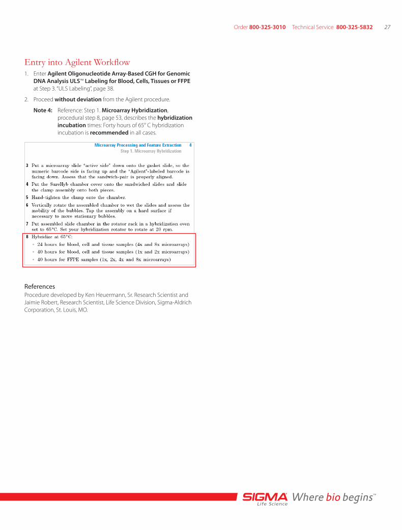
GenomePlex and Agilent 27-28
GenomePlex and Illumina 29-30
GenomePlex and NimbleGen-CGH 31-32
GenomePlex and NimbleGen-ChIP to Chip 33-34
Amplification Protocols 35
ChIP-CHIP Introduction 35
ChIP-CHIP Farhnam Lab Protocol 35-36
GenomePlex Whole Genome Amplification Kit (Cat No WGA1) 37
GenomePlex Complete Whole Genome Amplification Kit (Cat No WGA2) 37
GenomePlex Single Cell Whole Genome Amplification Kit (Cat No WGA4) 38
GenomePlex Tissue Whole Genome Amplification Kit (Cat No WGA5) 39-40
GenomePlex Whole Genome Reamplification Kit (Cat No WGA3) 41
Clean-Up Procedure for WGA Amplicons 42
GenomePlex Automation Resources 43
Troubleshooting Guide 44
Contact Information 44
FAQs 45
References 46-48
BiodiscoverWhole Genome Amplification Advisor
biomapping
Order 800-325-3010 Technical Service 800-325-5832 1
The availability of sufficient quantities of genomic DNA is crucial for numerous analyses used in the study of human and animal disorders In many cases the DNA source is limited or the available DNA is damaged or degraded preventing the researcher from performing crucial analyses
Genomic material is preserved in many ways including buccal swabs fresh tissue samples frozen tissue samples blood cards peripheral blood and formalin-fixed paraffin-embedded (FFPE) tissues Until recently the genetic information from some of these sources could only be amplified by gene specific polymerase chain reaction (PCR) Unfortunately there are drawbacks to gene specific PCR
The technique leads to rapid depletion of precious genomic material and only allows for analysis of specifically amplified genes An additional problem is that by only studying the amplified genes important genetic information essential for making prognostic decisions can go unnoticed As a result of the roadblocks presented by DNA starting material and the methods used to amplify DNA the desire to preserve complete genomic DNA samples led to the development of whole genome amplification (WGA) technology The technology emerged in the early 1990rsquos and quickly evolved into a field of its own within molecular cell biology WGA technology allows immortalization of a limited sample providing researchers with the capability to perform all desired analyses on one DNA sample
Introduction to Whole Genome Amplification
Unbiased amplification is desired and necessary for successful research employing whole genome amplification A WGA method must result in high quality representative samples every time The WGA method used should also be applicable to a wide spectrum of sources WGA methodology via PCR based on random fragmen-tation followed by the annealing of universal adapters not only results in high quality samples but it is also applicable to a variety of source materials This method provides a comparable yield to other WGA methods producing microgram quantities of DNA from as little as 1 nanogram of starting material however amplification is most successful with 10 nanograms of starting material yielding 5ndash10 micrograms of amplified product
The random fragmentation WGA method has been used to success-fully amplify genomic material from sources such as saliva whole blood blood card buccal swab commercially available human genomic DNA soil bacterial artificial chromosome formalin-fixed paraffin-embedded tissues from rat and human numerous plant sources and a variety of animal sources The range of this methodology appears to have no limit Random fragmentation is achieved by non-enzymatic digestioncleavage which leads to generation of a library of genomic fragments ranging in size from 100ndash1000 base pairs with an average size of approximately 400 base pairs The fragments are subsequently amplified with a limited number of PCR cycles by taking advantage of a robust DNA polymerase Annealing of universal adapters to the 5rsquo and 3rsquo ends of each of the fragments allows for specific priming with universal primers followed by amplification The use of adapter-specific primers and optimized DNA polymerase enzyme results in specific amplification of the desired product The GenomePlexreg Whole Genome Amplification Kit from Sigma-Aldrichreg utilizes the random fragmentation and annealing of adapters technique via PCR to generate high quality amplified genomic DNA from a wide variety of sources
Whole Genome Amplification allows for representative amplification of the whole genome preserving what was a limited sample and allowing for the analysis of any gene in the organism being studied Currently there are two different methodologies for performing WGA variations of PCR amplification and isothermal DNA amplification The PCR amplification category includes techniques to attach random primers degenerate primers and adaptors to genomic DNA frag-ments to create universal priming regions Whole genome ampli-fication emerged initially with degenerate oligonucleotide primer (DOP) PCR and primer extension pre-amplification (PEP) PCR which led to the development of the linker adaptor technique The linker adaptor technique takes advantage of the fragmentation of DNA by enzymatic and chemical means prior to the attachment of adaptors and amplification via PCR The second method for whole genome amplification isothermal DNA amplification utilizes multiple strand displacement (MSD) amplification using phi29 DNA polymerase
Both PCR amplification and isothermal DNA amplification method-ologies generate several micrograms of amplified DNA with as little as 1 nanogram of starting material The choice of methodology for WGA is often dictated by the source from which the DNA was obtained For instance isothermal DNA amplification using multiple strand displacement requires high quality DNA samples in excess of 2 kb in size for successful amplification Such DNA samples can be obtained from fresh tissues and blood samples but not from FFPE tissues or damaged DNA The PCR based methods are generally more tolerant of damaged DNA samples As a result PCR methods are applicable to a variety of sources
When implementing WGA it is critical to assess the entire research workflow and the desired results to assess which methodology may be more advantageous It is also important to remember that handling of the sample the DNA source the quantity and the quality of the DNA storage conditions and the extraction technique are all factors that can affect WGA directly or indirectly These factors should be taken into consideration when selecting the most appropriate WGA method Regardless of the methodology used to amplify the genomic material the ultimate result is that the amplified DNA is indistinguishable from the original genomic information
WGA Methodologies
Order 800-325-3010 Technical Service 800-325-5832 3
Fragmentation
Anneal primers
Extend
PCR amplify
2nd priming event
OmniPlex library formation
Amplified Genomic DNA
GenomePlexreg Whole Genome Amplification
Since its introduction to the market the GenomePlex technology has evolved into five powerful kits for whole genome amplification The GenomePlex Whole Genome Amplification Kit (Cat No WGA1) does not include a DNA polymerase providing researchers with the flexibility to use their polymerase of choice to proceed with amplification of the whole genome The GenomePlex Complete Whole Genome Amplification Kit (Cat No WGA2) includes a DNA polymerase mix designed for optimal amplification The GenomePlex Whole Genome Reamplification Kit (Cat No WGA3) allows for reamplification of products produced using any of the WGA kits resulting in an even higher yield of DNA The GenomePlex Single Cell WGA Kit (Cat No WGA4) was developed for use with single cells and includes an optimized cell lysis protocol which has been incorporated into the fragmentation step The newest member of the GenomePlex product family is the GenomePlex Tissue Whole Genome Amplification Kit (Cat No WGA5) This product allows whole genome amplification directly from formalin-fixed paraffin-embedded (FFPE) frozen RNAlaterreg-preserved or fresh tissue The kit includes optimized reagents for tissue disruption and cell lysis eliminating the need for tedious organic extractions to remove excess paraffin or DNA purification prior to amplification The GenomePlex technologies use the same amplification process whether using the WGA1 WGA2 WGA4 or the WGA5 Kit The processes involved in amplification of DNA include random fragmentation of input genomic DNA generation of the OmniPlexreg library and OmniPlex library amplification When amplification is complete it is strongly recommended to purify the amplified products Once WGA has been performed using WGA1 WGA2 WGA4 or WGA5 reamplification can be performed using WGA3 if desired
GenomePlex WGA Kit Process Overview
GenomePlexreg Whole Genome AmplificationOverview of the GenomePlex WGA Workflow
Random FragmentationFor successful whole genome amplification the input genomic DNA must be appropriately diluted with PCR grade water to a concentration of 1 ngmicrol The incubation step (4 minutes at 95 degC) denatures double stranded DNA into single stranded DNA and facilitates non-enzymatic fragmentation The incubation step is time and temperature sensitive and any deviation from the suggested incubation time and tempera ture may result in compromised amplification yields due to inappropriate fragmentation The result is genomic DNA that is randomly fragmented into overlapping segments The fragments range in size from 100ndash1000 base pairs with an average of about 400 base pairs The randomly fragmented DNA must be immediately processed to the next step generation of the OmniPlex library Unprocessed samples may degrade as the DNA fragments are not stable
Generation of the OmniPlexreg LibraryUpon addition of the preparation buffer that contains degenerate adapters and stabilization buffer the randomly fragmented DNA is incubated at 95 degC for 2 minutes The OmniPlex library is generated via a traditional PCR reaction This process takes a little over an hour The PCR reaction allows for annealing of universal adapters to 5rsquo and 3rsquo ends of each of the DNA fragments The universal adapters provide sites to which adapter-specific primers can anneal in order to facilitate amplification The OmniPlex library can immediately be amplified or it can be stored at ndash20 degC up to three days
Library AmplificationThe OmniPlex library is PCR amplified via a single proprietary primer The universal adapters at the 5rsquo and 3rsquo ends allow for priming to occur After 14 PCR cycles the OmniPlex Library will have been amplified 500ndash1000 fold in a total processing time of three hours generating 5ndash10 microg of amplified genomic DNA
Purification of Amplified ProductsIt is recommended to purify the amplified DNA with a PCR cleanup kit (Cat No NA1020) to ensure removal of salts and free nucleotides before quantitation or downstream processing GenomePlex amplified DNA can be stored at 2ndash8 degC for short periods of time but should be kept at ndash20 degC for long-term storage GenomePlex products can be used in any application using genomic DNA such as genotyping sequencing microarrays microsatellite analysis PCR comparative genomic hybridization (CGH) analysis TaqManreg assays or single nucleotide polymorphism (SNP) analysis GenomePlex Whole Genome Amplification technology can be applied in a wide range of fields including drug discovery environmental biology forensics and plant research
Reamplification with GenomePlexThe GenomePlex Whole Genome Reamplification Kit (Cat No WGA3) allows the user to obtain even higher yields of desired DNA without disturbing the original limited source of genomic DNA The products from WGA1 WGA2 WGA4 andor WGA5 can be reamplified using WGA3 WGA3 utilizes the same library amplification technology as WGA1 and WGA2 The procedure works optimally with at least 10 ng of input DNA Using less than 10 ng of input DNA may result in lower yields Additionally the products from WGA1 WGA2 WGA4 or WGA5 can be reamplified up to five cycles with minimal allele dropout producing an even higher yield It is important to note that a higher number of reamplification cycles may introduce slight bias representation
Order 800-325-3010 Technical Service 800-325-5832 5
Introduction The ultimate biological unit lies within a single cell Many biological disciplines have taken aim to elucidate the causes of cellular differentiation at this level The secret triggers that signal human maturation regeneration and genetic diseases all lie buried in a single cell that was originally part of the genetically clonal multicellular organism Despite careful work with sophisticated instrumentation available for the dissection of tissue samples several studies suggest that pooled cell samples thought to be homogenous are often composed of cells with quite different phenotypes (eg ref 1)
Development and commercialization of economical easier-to-use single cell tools have enabled more researchers to explore this novel area These include means to isolate single cells such as fluorescence activated cell sorting laser capture micro dissection optical tweezers and atomic force microscopy Once single cell samples were readily available applications such as fluorescent in situ hybridization or FISH2 and single cell PCR3 could be used to identify the differences between populations of single cells This has resulted in an explosion of work and speculation in the field of single-cell biology4-6
Adapting multicelltissue techniques to single cell study often have limited utility because of technical shortcomings ndash mostly problems related to sensitivity Despite the raw potential for single cell genomic analysis the field has been restricted to comparative analysis of relatively few genomic loci for large numbers of single-cell isolates Techniques such as FISH or single-cell PCR can be only used to probe a small number of DNA sequences before the cell is destroyed Likewise the small sample size of a single cell has so far allowed limited investigation of gene expression proteomic make up and the characterization of cell metabolites
Whole genome amplification (WGA) offers a means to overcome the above restrictions for single-cell genomic analyses WGA has been described as a non-specific amplification technique that affords an amplified product completely representative of the initial starting material Three different strategies for WGA have been described in the literature
Whole Genome Amplification for Single Cell Biology By Ernie Mueller and Chad Brueck Sigma-Aldrich Corporation St Louis MO USA
History Linker adapter PCR was first described in 19899 In this method the target DNA is digested with an appropriate restriction enzyme and then each end is ligated to an adaptor These known adaptor sequences are used to uniformly amplify each of the many DNA fragments representing the original sample The method relies on absolutely efficient ligation and unbiased amplification between the identical primed regions
Primer extension pre-amplification (PEP) PCR in contrast uses a set of random hexamers to prime template DNA10 The subsequent thermal cycling conditions use very low (permissive) annealing temperatures and fifty or more cycles to create a series of fragments representing the original input DNA Bias in the resulting PEP PCR product is due to non-uniformity of random hexamer annealing and extension -DNA sections with infrequent or distant priming events tend to be discriminated against in this method These shortfalls were largely overcome with multiple strand displacement (MSD) amplification11 The MSD technique employs a unique and highly processive mesophillic DNA polymerase phi29 The resulting product consists of long ~10-50kb fragments but good amplification and representation
Finally degenerative oligonucleotide primer (DOP) PCR also described as arbitrary PCR relies on a set of oligos with a random 3rsquo-end and partially fixed 5rsquo-sequence12 These primers are designed
to anneal relatively evenly throughout the DNA sample Once extended by a polymerase these products are amplified using oligos targeting their fixed sequences Primer design is critical for this technique ndash the oligo must bind relatively evenly throughout the DNA sequence but not bind to other oligonucleotides This method has also been successfully applied to give representative samples
The latter phenomenon is dependent on the method and the fact that damaged DNA can render certain loci unamplifiable
WGA methods that generate long amplicons like MSD can be less robust because priming events are necessarily few and therefore any error in a long amplicon causes a relatively large loss of information WGA that generates short amplicons such as PEP PCR linker-adaptor amplification and DOP PCR lose less information in such circumstances
Both DOP PCR and MSD amplification are now available in commercial molecular biology kits but only the GenomePlexreg kit offered by Sigma-Aldrich has been developed specifically for single cell applications The technique builds on a product line that was introduced first by Rubicon and acquired in the fall of 2004 by Sigma-Aldrich as the sole licensee and distributor Since that time Sigma-Aldrich has developed four WGA related products including the original kit (WGA1) a kit with an optimized DNA polymerase (WGA2) a kit to reamplify WGA DNA (WGA3) and a kit for single cell applications (WGA4) At the heart of this product line is a PCR-based WGA method that employs degenerate oligonucleotides coupled with universal adaptors in a combination of PEP and DOP amplification methods GenomePlex can faithfully amplify ten nanograms of genomic DNA in about three hours
The single cell WGA kit released in February 2006 is able to produce a million-fold amplification of a flow-sorted or laser micro-captured single cell resulting in approximately 5microg of final yield The single
Phi29 DNA Polymerase Extension Process
Random DNA hexamers act as primers on Genomic DNA target Isothermal amplification conditions enable the extension of new DNA templates with the addition of dNTPs and phi29 DNA polymerase
Single Primer PCR Amplifies GenomePlex Library
Traditional PCR annealing and extension are essentially single primer PCR amplification once the GenomePlex library has been created A pool of amplicons all containing a universal priming site are cycled using a single amplification primer
Single Cell Analysis Each of these techniques has been applied to the problem of amplifying the genetic material in a single cell and has met with some success PEP PCR was the first to be applied to single cell WGA and was successfully applied in several subsequent applications13-14 A variant of the DOP PCR developed by Rubicon Genomics was used to amplify single chromosomes15 a feat very shortly followed by the use of a linker-adaptor PCR method to also completely amplify a single chromosome16 Finally MSD with phi29 was used to amplify a series of single cells817
WGA methods differ in two respects the amount of bias in the product when using limiting amounts of input template and the quality requirements for the input template The former issue which in single cell applications manifests itself as apparent loss of information or allelic drop out (ADO) is thought to be due to inequities of local distribution of the reagents near the target28
Whole Genome Amplification for Single Cell Biology
Order 800-325-3010 Technical Service 800-325-5832 7
cell method differs from the original kit in three ways First of all WGA4 includes a cell lysis protocol that combines an efficient lysis procedure with the original fragmentation Secondly the subsequent isothermal library preparation steps use a newly optimized primer that gives better coverage at low template but maintains low self-annealing and thus undetectable primer elongation Finally the amplification cycling protocol has been modified to use more cycles allowing a greater total amplification The single cell WGA product was tested using 96 separate SYBRreg Green qPCR loci and has a demonstrated 25-33 random Allelic Drop Out (ADO) The single cell WGA work was validated with multiple beta-testers using STR analysis RPLF assays quantitative PCR and microarray analysis
Advances in single cell WGA will allow the researcher to uncover the contribution of genomics to single cell biology Specifically cancer and drug discovery research within genomics shows the greatest
MicroArray Workflow A DNA microarray is a proven technology used by molecular biology researchers to study a large number of genetic signatures at one time It consists of an arrayed series of thousands of microscopic spots of DNA oligos called features each containing a specific DNA sequence known as a probe These can be a short section of a gene or other DNA element that are used to a DNA sample under high-stringency conditions Probe-target hybridization is usually detected and quanti-fied by detection of flourescent probe or chemiluminescence labeled targets to determine relative abundance of nucleic acid sequences in the target Arrays can contain millions of probes accelerating many types of investigation
potential for opportunity Chromosomal aberrations as a result of cancer could be better catalogued when comparing a single cancerous cell to its normal counterpart In addition comparing single cells from the lsquotreatedrsquo population to the lsquountreatedrsquo to evaluate genomic effects can be used to screen drug candidates
Understanding differences at the level of a single cell is the ultimate goal of biology New commercialized techniques such as single cell WGA are opening a new frontier for further study Considerable work has already been accomplished toward the sensitive unbiased amplification of single cell RNA to allow for single cell gene expression722-27 and this area has already seen the development of commercialized kits to respond to this customer need As researchers continue to find sensitive means to explore epigenetics proteomics18-19 metabolomics and cell signaling20-21 the whole world of single cell biology will be revealed
In standard microarrays the probes are attached to a solid surface by a covalent bond to a chemical The solid surface can be solid glass or a silicon chip Other microarray platforms use microscopic beads instead of the large solid support
DNA microarrays can be used to measure changes in gene expres-sion levels to detect single nucleotide polymorphisms (SNPs) or to genotype or resequence mutant genomes
Extraction Protocols 9
Blood Card 9
Whole Blood 10-11
SerumPlasma 12-13
Buccal Swab 14
Plant Tissues 15
Formalin-fixed Paraffin-embedded (FFPE) Tissues 16-17
Qualitative Multiplex PCR Assay 18-19
Animal Tissues 20
Saliva 21
Soil 22
MicroArray Labeling Protocols 23
GenomePlexreg and Affymetrix 23-24
GenomePlex and Affymetrix - ChIP to Chip 25-26
GenomePlex and Agilent 27-28
GenomePlex and Illumina 29-30
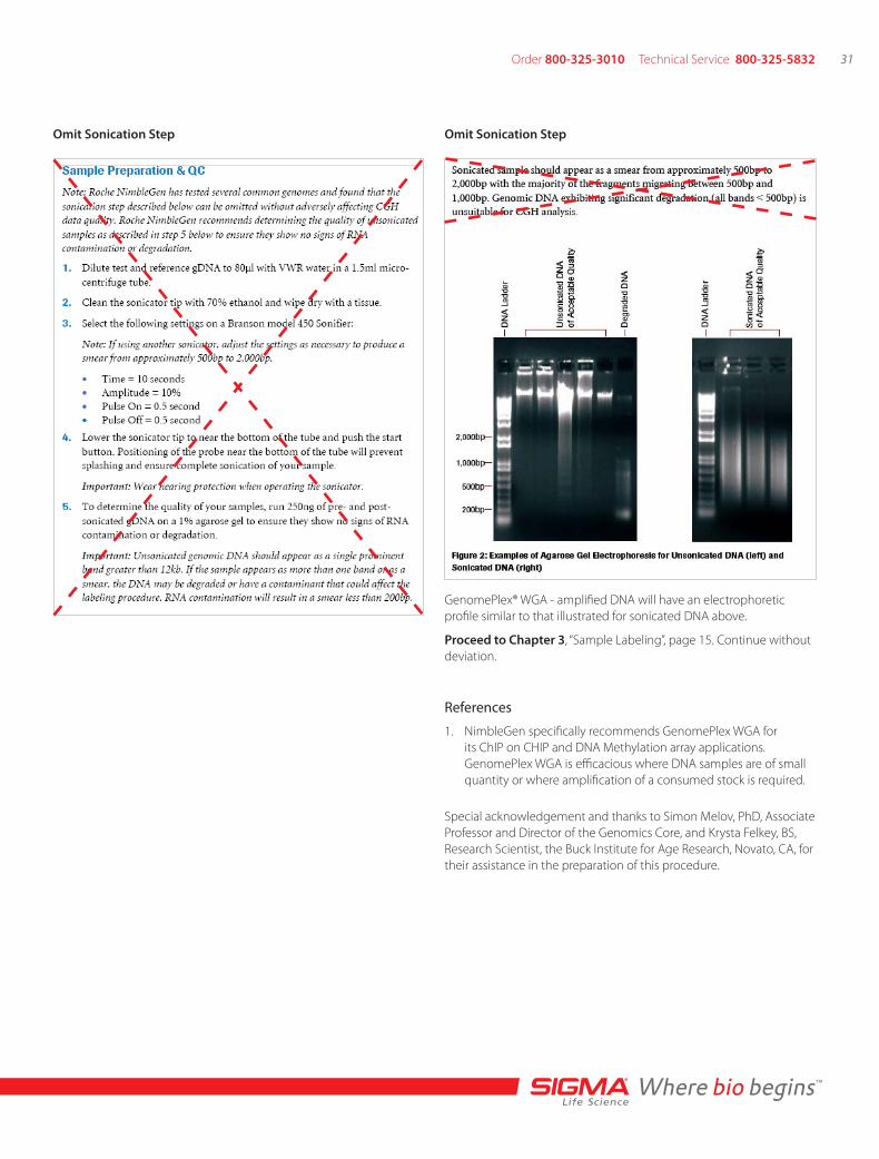
GenomePlex and NimbleGen-CGH 31-32
GenomePlex and NimbleGen-ChIP to Chip 33-34
Amplification Protocols 35
ChIP-chip Introduction 35
ChIP-chip Farhnam Lab Protocol 35-36
GenomePlex Whole Genome Amplification Kit (Cat No WGA1) 37
GenomePlex Complete Whole Genome Amplification Kit (Cat No WGA2) 37
GenomePlex Single Cell Whole Genome Amplification Kit (Cat No WGA4) 38
GenomePlex Tissue Whole Genome Amplification Kit (Cat No WGA5) 39-40
GenomePlex Whole Genome Reamplification Kit (Cat No WGA3) 41
Clean-Up Procedure for WGA Amplicons 42
GenomePlex Automation Resources 43
Protocols
Order 800-325-3010 Technical Service 800-325-5832 9
Blood cards provide the convenience of archiving small volumes of blood However many times genomic DNA from these samples is limited which may hinder the researcherrsquos ability to perform downstream analysis This protocol provides a simple and convenient method to extract genomic DNA from a blood card Once the DNA has been extracted it can then be amplified using the amplification protocol on page 21
Required ProductsGenElutetrade Blood Genomic DNA Kit (bull Cat No NA2000)
Materials to be Supplied by the UserBlood cardbull15 ml microcentrifuge tubesbullEthanol (bull Cat No E7023)
Microcentrifuge (with rotor for 2 ml tubes)bullWater molecular biology reagent (bull Cat No W4502)
55 degC water bath or heat blockbullExtraction of DNA from a Blood CardThe GenElute Blood Genomic DNA Kit (Cat No NA2000) is recom-mended for this procedure
1 Cut a disc from a dried blood card (200 microl spotted) into several 2 mm by 2 mm pieces and place the pieces into a 15 ml microcentrifuge tube
2 Add 40 microl Proteinase K and 10 ml Resuspension Solution
3 Add 300 microl of Lysis Solution C and vortex thoroughly for 15 seconds
4 Incubate at 55 degC for 10 minutes
5 After the incubation transfer the liquid (discard blood card remaining in the microcentrifuge tube) to a 15 ml conical tube
6 Add 500 microl of Column Preparation Solution to the GenElute Mini-prep Binding Column (red o-ring) and centrifuge at 12000 times g for 1 minute
7 Discard the flow-through liquid
Note The Column Preparation Solution maximizes binding of DNA to the membrane resulting in more consistent yields
8 Add 900 microl of 95ndash100 ethanol to the lysate in the 15 ml conical tube and mix thoroughly by vortexing 5ndash10 seconds
9 Transfer the contents of the tube into the treated column from step 6 Centrifuge at ge6500 times g for 1 minute Repeat until all of the lysate has been passed through the column
10 Discard the collection tube and flow-through Place the column in a new 2 ml collection tube
11 Add 500 microl of Prewash Solution (be sure to dilute with ethanol prior to first use) and centrifuge for 1 minute at ge6500 times g
12 Discard the collection tube containing the flow-through and place the binding column into a new 2 ml collection tube
13 Add 500 microl of Wash Solution (be sure to dilute with ethanol prior to first use) to the binding column and centrifuge at maximum speed (12000ndash16000 times g) for 3 minutes to dry the binding column
14 Pipette 400 microl of Elution Solution onto the column and centrifuge for 1 minute at ge6500 times g to elute the DNA
15 Store the eluted DNA at ndash20 degC or proceed to the amplification step
Amplification ProtocolSee page 21
Extraction ProtocolmdashBlood Card
Application Data
Amplified Human Genomic DNA from Blood Card
Products were amplified using the GenomePlexreg Whole Genome Amplification Kit (Cat No WGA1) from Sigma Supplier Arsquos kit and Supplier Qrsquos kit Products were resolved on a 15 agarose gel 5 microl of amplified product was added to each well The products amplified using GenomePlex technology were of a smaller molecular weight as shown on the gel when compared to Supplier A and Q This is due to the random fragmentation of genomic DNA prior to amplification Sigmarsquos amplified products are specific and there is no amplicon visible in the negative control (lane 2) indicating that only the desired genomic DNA is amplified Both Suppliers A and Q yield a nonspecific signal in the negative control which is equal in size and intensity to the signal for the suppliersrsquo positive control
Lane 1mdash1 kb Marker
Lane 2mdashSigma Positive Control
Lane 3mdashSigma Negative Control
Lane 4mdashSigma Blood Card
Lane 5mdashSigma Blood Card
Lane 6mdashSupplier A Positive Control
Lane 7mdashSupplier A Negative Control
Lane 8mdashSupplier A Blood Card
Lane 9mdashSupplier A Blood Card
Lane 10mdashSupplier Q Positive Control
Lane 11mdashSupplier Q Negative Control
Lane 12mdashSupplier Q Blood Card
Lane 13mdashSupplier Q Blood Card
Lane 14mdash1 kb Marker
Whole blood is a common source of material used to perform genetic analysis Many times genomic DNA isolated from whole blood samples is of low yield This can hinder the researcherrsquos ability to perform downstream analysis The following protocol is a simple method to isolate DNA from fresh or aged whole blood products Once the DNA is isolated it can be amplified using the GenomePlexreg Whole Genome Amplification protocol on page 21
Required ProductsGenElutetrade Blood Genomic DNA Kit (bull Cat No NA2000)
Materials to be Supplied by the UserWhole bloodbull15 ml microcentrifuge tubesbullEthanol (bull Cat No E7023)
Microcentrifuge (with rotor for 2 ml tubes)bullWater molecular biology reagent (bull Cat No W4502)
55 degC water bath or heat blockbullExtraction of DNA from Whole BloodThe GenElute Blood Genomic DNA Kit (Cat No NA2000) is recom-mended for this process
1 Place 20 microl of Proteinase K into a 15 ml microcentrifuge tube and add 200 microl of whole blood to the tube
2 Add 200 microl of Lysis Solution C and vortex thoroughly for 15 seconds
3 Incubate at 55 degC for 10 minutes
4 Add 500 microl of Column Preparation Solution to the GenElute Mini-prep Binding Column (red o-ring) and centrifuge at 12000 times g for 1 minute
Note The Column Preparation Solution maximizes binding of DNA to the membrane resulting in more consistent yields
5 Discard the flow-through liquid
6 Add 200 microl of 95ndash100 ethanol to the lysate from step 3 and mix thoroughly by vortexing 5ndash10 seconds
7 Transfer the entire contents of the tube into the treated column (from step 4) Centrifuge at ge6500 times g for 1 minute
8 Discard the collection tube and flow-through Place the column into a new 2 ml collection tube
9 Add 500 microl of Prewash Solution (be sure to dilute with ethanol prior to first use) and centrifuge at ge6500 times g for 1 minute
10 Discard the collection tube containing the flow-through and place the column into a new 2 ml collection tube
11 Add 500 microl of Wash Solution (be sure to dilute with ethanol prior to first use) to the binding column and centrifuge at maximum speed (12000ndash16000 times g) for 3 minutes to dry the binding column
12 Pipette 200 microl of Elution Solution onto the column and centrifuge for 1 minute at ge6500 times g to elute the DNA
13 Store the eluted DNA at ndash20 degC or proceed with the amplification step
Application Data
Human Whole Blood Amplified DNA-Comparison Data
Products were amplified using the GenomePlex Whole Genome Amplification Kit (Cat No WGA1) from Sigma Supplier Arsquos kit and Supplier Qrsquos kit Products were resolved on a 15 agarose gel 5 microl of amplified product was added to each well The products amplified using GenomePlex technology were of a smaller molecular weight as shown on the gel when compared to Supplier A and Q This is due to the random fragmentation of genomic DNA prior to amplification Sigmarsquos amplified products are specific and there is no amplicon visible in the negative control (lane 2) indicating that only the desired genomic DNA is amplified Both Suppliers A and Q yield a nonspecific signal in the negative control which is equal in size and intensity to the signal for the suppliersrsquo positive control
Extraction ProtocolmdashWhole Blood
Lane 1mdash1 kb Marker
Lane 2mdashSigma Positive Control
Lane 3mdashSigma Negative Control
Lane 4mdashSigma Blood Card
Lane 5mdashSigma Blood Card
Lane 6mdashSupplier A Positive Control
Lane 7mdashSupplier A Negative Control
Lane 8mdashSupplier A Blood Card
Lane 9mdashSupplier A Blood Card
Lane 10mdashSupplier Q Positive Control
Lane 11mdashSupplier Q Negative Control
Lane 12mdashSupplier Q Blood Card
Lane 13mdashSupplier Q Blood Card
Lane 14mdash1 kb Marker
Order 800-325-3010 Technical Service 800-325-5832 11
Extraction ProtocolmdashWhole Blood
HeterozygousTC
HomozygousAL 1CC
HomozygousAL 2TT
gD
NA
pro
du
ct after WG
A
Star
tin
g g
DN
A b
efo
re W
GA
Performance of DNA Amplified with GenomePlexreg WGA Identical to Non-Amplified DNA
Genomic DNA was extracted from a whole blood sample using the GenElutetrade Blood Genomic DNA Kit (Cat No NA2000) 10 ng of genomic DNA was amplified using the GenomePlex WGA Kit (Cat No WGA1) followed by purification using the GenElute PCR Clean-up Kit (Cat No NA1020) SNP genotyping analysis was performed on non-amplified DNA and GenomePlex amplified DNA GenomePlex WGA DNA genotyping provided the same accuracy and quality of scores to non-amplified DNA indicating that the amplification process did not alter the original genomic sequence
Whole genome amplification (WGA) of plasma and serum DNA presents a unique challenge due to the small amount of nucleic acid in such samples One must use a robust DNA purification scheme capable of removing inhibitory substances while at the same time concentrating the DNA within the sample The use of GenElutetrade Blood Genomic DNA Kit in combination with one of two modified WGA protocols has been shown to work well with DNA isolated from serum or plasma Both procedures typically produce approximately 5 microg of WGA product Old serum and plasma samples may contain degraded DNA and therefore may not be suitable for downstream applications such as WGA The WGA procedures described below are modifications of the WGA2 and WGA4 protocols The procedures work equally well each typically producing approximately 5 microg of WGA product
Required ProductsGenElutetrade Blood Genomic DNA Kit (bull Cat No NA2000)
Materials to be Supplied by the UserSerum or Plasmabull15 ml microcentrifuge tubesbullEthanol (bull Cat No E7023)
Microcentrifuge (with rotor for 2 ml tubes)bullWater molecular biology reagent (bull Cat No W4502)
55 degC water bath or heat blockbullExtraction of DNA from Serum or PlasmaThe GenElute Blood Genomic DNA Kit (Cat No NA2000) is recommended for this process It is recommended to start with 01 ml to 1 ml serum or plasma The protocol below is written for 01 ml to 02 ml serum or plasma Scale GenElute reagents proportionally if using more than 02 ml serum or plasma
1 Place 20 microl of Proteinase K into a 15 ml microcentrifuge tube and add 200 microl of plasma or serum to the tube
2 Add 200 microl of Lysis Solution C and vortex thoroughly for 15 seconds
3 Incubate at 55 degC for 10 minutes
4 Add 500 microl of Column Preparation Solution to the GenElute Mini-prep Binding Column (red o-ring) and centrifuge at 12000 times g for 1 minute
Note The Column Preparation Solution maximizes binding of DNA to the membrane resulting in more consistent yields
5 Discard the flow-through liquid
6 Add 200 microl of 95ndash100 ethanol to the lysate from step 3 and mix thoroughly by vortexing 5ndash10 seconds
7 Transfer the entire contents of the tube into the treated column from step 4 Centrifuge at 6500 times g for 1 minute
8 Discard the collection tube and flow-through Place the column into a new 2 ml collection tube
9 Add 500 microl of Prewash Solution (be sure to dilute with ethanol prior to first use) and centrifuge at 6500 times g for 1 minute
10 Discard the collection tube containing the flow-through and place the column into a new 2 ml collection tube
11 Add 500 microl of Wash Solution (be sure to dilute with ethanol prior to first use) to the binding column and centrifuge at maximum speed (12000ndash16000 times g) for 3 minutes to dry the binding column
12 Pipette 50 microl of Elution Solution onto the column and centrifuge for 1 minute at 6500 times g to elute the DNA
13 Store the eluted DNA at ndash20 degC or proceed to the amplification step with WGA2 or WGA4
Amplification with WGA21 Combine 10 microl of serum or plasma DNA purified as outlined above
and 1 microl of 10times Fragmentation Buffer to a PCR tube or multiwell stripplate
Note Use 10 microl of DNA regardless of concentration since spectro-photometric quantitation of very dilute DNA is not accurate 10 microl is the maximum volume that can be used in WGA
2 Continue with step 3 of the WGA2 protocol however perform 25 amplification cycles in step 13 as opposed to 14 cycles
Amplification with WGA41 Combine 10 microl of serum or plasma DNA purified as outlined above
and 1 microl of 10times Single Cell Lysis amp Fragmentation Buffer to a PCR tube or multiwell stripplate Mix thoroughly
Note Use 10 microl of DNA regardless of concentration since spectro-photometric quantitation of very dilute DNA is not accurate 10 microl is the maximum volume that can be used in WGA
2 Place the tubeplate in a thermal block or cycler at 95 degC for EXACTLY 4 minutes Immediately cool on ice Spin down sample prior to proceeding to Library Preparation
Note This incubation is very time sensitive Any deviation may alter results
3 Continue with the Library Preparation section (step 6) of the WGA4 technical bulletin
Whole Genome Amplification from Serum or Plasma
Order 800-325-3010 Technical Service 800-325-5832 13
This protocol provides a simple and convenient method to isolate amplify and purify genomic DNA from buccal swabs Buccal swabs are a convenient method of acquiring a DNA sample Once the DNA is isolated using the following extraction protocol it can be ampli-fied using the GenomePlexreg Whole Genome Amplification Protocol on page 21 to provide enough DNA for numerous downstream applications
Required ProductsGenElutetrade Mammalian Genomic DNA Miniprep Kit bull(Cat No G1N10)
Materials to be Supplied by the UserBuccal swabbull15 ml microcentrifuge tubesbullEthanol (bull Cat No E7023)
Microcentrifuge (with rotor for 2 ml tubes)bullWater molecular biology reagent (bull Cat No W4502)
55 degC water bath or heat blockbullExtraction of DNA from Buccal SwabIt is recommended to use the GenElute Mammalian Genomic DNA Miniprep Kit (Cat No G1N10) for this process
1 Dry the collected swabs at room temperature for 15 minutes
2 Add 280 microl of Lysis Solution T and 20 microl of Proteinase K Insert the swab and gently spin Cap the tube and mix by vortexing
3 Incubate the sample at 55 degC for 20 minutes with occasional vortexing
4 Add 200 microl of Lysis Solution C and vortex thoroughly for 15 seconds
5 Incubate at 70 degC for 10 minutes
6 Add 500 microl of Column Preparation Solution to each GenElute Mini-prep Binding Column (red o-ring) and centrifuge at 12000 times g for 1 minute Discard the flow-though liquid
Note The Column Preparation Solution maximizes binding of DNA to the membrane resulting in more consistent yields
7 Add 200 microl of 95ndash100 ethanol to the lysate from step 5
8 Mix thoroughly by vortexing and add the entire contents of the tube into the binding column
9 Centrifuge at ge6500 times g for 1 minute
10 Discard the collection tube containing the flow-through and place the binding column in a new 2 ml collection tube
11 Add 500 microl of Wash Solution (be sure to dilute with ethanol prior to first use) and centrifuge at ge6500 times g for 1 minute
12 Discard the collection tube and flow-through and place the binding column in a new 2 ml collection tube
13 Add another 500 microl of Wash Solution to the binding column and centrifuge at maximum speed (12000ndash16000 times g) for 3 minutes to dry the binding column
14 Pipette 200 microl of Elution Solution onto the binding column and centrifuge for 1 minute at ge6500 times g
15 Store the eluted DNA at ndash20 degC or proceed to the amplification step
Application Data
GenomePlex Whole Genome Amplification Performed on a Buccal Swab Sample
Amplified Sigma products are visualized on 15 agarose gel 5 microl of amplified product was loaded per well The GenomePlex amplified products result in an average size of 400 bp The smear pattern varies by source as shown on the gel
Extraction ProtocolmdashBuccal Swab
Lane 1mdash1 kb Ladder
Lane 2mdashBlood
Lane 3mdashPlant
Lane 4mdashBuccal Swab
Lane 5mdashSoil
Lane 6mdashPositive Control
Lane 7mdash1 kb Ladder
Extracting DNA from plant tissue is a complicated process due to the tough cell wall that surrounds most plant cells Genomic DNA from plant material can be damaged during the extraction process resulting in low yields of high quality genomic material As a result the researcherrsquos ability to perform downstream analysis is challenged GenomePlex Whole Genome Amplification has been used to amplify genomic DNA from soybean corn tomato purple coneflower and ginseng
Required ProductsGenElutetrade Plant Genomic DNA Miniprep (bull Cat No G2N10)
Materials to be Supplied by the UserPlant samplesbull15 ml microcentrifuge tubes (bull Cat No T9661)
Ethanol (bull Cat No E7023)
Microcentrifuge (with rotor for 2 ml tubes)bullWater molecular biology reagent (bull Cat No W4502)
55 degC water bath or heat blockbullExtraction of DNA from Plant TissuesIt is recommended to use the Sigma GenElute Plant Genomic DNA Miniprep Kit (Cat No G2N10) for this procedure
1 Grind approximately 50 mg leaf punch into a fine powder with liquid nitrogen Keep the sample on ice for immediate use or freeze at ndash70 degC
2 Add 350 microl of Lysis Solution (Part A) and 50 microl of Lysis Solution (Part B) and thoroughly mix by vortexing A white precipitate will form upon the addition of Lysis Solution Part B
3 Incubate the mixture at 65 degC for 10 minutes with occasional inversion to dissolve the precipitate
4 Add 130 microl of Precipitation Solution mix by inversion and place the sample on ice for 5 minutes
5 Centrifuge at maximum speed (12000ndash16000 times g) for 5 minutes to pellet the cellular debris proteins and polysaccharides
6 Carefully pipette the supernatant onto a GenElute Filtration Column (blue insert with a 2 ml collection tube)
7 Centrifuge at maximum speed for 1 minute Discard the Filtration Column and retain the collection tube
8 Add 700 microl of Binding Solution directly to the flow-through (liquid from step 7) Mix thoroughly by inversion
9 Insert the GenElute Miniprep Binding Column (red o-ring) into the provided microcentrifuge tube
10 Add 500 microl of the Column Preparation Solution to each Miniprep Column and centrifuge at 12000 times g for 1 minute Discard the flow-through liquid
11 Pipette 700 microl flow-through (from step 8) onto the Miniprep Column prepared in the previous step
12 Centrifuge at maximum speed for 1 minute and discard the flow-through
13 Apply the remaining lysate (from step 8) and repeat centrifugation for 1 minute at maximum speed and discard the flow-through
14 Place the Binding Column in a fresh 2 ml collection tube and apply 500 microl diluted Wash Solution to the column (be sure to add ethanol to the Wash Solution Concentrate prior to first time use)
15 Centrifuge at maximum speed for 1 minute Discard flow-through and retain the collection tube
16 Add another 500 microl of diluted Wash Solution to the column and centrifuge at maximum speed for 3 minutes to dry the column
17 Transfer the binding column to a fresh 2 ml collection tube
18 Apply 100 microl of pre-warmed (65 degC) Elution Solution to the column and centrifuge at maximum speed for 1 minute Repeat the elution
19 Store the eluted DNA at ndash20 degC or proceed to the amplification step
Extraction ProtocolmdashPlant Tissues
Application Data
GenomePlexreg Whole Genome Amplification Performed on Plant Samples
Amplified Sigma products are resolved on a 15 agarose gel 5 microl of amplified product was loaded per well The GenomePlex amplified products result in an average size of 400 bp The smear pattern varies by source as shown on the gel Products amplified using GenomePlex technology are specific as evidenced by the lack of signal in the negative control (lane 3)
Lane 1mdash1 kb Marker
Lane 2mdashPositive Control
Lane 3mdashNegative Control
Lane 4mdashCorn
Lane 5mdashGinseng
Lane 6mdashPurple Cornflower
Lane 7mdashTomato
Lane 8mdashSoybean
Order 800-325-3010 Technical Service 800-325-5832 15
Archived Formalin-fixed Paraffin-embedded (FFPE) tissue samples are invaluable resources for profiling gene expression and studying a variety of diseases Since the archived DNA is usually available in limited quantities amplification of the samples is essential Amplifying the FFPE tissue can be a difficult task due to the damaged template that results from the archiving process This protocol provides a convenient method to purify and amplify genomic DNA from FFPE tissue GenomePlexreg Whole Genome Amplification has been used to amplify genomic DNA from rat and human FFPE tissue samples
Note The protocol below describes the process of DNA purification and subsequent whole genome amplification We have dramatically streamlined this process with our GenomePlex Tissue Whole Genome Amplification Kit (Cat No WGA5) This kit includes optimized reagents for tissue disruption and cell lysis eliminating the need for tedious organic extractions to remove excess paraffin or DNA purification prior to amplification For more information on WGA5 Amplification Protocol see p 23
Required ProductsGenElutetrade Mammalian Genomic DNA Miniprep Kit bull(Cat No G1N10)
Materials to be Supplied by the UserFFPE tissuebull37 degC water bath or heat blockbullXylenebull55 degC water bath or heat blockbullEthanol (bull Cat No E7023)
70 degC water bath or heat blockbullWater molecular biology reagent (bull Cat No W4502)
Microcentrifuge (with rotor for 2 ml tubes)bullExtraction of DNA from Formalin-Fixed Paraffin-Embedded (FFPE) TissueThis protocol was performed on rat liver tissue The GenElute Mammalian Genomic DNA Miniprep Kit (Cat No G1N10) is recommended for this procedure
1 Place a small section (20 mg) of paraffin-embedded tissue in a 2 ml microcentrifuge tube
2 Add 1200 microl of xylene and vortex for 30 seconds
3 Centrifuge at full speed for 5 minutes at room temperature
4 Remove supernatant by pipetting Do not remove any of the pellet
5 Add 1200 microl of ethanol to the pellet to remove the residual xylene Mix by vortexing
6 Centrifuge at full speed for 5 minutes at room temperature
7 Carefully remove the ethanol by pipetting Do not remove any of the pellet
8 Repeat steps 5ndash7 again
9 Incubate the open microcentrifuge tube at 37 degC for 10ndash15 minutes to remove any residual ethanol by evaporation
10 Digest Tissue Resuspend the tissue pellet in 180 microl of Lysis Solution T
11 Add 20 microl of Proteinase K Mix by vortexing Incubate at 55 degC overnight or until the tissue is completely lysed Vortex occasionally during incubation
Optional RNase treatment If residual RNA is a concern add 20 microl of RNase A solution and incubate at room temperature for 2 minutes
12 Lyse cells Vortex for 15 seconds Add 200 microl of Lysis Solution C to the sample Vortex thoroughly as a homogenous mixture is essential for efficient lysis Incubate at 70 degC for 10 minutes
13 Prepare column Add 500 microl of the Column Preparation Solution to each pre-assembled GenElute Miniprep Binding Column and centrifuge at 12000 times g for 1 minute
14 Prepare for binding Add 200 microl of ethanol to the lysed sample and mix by vortexing
15 Load lysate Transfer the entire contents of the sample tube into the treated binding column from step 14 Centrifuge at ge6500 times g for 1 minute Discard the collection tube containing the flow-through liquid and place the binding column in a new 2 ml collection tube
16 First wash Prior to first use dilute the Wash Solution Concentrate with ethanol as described under preparation instructions Add 500 microl of Wash Solution to the binding column and centrifuge for 1 minute at ge6500 times g Discard the collection tube containing flow-through liquid and place the binding column in a new 2 ml collection tube
17 Second wash Add another 500 microl of Wash Solution to the binding column and centrifuge for 3 minutes at maximum speed (12000ndash18000 times g) to dry the binding column It is crucial that the binding column is free of ethanol before eluting DNA off the column Centrifuge the column for an additional minute if residual ethanol is visible Discard the collection tube containing the flow- through liquid and place the binding column in a new 2 ml collection tube
Extraction ProtocolmdashFormalin-Fixed Paraffin-Embedded (FFPE) Tissue and Diagnostic
18 Elute DNA Pipette 200 microl of the Elution Solution directly into the center of the binding column and incubate at room temperature for 5 minutes Centrifuge for 1 minute at ge6500 times g to elute the DNA
19 Store DNA samples at ndash20 degC
Note This protocol can be performed without using xylene starting with step number 10 As a result of omitting the xylene treatment step the amount of DNA will decrease by approximately 50 when compared to the protocol with a xylene step
Application Data
GenomePlexreg Whole Genome Amplification FFPE Tissue
Products were amplified using the GenomePlex Whole Genome Amplification Kit (Cat No WGA1) from Sigma Supplier A and Supplier Q Products were resolved on a 15 agarose gel 5 microl of amplified product was added to each well The products amplified using GenomePlex technology were of a smaller molecular weight as shown on the gel when compared to Supplier A and Q This is due to the random fragmentation of genomic DNA prior to amplification Sigmarsquos amplified products are specific and there is no amplicon visible in the negative control (lane 2) indicating that only the desired genomic DNA is amplified Both Suppliers A and Q yield a nonspecific signal in the negative control which is equal in size and intensity to the signal for the suppliersrsquo positive control
Nu
mb
er o
f C
ycle
s minus
Ct
Val
ues
Nu
mb
er o
f C
ycle
s minus
Ct
Val
ues
Loci
num
ber
Prim
er se
t
0
25
20
15
10
5
08 7 6 5
4 32
11
23
45
67
8910
1112
Sigm
a minus
10 n
gSig
ma
minus 10
0 ng
Supp
lier A
minus 0
ng
Supp
lier A
minus 1
0 ng
Supp
lier A
minus 1
00 n
g
Supp
lier Q
minus 0
ng
Supp
lier Q
minus 1
0 ng
Supp
lier Q
minus 1
00 n
g
Performance of FFPE DNA Amplified with GenomePlex WGA
DNA was extracted from a sample of formalin-fixed paraffin-embedded rat liver 10 ng and 100 ng of genomic DNA was amplified using GenomePlex Complete WGA Kit (Cat No WGA2) followed by purification using the GenElutetrade PCR Clean-up Kit (Cat No NA1020) Quantitative PCR (45 cycles) was performed on the WGA reaction using 12 different primer sets GenomePlex demonstrates ~1000 fold (10 Ct) better representation compared to suppliers
Lane 1mdash1 kb Marker
Lane 2mdashSigma Negative Control
Lane 3mdashSigma 10 ng input DNA
Lane 4mdashSigma 100 ng input DNA
Lane 5mdashSupplier A Negative Control
Lane 6mdashSupplier A 10 ng input DNA
Lane 7mdashSupplier A 100 ng input DNA
Lane 8mdashSupplier Q Negative Control
Lane 9mdashSupplier Q 10 ng input DNA
Lane 10mdashSupplier Q 100 ng input DNA
Order 800-325-3010 Technical Service 800-325-5832 17
Qualitative Multiplex PCR Assay for Assessing DNA Quality from FFPE Tissues and Other Sources of DamagedThe assessment of DNA quality is a crucial first step in acquiring meaningful data from formalin-fixed paraffin-embedded (FFPE) tissues and other sources of damaged DNA1 Formalin reacts with nucleic acids to cause irreversible damage resulting in DNA samples of poor quality that may not work in downstream processes2-3 To address this issue wersquove developed a simple qualitative gel-based multiplex PCR assay that can be used to determine DNA quality prior to performing tedious and expensive downstream processes such as array-based comparative genomic hybridization (aCGH) The assay consists of five primer sets derived from the NCBI UniSTS database that amplify products ranging from 132 bp to 295 bp (see table below) Some or all of these products will fail to amplify as DNA sample quality fades allowing the classification of genomic DNA quality based on the number and size of fragments amplified with high quality genomic DNA producing all five amplicons (see figure 1) This assay cannot predict the usefulness of damaged DNA without prior validation work By performing correlative experiments to empirically determine a quality threshold one can ensure the repeatability of their downstream experiments For example some applications may require that all five amplicons be produced in order to predict a successful outcome where as others such as qPCR may only require the presence of a single band In this way one can compare samples from different sources and repeatability of experiments is ensured
Materials to be Supplied by the UserWater molecular biology reagent (bull Cat No W4502)
25 mM MgClbull 2 (Cat No M8787)
4 agarose gels (bull Cat No P6097)
10X TBE buffer (bull Cat No T4323)
JumpStarttrade REDTaqreg ReadyMixtrade PCR reaction mix bull(Cat No P0982)
02 microM fi nal concentration each primer (see table below) bullMultiplex PCR Amplification of Damaged DNA (eg FFPE tissue DNA)JumpStart REDTaq ReadyMix PCR reaction mix (Cat No P0982) is recommended for this process Reagents may be scaled proportionally if performing PCR reactions of smaller volume
UniSTS number Forward Primer Sequence Reverse Primer Sequence Amplicon Size (bp) Chr
STB39J12SP6 GCAAAATCCATACCCTTTCTGC TCTTTCCCTCTACAACCCTCTAACC 132 4
STSG50529 GCTGTTAGAGCTTTTATTGCAGC CTAGAAATTTCTGCATAAACCAACC 150 22
CSNPHARP GCTGTTAGAGCTTTTATTGCAGC TTGCCTCTTACAGAGGAGCAG 196 2
SHGC147491 TTTGATGTTAGGACACGCTGAAA AAAAACGGAAGAAGTCTCTTGGC 235 12
SHGC105883 GTCAGAAGACTGAAAACGAAGCC GCTTGCCACACTCTTCTTCAAGT 295 13
Primer sequences
Reagent Name Sigma Cat No Final Concentration microl per Reaction
Water W4502 or
equivalent
NA 16
JumpStart RedTaq
ReadyMix
P0982 1X 25
25 mM MgCl2 M8787 35 mM 3
10 microM MultiPlex
Primer Mix
NA 02 microM each primer 1
TOTAL 45
Prepare 10 μM MultiPlex Primer Mix by combining all ten primers 1 in a suitable vessel at a final concentration of 10 μM each primer Refer to the table above for primer sequences
Combine the following reaction components in a suitable sized 2 tube Scale-up master mix appropriately for the number of reactions being performed Make extra master mix to account for pipetting loss Note The final PCR reaction volume may be scaled up or down as long as reagent concentrations are unchanged JumpStarttrade RedTaqreg ReadyMixtrade contains 2mM MgCl2 The final MgCl2 concentration is 35 mM after supplementing with 15 mM M8787
Add 45 μl of the resulting master mix to an appropriate PCR 3 tube or plate
Add 5 μl of genomic DNA and mix until homogenous For best 4 results use between 10 and 100 ng of template DNA per reaction Note Alternatively 5μl of undiluted WGA5 tissue lysate or 100 ng of WGA amplicons generated with any of Sigmarsquos GenomePlexreg products can be added instead of purified genomic DNA
Place PCR tube(s) or plate in thermal cycler and amplify DNA 5 using the following cycling parameters
94 degC for 2 minutes to denaturebull35 cycles ofbull
94 degC for 1 minutebull60 degC for 1 minutebull72 degC for 1 minutebull
72 degC for 7 minutesbull4 degC holdbull
Resolve 5 μl of resulting amplicons on a 4 agarose gel 6 Note JumpStart RedTaq ReadyMix contains gel-loading solution which allows immediate sample loading onto an agarose gel after PCR All five PCR amplicons (132 bp 150 bp 196 bp 235 bp and 295 bp) will be generated with high quality genomic DNA Low quality DNA may fail to produce any amplicons or may present as faint bands for some amplicons (See Fig 1)
Figure 1 Approximately 1mg of tissue was collected from five FFPE tissue samples followed by processing with the GenomePlex Tissue Whole Genome Amplifi cation Kit (Cat No WGA5) as outlined in the technical bulletin (A)5 μl of undiluted WGA5 tissue lysate was subjected to multiplex PCR amplifi cation as outlined above and 5 μl of each reaction was resolved on a 4 agarose gel (gel info here) The 123 bp ladder (Cat No D5042) was used as a size standard All five bands were amplified in lanes 1 and 2 indicating that these FFPE tissue lysates contain high quality genomic DNA where as lanes 3 4 and 5 contain low quality DNA since all or most of the multiplex PCR fragments were not amplifiable Similar results were observed when purifi ed DNA or amplified WGA product derived from these FFPE tissues were used directly in the multiplex qPCR assay (data not shown) (B) aCGH was performed to demonstrate a correlation between the multiplex PCR results and aCGH performance 1μg of WGA5 products were used for BAC aCGH analysis using PerkinElmerrsquos Spectral Labeling Kit and Spectral Chiptrade 2600 array platform per manufacturerrsquos recommendations The ideograms above are representative of the data obtained with this sample set They were generated using PerkinElmerrsquos SpectralWaretrade BAC array analysis software High quality array statistics and QC metrics were obtained with samples 1 amp 2 where as samples 3 4 and 5 produced irregular array statistics and poor QC metrics Test and control hybridization samples are labeled in the figure above
A
B Sample 1 Sample 5 Control DNA
Order 800-325-3010 Technical Service 800-325-5832 19
Extraction ProtocolmdashAnimal Tissue
Animal tissue is a common source of material when performing genetic analysis The protocol below is a simple method of extracting DNA from the animal sample Once the DNA has been isolated it can then be amplified using the GenomePlexreg Whole Genome Amplifica-tion Protocol on page 27 The GenomePlex products have been used to amplify genomic DNA from chicken porcine bovine fish and shrimp sources
Note The protocol below describes the process of DNA purification and subsequent whole genome amplification We have dramatically streamlined this process with our GenomePlex Tissue Whole Genome Amplification Kit (Cat No WGA5) This kit includes optimized reagents for tissue disruption and cell lysis eliminating the need for DNA purification prior to amplification For more information on WGA5 Amplification Protocol see p 23
Required ProductsGenElutetrade Mammalian Genomic DNA Miniprep Kit bull(Cat No G1N10)
Materials to be Supplied by the UserAnimal tissuebullMicrocentrifuge (with rotor for 2 ml tubes)bullEthanol (bull Cat No E7023)
55 degC water bath or heat blockbullWater molecular biology reagent (bull Cat No W4502)
70 degC water bath or heat blockbullExtraction of DNA from Animal TissueIt is suggested to use the GenElute Mammalian Genomic DNA Miniprep Kit (Cat No G1N10) for this procedure
1 Place 20 mg of tissue into a microcentrifuge tube
2 Digest Tissue Resuspend the tissue pellet in 180 microl of Lysis Solution T
3 Add 20 microl of proteinase K mix by vortexing
4 Incubate at 55 degC for 2ndash4 hours or overnight until the tissue is completely lysed Vortex occasionally during incubation
Optional RNase treatment If residual RNA is a concern add 20 microl of RNase A solution and incubate at room temperature for 2 minutes
5 Lyse cells Vortex for 15 seconds Add 200 microl of Lysis Solution C to the sample Vortex thoroughly as a homogenous mixture is essential for efficient lysis Incubate at 70 degC for 10 minutes
6 Prepare column Add 500 microl of the Column Preparation Solution to each pre-assembled GenElute MiniPrep Binding Column and centrifuge at 12000 times g for 1 minute
7 Prepare for binding Add 200 microl of ethanol to the lysed sample and mix by vortexing
8 Load lysate Transfer the entire contents of the sample tube into the treated binding column from step 5 Centrifuge at ge6500 times g for 1 minute Discard the collection tube containing the flow-through liquid and place the binding column in a new 2 ml collection tube
9 First wash Prior to first use dilute the Wash Solution Concentrate with ethanol as described under preparation instructions Add 500 microl of Wash Solution to the binding column and centrifuge for 1 minute at ge6500 times g Discard the collection tube containing flow-through liquid and place the binding column in a new 2 ml collection tube
10 Second wash Add another 500 microl of Wash Solution to the binding column and centrifuge for 3 minutes at maximum speed (12000ndash18000 times g) to dry the binding column It is crucial that the binding column is free of ethanol before eluting DNA off the column Centrifuge the column for an additional minute if residual ethanol is visible Finally discard the collection tube containing the flow-through liquid and place the binding column in a new 2 ml collection tube
11 Elute DNA Pipette 200 microl of the Elution Solution directly into the center of the binding column and incubate at room temperature for 5 minutes Centrifuge for 1 minute at ge6500 times g to elute the DNA
12 Store DNA samples at ndash20 degC
Application Data
GenomePlex Whole Genome Amplification Performed on Various Animal Tissues
Amplified Sigma products are visualized on 15 agarose gel 5 microl of amplified product was loaded per well The GenomePlex amplified products result in an average size of 400 bp The smear pattern varies by animal as shown on the gel Products amplified using GenomePlex technology are specific as evidenced by the lack of signal in the negative control (lane 3)
Lane 1mdash1 kb Marker
Lane 2mdashPositive Control
Lane 3mdashNegative Control
Lane 4mdashPorcine
Lane 5mdashBovine
Lane 6mdashChicken
Lane 7mdashCatfish
Lane 8mdashShrimp
Extraction of DNA from Saliva
Extraction TechniquesGenomePlexreg Whole Genome Amplification can be performed on DNA extracted in many ways As outlined in the Protocols section Sigma-Aldrich offers many products for DNA extraction including the GenElutetrade Blood Genomic DNA Kit (Cat No NA2000) GenElute Mammalian Genomic DNA Miniprep Kit (Cat No G1N10) and the GenElute Plant Genomic DNA M iniprep (Cat No G2N10) Genome-Plex Whole Genome Amplification has also been used to amplify DNA from soil and saliva samples DNA from saliva can be extracted using DNA Genotekrsquos Oragenetrade DNA Self-Collection Kit DNA from soil can be extracted using the UltraCleantrade Soil Kit by Mo Bio Laboratories Inc (12800-50) The protocols are included below
Extraction of DNA from SalivaThis protocol provides a simple and convenient method to isolate amplify and purify genomic DNA from saliva
Required ProductsDNA Genotekrsquos Oragene DNA Self-Collection KitbullGenomePlex WGA Kit (bull Cat No WGA1)
Materials to be Supplied by the UserSalivabull95 ethanol (bull Cat No E7023)
Water molecular biology reagent (bull Cat No W4502)
JumpStarttrade Taq DNA Polymerase (bull Cat No D9307) Note If using WGA2 there is no need to supply DNA polymerase as the enzyme is provided with the kit
Microcentrifuge (with rotor for 2 ml tubes)bull50 degC water bath or heat blockbull
Protocol for Extraction of DNA from SalivaIt is recommended to use the Oragenetrade DNA Self-Collection Kit from DNA Genotek Inc for this procedure
Purification from 500 microl aliquot 1 Incubate the Oragenesaliva sample in the Oragene vial at 50 degC
in a water bath or in an air incubator for a minimum of 1 hour The sample may be incubated overnight if this is more convenient
2 Transfer 500 microl of the Oragenesaliva sample to a 15 ml microcen-trifuge tube The remainder of the Oragenesaliva sample can be stored at room temperature until ready for further use
3 Add 20 microl (or 125th of the total volume) of Oragene Purifier (supplied with kit) and mix gently by inversion The sample will become turbid as impurities are precipitated
4 Incubate on ice for 10 minutes
5 Centrifuge for 3 minutes at 13000 rpm at room temperature Carefully pipette the clear supernatant into a fresh microcentri-fuge tube without disturbing the pellet Discard the pellet
6 Add 500 microl (or an equal volume) of room temperature 95 ethanol to the supernatant and mix gently by inverting at least 5 times A clot of DNA may be visible
7 Let the solution stand for 10 minutes at room temperature so that the DNA is fully precipitated Do not incubate at ndash20 degC because impurities will co-precipitate with the DNA
8 Centrifuge for 1 minute at 13000 rpm at room temperature Discard the supernatant without disturbing the DNA pellet (may or may not be visible)
9 If necessary centrifuge again for 10 seconds to remove excess ethanol
10 Once all of the ethanol has been removed dissolve the DNA pellet in 100 microl of TE buffer or other standard buffer The expected concentration of the rehydrated DNA is 10 to 100 ngmicrol
11 To fully dissolve the DNA vigorous vortexing followed by incuba-tion for a minimum of 1 hour at room temperature or prefer-ably overnight is recommended Alternatively incubation for 10 minutes at 50 degC is also effective
12 Store the DNA at ndash20 degC or proceed to the amplification step
Order 800-325-3010 Technical Service 800-325-5832 21
Genomic DNA from soil samples can be easily damaged by nucleases and contaminating debris resulting in low DNA yields As a result the researcherrsquos ability to perform downstream analysis may be compro-mised After isolating DNA from the soil sample the GenomePlexreg Whole Genome Amplification Protocol on page 27 can be used to amplify the genomic DNA resulting in a higher yield of DNA
Required ProductsUltraCleantrade Soil Kit (by Mo Bio Laboratories Inc 12800-50)bull
Materials to be Supplied by the UserSoil samplesbullEthanol (bull Cat No E7023)
Water molecular biology reagent (bull Cat No W4502)
15 ml microcentrifuge tubes (bull Cat No T9661)
Microcentrifuge (with rotor for 2 ml tubes)bullProtocol for Extraction of DNA from Soil The UltraClean Soil Kit by MO Bio Laboratories Inc is recommended for this procedure
1 Add 05 gm of soil sample to the 2 ml Bead Solution tube provided
2 Gently vortex to mix
3 Add 60 microl of Solution 1 and invert several times
4 Add 200 microl of Solution IRS (Inhibitor Removal Solution)
5 Vortex at maximum speed for 10 minutes
6 Centrifuge at 10000 times g for 30 seconds (be sure NOT to exceed 10000 times g or the tubes may break)
7 Transfer the supernatant to a clean microcentrifuge tube (super-natant may contain some soil particles)
8 Add 250 microl of Solution S2 and vortex for 5 seconds
9 Incubate at 4 degC for 5 minutes
10 Centrifuge the tubes at 10000 times g for 1 minute
11 Transfer 450 microl of supernatant to a clean microcentrifuge tube
12 Add 900 microl of Solution S3 to the supernatant and vortex for 5 seconds
13 Load 700 microl of the solution onto a spin filter and centrifuge at 10000 times g for 1 minute
14 Discard the flow-through and add the remaining supernatant to the spin filter and centrifuge at 10000 times g for 1 minute
15 Add 300 microl of Solution S4 and centrifuge for 30 seconds at 10000 times g Discard the flow-through
16 Centrifuge for 1 minute
17 Carefully place the spin filter into a clean microcentrifuge tube
18 Add 50 microl of Solution S5 to the filter membrane
19 Centrifuge for 30 seconds and discard the spin filter
20 Store the eluted DNA at ndash20 degC or proceed to the amplification step
Application Data
GenomePlex Whole Genome Amplification Performed on a Soil Sample
Amplified Sigma products are resolved on a 15 agarose gel 5 microl of amplified product was loaded per well The GenomePlex amplified products result in an average size of 400 bp The smear pattern is source specific as shown on the gel
Quantitation TechniquesThe amount of DNA amplified using GenomePlex Whole Genome Amplification can be detected with or without purification For the highest quality samples of DNA it is strongly recommended to purify the samples after amplification The amplified products can be measured with the PicoGreenreg dsDNA Quantitation Assay from Molecular Probes Inc (P-7589) Another method of detecting the amplified products is spectrophotometric absorption (OD260) on a NanoDropreg spectrophotometer This instrument can measure absor-bance on 1 microl of sample over a large dynamic range from 2ndash3700 ng
Extraction of DNA from Soil
Lane 1mdash1 kb Ladder
Lane 2mdashBlood
Lane 3mdashPlant
Lane 4mdashBuccal Swab
Lane 5mdashSoil
Lane 6mdashPositive Control
Lane 7mdash1 kb Ladder
The GenomePlexreg WGA amplification product is suitable as a microarray target for expression analysis on the Affymetrix platform and can be readily integrated into existing Affymetrix workflows for Comparative Genomic Hybridization (CGH) analysis1 The following modification is required
The GenomePlex WGA amplification product is bull double-stranded cDNA Fragmentation and labeling is accomplished using the GeneChipreg WT Double-Stranded DNA Labeling Kit (Affymetrix Cat No 900812)
Integration of Sigmareg GenomePlexreg WGA with the Affymetrix CGH Microarray Workflow
Replace with
Omit
Place 10 microL of DNA (1 ng microL) sample (in nuclease-free H1 2O) in a PCR tube or multiwell stripplate
FragmentationIsolate DNA sample and quantity concentration by UV absorption (260 1 nm) Prepare DNA solution of 1ngmicroL
Add 1 microL of 10X Fragmentation Buffer to 10 microLof DNA (1 ngmicroL) sample 2 in a PCR tube or multiwell stripplate
Place the tubeplate in a thermal block or cycler at 95˚C for EXACTLY 4 3 minutes NOTE The incubation is very time sensitive Any deviation may alter results
Immediately cool the sample on ice then centrifuge briefly to 4 consolidate the contents
Enter ldquoLibrary Preparationrdquo at Step 5
Library PreparationAdd 2 microL of 1X Library Preparation Buffer to each sample5
Add 1 microL of Library Stabilization Solution6
Vortex thoroughly consolidate by centrifugation and place in thermal 7 cycler at 95degC for 2 minutes
Cool the sample on ice consolidate the sample by centrifugation and 8 return to ice
Add 1 microL of Library Preparation Enzyme vortex thoroughly and 9 centrifuge briefly
Place sample in a thermal cycler and incubate as follows 10 16degC for 20 minutes 24degC for 20 minutes 37degC for 20 minutes 75degC for 5 minutes 4degC hold
Remove samples from thermal cycler and certrifuge briefly Samples 11 may be amplified immediately or stored at -20degC for three days
Preparation of GenomePlex WGA Amplification Product for Labeling
Perform a 1 modified GenomePlex WGA amplification as described in the product bulletin for GenomePlex WGA Kits Enter GenomePlex WGA2 Kit Procedure page 2 Fragmentation Modification 1 Fragmentation is not performed at this point in the procedure1
Order 800-325-3010 Technical Service 800-325-5832 23
Modification 2 1 During the amplification step dUTP is incorporated for subsequent Uracil-DNA glycosylase (UNG) fragmentation2
Replace Step 12 of Amplification Procedure
With
A master mix may be prepared by adding the following reagents to the 1 15 microL reaction from step 11 75 microL of 10X Amplification Master Mix 475 microL of Nuclease-Free Water 5 microL of WGA DNA Polymerase
Amplification
12 Create amplificatoin mix For each re-amplification reaction add the following reagents to the 15microL reaction from step 11 469 microL of Nuclease-Free Water 75 microL of 10X Amplification Master Mix (final volume = 60 microL) 06 microL of dUTP (10 mM) final conc = 80 microM 50 microL of WGA DNA Polymerase
Purify the amplification product using the GenElute2 trade PCR Cleanup kit (Cat No NA1020) eluting with sterile RNase-DNase-free water (Cat No W4502 or W1754) Note 1 Elute with lt 39 microL nuclease-free water Thirty microliters is the absolute minimum elution volume Note 2 The absolute capacity of the GenElute PCR Cleanup filter cartridge is 10 microg equivalent to the typical output of a single GenomePlexreg WGA amplification reaction
If concentration of the amplification product is required use 3 vacuum-centrifugation to avoid loss of amplified product
Table 211Fragmentation of Double-Stranded DNA
Entry into Affymetrix Workflow Enter the GeneChipreg WT Double-Stranded DNA Target Assay Chapter Two ldquoTarget Preparation for Samples Requiring Amplificationrdquo Procedure M ldquoFragmentation of Double-Stranded DNArdquo page 34
Proceed without deviation starting at step 1
Procedures M and N require the use of the GeneChipreg WT Double-Stranded DNA Terminal Labeling Kit (Affymatrix Cat No 900182)
Fragment the samples using the reactions described in 1 Table 211
Component Volume or Amount in 1 ReactionDouble-Stranded DNA 75 microg10X Fragmentation Buffer 48 μL UDG 10 UmicroL 15 μlAPE 1 100 UmicroL 225 microLRNase-free Water up to 48 microLTotal Volume 480 microL
References Clifford Tepper PhD Assistant Research 2 Biochemist UCD Med 1 Biochemistry and Molecular Medicine and Ryan Davis Staff Research Assistant University of Chalifornia-Davis Cancer Center Sacramento CA
KDM8 a H3K36me2 histone demethylase that acts in the cyclin 2 A1 coding region to regulate cancer cell proliferation Datsun A Hsia Clifford G Tepper Mamata R Pochampalli Elaine Y C Hsia Chie Izumiya Steve B Huerta Michael E Wright Hong-Wu Chen Hsing-Jien Kung and Yoshihiro Izumiya PNAS May 2010 107 9671 - 9676
The GenomePlexreg WGA amplification product is suitable as a microarray target for expression analysis on the Affymetrix platform and can be readily integrated into existing Affymetrix workflows for ChIP on CHIP analysis12 The following adaptation is required
The GenomePlex WGA amplification product is double-bullstranded cDNA Fragmentation and labeling is accomplished using the GeneChipreg WT Double-Stranded DNA Labeling Kit (Affymetrix Cat No 900812)
Preparation of GenomePlex WGA Amplification Product for Labeling
Perform a 1 modified GenomePlex WGA amplification as described in the product bulletin for GenomePlex WGA Kits Enter GenomePlex WGA2 Kit Procedure page 2 ldquoFragmentationrdquo Modification 1 Fragmentation is not performed at this point in the procedure1 2
Integration of Sigmareg GenomePlexreg WGA with the Affymetrix Microarray ChIP-chip Workflow
Proceed with
Omit
Place 10 microL of DNA (1 ng microL) sample (in nuclease-free H1 2O) in a PCR tube or multiwell stripplate
FragmentationIsolate DNA sample and quantity concentration by UV absorption (260 1 nm) Prepare DNA solution of 1ngmicroL
Add 1 microL of 10X Fragmentation Buffer to 10 microLof DNA (1 ngmicroL) sample 2 in a PCR tube or multiwell stripplate
Place the tubeplate in a thermal block or cycler at 95˚C for EXACTLY 4 3 minutes NOTE The incubation is very time sensitive Any deviation may alter results
Immediately cool the sample on ice then centrifuge briefly to 4 consolidate the contents
Enter ldquoLibrary Preparationrdquo at Step 5
Library PreparationAdd 2 microL of 1X Library Preparation Buffer to each sample5
Add 1 microL of Library Stabilization Solution6
Vortex thoroughly consolidate by centrifugation and place in thermal 7 cycler at 95degC for 2 minutes
Cool the sample on ice consolidate the sample by centrifugation and 8 return to ice
Add 1 microL of Library Preparation Enzyme vortex thoroughly and 9 centrifuge briefly
Place sample in a thermal cycler and incubate as follows 10 16degC for 20 minutes 24degC for 20 minutes 37degC for 20 minutes 75degC for 5 minutes 4degC hold
Remove samples from thermal cycler and certrifuge briefly Samples 11 may be amplified immediately or stored at -20degC for three days
Order 800-325-3010 Technical Service 800-325-5832 25
Modification 2 1 Amplification product is re-amplified with the GenomePlexreg WGA 3 Reamplification Kit1 During amplification dUTP is incorporated for subsequent Uracil-DNA glycosylase (UNG) fragmentation2
Replace Step 2 of Re-Amplification Procedure A
With
Amplification
Create amplificatoin mix For each re-amplification reaction add the 1 following to the WGA amplified DNA (step A1)3 489 microL of Nuclease-Free Water 75 microL of 10X Amplification Master Mix (final volume = 60 microL) 06 microL of dUTP (10 mM) final conc = 80 microM 30 microL of the 10 mM dNTP mix 50 microL of WGA DNA Polymerase
Purify the amplification product using the GenElute2 trade PCR Cleanup kit (Cat No NA1020) eluting with sterile RNase-DNase-free water (Cat No W4502 or W1754) Note 1 Elute with lt 39 microL nuclease-free water Thirty microliters is the absolute minimum elution volume Note 2 The absolute capacity of the GenElute PCR Cleanup filter cartridge is 10 microg equivalent to the typical output of a single GenomePlex WGA amplification reactionIf concentration of the amplification product is required use 3 vacuum-centrifugation to avoid loss of amplified product
Table 211Fragmentation of Double-Stranded DNA
Create amplification mix For each reamplification reaction add the 1 following to the WGA amplified DNA (step A1) 495 microL of Nuclease-Free Water 75 microL of 10X Amplification Master Mix (final volume = 60 microL) 30 microL of the 10 mM dNTP mix 50 microL of WGA DNA Polymerase
Entry into Affymetrix Workflow Enter the GeneChipreg WT Double-Stranded DNA Target Assay Chapter Two ldquoTarget Preparation for Samples Requiring Amplificationrdquo Procedure M ldquoFragmentation of Double-Stranded DNArdquo page 342
Proceed without deviation starting at step 1
Procedures M and N require the use of the GeneChipreg WT Double-Stranded DNA Terminal Labeling Kit (PN 900812)
Fragment the samples using the reactions described in 1 Table 211
Component Volume or Amount in 1 ReactionDouble-Stranded DNA 75 microg10X Fragmentation Buffer
48 μL
UDG 10 UmicroL 15 μlAPE 1 100 UmicroL 225 microLRNase-free Water up to 48 microLTotal Volume 480 microL
References OrsquoGeen et al 1 Biotechniques (November 2006) 41 (5) 577-580
Clifford Tepper PhD Assistant Research Biochemist UCD Med 2 Biochemistry and Molecular Medicine and Ryan Davis Staff Research Assistant University of California-Davis Cancer Center Sacramento CA
Preparation of GenomePlex WGA Amplification Product for Labeling
Perform GenomePlex WGA amplification as described in the 1 product bulletin for GenomePlex WGA Kits
Purify the amplification product using the GenElutetrade PCR 2 Cleanup kit (Cat No NA1020) eluting with sterile RNase-DNase-free water (Cat No W4502 or W1754)
Note 1 Thirty microliters is the absolute minimum elution volume
Note 2 The absolute capacity of the GenElute PCR Cleanup filter cartridge is 10 microg equivalent to the typical output of a single Genomeplex WGA amplification reaction
Note 3 Divalent cation contamination (eg Mg2+) will negatively affect ULS labeling efficiency The GenElute PCR Cleanup kit adequately removes both chaotropic salt contaminants and divalent cation contaminants
Determine concentration of purified amplification product using 3 NanoDropreg spectrophotometry Use the following table (adapted from the Agilent procedure) to assure you have the appropriate DNA concentration for target labeling for the respective Agilent array formats
If concentration of the amplification product is required use 4 vacuum-centrifugation to avoid loss of amplified product
Fragmentation5 of GenomePlex WGA amplification product is unnecessary
Integration of Sigmareg GenomePlexreg WGA with the Agilent Genomic Microarray Workflow
DNA input Volume of Minimum DNA Microarray format amount [ng]dagger DNA [μL] concentration [ngμL]
1x microarray (non-FFPE samples) 1500 165 91 1x microarray (FFPE samples) 2000 16 125
2x microarray 1000 17 59 4x microarray 500 8 625 8x microarray 250 8 32
Input DNA requirements and volumes are the same for both FFPE and non-FFPE samples for the 2x 4x and 8x arrays
dagger You can use more DNA but you will also need to use more ULS dye Always use a ratio of 1 μL ULS dye per microgram DNA (see procedure below)
The GenomePlexreg WGA amplification product is suitable as a microarray target for genomic analysis on the Agilent platform and can be readily integrated into existing Agilent workflows The following modifications are required
The GenomePlex WGA amplification product is double-bullstranded cDNA Labeling hybridization and wash procedures are performed using the workflow outlined in the Agilent Oligonucleotide Array-Based CGH for Genomic DNA Analysis ULS Labeling for Blood Cells Tissues or FFPE (with a High Throughput option) (Version 31 August 2009)
Fragmentation of the amplification product is bull not required
The bull Agilent Genomic DNA ULStrade (Universal Linkage System) Labeling Kit procedure (Agilent part 5190-0419 allows for direct labeling of the microarray target preferable to enzymatic incorporation of modified nucleotides during or following amplification
Order 800-325-3010 Technical Service 800-325-5832 27
Entry into Agilent Workflow Enter 1 Agilent Oligonucleotide Array-Based CGH for Genomic DNA Analysis ULStrade Labeling for Blood Cells Tissues or FFPE at Step 3 ldquoULS Labelingrdquo page 38
Proceed2 without deviation from the Agilent procedure
Note 4 Reference Step 1 Microarray Hybridization procedural step 8 page 53 describes the hybridization incubation times Forty hours of 65deg C hybridization incubation is recommended in all cases
References Procedure developed by Ken Heuermann Sr Research Scientist and Jaimie Robert Research Scientist Life Science Division Sigma-Aldrich Corporation St Louis MO
Preparation of GenomePlex WGA Amplification Product for Labeling
Perform GenomePlex WGA amplification as described in the 1 product bulletin for GenomePlex WGA Kits
Purify the amplification product using the GenElutetrade PCR 2 Cleanup kit (Cat No NA1020) eluting with sterile RNase-DNase-free water (Cat No W4502 or W1754)
Note 1 Thirty microliters is the absolute minimum elution volume
Note 2 The ablolute capacity of the GenElute PCR Cleanup filter cartridge is 10 microg equivalent to the typical output of a single GenomePlex WGA amplification reaction
If concentration of the amplification product is required use 3 vacuum-centrifugation to avoid loss of amplified product
Fragmentation of GenomePlex WGA amplification product 4 is unnecessary
Entry into Illumina Workflow To include a DNA quantitation step (optional)
(Manual Protocol) Enter the Illumina GoldenGate Genotyping Assay at Chapter 2 Make DNA Quantitation Plate ldquoPrepare Sample QDNA Fluotrac Plate with PicoGreentrade and DNArdquo Step 2 page 18
(Manual)
OR
Continued on next page
Integration of Sigmareg GenomePlexreg WGA with the Illumina Genomic Microarray WorkflowThe GenomePlexreg WGA amplification product is suitable as a microarray target for genomic analysis on the Illumina platform and can be readily integrated into the existing Illumina Golden Gate workflow1
bullThe GenomePlex WGA amplification product is double-stranded cDNA Labeling hybridization and wash procedures are preformed using the workflow outlined in the Illumina GoldenGate Genotyping Assay Guide (Cat No GT-901-1001 Part 15004065 Rev A June 2009)
Fragmentation of the amplification product is bull not required
Order 800-325-3010 Technical Service 800-325-5832 29
(Automated Protocol) Enter at Chapter 3 Quantitate DNA ldquoPreparationrdquo bullet 2 page 121
If DNA samples have been previously quantitated
(Manual Protocol) Enter Make Single-Use DNA (SUD) Plate Step 1 page 23
(Make Single-Use DNA (SUD) Plate)
(Manual Protocol) Enter Chapter 2 Make Multi-Use DNA (MUD) Plate Step 2 page 29
(Make Single-Use DNA (SUD) Plate)
OR
(Automated Protocol) Enter Chapter 3 Make Single-Use DNA (SUD) Plate Step 1 page 131
OR
(Automated Protocol) Enter Chapter 3 Make Single-Use DNA (MUD) Plate Step 2 page 169
Proceed without deviation from the Illumina procedure
References I Agalliu PA Schweitzer SM Leanza RD Burk and TE Rohan 1 (Jan 2009) Illumina DNA test panel-based genotyping of whole genome amplified-DNA extracted from hair samples performance and agreement with genotyping results from genomic DNA from buccal cells Clin Chem Lab Med 47(5) 516-22
Special acknowledgement and thanks to Dr Ilir Agalliu of the Albert Einstein College of Medicine Bronx NY and Dr Peter A Schweitzer Life Sciences Core Laboratories Center Cornell University Ithaca NY for their assistance in the assessment and preparation of this procedure
Integration of Sigmareg GenomePlexreg WGA with the NimbleGentrade Comparative Genomic Hybridization Microarray WorkflowThe GenomePlexreg WGA amplification product is suitable as a microarray target for genomic analysis on the NimbleGentrade platform and can be readily integrated into existing NimbleGen workflows NimbleGen recommends the use of GenomPlex WGA for amplification of genomic DNA for CGH microarray applications1
If your experimental genomic DNA sample quantity is less than the bullamount listed below amplify the experimental and control (input) samples using the Sigma GenomePlex Complete WGA 2 Kit (Cat No WGA2-50RXN) before labeling
The GenomePlex WGA amplification product size ranges from bull~200 to 1000 base pairs averaging ~400 base pairs making sonication unnecessary
Purify samples using the GenElutetrade PCR Cleanup kit bull(Cat No NA1020)
Preparation of GenomePlex WGA Amplification Product for Labeling
Perform GenomePlex WGA amplification as described in the 1 product bulletin for GenomePlex WGA Kits
Purify the amplification product using the GenElute PCR Cleanup 2 kit (Cat No NA1020) eluting with sterile RNase-DNase-free water (Cat No W4502 or W1754)
Note 1 Thirty microliters is the absolute minimum elution volume
Note 2 The ablosute capacity of the GenElute PCR Cleanup filter cartridge is 10 microg equivalent to the typical output of a single GenomePlex WGA amplification reaction
If concentration of the amplification product is required use 3 vacuum-centrifugation to avoid loss of amplified product
Further fragmentation of GenomePlex WGA amplification 4 product is unnecessary Omit the sonification step found in the NimbleGen CGH Analysis procedure below
Entry into NimbleGen Workflow Enter NimbleGen Arrays Userrsquos Guide CGH Analysis v 60 Chapter 2
Order 800-325-3010 Technical Service 800-325-5832 31
Omit Sonication Step Omit Sonication Step
GenomePlexreg WGA - amplified DNA will have an electrophoretic profile similar to that illustrated for sonicated DNA above
Proceed to Chapter 3 ldquoSample Labelingrdquo page 15 Continue without deviation
References
NimbleGen specifically recommends GenomePlex WGA for 1 its ChIP on CHIP and DNA Methylation array applications GenomePlex WGA is efficacious where DNA samples are of small quantity or where amplification of a consumed stock is required
Special acknowledgement and thanks to Simon Melov PhD Associate Professor and Director of the Genomics Core and Krysta Felkey BS Research Scientist the Buck Institute for Age Research Novato CA for their assistance in the preparation of this procedure
Integration of Sigmareg GenomePlexreg WGA with the NimbleGentrade Microarray ChIP-chip WorkflowThe GenomePlexreg WGA amplification product is suitable as a microarray target for genomic analysis on the NimbleGentrade platform and can be readily integrated into existing NimbleGen workflows NimbleGen recommends the use of GenomePlex WGA for amplification of immunoprecipitated genomic DNA for microarray applications ChIP on CHIP123
Prepare samples by following the bull NimbleGen Sample Preparation Protocol for ChIP-chip Step 7 Amplify the experimental and control input DNA samples using the Sigma GenomePlex Complete WGA 2 Kit (Cat No WGA2-50RXN) with additional amplification using GenomePlex WGA 3 or WGA 4 if necessary before proceeding to the NimbleGen labeling procedure
The GenomePlex WGA amplification product size ranges from ~200 bullto 1000 base pairs averaging ~400 base pairs making sonication unnecessary
Purify samples using the GenElutetrade PCR Cleanup kit bull(Cat No NA1020)
Preparation of GenomePlex WGA Amplification Product for Labeling
Perform a modified GenomePlex WGA amplification as described 1 in the product bulletin for GenomePlex WGA Kits Modification 1 Omit fragmentation steps 1-423
Omit
Modification 2 If insufficient amplification product is generated re-amplify with the GenomePlex WGA Reamplification Kit23
Purify the amplification product using the GenElute PCR Cleanup 2 kit (Cat No NA1020) eluting with sterile RNase-DNase-free water (Cat No W4502 or W1754)
Note 1 Thirty microliters is the absolute minimum elution volume
Note 2 The absolute capacity of the GenElute PCR Cleanup filter cartridge is 10 microg equivalent to the typical output of a single GenomePlex WGA amplification reaction
If concentration of the amplification product is required use 3 vacuum-centrifugation to avoid loss of amplified product
Fragmentation of GenomePlex WGA amplification product is 4 unnecessary
Order 800-325-3010 Technical Service 800-325-5832 33
We recommend using the GenElutetrade PCR Cleanup kit (CatNo NA1020)
Entry into NimbleGentrade Workflow Enter NimbleGen Array Userrsquos Guide ChIP-chip Analysis v50 Chapter 2 ldquoSample Preparation amp QCrdquo pp 9-11
Proceed to Chapter 3 ldquoSample Labelingrdquo Continue without deviation
References and Acknowledgements 1 NimbleGentrade ChIP-on-Chip Product Information httpwwwnimblegencomproductschipindexhtml
2 NimbleGen Arrays Userrsquos Guide ChIP-chip Analysis httpwwwnimblegencomproductslitchip_userguide_v6p0pdf
3 OrsquoGeen et al Biotechniques (November 2006) 41 (5) 577-580
Special acknowledgement and thanks to Simon Melov PhD Associate Professor and Director of the Genomics Core and Krysta Felkey BS Research Scientist the Buck Institute for Age Research Novato CA for their assistance in the preparation of this procedure
ChIP-chip Introduction
ChIP-chip is a powerful tool that combines chromatin immunoprecipitation (ChIP) with microarrays to study protein-DNA interactions genome-wide The ChIP step utilizes an antibody directed against a target protein of interest such as a transcription factor polymerase or modified histone present in nucleosomes The ChIP step results in the enrichment of DNA fragments bound by the target protein Normally ChIP DNA samples contain very small amounts of DNA necessitating the need to amplify the samples before labeling and hybridization to a microarray For researchers the GenomePlexreg Whole-genome Amplification Kits from Sigma-Aldrichreg are the preferred method for the efficient unbiased amplification of these precious samples Once amplified the ChIP DNA is labeled and hybridized to a micro array such as Roche NimbleGens sensitive high-density (with le21 million long-oligonucleotides probes) ChIP-chip microarrays GenomePlex has been used with Agilent and Affymetrix ChIP-chip arrays as well
I have used WGA2 kit to amplify my Chipped DNA I got brilliant results and amplification was very efficient Recommend others to use the same kit in case they want to have reliable and fast results WGA4 was very good on a small-scale when I used micro amounts of Chipped DNA from Cell culture I work with plants and its not easy sometimes to get a proper kit but WGA kits were perfect
Lena MSwiss Federal Institute of Technology Zurich (ETH)Institute of Plant Animal and Agroecosystem Sciences (IPAS)
Farnham Lab Whole Genome Amplification Protocol for ChIP-chipFrom OrsquoGeen et al BioTechniques 41(5) (November 2006) (adapted from protocol provided with Sigma GenomePlex Kit)
A Library Preparation
1 Add 2 ul 1X Library Preparation Buffer to 10 microl of input material1
[For the ldquoinputrdquo sample measure the concentration of reverse crosslinked QIAquick purified DNA and add 10 ng to a total volume of 10 microl with H2O For the ChIP sample the concentration of nucleic acid is usually too low to get an accurate quantitation Typically the entire 50 microl of reverse crosslinked QIAquick purified DNA is lyophilized and resuspended in 10 microl of H20]
Transfer samples to strip tubes or individual thin walled 02 ml PCR tubes
Add 1 ul Library Stabilization Solution vortex or mix by pipetting 2 Quick spin and place at 95 degC for 2 minutes in thermal cycler
Immediately cool on ice quick spin again3
Add 1 microl Library Preparation Enzyme vortex or mix by pipetting 4 and quick spin if necessary
Incubate in thermal cycler as follows5 16 degC for 20rsquo (cycler should be precooled to this temperature)bull24 degC for 20rsquobull37 degC for 20rsquobull75 degC for 5rsquobull4 degC holdbull
Quick spin if necessary and either proceed to first amplification 6 or freeze at ndash20 degC for up to three days
Order 800-325-3010 Technical Service 800-325-5832 35
B Amplification (round 1)
Prepare master mix for each sample containing7 75 microl of 10times Amplification MasterMixbull475 microl Nuclease-free Hbull 205 microl WGA DNA polymerasebull
[For multiple samples multiply above volumes by the number of samples then add 110 volume extra of each component]
Add 60 microl master mix to each sample vortex or mix by pipetting 8 and quick spin if necessary
Incubate in thermal cycler as follows9 95 degC for 3rsquo then 14 cycles ofbull94 degC for 15rdquobull65 degC for 5rsquo thenbull4 degC holdbull
At this point amplified material is stable and can be stored at ndash20 degC indefinitely
Purify samples using QIAquick PCR cleanup columns or analogous 10 product It is important to elute the samples in water so that the subsequent labeling reactions are efficient
[Since the amplified material contains both single- and double-stranded DNA that can be effectively labeled the column purification method used should recover both]
[At this stage the purification column eluates for total and immunoprecipitated samples should be readily quantifiable by NanoDropreg spectrometer or dye intercalation eg PicoGreenreg (dye intercalation may underestimate amount due to single strand product) Optimally total recovery for immunoprecipitated samples will be in the 1-4 microg range This gives enough material for several labelings for downstream microarray analysis If yields are less or more product is desired re-amplify material using Sigma GenomePlexreg WGA Reamplification Kit]
C Reamplification (round 2)
Add 15 ng purified amplification product in 10 microl volume to strip 11 tubes or individual thin walled 02 ml PCR tubes
[For input material start with the high concentration primary amplified stock]
Prepare master mix for each sample containing12 75 microl of 10times Amplification MasterMixbull475 microl Nuclease-free Hbull 205 microl WGA DNA polymerasebull
For multiple samples multiply above volumes by the number of samples then add 110 volume extra of each component
Add 60 microl master mix vortex or mix by pipetting and quick spin if 13 necessary
Incubate in thermal cycler as follows14 95 degC for 3rsquo then 14 cycles ofbull94 degC for 15rdquobull65 degC for 5rsquo thenbull4 degC holdbull
At this point amplified material is stable and can be stored at ndash20 degC indefinitely
Purify samples using QIAquick PCR cleanup columns or analogous 15 product
[Since the amplified material contains both single- and double-stranded DNA that can be effectively labeled the column purification method used should recover both]
GenomePlexreg Whole Genome Amplification (WGA1 or WGA2) ProtocolWhole genome amplification is performed with GenomePlex Whole Genome Amplification Kit (Cat No WGA1) andor GenomePlex Complete Whole Genome Amplification Kit (Cat No WGA2)
Random Fragmentation 1 Prepare DNA solution of 1 ngmicrol from extraction protocol
described above
2 Add 1 microl of 10times Fragmentation Buffer to 10 microl DNA (1 ngmicrol) in a PCR tube
3 Place the tube in a thermal cycler at 94 degC for exactly 4 minutes Note The incubation is time sensitive and any deviation may alter results
4 Immediately cool the sample on ice and centrifuge briefly
OmniPlexreg Library Preparation 5 Add 2 microl of 1times Library Preparation Buffer
6 Add 1 microl of Library Stabilization Solution
7 Mix thoroughly and place in thermal cycler at 95 degC for 2 minutes
8 Cool the sample on ice and centrifuge briefly
9 Add 1 microl of Library Preparation Enzyme mix thoroughly and centrifuge briefly
10 Place sample in thermal cycler and incubate as follows 16 degC for 20 minutes 24 degC for 20 minutes 37 degC for 20 minutes 75 degC for 5 minutes 4 degC hold
11 Remove samples from thermal cycler and centrifuge briefly
12 Samples may be amplified immediately or stored at ndash20 degC for up to three days
Whole Genome Amplification13 Add the following reagents to the entire 15 microl reaction
75 microl 10times Amplification Master Mix 475 microl Nuclease Free Water 50 microl JumpStarttrade Taq DNA Polymerase (125 units) for WGA1
-or-
50 microl WGA DNA Polymerase for WGA2
14 Mix thoroughly centrifuge briefly and begin thermocycling
Initial Denaturation 95 degC for 3 minutes
Perform 14 cycles as follows Denature 94 degC for 15 seconds AnnealExtend 65 degC for 5 minutes
15 After cycling is complete maintain the reactions at 4 degC or store at ndash20 degC until ready for analysis or purification
Achieve Robust Amplification Representative of the Original Input Genome
GenomePlex WGA was performed on genomic DNA isolated from HT29 colon carcinoma cells and from a healthy human male 25 ug of WGA product was labeled with Cy3reg or Cy5reg dye using the Genomic DNA Labeling Kit PLUS (Agilent) The entire labeled sample was loaded onto an Agilent Human Genome CGH Microarray 105A Specific activities were between 28 and 43 for all samples and always within 50 of samples being compared The dye swaps (A amp B) demonstrate that there was no bias in the DNA labeling and the aberrations detected are consistent with the HT-29 karyotype
Order 800-325-3010 Technical Service 800-325-5832 37
Whole Genome Amplification from a Single Cell
Whole genome amplification from a single cell is now possible with our optimized GenomePlexreg Single Cell Whole Genome Amplification Kit (Cat No WGA4) The single cell procedure differs very little from the previously described GenomePlex system but for three procedural changes (1) the kit includes a robust optimized cell lysis protocol that is incorporated into the fragmentation steps (2) the primers that have been optimized for increased sensitivity and (3) the number of cycles have been increased to account for the minute amount of starting template Single cells can be isolated by fluorescence-activated cell sorting (FACS) laser capture microdisection dilution or any other applicable method Single Cell WGA has been successfully applied to single cell Comparative Genome Hybridization STR analysis of amniocentesis samples and genomic analysis of in vitro fertilized embryos
WGA4 ProtocolSingle Cell Lysis and Fragmentation
1 Isolate a single cell into a PCR-ready vessel using laser capture micro-dissection cell sorting or other method If sorted the buffer should be of low ionic strength such as Tris EDTA (TE) buffer and in the minimal sort volume
2 Add a sufficient volume of water to the single cell sample for a final volume of 9 microl
3 Prepare a working Lysis and Fragmentation Buffer Solution by adding 2 microl of Proteinase K Solution into 32 microl of the 10times Single Cell Lysis and Fragmentation Buffer Vortex thoroughly
4 Add 1 microl of the freshly prepared Proteinase K Solution-10times Single Cell Lysis and Fragmentation Buffer to the single cell sample Mix thoroughly
5 Incubate DNA mix at 50 degC for 1 hour then heat to 99 degC for EXACTLY four minutes Note that the incubation is very time sensitive Any deviation may alter results Cool on ice Spin down sample prior to proceeding to Library Preparation
Library Preparation 6 Add 2 microl of 1times Single Cell Library Preparation Buffer to each sample
7 Add 1 microl of Library Stabilization Solution
8 Mix thoroughly and place in thermal cycler at 95 degC for 2 minutes
9 Cool the sample on ice consolidate the sample by centrifugation and replace on ice
10 Add 1 microl Library Preparation Enzyme mix thoroughly and centri-fuge briefly
11 Place sample in a thermal cycler and incubate as follows 16 degC for 20 minutes 24 degC for 20 minutes 37 degC for 20 minutes 75 degC for 5 minutes 4 degC hold
12 Remove samples from thermal cycler and centrifuge briefly Samples may be amplified immediately or stored at ndash20 degC for three days
Amplification13 Add the following reagents to the entire 14 microl reaction
75 microl of 10times Amplification Master Mix 485 microl of Nuclease-Free Water 50 microl of WGA DNA Polymerase
14 Mix thoroughly centrifuge briefly and begin thermocycling The following profile has been optimized for a PE 9700 or equivalent thermal cycler
Initial Denaturation 95 degC for 3 minutes
Perform 20 cycles as follows Denature 94 degC for 30 seconds AnnealExtend 65 degC for 5 minutes Hold 4 degC
After cycling is complete maintain the reactions at 4 degC or store at ndash20 degC until ready for analysis or purification The stability of WGA DNA is equivalent to genomic DNA stored under the same conditions
Whole Genome Amplification Directly from Archived TissueThe flexibility of the GenomePlexreg technology enables the use of compromised starting material such as DNA isolated from FFPE tissue for whole genome amplification Whole genome amplifica-tion directly from formalin-fixed paraffin-embedded (FFPE) frozen RNAlaterreg-preserved or fresh tissue is now possible with our Genom-ePlex Tissue Whole Genome Amplification Kit (Cat No WGA5) The kit includes optimized reagents for tissue disruption and cell lysis eliminating the need for tedious organic extractions to remove excess paraffin or DNA purification prior to amplification This rapid and straightforward method yields microgram quantities of genomic DNA from one milligram of tissue
WGA5 ProtocolLysis and Fragmentation
1 Weigh out a 1 mg sample of fresh frozen RNAlater-preserved or FFPE tissue into a PCR-ready vessel See step 4 for instructions on setting up a positive control reaction
Note As little as 01 mg of tissue has been successfully used with this kit If use of less than 1 mg of FFPE tissue is desired it is recommended to start with 1 mg of tissue and then empirically determine the minimum amount of tissue that can be processed
2 To the sample add 24 microL of CelLytic Y Lysis Solution (Cat No C8367) and 6 microL of Proteinase K Solution (Cat No P4850)
3 Place the tube in a thermal cycler at 60 degC for 60 minutes and then 99 degC for 4 minutes
Library Preparation 4 Combine 9 microL of Nuclease-Free Water (Cat No W4765) and 1 microL
of the tissue lysate from step 3 into a fresh PCR-ready vessel For a positive control reaction combine 7 microL of Nuclease-Free Water (Cat No W4765) 2 microL of Control Human Genomic DNA (Cat No D7192) and 1 microL of CelLytic Y Lysis Solution (Cat No C8367)
5 Add 2 microL of 1times Library Preparation Buffer (Cat No L7167)
6 Add 1 microL of Library Stabilization Solution (Cat No L7292)
7 Mix thoroughly and place in thermal cycler at 95 degC for 2 minutes
8 Immediately cool the sample on ice consolidate the sample by centrifugation and replace on ice
9 Add 1 μL of Library Preparation Enzyme (Cat No E0531) mix thoroughly and centrifuge briefly
10 Place sample in a thermal cycler and incubate as follows 16 degC for 20 minutes 24 degC for 20 minutes 37 degC for 20 minutes 75 degC for 5 minutes 4 degC hold
11 Remove samples from thermal cycler and centrifuge briefly Samples may be amplified immediately or stored at ndash20 degC for three days
Amplification12 Add the following reagents to the entire 14 microl reaction
75 microl of 10times Amplification Master Mix 485 microl of Nuclease-Free Water 50 microl of WGA DNA Polymerase
13 Mix thoroughly centrifuge briefly and begin thermocycling The following profile has been optimized for a PE 9700 or equivalent thermal cycler
Initial Denaturation 95 degC for 2 minutes
Perform 20 cycles as follows Denature 94 degC for 15 seconds AnnealExtend 65 degC for 4 minutes Hold 4 degC
After cycling is complete maintain the reactions at 4 degC or store at ndash20 degC until ready for analysis or purification The stability of WGA DNA is equivalent to genomic DNA stored under the same conditions
Order 800-325-3010 Technical Service 800-325-5832 39
Application Data
Robust Amplification of FFPE Tissues
DNA was amplified directly from 1 mg of various formalin-fixed paraffin-embedded human tissues with the GenomePlex Tissue WGA Kit (Cat No WGA5) DNA was purified with the GenElutetrade PCR Clean-up Kit and analyzed on a 1 Agarose Gel WGA yields were 5ndash6 micrograms for all samples
1000
Fold
Ch
ang
e
Chromosome Location (Each color = different chromosome)
100
10
01
00
00
1 2 3 4 56 7 8 9 10 11 12 13 14 15 16 17 18 19 2021
100800 60
Comparison of WGA5 Amplified amp Unamplified GenElute Purified FFPE Gallbladder DNA
Real-time qPCR was performed targeting 96 loci on unamplified human genomic DNA isolated directly from formalin-fixed paraffin-embedded (FFPE) gallbladder tissue or DNA products generated with the GenomePlexreg Tissue Whole Genome Amplification Kit starting with 1 mg of the same FFPE gallbladder tissue Fold change was calculated as 2CT where CT is the average CT value generated from GenomePlex amplified DNA minus the average CT value generated from unamplified DNA
Whole Genome Amplification Directly from Archived Tissue
1 Wide Range Marker
2 Uterus sample 1
3 Uterus sample 2
4 Gall Bladder
5 Colon
6 No Template control
7 Wide Range Marker
GenomePlexreg Whole Genome Reamplification (WGA3) Protocol1 Add 10 microl of 1 ngmicrol WGA amplified DNA (amplified using WGA1
WGA2 or WGA4 Kit) to a PCR tube or multiwell plate
Note It is necessary to clean up the WGA reaction to decrease possible bias in the reamplification We recommend using the Sigma GenElutetrade PCR Clean-up Kit (Cat No NA1020) or a standard purification method that isolates single and double stranded DNA
2 Create amplification mix For each reamplification reaction add the following to the WGA amplified DNA (step 1)
475 microl of Nuclease-Free Water 75 microl of 10times Amplification Master Mix 5 microl of WGA DNA Polymerase
3 Vortex thoroughly centrifuge briefly and begin thermocycling The following profile has been optimized for a PEreg 9700 or equivalent thermal cycler
Initial Denaturation 95 degC for 3 minutes
Perform 14 cycles as follows Denature 94 degC for 15 seconds AnnealExtend 65 degC for 5 minutes
4 After cycling is complete maintain the reactions at 4 degC or store at ndash20 degC until ready for analysis or purification The stability of WGA DNA is equivalent to genomic DNA stored under the same conditions
Application Data
23
235
24
245
25
255
26
265
27
275
28
0 2 4 6
Number of Reamplifications
Avg
Ct V
alu
e
WGA sample 1
WGA sample 2
qPCR Effects of Reamplification
Two 10 ng samples of purified human genomic DNA were amplified with WGA2 Each WGA product was then reamplified five successive times with WGA3 QPCR was performed on each sample using primer pairs representative of eight different single copy loci and the average Ct was determined Genomic representation was maintained after five successive reamplifications
Order 800-325-3010 Technical Service 800-325-5832 41
Clean-Up Procedure for WGA AmpliconsPurification of Amplified Products performed with GenElutetrade PCR Clean-up Kit (Cat No NA1020)
1 Insert a GenElute Miniprep Binding Column (with a blue o-ring) into a provided collection tube
2 Add 05 ml of the Column Preparation Solution to each Miniprep column and centrifuge at 12000 times g for 30 seconds to 1 minute Discard the eluate
Note The Column Preparation Solution maximizes binding of the DNA to the membrane resulting in more consistent yields
3 Add 5 volumes of Binding Solution to 1 volume of the PCR reaction and mix Example add 500 microl of Binding Solution to 100 microl of the PCR reaction
4 Transfer the solution into the binding column Centrifuge the column at maximum speed (12000ndash16000 times g) for 1 minute Discard the eluate but retain the collection tube
5 Replace the binding column in the collection tube Apply 05 ml of diluted Wash Solution to the column and centrifuge at maximum speed for 1 minute Discard the eluate but retain the collection tube
Note Be sure to add ethanol to the Wash Solution Concentrate prior to first time use See Preparation Instructions
6 Replace the column in the collection tube Centrifuge the column at maximum speed for 2 minutes without any additional wash solu-tion to remove excess ethanol Discard any residual eluate as well as the collection tube
7 Transfer the column to a fresh 2 ml collection tube Apply 50 microl of Elution Solution or water to the center of each column Incubate at room temperature for 1 minute
Note When eluting with water make sure that the pH of the water is between 55 and 85 Elution may also be performed using the Elution Solution diluted 10-fold with water
8 To elute the DNA centrifuge the column at maximum speed for 1 minute The PCR amplification product is now present in the eluate and is ready for immediate use or storage at ndash20 degC
Products Cat NoGenomePlex Whole Genome Amplification Kit WGA1GenomePlex Complete Whole Genome Amplification Kit WGA2GenomePlex Whole Genome Reamplification Kit WGA3GenomePlex Single Cell Whole Genome Amplification Kit WGA4GenomePlex Tissue Whole Genome Amplification Kit WGA5GenElute Blood Genomic DNA Miniprep Kit NA0200GenElute Mammalian Genomic DNA Miniprep Kit G1N10GenElute Plant Genomic DNA Miniprep Kit G2N10GenElute PCR Clean-up Kit NA1020Ethanol E7023Water molecular biology reagent W4502SYBRreg Green JumpStarttrade Taq ReadyMixtrade for High Throughput QPCR S9194SYBR Green JumpStart Taq ReadyMix for QPCR S4438SYBR Green JumpStart Taq ReadyMix without MgCl2 S5193JumpStart Taq ReadyMix for High Throughput Quantitative PCR D6442JumpStart Taq ReadyMix for Quantitative PCR D7440
GenomePlexreg Whole Genome Amplification
GenomePlexreg Automation Resources
Genomic testing and characterization has become an important tool for understanding biological systems Often such analysis is hampered by the number of samples to be examined and the availability of sufficient quantities of genomic DNA This is particularly a problem for rare and archived sources of DNA The GenomePlexreg Whole Genome Amplification (WGA) kits have been developed for use as a high-throughput system for the rapid and highly representative amplification of genomic DNA from trace amounts of starting material A semi-automated method has been developed and validated for the GenomePlexreg WGA Kits The following resources are available for download
Automated Method for use with the Sciclone ALH 3000 Workstation bull(Caliper Life Sciences)
Automation Protocol (183 Mb pdf ) for use with the Sciclone ALH bull3000 Workstation (Caliper Life Sciences)
Automated Method for use with the Biomek FXreg Workstation bull(Beckman Coulter)
Automation Protocol (186 Mb pdf ) for use with the Biomek FXreg bullWorkstation (Beckman Coulter)
Features and Benefits
Rapid Procedure96 samples can be prepared for WGA and amplified in under 4 hours using the Sciclone ALH 3000 Workstation
CompatibilityThe automated method for GenomePlexreg WGA is suitable for use with a variety of source samples including buccal swabs whole blood blood cards plant tissue and formalin-fixed paraffin-em-bedded tissues
Genomic DNA amplified using GenomePlexreg WGA can be used in a variety of downstream applications including TaqManreg assays SNP analysis sequencing CGH analysis and microarray analysis
Immortalizes Source SamplesNanogram quantities of genomic DNA can be amplified up to 500-fold
Representative AmplificationWGA is accomplished with minimal allelic drop out
ReproducibleThe automated GenomePlexreg WGA method provides accurate reac-tion setup while eliminating human error and cross contamination
Application DataM
A B
C D
E F
G H
1 2 3 4 5 6 7 8 9 10 11 12 M 1 2 3 4 5 6 7 8 9 10 11 12 M
Cross Contamination Analysis
10 ng of human genomic DNA samples or 8 microl of water were placed in alternating wells of an amplification plate The plate was processed using the automated WGA procedure on the Sciclone ALH 3000 workstation All samples were then subjected to amplification and 6 microl of the resultant products were electrophoresed on a 1 agarose gel Amplified products were not observed in the wells containing water
Order 800-325-3010 Technical Service 800-325-5832 43
Troubleshooting Guide
Problem No amplification or poor amplification
Possible Solutions 1 The DNA quantitation is incorrect
Try quantifying the amplified DNA in a different manner If the absorbance readings and PicoGreenreg assay fail to give accurate quantification visualize the samples on a 1 agarose gel
2 Low quality DNA The damage done to the DNA is too excessive for the amplifica-tion reaction to occur More than likely the DNA has been sheared to fragments smaller than 200 bp preventing amplification from occurring or there are too many contaminants in the original sample
3 Excessive contaminants are present The best optimization step is to perform a dilution series of starting material This will dilute contaminants and allow for the amplifica-tion to occur Loss of DNA may occur in this process so quantita-tion of dialyzed product is strongly recommended
4 Excessive inhibitors are present Dialysis in a suitable microdialysis unit may dilute the inhibitors Loss of DNA may occur in this process so quantitation of dialyzed product is strongly recommended
5 Inactive enzyme It is critical that the enzyme is stored properly at ndash20 degC The freezer used to store the enzyme must be a frost-free unit
6 Insufficient amount of starting material Depending on the original source the best results are obtained with 1ndash10 ng of input DNA for nondegraded DNA samples Successful WGA amplification has been performed with degraded samples by increasing the starting template to 25ndash100 ng Perform a broad titration of your starting material to establish the optimal conditions
7 Inefficient denaturation The fragmentation and library preparation steps are time and temperature sensitive and any deviation from the protocol may alter final results
8 Post purification was inappropriate We recommend using Sigmarsquos PCR Clean-up Kit (Cat No NA1020)
Problem No reamplification or poor reamplification
Possible Solutions 1 DNA that was amplified using a different WGA method (not
GenomePlexreg) was reamplified using Sigma-Aldrichrsquos GenomePlex Whole Genome Reamplification Kit (Cat No WGA3) Reamplifica-tion can only be performed on products initially amplified with Sigmarsquos GenomePlex WGA1 or WGA2 Kit The reamplification kit is not compatible with any other commercially available WGA kit
2 Amplified DNA was not purified prior to reamplification Prior to reamplification GenomePlex WGA1WGA2 products must be cleaned up with a PCR Clean-up Kit (Cat No NA1020)
3 One or more reagents may have been contaminated The best solu-tion is to start with fresh components
4 The DNA polymerase was potentially contaminated When using WGA1 the user must provide the DNA polymerase It is possible that the DNA polymerase is contaminated Minute amounts of any contaminating DNA will result in amplification
Problem Negative (no template) control affords product
Possible Solution 1 One or more reagents have been contaminated by outside source
While this may not affect your results a clean no-template control can be re-achieved only by replacing the affected component
Contact InformationTechnical Service (800) 325-5832wwwsigmaaldrichcom
1 How does GenomePlex work Genomic DNA is randomly fragmented and the resulting product
is manipulated to attach a common sequence at each DNA end This library of fragments is amplified using 14 rounds of PCR
2 What if fragmentation is allowed to proceed for less or more than 4 minutes
The 4 minute fragmentation time was found to give optimal results over a wide variety of DNA samples Too little or no frag-mentation will afford low yields and poor gene representation in the resulting WGA product A 10 minute fragmentation step will also give poor WGA yields in almost all cases because a signifi-cant fraction of the DNA is now too small to allow efficient library production
3 What is the average size of fragmented DNA The mean size after the fragmentation step is ~04 kb
4 Can GenomePlex be used on archival fixed tissue DNA or degraded samples
Yes provided that the extracted DNA is 200 bp or greater in size However more damaged DNA is required to afford acceptable yields of final product We recommend using up to 10 ng of fixed tissue DNA and following the complete protocol including frag-mentation See the troubleshooting guide under low yield input DNA is degraded
5 Can I use less input DNA in the GenomePlex protocol When using a complex starting material such as human genomic
DNA the gene bias in the resulting GenomePlex output is signifi-cantly altered if the input DNA is reduced An input of 1 ng of human genomic DNA affords product with gene representation that varies 2ndash10-fold over the original material even though the yield is only ~50 lower However less complex genomics such as bacterial DNA can give good representation with only 1 ng of input DNA
6 How should WGA DNA be purified Is there a preferred way to quantify GenomePlex DNA
We recommend purifying GenomePlex DNA using Sigmarsquos PCR Clean-up Kit (Cat No NA1020) before it is used in any down-stream process Once purified the DNA can be quantified by measuring absorbance assuming that one A260 unit is equivalent to 50 ngmicrol DNA Measurement techniques such as PicoGreenreg will often underestimate the actual WGA DNA yield since single stranded DNA might be generated during amplification
7 How can I store GenomePlex DNA Where can I stop during the GenomePlex process
The WGA process can be divided into fragmentation generation of the OmniPlexreg library and PCR amplification Fragmented DNA should be processed immediately as the ends of this DNA can degrade and will affect subsequent steps OmniPlex library DNA generated in the stepped isothermal reactions can be stored up to three days at ndash20 degC without any detectable differences to the process The final WGA DNA should be stored at ndash20 degC and is as stable as any comparably stored genomic DNA sample
8 Will the GenomePlex process afford product with a negative control (no input DNA)
No-template controls will occasionally afford product This no-template product will not contain genes of interest if probed using PCR or hybridization techniques
9 What studies were conducted to determine sequence fidelity for this whole genome amplification method
The GenomePlex method was tested for representation during development of the product by using 107 different human primer sets along with PCR and quantitative PCR The sets are all from the National Center for Biotechnology UniSTS database While this subset of 107 DNA sequences represents a small fraction of the 20ndash30000 human genes it is a good statistical representation of the human genome
As part of our QC protocol every lot of our whole genome amplification (WGA) kit is run on human genomic DNA which is subsequently tested using QPCR and eight different UniSTS primer sets The results are compared to an equal amount of unamplified DNA This test demonstrates equal representation over several different regions
View application data and recent protocols using Genom-ePlex WGA products by visiting the WGA home page at sigmaaldrichcomwga
10 I have analyzed my sample for gene representation and have observed allelic dropout
Studies have shown that allelic dropout is inherent in single cell amplification Sigmarsquos Single Cell WGA Kit minimizes allelic dropout Our WGA methodology has been tested via quantitative PCR on multiple single cell WGA samples resulting in 30 allelic dropout
GenomePlexreg Whole Genome Amplification FAQSFrequently Asked Questions
Order 800-325-3010 Technical Service 800-325-5832 45
References
GenomePlexreg WGA products have been utilized in a number of exciting publications since the product release in 2006 For an updated literature list search popular data bases such as httphighwirestanfordedu using the following keywords GenomePlex single cell microarray next generation sequencing
Selected open access PLoS-- Public Library of Science Publications
Reconstruction of Cell Lineage Trees in MiceAdam Wasserstrom Rivka Adar Gabi Shefer Dan Frumkin Shalev Itzkovitz Tomer Stern Irena Shur Lior Zangi Shai Kaplan Alon Harmelin Yair Reisner Dafna Benayahu Eldad Tzahor Eran Segal Ehud Shapiro [+]PLoS ONE Volume 3 (4) e1939Public Library of Science (PLoS) Journal ndash Apr 9 2008 wwwplosoneorgarticleinfodoi101371journalpone0001939
HECTD2 Is Associated with Susceptibility to Mouse and Human Prion DiseaseSarah E Lloyd Emma G Maytham Hirva Pota Julia Grizenkova Eleni Molou James Uphill Holger Hummerich Jerome Whitfield Michael P Alpers Simon Mead John Collinge [+]PLoS Genetics Volume 5 (2) e1000383Public Library of Science (PLoS) Journal ndash Feb 13 2009 wwwplosgeneticsorgarticleinfodoi101371journalpgen1000383
Detection of Mycobacterium leprae DNA from Archaeological Skeletal Remains in Japan Using Whole Genome Amplification and Polymerase Chain ReactionKoichi Suzuki Wataru Takigawa Kazunari Tanigawa Kazuaki Nakamura Yuko Ishido Akira Kawashima Huhehasi Wu Takeshi Akama Mariko Sue Aya Yoshihara Shuichi Mori Norihisa Ishii [+]PLoS ONE Volume 5 (8) e12422Public Library of Science (PLoS) Journal ndash Aug 26 2010 wwwplosoneorgarticleinfodoi101371journalpone0012422
Transcriptome Methylome and Genomic Variations Analysis of Ectopic Thyroid GlandsRasha Abu-Khudir Jean Paquette Anne Lefort Frederick Libert Jean-Pierre Chanoine Gilbert Vassart Johnny Deladoeumly [+]PLoS ONE Volume 5 (10) e13420Public Library of Science (PLoS) Journal ndash Oct 15 2010 wwwplosoneorgarticleinfodoi101371journalpone0013420
Genome-Wide Analysis of KAP1 Binding Suggests Autoregulation of KRAB-ZNFsHenriette OGeen Sharon L Squazzo Sushma Iyengar Kim Blahnik John L Rinn Howard Y Chang Roland Green Peggy J Farnham [+]PLoS Genetics Volume 3 (6) e89Public Library of Science (PLoS) Journal ndash Jun 1 2007 wwwplosgeneticsorgarticleinfodoi101371journalpgen0030089
Histone H3 Lysine 27 Methylation Asymmetry on Developmentally-Regulated Promoters Distinguish the First Two Lineages in Mouse Preimplantation EmbryosJohn Arne Dahl Andrew H Reiner Arne Klungland Teruhiko Wakayama Philippe Collas [+]PLoS ONE Volume 5 (2) e9150Public Library of Science (PLoS) Journal ndash Feb 10 2010 wwwplosoneorgarticleinfodoi101371journalpone0009150
Evaluation of Group Genetic Ancestry of Populations from Philadelphia and Dakar in the Context of Sex-Biased Admixture in the AmericasKlara Stefflova Matthew C Dulik Athma A Pai Amy H Walker Charnita M Zeigler-Johnson Serigne M Gueye Theodore G Schurr Timothy R Rebbeck [+]PLoS ONE Volume 4 (11) e7842Public Library of Science (PLoS) Journal ndash Nov 25 2009 wwwplosoneorgarticleinfodoi101371journalpone0007842
Molecular Landscape of Modified Histones in Drosophila Heterochromatic Genes and Euchromatin-Heterochromatin Transition ZonesJiro C Yasuhara Barbara T Wakimoto PLoS Genetics Volume 4 (1) e16Public Library of Science (PLoS) Journal ndash Jan 18 2008 wwwplosgeneticsorgarticleinfodoi101371journalpgen0040016
Identification of Thyroid Hormone Receptor Binding Sites and Target Genes Using ChIP-on-Chip in Developing Mouse CerebellumHongyan Dong Carole L Yauk Andrea Rowan-Carroll Seo-Hee You R Thomas Zoeller Iain Lambert Michael G Wade [+]PLoS ONE Volume 4 (2) e4610Public Library of Science (PLoS) Journal ndash Feb 25 2009 wwwplosoneorgarticleinfodoi101371journalpone0004610
Repression of Flowering by the miR172 Target SMZJohannes Mathieu Levi J Yant Felix Muumlrdter Frank Kuumlttner Markus Schmid [+]PLoS Biology Volume 7 (7) e1000148Public Library of Science (PLoS) Journal ndash Jul 7 2009 wwwplosbiologyorgarticleinfodoi101371journalpbio1000148
Defining the Plasticity of Transcription Factor Binding Sites by Deconstructing DNA Consensus Sequences The PhoP-Binding Sites among GammaEnterobacteriaOscar Harari Sun-Yang Park Henry Huang Eduardo A Groisman Igor Zwir [+]PLoS Computational Biology Volume 6 (7) e1000862Public Library of Science (PLoS) Journal ndash Jul 22 2010 wwwploscompbiolorgarticleinfodoi101371journalpcbi1000862
A Systems Approach Reveals Regulatory Circuitry for Arabidopsis Trichome Initiation by the GL3 and GL1 SelectorsKengo Morohashi Erich Grotewold PLoS Genetics Volume 5 (2) e1000396Public Library of Science (PLoS) Journal ndash Feb 27 2009 wwwplosgeneticsorgarticleinfodoi101371journalpgen1000396
Global Reorganization of Replication Domains During Embryonic Stem Cell DifferentiationIchiro Hiratani Tyrone Ryba Mari Itoh Tomoki Yokochi Michaela Schwaiger Chia-Wei Chang Yung Lyou Tim M Townes Dirk Schuumlbeler David M Gilbert [+]PLoS Biology Volume 6 (10) e245Public Library of Science (PLoS) Journal ndash Oct 7 2008 wwwplosbiologyorgarticleinfodoi101371journalpbio0060245
Bartkuhn M et al Active promoters and insulators are marked by the centrosomal protein 190 EMBO J 2009 Apr 8 28(7) 877-88
Bernatavichute Y V et al Genome-Wide Association of Histone H3 Lysine Nine Methylation with CHG DNA Methylation in Arabidopsis thaliana PLoS ONE 2008 Sept 8 3(9)
Bushey A M et al Three subclasses of a Drosophila insulator show distinct and cell type-specific genomic distributions Genes Dev 2009 Jun 23(11) 1338ndash50
Capozzi O et al Evolutionary descent of a human chromosome 6 neocentromere A jump back to 17 million years ago Genome Res 2009 May 19(5) 778ndash84
Cotterman R et al N-Myc Regulates a Widespread Euchromatic Program in the Human Genome Partially Independent of Its Role as a Classical Transcription Factor Cancer Res 2008 Dec 1 68(23) 9654ndash62
Dahl J A et al Fast genomic microChIP-chip from 1000 cells Genome Biology 2009 Feb 10 10(2)R13
Hiratani I et al Global Reorganization of Replication Domains During Embryonic Stem Cell Differentiation PLoS Biol 2008 Oct 7 6(10)e245
Hung T et al ING4 Mediates Crosstalk between Histone H3K4 Trimethylation and H3 Acetylation to Attenuate Cellular Transformation Molecular Cell 2009 Jan 30 33(2) 248ndash56
Jacobs F M et al Identification of Dlk1 Ptpru and Klhl1 as novel Nurr1 target genes in meso-diencephalic dopamine neurons Development 2009 Jul 136(14) 2363ndash73
Jiang N et al Genome-Wide Mapping of Boundary Element-Associated Factor (BEAF) Binding Sites in Drosophila melanogaster Links BEAF to Transcription Mol Cell Bio 2009 Jul 29(13) 3556ndash68
Knott S R et al Genome-wide replication profiles indicate an expansive role for Rpd3L in regulating replication initiation timing or efficiency and reveal genomic loci of Rpd3 function in Saccharomyces cerevisiae Genes Dev 2009 May 1 23(9) 1077ndash90
Wahlberg K et al The HBS1L-MYB intergenic interval associated with elevated HbF levels shows characteristics of a distal regulatory region in erythroid cells Blood 2009 Jun 15 Epub ahead of print
Wen B et al Large histone H3 lysine 9 dimethylated chromatin blocks distinguish differentiated from embryonic stem cells Nat Genet 2009 Feb 41(2) 246ndash50
OGeen H et al Comparison of sample preparation methods for ChIP-chip assays BioTechniques 2006 Nov 41(5) 577ndash80
Sabbattini P et al A novel role for the Aurora B kinase in epigenetic marking of silent chromatin in differentiated postmitotic cells EMBO J 2007 Nov 14 26(22) 4657ndash69
Acevedo L G et al Genome-scale ChIP-chip analysis using 10000 human cells BioTechniques 2007 Dec 43(6) 791ndash7
Heike Fiegler Jochen B Geigl Sabine Langer Diane Rigler Keith Porter Kristian Unger Nigel P Carter and Michael R Speicher High resolution array-CGH analysis of single cells Nucleic Acids Res Feb 2007 35 e15
Siri Tveito Leonardo Meza-Zepeda and Oystein Fodstad Abstract 4997 Array CGH analysis of immunomagnetically isolated breast cancer cells from sentinel lymph node and bone marrow AACR Meeting Abstracts Apr 2009 2009 4997
Jochen B Geigl Anna C Obenauf Julie Waldispuehl-Geigl Eva M Hoffmann Martina Auer Martina Houmlrmann Maria Fischer Zlatko Trajanoski Michael A Schenk Lars O Baumbusch and Michael R Speicher Identification of small gains and losses in single cells after whole genome amplification on tiling oligo arrays Nucleic Acids Res Aug 2009 37 e105
Joris van Arensbergen Javier Garciacutea-Hurtado Ignasi Moran Miguel Angel Maestro Xiaobo Xu Mark Van de Casteele Anouchka L Skoudy Matteo Palassini Harry Heimberg and Jorge Ferrer Derepression of Polycomb targets during pancreatic organogenesis allows insulin-producing beta-cells to adopt a neural gene activity program Genome Res Jun 2010 20 722 - 732
L Yates K Kohut A Jones H Patel C Gillet I Tomlinson S Pinder R Roylance and E Sawyer SNP-LOH Analysis of Lobular Carcinoma In Situ and Associated Invasive Lobular Cancer Cancer Res Dec 2009 69 5169
Hilde Brems Caroline Park Opheacutelia Maertens Alexander Pemov Ludwine Messiaen Meena Upadhyaya Kathleen Claes Eline Beert Kristel Peeters Victor Mautner Jennifer L Sloan Lawrence Yao Chyi-Chia Richard Lee Raf Sciot Luc De Smet Eric Legius and Douglas R Stewart Glomus Tumors in Neurofibromatosis Type 1 Genetic Functional and Clinical Evidence of a Novel Association Cancer Res Sep 2009 69 7393 - 7401
Raisa Vuononvirta Neil J Sebire Anthony R Dallosso Jorge S Reis-Filho Richard D Williams Alan Mackay Kerry Fenwick Anita Grigoriadis Alan Ashworth Kathy Pritchard-Jones Keith W Brown Gordan M Vujanic and Chris Jones Perilobar Nephrogenic Rests Are Nonobligate Molecular Genetic Precursor Lesions of Insulin-Like Growth Factor-II-Associated Wilms Tumors Clin Cancer Res Dec 2008 14 7635 - 7644
Chad Brueck Sunny Song and Anniek De Witte Array CGH analysis of challenging samples AACR Meeting Abstracts Apr 2007 2007 3792
L Yates K Kohut A Jones H Patel C Gillet I Tomlinson S Pinder R Roylance and E Sawyer SNP-LOH Analysis of Lobular Carcinoma In Situ and Associated Invasive Lobular Cancer Cancer Res Dec 2009 69 5169
Elisa Benedettini Lynette M Sholl Michael Peyton John Reilly Christopher Ware Lenora Davis Natalie Vena Dyane Bailey Beow Y Yeap Michelangelo Fiorentino Azra H Ligon Bo-Sheng Pan Victoria Richon John D Minna Adi F Gazdar Giulio Draetta Silvano Bosari Lucian R Chirieac Bart Lutterbach and Massimo Loda Met Activation in Non-Small Cell Lung Cancer Is Associated with de Novo Resistance to EGFR Inhibitors and the Development of Brain Metastasis Am J Pathol Jul 2010 177 415 - 423
Raisa Vuononvirta Neil J Sebire Anthony R Dallosso Jorge S Reis-Filho Richard D Williams Alan Mackay Kerry Fenwick Anita Grigoriadis Alan Ashworth Kathy Pritchard-Jones Keith W Brown Gordan M Vujanic and Chris Jones Perilobar Nephrogenic Rests Are Nonobligate Molecular Genetic Precursor Lesions of Insulin-Like Growth Factor-II-Associated Wilms Tumors Clin Cancer Res Dec 2008 14 7635 - 7644
Jochen B Geigl Anna C Obenauf Julie Waldispuehl-Geigl Eva M Hoffmann Martina Auer Martina Houmlrmann Maria Fischer Zlatko Trajanoski Michael A Schenk Lars O Baumbusch and Michael R Speicher Identification of small gains and losses in single cells after whole genome amplification on tiling oligo arrays Nucleic Acids Res Aug 2009 37 e105
Christian Steidl Adele Telenius Sohrab P Shah Pedro Farinha Lorena Barclay Merrill Boyle Joseph M Connors Douglas E Horsman and Randy D Gascoyne Genome-wide copy number analysis of Hodgkin Reed-Sternberg cells identifies recurrent imbalances with correlations to treatment outcome Blood Jul 2010 116 418 - 427
References
1 Reyes F Gourdin MF Lejonc JL Cartron JP Gorius JB Dreyfus B The heterogeneity of erythrocyte antigen distribution in human normal phenotypes an immunoelectron microscopy study Br J Haematol 34(4) 613-21 (1976)
2 Cheung SW Tishler PV Atkins L Sengupta SK Modest EJ Forget BG Gene mapping by fluorescent in situ hybridization Cell Biol Int Rep 1(3) 255-62 (1977)
3 Mariani BD Schimke RT Gene amplification in a single cell cycle in Chinese hamster ovary cells J Biol Chem 259(3) 1901-10 (1984)
4 Lange B M Single-cell genomics Curr Opin Plant Biol 8(3) 236-41 (2005)
5 Liu WT Zhu L Environmental microbiology-on-a-chip and its future impacts Trends Biotechnol 23(4)174not9 (2005)
6 Diks SH Peppelenbosch MP Single cell proteomics for personalised medicine Trends Mol Med 10(12) 574-7 (2004)
7 Subkhankulova T Livesey FJ Comparative evaluation of linear and exponential amplification techniques for expression profiling at the single cell level Genome Biol 7(3) R18 (2006)
8 Paul P Apgar J Single-molecule dilution and multiple displacement amplification for molecular haplotyping Biotechniques 38(4) 553-9 (2005)
9 Ludecke HJ Senger G Claussen U Horsthemke B Cloning defined regions of the human genome by microdissection of banded chromosomes and enzymatic amplification Nature 338(6213) 348-50 (1989)
10 Zhang L Cui X Schmitt K Hubert R Navidi W Arnheim N Whole genome amplification from a single cell implications for genetic analysis Proc Natl Acad Sci USA 89(13) 5847-51 (1992)
11 Dean FB Nelson JR Giesler TL Lasken RS Rapid amplification of plasmid and phage DNA using Phi 29 DNA polymerase and multiply primed rolling circle amplification Genome Res 11(6) 1095-9 (2001)
References - Qualitative Multiplex PCR Assay
Order 800-325-3010 Technical Service 800-325-5832 47
References
1 Van Beers EH et al (2006) A multiplex PCR predictor for aCGH success of FFPE samples Br J Cancer 94 333-337
2 Schander C Halanych KM (2003) DNA PCR and formalinized animal tissue ndash a short review and protocols Org Divers Evol 3 195-205
3 Srinivasan M et al (2002) Effect of fi xatives and tissue processing on the content and integrity of nucleic acids Am J Pathol 161 1961-1971
12 Telenius H Carter NP Bebb CE Nordenskjold M Ponder BA Tunnacliffe A Degenerate oligonucleotide-primed PCR general amplification of target DNA by a single degenerate primer Genomics 13(3) 718-25 (1992)
13 Barrett MT Reid B J and Joslyn G Genotypic analysis of multiple loci in somatic cells by whole genome amplification Nucleic Acids Res 23(17) 3488ndash3492 (1995)
14 Snabes MC Chong SS Subramanian SB Kristjansson K DiSepio D Hughes MR Preimplantation single-cell analysis of multiple genetic loci by whole-genome amplification Proc Natl Acad Sci USA 91(13) 6181-5 (1995)
15 Gribble S Bee L N Prigmore E Burford D C and Carter N P Chromo-some paints from single copies of chromosomes Chromosome Res 12 (2) 143-51 (2004)
16 Thalhammer S Langer S Speicher MR Heckl W M and Geigl J B Generation of chromosome painting probes from single chromosomes by laser microdissection and linker-adaptor PCR Chromosome Res 12 (4) 337-43 (2004)
17 Hellani A Coskun S Benkhalifa M Tbakhi A Sakati N Al-Odaib A Ozand P Multiple displacement amplification on single cell and possible PGD applications Mol Hum Reprod 10(11) 847-52 (2004)
18 Irish JM Kotecha N Nolan GP Mapping normal and cancer cell signaling networks towards single-cell proteomics Nat Rev Cancer 6(2) 146not55 (2006)
19 Hellmich W Pelargus C Leffhalm K Ros A Anselmetti D Single cell manipulation analytics and label-free protein detection in microfluidic devices for systems nanobiology Electrophoresis 26(19) 3689-96 (2005)
20 Irish JM Hovland R Krutzik PO Perez OD Bruserud O Gjertsen BT Nolan GP Single cell profiling of potentiated phospho-protein networks in cancer cells Cell 118(2) 217-28 (2004)
21 Krutzik PO Nolan GP Intracellular phospho-protein staining techniques for flow cytometry monitoring single cell signaling events Cytometry A 55(2) 61-70 (2003)
22 Ginsberg SD RNA amplification strategies for small sample populations Methods 37(3) 229-37 (2005)
23 Kurimoto K Yabuta Y Ohinata Y Ono Y Uno KD Yamada RG Ueda HR Saitou M An improved single-cell cDNA amplification method for efficient high-density oligonucleotide microarray analysis Nucleic Acids Res 34(5) e42 (2006)
24 Van Gelder RN von Xastrow ME Yool A Dement DC Barchas JD Eberwine JH Amplified RNA Synthesized from Limited Quantities of Heterogeneous cDNA Proc Natl Acad Sci USA 871663-1667 (1990)
25 Dixon AK Richardson PJ Pinnock RD Lee K Gene-Expression Analysis at the Single-Cell Level Trends Pharmacol Sci 21 65-70 (2000)
26 Ginsberg SD Elarova I Ruben M Tan F Counts SE Eberwine JH Trojanowski JQ Hemby SE Mufson EJ Che S Single-Cell Gene Expression Analysis Implications for Neurodegenerative and Neuropsychiatric Disorders Neurochem Res 291053-64 (2004)
27 Levsky JM Shenoy SM Pezo RC Singer RH Single-Cell Gene Expression Profiling Science 297836-40 (2002)
28 Findlay I Ray P Quirke P Rutherford A Lilford R Allelic drop-out and preferential amplification in single cells and human blastomeres implications for preimplantation diagnosis of sex and cystic fibrosis Hum Reprod 10(6)1609-18 (1995)
References - ChIP-chip Introduction
74844-5101321110
OrderCustomer Service (800) 325-3010 bull Fax (800) 325-5052 Technical Service (800) 325-5832 bull sigma-aldrichcomtechservice DevelopmentCustom Manufacturing Inquiries (800) 244-1173 Safety-related Information sigma-aldrichcomsafetycenter
Sigma-Aldrichreg Worldwide Offices
copy2011 Sigma-Aldrich Co All rights reserved SIGMA SAFC SIGMA-ALDRICH ALDRICH FLUKA and SUPELCO are trademarks belonging to Sigma-Aldrich Co and its affiliate Sigma-Aldrich Biotechnology LP Sigma brand products are sold through Sigma-Aldrich Inc REDTaq is a registered trademark of Sigma-Aldrich Biotechnology LP and Sigma-Aldrich Co GenElute JumpStart and ReadyMix are trademarks of Sigma-Aldrich Biotechnology LP and Sigma-Aldrich Co GenomePlex and OmniPlex are reg-istered trademarks of Rubicon Genomics Inc RNAlater is a registered trademark of Ambion Inc TaqMan is a registered trademark of Roche Molecular Systems Inc SYBR and PicoGreen are registered trademarks of Molecular Probes Inc Spectral Chip is a trademark of Perkin Elmer Inc SpectralWare is a registered trademark of Perkin Elmer Inc Oragene is a trademark of DNA Genotek Inc UltraClean is a trademark of Mo Bio Laboratories NanoDrop is a trademark of Thermo Fisher Scientific Inc GeneChip is a registered trademark of Affymetrix ULS (Universal Linking System) is a trademark of Agilent NimbleGen is a trademark of Roche Cy3 and Cy5 are registered trademarks of Biological Detection Systems now part of Amersham Pharmacia Biotech Biomek FX is a registered trademark of Beckman Coulter
World Headquarters 3050 Spruce St
St Louis MO 63103 (314) 771-5765
sigma-aldrichcom
ArgentinaFree Tel 0810 888 7446 Tel (+54) 11 4556 1472 Fax (+54) 11 4552 1698
AustraliaFree Tel 1800 800 097 Free Fax 1800 800 096 Tel (+61) 2 9841 0555 Fax (+61) 2 9841 0500
AustriaTel (+43) 1 605 81 10 Fax (+43) 1 605 81 20
BelgiumFree Tel 0800 14747 Free Fax 0800 14745 Tel (+32) 3 899 13 01 Fax (+32) 3 899 13 11
BrazilFree Tel 0800 701 7425 Tel (+55) 11 3732 3100 Fax (+55) 11 5522 9895
CanadaFree Tel 1800 565 1400 Free Fax 1800 265 3858 Tel (+1) 905 829 9500 Fax (+1) 905 829 9292
ChileTel (+56) 2 495 7395 Fax (+56) 2 495 7396
ChinaFree Tel 800 819 3336 Tel (+86) 21 6141 5566 Fax (+86) 21 6141 5567
Czech RepublicTel (+420) 246 003 200 Fax (+420) 246 003 291
DenmarkTel (+45) 43 56 59 00 Fax (+45) 43 56 59 05
FinlandTel (+358) 9 350 9250 Fax (+358) 9 350 92555
FranceFree Tel 0800 211 408 Free Fax 0800 031 052 Tel (+33) 474 82 28 88 Fax (+33) 474 95 68 08
GermanyFree Tel 0800 51 55 000 Free Fax 0800 64 90 000 Tel (+49) 89 6513 0 Fax (+49) 89 6513 1160
HungaryIngyenes telefonszaacutem 06 80 355 355 Ingyenes fax szaacutem 06 80 344 344 Tel (+36) 1 235 9063 Fax (+36) 1 269 6470
IndiaTelephone Bangalore (+91) 80 6621 9400 New Delhi (+91) 11 4358 8000 Mumbai (+91) 22 2570 2364 Hyderabad (+91) 40 4015 5488 Kolkata (+91) 33 4013 8003 Fax Bangalore (+91) 80 6621 9550 New Delhi (+91) 11 4358 8001 Mumbai (+91) 22 4087 2364 Hyderabad (+91) 40 4015 5488 Kolkata (+91) 33 4013 8000
IrelandFree Tel 1800 200 888 Free Fax 1800 600 222 Tel (+353) 402 20370 Fax (+ 353) 402 20375
IsraelFree Tel 1 800 70 2222 Tel (+972) 8 948 4100 Fax (+972) 8 948 4200
ItalyFree Tel 800 827 018 Tel (+39) 02 3341 7310 Fax (+39) 02 3801 0737
JapanTel (+81) 3 5796 7300 Fax (+81) 3 5796 7315
Korea Free Tel (+82) 80 023 7111 Free Fax (+82) 80 023 8111 Tel (+82) 31 329 9000 Fax (+82) 31 329 9090
MalaysiaTel (+60) 3 5635 3321 Fax (+60) 3 5635 4116
MexicoFree Tel 01 800 007 5300 Free Fax 01 800 712 9920 Tel (+52) 722 276 1600 Fax (+52) 722 276 1601
The NetherlandsFree Tel 0800 022 9088 Free Fax 0800 022 9089 Tel (+31) 78 620 5411 Fax (+31) 78 620 5421
New ZealandFree Tel 0800 936 666 Free Fax 0800 937 777 Tel (+61) 2 9841 0555 Fax (+61) 2 9841 0500
NorwayTel (+47) 23 17 60 00 Fax (+47) 23 17 60 10
PolandTel (+48) 61 829 01 00 Fax (+48) 61 829 01 20
PortugalFree Tel 800 202 180 Free Fax 800 202 178 Tel (+351) 21 924 2555 Fax (+351) 21 924 2610
RussiaTel (+7) 495 621 5828 Fax (+7) 495 621 6037
SingaporeTel (+65) 6779 1200 Fax (+65) 6779 1822
SlovakiaTel (+421) 255 571 562 Fax (+421) 255 571 564
South AfricaFree Tel 0800 1100 75 Free Fax 0800 1100 79 Tel (+27) 11 979 1188 Fax (+27) 11 979 1119
SpainFree Tel 900 101 376 Free Fax 900 102 028 Tel (+34) 91 661 99 77 Fax (+34) 91 661 96 42
SwedenTel (+46) 8 742 4200 Fax (+46) 8 742 4243
SwitzerlandFree Tel 0800 80 00 80 Free Fax 0800 80 00 81 Tel (+41) 81 755 2828 Fax (+41) 81 755 2815
United KingdomFree Tel 0800 717 181 Free Fax 0800 378 785 Tel (+44) 1747 833 000 Fax (+44) 1747 833 313
United StatesToll-Free 800 325 3010 Toll-Free Fax 800 325 5052 Tel (+1) 314 771 5765 Fax (+1) 314 771 5757
VietnamTel (+84) 3516 2810 Fax (+84) 6258 4238
Internet sigma-aldrichcom
Enabling Science to Improve the Quality of Life

Table of Contents
Introduction 1
Whole Genome Amplification Methodologies 2
PCR Amplification 2
DNA Amplification Using Random Fragmentation 2
GenomePlexreg Whole Genome Amplification 3
Introduction to WGA1 WGA2 WGA3 WGA4 and WGA5 3
Overview of the GenomePlex Whole Genome Amplification Workflow 4
Overview of Single Cell WGA 5
Overview of the use of MicroArray Workflow 7
Extraction Protocols 9
Blood Card 9
Whole Blood 10-11
SerumPlasma 12-13
Buccal Swab 14
Plant Tissues 15
Formalin-fixed Paraffin-embedded (FFPE) Tissues 16-17
Qualitative Multiplex PCR Assay 18-19
Animal Tissues 20
Saliva 21
Soil 22
MicroArray Labeling Protocols 23
GenomePlex and Affymetrix 23-24
GenomePlex and Affymetrix - ChIP to Chip 25-26
GenomePlex and Agilent 27-28
GenomePlex and Illumina 29-30
GenomePlex and NimbleGen-CGH 31-32
GenomePlex and NimbleGen-ChIP to Chip 33-34
Amplification Protocols 35
ChIP-CHIP Introduction 35
ChIP-CHIP Farhnam Lab Protocol 35-36
GenomePlex Whole Genome Amplification Kit (Cat No WGA1) 37
GenomePlex Complete Whole Genome Amplification Kit (Cat No WGA2) 37
GenomePlex Single Cell Whole Genome Amplification Kit (Cat No WGA4) 38
GenomePlex Tissue Whole Genome Amplification Kit (Cat No WGA5) 39-40
GenomePlex Whole Genome Reamplification Kit (Cat No WGA3) 41
Clean-Up Procedure for WGA Amplicons 42
GenomePlex Automation Resources 43
Troubleshooting Guide 44
Contact Information 44
FAQs 45
References 46-48
BiodiscoverWhole Genome Amplification Advisor
biomapping
Order 800-325-3010 Technical Service 800-325-5832 1
The availability of sufficient quantities of genomic DNA is crucial for numerous analyses used in the study of human and animal disorders In many cases the DNA source is limited or the available DNA is damaged or degraded preventing the researcher from performing crucial analyses
Genomic material is preserved in many ways including buccal swabs fresh tissue samples frozen tissue samples blood cards peripheral blood and formalin-fixed paraffin-embedded (FFPE) tissues Until recently the genetic information from some of these sources could only be amplified by gene specific polymerase chain reaction (PCR) Unfortunately there are drawbacks to gene specific PCR
The technique leads to rapid depletion of precious genomic material and only allows for analysis of specifically amplified genes An additional problem is that by only studying the amplified genes important genetic information essential for making prognostic decisions can go unnoticed As a result of the roadblocks presented by DNA starting material and the methods used to amplify DNA the desire to preserve complete genomic DNA samples led to the development of whole genome amplification (WGA) technology The technology emerged in the early 1990rsquos and quickly evolved into a field of its own within molecular cell biology WGA technology allows immortalization of a limited sample providing researchers with the capability to perform all desired analyses on one DNA sample
Introduction to Whole Genome Amplification
Unbiased amplification is desired and necessary for successful research employing whole genome amplification A WGA method must result in high quality representative samples every time The WGA method used should also be applicable to a wide spectrum of sources WGA methodology via PCR based on random fragmen-tation followed by the annealing of universal adapters not only results in high quality samples but it is also applicable to a variety of source materials This method provides a comparable yield to other WGA methods producing microgram quantities of DNA from as little as 1 nanogram of starting material however amplification is most successful with 10 nanograms of starting material yielding 5ndash10 micrograms of amplified product
The random fragmentation WGA method has been used to success-fully amplify genomic material from sources such as saliva whole blood blood card buccal swab commercially available human genomic DNA soil bacterial artificial chromosome formalin-fixed paraffin-embedded tissues from rat and human numerous plant sources and a variety of animal sources The range of this methodology appears to have no limit Random fragmentation is achieved by non-enzymatic digestioncleavage which leads to generation of a library of genomic fragments ranging in size from 100ndash1000 base pairs with an average size of approximately 400 base pairs The fragments are subsequently amplified with a limited number of PCR cycles by taking advantage of a robust DNA polymerase Annealing of universal adapters to the 5rsquo and 3rsquo ends of each of the fragments allows for specific priming with universal primers followed by amplification The use of adapter-specific primers and optimized DNA polymerase enzyme results in specific amplification of the desired product The GenomePlexreg Whole Genome Amplification Kit from Sigma-Aldrichreg utilizes the random fragmentation and annealing of adapters technique via PCR to generate high quality amplified genomic DNA from a wide variety of sources
Whole Genome Amplification allows for representative amplification of the whole genome preserving what was a limited sample and allowing for the analysis of any gene in the organism being studied Currently there are two different methodologies for performing WGA variations of PCR amplification and isothermal DNA amplification The PCR amplification category includes techniques to attach random primers degenerate primers and adaptors to genomic DNA frag-ments to create universal priming regions Whole genome ampli-fication emerged initially with degenerate oligonucleotide primer (DOP) PCR and primer extension pre-amplification (PEP) PCR which led to the development of the linker adaptor technique The linker adaptor technique takes advantage of the fragmentation of DNA by enzymatic and chemical means prior to the attachment of adaptors and amplification via PCR The second method for whole genome amplification isothermal DNA amplification utilizes multiple strand displacement (MSD) amplification using phi29 DNA polymerase
Both PCR amplification and isothermal DNA amplification method-ologies generate several micrograms of amplified DNA with as little as 1 nanogram of starting material The choice of methodology for WGA is often dictated by the source from which the DNA was obtained For instance isothermal DNA amplification using multiple strand displacement requires high quality DNA samples in excess of 2 kb in size for successful amplification Such DNA samples can be obtained from fresh tissues and blood samples but not from FFPE tissues or damaged DNA The PCR based methods are generally more tolerant of damaged DNA samples As a result PCR methods are applicable to a variety of sources
When implementing WGA it is critical to assess the entire research workflow and the desired results to assess which methodology may be more advantageous It is also important to remember that handling of the sample the DNA source the quantity and the quality of the DNA storage conditions and the extraction technique are all factors that can affect WGA directly or indirectly These factors should be taken into consideration when selecting the most appropriate WGA method Regardless of the methodology used to amplify the genomic material the ultimate result is that the amplified DNA is indistinguishable from the original genomic information
WGA Methodologies
Order 800-325-3010 Technical Service 800-325-5832 3
Fragmentation
Anneal primers
Extend
PCR amplify
2nd priming event
OmniPlex library formation
Amplified Genomic DNA
GenomePlexreg Whole Genome Amplification
Since its introduction to the market the GenomePlex technology has evolved into five powerful kits for whole genome amplification The GenomePlex Whole Genome Amplification Kit (Cat No WGA1) does not include a DNA polymerase providing researchers with the flexibility to use their polymerase of choice to proceed with amplification of the whole genome The GenomePlex Complete Whole Genome Amplification Kit (Cat No WGA2) includes a DNA polymerase mix designed for optimal amplification The GenomePlex Whole Genome Reamplification Kit (Cat No WGA3) allows for reamplification of products produced using any of the WGA kits resulting in an even higher yield of DNA The GenomePlex Single Cell WGA Kit (Cat No WGA4) was developed for use with single cells and includes an optimized cell lysis protocol which has been incorporated into the fragmentation step The newest member of the GenomePlex product family is the GenomePlex Tissue Whole Genome Amplification Kit (Cat No WGA5) This product allows whole genome amplification directly from formalin-fixed paraffin-embedded (FFPE) frozen RNAlaterreg-preserved or fresh tissue The kit includes optimized reagents for tissue disruption and cell lysis eliminating the need for tedious organic extractions to remove excess paraffin or DNA purification prior to amplification The GenomePlex technologies use the same amplification process whether using the WGA1 WGA2 WGA4 or the WGA5 Kit The processes involved in amplification of DNA include random fragmentation of input genomic DNA generation of the OmniPlexreg library and OmniPlex library amplification When amplification is complete it is strongly recommended to purify the amplified products Once WGA has been performed using WGA1 WGA2 WGA4 or WGA5 reamplification can be performed using WGA3 if desired
GenomePlex WGA Kit Process Overview
GenomePlexreg Whole Genome AmplificationOverview of the GenomePlex WGA Workflow
Random FragmentationFor successful whole genome amplification the input genomic DNA must be appropriately diluted with PCR grade water to a concentration of 1 ngmicrol The incubation step (4 minutes at 95 degC) denatures double stranded DNA into single stranded DNA and facilitates non-enzymatic fragmentation The incubation step is time and temperature sensitive and any deviation from the suggested incubation time and tempera ture may result in compromised amplification yields due to inappropriate fragmentation The result is genomic DNA that is randomly fragmented into overlapping segments The fragments range in size from 100ndash1000 base pairs with an average of about 400 base pairs The randomly fragmented DNA must be immediately processed to the next step generation of the OmniPlex library Unprocessed samples may degrade as the DNA fragments are not stable
Generation of the OmniPlexreg LibraryUpon addition of the preparation buffer that contains degenerate adapters and stabilization buffer the randomly fragmented DNA is incubated at 95 degC for 2 minutes The OmniPlex library is generated via a traditional PCR reaction This process takes a little over an hour The PCR reaction allows for annealing of universal adapters to 5rsquo and 3rsquo ends of each of the DNA fragments The universal adapters provide sites to which adapter-specific primers can anneal in order to facilitate amplification The OmniPlex library can immediately be amplified or it can be stored at ndash20 degC up to three days
Library AmplificationThe OmniPlex library is PCR amplified via a single proprietary primer The universal adapters at the 5rsquo and 3rsquo ends allow for priming to occur After 14 PCR cycles the OmniPlex Library will have been amplified 500ndash1000 fold in a total processing time of three hours generating 5ndash10 microg of amplified genomic DNA
Purification of Amplified ProductsIt is recommended to purify the amplified DNA with a PCR cleanup kit (Cat No NA1020) to ensure removal of salts and free nucleotides before quantitation or downstream processing GenomePlex amplified DNA can be stored at 2ndash8 degC for short periods of time but should be kept at ndash20 degC for long-term storage GenomePlex products can be used in any application using genomic DNA such as genotyping sequencing microarrays microsatellite analysis PCR comparative genomic hybridization (CGH) analysis TaqManreg assays or single nucleotide polymorphism (SNP) analysis GenomePlex Whole Genome Amplification technology can be applied in a wide range of fields including drug discovery environmental biology forensics and plant research
Reamplification with GenomePlexThe GenomePlex Whole Genome Reamplification Kit (Cat No WGA3) allows the user to obtain even higher yields of desired DNA without disturbing the original limited source of genomic DNA The products from WGA1 WGA2 WGA4 andor WGA5 can be reamplified using WGA3 WGA3 utilizes the same library amplification technology as WGA1 and WGA2 The procedure works optimally with at least 10 ng of input DNA Using less than 10 ng of input DNA may result in lower yields Additionally the products from WGA1 WGA2 WGA4 or WGA5 can be reamplified up to five cycles with minimal allele dropout producing an even higher yield It is important to note that a higher number of reamplification cycles may introduce slight bias representation
Order 800-325-3010 Technical Service 800-325-5832 5
Introduction The ultimate biological unit lies within a single cell Many biological disciplines have taken aim to elucidate the causes of cellular differentiation at this level The secret triggers that signal human maturation regeneration and genetic diseases all lie buried in a single cell that was originally part of the genetically clonal multicellular organism Despite careful work with sophisticated instrumentation available for the dissection of tissue samples several studies suggest that pooled cell samples thought to be homogenous are often composed of cells with quite different phenotypes (eg ref 1)
Development and commercialization of economical easier-to-use single cell tools have enabled more researchers to explore this novel area These include means to isolate single cells such as fluorescence activated cell sorting laser capture micro dissection optical tweezers and atomic force microscopy Once single cell samples were readily available applications such as fluorescent in situ hybridization or FISH2 and single cell PCR3 could be used to identify the differences between populations of single cells This has resulted in an explosion of work and speculation in the field of single-cell biology4-6
Adapting multicelltissue techniques to single cell study often have limited utility because of technical shortcomings ndash mostly problems related to sensitivity Despite the raw potential for single cell genomic analysis the field has been restricted to comparative analysis of relatively few genomic loci for large numbers of single-cell isolates Techniques such as FISH or single-cell PCR can be only used to probe a small number of DNA sequences before the cell is destroyed Likewise the small sample size of a single cell has so far allowed limited investigation of gene expression proteomic make up and the characterization of cell metabolites
Whole genome amplification (WGA) offers a means to overcome the above restrictions for single-cell genomic analyses WGA has been described as a non-specific amplification technique that affords an amplified product completely representative of the initial starting material Three different strategies for WGA have been described in the literature
Whole Genome Amplification for Single Cell Biology By Ernie Mueller and Chad Brueck Sigma-Aldrich Corporation St Louis MO USA
History Linker adapter PCR was first described in 19899 In this method the target DNA is digested with an appropriate restriction enzyme and then each end is ligated to an adaptor These known adaptor sequences are used to uniformly amplify each of the many DNA fragments representing the original sample The method relies on absolutely efficient ligation and unbiased amplification between the identical primed regions
Primer extension pre-amplification (PEP) PCR in contrast uses a set of random hexamers to prime template DNA10 The subsequent thermal cycling conditions use very low (permissive) annealing temperatures and fifty or more cycles to create a series of fragments representing the original input DNA Bias in the resulting PEP PCR product is due to non-uniformity of random hexamer annealing and extension -DNA sections with infrequent or distant priming events tend to be discriminated against in this method These shortfalls were largely overcome with multiple strand displacement (MSD) amplification11 The MSD technique employs a unique and highly processive mesophillic DNA polymerase phi29 The resulting product consists of long ~10-50kb fragments but good amplification and representation
Finally degenerative oligonucleotide primer (DOP) PCR also described as arbitrary PCR relies on a set of oligos with a random 3rsquo-end and partially fixed 5rsquo-sequence12 These primers are designed
to anneal relatively evenly throughout the DNA sample Once extended by a polymerase these products are amplified using oligos targeting their fixed sequences Primer design is critical for this technique ndash the oligo must bind relatively evenly throughout the DNA sequence but not bind to other oligonucleotides This method has also been successfully applied to give representative samples
The latter phenomenon is dependent on the method and the fact that damaged DNA can render certain loci unamplifiable
WGA methods that generate long amplicons like MSD can be less robust because priming events are necessarily few and therefore any error in a long amplicon causes a relatively large loss of information WGA that generates short amplicons such as PEP PCR linker-adaptor amplification and DOP PCR lose less information in such circumstances
Both DOP PCR and MSD amplification are now available in commercial molecular biology kits but only the GenomePlexreg kit offered by Sigma-Aldrich has been developed specifically for single cell applications The technique builds on a product line that was introduced first by Rubicon and acquired in the fall of 2004 by Sigma-Aldrich as the sole licensee and distributor Since that time Sigma-Aldrich has developed four WGA related products including the original kit (WGA1) a kit with an optimized DNA polymerase (WGA2) a kit to reamplify WGA DNA (WGA3) and a kit for single cell applications (WGA4) At the heart of this product line is a PCR-based WGA method that employs degenerate oligonucleotides coupled with universal adaptors in a combination of PEP and DOP amplification methods GenomePlex can faithfully amplify ten nanograms of genomic DNA in about three hours
The single cell WGA kit released in February 2006 is able to produce a million-fold amplification of a flow-sorted or laser micro-captured single cell resulting in approximately 5microg of final yield The single
Phi29 DNA Polymerase Extension Process
Random DNA hexamers act as primers on Genomic DNA target Isothermal amplification conditions enable the extension of new DNA templates with the addition of dNTPs and phi29 DNA polymerase
Single Primer PCR Amplifies GenomePlex Library
Traditional PCR annealing and extension are essentially single primer PCR amplification once the GenomePlex library has been created A pool of amplicons all containing a universal priming site are cycled using a single amplification primer
Single Cell Analysis Each of these techniques has been applied to the problem of amplifying the genetic material in a single cell and has met with some success PEP PCR was the first to be applied to single cell WGA and was successfully applied in several subsequent applications13-14 A variant of the DOP PCR developed by Rubicon Genomics was used to amplify single chromosomes15 a feat very shortly followed by the use of a linker-adaptor PCR method to also completely amplify a single chromosome16 Finally MSD with phi29 was used to amplify a series of single cells817
WGA methods differ in two respects the amount of bias in the product when using limiting amounts of input template and the quality requirements for the input template The former issue which in single cell applications manifests itself as apparent loss of information or allelic drop out (ADO) is thought to be due to inequities of local distribution of the reagents near the target28
Whole Genome Amplification for Single Cell Biology
Order 800-325-3010 Technical Service 800-325-5832 7
cell method differs from the original kit in three ways First of all WGA4 includes a cell lysis protocol that combines an efficient lysis procedure with the original fragmentation Secondly the subsequent isothermal library preparation steps use a newly optimized primer that gives better coverage at low template but maintains low self-annealing and thus undetectable primer elongation Finally the amplification cycling protocol has been modified to use more cycles allowing a greater total amplification The single cell WGA product was tested using 96 separate SYBRreg Green qPCR loci and has a demonstrated 25-33 random Allelic Drop Out (ADO) The single cell WGA work was validated with multiple beta-testers using STR analysis RPLF assays quantitative PCR and microarray analysis
Advances in single cell WGA will allow the researcher to uncover the contribution of genomics to single cell biology Specifically cancer and drug discovery research within genomics shows the greatest
MicroArray Workflow A DNA microarray is a proven technology used by molecular biology researchers to study a large number of genetic signatures at one time It consists of an arrayed series of thousands of microscopic spots of DNA oligos called features each containing a specific DNA sequence known as a probe These can be a short section of a gene or other DNA element that are used to a DNA sample under high-stringency conditions Probe-target hybridization is usually detected and quanti-fied by detection of flourescent probe or chemiluminescence labeled targets to determine relative abundance of nucleic acid sequences in the target Arrays can contain millions of probes accelerating many types of investigation
potential for opportunity Chromosomal aberrations as a result of cancer could be better catalogued when comparing a single cancerous cell to its normal counterpart In addition comparing single cells from the lsquotreatedrsquo population to the lsquountreatedrsquo to evaluate genomic effects can be used to screen drug candidates
Understanding differences at the level of a single cell is the ultimate goal of biology New commercialized techniques such as single cell WGA are opening a new frontier for further study Considerable work has already been accomplished toward the sensitive unbiased amplification of single cell RNA to allow for single cell gene expression722-27 and this area has already seen the development of commercialized kits to respond to this customer need As researchers continue to find sensitive means to explore epigenetics proteomics18-19 metabolomics and cell signaling20-21 the whole world of single cell biology will be revealed
In standard microarrays the probes are attached to a solid surface by a covalent bond to a chemical The solid surface can be solid glass or a silicon chip Other microarray platforms use microscopic beads instead of the large solid support
DNA microarrays can be used to measure changes in gene expres-sion levels to detect single nucleotide polymorphisms (SNPs) or to genotype or resequence mutant genomes
Extraction Protocols 9
Blood Card 9
Whole Blood 10-11
SerumPlasma 12-13
Buccal Swab 14
Plant Tissues 15
Formalin-fixed Paraffin-embedded (FFPE) Tissues 16-17
Qualitative Multiplex PCR Assay 18-19
Animal Tissues 20
Saliva 21
Soil 22
MicroArray Labeling Protocols 23
GenomePlexreg and Affymetrix 23-24
GenomePlex and Affymetrix - ChIP to Chip 25-26
GenomePlex and Agilent 27-28
GenomePlex and Illumina 29-30
GenomePlex and NimbleGen-CGH 31-32
GenomePlex and NimbleGen-ChIP to Chip 33-34
Amplification Protocols 35
ChIP-chip Introduction 35
ChIP-chip Farhnam Lab Protocol 35-36
GenomePlex Whole Genome Amplification Kit (Cat No WGA1) 37
GenomePlex Complete Whole Genome Amplification Kit (Cat No WGA2) 37
GenomePlex Single Cell Whole Genome Amplification Kit (Cat No WGA4) 38
GenomePlex Tissue Whole Genome Amplification Kit (Cat No WGA5) 39-40
GenomePlex Whole Genome Reamplification Kit (Cat No WGA3) 41
Clean-Up Procedure for WGA Amplicons 42
GenomePlex Automation Resources 43
Protocols
Order 800-325-3010 Technical Service 800-325-5832 9
Blood cards provide the convenience of archiving small volumes of blood However many times genomic DNA from these samples is limited which may hinder the researcherrsquos ability to perform downstream analysis This protocol provides a simple and convenient method to extract genomic DNA from a blood card Once the DNA has been extracted it can then be amplified using the amplification protocol on page 21
Required ProductsGenElutetrade Blood Genomic DNA Kit (bull Cat No NA2000)
Materials to be Supplied by the UserBlood cardbull15 ml microcentrifuge tubesbullEthanol (bull Cat No E7023)
Microcentrifuge (with rotor for 2 ml tubes)bullWater molecular biology reagent (bull Cat No W4502)
55 degC water bath or heat blockbullExtraction of DNA from a Blood CardThe GenElute Blood Genomic DNA Kit (Cat No NA2000) is recom-mended for this procedure
1 Cut a disc from a dried blood card (200 microl spotted) into several 2 mm by 2 mm pieces and place the pieces into a 15 ml microcentrifuge tube
2 Add 40 microl Proteinase K and 10 ml Resuspension Solution
3 Add 300 microl of Lysis Solution C and vortex thoroughly for 15 seconds
4 Incubate at 55 degC for 10 minutes
5 After the incubation transfer the liquid (discard blood card remaining in the microcentrifuge tube) to a 15 ml conical tube
6 Add 500 microl of Column Preparation Solution to the GenElute Mini-prep Binding Column (red o-ring) and centrifuge at 12000 times g for 1 minute
7 Discard the flow-through liquid
Note The Column Preparation Solution maximizes binding of DNA to the membrane resulting in more consistent yields
8 Add 900 microl of 95ndash100 ethanol to the lysate in the 15 ml conical tube and mix thoroughly by vortexing 5ndash10 seconds
9 Transfer the contents of the tube into the treated column from step 6 Centrifuge at ge6500 times g for 1 minute Repeat until all of the lysate has been passed through the column
10 Discard the collection tube and flow-through Place the column in a new 2 ml collection tube
11 Add 500 microl of Prewash Solution (be sure to dilute with ethanol prior to first use) and centrifuge for 1 minute at ge6500 times g
12 Discard the collection tube containing the flow-through and place the binding column into a new 2 ml collection tube
13 Add 500 microl of Wash Solution (be sure to dilute with ethanol prior to first use) to the binding column and centrifuge at maximum speed (12000ndash16000 times g) for 3 minutes to dry the binding column
14 Pipette 400 microl of Elution Solution onto the column and centrifuge for 1 minute at ge6500 times g to elute the DNA
15 Store the eluted DNA at ndash20 degC or proceed to the amplification step
Amplification ProtocolSee page 21
Extraction ProtocolmdashBlood Card
Application Data
Amplified Human Genomic DNA from Blood Card
Products were amplified using the GenomePlexreg Whole Genome Amplification Kit (Cat No WGA1) from Sigma Supplier Arsquos kit and Supplier Qrsquos kit Products were resolved on a 15 agarose gel 5 microl of amplified product was added to each well The products amplified using GenomePlex technology were of a smaller molecular weight as shown on the gel when compared to Supplier A and Q This is due to the random fragmentation of genomic DNA prior to amplification Sigmarsquos amplified products are specific and there is no amplicon visible in the negative control (lane 2) indicating that only the desired genomic DNA is amplified Both Suppliers A and Q yield a nonspecific signal in the negative control which is equal in size and intensity to the signal for the suppliersrsquo positive control
Lane 1mdash1 kb Marker
Lane 2mdashSigma Positive Control
Lane 3mdashSigma Negative Control
Lane 4mdashSigma Blood Card
Lane 5mdashSigma Blood Card
Lane 6mdashSupplier A Positive Control
Lane 7mdashSupplier A Negative Control
Lane 8mdashSupplier A Blood Card
Lane 9mdashSupplier A Blood Card
Lane 10mdashSupplier Q Positive Control
Lane 11mdashSupplier Q Negative Control
Lane 12mdashSupplier Q Blood Card
Lane 13mdashSupplier Q Blood Card
Lane 14mdash1 kb Marker
Whole blood is a common source of material used to perform genetic analysis Many times genomic DNA isolated from whole blood samples is of low yield This can hinder the researcherrsquos ability to perform downstream analysis The following protocol is a simple method to isolate DNA from fresh or aged whole blood products Once the DNA is isolated it can be amplified using the GenomePlexreg Whole Genome Amplification protocol on page 21
Required ProductsGenElutetrade Blood Genomic DNA Kit (bull Cat No NA2000)
Materials to be Supplied by the UserWhole bloodbull15 ml microcentrifuge tubesbullEthanol (bull Cat No E7023)
Microcentrifuge (with rotor for 2 ml tubes)bullWater molecular biology reagent (bull Cat No W4502)
55 degC water bath or heat blockbullExtraction of DNA from Whole BloodThe GenElute Blood Genomic DNA Kit (Cat No NA2000) is recom-mended for this process
1 Place 20 microl of Proteinase K into a 15 ml microcentrifuge tube and add 200 microl of whole blood to the tube
2 Add 200 microl of Lysis Solution C and vortex thoroughly for 15 seconds
3 Incubate at 55 degC for 10 minutes
4 Add 500 microl of Column Preparation Solution to the GenElute Mini-prep Binding Column (red o-ring) and centrifuge at 12000 times g for 1 minute
Note The Column Preparation Solution maximizes binding of DNA to the membrane resulting in more consistent yields
5 Discard the flow-through liquid
6 Add 200 microl of 95ndash100 ethanol to the lysate from step 3 and mix thoroughly by vortexing 5ndash10 seconds
7 Transfer the entire contents of the tube into the treated column (from step 4) Centrifuge at ge6500 times g for 1 minute
8 Discard the collection tube and flow-through Place the column into a new 2 ml collection tube
9 Add 500 microl of Prewash Solution (be sure to dilute with ethanol prior to first use) and centrifuge at ge6500 times g for 1 minute
10 Discard the collection tube containing the flow-through and place the column into a new 2 ml collection tube
11 Add 500 microl of Wash Solution (be sure to dilute with ethanol prior to first use) to the binding column and centrifuge at maximum speed (12000ndash16000 times g) for 3 minutes to dry the binding column
12 Pipette 200 microl of Elution Solution onto the column and centrifuge for 1 minute at ge6500 times g to elute the DNA
13 Store the eluted DNA at ndash20 degC or proceed with the amplification step
Application Data
Human Whole Blood Amplified DNA-Comparison Data
Products were amplified using the GenomePlex Whole Genome Amplification Kit (Cat No WGA1) from Sigma Supplier Arsquos kit and Supplier Qrsquos kit Products were resolved on a 15 agarose gel 5 microl of amplified product was added to each well The products amplified using GenomePlex technology were of a smaller molecular weight as shown on the gel when compared to Supplier A and Q This is due to the random fragmentation of genomic DNA prior to amplification Sigmarsquos amplified products are specific and there is no amplicon visible in the negative control (lane 2) indicating that only the desired genomic DNA is amplified Both Suppliers A and Q yield a nonspecific signal in the negative control which is equal in size and intensity to the signal for the suppliersrsquo positive control
Extraction ProtocolmdashWhole Blood
Lane 1mdash1 kb Marker
Lane 2mdashSigma Positive Control
Lane 3mdashSigma Negative Control
Lane 4mdashSigma Blood Card
Lane 5mdashSigma Blood Card
Lane 6mdashSupplier A Positive Control
Lane 7mdashSupplier A Negative Control
Lane 8mdashSupplier A Blood Card
Lane 9mdashSupplier A Blood Card
Lane 10mdashSupplier Q Positive Control
Lane 11mdashSupplier Q Negative Control
Lane 12mdashSupplier Q Blood Card
Lane 13mdashSupplier Q Blood Card
Lane 14mdash1 kb Marker
Order 800-325-3010 Technical Service 800-325-5832 11
Extraction ProtocolmdashWhole Blood
HeterozygousTC
HomozygousAL 1CC
HomozygousAL 2TT
gD
NA
pro
du
ct after WG
A
Star
tin
g g
DN
A b
efo
re W
GA
Performance of DNA Amplified with GenomePlexreg WGA Identical to Non-Amplified DNA
Genomic DNA was extracted from a whole blood sample using the GenElutetrade Blood Genomic DNA Kit (Cat No NA2000) 10 ng of genomic DNA was amplified using the GenomePlex WGA Kit (Cat No WGA1) followed by purification using the GenElute PCR Clean-up Kit (Cat No NA1020) SNP genotyping analysis was performed on non-amplified DNA and GenomePlex amplified DNA GenomePlex WGA DNA genotyping provided the same accuracy and quality of scores to non-amplified DNA indicating that the amplification process did not alter the original genomic sequence
Whole genome amplification (WGA) of plasma and serum DNA presents a unique challenge due to the small amount of nucleic acid in such samples One must use a robust DNA purification scheme capable of removing inhibitory substances while at the same time concentrating the DNA within the sample The use of GenElutetrade Blood Genomic DNA Kit in combination with one of two modified WGA protocols has been shown to work well with DNA isolated from serum or plasma Both procedures typically produce approximately 5 microg of WGA product Old serum and plasma samples may contain degraded DNA and therefore may not be suitable for downstream applications such as WGA The WGA procedures described below are modifications of the WGA2 and WGA4 protocols The procedures work equally well each typically producing approximately 5 microg of WGA product
Required ProductsGenElutetrade Blood Genomic DNA Kit (bull Cat No NA2000)
Materials to be Supplied by the UserSerum or Plasmabull15 ml microcentrifuge tubesbullEthanol (bull Cat No E7023)
Microcentrifuge (with rotor for 2 ml tubes)bullWater molecular biology reagent (bull Cat No W4502)
55 degC water bath or heat blockbullExtraction of DNA from Serum or PlasmaThe GenElute Blood Genomic DNA Kit (Cat No NA2000) is recommended for this process It is recommended to start with 01 ml to 1 ml serum or plasma The protocol below is written for 01 ml to 02 ml serum or plasma Scale GenElute reagents proportionally if using more than 02 ml serum or plasma
1 Place 20 microl of Proteinase K into a 15 ml microcentrifuge tube and add 200 microl of plasma or serum to the tube
2 Add 200 microl of Lysis Solution C and vortex thoroughly for 15 seconds
3 Incubate at 55 degC for 10 minutes
4 Add 500 microl of Column Preparation Solution to the GenElute Mini-prep Binding Column (red o-ring) and centrifuge at 12000 times g for 1 minute
Note The Column Preparation Solution maximizes binding of DNA to the membrane resulting in more consistent yields
5 Discard the flow-through liquid
6 Add 200 microl of 95ndash100 ethanol to the lysate from step 3 and mix thoroughly by vortexing 5ndash10 seconds
7 Transfer the entire contents of the tube into the treated column from step 4 Centrifuge at 6500 times g for 1 minute
8 Discard the collection tube and flow-through Place the column into a new 2 ml collection tube
9 Add 500 microl of Prewash Solution (be sure to dilute with ethanol prior to first use) and centrifuge at 6500 times g for 1 minute
10 Discard the collection tube containing the flow-through and place the column into a new 2 ml collection tube
11 Add 500 microl of Wash Solution (be sure to dilute with ethanol prior to first use) to the binding column and centrifuge at maximum speed (12000ndash16000 times g) for 3 minutes to dry the binding column
12 Pipette 50 microl of Elution Solution onto the column and centrifuge for 1 minute at 6500 times g to elute the DNA
13 Store the eluted DNA at ndash20 degC or proceed to the amplification step with WGA2 or WGA4
Amplification with WGA21 Combine 10 microl of serum or plasma DNA purified as outlined above
and 1 microl of 10times Fragmentation Buffer to a PCR tube or multiwell stripplate
Note Use 10 microl of DNA regardless of concentration since spectro-photometric quantitation of very dilute DNA is not accurate 10 microl is the maximum volume that can be used in WGA
2 Continue with step 3 of the WGA2 protocol however perform 25 amplification cycles in step 13 as opposed to 14 cycles
Amplification with WGA41 Combine 10 microl of serum or plasma DNA purified as outlined above
and 1 microl of 10times Single Cell Lysis amp Fragmentation Buffer to a PCR tube or multiwell stripplate Mix thoroughly
Note Use 10 microl of DNA regardless of concentration since spectro-photometric quantitation of very dilute DNA is not accurate 10 microl is the maximum volume that can be used in WGA
2 Place the tubeplate in a thermal block or cycler at 95 degC for EXACTLY 4 minutes Immediately cool on ice Spin down sample prior to proceeding to Library Preparation
Note This incubation is very time sensitive Any deviation may alter results
3 Continue with the Library Preparation section (step 6) of the WGA4 technical bulletin
Whole Genome Amplification from Serum or Plasma
Order 800-325-3010 Technical Service 800-325-5832 13
This protocol provides a simple and convenient method to isolate amplify and purify genomic DNA from buccal swabs Buccal swabs are a convenient method of acquiring a DNA sample Once the DNA is isolated using the following extraction protocol it can be ampli-fied using the GenomePlexreg Whole Genome Amplification Protocol on page 21 to provide enough DNA for numerous downstream applications
Required ProductsGenElutetrade Mammalian Genomic DNA Miniprep Kit bull(Cat No G1N10)
Materials to be Supplied by the UserBuccal swabbull15 ml microcentrifuge tubesbullEthanol (bull Cat No E7023)
Microcentrifuge (with rotor for 2 ml tubes)bullWater molecular biology reagent (bull Cat No W4502)
55 degC water bath or heat blockbullExtraction of DNA from Buccal SwabIt is recommended to use the GenElute Mammalian Genomic DNA Miniprep Kit (Cat No G1N10) for this process
1 Dry the collected swabs at room temperature for 15 minutes
2 Add 280 microl of Lysis Solution T and 20 microl of Proteinase K Insert the swab and gently spin Cap the tube and mix by vortexing
3 Incubate the sample at 55 degC for 20 minutes with occasional vortexing
4 Add 200 microl of Lysis Solution C and vortex thoroughly for 15 seconds
5 Incubate at 70 degC for 10 minutes
6 Add 500 microl of Column Preparation Solution to each GenElute Mini-prep Binding Column (red o-ring) and centrifuge at 12000 times g for 1 minute Discard the flow-though liquid
Note The Column Preparation Solution maximizes binding of DNA to the membrane resulting in more consistent yields
7 Add 200 microl of 95ndash100 ethanol to the lysate from step 5
8 Mix thoroughly by vortexing and add the entire contents of the tube into the binding column
9 Centrifuge at ge6500 times g for 1 minute
10 Discard the collection tube containing the flow-through and place the binding column in a new 2 ml collection tube
11 Add 500 microl of Wash Solution (be sure to dilute with ethanol prior to first use) and centrifuge at ge6500 times g for 1 minute
12 Discard the collection tube and flow-through and place the binding column in a new 2 ml collection tube
13 Add another 500 microl of Wash Solution to the binding column and centrifuge at maximum speed (12000ndash16000 times g) for 3 minutes to dry the binding column
14 Pipette 200 microl of Elution Solution onto the binding column and centrifuge for 1 minute at ge6500 times g
15 Store the eluted DNA at ndash20 degC or proceed to the amplification step
Application Data
GenomePlex Whole Genome Amplification Performed on a Buccal Swab Sample
Amplified Sigma products are visualized on 15 agarose gel 5 microl of amplified product was loaded per well The GenomePlex amplified products result in an average size of 400 bp The smear pattern varies by source as shown on the gel
Extraction ProtocolmdashBuccal Swab
Lane 1mdash1 kb Ladder
Lane 2mdashBlood
Lane 3mdashPlant
Lane 4mdashBuccal Swab
Lane 5mdashSoil
Lane 6mdashPositive Control
Lane 7mdash1 kb Ladder
Extracting DNA from plant tissue is a complicated process due to the tough cell wall that surrounds most plant cells Genomic DNA from plant material can be damaged during the extraction process resulting in low yields of high quality genomic material As a result the researcherrsquos ability to perform downstream analysis is challenged GenomePlex Whole Genome Amplification has been used to amplify genomic DNA from soybean corn tomato purple coneflower and ginseng
Required ProductsGenElutetrade Plant Genomic DNA Miniprep (bull Cat No G2N10)
Materials to be Supplied by the UserPlant samplesbull15 ml microcentrifuge tubes (bull Cat No T9661)
Ethanol (bull Cat No E7023)
Microcentrifuge (with rotor for 2 ml tubes)bullWater molecular biology reagent (bull Cat No W4502)
55 degC water bath or heat blockbullExtraction of DNA from Plant TissuesIt is recommended to use the Sigma GenElute Plant Genomic DNA Miniprep Kit (Cat No G2N10) for this procedure
1 Grind approximately 50 mg leaf punch into a fine powder with liquid nitrogen Keep the sample on ice for immediate use or freeze at ndash70 degC
2 Add 350 microl of Lysis Solution (Part A) and 50 microl of Lysis Solution (Part B) and thoroughly mix by vortexing A white precipitate will form upon the addition of Lysis Solution Part B
3 Incubate the mixture at 65 degC for 10 minutes with occasional inversion to dissolve the precipitate
4 Add 130 microl of Precipitation Solution mix by inversion and place the sample on ice for 5 minutes
5 Centrifuge at maximum speed (12000ndash16000 times g) for 5 minutes to pellet the cellular debris proteins and polysaccharides
6 Carefully pipette the supernatant onto a GenElute Filtration Column (blue insert with a 2 ml collection tube)
7 Centrifuge at maximum speed for 1 minute Discard the Filtration Column and retain the collection tube
8 Add 700 microl of Binding Solution directly to the flow-through (liquid from step 7) Mix thoroughly by inversion
9 Insert the GenElute Miniprep Binding Column (red o-ring) into the provided microcentrifuge tube
10 Add 500 microl of the Column Preparation Solution to each Miniprep Column and centrifuge at 12000 times g for 1 minute Discard the flow-through liquid
11 Pipette 700 microl flow-through (from step 8) onto the Miniprep Column prepared in the previous step
12 Centrifuge at maximum speed for 1 minute and discard the flow-through
13 Apply the remaining lysate (from step 8) and repeat centrifugation for 1 minute at maximum speed and discard the flow-through
14 Place the Binding Column in a fresh 2 ml collection tube and apply 500 microl diluted Wash Solution to the column (be sure to add ethanol to the Wash Solution Concentrate prior to first time use)
15 Centrifuge at maximum speed for 1 minute Discard flow-through and retain the collection tube
16 Add another 500 microl of diluted Wash Solution to the column and centrifuge at maximum speed for 3 minutes to dry the column
17 Transfer the binding column to a fresh 2 ml collection tube
18 Apply 100 microl of pre-warmed (65 degC) Elution Solution to the column and centrifuge at maximum speed for 1 minute Repeat the elution
19 Store the eluted DNA at ndash20 degC or proceed to the amplification step
Extraction ProtocolmdashPlant Tissues
Application Data
GenomePlexreg Whole Genome Amplification Performed on Plant Samples
Amplified Sigma products are resolved on a 15 agarose gel 5 microl of amplified product was loaded per well The GenomePlex amplified products result in an average size of 400 bp The smear pattern varies by source as shown on the gel Products amplified using GenomePlex technology are specific as evidenced by the lack of signal in the negative control (lane 3)
Lane 1mdash1 kb Marker
Lane 2mdashPositive Control
Lane 3mdashNegative Control
Lane 4mdashCorn
Lane 5mdashGinseng
Lane 6mdashPurple Cornflower
Lane 7mdashTomato
Lane 8mdashSoybean
Order 800-325-3010 Technical Service 800-325-5832 15
Archived Formalin-fixed Paraffin-embedded (FFPE) tissue samples are invaluable resources for profiling gene expression and studying a variety of diseases Since the archived DNA is usually available in limited quantities amplification of the samples is essential Amplifying the FFPE tissue can be a difficult task due to the damaged template that results from the archiving process This protocol provides a convenient method to purify and amplify genomic DNA from FFPE tissue GenomePlexreg Whole Genome Amplification has been used to amplify genomic DNA from rat and human FFPE tissue samples
Note The protocol below describes the process of DNA purification and subsequent whole genome amplification We have dramatically streamlined this process with our GenomePlex Tissue Whole Genome Amplification Kit (Cat No WGA5) This kit includes optimized reagents for tissue disruption and cell lysis eliminating the need for tedious organic extractions to remove excess paraffin or DNA purification prior to amplification For more information on WGA5 Amplification Protocol see p 23
Required ProductsGenElutetrade Mammalian Genomic DNA Miniprep Kit bull(Cat No G1N10)
Materials to be Supplied by the UserFFPE tissuebull37 degC water bath or heat blockbullXylenebull55 degC water bath or heat blockbullEthanol (bull Cat No E7023)
70 degC water bath or heat blockbullWater molecular biology reagent (bull Cat No W4502)
Microcentrifuge (with rotor for 2 ml tubes)bullExtraction of DNA from Formalin-Fixed Paraffin-Embedded (FFPE) TissueThis protocol was performed on rat liver tissue The GenElute Mammalian Genomic DNA Miniprep Kit (Cat No G1N10) is recommended for this procedure
1 Place a small section (20 mg) of paraffin-embedded tissue in a 2 ml microcentrifuge tube
2 Add 1200 microl of xylene and vortex for 30 seconds
3 Centrifuge at full speed for 5 minutes at room temperature
4 Remove supernatant by pipetting Do not remove any of the pellet
5 Add 1200 microl of ethanol to the pellet to remove the residual xylene Mix by vortexing
6 Centrifuge at full speed for 5 minutes at room temperature
7 Carefully remove the ethanol by pipetting Do not remove any of the pellet
8 Repeat steps 5ndash7 again
9 Incubate the open microcentrifuge tube at 37 degC for 10ndash15 minutes to remove any residual ethanol by evaporation
10 Digest Tissue Resuspend the tissue pellet in 180 microl of Lysis Solution T
11 Add 20 microl of Proteinase K Mix by vortexing Incubate at 55 degC overnight or until the tissue is completely lysed Vortex occasionally during incubation
Optional RNase treatment If residual RNA is a concern add 20 microl of RNase A solution and incubate at room temperature for 2 minutes
12 Lyse cells Vortex for 15 seconds Add 200 microl of Lysis Solution C to the sample Vortex thoroughly as a homogenous mixture is essential for efficient lysis Incubate at 70 degC for 10 minutes
13 Prepare column Add 500 microl of the Column Preparation Solution to each pre-assembled GenElute Miniprep Binding Column and centrifuge at 12000 times g for 1 minute
14 Prepare for binding Add 200 microl of ethanol to the lysed sample and mix by vortexing
15 Load lysate Transfer the entire contents of the sample tube into the treated binding column from step 14 Centrifuge at ge6500 times g for 1 minute Discard the collection tube containing the flow-through liquid and place the binding column in a new 2 ml collection tube
16 First wash Prior to first use dilute the Wash Solution Concentrate with ethanol as described under preparation instructions Add 500 microl of Wash Solution to the binding column and centrifuge for 1 minute at ge6500 times g Discard the collection tube containing flow-through liquid and place the binding column in a new 2 ml collection tube
17 Second wash Add another 500 microl of Wash Solution to the binding column and centrifuge for 3 minutes at maximum speed (12000ndash18000 times g) to dry the binding column It is crucial that the binding column is free of ethanol before eluting DNA off the column Centrifuge the column for an additional minute if residual ethanol is visible Discard the collection tube containing the flow- through liquid and place the binding column in a new 2 ml collection tube
Extraction ProtocolmdashFormalin-Fixed Paraffin-Embedded (FFPE) Tissue and Diagnostic
18 Elute DNA Pipette 200 microl of the Elution Solution directly into the center of the binding column and incubate at room temperature for 5 minutes Centrifuge for 1 minute at ge6500 times g to elute the DNA
19 Store DNA samples at ndash20 degC
Note This protocol can be performed without using xylene starting with step number 10 As a result of omitting the xylene treatment step the amount of DNA will decrease by approximately 50 when compared to the protocol with a xylene step
Application Data
GenomePlexreg Whole Genome Amplification FFPE Tissue
Products were amplified using the GenomePlex Whole Genome Amplification Kit (Cat No WGA1) from Sigma Supplier A and Supplier Q Products were resolved on a 15 agarose gel 5 microl of amplified product was added to each well The products amplified using GenomePlex technology were of a smaller molecular weight as shown on the gel when compared to Supplier A and Q This is due to the random fragmentation of genomic DNA prior to amplification Sigmarsquos amplified products are specific and there is no amplicon visible in the negative control (lane 2) indicating that only the desired genomic DNA is amplified Both Suppliers A and Q yield a nonspecific signal in the negative control which is equal in size and intensity to the signal for the suppliersrsquo positive control
Nu
mb
er o
f C
ycle
s minus
Ct
Val
ues
Nu
mb
er o
f C
ycle
s minus
Ct
Val
ues
Loci
num
ber
Prim
er se
t
0
25
20
15
10
5
08 7 6 5
4 32
11
23
45
67
8910
1112
Sigm
a minus
10 n
gSig
ma
minus 10
0 ng
Supp
lier A
minus 0
ng
Supp
lier A
minus 1
0 ng
Supp
lier A
minus 1
00 n
g
Supp
lier Q
minus 0
ng
Supp
lier Q
minus 1
0 ng
Supp
lier Q
minus 1
00 n
g
Performance of FFPE DNA Amplified with GenomePlex WGA
DNA was extracted from a sample of formalin-fixed paraffin-embedded rat liver 10 ng and 100 ng of genomic DNA was amplified using GenomePlex Complete WGA Kit (Cat No WGA2) followed by purification using the GenElutetrade PCR Clean-up Kit (Cat No NA1020) Quantitative PCR (45 cycles) was performed on the WGA reaction using 12 different primer sets GenomePlex demonstrates ~1000 fold (10 Ct) better representation compared to suppliers
Lane 1mdash1 kb Marker
Lane 2mdashSigma Negative Control
Lane 3mdashSigma 10 ng input DNA
Lane 4mdashSigma 100 ng input DNA
Lane 5mdashSupplier A Negative Control
Lane 6mdashSupplier A 10 ng input DNA
Lane 7mdashSupplier A 100 ng input DNA
Lane 8mdashSupplier Q Negative Control
Lane 9mdashSupplier Q 10 ng input DNA
Lane 10mdashSupplier Q 100 ng input DNA
Order 800-325-3010 Technical Service 800-325-5832 17
Qualitative Multiplex PCR Assay for Assessing DNA Quality from FFPE Tissues and Other Sources of DamagedThe assessment of DNA quality is a crucial first step in acquiring meaningful data from formalin-fixed paraffin-embedded (FFPE) tissues and other sources of damaged DNA1 Formalin reacts with nucleic acids to cause irreversible damage resulting in DNA samples of poor quality that may not work in downstream processes2-3 To address this issue wersquove developed a simple qualitative gel-based multiplex PCR assay that can be used to determine DNA quality prior to performing tedious and expensive downstream processes such as array-based comparative genomic hybridization (aCGH) The assay consists of five primer sets derived from the NCBI UniSTS database that amplify products ranging from 132 bp to 295 bp (see table below) Some or all of these products will fail to amplify as DNA sample quality fades allowing the classification of genomic DNA quality based on the number and size of fragments amplified with high quality genomic DNA producing all five amplicons (see figure 1) This assay cannot predict the usefulness of damaged DNA without prior validation work By performing correlative experiments to empirically determine a quality threshold one can ensure the repeatability of their downstream experiments For example some applications may require that all five amplicons be produced in order to predict a successful outcome where as others such as qPCR may only require the presence of a single band In this way one can compare samples from different sources and repeatability of experiments is ensured
Materials to be Supplied by the UserWater molecular biology reagent (bull Cat No W4502)
25 mM MgClbull 2 (Cat No M8787)
4 agarose gels (bull Cat No P6097)
10X TBE buffer (bull Cat No T4323)
JumpStarttrade REDTaqreg ReadyMixtrade PCR reaction mix bull(Cat No P0982)
02 microM fi nal concentration each primer (see table below) bullMultiplex PCR Amplification of Damaged DNA (eg FFPE tissue DNA)JumpStart REDTaq ReadyMix PCR reaction mix (Cat No P0982) is recommended for this process Reagents may be scaled proportionally if performing PCR reactions of smaller volume
UniSTS number Forward Primer Sequence Reverse Primer Sequence Amplicon Size (bp) Chr
STB39J12SP6 GCAAAATCCATACCCTTTCTGC TCTTTCCCTCTACAACCCTCTAACC 132 4
STSG50529 GCTGTTAGAGCTTTTATTGCAGC CTAGAAATTTCTGCATAAACCAACC 150 22
CSNPHARP GCTGTTAGAGCTTTTATTGCAGC TTGCCTCTTACAGAGGAGCAG 196 2
SHGC147491 TTTGATGTTAGGACACGCTGAAA AAAAACGGAAGAAGTCTCTTGGC 235 12
SHGC105883 GTCAGAAGACTGAAAACGAAGCC GCTTGCCACACTCTTCTTCAAGT 295 13
Primer sequences
Reagent Name Sigma Cat No Final Concentration microl per Reaction
Water W4502 or
equivalent
NA 16
JumpStart RedTaq
ReadyMix
P0982 1X 25
25 mM MgCl2 M8787 35 mM 3
10 microM MultiPlex
Primer Mix
NA 02 microM each primer 1
TOTAL 45
Prepare 10 μM MultiPlex Primer Mix by combining all ten primers 1 in a suitable vessel at a final concentration of 10 μM each primer Refer to the table above for primer sequences
Combine the following reaction components in a suitable sized 2 tube Scale-up master mix appropriately for the number of reactions being performed Make extra master mix to account for pipetting loss Note The final PCR reaction volume may be scaled up or down as long as reagent concentrations are unchanged JumpStarttrade RedTaqreg ReadyMixtrade contains 2mM MgCl2 The final MgCl2 concentration is 35 mM after supplementing with 15 mM M8787
Add 45 μl of the resulting master mix to an appropriate PCR 3 tube or plate
Add 5 μl of genomic DNA and mix until homogenous For best 4 results use between 10 and 100 ng of template DNA per reaction Note Alternatively 5μl of undiluted WGA5 tissue lysate or 100 ng of WGA amplicons generated with any of Sigmarsquos GenomePlexreg products can be added instead of purified genomic DNA
Place PCR tube(s) or plate in thermal cycler and amplify DNA 5 using the following cycling parameters
94 degC for 2 minutes to denaturebull35 cycles ofbull
94 degC for 1 minutebull60 degC for 1 minutebull72 degC for 1 minutebull
72 degC for 7 minutesbull4 degC holdbull
Resolve 5 μl of resulting amplicons on a 4 agarose gel 6 Note JumpStart RedTaq ReadyMix contains gel-loading solution which allows immediate sample loading onto an agarose gel after PCR All five PCR amplicons (132 bp 150 bp 196 bp 235 bp and 295 bp) will be generated with high quality genomic DNA Low quality DNA may fail to produce any amplicons or may present as faint bands for some amplicons (See Fig 1)
Figure 1 Approximately 1mg of tissue was collected from five FFPE tissue samples followed by processing with the GenomePlex Tissue Whole Genome Amplifi cation Kit (Cat No WGA5) as outlined in the technical bulletin (A)5 μl of undiluted WGA5 tissue lysate was subjected to multiplex PCR amplifi cation as outlined above and 5 μl of each reaction was resolved on a 4 agarose gel (gel info here) The 123 bp ladder (Cat No D5042) was used as a size standard All five bands were amplified in lanes 1 and 2 indicating that these FFPE tissue lysates contain high quality genomic DNA where as lanes 3 4 and 5 contain low quality DNA since all or most of the multiplex PCR fragments were not amplifiable Similar results were observed when purifi ed DNA or amplified WGA product derived from these FFPE tissues were used directly in the multiplex qPCR assay (data not shown) (B) aCGH was performed to demonstrate a correlation between the multiplex PCR results and aCGH performance 1μg of WGA5 products were used for BAC aCGH analysis using PerkinElmerrsquos Spectral Labeling Kit and Spectral Chiptrade 2600 array platform per manufacturerrsquos recommendations The ideograms above are representative of the data obtained with this sample set They were generated using PerkinElmerrsquos SpectralWaretrade BAC array analysis software High quality array statistics and QC metrics were obtained with samples 1 amp 2 where as samples 3 4 and 5 produced irregular array statistics and poor QC metrics Test and control hybridization samples are labeled in the figure above
A
B Sample 1 Sample 5 Control DNA
Order 800-325-3010 Technical Service 800-325-5832 19
Extraction ProtocolmdashAnimal Tissue
Animal tissue is a common source of material when performing genetic analysis The protocol below is a simple method of extracting DNA from the animal sample Once the DNA has been isolated it can then be amplified using the GenomePlexreg Whole Genome Amplifica-tion Protocol on page 27 The GenomePlex products have been used to amplify genomic DNA from chicken porcine bovine fish and shrimp sources
Note The protocol below describes the process of DNA purification and subsequent whole genome amplification We have dramatically streamlined this process with our GenomePlex Tissue Whole Genome Amplification Kit (Cat No WGA5) This kit includes optimized reagents for tissue disruption and cell lysis eliminating the need for DNA purification prior to amplification For more information on WGA5 Amplification Protocol see p 23
Required ProductsGenElutetrade Mammalian Genomic DNA Miniprep Kit bull(Cat No G1N10)
Materials to be Supplied by the UserAnimal tissuebullMicrocentrifuge (with rotor for 2 ml tubes)bullEthanol (bull Cat No E7023)
55 degC water bath or heat blockbullWater molecular biology reagent (bull Cat No W4502)
70 degC water bath or heat blockbullExtraction of DNA from Animal TissueIt is suggested to use the GenElute Mammalian Genomic DNA Miniprep Kit (Cat No G1N10) for this procedure
1 Place 20 mg of tissue into a microcentrifuge tube
2 Digest Tissue Resuspend the tissue pellet in 180 microl of Lysis Solution T
3 Add 20 microl of proteinase K mix by vortexing
4 Incubate at 55 degC for 2ndash4 hours or overnight until the tissue is completely lysed Vortex occasionally during incubation
Optional RNase treatment If residual RNA is a concern add 20 microl of RNase A solution and incubate at room temperature for 2 minutes
5 Lyse cells Vortex for 15 seconds Add 200 microl of Lysis Solution C to the sample Vortex thoroughly as a homogenous mixture is essential for efficient lysis Incubate at 70 degC for 10 minutes
6 Prepare column Add 500 microl of the Column Preparation Solution to each pre-assembled GenElute MiniPrep Binding Column and centrifuge at 12000 times g for 1 minute
7 Prepare for binding Add 200 microl of ethanol to the lysed sample and mix by vortexing
8 Load lysate Transfer the entire contents of the sample tube into the treated binding column from step 5 Centrifuge at ge6500 times g for 1 minute Discard the collection tube containing the flow-through liquid and place the binding column in a new 2 ml collection tube
9 First wash Prior to first use dilute the Wash Solution Concentrate with ethanol as described under preparation instructions Add 500 microl of Wash Solution to the binding column and centrifuge for 1 minute at ge6500 times g Discard the collection tube containing flow-through liquid and place the binding column in a new 2 ml collection tube
10 Second wash Add another 500 microl of Wash Solution to the binding column and centrifuge for 3 minutes at maximum speed (12000ndash18000 times g) to dry the binding column It is crucial that the binding column is free of ethanol before eluting DNA off the column Centrifuge the column for an additional minute if residual ethanol is visible Finally discard the collection tube containing the flow-through liquid and place the binding column in a new 2 ml collection tube
11 Elute DNA Pipette 200 microl of the Elution Solution directly into the center of the binding column and incubate at room temperature for 5 minutes Centrifuge for 1 minute at ge6500 times g to elute the DNA
12 Store DNA samples at ndash20 degC
Application Data
GenomePlex Whole Genome Amplification Performed on Various Animal Tissues
Amplified Sigma products are visualized on 15 agarose gel 5 microl of amplified product was loaded per well The GenomePlex amplified products result in an average size of 400 bp The smear pattern varies by animal as shown on the gel Products amplified using GenomePlex technology are specific as evidenced by the lack of signal in the negative control (lane 3)
Lane 1mdash1 kb Marker
Lane 2mdashPositive Control
Lane 3mdashNegative Control
Lane 4mdashPorcine
Lane 5mdashBovine
Lane 6mdashChicken
Lane 7mdashCatfish
Lane 8mdashShrimp
Extraction of DNA from Saliva
Extraction TechniquesGenomePlexreg Whole Genome Amplification can be performed on DNA extracted in many ways As outlined in the Protocols section Sigma-Aldrich offers many products for DNA extraction including the GenElutetrade Blood Genomic DNA Kit (Cat No NA2000) GenElute Mammalian Genomic DNA Miniprep Kit (Cat No G1N10) and the GenElute Plant Genomic DNA M iniprep (Cat No G2N10) Genome-Plex Whole Genome Amplification has also been used to amplify DNA from soil and saliva samples DNA from saliva can be extracted using DNA Genotekrsquos Oragenetrade DNA Self-Collection Kit DNA from soil can be extracted using the UltraCleantrade Soil Kit by Mo Bio Laboratories Inc (12800-50) The protocols are included below
Extraction of DNA from SalivaThis protocol provides a simple and convenient method to isolate amplify and purify genomic DNA from saliva
Required ProductsDNA Genotekrsquos Oragene DNA Self-Collection KitbullGenomePlex WGA Kit (bull Cat No WGA1)
Materials to be Supplied by the UserSalivabull95 ethanol (bull Cat No E7023)
Water molecular biology reagent (bull Cat No W4502)
JumpStarttrade Taq DNA Polymerase (bull Cat No D9307) Note If using WGA2 there is no need to supply DNA polymerase as the enzyme is provided with the kit
Microcentrifuge (with rotor for 2 ml tubes)bull50 degC water bath or heat blockbull
Protocol for Extraction of DNA from SalivaIt is recommended to use the Oragenetrade DNA Self-Collection Kit from DNA Genotek Inc for this procedure
Purification from 500 microl aliquot 1 Incubate the Oragenesaliva sample in the Oragene vial at 50 degC
in a water bath or in an air incubator for a minimum of 1 hour The sample may be incubated overnight if this is more convenient
2 Transfer 500 microl of the Oragenesaliva sample to a 15 ml microcen-trifuge tube The remainder of the Oragenesaliva sample can be stored at room temperature until ready for further use
3 Add 20 microl (or 125th of the total volume) of Oragene Purifier (supplied with kit) and mix gently by inversion The sample will become turbid as impurities are precipitated
4 Incubate on ice for 10 minutes
5 Centrifuge for 3 minutes at 13000 rpm at room temperature Carefully pipette the clear supernatant into a fresh microcentri-fuge tube without disturbing the pellet Discard the pellet
6 Add 500 microl (or an equal volume) of room temperature 95 ethanol to the supernatant and mix gently by inverting at least 5 times A clot of DNA may be visible
7 Let the solution stand for 10 minutes at room temperature so that the DNA is fully precipitated Do not incubate at ndash20 degC because impurities will co-precipitate with the DNA
8 Centrifuge for 1 minute at 13000 rpm at room temperature Discard the supernatant without disturbing the DNA pellet (may or may not be visible)
9 If necessary centrifuge again for 10 seconds to remove excess ethanol
10 Once all of the ethanol has been removed dissolve the DNA pellet in 100 microl of TE buffer or other standard buffer The expected concentration of the rehydrated DNA is 10 to 100 ngmicrol
11 To fully dissolve the DNA vigorous vortexing followed by incuba-tion for a minimum of 1 hour at room temperature or prefer-ably overnight is recommended Alternatively incubation for 10 minutes at 50 degC is also effective
12 Store the DNA at ndash20 degC or proceed to the amplification step
Order 800-325-3010 Technical Service 800-325-5832 21
Genomic DNA from soil samples can be easily damaged by nucleases and contaminating debris resulting in low DNA yields As a result the researcherrsquos ability to perform downstream analysis may be compro-mised After isolating DNA from the soil sample the GenomePlexreg Whole Genome Amplification Protocol on page 27 can be used to amplify the genomic DNA resulting in a higher yield of DNA
Required ProductsUltraCleantrade Soil Kit (by Mo Bio Laboratories Inc 12800-50)bull
Materials to be Supplied by the UserSoil samplesbullEthanol (bull Cat No E7023)
Water molecular biology reagent (bull Cat No W4502)
15 ml microcentrifuge tubes (bull Cat No T9661)
Microcentrifuge (with rotor for 2 ml tubes)bullProtocol for Extraction of DNA from Soil The UltraClean Soil Kit by MO Bio Laboratories Inc is recommended for this procedure
1 Add 05 gm of soil sample to the 2 ml Bead Solution tube provided
2 Gently vortex to mix
3 Add 60 microl of Solution 1 and invert several times
4 Add 200 microl of Solution IRS (Inhibitor Removal Solution)
5 Vortex at maximum speed for 10 minutes
6 Centrifuge at 10000 times g for 30 seconds (be sure NOT to exceed 10000 times g or the tubes may break)
7 Transfer the supernatant to a clean microcentrifuge tube (super-natant may contain some soil particles)
8 Add 250 microl of Solution S2 and vortex for 5 seconds
9 Incubate at 4 degC for 5 minutes
10 Centrifuge the tubes at 10000 times g for 1 minute
11 Transfer 450 microl of supernatant to a clean microcentrifuge tube
12 Add 900 microl of Solution S3 to the supernatant and vortex for 5 seconds
13 Load 700 microl of the solution onto a spin filter and centrifuge at 10000 times g for 1 minute
14 Discard the flow-through and add the remaining supernatant to the spin filter and centrifuge at 10000 times g for 1 minute
15 Add 300 microl of Solution S4 and centrifuge for 30 seconds at 10000 times g Discard the flow-through
16 Centrifuge for 1 minute
17 Carefully place the spin filter into a clean microcentrifuge tube
18 Add 50 microl of Solution S5 to the filter membrane
19 Centrifuge for 30 seconds and discard the spin filter
20 Store the eluted DNA at ndash20 degC or proceed to the amplification step
Application Data
GenomePlex Whole Genome Amplification Performed on a Soil Sample
Amplified Sigma products are resolved on a 15 agarose gel 5 microl of amplified product was loaded per well The GenomePlex amplified products result in an average size of 400 bp The smear pattern is source specific as shown on the gel
Quantitation TechniquesThe amount of DNA amplified using GenomePlex Whole Genome Amplification can be detected with or without purification For the highest quality samples of DNA it is strongly recommended to purify the samples after amplification The amplified products can be measured with the PicoGreenreg dsDNA Quantitation Assay from Molecular Probes Inc (P-7589) Another method of detecting the amplified products is spectrophotometric absorption (OD260) on a NanoDropreg spectrophotometer This instrument can measure absor-bance on 1 microl of sample over a large dynamic range from 2ndash3700 ng
Extraction of DNA from Soil
Lane 1mdash1 kb Ladder
Lane 2mdashBlood
Lane 3mdashPlant
Lane 4mdashBuccal Swab
Lane 5mdashSoil
Lane 6mdashPositive Control
Lane 7mdash1 kb Ladder
The GenomePlexreg WGA amplification product is suitable as a microarray target for expression analysis on the Affymetrix platform and can be readily integrated into existing Affymetrix workflows for Comparative Genomic Hybridization (CGH) analysis1 The following modification is required
The GenomePlex WGA amplification product is bull double-stranded cDNA Fragmentation and labeling is accomplished using the GeneChipreg WT Double-Stranded DNA Labeling Kit (Affymetrix Cat No 900812)
Integration of Sigmareg GenomePlexreg WGA with the Affymetrix CGH Microarray Workflow
Replace with
Omit
Place 10 microL of DNA (1 ng microL) sample (in nuclease-free H1 2O) in a PCR tube or multiwell stripplate
FragmentationIsolate DNA sample and quantity concentration by UV absorption (260 1 nm) Prepare DNA solution of 1ngmicroL
Add 1 microL of 10X Fragmentation Buffer to 10 microLof DNA (1 ngmicroL) sample 2 in a PCR tube or multiwell stripplate
Place the tubeplate in a thermal block or cycler at 95˚C for EXACTLY 4 3 minutes NOTE The incubation is very time sensitive Any deviation may alter results
Immediately cool the sample on ice then centrifuge briefly to 4 consolidate the contents
Enter ldquoLibrary Preparationrdquo at Step 5
Library PreparationAdd 2 microL of 1X Library Preparation Buffer to each sample5
Add 1 microL of Library Stabilization Solution6
Vortex thoroughly consolidate by centrifugation and place in thermal 7 cycler at 95degC for 2 minutes
Cool the sample on ice consolidate the sample by centrifugation and 8 return to ice
Add 1 microL of Library Preparation Enzyme vortex thoroughly and 9 centrifuge briefly
Place sample in a thermal cycler and incubate as follows 10 16degC for 20 minutes 24degC for 20 minutes 37degC for 20 minutes 75degC for 5 minutes 4degC hold
Remove samples from thermal cycler and certrifuge briefly Samples 11 may be amplified immediately or stored at -20degC for three days
Preparation of GenomePlex WGA Amplification Product for Labeling
Perform a 1 modified GenomePlex WGA amplification as described in the product bulletin for GenomePlex WGA Kits Enter GenomePlex WGA2 Kit Procedure page 2 Fragmentation Modification 1 Fragmentation is not performed at this point in the procedure1
Order 800-325-3010 Technical Service 800-325-5832 23
Modification 2 1 During the amplification step dUTP is incorporated for subsequent Uracil-DNA glycosylase (UNG) fragmentation2
Replace Step 12 of Amplification Procedure
With
A master mix may be prepared by adding the following reagents to the 1 15 microL reaction from step 11 75 microL of 10X Amplification Master Mix 475 microL of Nuclease-Free Water 5 microL of WGA DNA Polymerase
Amplification
12 Create amplificatoin mix For each re-amplification reaction add the following reagents to the 15microL reaction from step 11 469 microL of Nuclease-Free Water 75 microL of 10X Amplification Master Mix (final volume = 60 microL) 06 microL of dUTP (10 mM) final conc = 80 microM 50 microL of WGA DNA Polymerase
Purify the amplification product using the GenElute2 trade PCR Cleanup kit (Cat No NA1020) eluting with sterile RNase-DNase-free water (Cat No W4502 or W1754) Note 1 Elute with lt 39 microL nuclease-free water Thirty microliters is the absolute minimum elution volume Note 2 The absolute capacity of the GenElute PCR Cleanup filter cartridge is 10 microg equivalent to the typical output of a single GenomePlexreg WGA amplification reaction
If concentration of the amplification product is required use 3 vacuum-centrifugation to avoid loss of amplified product
Table 211Fragmentation of Double-Stranded DNA
Entry into Affymetrix Workflow Enter the GeneChipreg WT Double-Stranded DNA Target Assay Chapter Two ldquoTarget Preparation for Samples Requiring Amplificationrdquo Procedure M ldquoFragmentation of Double-Stranded DNArdquo page 34
Proceed without deviation starting at step 1
Procedures M and N require the use of the GeneChipreg WT Double-Stranded DNA Terminal Labeling Kit (Affymatrix Cat No 900182)
Fragment the samples using the reactions described in 1 Table 211
Component Volume or Amount in 1 ReactionDouble-Stranded DNA 75 microg10X Fragmentation Buffer 48 μL UDG 10 UmicroL 15 μlAPE 1 100 UmicroL 225 microLRNase-free Water up to 48 microLTotal Volume 480 microL
References Clifford Tepper PhD Assistant Research 2 Biochemist UCD Med 1 Biochemistry and Molecular Medicine and Ryan Davis Staff Research Assistant University of Chalifornia-Davis Cancer Center Sacramento CA
KDM8 a H3K36me2 histone demethylase that acts in the cyclin 2 A1 coding region to regulate cancer cell proliferation Datsun A Hsia Clifford G Tepper Mamata R Pochampalli Elaine Y C Hsia Chie Izumiya Steve B Huerta Michael E Wright Hong-Wu Chen Hsing-Jien Kung and Yoshihiro Izumiya PNAS May 2010 107 9671 - 9676
The GenomePlexreg WGA amplification product is suitable as a microarray target for expression analysis on the Affymetrix platform and can be readily integrated into existing Affymetrix workflows for ChIP on CHIP analysis12 The following adaptation is required
The GenomePlex WGA amplification product is double-bullstranded cDNA Fragmentation and labeling is accomplished using the GeneChipreg WT Double-Stranded DNA Labeling Kit (Affymetrix Cat No 900812)
Preparation of GenomePlex WGA Amplification Product for Labeling
Perform a 1 modified GenomePlex WGA amplification as described in the product bulletin for GenomePlex WGA Kits Enter GenomePlex WGA2 Kit Procedure page 2 ldquoFragmentationrdquo Modification 1 Fragmentation is not performed at this point in the procedure1 2
Integration of Sigmareg GenomePlexreg WGA with the Affymetrix Microarray ChIP-chip Workflow
Proceed with
Omit
Place 10 microL of DNA (1 ng microL) sample (in nuclease-free H1 2O) in a PCR tube or multiwell stripplate
FragmentationIsolate DNA sample and quantity concentration by UV absorption (260 1 nm) Prepare DNA solution of 1ngmicroL
Add 1 microL of 10X Fragmentation Buffer to 10 microLof DNA (1 ngmicroL) sample 2 in a PCR tube or multiwell stripplate
Place the tubeplate in a thermal block or cycler at 95˚C for EXACTLY 4 3 minutes NOTE The incubation is very time sensitive Any deviation may alter results
Immediately cool the sample on ice then centrifuge briefly to 4 consolidate the contents
Enter ldquoLibrary Preparationrdquo at Step 5
Library PreparationAdd 2 microL of 1X Library Preparation Buffer to each sample5
Add 1 microL of Library Stabilization Solution6
Vortex thoroughly consolidate by centrifugation and place in thermal 7 cycler at 95degC for 2 minutes
Cool the sample on ice consolidate the sample by centrifugation and 8 return to ice
Add 1 microL of Library Preparation Enzyme vortex thoroughly and 9 centrifuge briefly
Place sample in a thermal cycler and incubate as follows 10 16degC for 20 minutes 24degC for 20 minutes 37degC for 20 minutes 75degC for 5 minutes 4degC hold
Remove samples from thermal cycler and certrifuge briefly Samples 11 may be amplified immediately or stored at -20degC for three days
Order 800-325-3010 Technical Service 800-325-5832 25
Modification 2 1 Amplification product is re-amplified with the GenomePlexreg WGA 3 Reamplification Kit1 During amplification dUTP is incorporated for subsequent Uracil-DNA glycosylase (UNG) fragmentation2
Replace Step 2 of Re-Amplification Procedure A
With
Amplification
Create amplificatoin mix For each re-amplification reaction add the 1 following to the WGA amplified DNA (step A1)3 489 microL of Nuclease-Free Water 75 microL of 10X Amplification Master Mix (final volume = 60 microL) 06 microL of dUTP (10 mM) final conc = 80 microM 30 microL of the 10 mM dNTP mix 50 microL of WGA DNA Polymerase
Purify the amplification product using the GenElute2 trade PCR Cleanup kit (Cat No NA1020) eluting with sterile RNase-DNase-free water (Cat No W4502 or W1754) Note 1 Elute with lt 39 microL nuclease-free water Thirty microliters is the absolute minimum elution volume Note 2 The absolute capacity of the GenElute PCR Cleanup filter cartridge is 10 microg equivalent to the typical output of a single GenomePlex WGA amplification reactionIf concentration of the amplification product is required use 3 vacuum-centrifugation to avoid loss of amplified product
Table 211Fragmentation of Double-Stranded DNA
Create amplification mix For each reamplification reaction add the 1 following to the WGA amplified DNA (step A1) 495 microL of Nuclease-Free Water 75 microL of 10X Amplification Master Mix (final volume = 60 microL) 30 microL of the 10 mM dNTP mix 50 microL of WGA DNA Polymerase
Entry into Affymetrix Workflow Enter the GeneChipreg WT Double-Stranded DNA Target Assay Chapter Two ldquoTarget Preparation for Samples Requiring Amplificationrdquo Procedure M ldquoFragmentation of Double-Stranded DNArdquo page 342
Proceed without deviation starting at step 1
Procedures M and N require the use of the GeneChipreg WT Double-Stranded DNA Terminal Labeling Kit (PN 900812)
Fragment the samples using the reactions described in 1 Table 211
Component Volume or Amount in 1 ReactionDouble-Stranded DNA 75 microg10X Fragmentation Buffer
48 μL
UDG 10 UmicroL 15 μlAPE 1 100 UmicroL 225 microLRNase-free Water up to 48 microLTotal Volume 480 microL
References OrsquoGeen et al 1 Biotechniques (November 2006) 41 (5) 577-580
Clifford Tepper PhD Assistant Research Biochemist UCD Med 2 Biochemistry and Molecular Medicine and Ryan Davis Staff Research Assistant University of California-Davis Cancer Center Sacramento CA
Preparation of GenomePlex WGA Amplification Product for Labeling
Perform GenomePlex WGA amplification as described in the 1 product bulletin for GenomePlex WGA Kits
Purify the amplification product using the GenElutetrade PCR 2 Cleanup kit (Cat No NA1020) eluting with sterile RNase-DNase-free water (Cat No W4502 or W1754)
Note 1 Thirty microliters is the absolute minimum elution volume
Note 2 The absolute capacity of the GenElute PCR Cleanup filter cartridge is 10 microg equivalent to the typical output of a single Genomeplex WGA amplification reaction
Note 3 Divalent cation contamination (eg Mg2+) will negatively affect ULS labeling efficiency The GenElute PCR Cleanup kit adequately removes both chaotropic salt contaminants and divalent cation contaminants
Determine concentration of purified amplification product using 3 NanoDropreg spectrophotometry Use the following table (adapted from the Agilent procedure) to assure you have the appropriate DNA concentration for target labeling for the respective Agilent array formats
If concentration of the amplification product is required use 4 vacuum-centrifugation to avoid loss of amplified product
Fragmentation5 of GenomePlex WGA amplification product is unnecessary
Integration of Sigmareg GenomePlexreg WGA with the Agilent Genomic Microarray Workflow
DNA input Volume of Minimum DNA Microarray format amount [ng]dagger DNA [μL] concentration [ngμL]
1x microarray (non-FFPE samples) 1500 165 91 1x microarray (FFPE samples) 2000 16 125
2x microarray 1000 17 59 4x microarray 500 8 625 8x microarray 250 8 32
Input DNA requirements and volumes are the same for both FFPE and non-FFPE samples for the 2x 4x and 8x arrays
dagger You can use more DNA but you will also need to use more ULS dye Always use a ratio of 1 μL ULS dye per microgram DNA (see procedure below)
The GenomePlexreg WGA amplification product is suitable as a microarray target for genomic analysis on the Agilent platform and can be readily integrated into existing Agilent workflows The following modifications are required
The GenomePlex WGA amplification product is double-bullstranded cDNA Labeling hybridization and wash procedures are performed using the workflow outlined in the Agilent Oligonucleotide Array-Based CGH for Genomic DNA Analysis ULS Labeling for Blood Cells Tissues or FFPE (with a High Throughput option) (Version 31 August 2009)
Fragmentation of the amplification product is bull not required
The bull Agilent Genomic DNA ULStrade (Universal Linkage System) Labeling Kit procedure (Agilent part 5190-0419 allows for direct labeling of the microarray target preferable to enzymatic incorporation of modified nucleotides during or following amplification
Order 800-325-3010 Technical Service 800-325-5832 27
Entry into Agilent Workflow Enter 1 Agilent Oligonucleotide Array-Based CGH for Genomic DNA Analysis ULStrade Labeling for Blood Cells Tissues or FFPE at Step 3 ldquoULS Labelingrdquo page 38
Proceed2 without deviation from the Agilent procedure
Note 4 Reference Step 1 Microarray Hybridization procedural step 8 page 53 describes the hybridization incubation times Forty hours of 65deg C hybridization incubation is recommended in all cases
References Procedure developed by Ken Heuermann Sr Research Scientist and Jaimie Robert Research Scientist Life Science Division Sigma-Aldrich Corporation St Louis MO
Preparation of GenomePlex WGA Amplification Product for Labeling
Perform GenomePlex WGA amplification as described in the 1 product bulletin for GenomePlex WGA Kits
Purify the amplification product using the GenElutetrade PCR 2 Cleanup kit (Cat No NA1020) eluting with sterile RNase-DNase-free water (Cat No W4502 or W1754)
Note 1 Thirty microliters is the absolute minimum elution volume
Note 2 The ablolute capacity of the GenElute PCR Cleanup filter cartridge is 10 microg equivalent to the typical output of a single GenomePlex WGA amplification reaction
If concentration of the amplification product is required use 3 vacuum-centrifugation to avoid loss of amplified product
Fragmentation of GenomePlex WGA amplification product 4 is unnecessary
Entry into Illumina Workflow To include a DNA quantitation step (optional)
(Manual Protocol) Enter the Illumina GoldenGate Genotyping Assay at Chapter 2 Make DNA Quantitation Plate ldquoPrepare Sample QDNA Fluotrac Plate with PicoGreentrade and DNArdquo Step 2 page 18
(Manual)
OR
Continued on next page
Integration of Sigmareg GenomePlexreg WGA with the Illumina Genomic Microarray WorkflowThe GenomePlexreg WGA amplification product is suitable as a microarray target for genomic analysis on the Illumina platform and can be readily integrated into the existing Illumina Golden Gate workflow1
bullThe GenomePlex WGA amplification product is double-stranded cDNA Labeling hybridization and wash procedures are preformed using the workflow outlined in the Illumina GoldenGate Genotyping Assay Guide (Cat No GT-901-1001 Part 15004065 Rev A June 2009)
Fragmentation of the amplification product is bull not required
Order 800-325-3010 Technical Service 800-325-5832 29
(Automated Protocol) Enter at Chapter 3 Quantitate DNA ldquoPreparationrdquo bullet 2 page 121
If DNA samples have been previously quantitated
(Manual Protocol) Enter Make Single-Use DNA (SUD) Plate Step 1 page 23
(Make Single-Use DNA (SUD) Plate)
(Manual Protocol) Enter Chapter 2 Make Multi-Use DNA (MUD) Plate Step 2 page 29
(Make Single-Use DNA (SUD) Plate)
OR
(Automated Protocol) Enter Chapter 3 Make Single-Use DNA (SUD) Plate Step 1 page 131
OR
(Automated Protocol) Enter Chapter 3 Make Single-Use DNA (MUD) Plate Step 2 page 169
Proceed without deviation from the Illumina procedure
References I Agalliu PA Schweitzer SM Leanza RD Burk and TE Rohan 1 (Jan 2009) Illumina DNA test panel-based genotyping of whole genome amplified-DNA extracted from hair samples performance and agreement with genotyping results from genomic DNA from buccal cells Clin Chem Lab Med 47(5) 516-22
Special acknowledgement and thanks to Dr Ilir Agalliu of the Albert Einstein College of Medicine Bronx NY and Dr Peter A Schweitzer Life Sciences Core Laboratories Center Cornell University Ithaca NY for their assistance in the assessment and preparation of this procedure
Integration of Sigmareg GenomePlexreg WGA with the NimbleGentrade Comparative Genomic Hybridization Microarray WorkflowThe GenomePlexreg WGA amplification product is suitable as a microarray target for genomic analysis on the NimbleGentrade platform and can be readily integrated into existing NimbleGen workflows NimbleGen recommends the use of GenomPlex WGA for amplification of genomic DNA for CGH microarray applications1
If your experimental genomic DNA sample quantity is less than the bullamount listed below amplify the experimental and control (input) samples using the Sigma GenomePlex Complete WGA 2 Kit (Cat No WGA2-50RXN) before labeling
The GenomePlex WGA amplification product size ranges from bull~200 to 1000 base pairs averaging ~400 base pairs making sonication unnecessary
Purify samples using the GenElutetrade PCR Cleanup kit bull(Cat No NA1020)
Preparation of GenomePlex WGA Amplification Product for Labeling
Perform GenomePlex WGA amplification as described in the 1 product bulletin for GenomePlex WGA Kits
Purify the amplification product using the GenElute PCR Cleanup 2 kit (Cat No NA1020) eluting with sterile RNase-DNase-free water (Cat No W4502 or W1754)
Note 1 Thirty microliters is the absolute minimum elution volume
Note 2 The ablosute capacity of the GenElute PCR Cleanup filter cartridge is 10 microg equivalent to the typical output of a single GenomePlex WGA amplification reaction
If concentration of the amplification product is required use 3 vacuum-centrifugation to avoid loss of amplified product
Further fragmentation of GenomePlex WGA amplification 4 product is unnecessary Omit the sonification step found in the NimbleGen CGH Analysis procedure below
Entry into NimbleGen Workflow Enter NimbleGen Arrays Userrsquos Guide CGH Analysis v 60 Chapter 2
Order 800-325-3010 Technical Service 800-325-5832 31
Omit Sonication Step Omit Sonication Step
GenomePlexreg WGA - amplified DNA will have an electrophoretic profile similar to that illustrated for sonicated DNA above
Proceed to Chapter 3 ldquoSample Labelingrdquo page 15 Continue without deviation
References
NimbleGen specifically recommends GenomePlex WGA for 1 its ChIP on CHIP and DNA Methylation array applications GenomePlex WGA is efficacious where DNA samples are of small quantity or where amplification of a consumed stock is required
Special acknowledgement and thanks to Simon Melov PhD Associate Professor and Director of the Genomics Core and Krysta Felkey BS Research Scientist the Buck Institute for Age Research Novato CA for their assistance in the preparation of this procedure
Integration of Sigmareg GenomePlexreg WGA with the NimbleGentrade Microarray ChIP-chip WorkflowThe GenomePlexreg WGA amplification product is suitable as a microarray target for genomic analysis on the NimbleGentrade platform and can be readily integrated into existing NimbleGen workflows NimbleGen recommends the use of GenomePlex WGA for amplification of immunoprecipitated genomic DNA for microarray applications ChIP on CHIP123
Prepare samples by following the bull NimbleGen Sample Preparation Protocol for ChIP-chip Step 7 Amplify the experimental and control input DNA samples using the Sigma GenomePlex Complete WGA 2 Kit (Cat No WGA2-50RXN) with additional amplification using GenomePlex WGA 3 or WGA 4 if necessary before proceeding to the NimbleGen labeling procedure
The GenomePlex WGA amplification product size ranges from ~200 bullto 1000 base pairs averaging ~400 base pairs making sonication unnecessary
Purify samples using the GenElutetrade PCR Cleanup kit bull(Cat No NA1020)
Preparation of GenomePlex WGA Amplification Product for Labeling
Perform a modified GenomePlex WGA amplification as described 1 in the product bulletin for GenomePlex WGA Kits Modification 1 Omit fragmentation steps 1-423
Omit
Modification 2 If insufficient amplification product is generated re-amplify with the GenomePlex WGA Reamplification Kit23
Purify the amplification product using the GenElute PCR Cleanup 2 kit (Cat No NA1020) eluting with sterile RNase-DNase-free water (Cat No W4502 or W1754)
Note 1 Thirty microliters is the absolute minimum elution volume
Note 2 The absolute capacity of the GenElute PCR Cleanup filter cartridge is 10 microg equivalent to the typical output of a single GenomePlex WGA amplification reaction
If concentration of the amplification product is required use 3 vacuum-centrifugation to avoid loss of amplified product
Fragmentation of GenomePlex WGA amplification product is 4 unnecessary
Order 800-325-3010 Technical Service 800-325-5832 33
We recommend using the GenElutetrade PCR Cleanup kit (CatNo NA1020)
Entry into NimbleGentrade Workflow Enter NimbleGen Array Userrsquos Guide ChIP-chip Analysis v50 Chapter 2 ldquoSample Preparation amp QCrdquo pp 9-11
Proceed to Chapter 3 ldquoSample Labelingrdquo Continue without deviation
References and Acknowledgements 1 NimbleGentrade ChIP-on-Chip Product Information httpwwwnimblegencomproductschipindexhtml
2 NimbleGen Arrays Userrsquos Guide ChIP-chip Analysis httpwwwnimblegencomproductslitchip_userguide_v6p0pdf
3 OrsquoGeen et al Biotechniques (November 2006) 41 (5) 577-580
Special acknowledgement and thanks to Simon Melov PhD Associate Professor and Director of the Genomics Core and Krysta Felkey BS Research Scientist the Buck Institute for Age Research Novato CA for their assistance in the preparation of this procedure
ChIP-chip Introduction
ChIP-chip is a powerful tool that combines chromatin immunoprecipitation (ChIP) with microarrays to study protein-DNA interactions genome-wide The ChIP step utilizes an antibody directed against a target protein of interest such as a transcription factor polymerase or modified histone present in nucleosomes The ChIP step results in the enrichment of DNA fragments bound by the target protein Normally ChIP DNA samples contain very small amounts of DNA necessitating the need to amplify the samples before labeling and hybridization to a microarray For researchers the GenomePlexreg Whole-genome Amplification Kits from Sigma-Aldrichreg are the preferred method for the efficient unbiased amplification of these precious samples Once amplified the ChIP DNA is labeled and hybridized to a micro array such as Roche NimbleGens sensitive high-density (with le21 million long-oligonucleotides probes) ChIP-chip microarrays GenomePlex has been used with Agilent and Affymetrix ChIP-chip arrays as well
I have used WGA2 kit to amplify my Chipped DNA I got brilliant results and amplification was very efficient Recommend others to use the same kit in case they want to have reliable and fast results WGA4 was very good on a small-scale when I used micro amounts of Chipped DNA from Cell culture I work with plants and its not easy sometimes to get a proper kit but WGA kits were perfect
Lena MSwiss Federal Institute of Technology Zurich (ETH)Institute of Plant Animal and Agroecosystem Sciences (IPAS)
Farnham Lab Whole Genome Amplification Protocol for ChIP-chipFrom OrsquoGeen et al BioTechniques 41(5) (November 2006) (adapted from protocol provided with Sigma GenomePlex Kit)
A Library Preparation
1 Add 2 ul 1X Library Preparation Buffer to 10 microl of input material1
[For the ldquoinputrdquo sample measure the concentration of reverse crosslinked QIAquick purified DNA and add 10 ng to a total volume of 10 microl with H2O For the ChIP sample the concentration of nucleic acid is usually too low to get an accurate quantitation Typically the entire 50 microl of reverse crosslinked QIAquick purified DNA is lyophilized and resuspended in 10 microl of H20]
Transfer samples to strip tubes or individual thin walled 02 ml PCR tubes
Add 1 ul Library Stabilization Solution vortex or mix by pipetting 2 Quick spin and place at 95 degC for 2 minutes in thermal cycler
Immediately cool on ice quick spin again3
Add 1 microl Library Preparation Enzyme vortex or mix by pipetting 4 and quick spin if necessary
Incubate in thermal cycler as follows5 16 degC for 20rsquo (cycler should be precooled to this temperature)bull24 degC for 20rsquobull37 degC for 20rsquobull75 degC for 5rsquobull4 degC holdbull
Quick spin if necessary and either proceed to first amplification 6 or freeze at ndash20 degC for up to three days
Order 800-325-3010 Technical Service 800-325-5832 35
B Amplification (round 1)
Prepare master mix for each sample containing7 75 microl of 10times Amplification MasterMixbull475 microl Nuclease-free Hbull 205 microl WGA DNA polymerasebull
[For multiple samples multiply above volumes by the number of samples then add 110 volume extra of each component]
Add 60 microl master mix to each sample vortex or mix by pipetting 8 and quick spin if necessary
Incubate in thermal cycler as follows9 95 degC for 3rsquo then 14 cycles ofbull94 degC for 15rdquobull65 degC for 5rsquo thenbull4 degC holdbull
At this point amplified material is stable and can be stored at ndash20 degC indefinitely
Purify samples using QIAquick PCR cleanup columns or analogous 10 product It is important to elute the samples in water so that the subsequent labeling reactions are efficient
[Since the amplified material contains both single- and double-stranded DNA that can be effectively labeled the column purification method used should recover both]
[At this stage the purification column eluates for total and immunoprecipitated samples should be readily quantifiable by NanoDropreg spectrometer or dye intercalation eg PicoGreenreg (dye intercalation may underestimate amount due to single strand product) Optimally total recovery for immunoprecipitated samples will be in the 1-4 microg range This gives enough material for several labelings for downstream microarray analysis If yields are less or more product is desired re-amplify material using Sigma GenomePlexreg WGA Reamplification Kit]
C Reamplification (round 2)
Add 15 ng purified amplification product in 10 microl volume to strip 11 tubes or individual thin walled 02 ml PCR tubes
[For input material start with the high concentration primary amplified stock]
Prepare master mix for each sample containing12 75 microl of 10times Amplification MasterMixbull475 microl Nuclease-free Hbull 205 microl WGA DNA polymerasebull
For multiple samples multiply above volumes by the number of samples then add 110 volume extra of each component
Add 60 microl master mix vortex or mix by pipetting and quick spin if 13 necessary
Incubate in thermal cycler as follows14 95 degC for 3rsquo then 14 cycles ofbull94 degC for 15rdquobull65 degC for 5rsquo thenbull4 degC holdbull
At this point amplified material is stable and can be stored at ndash20 degC indefinitely
Purify samples using QIAquick PCR cleanup columns or analogous 15 product
[Since the amplified material contains both single- and double-stranded DNA that can be effectively labeled the column purification method used should recover both]
GenomePlexreg Whole Genome Amplification (WGA1 or WGA2) ProtocolWhole genome amplification is performed with GenomePlex Whole Genome Amplification Kit (Cat No WGA1) andor GenomePlex Complete Whole Genome Amplification Kit (Cat No WGA2)
Random Fragmentation 1 Prepare DNA solution of 1 ngmicrol from extraction protocol
described above
2 Add 1 microl of 10times Fragmentation Buffer to 10 microl DNA (1 ngmicrol) in a PCR tube
3 Place the tube in a thermal cycler at 94 degC for exactly 4 minutes Note The incubation is time sensitive and any deviation may alter results
4 Immediately cool the sample on ice and centrifuge briefly
OmniPlexreg Library Preparation 5 Add 2 microl of 1times Library Preparation Buffer
6 Add 1 microl of Library Stabilization Solution
7 Mix thoroughly and place in thermal cycler at 95 degC for 2 minutes
8 Cool the sample on ice and centrifuge briefly
9 Add 1 microl of Library Preparation Enzyme mix thoroughly and centrifuge briefly
10 Place sample in thermal cycler and incubate as follows 16 degC for 20 minutes 24 degC for 20 minutes 37 degC for 20 minutes 75 degC for 5 minutes 4 degC hold
11 Remove samples from thermal cycler and centrifuge briefly
12 Samples may be amplified immediately or stored at ndash20 degC for up to three days
Whole Genome Amplification13 Add the following reagents to the entire 15 microl reaction
75 microl 10times Amplification Master Mix 475 microl Nuclease Free Water 50 microl JumpStarttrade Taq DNA Polymerase (125 units) for WGA1
-or-
50 microl WGA DNA Polymerase for WGA2
14 Mix thoroughly centrifuge briefly and begin thermocycling
Initial Denaturation 95 degC for 3 minutes
Perform 14 cycles as follows Denature 94 degC for 15 seconds AnnealExtend 65 degC for 5 minutes
15 After cycling is complete maintain the reactions at 4 degC or store at ndash20 degC until ready for analysis or purification
Achieve Robust Amplification Representative of the Original Input Genome
GenomePlex WGA was performed on genomic DNA isolated from HT29 colon carcinoma cells and from a healthy human male 25 ug of WGA product was labeled with Cy3reg or Cy5reg dye using the Genomic DNA Labeling Kit PLUS (Agilent) The entire labeled sample was loaded onto an Agilent Human Genome CGH Microarray 105A Specific activities were between 28 and 43 for all samples and always within 50 of samples being compared The dye swaps (A amp B) demonstrate that there was no bias in the DNA labeling and the aberrations detected are consistent with the HT-29 karyotype
Order 800-325-3010 Technical Service 800-325-5832 37
Whole Genome Amplification from a Single Cell
Whole genome amplification from a single cell is now possible with our optimized GenomePlexreg Single Cell Whole Genome Amplification Kit (Cat No WGA4) The single cell procedure differs very little from the previously described GenomePlex system but for three procedural changes (1) the kit includes a robust optimized cell lysis protocol that is incorporated into the fragmentation steps (2) the primers that have been optimized for increased sensitivity and (3) the number of cycles have been increased to account for the minute amount of starting template Single cells can be isolated by fluorescence-activated cell sorting (FACS) laser capture microdisection dilution or any other applicable method Single Cell WGA has been successfully applied to single cell Comparative Genome Hybridization STR analysis of amniocentesis samples and genomic analysis of in vitro fertilized embryos
WGA4 ProtocolSingle Cell Lysis and Fragmentation
1 Isolate a single cell into a PCR-ready vessel using laser capture micro-dissection cell sorting or other method If sorted the buffer should be of low ionic strength such as Tris EDTA (TE) buffer and in the minimal sort volume
2 Add a sufficient volume of water to the single cell sample for a final volume of 9 microl
3 Prepare a working Lysis and Fragmentation Buffer Solution by adding 2 microl of Proteinase K Solution into 32 microl of the 10times Single Cell Lysis and Fragmentation Buffer Vortex thoroughly
4 Add 1 microl of the freshly prepared Proteinase K Solution-10times Single Cell Lysis and Fragmentation Buffer to the single cell sample Mix thoroughly
5 Incubate DNA mix at 50 degC for 1 hour then heat to 99 degC for EXACTLY four minutes Note that the incubation is very time sensitive Any deviation may alter results Cool on ice Spin down sample prior to proceeding to Library Preparation
Library Preparation 6 Add 2 microl of 1times Single Cell Library Preparation Buffer to each sample
7 Add 1 microl of Library Stabilization Solution
8 Mix thoroughly and place in thermal cycler at 95 degC for 2 minutes
9 Cool the sample on ice consolidate the sample by centrifugation and replace on ice
10 Add 1 microl Library Preparation Enzyme mix thoroughly and centri-fuge briefly
11 Place sample in a thermal cycler and incubate as follows 16 degC for 20 minutes 24 degC for 20 minutes 37 degC for 20 minutes 75 degC for 5 minutes 4 degC hold
12 Remove samples from thermal cycler and centrifuge briefly Samples may be amplified immediately or stored at ndash20 degC for three days
Amplification13 Add the following reagents to the entire 14 microl reaction
75 microl of 10times Amplification Master Mix 485 microl of Nuclease-Free Water 50 microl of WGA DNA Polymerase
14 Mix thoroughly centrifuge briefly and begin thermocycling The following profile has been optimized for a PE 9700 or equivalent thermal cycler
Initial Denaturation 95 degC for 3 minutes
Perform 20 cycles as follows Denature 94 degC for 30 seconds AnnealExtend 65 degC for 5 minutes Hold 4 degC
After cycling is complete maintain the reactions at 4 degC or store at ndash20 degC until ready for analysis or purification The stability of WGA DNA is equivalent to genomic DNA stored under the same conditions
Whole Genome Amplification Directly from Archived TissueThe flexibility of the GenomePlexreg technology enables the use of compromised starting material such as DNA isolated from FFPE tissue for whole genome amplification Whole genome amplifica-tion directly from formalin-fixed paraffin-embedded (FFPE) frozen RNAlaterreg-preserved or fresh tissue is now possible with our Genom-ePlex Tissue Whole Genome Amplification Kit (Cat No WGA5) The kit includes optimized reagents for tissue disruption and cell lysis eliminating the need for tedious organic extractions to remove excess paraffin or DNA purification prior to amplification This rapid and straightforward method yields microgram quantities of genomic DNA from one milligram of tissue
WGA5 ProtocolLysis and Fragmentation
1 Weigh out a 1 mg sample of fresh frozen RNAlater-preserved or FFPE tissue into a PCR-ready vessel See step 4 for instructions on setting up a positive control reaction
Note As little as 01 mg of tissue has been successfully used with this kit If use of less than 1 mg of FFPE tissue is desired it is recommended to start with 1 mg of tissue and then empirically determine the minimum amount of tissue that can be processed
2 To the sample add 24 microL of CelLytic Y Lysis Solution (Cat No C8367) and 6 microL of Proteinase K Solution (Cat No P4850)
3 Place the tube in a thermal cycler at 60 degC for 60 minutes and then 99 degC for 4 minutes
Library Preparation 4 Combine 9 microL of Nuclease-Free Water (Cat No W4765) and 1 microL
of the tissue lysate from step 3 into a fresh PCR-ready vessel For a positive control reaction combine 7 microL of Nuclease-Free Water (Cat No W4765) 2 microL of Control Human Genomic DNA (Cat No D7192) and 1 microL of CelLytic Y Lysis Solution (Cat No C8367)
5 Add 2 microL of 1times Library Preparation Buffer (Cat No L7167)
6 Add 1 microL of Library Stabilization Solution (Cat No L7292)
7 Mix thoroughly and place in thermal cycler at 95 degC for 2 minutes
8 Immediately cool the sample on ice consolidate the sample by centrifugation and replace on ice
9 Add 1 μL of Library Preparation Enzyme (Cat No E0531) mix thoroughly and centrifuge briefly
10 Place sample in a thermal cycler and incubate as follows 16 degC for 20 minutes 24 degC for 20 minutes 37 degC for 20 minutes 75 degC for 5 minutes 4 degC hold
11 Remove samples from thermal cycler and centrifuge briefly Samples may be amplified immediately or stored at ndash20 degC for three days
Amplification12 Add the following reagents to the entire 14 microl reaction
75 microl of 10times Amplification Master Mix 485 microl of Nuclease-Free Water 50 microl of WGA DNA Polymerase
13 Mix thoroughly centrifuge briefly and begin thermocycling The following profile has been optimized for a PE 9700 or equivalent thermal cycler
Initial Denaturation 95 degC for 2 minutes
Perform 20 cycles as follows Denature 94 degC for 15 seconds AnnealExtend 65 degC for 4 minutes Hold 4 degC
After cycling is complete maintain the reactions at 4 degC or store at ndash20 degC until ready for analysis or purification The stability of WGA DNA is equivalent to genomic DNA stored under the same conditions
Order 800-325-3010 Technical Service 800-325-5832 39
Application Data
Robust Amplification of FFPE Tissues
DNA was amplified directly from 1 mg of various formalin-fixed paraffin-embedded human tissues with the GenomePlex Tissue WGA Kit (Cat No WGA5) DNA was purified with the GenElutetrade PCR Clean-up Kit and analyzed on a 1 Agarose Gel WGA yields were 5ndash6 micrograms for all samples
1000
Fold
Ch
ang
e
Chromosome Location (Each color = different chromosome)
100
10
01
00
00
1 2 3 4 56 7 8 9 10 11 12 13 14 15 16 17 18 19 2021
100800 60
Comparison of WGA5 Amplified amp Unamplified GenElute Purified FFPE Gallbladder DNA
Real-time qPCR was performed targeting 96 loci on unamplified human genomic DNA isolated directly from formalin-fixed paraffin-embedded (FFPE) gallbladder tissue or DNA products generated with the GenomePlexreg Tissue Whole Genome Amplification Kit starting with 1 mg of the same FFPE gallbladder tissue Fold change was calculated as 2CT where CT is the average CT value generated from GenomePlex amplified DNA minus the average CT value generated from unamplified DNA
Whole Genome Amplification Directly from Archived Tissue
1 Wide Range Marker
2 Uterus sample 1
3 Uterus sample 2
4 Gall Bladder
5 Colon
6 No Template control
7 Wide Range Marker
GenomePlexreg Whole Genome Reamplification (WGA3) Protocol1 Add 10 microl of 1 ngmicrol WGA amplified DNA (amplified using WGA1
WGA2 or WGA4 Kit) to a PCR tube or multiwell plate
Note It is necessary to clean up the WGA reaction to decrease possible bias in the reamplification We recommend using the Sigma GenElutetrade PCR Clean-up Kit (Cat No NA1020) or a standard purification method that isolates single and double stranded DNA
2 Create amplification mix For each reamplification reaction add the following to the WGA amplified DNA (step 1)
475 microl of Nuclease-Free Water 75 microl of 10times Amplification Master Mix 5 microl of WGA DNA Polymerase
3 Vortex thoroughly centrifuge briefly and begin thermocycling The following profile has been optimized for a PEreg 9700 or equivalent thermal cycler
Initial Denaturation 95 degC for 3 minutes
Perform 14 cycles as follows Denature 94 degC for 15 seconds AnnealExtend 65 degC for 5 minutes
4 After cycling is complete maintain the reactions at 4 degC or store at ndash20 degC until ready for analysis or purification The stability of WGA DNA is equivalent to genomic DNA stored under the same conditions
Application Data
23
235
24
245
25
255
26
265
27
275
28
0 2 4 6
Number of Reamplifications
Avg
Ct V
alu
e
WGA sample 1
WGA sample 2
qPCR Effects of Reamplification
Two 10 ng samples of purified human genomic DNA were amplified with WGA2 Each WGA product was then reamplified five successive times with WGA3 QPCR was performed on each sample using primer pairs representative of eight different single copy loci and the average Ct was determined Genomic representation was maintained after five successive reamplifications
Order 800-325-3010 Technical Service 800-325-5832 41
Clean-Up Procedure for WGA AmpliconsPurification of Amplified Products performed with GenElutetrade PCR Clean-up Kit (Cat No NA1020)
1 Insert a GenElute Miniprep Binding Column (with a blue o-ring) into a provided collection tube
2 Add 05 ml of the Column Preparation Solution to each Miniprep column and centrifuge at 12000 times g for 30 seconds to 1 minute Discard the eluate
Note The Column Preparation Solution maximizes binding of the DNA to the membrane resulting in more consistent yields
3 Add 5 volumes of Binding Solution to 1 volume of the PCR reaction and mix Example add 500 microl of Binding Solution to 100 microl of the PCR reaction
4 Transfer the solution into the binding column Centrifuge the column at maximum speed (12000ndash16000 times g) for 1 minute Discard the eluate but retain the collection tube
5 Replace the binding column in the collection tube Apply 05 ml of diluted Wash Solution to the column and centrifuge at maximum speed for 1 minute Discard the eluate but retain the collection tube
Note Be sure to add ethanol to the Wash Solution Concentrate prior to first time use See Preparation Instructions
6 Replace the column in the collection tube Centrifuge the column at maximum speed for 2 minutes without any additional wash solu-tion to remove excess ethanol Discard any residual eluate as well as the collection tube
7 Transfer the column to a fresh 2 ml collection tube Apply 50 microl of Elution Solution or water to the center of each column Incubate at room temperature for 1 minute
Note When eluting with water make sure that the pH of the water is between 55 and 85 Elution may also be performed using the Elution Solution diluted 10-fold with water
8 To elute the DNA centrifuge the column at maximum speed for 1 minute The PCR amplification product is now present in the eluate and is ready for immediate use or storage at ndash20 degC
Products Cat NoGenomePlex Whole Genome Amplification Kit WGA1GenomePlex Complete Whole Genome Amplification Kit WGA2GenomePlex Whole Genome Reamplification Kit WGA3GenomePlex Single Cell Whole Genome Amplification Kit WGA4GenomePlex Tissue Whole Genome Amplification Kit WGA5GenElute Blood Genomic DNA Miniprep Kit NA0200GenElute Mammalian Genomic DNA Miniprep Kit G1N10GenElute Plant Genomic DNA Miniprep Kit G2N10GenElute PCR Clean-up Kit NA1020Ethanol E7023Water molecular biology reagent W4502SYBRreg Green JumpStarttrade Taq ReadyMixtrade for High Throughput QPCR S9194SYBR Green JumpStart Taq ReadyMix for QPCR S4438SYBR Green JumpStart Taq ReadyMix without MgCl2 S5193JumpStart Taq ReadyMix for High Throughput Quantitative PCR D6442JumpStart Taq ReadyMix for Quantitative PCR D7440
GenomePlexreg Whole Genome Amplification
GenomePlexreg Automation Resources
Genomic testing and characterization has become an important tool for understanding biological systems Often such analysis is hampered by the number of samples to be examined and the availability of sufficient quantities of genomic DNA This is particularly a problem for rare and archived sources of DNA The GenomePlexreg Whole Genome Amplification (WGA) kits have been developed for use as a high-throughput system for the rapid and highly representative amplification of genomic DNA from trace amounts of starting material A semi-automated method has been developed and validated for the GenomePlexreg WGA Kits The following resources are available for download
Automated Method for use with the Sciclone ALH 3000 Workstation bull(Caliper Life Sciences)
Automation Protocol (183 Mb pdf ) for use with the Sciclone ALH bull3000 Workstation (Caliper Life Sciences)
Automated Method for use with the Biomek FXreg Workstation bull(Beckman Coulter)
Automation Protocol (186 Mb pdf ) for use with the Biomek FXreg bullWorkstation (Beckman Coulter)
Features and Benefits
Rapid Procedure96 samples can be prepared for WGA and amplified in under 4 hours using the Sciclone ALH 3000 Workstation
CompatibilityThe automated method for GenomePlexreg WGA is suitable for use with a variety of source samples including buccal swabs whole blood blood cards plant tissue and formalin-fixed paraffin-em-bedded tissues
Genomic DNA amplified using GenomePlexreg WGA can be used in a variety of downstream applications including TaqManreg assays SNP analysis sequencing CGH analysis and microarray analysis
Immortalizes Source SamplesNanogram quantities of genomic DNA can be amplified up to 500-fold
Representative AmplificationWGA is accomplished with minimal allelic drop out
ReproducibleThe automated GenomePlexreg WGA method provides accurate reac-tion setup while eliminating human error and cross contamination
Application DataM
A B
C D
E F
G H
1 2 3 4 5 6 7 8 9 10 11 12 M 1 2 3 4 5 6 7 8 9 10 11 12 M
Cross Contamination Analysis
10 ng of human genomic DNA samples or 8 microl of water were placed in alternating wells of an amplification plate The plate was processed using the automated WGA procedure on the Sciclone ALH 3000 workstation All samples were then subjected to amplification and 6 microl of the resultant products were electrophoresed on a 1 agarose gel Amplified products were not observed in the wells containing water
Order 800-325-3010 Technical Service 800-325-5832 43
Troubleshooting Guide
Problem No amplification or poor amplification
Possible Solutions 1 The DNA quantitation is incorrect
Try quantifying the amplified DNA in a different manner If the absorbance readings and PicoGreenreg assay fail to give accurate quantification visualize the samples on a 1 agarose gel
2 Low quality DNA The damage done to the DNA is too excessive for the amplifica-tion reaction to occur More than likely the DNA has been sheared to fragments smaller than 200 bp preventing amplification from occurring or there are too many contaminants in the original sample
3 Excessive contaminants are present The best optimization step is to perform a dilution series of starting material This will dilute contaminants and allow for the amplifica-tion to occur Loss of DNA may occur in this process so quantita-tion of dialyzed product is strongly recommended
4 Excessive inhibitors are present Dialysis in a suitable microdialysis unit may dilute the inhibitors Loss of DNA may occur in this process so quantitation of dialyzed product is strongly recommended
5 Inactive enzyme It is critical that the enzyme is stored properly at ndash20 degC The freezer used to store the enzyme must be a frost-free unit
6 Insufficient amount of starting material Depending on the original source the best results are obtained with 1ndash10 ng of input DNA for nondegraded DNA samples Successful WGA amplification has been performed with degraded samples by increasing the starting template to 25ndash100 ng Perform a broad titration of your starting material to establish the optimal conditions
7 Inefficient denaturation The fragmentation and library preparation steps are time and temperature sensitive and any deviation from the protocol may alter final results
8 Post purification was inappropriate We recommend using Sigmarsquos PCR Clean-up Kit (Cat No NA1020)
Problem No reamplification or poor reamplification
Possible Solutions 1 DNA that was amplified using a different WGA method (not
GenomePlexreg) was reamplified using Sigma-Aldrichrsquos GenomePlex Whole Genome Reamplification Kit (Cat No WGA3) Reamplifica-tion can only be performed on products initially amplified with Sigmarsquos GenomePlex WGA1 or WGA2 Kit The reamplification kit is not compatible with any other commercially available WGA kit
2 Amplified DNA was not purified prior to reamplification Prior to reamplification GenomePlex WGA1WGA2 products must be cleaned up with a PCR Clean-up Kit (Cat No NA1020)
3 One or more reagents may have been contaminated The best solu-tion is to start with fresh components
4 The DNA polymerase was potentially contaminated When using WGA1 the user must provide the DNA polymerase It is possible that the DNA polymerase is contaminated Minute amounts of any contaminating DNA will result in amplification
Problem Negative (no template) control affords product
Possible Solution 1 One or more reagents have been contaminated by outside source
While this may not affect your results a clean no-template control can be re-achieved only by replacing the affected component
Contact InformationTechnical Service (800) 325-5832wwwsigmaaldrichcom
1 How does GenomePlex work Genomic DNA is randomly fragmented and the resulting product
is manipulated to attach a common sequence at each DNA end This library of fragments is amplified using 14 rounds of PCR
2 What if fragmentation is allowed to proceed for less or more than 4 minutes
The 4 minute fragmentation time was found to give optimal results over a wide variety of DNA samples Too little or no frag-mentation will afford low yields and poor gene representation in the resulting WGA product A 10 minute fragmentation step will also give poor WGA yields in almost all cases because a signifi-cant fraction of the DNA is now too small to allow efficient library production
3 What is the average size of fragmented DNA The mean size after the fragmentation step is ~04 kb
4 Can GenomePlex be used on archival fixed tissue DNA or degraded samples
Yes provided that the extracted DNA is 200 bp or greater in size However more damaged DNA is required to afford acceptable yields of final product We recommend using up to 10 ng of fixed tissue DNA and following the complete protocol including frag-mentation See the troubleshooting guide under low yield input DNA is degraded
5 Can I use less input DNA in the GenomePlex protocol When using a complex starting material such as human genomic
DNA the gene bias in the resulting GenomePlex output is signifi-cantly altered if the input DNA is reduced An input of 1 ng of human genomic DNA affords product with gene representation that varies 2ndash10-fold over the original material even though the yield is only ~50 lower However less complex genomics such as bacterial DNA can give good representation with only 1 ng of input DNA
6 How should WGA DNA be purified Is there a preferred way to quantify GenomePlex DNA
We recommend purifying GenomePlex DNA using Sigmarsquos PCR Clean-up Kit (Cat No NA1020) before it is used in any down-stream process Once purified the DNA can be quantified by measuring absorbance assuming that one A260 unit is equivalent to 50 ngmicrol DNA Measurement techniques such as PicoGreenreg will often underestimate the actual WGA DNA yield since single stranded DNA might be generated during amplification
7 How can I store GenomePlex DNA Where can I stop during the GenomePlex process
The WGA process can be divided into fragmentation generation of the OmniPlexreg library and PCR amplification Fragmented DNA should be processed immediately as the ends of this DNA can degrade and will affect subsequent steps OmniPlex library DNA generated in the stepped isothermal reactions can be stored up to three days at ndash20 degC without any detectable differences to the process The final WGA DNA should be stored at ndash20 degC and is as stable as any comparably stored genomic DNA sample
8 Will the GenomePlex process afford product with a negative control (no input DNA)
No-template controls will occasionally afford product This no-template product will not contain genes of interest if probed using PCR or hybridization techniques
9 What studies were conducted to determine sequence fidelity for this whole genome amplification method
The GenomePlex method was tested for representation during development of the product by using 107 different human primer sets along with PCR and quantitative PCR The sets are all from the National Center for Biotechnology UniSTS database While this subset of 107 DNA sequences represents a small fraction of the 20ndash30000 human genes it is a good statistical representation of the human genome
As part of our QC protocol every lot of our whole genome amplification (WGA) kit is run on human genomic DNA which is subsequently tested using QPCR and eight different UniSTS primer sets The results are compared to an equal amount of unamplified DNA This test demonstrates equal representation over several different regions
View application data and recent protocols using Genom-ePlex WGA products by visiting the WGA home page at sigmaaldrichcomwga
10 I have analyzed my sample for gene representation and have observed allelic dropout
Studies have shown that allelic dropout is inherent in single cell amplification Sigmarsquos Single Cell WGA Kit minimizes allelic dropout Our WGA methodology has been tested via quantitative PCR on multiple single cell WGA samples resulting in 30 allelic dropout
GenomePlexreg Whole Genome Amplification FAQSFrequently Asked Questions
Order 800-325-3010 Technical Service 800-325-5832 45
References
GenomePlexreg WGA products have been utilized in a number of exciting publications since the product release in 2006 For an updated literature list search popular data bases such as httphighwirestanfordedu using the following keywords GenomePlex single cell microarray next generation sequencing
Selected open access PLoS-- Public Library of Science Publications
Reconstruction of Cell Lineage Trees in MiceAdam Wasserstrom Rivka Adar Gabi Shefer Dan Frumkin Shalev Itzkovitz Tomer Stern Irena Shur Lior Zangi Shai Kaplan Alon Harmelin Yair Reisner Dafna Benayahu Eldad Tzahor Eran Segal Ehud Shapiro [+]PLoS ONE Volume 3 (4) e1939Public Library of Science (PLoS) Journal ndash Apr 9 2008 wwwplosoneorgarticleinfodoi101371journalpone0001939
HECTD2 Is Associated with Susceptibility to Mouse and Human Prion DiseaseSarah E Lloyd Emma G Maytham Hirva Pota Julia Grizenkova Eleni Molou James Uphill Holger Hummerich Jerome Whitfield Michael P Alpers Simon Mead John Collinge [+]PLoS Genetics Volume 5 (2) e1000383Public Library of Science (PLoS) Journal ndash Feb 13 2009 wwwplosgeneticsorgarticleinfodoi101371journalpgen1000383
Detection of Mycobacterium leprae DNA from Archaeological Skeletal Remains in Japan Using Whole Genome Amplification and Polymerase Chain ReactionKoichi Suzuki Wataru Takigawa Kazunari Tanigawa Kazuaki Nakamura Yuko Ishido Akira Kawashima Huhehasi Wu Takeshi Akama Mariko Sue Aya Yoshihara Shuichi Mori Norihisa Ishii [+]PLoS ONE Volume 5 (8) e12422Public Library of Science (PLoS) Journal ndash Aug 26 2010 wwwplosoneorgarticleinfodoi101371journalpone0012422
Transcriptome Methylome and Genomic Variations Analysis of Ectopic Thyroid GlandsRasha Abu-Khudir Jean Paquette Anne Lefort Frederick Libert Jean-Pierre Chanoine Gilbert Vassart Johnny Deladoeumly [+]PLoS ONE Volume 5 (10) e13420Public Library of Science (PLoS) Journal ndash Oct 15 2010 wwwplosoneorgarticleinfodoi101371journalpone0013420
Genome-Wide Analysis of KAP1 Binding Suggests Autoregulation of KRAB-ZNFsHenriette OGeen Sharon L Squazzo Sushma Iyengar Kim Blahnik John L Rinn Howard Y Chang Roland Green Peggy J Farnham [+]PLoS Genetics Volume 3 (6) e89Public Library of Science (PLoS) Journal ndash Jun 1 2007 wwwplosgeneticsorgarticleinfodoi101371journalpgen0030089
Histone H3 Lysine 27 Methylation Asymmetry on Developmentally-Regulated Promoters Distinguish the First Two Lineages in Mouse Preimplantation EmbryosJohn Arne Dahl Andrew H Reiner Arne Klungland Teruhiko Wakayama Philippe Collas [+]PLoS ONE Volume 5 (2) e9150Public Library of Science (PLoS) Journal ndash Feb 10 2010 wwwplosoneorgarticleinfodoi101371journalpone0009150
Evaluation of Group Genetic Ancestry of Populations from Philadelphia and Dakar in the Context of Sex-Biased Admixture in the AmericasKlara Stefflova Matthew C Dulik Athma A Pai Amy H Walker Charnita M Zeigler-Johnson Serigne M Gueye Theodore G Schurr Timothy R Rebbeck [+]PLoS ONE Volume 4 (11) e7842Public Library of Science (PLoS) Journal ndash Nov 25 2009 wwwplosoneorgarticleinfodoi101371journalpone0007842
Molecular Landscape of Modified Histones in Drosophila Heterochromatic Genes and Euchromatin-Heterochromatin Transition ZonesJiro C Yasuhara Barbara T Wakimoto PLoS Genetics Volume 4 (1) e16Public Library of Science (PLoS) Journal ndash Jan 18 2008 wwwplosgeneticsorgarticleinfodoi101371journalpgen0040016
Identification of Thyroid Hormone Receptor Binding Sites and Target Genes Using ChIP-on-Chip in Developing Mouse CerebellumHongyan Dong Carole L Yauk Andrea Rowan-Carroll Seo-Hee You R Thomas Zoeller Iain Lambert Michael G Wade [+]PLoS ONE Volume 4 (2) e4610Public Library of Science (PLoS) Journal ndash Feb 25 2009 wwwplosoneorgarticleinfodoi101371journalpone0004610
Repression of Flowering by the miR172 Target SMZJohannes Mathieu Levi J Yant Felix Muumlrdter Frank Kuumlttner Markus Schmid [+]PLoS Biology Volume 7 (7) e1000148Public Library of Science (PLoS) Journal ndash Jul 7 2009 wwwplosbiologyorgarticleinfodoi101371journalpbio1000148
Defining the Plasticity of Transcription Factor Binding Sites by Deconstructing DNA Consensus Sequences The PhoP-Binding Sites among GammaEnterobacteriaOscar Harari Sun-Yang Park Henry Huang Eduardo A Groisman Igor Zwir [+]PLoS Computational Biology Volume 6 (7) e1000862Public Library of Science (PLoS) Journal ndash Jul 22 2010 wwwploscompbiolorgarticleinfodoi101371journalpcbi1000862
A Systems Approach Reveals Regulatory Circuitry for Arabidopsis Trichome Initiation by the GL3 and GL1 SelectorsKengo Morohashi Erich Grotewold PLoS Genetics Volume 5 (2) e1000396Public Library of Science (PLoS) Journal ndash Feb 27 2009 wwwplosgeneticsorgarticleinfodoi101371journalpgen1000396
Global Reorganization of Replication Domains During Embryonic Stem Cell DifferentiationIchiro Hiratani Tyrone Ryba Mari Itoh Tomoki Yokochi Michaela Schwaiger Chia-Wei Chang Yung Lyou Tim M Townes Dirk Schuumlbeler David M Gilbert [+]PLoS Biology Volume 6 (10) e245Public Library of Science (PLoS) Journal ndash Oct 7 2008 wwwplosbiologyorgarticleinfodoi101371journalpbio0060245
Bartkuhn M et al Active promoters and insulators are marked by the centrosomal protein 190 EMBO J 2009 Apr 8 28(7) 877-88
Bernatavichute Y V et al Genome-Wide Association of Histone H3 Lysine Nine Methylation with CHG DNA Methylation in Arabidopsis thaliana PLoS ONE 2008 Sept 8 3(9)
Bushey A M et al Three subclasses of a Drosophila insulator show distinct and cell type-specific genomic distributions Genes Dev 2009 Jun 23(11) 1338ndash50
Capozzi O et al Evolutionary descent of a human chromosome 6 neocentromere A jump back to 17 million years ago Genome Res 2009 May 19(5) 778ndash84
Cotterman R et al N-Myc Regulates a Widespread Euchromatic Program in the Human Genome Partially Independent of Its Role as a Classical Transcription Factor Cancer Res 2008 Dec 1 68(23) 9654ndash62
Dahl J A et al Fast genomic microChIP-chip from 1000 cells Genome Biology 2009 Feb 10 10(2)R13
Hiratani I et al Global Reorganization of Replication Domains During Embryonic Stem Cell Differentiation PLoS Biol 2008 Oct 7 6(10)e245
Hung T et al ING4 Mediates Crosstalk between Histone H3K4 Trimethylation and H3 Acetylation to Attenuate Cellular Transformation Molecular Cell 2009 Jan 30 33(2) 248ndash56
Jacobs F M et al Identification of Dlk1 Ptpru and Klhl1 as novel Nurr1 target genes in meso-diencephalic dopamine neurons Development 2009 Jul 136(14) 2363ndash73
Jiang N et al Genome-Wide Mapping of Boundary Element-Associated Factor (BEAF) Binding Sites in Drosophila melanogaster Links BEAF to Transcription Mol Cell Bio 2009 Jul 29(13) 3556ndash68
Knott S R et al Genome-wide replication profiles indicate an expansive role for Rpd3L in regulating replication initiation timing or efficiency and reveal genomic loci of Rpd3 function in Saccharomyces cerevisiae Genes Dev 2009 May 1 23(9) 1077ndash90
Wahlberg K et al The HBS1L-MYB intergenic interval associated with elevated HbF levels shows characteristics of a distal regulatory region in erythroid cells Blood 2009 Jun 15 Epub ahead of print
Wen B et al Large histone H3 lysine 9 dimethylated chromatin blocks distinguish differentiated from embryonic stem cells Nat Genet 2009 Feb 41(2) 246ndash50
OGeen H et al Comparison of sample preparation methods for ChIP-chip assays BioTechniques 2006 Nov 41(5) 577ndash80
Sabbattini P et al A novel role for the Aurora B kinase in epigenetic marking of silent chromatin in differentiated postmitotic cells EMBO J 2007 Nov 14 26(22) 4657ndash69
Acevedo L G et al Genome-scale ChIP-chip analysis using 10000 human cells BioTechniques 2007 Dec 43(6) 791ndash7
Heike Fiegler Jochen B Geigl Sabine Langer Diane Rigler Keith Porter Kristian Unger Nigel P Carter and Michael R Speicher High resolution array-CGH analysis of single cells Nucleic Acids Res Feb 2007 35 e15
Siri Tveito Leonardo Meza-Zepeda and Oystein Fodstad Abstract 4997 Array CGH analysis of immunomagnetically isolated breast cancer cells from sentinel lymph node and bone marrow AACR Meeting Abstracts Apr 2009 2009 4997
Jochen B Geigl Anna C Obenauf Julie Waldispuehl-Geigl Eva M Hoffmann Martina Auer Martina Houmlrmann Maria Fischer Zlatko Trajanoski Michael A Schenk Lars O Baumbusch and Michael R Speicher Identification of small gains and losses in single cells after whole genome amplification on tiling oligo arrays Nucleic Acids Res Aug 2009 37 e105
Joris van Arensbergen Javier Garciacutea-Hurtado Ignasi Moran Miguel Angel Maestro Xiaobo Xu Mark Van de Casteele Anouchka L Skoudy Matteo Palassini Harry Heimberg and Jorge Ferrer Derepression of Polycomb targets during pancreatic organogenesis allows insulin-producing beta-cells to adopt a neural gene activity program Genome Res Jun 2010 20 722 - 732
L Yates K Kohut A Jones H Patel C Gillet I Tomlinson S Pinder R Roylance and E Sawyer SNP-LOH Analysis of Lobular Carcinoma In Situ and Associated Invasive Lobular Cancer Cancer Res Dec 2009 69 5169
Hilde Brems Caroline Park Opheacutelia Maertens Alexander Pemov Ludwine Messiaen Meena Upadhyaya Kathleen Claes Eline Beert Kristel Peeters Victor Mautner Jennifer L Sloan Lawrence Yao Chyi-Chia Richard Lee Raf Sciot Luc De Smet Eric Legius and Douglas R Stewart Glomus Tumors in Neurofibromatosis Type 1 Genetic Functional and Clinical Evidence of a Novel Association Cancer Res Sep 2009 69 7393 - 7401
Raisa Vuononvirta Neil J Sebire Anthony R Dallosso Jorge S Reis-Filho Richard D Williams Alan Mackay Kerry Fenwick Anita Grigoriadis Alan Ashworth Kathy Pritchard-Jones Keith W Brown Gordan M Vujanic and Chris Jones Perilobar Nephrogenic Rests Are Nonobligate Molecular Genetic Precursor Lesions of Insulin-Like Growth Factor-II-Associated Wilms Tumors Clin Cancer Res Dec 2008 14 7635 - 7644
Chad Brueck Sunny Song and Anniek De Witte Array CGH analysis of challenging samples AACR Meeting Abstracts Apr 2007 2007 3792
L Yates K Kohut A Jones H Patel C Gillet I Tomlinson S Pinder R Roylance and E Sawyer SNP-LOH Analysis of Lobular Carcinoma In Situ and Associated Invasive Lobular Cancer Cancer Res Dec 2009 69 5169
Elisa Benedettini Lynette M Sholl Michael Peyton John Reilly Christopher Ware Lenora Davis Natalie Vena Dyane Bailey Beow Y Yeap Michelangelo Fiorentino Azra H Ligon Bo-Sheng Pan Victoria Richon John D Minna Adi F Gazdar Giulio Draetta Silvano Bosari Lucian R Chirieac Bart Lutterbach and Massimo Loda Met Activation in Non-Small Cell Lung Cancer Is Associated with de Novo Resistance to EGFR Inhibitors and the Development of Brain Metastasis Am J Pathol Jul 2010 177 415 - 423
Raisa Vuononvirta Neil J Sebire Anthony R Dallosso Jorge S Reis-Filho Richard D Williams Alan Mackay Kerry Fenwick Anita Grigoriadis Alan Ashworth Kathy Pritchard-Jones Keith W Brown Gordan M Vujanic and Chris Jones Perilobar Nephrogenic Rests Are Nonobligate Molecular Genetic Precursor Lesions of Insulin-Like Growth Factor-II-Associated Wilms Tumors Clin Cancer Res Dec 2008 14 7635 - 7644
Jochen B Geigl Anna C Obenauf Julie Waldispuehl-Geigl Eva M Hoffmann Martina Auer Martina Houmlrmann Maria Fischer Zlatko Trajanoski Michael A Schenk Lars O Baumbusch and Michael R Speicher Identification of small gains and losses in single cells after whole genome amplification on tiling oligo arrays Nucleic Acids Res Aug 2009 37 e105
Christian Steidl Adele Telenius Sohrab P Shah Pedro Farinha Lorena Barclay Merrill Boyle Joseph M Connors Douglas E Horsman and Randy D Gascoyne Genome-wide copy number analysis of Hodgkin Reed-Sternberg cells identifies recurrent imbalances with correlations to treatment outcome Blood Jul 2010 116 418 - 427
References
1 Reyes F Gourdin MF Lejonc JL Cartron JP Gorius JB Dreyfus B The heterogeneity of erythrocyte antigen distribution in human normal phenotypes an immunoelectron microscopy study Br J Haematol 34(4) 613-21 (1976)
2 Cheung SW Tishler PV Atkins L Sengupta SK Modest EJ Forget BG Gene mapping by fluorescent in situ hybridization Cell Biol Int Rep 1(3) 255-62 (1977)
3 Mariani BD Schimke RT Gene amplification in a single cell cycle in Chinese hamster ovary cells J Biol Chem 259(3) 1901-10 (1984)
4 Lange B M Single-cell genomics Curr Opin Plant Biol 8(3) 236-41 (2005)
5 Liu WT Zhu L Environmental microbiology-on-a-chip and its future impacts Trends Biotechnol 23(4)174not9 (2005)
6 Diks SH Peppelenbosch MP Single cell proteomics for personalised medicine Trends Mol Med 10(12) 574-7 (2004)
7 Subkhankulova T Livesey FJ Comparative evaluation of linear and exponential amplification techniques for expression profiling at the single cell level Genome Biol 7(3) R18 (2006)
8 Paul P Apgar J Single-molecule dilution and multiple displacement amplification for molecular haplotyping Biotechniques 38(4) 553-9 (2005)
9 Ludecke HJ Senger G Claussen U Horsthemke B Cloning defined regions of the human genome by microdissection of banded chromosomes and enzymatic amplification Nature 338(6213) 348-50 (1989)
10 Zhang L Cui X Schmitt K Hubert R Navidi W Arnheim N Whole genome amplification from a single cell implications for genetic analysis Proc Natl Acad Sci USA 89(13) 5847-51 (1992)
11 Dean FB Nelson JR Giesler TL Lasken RS Rapid amplification of plasmid and phage DNA using Phi 29 DNA polymerase and multiply primed rolling circle amplification Genome Res 11(6) 1095-9 (2001)
References - Qualitative Multiplex PCR Assay
Order 800-325-3010 Technical Service 800-325-5832 47
References
1 Van Beers EH et al (2006) A multiplex PCR predictor for aCGH success of FFPE samples Br J Cancer 94 333-337
2 Schander C Halanych KM (2003) DNA PCR and formalinized animal tissue ndash a short review and protocols Org Divers Evol 3 195-205
3 Srinivasan M et al (2002) Effect of fi xatives and tissue processing on the content and integrity of nucleic acids Am J Pathol 161 1961-1971
12 Telenius H Carter NP Bebb CE Nordenskjold M Ponder BA Tunnacliffe A Degenerate oligonucleotide-primed PCR general amplification of target DNA by a single degenerate primer Genomics 13(3) 718-25 (1992)
13 Barrett MT Reid B J and Joslyn G Genotypic analysis of multiple loci in somatic cells by whole genome amplification Nucleic Acids Res 23(17) 3488ndash3492 (1995)
14 Snabes MC Chong SS Subramanian SB Kristjansson K DiSepio D Hughes MR Preimplantation single-cell analysis of multiple genetic loci by whole-genome amplification Proc Natl Acad Sci USA 91(13) 6181-5 (1995)
15 Gribble S Bee L N Prigmore E Burford D C and Carter N P Chromo-some paints from single copies of chromosomes Chromosome Res 12 (2) 143-51 (2004)
16 Thalhammer S Langer S Speicher MR Heckl W M and Geigl J B Generation of chromosome painting probes from single chromosomes by laser microdissection and linker-adaptor PCR Chromosome Res 12 (4) 337-43 (2004)
17 Hellani A Coskun S Benkhalifa M Tbakhi A Sakati N Al-Odaib A Ozand P Multiple displacement amplification on single cell and possible PGD applications Mol Hum Reprod 10(11) 847-52 (2004)
18 Irish JM Kotecha N Nolan GP Mapping normal and cancer cell signaling networks towards single-cell proteomics Nat Rev Cancer 6(2) 146not55 (2006)
19 Hellmich W Pelargus C Leffhalm K Ros A Anselmetti D Single cell manipulation analytics and label-free protein detection in microfluidic devices for systems nanobiology Electrophoresis 26(19) 3689-96 (2005)
20 Irish JM Hovland R Krutzik PO Perez OD Bruserud O Gjertsen BT Nolan GP Single cell profiling of potentiated phospho-protein networks in cancer cells Cell 118(2) 217-28 (2004)
21 Krutzik PO Nolan GP Intracellular phospho-protein staining techniques for flow cytometry monitoring single cell signaling events Cytometry A 55(2) 61-70 (2003)
22 Ginsberg SD RNA amplification strategies for small sample populations Methods 37(3) 229-37 (2005)
23 Kurimoto K Yabuta Y Ohinata Y Ono Y Uno KD Yamada RG Ueda HR Saitou M An improved single-cell cDNA amplification method for efficient high-density oligonucleotide microarray analysis Nucleic Acids Res 34(5) e42 (2006)
24 Van Gelder RN von Xastrow ME Yool A Dement DC Barchas JD Eberwine JH Amplified RNA Synthesized from Limited Quantities of Heterogeneous cDNA Proc Natl Acad Sci USA 871663-1667 (1990)
25 Dixon AK Richardson PJ Pinnock RD Lee K Gene-Expression Analysis at the Single-Cell Level Trends Pharmacol Sci 21 65-70 (2000)
26 Ginsberg SD Elarova I Ruben M Tan F Counts SE Eberwine JH Trojanowski JQ Hemby SE Mufson EJ Che S Single-Cell Gene Expression Analysis Implications for Neurodegenerative and Neuropsychiatric Disorders Neurochem Res 291053-64 (2004)
27 Levsky JM Shenoy SM Pezo RC Singer RH Single-Cell Gene Expression Profiling Science 297836-40 (2002)
28 Findlay I Ray P Quirke P Rutherford A Lilford R Allelic drop-out and preferential amplification in single cells and human blastomeres implications for preimplantation diagnosis of sex and cystic fibrosis Hum Reprod 10(6)1609-18 (1995)
References - ChIP-chip Introduction
74844-5101321110
OrderCustomer Service (800) 325-3010 bull Fax (800) 325-5052 Technical Service (800) 325-5832 bull sigma-aldrichcomtechservice DevelopmentCustom Manufacturing Inquiries (800) 244-1173 Safety-related Information sigma-aldrichcomsafetycenter
Sigma-Aldrichreg Worldwide Offices
copy2011 Sigma-Aldrich Co All rights reserved SIGMA SAFC SIGMA-ALDRICH ALDRICH FLUKA and SUPELCO are trademarks belonging to Sigma-Aldrich Co and its affiliate Sigma-Aldrich Biotechnology LP Sigma brand products are sold through Sigma-Aldrich Inc REDTaq is a registered trademark of Sigma-Aldrich Biotechnology LP and Sigma-Aldrich Co GenElute JumpStart and ReadyMix are trademarks of Sigma-Aldrich Biotechnology LP and Sigma-Aldrich Co GenomePlex and OmniPlex are reg-istered trademarks of Rubicon Genomics Inc RNAlater is a registered trademark of Ambion Inc TaqMan is a registered trademark of Roche Molecular Systems Inc SYBR and PicoGreen are registered trademarks of Molecular Probes Inc Spectral Chip is a trademark of Perkin Elmer Inc SpectralWare is a registered trademark of Perkin Elmer Inc Oragene is a trademark of DNA Genotek Inc UltraClean is a trademark of Mo Bio Laboratories NanoDrop is a trademark of Thermo Fisher Scientific Inc GeneChip is a registered trademark of Affymetrix ULS (Universal Linking System) is a trademark of Agilent NimbleGen is a trademark of Roche Cy3 and Cy5 are registered trademarks of Biological Detection Systems now part of Amersham Pharmacia Biotech Biomek FX is a registered trademark of Beckman Coulter
World Headquarters 3050 Spruce St
St Louis MO 63103 (314) 771-5765
sigma-aldrichcom
ArgentinaFree Tel 0810 888 7446 Tel (+54) 11 4556 1472 Fax (+54) 11 4552 1698
AustraliaFree Tel 1800 800 097 Free Fax 1800 800 096 Tel (+61) 2 9841 0555 Fax (+61) 2 9841 0500
AustriaTel (+43) 1 605 81 10 Fax (+43) 1 605 81 20
BelgiumFree Tel 0800 14747 Free Fax 0800 14745 Tel (+32) 3 899 13 01 Fax (+32) 3 899 13 11
BrazilFree Tel 0800 701 7425 Tel (+55) 11 3732 3100 Fax (+55) 11 5522 9895
CanadaFree Tel 1800 565 1400 Free Fax 1800 265 3858 Tel (+1) 905 829 9500 Fax (+1) 905 829 9292
ChileTel (+56) 2 495 7395 Fax (+56) 2 495 7396
ChinaFree Tel 800 819 3336 Tel (+86) 21 6141 5566 Fax (+86) 21 6141 5567
Czech RepublicTel (+420) 246 003 200 Fax (+420) 246 003 291
DenmarkTel (+45) 43 56 59 00 Fax (+45) 43 56 59 05
FinlandTel (+358) 9 350 9250 Fax (+358) 9 350 92555
FranceFree Tel 0800 211 408 Free Fax 0800 031 052 Tel (+33) 474 82 28 88 Fax (+33) 474 95 68 08
GermanyFree Tel 0800 51 55 000 Free Fax 0800 64 90 000 Tel (+49) 89 6513 0 Fax (+49) 89 6513 1160
HungaryIngyenes telefonszaacutem 06 80 355 355 Ingyenes fax szaacutem 06 80 344 344 Tel (+36) 1 235 9063 Fax (+36) 1 269 6470
IndiaTelephone Bangalore (+91) 80 6621 9400 New Delhi (+91) 11 4358 8000 Mumbai (+91) 22 2570 2364 Hyderabad (+91) 40 4015 5488 Kolkata (+91) 33 4013 8003 Fax Bangalore (+91) 80 6621 9550 New Delhi (+91) 11 4358 8001 Mumbai (+91) 22 4087 2364 Hyderabad (+91) 40 4015 5488 Kolkata (+91) 33 4013 8000
IrelandFree Tel 1800 200 888 Free Fax 1800 600 222 Tel (+353) 402 20370 Fax (+ 353) 402 20375
IsraelFree Tel 1 800 70 2222 Tel (+972) 8 948 4100 Fax (+972) 8 948 4200
ItalyFree Tel 800 827 018 Tel (+39) 02 3341 7310 Fax (+39) 02 3801 0737
JapanTel (+81) 3 5796 7300 Fax (+81) 3 5796 7315
Korea Free Tel (+82) 80 023 7111 Free Fax (+82) 80 023 8111 Tel (+82) 31 329 9000 Fax (+82) 31 329 9090
MalaysiaTel (+60) 3 5635 3321 Fax (+60) 3 5635 4116
MexicoFree Tel 01 800 007 5300 Free Fax 01 800 712 9920 Tel (+52) 722 276 1600 Fax (+52) 722 276 1601
The NetherlandsFree Tel 0800 022 9088 Free Fax 0800 022 9089 Tel (+31) 78 620 5411 Fax (+31) 78 620 5421
New ZealandFree Tel 0800 936 666 Free Fax 0800 937 777 Tel (+61) 2 9841 0555 Fax (+61) 2 9841 0500
NorwayTel (+47) 23 17 60 00 Fax (+47) 23 17 60 10
PolandTel (+48) 61 829 01 00 Fax (+48) 61 829 01 20
PortugalFree Tel 800 202 180 Free Fax 800 202 178 Tel (+351) 21 924 2555 Fax (+351) 21 924 2610
RussiaTel (+7) 495 621 5828 Fax (+7) 495 621 6037
SingaporeTel (+65) 6779 1200 Fax (+65) 6779 1822
SlovakiaTel (+421) 255 571 562 Fax (+421) 255 571 564
South AfricaFree Tel 0800 1100 75 Free Fax 0800 1100 79 Tel (+27) 11 979 1188 Fax (+27) 11 979 1119
SpainFree Tel 900 101 376 Free Fax 900 102 028 Tel (+34) 91 661 99 77 Fax (+34) 91 661 96 42
SwedenTel (+46) 8 742 4200 Fax (+46) 8 742 4243
SwitzerlandFree Tel 0800 80 00 80 Free Fax 0800 80 00 81 Tel (+41) 81 755 2828 Fax (+41) 81 755 2815
United KingdomFree Tel 0800 717 181 Free Fax 0800 378 785 Tel (+44) 1747 833 000 Fax (+44) 1747 833 313
United StatesToll-Free 800 325 3010 Toll-Free Fax 800 325 5052 Tel (+1) 314 771 5765 Fax (+1) 314 771 5757
VietnamTel (+84) 3516 2810 Fax (+84) 6258 4238
Internet sigma-aldrichcom
Enabling Science to Improve the Quality of Life