whole brain and localized magnetization transfer … · 2007-10-10 · of education (15.5 2.4...
TRANSCRIPT

ORIGINALRESEARCH
Whole Brain and Localized MagnetizationTransfer Measurements Are Associated withCognitive Impairment in Patients Infected withHuman Immunodeficiency Virus
Y. WuP. Storey
A. CarrilloC. SaglamerB.A. Cohen
L.G. EpsteinR.R. Edelman
A.B. Ragin
BACKGROUND AND PURPOSE: Patients infected with human immunodeficiency virus (HIV) are sus-ceptible to cognitive deterioration. This study investigated the utility of magnetization transfer (MT)imaging for quantification of brain tissue alterations associated with cognitive deficits in patients withHIV.
MATERIALS AND METHODS: MT ratios (MTR) were derived for whole brain and for regions of interest(ROIs) in the basal ganglia and white matter in 11 HIV and 12 control subjects. Relationships withseverity of cognitive impairment and specific neuropsychological deficits were also evaluated.
RESULTS: MTR values for normalized whole brain histogram peak height, whole brain histogrammean, and all examined ROIs were reduced in the HIV subjects. Normalized histogram peak height andmean for whole brain, as well as means for the corpus callosum, basal ganglia, and frontal white matter(FWM), were significantly correlated with severity of cognitive impairment. MTR values for whitematter regions (corpus callosum, FWM, and centrum semiovale) were correlated with specific cogni-tive deficits.
CONCLUSION: Quantitative MTR measurements, determined for the whole brain and for vulnerableROIs, are sensitive to neuropathologic changes associated with cognitive impairment in HIV-infectedpatients.
Human immunodeficiency virus (HIV)-associated cogni-tive impairment involves behavioral, motor, and neuro-
psychological deficits that may eventually progress to demen-tia.1 Cognitive deficits are secondary to brain injury inresponse to proinflammatory cytokines, chemokines, andneurotoxic HIV viral proteins (eg, gp120).2,3 Microscopic pa-thologies include inflammatory infiltrates, myelin pallor, den-dritic simplification, and neuronal loss.4,5 Injury is prominentin the basal ganglia and deep white matter, and the pattern ofcognitive deficits is consistent with subcortical dementia.1,4,6
Conventional MR imaging findings in patients with HIV,however, have limited prognostic significance for histopatho-logic and clinical neuropsychological outcome.7
Magnetization transfer (MT) imaging is a noninvasivequantitative MR imaging strategy that has been used to detectsubtle or occult alterations in normal-appearing brain tissuein neurologic disorders. The MT effect results from macromo-lecular proteins and lipids in myelin membranes,8,9 which areundetectable on conventional T1- and T2-weighted brain im-ages because their signal intensity decays rapidly. MT selec-tively saturates the macromolecular-bound protons to strate-
gically probe tissue integrity at the microstructural level.10 TheMT ratio (MTR) is computed on the basis of the difference of2 serial images; 1 with MT saturation and 1 without. MT canbe used to quantify pathologic changes in macromolecules dueto tissue injury and destruction. This noninvasive strategy hasbeen used to identify injury in various neurologic pathologies,including HIV encephalitis.11-16
An advantage of MT is the possibility of acquiring mea-surements of the brain at different levels of analysis. This strat-egy can be used to derive quantitative MTR measurements forthe whole brain as well as for localized regions of interest(ROIs). In patients with HIV, the whole brain histogramMTR, a putative measure of remaining normal tissue, is re-duced17,18 and is significantly correlated with severity of cog-nitive impairment and psychomotor losses.18 However, MTmeasurements for localized regions and the relationship ofthese measurements to specific cognitive deficits have notbeen systematically investigated in patients with HIV, to ourknowledge. MT measurements, acquired in vulnerable sub-cortical brain regions, may be more sensitive to subtle local-ized tissue damage and may better correlate with cognitivedeficits in patients with HIV. The purpose of this study was toderive MT measurements in the basal ganglia and deep whitematter to evaluate relationships with the deficits characteristicof HIV-associated cognitive deterioration. In addition, severalwhole brain MT indices were assessed.
Materials and Methods
ParticipantsEleven HIV-seropositive subjects (mean age, 49.4 � 7.27; 9 men and
2 women) participating in an investigation of the natural history of
neurologic impairment in advanced HIV infection19 were included.
Received February 21, 2007; accepted after revision April 30.
From the Department of Radiology (Y.W., P.S., A.C., R.R.E., A.B.R.), Evanston NorthwesternHealthcare, Evanston, Ill; and Departments of Radiology (Y.W., P.S., C.S., R.R.E., A.B.R.) andPediatrics (L.G.E.) and Neurology (B.A.C., L.G.E.), Feinberg School of Medicine, Northwest-ern University, Chicago, Ill.
This work was supported by NIH grants K23 MH66705 (A.R.) and NS36519 (L.E.), and theNational Institute of Mental Health (grants MH66705 and MH63039) and the NationalInstitute of Neurologic Disorders and Stroke (grants NS36519 and NS049465).
Paper previously presented in part at: Annual Meeting of the International Society forMagnetic Resonance in Medicine, May 6 –12, 2006; Seattle, Wash.
Please address correspondence to Ying Wu, MD, 2650 Ridge Ave, Walgreen G507,Evanston, IL 60201; e-mail: [email protected]
DOI 10.3174/ajnr.A0740
BRA
INORIGIN
ALRESEARCH
AJNR Am J Neuroradiol 29:● � Jan 2008 � www.ajnr.org 1
Published October 10, 2007 as 10.3174/ajnr.A0740
Copyright 2007 by American Society of Neuroradiology.

Control subjects included 12 seronegative volunteers without a his-
tory of neurologic illness (mean age, 43.0 � 10.36; 10 men and 2
women). Demographic variables were compared in the 2 groups, and
no significant difference was found, though the HIV subjects (47.0 �
7.27) were older on average than control participants (43.0 � 10.36).
There were also no significant differences between the groups in years
of education (15.5 � 2.4 versus 15.5 � 2.7).
Study exclusion criteria were chronic neurologic disorders, cur-
rent or past opportunistic central nervous system (CNS) infection,
psychosis at study entry, schizophrenia, history of head injury, or
chronic neurologic disorders. All HIV subjects were receiving antiret-
roviral therapy. Clinical assessments of the HIV subjects included the
macroneurologic examination created by the AIDS Clinical Trials
Group and the motor portion of the Unified Parkinson’s Disease
Rating Scale, used to assess extrapyramidal signs. The neuropsycho-
logical examination evaluated working memory; verbal memory; vi-
sual memory; constructional ability; and psychomotor, motor speed,
and frontal/executive systems on the basis of composites of individual
subtests included in the battery. The severity of cognitive impairment
was determined on the basis of criteria defined by the Memorial
Sloan-Kettering (MSK) rating scale,20 operationalized for uniform
staging across multiple research sites.21,22 The operationalized MSK
scoring takes into account the presence of CNS abnormalities on ex-
amination; the results of the neuropsychological testing; and the de-
gree of impairment in work, self-care, and mobility status reported by
the patient. A reported deficit in at least 1 of the 8 instrumental activ-
ities of daily living is required to meet the minimal functional crite-
rion for MSK staging. The derivation of the cognitive domain mea-
sures and the operational definitions of the severity of cognitive
impairment ratings have been described in more detail in prior re-
ports.21,22 MSK scores for the HIV subjects ranged from 0.5 to 2
versus 0 – 0.5 for healthy controls. CD4 counts for the HIV subjects
ranged from 24 to 427; plasma viral load ranged from undetectable to
154,938 copies/mL. The investigation was conducted with approval
from the institutional review board.
MR Imaging and Image ProcessingMT imaging was performed with a fast gradient-echo sequence by
using a low flip angle (20°) and a TR of 1000 ms to achieve minimal
T1-weighting. Twenty-four contiguous 7-mm axial sections covering
the entire brain were used for the MT scans. In-plane resolution was
0.9375 � 0.9375 mm2. The sequence was run twice, once preceded by
an off-resonant saturation pulse (MS) and once without the satura-
tion pulse (M0). The frequency offset of the saturation pulse was 1200
Hz, and its duration was 16 ms. Identical prescanning settings (center
frequency, shim parameters, transmit gain, and receiver gain) were
maintained between the 2 acquisitions. Other conventional imaging
acquisitions included T2- and proton attenuation–weighted spin-
echo sequences. Quantitative image analysis was performed off-line.
The whole brain histogram analysis was performed by using custom-
ized image processing software written in Matlab (Mathworks,
Natick, Mass). Maps of the MTR were obtained by using the relation,
MTR �M0 � Ms
M0
where MS and M0 are the signal intensities in a given voxel obtained
with and without the MT saturation pulse. The background noise,
skull, extracranial tissues, and CSF were segmented out. The MTR
histogram (Fig 1A) was produced to obtain the normalized peak
height, peak site, and mean MTR. The peak height of the histogram
was divided by the number of voxels of brain parenchyma to normal-
ize head size variation and atrophy. MTR measurements for the ROIs
were determined by using an Advanced Workstation (GE Healthcare,
Milwaukee, Wis). The ROIs were placed by a radiologist who was
blinded to the clinical and cognitive status of the subjects. MT color
maps were computed by using the previously mentioned relation with
the color spectrum indicating the range of MTR values. Mean MTR
was determined by averaging the MTR of pixels in the ROI. Uniform-
sized (33 � 3 mm2) ROIs were positioned on the MT raw images. The
MTR color maps were used to constrain ROI placement. Partial vol-
ume artifact, shown as a rim (Fig 2) surrounding ventricular CSF (in
green), was avoided. The centrum semiovale was identified 1 section
above the ceiling of the bilateral ventricles by using the MTR color
map to rule out possible CSF contamination. The mean MTR was
acquired for ROIs in the corpus callosum (genu and splenium),
FWM, centrum semiovale, caudate, putamen, and thalamus (Fig 2A).
Routine visual inspection of the images indicated the atrophic
changes, some punctate focal hyperintensities, and diffuse subtle hy-
Fig 1. A, In MTR histograms, the normalized whole brain peakfor patients with HIV is lower and shifted to the left, dem-onstrating significantly reduced MTR value compared withthat of control subjects. AU indicates arbitrary unit; asterisk,P � .05. B, Whole brain mean MTR and peak site for HIV andcontrol subjects. Asterisk indicates P � .05; double asterisks,P � .01.
Fig 2. A, ROIs. From left to right, MT without saturation, MTwith saturation, and MTR color maps. B, Group comparisonsof the HIV and controls for the studied ROIs. Splen indicatessplenium; CSEM, centrum semiovale; Put, putamen; Caud,caudate; Thal, thalamus; asterisk, P � .05; double asterisks,P � .01.
2 Wu � AJNR 29 � Jan 2008 � www.ajnr.org
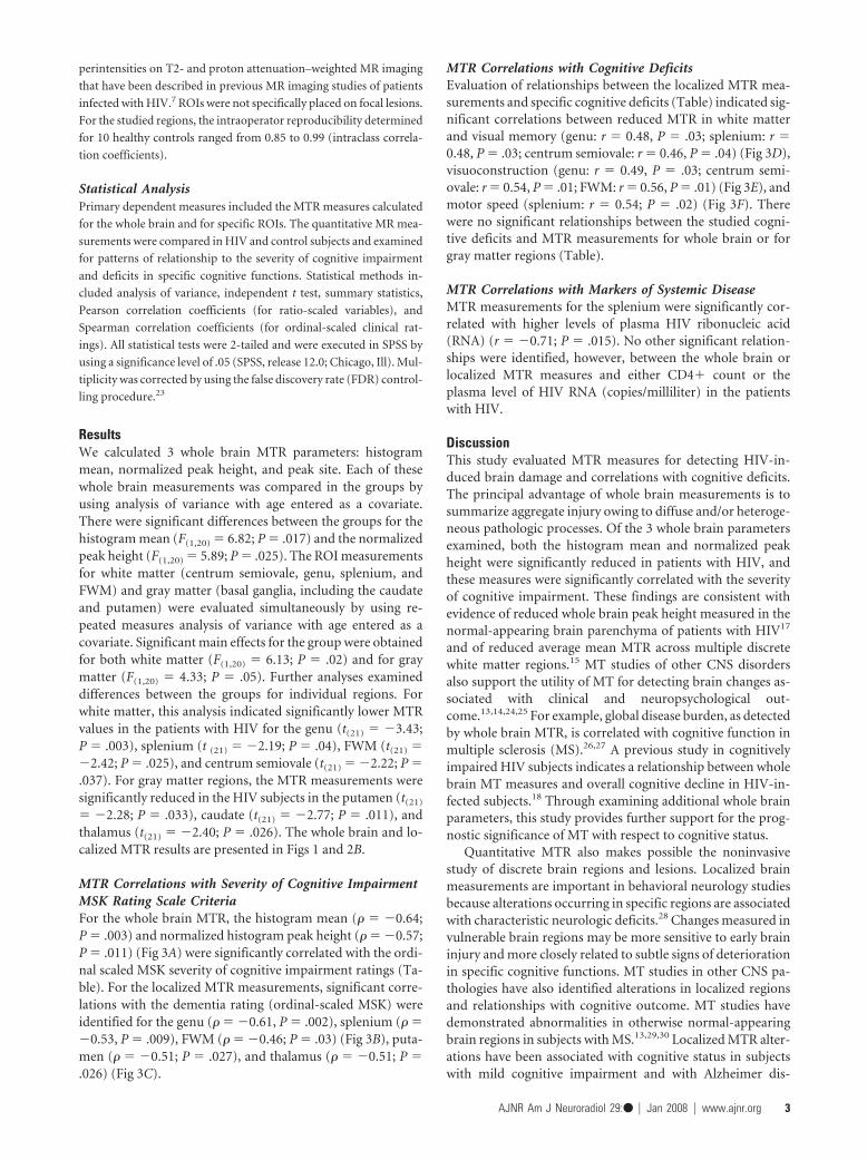
perintensities on T2- and proton attenuation–weighted MR imaging
that have been described in previous MR imaging studies of patients
infected with HIV.7 ROIs were not specifically placed on focal lesions.
For the studied regions, the intraoperator reproducibility determined
for 10 healthy controls ranged from 0.85 to 0.99 (intraclass correla-
tion coefficients).
Statistical AnalysisPrimary dependent measures included the MTR measures calculated
for the whole brain and for specific ROIs. The quantitative MR mea-
surements were compared in HIV and control subjects and examined
for patterns of relationship to the severity of cognitive impairment
and deficits in specific cognitive functions. Statistical methods in-
cluded analysis of variance, independent t test, summary statistics,
Pearson correlation coefficients (for ratio-scaled variables), and
Spearman correlation coefficients (for ordinal-scaled clinical rat-
ings). All statistical tests were 2-tailed and were executed in SPSS by
using a significance level of .05 (SPSS, release 12.0; Chicago, Ill). Mul-
tiplicity was corrected by using the false discovery rate (FDR) control-
ling procedure.23
ResultsWe calculated 3 whole brain MTR parameters: histogrammean, normalized peak height, and peak site. Each of thesewhole brain measurements was compared in the groups byusing analysis of variance with age entered as a covariate.There were significant differences between the groups for thehistogram mean (F(1,20) � 6.82; P � .017) and the normalizedpeak height (F(1,20) � 5.89; P � .025). The ROI measurementsfor white matter (centrum semiovale, genu, splenium, andFWM) and gray matter (basal ganglia, including the caudateand putamen) were evaluated simultaneously by using re-peated measures analysis of variance with age entered as acovariate. Significant main effects for the group were obtainedfor both white matter (F(1,20) � 6.13; P � .02) and for graymatter (F(1,20) � 4.33; P � .05). Further analyses examineddifferences between the groups for individual regions. Forwhite matter, this analysis indicated significantly lower MTRvalues in the patients with HIV for the genu (t(21) � �3.43;P � .003), splenium (t (21) � �2.19; P � .04), FWM (t(21) ��2.42; P � .025), and centrum semiovale (t(21) � �2.22; P �.037). For gray matter regions, the MTR measurements weresignificantly reduced in the HIV subjects in the putamen (t(21)
� �2.28; P � .033), caudate (t(21) � �2.77; P � .011), andthalamus (t(21) � �2.40; P � .026). The whole brain and lo-calized MTR results are presented in Figs 1 and 2B.
MTR Correlations with Severity of Cognitive ImpairmentMSK Rating Scale CriteriaFor the whole brain MTR, the histogram mean (� � �0.64;P � .003) and normalized histogram peak height (� � �0.57;P � .011) (Fig 3A) were significantly correlated with the ordi-nal scaled MSK severity of cognitive impairment ratings (Ta-ble). For the localized MTR measurements, significant corre-lations with the dementia rating (ordinal-scaled MSK) wereidentified for the genu (� � �0.61, P � .002), splenium (� ��0.53, P � .009), FWM (� � �0.46; P � .03) (Fig 3B), puta-men (� � �0.51; P � .027), and thalamus (� � �0.51; P �.026) (Fig 3C).
MTR Correlations with Cognitive DeficitsEvaluation of relationships between the localized MTR mea-surements and specific cognitive deficits (Table) indicated sig-nificant correlations between reduced MTR in white matterand visual memory (genu: r � 0.48, P � .03; splenium: r �0.48, P � .03; centrum semiovale: r � 0.46, P � .04) (Fig 3D),visuoconstruction (genu: r � 0.49, P � .03; centrum semi-ovale: r � 0.54, P � .01; FWM: r � 0.56, P � .01) (Fig 3E), andmotor speed (splenium: r � 0.54; P � .02) (Fig 3F). Therewere no significant relationships between the studied cogni-tive deficits and MTR measurements for whole brain or forgray matter regions (Table).
MTR Correlations with Markers of Systemic DiseaseMTR measurements for the splenium were significantly cor-related with higher levels of plasma HIV ribonucleic acid(RNA) (r � �0.71; P � .015). No other significant relation-ships were identified, however, between the whole brain orlocalized MTR measures and either CD4� count or theplasma level of HIV RNA (copies/milliliter) in the patientswith HIV.
DiscussionThis study evaluated MTR measures for detecting HIV-in-duced brain damage and correlations with cognitive deficits.The principal advantage of whole brain measurements is tosummarize aggregate injury owing to diffuse and/or heteroge-neous pathologic processes. Of the 3 whole brain parametersexamined, both the histogram mean and normalized peakheight were significantly reduced in patients with HIV, andthese measures were significantly correlated with the severityof cognitive impairment. These findings are consistent withevidence of reduced whole brain peak height measured in thenormal-appearing brain parenchyma of patients with HIV17
and of reduced average mean MTR across multiple discretewhite matter regions.15 MT studies of other CNS disordersalso support the utility of MT for detecting brain changes as-sociated with clinical and neuropsychological out-come.13,14,24,25 For example, global disease burden, as detectedby whole brain MTR, is correlated with cognitive function inmultiple sclerosis (MS).26,27 A previous study in cognitivelyimpaired HIV subjects indicates a relationship between wholebrain MT measures and overall cognitive decline in HIV-in-fected subjects.18 Through examining additional whole brainparameters, this study provides further support for the prog-nostic significance of MT with respect to cognitive status.
Quantitative MTR also makes possible the noninvasivestudy of discrete brain regions and lesions. Localized brainmeasurements are important in behavioral neurology studiesbecause alterations occurring in specific regions are associatedwith characteristic neurologic deficits.28 Changes measured invulnerable brain regions may be more sensitive to early braininjury and more closely related to subtle signs of deteriorationin specific cognitive functions. MT studies in other CNS pa-thologies have also identified alterations in localized regionsand relationships with cognitive outcome. MT studies havedemonstrated abnormalities in otherwise normal-appearingbrain regions in subjects with MS.13,29,30 Localized MTR alter-ations have been associated with cognitive status in subjectswith mild cognitive impairment and with Alzheimer dis-
AJNR Am J Neuroradiol 29:● � Jan 2008 � www.ajnr.org 3

ease.16,25 Information concerning the sensitivity of MT to lo-calized brain alterations in patients with HIV is very limited,and available information was acquired before widespread useof highly active antiretroviral therapy (HAART).15,31 This in-vestigation evaluated localized MT measurements in regionsin which injury has been identified by postmortem studies ofHIV encephalopathy, the pathologic correlate of dementia.5,32
MTR values were significantly reduced in HIV subjects in allbrain regions studied, including the basal ganglia (putamen,caudate, and thalamus) and white matter (genu, splenium,FWM, and centrum semiovale). These findings support the
sensitivity of MT to localized neuropathologic changes inHIV-infected subjects.
The localized MT measurements were also significantlycorrelated with cognitive status. MTR values for the corpuscallosum (genu and splenium), FWM, and the basal ganglia(putamen and thalamus) were significantly correlated withseverity of cognitive impairment, as determined by a clinicaldementia scale.20 Relationships with specific cognitive deficits,determined by neuropsychological testing, were generallymore pronounced for white matter regions. Most notably, re-duced MTR measurements in the corpus callosum were sig-
Fig 3. Scatterplots of significant correlations between MTRmeasurements and cognitive status measures, including theMSK ordinal-scale dementia rating (A–C ) and continuouscognitive function variables (D–F )
.
Correlations of MTR measurements and cognitive status measures
Whole Brain MTR Localized MTR
Mmtr NPH PSite Genu Splen FWM CSEM Put Caud ThalMSK �0.64* �0.57* �0.36 �0.61* �0.53* �0.46* �0.38 �0.51* �0.39 �0.51*Working memory 0.03 �0.51 0.32 0.11 0.29 0.25 0.31 0.06 0.08 0.09Verbal memory �0.14 0.51 �0.24 �0.23 �0.01 �0.28 �0.32 �0.02 �0.33 �0.05Visual memory 0.47 0.18 0.25 0.48* 0.48* 0.42 0.46* 0.10 0.30 0.30Visuoconstruction 0.43 �0.05 0.43 0.49* 0.20 0.56* 0.54* 0.28 0.41 0.37Psychomotor 0.11 0.27 0.07 0.24 0.19 0.04 �0.12 0.15 �0.25 0.23Motor speed 0.40 0.46 0.11 0.24 0.54* 0.24 0.11 0.26 0.05 0.42Executive function �0.26 0.14 �0.25 �0.37 �0.14 �0.26 �0.22 �0.36 �0.24 �0.16
Note:—Mmtr � mean MTR; NPH, normalized peak height; Psite, peak site; Splen, splenium; FWM, frontal white matter; CSEM, centrum semiovale; Put, putamen; Caud, caudate; Tha;thalamus.* Significant using FDR-adjusted P value. Spearman correlation coefficients for MSK; all others, Pearson correlation coefficients.
4 Wu � AJNR 29 � Jan 2008 � www.ajnr.org

nificantly correlated with motor speed (splenium), visualmemory (splenium), and visuoconstruction (genu). Alter-ations in the corpus callosum have also been identified inHIV-infected subjects with diffusion tensor imaging,33-35 andthese changes correlate with the severity of cognitive impair-ment and motor function.34,35 Structural studies indicatethinning of the corpus callosum in patients with HIV.36 It hasbeen suggested that the vulnerability of the corpus callosum toinjury in HIV dementia has not been adequately recognized,and this brain region may be an HIV predilection site.37 More-over, only MTR measurements for the splenium of the corpuscallosum were significantly correlated with higher levels ofplasma HIV RNA. Taken together, these findings suggest thatquantitative MR imaging measurements acquired in the cor-pus callosum may be informative in studies of HIV-associatedcognitive impairment.
The localized MTR measurements also indicated signifi-cant alterations in the basal ganglia, including the caudate,thalamus, and putamen. Moreover, MTR measurements forthe thalamus and putamen were significantly correlated withthe severity of cognitive impairment. The basal ganglia havebeen implicated in cognitive deterioration in HIV-infected pa-tients by histopathologic findings at autopsy.1 MR spectro-scopic and positron-emission tomography (PET) studies havefound abnormal hypermetabolism in basal ganglia regions inpatients with HIV dementia.38,39 Measurements of the basalganglia acquired with diffusion-tensor imaging demonstratesignificant relationships with cognitive and clinical parame-ters in HIV-infected patients.40 MTR may have considerablepractical significance for studying changes in these subcorticalgray matter regions. MT affords higher spatial resolution andis easier to implement and less labor-intensive than techniquessuch as MR spectroscopy and PET. A comparative study of 5different quantitative MR imaging measures, including totalwater content, myelin water content, mean T2 relaxation time,T1 relaxation time, and MTR, identified MTR as the mostreliable and sensitive for detecting abnormalities in tissue.41
Findings from this investigation indicate that MT is apromising method for summarizing both aggregate and local-ized neuropathologic changes associated with cognitive defi-cits in HIV-infected patients. Measurements acquired withMTR, both for whole brain and for selected ROIs, distin-guished HIV from control subjects and were significantly cor-related with severity of cognitive impairment. MTR measure-ments in studied white matter regions were correlated withspecific cognitive deficits. Findings from this investigationprovide further evidence implicating white matter injury inHIV-associated cognitive deterioration. Multifocal-distrib-uted neural networks interconnected by white matter path-ways are critical to intact higher order cognitive function.42
The corpus callosum plays a role in visuomotor integrationand may interact in important ways with subcortical struc-tures, notably basal ganglia, in response initiation.43 Injuryinvolving the corpus callosum and/or basal ganglia may bereflected in slowed response initiation and longer reactiontimes on tasks involving hemispheric transfer or integrationbetween regions.
Many neuroradiologic studies, particularly in MS, havefound that MT detects subtle changes that are not identifiedon conventional MR imaging.11-16 However, the technique
has not yet been adapted for clinical settings in the manage-ment of patients with HIV. Potential clinical applications ofMT include early detection of neurologic involvement andresponse to treatment. In MS, for example, MT measurementshave been used to evaluate drug effectiveness44 and have beenrecommended as objective end points in large-scale MS tri-als.45 It is possible, pending further study, that MT could beused to detect response to specific antiretrovirals. These mea-surements may be more sensitive to subtle or short-termchanges in status than measures based on clinical evaluation(eg, cognitive symptoms). The patients with HIV in this inves-tigation were all cognitively impaired and were on antiretro-viral regimens. Further studies are necessary to determine thepotential of localized MTR measurements for detecting neu-ropathologic changes in asymptomatic stages of infection, forstudying the impact of neuroprotective interventions, and formonitoring neurologic progression across the course of HIVinfection.
ConclusionMTR measurements are sensitive to the neuropathologic sub-strate in patients with HIV. In this investigation, aggregatechanges measured with whole brain MTR, as well as localizedMTR measurements, demonstrated significant correlationswith clinical ratings of overall cognitive function. Specific neu-ropsychological deficits were more highly correlated with lo-calized MT measurements.
AcknowledgmentsWe are grateful for the assistance of Linda Pierchala and LindaReisberg.
References1. McArthur JC, Brew BJ, Nath A. Neurological complications of HIV infection.
Lancet Neurol 2005;4:543–552. Kaul M, Garden GA, Lipton SA. Pathways to neuronal injury and apoptosis in
HIV-associated dementia. Nature 2001;410:988 –943. Power C, Gill MJ, Johnson RT. Progress in clinical neurosciences: the neuro-
pathogenesis of HIV infection– host-virus interaction and the impact of ther-apy. Can J Neurol Sci 2002;29:19 –32
4. Navia BA, Cho ES, Petito CK, et al. The AIDS dementia complex: II. Neuropa-thology. Ann Neurol 1986;19:525–35
5. Bell JE. An update on the neuropathology of HIV in the HAART era. Histopa-thology 2004;45:549 –59
6. Navia BA, Jordan BD, Price RW. The AIDS dementia complex: I. Clinical fea-tures. Ann Neurol 1986;19:517–24
7. Post MJ, Berger JR, Quencer RM. Asymptomatic and neurologically symptom-atic HIV-seropositive individuals: prospective evaluation with cranial MRimaging. Radiology 1991;178:131–39
8. Koenig SH. Cholesterol of myelin is the determinant of gray-white contrast inMRI of brain. Magn Reson Med 1991;20:285–91
9. Dousset V, Brochet B, Vital A, et al. Lysolecithin-induced demyelination inprimates: preliminary in vivo study with MR and magnetization transfer.AJNR Am J Neuroradiol 1995;16:225–31
10. Wolff SD, Balaban RS. Magnetization transfer contrast (MTC) and tissue wa-ter proton relaxation in vivo. Magn Reson Med 1989;10:135– 44
11. Dousset V, Grossman RI, Ramer KN, et al. Experimental allergic encephalo-myelitis and multiple sclerosis: lesion characterization with magnetizationtransfer imaging. Radiology 1992;182:483–91
12. Grossman RI, Gomori JM, Ramer KN, et al. Magnetization transfer: theory andclinical applications in neuroradiology. Radiographics 1994;14:279 –90
13. Filippi M, Campi A, Dousset V, et al. A magnetization transfer imaging studyof normal-appearing white matter in multiple sclerosis. Neurology1995;45:478 – 82
14. van Buchem MA, Grossman RI, Armstrong C, et al. Correlation of volumetricmagnetization transfer imaging with clinical data in MS. Neurology1998;50:1609 –17
15. Dousset V, Armand JP, Lacoste D, et al. Magnetization transfer study of HIV
AJNR Am J Neuroradiol 29:● � Jan 2008 � www.ajnr.org 5

encephalitis and progressive multifocal leukoencephalopathy: Grouped’Epidemiologie Clinique du SIDA en Aquitaine. AJNR Am J Neuroradiol1997;18:895–901
16. van der Flier WM, van den Heuvel DM, Weverling-Rijnsburger AW, et al. Mag-netization transfer imaging in normal aging, mild cognitive impairment, andAlzheimer’s disease. Ann Neurol 2002;52:62– 67
17. Ge Y, Kolson DL, Babb JS, et al. Whole brain imaging of HIV-infected patients:quantitative analysis of magnetization transfer ratio histogram and fractionalbrain volume. AJNR Am J Neuroradiol 2003;24:82– 87
18. Ragin AB, Storey P, Cohen BA, et al. Disease burden in HIV-associated cogni-tive impairment: a study of whole-brain imaging measures. Neurology2004;63:2293–97
19. McArthur JC, McDermott MP, McClernon D, et al. Attenuated central nervoussystem infection in advanced HIV/AIDS with combination antiretroviraltherapy. Arch Neurol 2004;61:1687–96
20. Price RW, Brew BJ. The AIDS dementia complex. J Infect Dis 1988;158:1079 – 8321. Marder K, Tang MX, Mejia H, et al. Risk of Parkinson’s disease among first-
degree relatives: a community-based study. Neurology 1996;47:155– 6022. Marder K, Albert SM, McDermott MP, et al. Inter-rater reliability of a clinical
staging of HIV-associated cognitive impairment. Neurology 2003;60:1467–7323. Benjamini Y, Drai D, Elmer G, et al. Controlling the false discovery rate in
behavior genetics research. Behav Brain Res 2001;125:279 – 8424. Grossman RI. Application of magnetization transfer imaging to multiple scle-
rosis. Neurology 1999;53:S8 –1125. Hentschel F, Kreis M, Damian M, et al. Quantification of microangiopathic
lesions in brain parenchyma and age-adjusted mean scores for the diagnosticseparation of normal from pathological values in senile dementia [ in Ger-man]. Rofo 2005;177:864 –71
26. van Buchem MA, McGowan JC, Grossman RI. Magnetization transfer histo-gram methodology: its clinical and neuropsychological correlates. Neurology1999;53:S23–28
27. Rovaris M, Filippi M, Falautano M, et al. Relation between MR abnormalitiesand patterns of cognitive impairment in multiple sclerosis. Neurology1998;50:1601– 08
28. Filley CM. White matter and behavioral neurology. Ann N Y Acad Sci2005;1064:162– 83
29. Filippi M, Rocca MA, Martino G, et al. Magnetization transfer changes in thenormal-appearing white matter precede the appearance of enhancing lesionsin patients with multiple sclerosis. Ann Neurol 1998;43:809 –14
30. Loevner LA, Grossman RI, Cohen JA, et al. Microscopic disease in normal-appearing white matter on conventional MR images in patients with multiplesclerosis: assessment with magnetization-transfer measurements. Radiology1995;196:511–15
31. Ernst T, Chang L, Witt M, et al. Progressive multifocal leukoencephalopathyand human immunodeficiency virus-associated white matter lesions in AIDS:magnetization transfer MR imaging. Radiology 1999;210:539 – 43
32. Everall I, Barnes H, Spargo E, et al. Assessment of neuronal density in theputamen in human immunodeficiency virus (HIV) infection: application ofstereology and spatial analysis of quadrats. J Neurovirol 1995;1:126 –29
33. Filippi CG, Ulug AM, Ryan E, et al. Diffusion tensor imaging of patients withHIV and normal-appearing white matter on MR images of the brain. AJNRAm J Neuroradiol 2001;22:277– 83
34. Wu Y, Storey P, Cohen BA, et al. Diffusion alterations in corpus callosum ofpatients with HIV. AJNR Am J Neuroradiol 2006;27:656 – 60
35. Pfefferbaum A, Rosenbloom MJ, Adalsteinsson E, et al. Diffusion tensor imag-ing with quantitative fibre tracking in HIV infection and alcoholismcomorbidity: synergistic white matter damage. Brain 2007;130:48 – 64. Epub2006 Sep 7
36. Thompson PM, Dutton RA, Hayashi KM, et al. 3D mapping of ventricular andcorpus callosum abnormalities in HIV/AIDS. Neuroimage 2006;2031:2012–23
37. Budka H. HIV-associated neuropathology. In: Gendelman HE, Igor, Everall IP,et al, eds. The Neurology of AIDS. New York: Chapman & Hall; 1998:241– 60
38. Chang L, Lee PL, Yiannoutsos CT, et al. A multicenter in vivo proton-MRSstudy of HIV-associated dementia and its relationship to age. Neuroimage2004;23:1336 – 47
39. von Giesen HJ, Wittsack HJ, Wenserski F, et al. Basal ganglia metabolite abnor-malities in minor motor disorders associated with human immunodeficiencyvirus type 1. Arch Neurol 2001;58:1281– 86
40. Ragin AB, Wu Y, Storey P, et al. Diffusion tensor imaging of subcortical braininjury in patients infected with human immunodeficiency virus. J Neurovirol2005;11:292–98
41. Vavasour IM, Clark CM, Li DK, et al. Reproducibility and reliability of MRmeasurements in white matter: clinical implications. Neuroimage 2006;2032:2637– 42
42. Mesulam M. Brain, mind, and the evolution of connectivity. Brain Cogn2000;42:4 – 6
43. Reuter-Lorenz, PA. Parallel processing in the bisected brain: implications forcallosal function. In: Zaidel E, Iacoboni M, eds. The Parallel Brain: The CognitiveNeuroscience of the Corpus Callosum. Cambridge, Mass; MIT Press; 2003
44. Inglese M, van Waesberghe JH, Rovaris M, et al. The effect of interferon beta-1bon quantities derived from MT MRI in secondary progressive MS. Neurology2003;60:853– 60
45. Filippi M, Dousset V, McFarland HF, et al. The role of MRI in the diagnosis andmonitoring of multiple sclerosis: consensus report of the White Matter StudyGroup. J Magn Reson Imaging 2002;15:499 –504
6 Wu � AJNR 29 � Jan 2008 � www.ajnr.org