whole body cardiovascular magnetic resonance imaging to
TRANSCRIPT

RESEARCH ARTICLE Open Access
Whole body cardiovascular magneticresonance imaging to stratify symptomaticand asymptomatic atherosclerotic burdenin patients with isolated cardiovasculardiseaseJonathan R. Weir-McCall1,2,5*, Suzanne L. Duce1, Stephen J. Gandy2,3, Shona Z. Matthew1, Patricia Martin2,Deirdre B. Cassidy1, Lynne McCormick1, Jill J. F. Belch1, Allan D. Struthers1, Helen M. Colhoun4
and J. Graeme Houston1,2
Abstract
Background: The aim of this study was to use whole body cardiovascular magnetic resonance imaging (WB CVMR)to assess the heart and arterial network in a single examination, so as to describe the burden of atherosclerosis andsubclinical disease in participants with symptomatic single site vascular disease.
Methods: 64 patients with a history of symptomatic single site vascular disease (38 coronary artery disease (CAD), 9cerebrovascular disease, 17 peripheral arterial disease (PAD)) underwent whole body angiogram and cardiac MR ina 3 T scanner. The arterial tree was subdivided into 31 segments and each scored according to the degree ofstenosis. From this a standardised atheroma score (SAS) was calculated. Cine and late gadolinium enhancementimages of the left ventricle were obtained.
Results: Asymptomatic atherosclerotic disease with greater than 50 % stenosis in arteries other than that responsiblefor their presenting complain was detected in 37 % of CAD, 33 % of cerebrovascular and 47 % of PAD patients.Unrecognised myocardial infarcts were observed in 29 % of PAD patients. SAS was significantly higher in PADpatients 24 (17.5-30.5) compared to CAD 4 (2–11.25) or cerebrovascular disease patients 6 (2-10) (ANCOVA p < 0.001).Standardised atheroma score positively correlated with age (β 0.36 p = 0.002), smoking status (β 0.34 p = 0.002), and LVmass (β -0.61 p = 0.001) on multiple linear regression.
Conclusion: WB CVMR is an effective method for the stratification of cardiovascular disease. The high prevalence ofasymptomatic arterial disease, and silent myocardial infarctions, particularly in the peripheral arterial disease group,demonstrates the importance of a systematic approach to the assessment of cardiovascular disease.
Keywords: Whole body magnetic resonance angiography, Atherosclerosis, Coronary artery disease, Peripheral arterialdisease, Cerebrovascular disease, Atheroma burden
* Correspondence: [email protected] R. Weir-McCall and Suzanne L. Duce are joint first authors.1Division of Cardiovascular and Diabetes Medicine, Medical ResearchInstitute, University of Dundee, DD1 9SY, UK2NHS Tayside Clinical Radiology, Ninewells Hospital, Dundee DD1 9SY, UKFull list of author information is available at the end of the article
© 2016 Weir-McCall et al. Open Access This article is distributed under the terms of the Creative Commons Attribution 4.0International License (http://creativecommons.org/licenses/by/4.0/), which permits unrestricted use, distribution, andreproduction in any medium, provided you give appropriate credit to the original author(s) and the source, provide a link tothe Creative Commons license, and indicate if changes were made. The Creative Commons Public Domain Dedication waiver(http://creativecommons.org/publicdomain/zero/1.0/) applies to the data made available in this article, unless otherwise stated.
Weir-McCall et al. BMC Medical Imaging (2016) 16:18 DOI 10.1186/s12880-016-0121-4

BackgroundAtherosclerosis with subsequent plaque formation is theunderlying pathophysiological process in the leadingcauses of morbidity and mortality in the western world[1]. The widely distributed nature of atherosclerosisacross the body has been appreciated for decades [2],however routine stratification or quantification of wholebody disease burden is not routinely performed. Multi-modal imaging studies of patients presenting with cor-onary artery disease [3, 4], cerebrovascular disease [5, 6]and peripheral arterial disease [7–9], have shown a highprevalence of atherosclerotic disease in other sites inaddition to the presenting disease. Multimodal studiesby their nature involved multiple examinations and typ-ically require visits to different healthcare departments.An alternative option is whole body cardiovascular mag-netic resonance imaging (WB CVMR), which can assessthe heart and arterial network in a single examination,has the advantages of being non-invasive and avoids ion-ising radiation.WB CVMR comprises of a suite of cardiac magnetic
resonance (CMR) and whole body MR angiography (WB-MRA) sequences, allowing the systemic assessment ofwhole body atheroma burden, with cardiac structure,function and the detection of myocardial scarring, in asingle imaging session. Global atheroma burden has beenshown to correlate well with traditional cardiac risk fac-tors, the prevalence of coronary artery disease and futuremajor adverse cardiovascular events [10–13]. Previousstudies have assessed the ability of WB-MRA to assessextra-site disease in coronary arterial disease, peripheralarterial disease and vasculitis [14–16]. However a directcomparison of extra-site disease and atheroma burden be-tween cardiovascular disease groups has not been previ-ously conducted. The aim of the study was therefore todetermine the yield of WB CVMR in detecting asymp-tomatic cardiovascular disease, at sites other than at theclinically apparent location in different patient cohorts,and to compare atheroma burden between the groups.
MethodsEthicsThe protocols were reviewed and approved by the Eastof Scotland Research Ethics Committee and was con-ducted in accordance with the Declaration of Helsinki.All volunteers gave written informed consent to partici-pate in this study.
ParticipantsPatients with isolated coronary, cerebrovascular or per-ipheral vascular disease were identified from existingclinical databases and from local cardiology, stroke, andvascular clinics. Sixty-four participants with a prior diag-nosis of single territory vascular disease attended MRI
appointments between March 2009 and December 2012.The subjects were categorised based on their history ofcardiovascular disease; their demographics are sum-marised in Table 1. Group 1 contained those with clin-ical evidence of coronary artery disease (n = 38). Group2 contained those who had had a cerebrovascular event(n = 9). Group 3 contained those with clinical evidenceof peripheral arterial disease (n = 17).Coronary arterial disease (Group 1) included non-
fatal acute myocardial infarction, hospitalised acute cor-onary syndrome, resuscitated cardiac arrest (not attrib-uted to a non CAD causes), coronary artery bypassgraft (CABG) or any other coronary revascularisationprocedure. Cerebrovascular disease (Group 2) inclusioncriteria were non-fatal strokes or transient ischemic at-tacks (TIA) confirmed by a specialist stroke physician.Peripheral arterial disease (Group 3) inclusion criteriawas ankle-brachial pressure index (ABPI) <0.9 withintermittent claudication, walking distance of not morethan 200 yards, or abnormal toe systolic pressure. Vol-unteers were excluded if they had a clinical history ofarterial disease at any site other than their primarydiagnosis location. Other exclusion criteria included thepossibility of metallic implants, history of claustropho-bia, pregnancy, renal replacement therapy, end stagerenal disease, therapy for any chronic inflammatory dis-ease, atrial fibrillation or malignancy.
Table 1 Demographics and clinical characteristics in the studypopulation
Group 1 Group 2 Group 3
CAD CVD PAD
Number 38 9 17
Male [%] 31 (81.6) 4 (44.4) 13 (76.5)
Age [years] 66.0 ± 8 61.2 ± 8 68.7 ± 10
Weight [kg] 87.2 ± 13 77.4 ± 12 89.7 ± 28
Height [m] 1.69 ± 0.1 1.64 ± 0.1 1.7 ± 0.1
BMI [kg/m2] 30.6 ± 4 28.7 ± 2 28.8 ± 4
Systolic BP [mmHg] 135 ± 15 132 ± 12 137 ± 14
Diastolic BP [mmHg] 76 ± 9 74 ± 8 78 ± 8
Hypertension 32 (84.2) 6 (66.7) 13 (76.5)
Antiplatelet 34 (89) 9 (100) 15 (88)
Antihypertensive 37 (97) 8 (89) 14 (82)
Statin prescription 30 (79) 8 (89) 16 (94)
Type 2 Diabetes 19 (50) 5 (55.6) 6 (35.3)
Non-Smokers 16 (42) 1 (11) 1 (6)
Former-Smokers 20 (53) 8 (89) 11 (65)
Smokers 2 (5) 0 (0) 5 (29)
Values expressed as Mean ± SD, or N (%)BMI body mass index, BP blood pressure, CAD coronary artery disease, CVDcerebrovascular disease, PAD peripheral arterial disease
Weir-McCall et al. BMC Medical Imaging (2016) 16:18 Page 2 of 10

Magnetic resonance imagingImages were acquired on a 32 RF receiver channel, 3Tesla MRI scanner (Magnetom Trio, Siemens, Erlangen,Germany) equipped with high-performance gradient sys-tem and electrocardiograph (ECG)-gating. For wholebody coverage, a combination of six RF coils were used:head matrix (12 elements); neck matrix (4 elements);spine matrix (up to 24 elements); two body matrix (6elements each); and peripheral angiography phasedarray RF surface coils (16 elements. Subjects wereplaced head first into the magnet bore and were exam-ined in the supine position. Total scan time was of theorder of 45 min.
Whole body magnetic resonance angiography protocolFor localisation, four low-resolution images were acquiredfrom head to foot using gradient echo fast low angle shot(FLASH) sequences with 500 mm field of view (FOV).Whole body magnetic resonance angiography (WB-MRA)images involved the acquisition of 4 overlapping 3D datasets using a coronal spoiled FLASH (fast low angle shot)sequence (see Table 2 for acquisition parameters). Fouranatomically distinct stations with field of view of 500 mmwere: head and thorax (station 1), thorax and abdomen(station 2), abdomen and upper legs (stations 3) and lowerlegs (station 4). These were positioned with an overlap ofat least 75 mm between each field of view. Anatomicalpaired images were acquired pre- and post-contrast. Astandard dose of 25 ml gadoterate meglumine contrastagent (Dotarem, Guerbet, Villepinte, France) was adminis-tered by intravenous injection in the antecubital fossa in 2separate boluses (10 ml and 15 ml respectively) using aSpectris Solaris power injector (MedRad, Pittsburgh,USA) at a rate of 1.5 ml/sec, each followed by a 20 mlbolus of saline. For station 1 and 4 imaging, post-contrastimages were acquired after an injection of 10 ml of0.5 mmol/ml gadoterate meglumine contrast agent. Sta-tion 1 image acquisition commenced when the bolus ofcontrast agent arrived at the top of the aortic arch in the‘2D Care Bolus’ images (Siemens, Erlangen, Germany).Station 4 image acquisition followed immediately afterthis, with three consecutive volumes of station 4 acquired
to ensure that peak arterial enhancement was caught.There was a delay before stations 2 and 3 image acquisi-tion of at least 10 min to allow contrast washout and min-imise venous contamination during the second injectionand image acquisition [17]. Post-contrast images wereacquired after an injection of 15 ml of 0.5 mmol/mlgadoterate meglumine contrast agent, again followed bya 20 ml saline bolus. Acquisition commenced once thecontrast agent bolus was seen in the descending aortain the coronal 2D Care Bolus image. Station 3 imagingcommenced immediately after the completion of theacquisition of station 2.
Cardiac magnetic resonance (CMR) protocolsCardiac magnetic resonance (CMR) imaging utilised aspine matrix and six-element body array matrix RF coils.Left ventricular assessment involved the acquisition ofshort axis, multi-slice 2D images from the atrio-ventricularring to the apex using a CINE TrueFISP sequence withretrospective ECG-gating, with repeated end-expiratorybreath-holds (Table 2). The slice thickness was 6 mmand inter-slice gap was 4 mm. Ten minutes after the in-jection of the first dose of contrast agent, the ECG-gated, breath-hold, end-diastole, short-axis, multi-slicelate gadolinium enhanced (LGE) CMR images were ac-quired using a 2D phase sensitive inversion recovery(PSIR) sequence (Table 2).
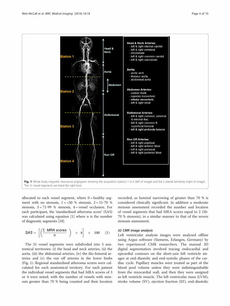
WB-MRA image analysisResearchers were blind to the participants’ clinical historyduring image analysis. The 3D WB-MRA datasets wereviewed offline as source images using both multi-planarreconstruction (MPR) and maximum intensity projections(MIP) (Carestream PACS Client Suite Version 10.1 sp1,Rochester, NY, USA) by a radiologist with experience ofreporting over 400 whole body magnetic resonance angio-grams. Based on previous pilot work, the arterial networkwas divided into 31 vessel segments extending from theinternal and external carotid arteries to the trifurcationvessels of the lower limb (Fig. 1). Each arterial segmentwas scored according to maximal luminal stenosis withinthe vessel lumen. Categorical MRA scores from 0-4 were
Table 2 Imaging parameters for MRI sequences used for the combined CMR and WB-MRA protocol
Location Sequence Plane TR (ms) TE (ms) Flip Angle (0) Pixel size (mm) Slice thickness (mm)
WB-MRA Station 1 FLASH Coronal 2.68 1 19 1.1x1 1.1
WB-MRA Station 2 FLASH Coronal 2.6 0.96 16 1.3x1.1 1.3
WB-MRA Station 3 FLASH Coronal 3.47 1.21 37 1.5x1 1.4
WB-MRA Station 4 FLASH Coronal 2.61 0.96 22 1.2x1.1 1
LVA Heart LV TrueFISP Short axis 3.40 1.48 50-60 1.9x1.4 6
LGE-CMR Heart LV PSIR Short axis 846.4/5.21 1.99 20 1.9x1.4 6
WB-MRA whole body magnetic resonance angiography, LVA left ventricular analysis, LGE-CMR Late gadolinium enhanced cardiac magnetic resonance images, TRrepetition time, TE echo time
Weir-McCall et al. BMC Medical Imaging (2016) 16:18 Page 3 of 10
allocated to each vessel segment, where 0 = healthy seg-ment with no stenosis, 1 = <50 % stenosis, 2 = 51-70 %stenosis, 3 = 71-99 % stenosis, 4 = vessel occlusion. Foreach participant, the ‘standardised atheroma score’ (SAS)was calculated using equation [1] where n is the numberof diagnostic segments [18].
SAS ¼ Σ MRA scores
n
� �� 4
� �� 100 ð1Þ
The 31 vessel segments were subdivided into 5 ana-tomical territories: (i) the head and neck arteries, (ii) theaorta, (iii) the abdominal arteries, (iv) the ilio-femoral ar-teries and (v) the run off arteries in the lower limbs(Fig. 1). Regional standardised atheroma scores were cal-culated for each anatomical territory. For each patientthe individual vessel segments that had MRA scores of 3or 4 were noted, with the number of vessels with sten-osis greater than 70 % being counted and their location
recorded, as luminal narrowing of greater than 70 % isconsidered clinically significant. In addition a moderatestenosis assessment recorded the number and locationof vessel segments that had MRA scores equal to 2 (50-70 % stenosis), in a similar manner to that of the severestenosis assessment.
(ii) CMR image analysisLeft ventricular analysis images were analysed offlineusing Argus software (Siemens, Erlangen, Germany) bytwo experienced CMR researchers. The manual 3Ddigital segmentation involved tracing endocardial andepicardial contours on the short-axis left ventricle im-ages at end-diastolic and end-systolic phases of the car-diac cycle. Papillary muscles were treated as part of theblood pool volume unless they were indistinguishablefrom the myocardial wall, and then they were assignedas left ventricle muscle. The left ventricular mass (LVM),stroke volume (SV), ejection fraction (EF), end-diastolic
Fig. 1 Whole body magnetic resonance angiogram showing the acquisition stations 1 to 4 (left of image) and the 5 arterial territories (right of image).The 31 vessel segments are listed (far right box)
Weir-McCall et al. BMC Medical Imaging (2016) 16:18 Page 4 of 10

(EDV) and end-systolic volumes (ESV) were determinedusing an algorithm based on the Simpson rule [19]. Re-sults were normalised to body surface area. Left ven-tricular hypertrophy was defined as an indexed LVMabove the normal range for sex [19]. Late gadoliniumenhanced images of the left ventricle were inspected forevidence of myocardial signal enhancement using aCarestream PACS workstation (Rochester, NY, USA).The location was recorded according to the AHA 17segment model [20]. Delayed enhancement was definedas <50 % or >50 % wall thickness according to the max-imum depth of delayed enhancement in any segment.
Statistical methodsDescriptive statistics were used for the analysis of thedemographic and clinical features of the cohorts with dataexpressed as mean ± standard deviation (sd) for normallydistributed data, and median (interquartile range) for non-normal distributed data. Normality tests were performed;if the test failed, where possible standard transformationssuch as square root, reciprocal or logarithmic transformswere used to generate a Gaussian distribution. To test thenull hypothesis to determine if samples originated fromthe same distribution, one-way analysis of variance(ANOVA) with the Bonferroni post hoc adjustment wasused for the parametric data, and Kruskal–Wallis ANOVAby ranks was used for the non-parametric data. ANCOVAwas performed to confirm differences between the groupswith the WB-SAS as the dependant variable. MANCOVAwas used to determine the relations of the LV metrics tothe demographic data with the LV metrics entered as thedependant variables. Pearson correlation coefficients be-tween WB-SAS, LVA and population demographic met-rics were reported. All variables with a p-value <0.3 onunivariate analysis were entered into a multivariate regres-sion analysis with the WB-SAS as the dependent variableand the remainder as independent variables. All data wereanalysed using SPSS statistical package (version 21.0, SPSSInc. Chicago, Illinois). Significance was assumed when p <0.05. A local statistician provided statistical support.
ResultsCMR and whole body MRA images were acquired from64 participants (75 % male, age 66.1 ± 8.5) with singlesite cardiovascular disease. There were no statisticallysignificant differences in the demographic metrics be-tween each of the diseased groups (Table 1), except forsmoking status with significantly more people in thePAD group being current smokers than in either theCAD or cerebrovascular groups.In the WB-MRA analysis, 1978 of the 1984 vessel seg-
ments (99.7 %) were interpretable. 6 segments in 4 ofthe 64 examinations were rated as ‘non-diagnostic’ dueto movement artefact or incomplete vessel visualisation.
619 (31.3 %) of the 1978 arterial segments had evidenceof luminal narrowing: 453 (22.9 %) had stenosis below50 %, 61 (3.1 %) had stenosis between 50-70 %, 63(3.2 %) had stenosis between 70-90 %, and 42 (2.1 %)had complete occlusion.The PAD group had the highest whole body standardised
atheroma score (WB-SAS) of 24.8 ± 9.9 and the CADgroup had the lowest WB-SAS of 7.0 ± 6.2. The WB-SASof the PAD patients was statistically significantly higherthan those of either the CAD or cerebrovascular diseasepatients (ANOVA test: P ≤ 0.001). Differences between thegroups persisted on ANCOVA (F = 15.18, p < 0.001), ac-counting for age, gender, smoking status, blood pressure,BMI and statin prescription. There was no significant dif-ference in the WB-SAS between the CAD and cerebrovas-cular disease groups. Across all 5 anatomical territories,the PAD subjects’ regional SAS were consistently highercompared to either the CAD or cerebrovascular groups’scores (Table 3). There were no statistically significant dif-ferences between any of the regional SAS of CAD andcerebrovascular group. On univariate analysis, there werepositive correlations of SAS with age (r = 0.37 P =0.002), smoking status (r = 0.397 P = 0.002), LV ejectionfraction (r = -0.26 P = 0.034) and LV mass (r = -0.42 P =0.001) and a trend towards an association with diastolicblood pressure (r = -0.23 P = 0.052) (Table 4). On multi-variate analysis, age (β 0.36 p = 0.002), smoking status(β 0.34 p = 0.002), and LV mass (β -0.61 p = 0.001) con-tinued to demonstrate a significant association withWB-SAS.For each patient, the extent of stenosis was further in-
vestigated by counting the number of vessel segmentsthat had either (a) severe stenosis with MRA score of 3or 4 (associated with luminal narrowing of greater than70 %) or (b) moderate stenosis with MRA score of 2 (as-sociated with luminal narrowing of between 50 and70 %). The results are summarised in Table 5. 100 % ofPAD patients have severe stenosis detectable in at leastone arterial vessel, with the ilio-femoral arteries beingmost affected. A third of the CAD patients had severestenosis present in their MRA images, with the run offarteries most commonly affected (18.4 % of patients),while severe arterial stenosis was detected in only 22 %of the cerebrovascular patients. With luminal narrowingof greater than 70 % considered clinically significant, thisanalysis is useful for screening patients and highlightingthose who require follow-up investigations.The left ventricular assessment (Table 3) revealed that
the PAD group have the largest mean left ventricular mass(LVM), end-diastolic (EDV), end-systolic volume (ESV),cardiac ejection fraction (EF) and stroke volume (SV), andthese differences reached statistical significance for theLVM, EDV and SV values (ANOVA P ≤ 0.007). These dif-ferences persisted on MANCOVA (F = 8.87, p = 0.001 for
Weir-McCall et al. BMC Medical Imaging (2016) 16:18 Page 5 of 10

LVM, F = 3.27, p = 0.047 for EDV and F = 4.43, p = 0.018for LVM). The differences between the CAD and cere-brovascular disease cohorts were relatively small anddid not reach statistical significance. Left ventricularhypertrophy was detected in 16 % of the study popula-tion, while impaired left ventricular systolic functionwas present in 13 %.Late gadolinium enhancement (LGE) was observed in
21 (32.8 %) of the 64 subjects. All subjects with LGE had
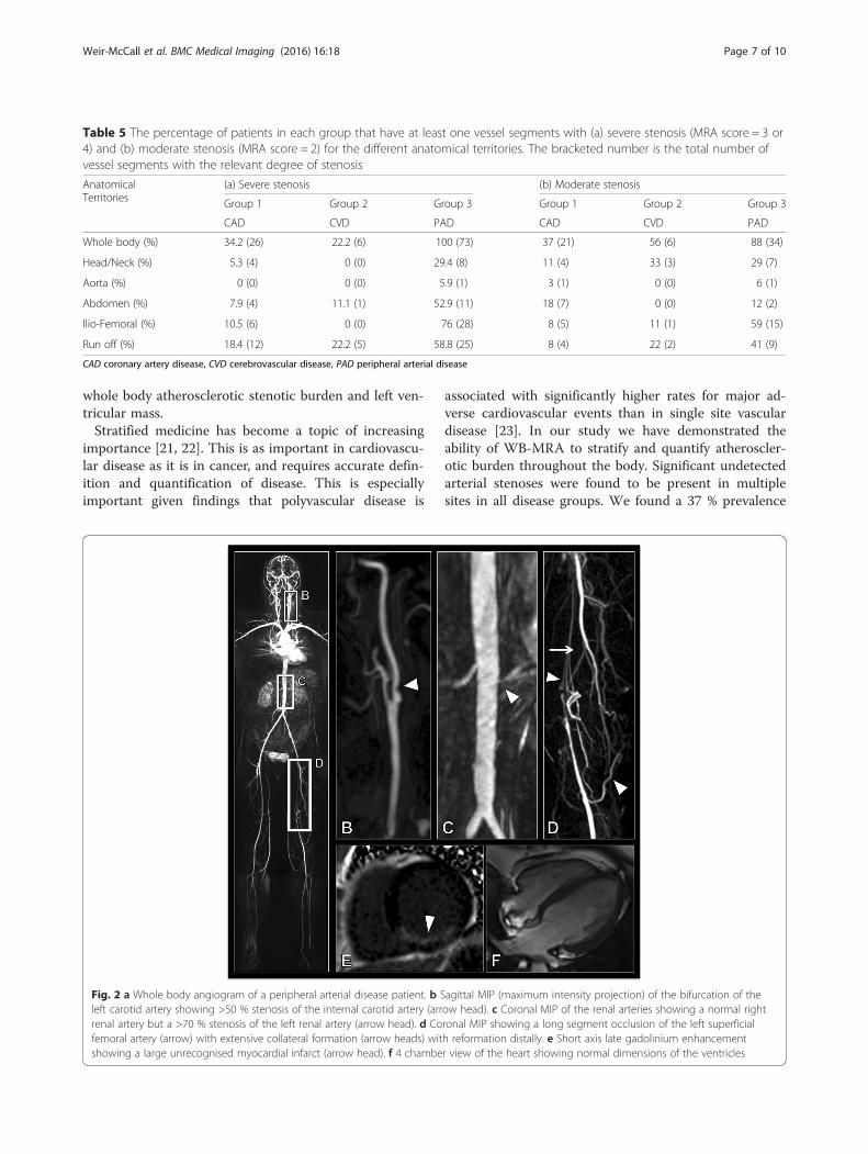
scarring in a subendocardial location with a territorial dis-tribution typical of an ischaemic aetiology. The majority ofthe enhancement occurred in the CAD group, with 16(42.1 %) CAD participants displaying evidence of myocar-dial scarring, affecting a total of 91 AHA segments. Nomyocardial LGE was observed in the cerebrovascular dis-ease group images. Five (29.4 %) of the PAD group hadevidence of unrecognised myocardial infarction (UMI), af-fecting a total of 21 AHA segments (See Figure 2). UMIstended to occur in the inferior wall with 9/21 UMIs occur-ring in the inferior segments, with the second most com-mon location being the inferoseptal segment with 4/21UMIs occurring in this region. Recognised MIs demon-strated no territorial predominance. UMIs were smaller,involving an average of 4.2 AHA segments, comparedwith 6.1 AHA segments in the recognised MI group. 40 %of UMIs involved less than 50 % of the myocardial thick-ness in the affected segments while the remaining 60 % in-volved greater than 50 % of the myocardial thickness. Incomparison, 73 % of recognised MIs involved >50 % ofthe myocardial thickness. No correlation was observed be-tween WB-SAS and either the presence or the severity ofthe late gadolinium myocardial enhancement.
DiscussionWe have shown that whole body cardiovascular MRI is afeasible solution to stratify the extent of atherosclerosisin arteries and the extent of cardiac dysfunction andmyocardial scarring in a 45-min exam. This is also thefirst study to show a positive correlation between the
Table 3 Whole body cardiovascular magnetic resonance imaging (WB CVMR) data for each group including magnetic resonanceangiography (MRA) scores, standardised stenosis scores (SAS), left ventricular analysis (LVA) and left ventricular late gadoliniumenhancement (LGE) results
Group 1 Group 2 Group 3
CAD CVD PAD
WB-SAS 4 (2–11.25) 6 (2-10) 24 (17.5-30.5)
Head/Neck-SAS 2.8 (0–5.6) 8.3 (1.4–9.7) 19.4 (9.7-25)
Aorta-SAS 8.3 (0–16.7) 8.3 (8.3-12.5) 8.3 (8.3-16.7)
Abdomen-SAS 8.3 (0–10) 0 (0–5) 20 (5-27.5)
Ilio-Femoral-SAS 6.25 (0–20.8) 4.2 (2.1–14.6) 37.5 (27.1-52.1)
Run Off-SAS 0 (0–10.2) 0 (0–18.7) 31.3 (11-48.5)
LVM (g/m2) 59.1 (54.1–64.5) 53.4 (47.4–71.1) 105.7 (67.8-124.5)
EDV (ml/m2) 74.7 ± 18.8 72.4 ± 15.2 104.1 ± 34.9
ESV (ml/m2) 25.3 (18.3–29.7) 22.4 (17.8–35.8) 33.8 (22.8-42.8)
EF (%) 64.5 ± 9.8 64.7 ± 13.4 66.4 ± 11.8
SV (ml/m2) 46.8 (40.4–52) 46 (40.2–52.7) 73.7 (39.4-92.7)
LGE 16 (42.1 %) 0 (0 %) 5 (29.4 %)
Values expressed as Mean ± SD, Median (Interquartile range) or N (%)CAD coronary artery disease, CVD cerebrovascular disease, PAD peripheral arterial disease, SAS standardised atheroma score, WB whole body, LVM indexed leftventricular mass, EDV indexed end diastolic volume, ESV indexed end systolic volume, EF ejection fraction, SV indexed stroke volume, LGE Late gadolinium enhancement
Table 4 Correlation of whole body standardised atheromascore with demographic and left ventricular parameters
Pearson correlation P-value
Age 0.461 <0.001
Gender -0.19 0.091
Systolic BP 0.19 0.095
Diastolic BP -0.23 0.052
BMI -0.22 0.06
Hypertension 0.13 0.18
History of Smoking 0.4 0.002
T2DM 0.05 0.37
Statins 0.008 0.48
Left ventricular metrics
LV End diastolic volume 0.11 0.22
LV End systolic volume 0.17 0.11
LV stroke volume -0.18 0.11
Ejection fraction -0.26 0.03
LV mass -0.42 0.001
Weir-McCall et al. BMC Medical Imaging (2016) 16:18 Page 6 of 10
whole body atherosclerotic stenotic burden and left ven-tricular mass.Stratified medicine has become a topic of increasing
importance [21, 22]. This is as important in cardiovascu-lar disease as it is in cancer, and requires accurate defin-ition and quantification of disease. This is especiallyimportant given findings that polyvascular disease is
associated with significantly higher rates for major ad-verse cardiovascular events than in single site vasculardisease [23]. In our study we have demonstrated theability of WB-MRA to stratify and quantify atheroscler-otic burden throughout the body. Significant undetectedarterial stenoses were found to be present in multiplesites in all disease groups. We found a 37 % prevalence
Table 5 The percentage of patients in each group that have at least one vessel segments with (a) severe stenosis (MRA score = 3 or4) and (b) moderate stenosis (MRA score = 2) for the different anatomical territories. The bracketed number is the total number ofvessel segments with the relevant degree of stenosis
AnatomicalTerritories
(a) Severe stenosis (b) Moderate stenosis
Group 1 Group 2 Group 3 Group 1 Group 2 Group 3
CAD CVD PAD CAD CVD PAD
Whole body (%) 34.2 (26) 22.2 (6) 100 (73) 37 (21) 56 (6) 88 (34)
Head/Neck (%) 5.3 (4) 0 (0) 29.4 (8) 11 (4) 33 (3) 29 (7)
Aorta (%) 0 (0) 0 (0) 5.9 (1) 3 (1) 0 (0) 6 (1)
Abdomen (%) 7.9 (4) 11.1 (1) 52.9 (11) 18 (7) 0 (0) 12 (2)
Ilio-Femoral (%) 10.5 (6) 0 (0) 76 (28) 8 (5) 11 (1) 59 (15)
Run off (%) 18.4 (12) 22.2 (5) 58.8 (25) 8 (4) 22 (2) 41 (9)
CAD coronary artery disease, CVD cerebrovascular disease, PAD peripheral arterial disease
Fig. 2 a Whole body angiogram of a peripheral arterial disease patient. b Sagittal MIP (maximum intensity projection) of the bifurcation of theleft carotid artery showing >50 % stenosis of the internal carotid artery (arrow head). c Coronal MIP of the renal arteries showing a normal rightrenal artery but a >70 % stenosis of the left renal artery (arrow head). d Coronal MIP showing a long segment occlusion of the left superficialfemoral artery (arrow) with extensive collateral formation (arrow heads) with reformation distally. e Short axis late gadolinium enhancementshowing a large unrecognised myocardial infarct (arrow head). f 4 chamber view of the heart showing normal dimensions of the ventricles
Weir-McCall et al. BMC Medical Imaging (2016) 16:18 Page 7 of 10

of extra-coronary arterial disease in CAD. This is lowerthan the 50 % reported in one study although this latterstudy did not exclude patients with known extra-coronary disease [16]. It is also lower than the 55 % re-ported in a more recent study [12], although this wasperformed at 1.5 T with a lower spatial resolution thanobtained in the current study, thus the improved arterialdefinition may have led to a more accurate quantifica-tion of the degree of stenosis due to improved spatialresolution found at 3 T. We found that 33 % of cerebro-vascular disease patients had significant disease outwiththe head and neck. This is significantly lower than thatreported by Paraskevas et al. [5], however their studyonly looked at patients with known unilateral internalcarotid artery occlusion which is at the extreme end ofthe carotid disease spectrum, while our study used priorcerebrovascular events as inclusion criteria. Our PADcohort demonstrated significant disease above the levelof the abdominal aortic bifurcation in 47 % which isslightly higher than that previously reported on WB-MRA [15].The standardised atheroma score was significantly
higher in the peripheral arterial disease group than ei-ther the cerebrovascular or the coronary arterial diseasegroup. Previous epidemiological studies have shown thatpatients with PAD have a 33 % higher composite risk ofCV death, myocardial infarction, stroke, or hospitalisationfor atherothrombotic event(s) than either cerebrovascularor coronary arterial disease groups [24]. As well as the in-trinsic risk of arterial stenosis, part of the causative mech-anism may be the effect the atherosclerotic burden has onthe heart. We have shown for the first time the associationbetween whole body arterial atheroma burden and leftventricular mass, which is known to be strongly associatedwith future cardiovascular events [25]. This may be due tothe stiffening nature of the atherosclerosis on the arteries,as total atheroma burden has been shown to correlatewith arterial stiffness, [26] which in turn is associated withleft ventricular hypertrophy [27]. Given that both left ven-tricular mass and atheroma burden are associated with in-creased future risk of cardiac events, further work isrequired to extricate the interaction between these mea-sures, to ascertain whether these need to be targeted indi-vidually, or whether there is a single linking aetiologywhich can be targeted.Unrecognised myocardial infarctions are present in
29.4 % of patients with peripheral arterial disease. The ratein peripheral arterial disease patients is significantly higherthan the 6 % reported in a previous PAD population usingECG and echocardiography during pre-operative work-up[28], or the 14 % reported in a series of patients undergo-ing pre-operative coronary angiography screening [29].However ECG has been shown to only detect 6-29 % ofunrecognised myocardial infarcts revealed on late
gadolinium enhancement [30, 31]. Our observed inci-dence is closer to that expected from a previous studylooking at whole body cardiovascular MR in a populationcohort study which showed rates of unrecognised myocar-dial infarction in 19.7 % of 70 year olds and 30 % in 75 yearolds [30, 32]. That the prevalence in the PAD populationwas on par with a 75 year old cohort despite having amean age of 68 is in keeping with the higher prevalence ofrisks factors in our population, although a recent studyhas called into question the link between unrecognisedmyocardial infarctions and traditional risk factors [33].None of the patients with cerebrovascular disease hadevidence of unrecognised myocardial infarcts. This issurprising given the results of previous studies showingunrecognised myocardial infarcts in 32 %-52 % of pa-tients but may be due to the small numbers of thisgroup in the current study.Recognition of unrecognised myocardial infarction is
important as these have the same prognostic implica-tions as recognised myocardial infarcts [34]. Further-more these patients respond well to both conventionalsecondary prevention medication and percutaneous cor-onary intervention [35–37].It could be argued that since patients with clinically ap-
parent cardiovascular disease in one site will result in pa-tients being treated for atherosclerotic risk factors thatfurther information about disease elsewhere is superflu-ous. However this ignores several factors. Despite ourstudy showing comparable rates of statin prescriptions be-tween the different groups, a previous population studyhas shown that community prescription of risk modifyingagents is markedly different between disease groups, withfewer patients with PAD being prescribed statins and anti-platelet agents compared with stroke or coronary arterydisease groups [38]. This suggests poor appreciation ofthe extensive disease present elsewhere in the body, in-deed, in our study the PAD population had the most ex-tensive extra-primary site disease. The second is theadditional prognostic information this provides. Multisitedisease is associated with a significantly raised risk of fu-ture major adverse cardiac and cerebrovascular events(MACCE) compared with single site disease, and had agreater detrimental effect on future prognosis than thepresence of diabetes [23, 38]. While these studies have fo-cused on symptomatic disease, the significance of asymp-tomatic disease is supported by recent studies showingincreased risk of MACCE in patients with higher globalatherosclerotic burden on whole body MR angiography[10, 11]. Thus patients with polyvascular disease may war-rant more intensive management and follow-up as well asbeing ideal candidates for future novel therapeutic agents[24]. For those being referred for a clinical MRA of a spe-cific vascular territory, extension of this to a whole bodycardiovascular MR would be a logical step, and indeed the
Weir-McCall et al. BMC Medical Imaging (2016) 16:18 Page 8 of 10

added cost of extending a clinically indicated MRA to in-clude the rest of the body is small when considered in re-lation to the high cost of the baseline exam, and in aperipheral arterial disease population has been shown tobe cost effective due to its reductions in requirements forother imaging investigations (such as echocardiographyand carotid Doppler) and alterations in patient manage-ment, although other cardiovascular disease cohorts havestill to be assessed [39].The limitations of the current study are: The study
groups are unequal in size with a relatively small num-ber of cerebrovascular patients and large number of cor-onary arterial disease. In addition the cerebrovasculargroup demonstrates a disproportionate number of fe-males which could bias the results, however we demon-strated no significant correlation between SAS and sex,and accounting for sex using an analysis of covariancedid not change the results. An intrinsic limitation ofWB-MRA is its ability to only provide information onthe prevalence of stenotic atherosclerosis, and will thusmiss the earliest stages of the disease including vesselstiffening and remodelling. Additionally, while the prog-nostic effect of symptomatic multisite atherosclerosis isknown, the effect of asymptomatic multisite disease stillrequires further work to elucidate.
ConclusionWB CVMR is an effective method for the stratificationof cardiovascular disease. The high prevalence ofasymptomatic arterial disease, and silent myocardial in-farctions, particularly in the peripheral arterial diseasegroup, demonstrates the importance of a systematic ap-proach to the assessment of cardiovascular disease.
AbbreviationsAHA: American Heart Association; CAD: coronary artery disease; CMR: cardiacmagnetic resonance imaging; CVD: cerebrovascular disease; EDV: enddiastolic volume; ESV: end systolic volume; LGE-CMR: late gadoliniumenhanced cardiac magnetic resonance images; LVA: left ventricular analysis;LVH: left ventricular hypertrophy; LVM: left ventricular mass; MRA: magneticresonance angiography; PAD: peripheral arterial disease; SAS: standardisedatheroma score; SV: stroke volume; UMI: unrecognised myocardial infarct;WB-CVMR: whole body magnetic cardiovascular magnetic resonance;WB-MRA: whole body magnetic resonance angiography.
Competing interestsJWM has received monies from Guerbet for attending symposia, and forrunning educational meetings. HMC received monies from Pfizer forattending symposia, a speaker’s bureau, as a member of staff and forconsultancy. HMC has received research funds from Pfizer, Roche, Eli-Lilly,Boehringer Ingelheim (BI) and Astra Zeneca. HMC has shares in Roche. GH isdirector and Shareholder of Vascular Flow Technologies Ltd, and has receivedresearch funds from Guerbet.
Authors’ contributionsJWM, SLD, HMC, JJF, ADS and GH conceived the study. JWM, SLD and SManalysed the MRI studies. JWM, SLD, DC analysed and interpreted the data.PM, SG and LM worked on the development of the MRI protocol. JWM andSLD drafted the manuscript. All authors revised the manuscript critically forimportant intellectual content, and read and approved the final manuscript.
Acknowledgements: Funding, disclosuresThis is sub study of the multicentre SUMMIT study. SUMMIT receives supportfrom the Innovative Medicines Initiative (IMI) Joint Undertaking under thegrant agreement n° [115006], resources of which are composed of financialcontribution from the European Union’s Seventh Framework Programme(FP7/2007-2013) and EFPIA companies’ in kind contribution. JRWM issupported by the Wellcome Trust through the Scottish TranslationalMedicine and Therapeutics Initiative (Grant no. WT 085664) in the form of aClinical Research Fellowship. Neither groups had any role in: study design,the collection, analysis, and interpretation of data; in the writing of themanuscript; nor in the decision to submit the manuscript for publication.
Author details1Division of Cardiovascular and Diabetes Medicine, Medical ResearchInstitute, University of Dundee, DD1 9SY, UK. 2NHS Tayside Clinical Radiology,Ninewells Hospital, Dundee DD1 9SY, UK. 3NHS Tayside Medical Physics,Ninewells Hospital, Dundee DD1 9SY, UK. 4Division of Population HealthSciences, Medical Research Institute, The Mackenzie Building, University ofDundee, DD2 4BF, UK. 5Division of Cardiovascular and Diabetic Medicine,Level 7, Ninewells Hospital, Dundee DD1 9SY, UK.
Received: 22 November 2014 Accepted: 16 February 2016
References1. Rosamond W, Flegal K, Furie K, Go A, Greenlund K, Haase N, Hailpern SM,
Ho M, Howard V, Kissela B, Kissela B, Kittner S, Lloyd-Jones D, McDermott M,Meigs J, Moy C, Nichol G, O’Donnell C, Roger V, Sorlie P, Steinberger J,Thom T, Wilson M, Hong Y. Heart disease and stroke statistics–2008 update:a report from the American Heart Association Statistics Committee andStroke Statistics Subcommittee. Circulation. 2008;117:e25–146.
2. Mitchell JR, Schwartz CJ. Relationship between arterial disease in differentsites. A study of the aorta and coronary, carotid, and iliac arteries. Br Med J.1962;1:1293–301.
3. Rigatelli G. Aortoiliac angiography during coronary artery angiography detectssignificant occult aortoiliac and renal artery atherosclerosis in patients withcoronary atherosclerosis. Int J Cardiovasc Imaging. 2004;20:299–303.
4. Newman AB, Shemanski L, Manolio TA, Cushman M, Mittelmark M, Polak JF,Powe NR, Siscovick D. Ankle-arm index as a predictor of cardiovasculardisease and mortality in the Cardiovascular Health Study. The CardiovascularHealth Study Group. Arterioscler Thromb Vasc Biol. 1999;19:538–45.
5. Paraskevas KI, Mikhailidis DP, Liapis CD. Internal carotid artery occlusion:association with atherosclerotic disease in other arterial beds and vascularrisk factors. Angiology. 2007;58:329–35.
6. Kawarada O, Yokoi Y, Morioka N, Nakata S, Higashiue S, Mori T, Iwahashi M,Hatada A. Carotid stenosis and peripheral artery disease in Japanesepatients with coronary artery disease undergoing coronary artery bypassgrafting. Circ J. 2003;67:1003–6.
7. Wachtell K, Ibsen H, Olsen MH, Laybourn C, Christoffersen JK, Nørgaard H,Mantoni M, Lund JO. Prevalence of renal artery stenosis in patients withperipheral vascular disease and hypertension. J Hum Hypertens. 1996;10:83–5.
8. Alexandrova NA, Gibson WC, Norris JW, Maggisano R. Carotid artery stenosisin peripheral vascular disease. J Vasc Surg. 1996;23:645–9.
9. Von Kemp K, van den Brande P, Peterson T, Waegeneers S, Scheerlinck T,Danau W, van Tussenbroek F, Debing E, Staelens I. Screening forconcomitant diseases in peripheral vascular patients. Results of a systematicapproach. Int Angiol. 1997;16:114–22.
10. Bamberg F, Parhofer KG, Lochner E, Marcus RP, Theisen D, Findeisen HM,Hoffmann U, Schönberg SO, Schlett CL, Reiser MF, Weckbach S. Diabetesmellitus: long-term prognostic value of whole-body MR imaging for theoccurrence of cardiac and cerebrovascular events. Radiology. 2013;269:730–7.
11. Lundberg C, Johansson L, Barbier CE, Lind L, Ahlström H, Hansen T. Totalatherosclerotic burden by whole body magnetic resonance angiographypredicts major adverse cardiovascular events. Atherosclerosis. 2013;228:148–52.
12. Lehrke S, Egenlauf B, Steen H, Lossnitzer D, Korosoglou G, Merten C, IvandicBT, Giannitsis E, Katus H a. Prediction of coronary artery disease by asystemic atherosclerosis score index derived from whole-body MRangiography. J Cardiovasc Magn Reson. 2009;11:36.
13. Hansen T, Ahlström H, Wikström J, Lind L, Johansson L. A total atheroscleroticscore for whole-body MRA and its relation to traditional cardiovascular riskfactors. Eur Radiol. 2008;18:1174–80.
Weir-McCall et al. BMC Medical Imaging (2016) 16:18 Page 9 of 10
14. Lin J, Chen B, Wang J-H, Zeng M-S, Wang Y-X. Whole-body three-dimensionalcontrast-enhanced magnetic resonance (MR) angiography with parallelimaging techniques on a multichannel MR system for the detection of varioussystemic arterial diseases. Heart Vessels. 2006;21:395–8.
15. Goyen M, Herborn CU, Kröger K, Ruehm SG, Debatin JF. Total-body 3Dmagnetic resonance angiography influences the management of patientswith peripheral arterial occlusive disease. Eur Radiol. 2006;16:685–91.
16. Ladd SC, Debatin JF, Stang A, Bromen K, Moebus S, Nuefer M, Gizewski E, WankeI, Doerfler A, Ladd ME, Benemann J, Erbel R, Forsting M, Schmermund A, JöckelK-H. Whole-body MR vascular screening detects unsuspected concomitantvascular disease in coronary heart disease patients. Eur Radiol. 2007;17:1035–45.
17. Waugh SA, Ramkumar PG, Gandy SJ, Nicholas RS, Martin P, Belch JJF,Struthers AD, Houston JG. Optimization of the contrast dose and injectionrates in whole-body MR angiography at 3.0 T. J Magn Reson Imaging.2009;30:1059–67.
18. Weir-McCall JR, Khan F, Lambert MA, Adamson CL, Gardner M, Gandy SJ,Ramkumar PG, Belch JJF, Struthers AD, Rauchhaus P, Morris AD, Houston JG.Common carotid intima media thickness and ankle-brachial pressure indexcorrelate with local but not global atheroma burden: a cross sectional studyusing whole body magnetic resonance angiography. PLoS One. 2014;9:e99190.
19. Natori S, Lai S, Finn JP, Gomes AS, Hundley WG, Jerosch-Herold M, PearsonG, Sinha S, Arai A, Lima JAC, Bluemke DA. Cardiovascular function in multi-ethnic study of atherosclerosis: normal values by age, sex, and ethnicity. AJRAm J Roentgenol. 2006;186(6 Suppl 2):S357–65.
20. Cerqueira MD, Weissman NJ, Dilsizian V, Jacobs AK, Kaul S, Laskey WK,Pennell DJ, Rumberger JA, Ryan T, Verani MS. Standardized myocardialsegmentation and nomenclature for tomographic imaging of the heart. Astatement for healthcare professionals from the Cardiac Imaging Committeeof the Council on Clinical Cardiology of the American Heart Association.Circulation. 2002;105:539–42.
21. The Academy of Medical Sciences. Realising the potential of stratifiedmedicine. 2013.
22. Trusheim MR, Berndt ER, Douglas FL. Stratified medicine: strategic andeconomic implications of combining drugs and clinical biomarkers. Nat RevDrug Discov. 2007;6:287–93.
23. Bhatt DL, Eagle K a, Ohman EM, Hirsch AT, Goto S, Mahoney EM, WilsonPWF, Alberts MJ, D’Agostino R, Liau C-S, Mas J-L, Röther J, Smith SC, SaletteG, Contant CF, Massaro JM, Steg PG. Comparative determinants of 4-yearcardiovascular event rates in stable outpatients at risk of or withatherothrombosis. JAMA. 2010;304:1350–7.
24. Steg PG, Bhatt DL, Wilson PWF, D’Agostino R, Ohman EM, Röther J, Liau C-S,Hirsch AT, Mas J-L, Ikeda Y, Pencina MJ, Goto S. One-year cardiovascularevent rates in outpatients with atherothrombosis. JAMA. 2007;297:1197–206.
25. Bluemke D a, Kronmal R a, Lima J a C, Liu K, Olson J, Burke GL, Folsom AR.The relationship of left ventricular mass and geometry to incidentcardiovascular events: the MESA (Multi-Ethnic Study of Atherosclerosis)study. J Am Coll Cardiol. 2008;52:2148–55.
26. Lind L, Andersson J, Hansen T, Johansson L, Ahlström H. Atherosclerosismeasured by whole body magnetic resonance angiography and carotidartery ultrasound is related to arterial compliance, but not to endothelium-dependent vasodilation - the Prospective Investigation of the Vasculature inUppsala Seniors (PIV. Clin Physiol Funct Imaging. 2009;29:321–9.
27. Laurent S, Cockcroft J, Van Bortel L, Boutouyrie P, Giannattasio C, Hayoz D,Pannier B, Vlachopoulos C, Wilkinson I, Struijker-Boudier H. Expert consensusdocument on arterial stiffness: methodological issues and clinicalapplications. Eur Heart J. 2006;27:2588–605.
28. Roghi A, Palmieri B, Crivellaro W, Faletra F, Puttini M. Relationship ofunrecognised myocardial infarction, diabetes mellitus and type of surgeryto postoperative cardiac outcomes in vascular surgery. Eur J VascEndovasc Surg. 2001;21:9–16.
29. Hertzer NR, Beven EG, Young JR, O’Hara PJ, Ruschhaupt WF, Graor RA,Dewolfe VG, Maljovec LC. Coronary artery disease in peripheral vascularpatients. A classification of 1000 coronary angiograms and results of surgicalmanagement. Ann Surg. 1984;199:223–33.
30. Barbier CE, Bjerner T, Johansson L, Lind L, Ahlström H. Myocardial scarsmore frequent than expected: magnetic resonance imaging detectspotential risk group. J Am Coll Cardiol. 2006;48:765–71.
31. Schelbert EB, Cao JJ, Sigurdsson S, Aspelund T, Kellman P, Aletras AH, Dyke CK,Thorgeirsson G, Eiriksdottir G, Launer LJ, Gudnason V, Harris TB, Arai AE.Prevalence and prognosis of unrecognized myocardial infarction determinedby cardiac magnetic resonance in older adults. JAMA. 2012;308:890–6.
32. Barbier CE, Nylander R, Themudo R, Ahlström H, Lind L, Larsson E-M, BjernerT, Johansson L. Prevalence of unrecognized myocardial infarction detectedwith magnetic resonance imaging and its relationship to cerebral ischemiclesions in both sexes. J Am Coll Cardiol. 2011;58:1372–7.
33. Ebeling Barbier C, Bjerner T, Hansen T, Andersson J, Lind L, Hulthe J,Johansson L, Ahlström H. Clinically unrecognized myocardial infarctiondetected at MR imaging may not be associated with atherosclerosis.Radiology. 2007;245:103–10.
34. Sheifer SE, Gersh BJ, Yanez ND, Ades PA, Burke GL, Manolio TA. Prevalence,predisposing factors, and prognosis of clinically unrecognized myocardialinfarction in the elderly. J Am Coll Cardiol. 2000;35:119–26.
35. Erne P, Schoenenberger AW, Burckhardt D, Zuber M, Kiowski W, Buser PT,Dubach P, Resink TJ, Pfisterer M. Effects of percutaneous coronaryinterventions in silent ischemia after myocardial infarction: the SWISSI IIrandomized controlled trial. JAMA. 2007;297:1985–91.
36. Erne P, Schoenenberger AW, Zuber M, Burckhardt D, Kiowski W, Dubach P,Resink T, Pfisterer M. Effects of anti-ischaemic drug therapy in silentmyocardial ischaemia type I: the Swiss Interventional Study on SilentIschaemia type I (SWISSI I): a randomized, controlled pilot study. Eur Heart J.2007;28:2110–7.
37. Faglia E, Manuela M, Antonella Q, Michela G, Vincenzo C, Maurizio C,Roberto M, Alberto M. Risk reduction of cardiac events by screening ofunknown asymptomatic coronary artery disease in subjects with type 2diabetes mellitus at high cardiovascular risk: an open-label randomized pilotstudy. Am Heart J. 2005;149:e1–6.
38. Hirsch AT, Criqui MH, Treat-Jacobson D, Regensteiner JG, Creager MA, OlinJW, Krook SH, Hunninghake DB, Comerota AJ, Walsh ME, McDermott MM,Hiatt WR. Peripheral arterial disease detection, awareness, and treatment inprimary care. JAMA. 2001;286:1317–24.
39. Gassull D, Schulthess D, Suttie S, Houston G. Whole-Body Magnetic ResonanceAngiography (WBMRA) as a tool for driving efficiency in the cost andtreatment of Claudication Co-morbities. Heal Policy Technol. 2013;2:181–7.
• We accept pre-submission inquiries
• Our selector tool helps you to find the most relevant journal
• We provide round the clock customer support
• Convenient online submission
• Thorough peer review
• Inclusion in PubMed and all major indexing services
• Maximum visibility for your research
Submit your manuscript atwww.biomedcentral.com/submit
Submit your next manuscript to BioMed Central and we will help you at every step:
Weir-McCall et al. BMC Medical Imaging (2016) 16:18 Page 10 of 10