white matter damage impairs access to consciousness in ... · white matter damage impairs access to...
TRANSCRIPT

NeuroImage 44 (2009) 590–599
Contents lists available at ScienceDirect
NeuroImage
j ourna l homepage: www.e lsev ie r.com/ locate /yn img
White matter damage impairs access to consciousness in multiple sclerosis
Françoise Reuter a,b, Antoine Del Cul c,d, Irina Malikova a,b, Lionel Naccache c,e, Sylviane Confort-Gouny a,Laurent Cohen c,e, André Ali Cherif b, Patrick J. Cozzone a, Jean Pelletier a,b, Jean-Philippe Ranjeva a,Stanislas Dehaene c,f, Bertrand Audoin a,b,⁎a Centre de Résonance Magnétique Biologique et Médicale (CRMBM) UMR CNRS 6612, Faculté de Médecine, Université de la Méditerranée, Aix-Marseille II, Marseille, Franceb Pôle de Neurosciences cliniques, Service de Neurologie, Assistance Publique Hôpitaux de Marseille, CHU Timone, Marseille, Francec Inserm-CEA Cognitive Neuroimaging Unit, CEA/SAC/DSV/DRM/NeuroSpin, Gif sur Yvette, Franced AP-HP, Albert Chenevier and Henri Mondor Hospitals, Psychiatry Department; University Paris XII and INSERM U513, Creteil, Francee Service de Neurologie, Hôpital de la Salpêtrière, Paris, Francef Collège de France, Paris, France
⁎ Corresponding author. Service de Neurologie, CHUPierre, 13005 Marseille, France. Fax: +33 (0)4 91 38 84 6
E-mail address: [email protected] (B. Audoi
1053-8119/$ – see front matter © 2008 Elsevier Inc. Alldoi:10.1016/j.neuroimage.2008.08.024
a b s t r a c t
a r t i c l e i n f oArticle history:
Global neuronal workspace Received 19 February 2008Revised 15 July 2008Accepted 13 August 2008Available online 28 August 2008Keywords:ConsciousnessMultiple sclerosisClinically isolated syndromeCognitive impairmentMagnetization transfer ratio imaging
theory predicts that damage to long-distance white matter (WM) tracts shouldimpair access to consciousness during the perception of brief stimuli. To address this issue, we studied visualbackward masking in 18 patients at the very first clinical stage of multiple sclerosis (MS), a neurologicaldisease characterized by extensive WM damage, and in 18 matched healthy subjects. In our maskingparadigm, the visibility of a digit stimulus increases non-linearly as a function of the interval durationbetween this target and a subsequent mask. In order to characterize quantitatively, for each subject, thetransition between non-conscious and conscious perception of the stimulus, we used non-linear regressionto fit a sigmoid curve to objective performance and subjective visibility reports as a function of target-maskdelay. The delay corresponding to the inflexion point of the sigmoid, where visibility suddenly increases, wastermed the “non-linear transition threshold” and used as a summary measure of masking efficiency.Objective and subjective non-linear transition thresholds were highly correlated across subjects in bothgroups, and were higher in patients compared to controls. In patients, variations in the non-linear transitionthreshold were inversely correlated to the Magnetization transfer ratio (MTR) values inside the rightdorsolateral prefrontal WM, the right occipito-frontal fasciculus and the left cerebellum. This study providesclinical evidence of a relationship between impairments of conscious access and integrity of large WMbundles, particularly involving prefrontal cortex, as predicted by global neuronal workspace theory.
© 2008 Elsevier Inc. All rights reserved.
Introduction
Understanding neural mechanisms underlying conscious percep-tion remains an unsolved question in cognitive neuroscience.Whether conscious perception is an early phenomenon localized inposterior occipital and temporal brain areas or whether it requires alarger network involving anterior frontal and parietal areas is stillcontroversial. In fact, some theoretical models support an essentialrole of distributed long-distance brain networks in visual awareness(Dehaene et al., 2006; Dehaene and Naccache, 2001; Dehaene et al.,2003; Di Lollo et al., 2000). These models have been corroborated byfunctional neuroimaging studies showing that access of sensorystimuli to conscious report correlated with the activation of higherassociative cortices, particularly parietal, prefrontal and anterior cin-gulate areas (Beck et al., 2001; Dehaene et al., 2001; Del Cul et al.,
Timone, 265 Boulevard Saint1.n).
rights reserved.
2007; Feinstein et al., 2004; Gross et al., 2004; Haynes et al., 2005;Kranczioch et al., 2007; Lumer and Rees, 1999; Marois et al., 2004;Sergent et al., 2005). In particular, backward masking paradigm, inwhich the visibility of a briefly flashed stimulus is reduced whenfollowed after a short delay by a second stimulus called the mask, hasbeen used to differentiate conscious from non-conscious processing ofvisual stimuli. Dehaene et al. (2001) showed in an fMRI study using amasking paradigm that the difference between masked andunmasked words was the presence of increased activity at distantparietal, prefrontal and cingulate sites, only whenwords were visible.Using a metacontrast backward masking task, Haynes et al. (2005)showed that activity in higher visually responsive areas (fusiformgyrus, V5/MT, and regions of parietal and prefrontal cortex) showed abetter correlationwith perception than did activity in stimulus-drivenregions of early visual cortex. In a recent study using high-densityevent-related potentials, Del Cul et al. (2007) found that masked digitselicited early activity in posterior occipito-temporal cortices whetherthey were subliminal or not. However, when they crossed thethreshold for conscious access, they were accompanied by late ex-

591F. Reuter et al. / NeuroImage 44 (2009) 590–599
tensive activation in anterior and inferior frontal cortex as well asother focal distant parietal, temporal and occipital sites (Del Cul et al.,2007).
According to global neuronal workspace theory (Dehaene et al.,1998; Dehaene et al., 2003), conscious access of visual stimuli restsupon the extension of the brain activation from perceptual areas tohigher associative cortices (prefrontal, cingulate and parietal regions)interconnected by long-distance connections and forming a rever-berating neuronal assembly. Thus, according to this theory, theintegrity of the white matter (WM) may be one main prerequisitefor access to consciousness because of the long-distance connectionsneeded to integrate the information from perception to higher cog-nitive processing.
Here we used a visual backward masking paradigm, adapted frommetacontrast masking (Breitmeyer and Ogmen, 2000), which hasbeen found to provide a reliable and highly reproducible experimentalmeasure of the efficiency of the transition between non-consciousand conscious perception. The perception of masked stimuli wasevaluated both objectively (objective performance in comparing thedigit to 5) and subjectively (introspective ratings of prime visibility ona continuous scale). For both objective performance and subjectivereports, performance varies as a sigmoidal function of the target-maskdelay, thus showing that conscious perception suddenly increases atdelays of around 50 ms (Del Cul et al., 2006; Reuter et al., 2007).Accordingly, we defined the “non-linear transition threshold” as thevalue of the target-mask delay at which the sigmoidal curve reachesits inflexion point (the relation of this measure to other classicalmeasures of “threshold” is studied in the discussion). According to the“conscious neuronal workspace” theory, the efficiency of consciousaccess should be affected by WM injury. In order to test thishypothesis, we applied this methodology on patients suffering frommultiple sclerosis (MS), an autoimmune disease of the central nervoussystem characterized by diffuse WM injury affecting long-distanceconnectivity (Au Duong et al., 2005a; Au Duong et al., 2005b; Caderet al., 2006).
Recently, we evidenced that patients with relapsing remitting MShave an impaired access to consciousness compared to healthycontrols whereas non-conscious processing, as measured by priming,was preserved (Reuter et al., 2007). These findings suggested thatconscious access to masked stimuli depends on the integrity oflarge-scale cortical integrative processes, which involve long-distanceWM projections. However, no direct evidence of such a relationshipbetween impaired access to consciousness and large bundles WMdamage has been provided yet. One way to evaluate the extent ofdamage in tissue matrix is to determine the reduction in magnetiza-tion transfer ratio (MTR) values, reflecting the decrease in the ex-change between the bounded pool of protons (macromolecules) andthe free pool of protons (free water). MTR decrease can be assigned toprocesses as various as edema, marked astrocytic proliferation, peri-vascular inflammation, demyelination and axonal loss (Evangelouet al., 2000; van Waesberghe et al., 1999), but in MS, MTR has beenfound to correlate more with the extent of demyelination inside theWM (Chen et al., 2007). Moreover, statistical mapping applied to MTRdata has recently been reported as a sensitive tool to evidence subtlelocal tissue damage in different phenotypes of MS in grey matter aswell as in WM (Audoin et al., 2007a; Audoin et al., 2006; Audoin et al.,2004; Ranjeva et al., 2005).
The first aim of the present study was to demonstrate thatimpaired access to consciousness assessed using a validated paradigm(Del Cul et al., 2006; Reuter et al., 2007) is present in patients with MSas soon as the very first clinical stage of the diseasewhere tissue injuryis mainly characterized by WM damage. Secondly, we aimed atevidencing, as predicted by the “conscious neuronal workspace”theory, the direct relationship between impairment of access toconsciousness and extent of WM damage assessed by statisticalmapping analysis applied to brain MTR imaging.
Materials and methods
Subjects
A group of 18 patients with clinically isolated syndrome (CIS) (atotally new population of patients relative to previous studiesreported (Audoin et al., 2004; Ranjeva et al., 2005; Reuter et al.,2007) was recruited between January 2007 and June 2007 at theDepartment of Neurology (Timone University Hospital of Marseille)according to the following criteria: (1) aged between 18 and 45 years;(2) occurrence of a first presumed inflammatory demyelinating eventin the central nervous system involving, the spinal cord, the cerebralhemisphere or the brainstem; (3) no previous history of neurologicalsymptoms suggestive of demyelination; (4) exclusion of alternativediagnoses (lupus erythematosus, anti-phospholipid antibody syn-drome, Behcet disease, sarcoidosis, Lyme disease, cerebral arteritis,brain lymphoma, etc.); (5) presence of oligoclonal bands on thecerebrospinal fluid (CSF) analysis; and (6) presence on initial MRI,performed before inclusion, of two ormore lesions located in the brainor spinal cord.
A control group of 18 age-, sex- and educational level-matchedhealthy subjects was also included during the same period. All wereright-handed and native French speakers.
Exclusion criteria included alcohol or other drug abuse, opticneuritis history, impaired visual acuity and the existence of scotomaon the visual field. None of the patients had experienced a relapse orreceived treatment with steroids in the preceding three months.Disability was assessed with the Kurtzke Expanded Disability StatusScale (EDSS) (Kurtzke, 1983). All participants gave their informedconsent for their participation in this study, which was approved bythe local Ethics Committee (Timone Hospital, Marseille, France).
Neuropsychological testing
Neuropsychological assessment was performed using the BriefRepeatable Battery (BRB) (Boringa et al., 2001).
Visual backward masking task
We performed a visual backward masking paradigm used inseveral previous studies of normal subjects as well as schizophrenicand MS patients (Del Cul et al., 2007; Del Cul et al., 2006; Reuter et al.,2007). The test was delivered through a PC laptop using the E-Primesoftware (Psychology Software Tools, Inc). Stimuli presentation beganwith a central cross fixation. Then, a first number (colour: black, Arabicnumber, font: courier new, size: 36, back-colour: white) that we willcall the “prime” appeared. It was followed, after a variable delay, by amasking shape surrounding the prime position without spatialoverlap. This mask contained in its structure a second number, the“target” (colour: black, Arabic number, font: courier new, size: 36,back-colour: white), and 3 letters (M, E, E) (colour; black, font: couriernew, size: 36, back-colour: white) (see Fig. 1). The prime was pre-sented for 16 ms and the target for 250 ms. The prime was positionedrandomly in one of the four positions at the square tops (1.4° visualangle) relative to the central fixation cross (upper left, upper right,bottom left, bottom right). Visual angle of the target was 1° relative tothe central fixation cross. We varied the interval between the primeand the subsequent mask, thus allowing us to progressively unmaskthe stimulus digit. The delay between the onset of the prime and theonset of the mask could take one out of eight values (0, 16, 33, 50, 66,83, 100 and 150 ms), referred as the stimulus onset asynchrony (SOA).For the SOAvalue of 0 ms, the prime and themask had the same onset,but the target persisted after the prime had disappeared. The stimulusset consisted of 16 pairs of prime and target numbers, consisting in allpairs of the numbers 1, 4, 6 and 9 written in Arabic format. As aconsequence, the following factors could be analyzed: response

Fig. 1. Experiment design. The prime was presented for 16 ms at one of four positions (1.4° above or below and 1.4° right or left to the fixation cross). The mask (duration ofpresentation 250 ms) was composed of three letters (M, M, and E) and the target number (1° from the fixation cross). Those four symbols surrounded the prime number withouttouching it. In the first experiment, referred to as the “priming experiment”, subjects were asked to compare each target number with 5, pressing the right-hand key as fast as possiblefor numbers larger than 5 and the left-hand key for numbers smaller than 5. The second experiment aimed at measuring objective performance and subjective visibility for eachdelay. We measured objective performance by examining subjects' ability to perform the number comparison task on the prime. Subjects were asked to compare the prime with 5,pressing the right-hand key for numbers larger than 5 and the left-hand key for numbers smaller than 5. We also measured subjective visibility by collecting introspective ratings ofprime visibility on a subjective continuous scale.
592 F. Reuter et al. / NeuroImage 44 (2009) 590–599
congruity (whether or not the prime and target fell on the same side of5), and repetition (within the congruent trials, whether or not theprime and target were the same number). Then, trials could be dividedin 3 conditions defined by the prime–target relation: congruent re-peated, congruent non-repeated and incongruent.
The experiment began with an explanation of the tasks and avisualization of examples. Two experiments were then performed in afixed order.
Priming experiment
In the first experiment referred to as the “priming experiment”,subjects were asked to compare each target number with 5, pressingthe right-hand key as fast as possible for numbers larger than 5 andthe left-hand key for numbers smaller than 5. This experiment con-sisted in 320 experimental trials (8 blocks of 40 trials, one block foreach delay). The different delays were blocked in order to facilitate thesubject's task; it was felt that, if the delays had been mixed, it wouldhave been too difficult for patients to avoid responding to the primeon conscious trials. Blocking helped them to learn to focus on thetarget and neglect the prime, regardless of its visibility.
Masking experiment
The experiment consisted in 20 trials for each delay, for a total of180 trials presented in random order. On each trial, subjects per-formed an objective and a subjective task. We measured objectiveperformance (% correct) for each SOA by examining subjects' ability toperform the number comparison task on the prime. In this task,subjects were asked to compare the prime with 5, pressing the right-hand key for numbers larger than 5 and the left-hand key for numberssmaller than 5. We also collected subjective ratings of prime visibility.Subjects were asked to move a cursor on a continuous scale from“seen” to “not seen” (Del Cul et al., 2006; Sergent and Dehaene, 2004).This scale was not graduated but was divided by the computer intotwenty-one positions (0 to 20).
Both objective and subjective measures were aimed at measuringthe delay at which a sudden transition occurs, putatively reflecting the
difference between non-conscious and conscious perception. Toobtain a summary measure characterizing, for each subject, thetransition period between non-conscious and conscious perception ofthe prime, we used non-linear regression to fit either the objectiveperformance or the subjective visibility curves as a function of SOAwith a sigmoid defined as f xð Þ¼ α1 þ α2
1þe−α3 x−α4ð Þ where the αi arefree parameters. The delay corresponding to the inflexion point ofthe sigmoid was used as a measure that best characterizes when thetransition between non-conscious and conscious perception occurs.This measure, which we term the “non-linear transition threshold”,reflects the target-mask delay where the variation in performanceor visibility is the highest. Determining such a delay in a region ofthe curve with large non-linear variation minimizes the errorsand allows a better sensitivity to compare the two populations. Ourdata gives rises to separate objective and subjective evaluations ofthis non-linear transition threshold, but as we shall see, the twomeasures are tightly correlated across subjects, confirming ourhypothesis that the inflection point marks a major transition pointin perception (see also Del Cul et al., 2007, for brain-imagingevidence).
MRI exploration
Patients were imaged with a 1.5-T commercially available unit(Magnetom Vision Plus; Siemens, Erlangen, Germany). The MRimaging protocol included localizer scout imaging, transverse fastspin-echo proton density-weighted and T2-weighted sequences(2600/15/85 [TR/TE1/TE2], 44 contiguous sections, 3-mm sectionthickness, 90° flip angle, 240-mm FOV, 256×256 matrix), andtransverse proton density-weighted spoiled gradient-echo sequences(500/4.7 [TR/TE], 44 contiguous sections, 3-mm section thickness, 30°flip angle, 240-mm FOV, 256×256 matrix) performed without andwith MT saturation (1.5-kHz off-water resonance, 500°). A T1-weighted spin-echo sequence (650/10 [TR/TE], 44 contiguous sections,3-mm section thickness, 90° flip angle, 240-mm FOV, 256×256matrix) was also performed 5 min after injection of a gadolinium-based contrast agent (0.1 mmol/L/kg Dotarem, Guerbet, Roissy,France).

593F. Reuter et al. / NeuroImage 44 (2009) 590–599
MTR image processing (Fig. 2)
MTR maps were calculated on a voxel-by-voxel basis according tothe following equation: MTR (%)=(M0−Mmt)/M0, where M0 and Mmt
were the images obtained without and with MT saturation pulse,respectively (Fig. 2). Mmt images were coregistered onto the corres-ponding T2-weighted images and spatial transformationswere appliedto the correspondingMTR images. MS lesionswere contoured onto theT2-weighted images using a semi automated method (interactivethresholding technique written on the interactive data language (IDL)platform; Research System, Inc.). The T2-weighted images were thennormalized (16 non-linear registration 7×6×7 basis functions) into theMontreal Neurology Institute (MNI) space using the T2 anatomicaltemplate provided by SPM2 software (Wellcome Institute, London,UK). This transformation applied to both coregistered MTR maps andlesion maps to obtain the normalized MTR maps and the normalizedlesion maps. The normalized normal appearing brain tissue maps(NABT=normalized T2-weighted images−normalized lesion maps)were segmented using the SPM2 software (Friston et al.,1999). A binarymask of the normalized normal appearingwhitematter (NAWM)meanwas obtained by selecting pixels composed by more than 75% in WMfrom the segmented compartment. The binary mask of normalizedlesions was combined to the binary mask of the normalized NAWM.This resulting mask of normalized NAWM and normalized lesions wasapplied to the normalized MTR map in order to obtain the individualnormalized WM MTR map. This map was smoothed with a Gaussianfilter (FWHM 12 mm) before statistical assessment.
Statistical mapping analysis
Correlations between local MTR values inside WM and theobjective non-linear transition threshold were assessed using statis-tical mapping analysis (SPM2) (regression with age as confoundingcovariate, pb0.005, k=20, uncorrected for multiple comparisons).Significant clusters were then re-tested using a Spearman rank test(pb0.05, corrected for multiple comparisons).
Fig. 2. Processing chain for magne
Results
Demographics and clinical characteristics of subjects
In patients, mean age was 31.2 years (SD=8.8), mean educationallevel was 13.4 years (SD=2.3), mean disease duration was 7.6 months(SD=3.8) andmedian EDSS was 1 (0–1.5). The mean T2 lesion load was2.45 mm3 (SD=2.18) in patients. In controls, mean age was 27 years(SD=9) and mean educational level was 14.3 (SD=2.8). No significantdifference was observed between patients and controls in term of ageand educational level (Mann Whitney U-test). Half of patients (9/18)had at least one post-gadolinium enhancing lesion.
Brief neuropsychological assessment
A significant between-group difference was found in Word ListGeneration from the BRB (23+21 in patients; 27+8.6 in controls;p=0.01) (see Table 1).
Subliminal and conscious priming effects
In the priming experiment, we measured the ability of the sub-liminal number prime to influence the processing of the subsequentconscious target. We selected the delays where the prime was alwaysprocessed non-consciously for both groups (SOA 16ms and SOA 33ms)and those where the prime was always processed consciously (SOA100 ms and SOA 150 ms). We then performed an analysis of variance(ANOVA) on mean reaction time (RT) with factors of group, prime–target relation (congruent repeated, congruent non-repeated, andincongruent) and type of process (conscious and non-conscious).
The RT analysis revealed that there was no significant diffe-rence between patients and controls both for conscious processing(669.35 ms versus 636.38 ms, p=0.18) and for non-conscious pro-cessing (586.38 ms versus 564 ms, p=0.13). The priming effect on RTwas significant in patients both for conscious processing (p=0.001)and for non-conscious processing (p=0.05). The priming effect was
tization transfer ratio images.

Table 1Performance to the brief repeatable battery
CIS patients Controls p value
SRT LTS 57 (10) 61 (7) 0.268SRT CLTR 54 (12) 56 (8) 0.808SRT DR 11 (1) 11 (0.5) N0.999SPART 20 (5) 20 (4) 0.653SPART DR 7 (2) 7 (2) 0.958SDMT 54 (9) 56 (7) 0.416PASAT 3 44 (8) 48 (7) 0.116WLG 19 (7)⁎ 27 (9) 0.01
pb0.05, Mann Whitney U-test.All scores are expressed as means (SD). SRT, selective reminding test; LTS, long termstorage; CLTR, consistent long term retrieval; DR, delayed recall; SPART, spatialreminding test; SDMT, symbol digit modalities test; PASAT, paced auditory serialaddition test; WLG, word list generation. Fig. 4. Objective performance in patients and controls. Fraction of good responses in
prime comparison to 5 as a function of SOA and sigmoidal fits (continuous line) arerepresented separately for patients and controls. The non-linear transition thresholdcorresponds to the inflexion point of the sigmoid. (The present sigmoidal curves wereobtained by averaging the individual subjects' fitting curves for each SOA). Error barsrepresent the standard error.
594 F. Reuter et al. / NeuroImage 44 (2009) 590–599
also significant in controls both for conscious processing (p=0.001)and for non-conscious processing (p=0.01). There was no group⁎-priming effect interaction neither for conscious processing (p=0.9)nor for non-conscious processing (p=0.7).
Characterisation of the transition between non-conscious and consciousperception
To measure the efficiency of the transition from non-consciousto conscious perception, we evaluated the target-mask delay cor-
Fig. 3. Distribution of subjective visibility ratings in controls (A) and in patients (B). We obserresponses close tomaximal visibility (scale scoreN16) and a second set of responses peaking anone process.
responding to the inflexion point of the visibility rate curve, i.e. thetime where the variation in performance and visibility is the highest(parameter α4, see Materials and methods). This measure, termedthe “non-linear transition threshold”, was obtained from both ob-jective performances and subjective visibility performances (Fig. 3).Themeanobjective non-linear transition thresholdwas 57ms (SD=10)
ved a bimodal distribution of scores in controls (A) and in patients (B), with a first set oft zero visibility (scale scoreb4) suggesting that conscious perception is driven by an all or

Table 2Location of significant inverse correlations between non-linear transition threshold values and local WMMTR (regression with age as confounding covariate pb0.005, k=20; SPM2)
Location Clusterextent
Corrected-Rho(Spearman rank)
Corrected P(Spearman rank)
Talairach coordinates
X Y Z
Right medial anterior prefrontal WM 69 −0.648 0.061 −14 54 −9−20 56 −4−14 56 3
Left superior prefrontal WM 18 −0.661 0.051 14 57 17Right dorsolateral prefrontal WM 34 −0.771 0.027⁎ −38 25 28
−38 32 24Right occipito-frontal fasciculus 395 −0.687 0.037⁎ −22 −14 30
−22 11 20−22 3 24
145 −0.635 0.070 −22 27 2Left occipito-frontal fasciculus 18 −0.639 0.067 24 −10 28Left inferior longitudinal fasciculus 139 −0.660 0.052 40 −45 −1
34 −62 3Brainstem 65 −0.635 0.070 −10 −20 −21Left cerebellum 15 −0.671 0.045⁎ 16 −54 −31
⁎ Statistical values surviving multiple comparison correction (Spearman rank corrected pb0.05; 8 comparisons).
595F. Reuter et al. / NeuroImage 44 (2009) 590–599
for controls (mean fraction of good response at threshold=0.724(SD=0.036)) and 69 ms (SD=16) for patients (mean fraction of goodresponse at threshold=0.716 (SD=0.047). This value was signifi-
Fig. 5. Statistical maps of the correlations between local MTR values and the non-linear transcovariate, uncorrected pb0.005, k=20) are displayed for the right hemisphere (A) and the le(pb0.05, corrected for multiple comparisons). DLPF WM: Dorsolateral Prefrontal White MattILF: Inferior Longitudinal fascicle; SPF WM: Superior Prefrontal white matter.
cantly higher for patients than for controls (p=0.01, ANOVA with ageand educational level as confounding covariates) (see Fig. 4). Themean value obtained from the subjective visibility curves was 59 ms
ition threshold in CIS patients. Results from voxel based analysis (regressionwith age asft hemisphere (B). Significant clusters were then re-tested using a Spearman rank tester; OFF: Occipito-Frontal fascicle; MAPF WM: Medial Anterior Prefrontal white matter;
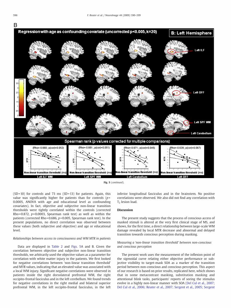
Fig. 5 (continued).
596 F. Reuter et al. / NeuroImage 44 (2009) 590–599
(SD=10) for controls and 73 ms (SD=13) for patients. Again, thisvalue was significantly higher for patients than for controls (p=0.0005, ANOVA with age and educational level as confoundingcovariates). In fact, objective and subjective non-linear transitionthresholds were tightly correlated within the controls (correctedRho=0.872, p=0.0003, Spearman rank test) as well as within thepatients (corrected Rho=0.686, p=0.005, Spearman rank test). In thepresent populations, no direct correlation was observed betweenthese values (both subjective and objective) and age or educationallevel.
Relationships between access to consciousness and WM MTR in patients
Data are displayed in Table 2 and Figs. 5A and B. Given thecorrelation between objective and subjective non-linear transitionthresholds, we arbitrarily used the objective values as a parameter forcorrelation with white matter injury in the patients. We first lookedfor negative correlations between ‘non-linear transition threshold’and MTR values, indicating that an elevated value was associated witha local WM injury. Significant negative correlations were observed inpatients inside the right dorsolateral prefrontal WM, the rightoccipito-frontal fasciculus and in the left cerebellum.We found trendsfor negative correlations in the right medial and bilateral superiorprefrontal WM, in the left occipito-frontal fasciculus, in the left
inferior longitudinal fasciculus and in the brainstem. No positivecorrelations were observed. We also did not find any correlation withT2 lesion load.
Discussion
The present study suggests that the process of conscious access ofmasked stimuli is altered at the very first clinical stage of MS, andshows, for the first time, a direct relationship between large-scaleWMdamage revealed by local MTR decrease and abnormal and delayedtransition towards conscious perception during masking.
Measuring a ‘non-linear transition threshold’ between non-consciousand conscious perception
The present work uses the measurement of the inflexion point ofthe sigmoidal curve relating either objective performance or sub-jective visibility to target-mask SOA as a marker of the transitionperiod between non-conscious and conscious perception. This aspectof our research is based on prior results, replicated here, which showsthat in some metacontrast masking, substitution masking andattentional blink tasks, participants' reports of seeing the stimulusevolve in a highly non-linear manner with SOA (Del Cul et al., 2007;Del Cul et al., 2006; Reuter et al., 2007; Sergent et al., 2005; Sergent

597F. Reuter et al. / NeuroImage 44 (2009) 590–599
and Dehaene, 2004). Several findings suggest that this non-linearityreflects a major event in the brain's processing of masked stimuli,justifying our attempt to use it as a marker of brain injury. Even whenparticipants are given an opportunity to respond with a continuousrating of visibility, their responses are distributed in a bimodalmanner: on any given trial, they either report very high visibility orvery reduced visibility. Thus, the smooth sigmoidal curve actuallyreflects the average of different proportions of seen and not-seenjudgments, and the inflexion point characterizes the point of maximalvariability in these proportions. Recordings of brain activation atseveral levels of masking (Del Cul et al., 2007) confirm that a sharpnon-linear phase transition underlies the sudden feeling of “seeing” amasked stimulus. Note that the inflexion point is just a convenient,robust measure of this shifted non-linearity. Determining such a delayin a region of the curve with large gradient variations minimizes themeasurement error and allows for a better sensitivity when compar-ing the two populations.
It is important to note that our definition of ‘non-linear transitionthreshold’ differs from the classical psychological definition of“objective threshold” and “subjective threshold”, Objective thresholdis classically defined as the target-mask delay at which performancein some objective task (e.g. target classification) drops down tochance level, and subjective threshold as the delay at which subjectsdeny detecting the stimulus (Cheesman and Merikle, 1984). Thesubjective threshold is usually higher than the objective threshold,thus defining a zone of objective “subliminal” performance withoutsubjective reportability. In our prior research (Del Cul et al., 2006),we found such a dissociation at short SOAs, where a non-negligibleproportion of subjectively not-seen trials were accompanied byabove-chance objective performance. In Cheesman and Merikle'sterminology, our results would indicate a very low “objectivethreshold”. The significance of these definitions, however, remainsheavily debated (Dehaene and Naccache, 2001; Holender, 1986;Merikle et al., 2001). Several authors have noted that the thresholdsare difficult to measure reliably since they depend on the exactinstructions, the statistical level of significance chose, and the amountof data collected. Accepting the null hypothesis of a zero d-prime in anobjective task can under-estimate conscious access. Conversely,higher-than-chance performance can occur while subjects denyseeing anything, and thus the objective threshold can over-estimateconscious access.
Our study sidesteps many of these difficulties, while focusing onwhat is, arguably, the most salient finding in the masking literaturesince over a century: the non-linearity of the curve relatingperception to the target-mask delay, indicating that beyond a criticaldelay, subjects exhibit a sudden ability to extract information andbecome aware of the masked stimulus. In the present, this non-linearity is also the most evident difference between the controls andthe patients: there is a systematic shift of both objective andsubjective curves towards higher SOA values in patients, and ourmeasure of ‘non-linear transition threshold’ adequately captures thisphenomenon. Future work should return to the largely orthogonalissue of whether the point of first occurrence of a deviation fromchance-level responding also differs between patients and controls(but see Fig. 4 for a clear illustration of the difficulty of assessing thispoint).
Subliminal priming effect
As reported in relapsing remittingMS (RR-MS) (Reuter et al., 2007),patients with CIS, showed significant subliminal priming effectsuggesting normal non-conscious processing. However, we foundnormal reaction times in CIS patients while RR-MS were slower thancontrols. The longer RT found in RR-MS patients could be due, at leastin part, to motor disability or/and to slower information processingspeed.
Impairment of access to consciousness in patients
As previously described, the “conscious neuronal workspace”theory emphasizes the role of long-distance connections necessaryfor the emergence of a self-amplified reverberant neuronal assemblywhich connects together distant brain areas, particularly prefrontal,cingulate and parietal cortices, and which is associated with con-scious reportability (Dehaene et al., 1998; Dehaene et al., 2003).Diffuse demyelinating processes in patients with MS may disturb theconnectivity between distant cortical areas (Au Duong et al., 2005a;Au Duong et al., 2005b; Audoin et al., 2007b), and thus provide amechanism for impaired conscious access. The load of brain MRI-visible lesions, which is generally low at the early stage of MS, maynot account significantly for the degree of impairment of access toconsciousness as suggested by the lack of correlation between the T2lesion load and the non-linear transition threshold in the presentstudy. This is not surprising considering the results of numerous MRIstudies (Allen and McKeown, 1979; Audoin et al., 2005; Audoin et al.,2007b; Cercignani et al., 2001; Cercignani et al., 2000; Evangelou etal., 2000; Tortorella et al., 2000) which demonstrated thatpathological processes could be present outside MRI-visible lesions,in the so called ‘normal appearing brain tissue’ since the very firststage of MS.
The deficit observed in patients is not due to damage to early visualpathways
As discussed in our previous report on relapsing remitting MS(Reuter et al., 2007), impairment of conscious access cannot berelated to a dysfunction along the visual pathway including the opticnerve. Because our measures are obtained from the variation inperformance as a function of target-mask interval, they should notbe affected by an additive optic nerve delay. A second argumentagainst putative low-level visual deficits is the finding of a normalsubliminal priming effect in MS patients (Reuter et al., 2007).Behavioral and neuroimaging studies of subliminal priming suggestthat masked invisible stimuli are processed extensively, includingvisual recognition, but also lexical and even semantic levels (Dehaene,2004; Dehaene et al., 2004; Dehaene et al., 2001; Greenwald et al.,1996; Naccache and Dehaene, 2001). The preservation of non-conscious priming in very early MS patients suggests that the fastfeed-forward processing stages supporting these priming effects mustbe largely intact, including conduction from optic nerve to visualcortex.
The fact that the present study used a visual task to assessnon-conscious and conscious processing in MS patients might raisethe question of a selection bias with the exclusion of patients withoptic neuritis, one of the most common clinical onsets of MS. How-ever, it is noteworthy to state that the first goal of this preliminarystudy was to demonstrate the important involvement of large WMbundles integrity on access to consciousness. To generalize thisapproach and allowing a clinical transfer of the method to the wholepopulation of MS patients, it will be crucial to develop paradigms inother sensory modalities, such as auditory perception, for whichperipheral injury is far less frequent in early clinical stage of MS.
White matter damage impairs access to consciousness
In the present study, significant negative correlations indicatedthat the severity of the anomaly in the transition to consciousperception (delayed inflexion point) was related to the impaired MTRvalues in the right dorsolateral prefrontal WM, the right occipito-frontal fasciculus and in the left cerebellum. We found trends fornegative correlations in the right medial and bilateral superior pre-frontal WM, in the left occipito-frontal fasciculus, in the left inferiorlongitudinal fasciculus and in the brainstem. The occipito-frontal

598 F. Reuter et al. / NeuroImage 44 (2009) 590–599
fasciculus is a cortical associative fiber tract which extends from thedorsal and medial part of the occipital lobes, the dorsal, medial andinferior parietal lobes to the dorsal and medial part of the prefrontaland premotor regions (Makris et al., 2007). The inverse correlationbetween the degree of tissue injury of the occipito-frontal fasciculusand the ‘non-linear transition threshold’ may be related to thestrategic anatomical situation of this WM bundle which may beinvolved in the extension of the brain activation from perceptualoccipital areas to associative prefrontal cortices. The inferior long-itudinal fasciculus is a WM associative tract connecting the occipitaland temporal lobes. Fibers originate from extrastriate visual associa-tive areas of the occipital lobe, sparing the primary visual cortex(Schmahmann and Pandya, 2006). At the occipital horn of the lateralventricle, the inferior longitudinal fasciculus fibers run laterally to theoptic radiation and callosal (tapetal) fibers and fibers terminate inlateral and medial temporal areas. In summary, both the inferiorlongitudinal fasciculus and the occipito-frontal fasciculus connectvisual inputs to higher cortical areas in the occipito-temporal andfrontal pathways. In the context of the ‘conscious neuronal workspace’theory, these bundles may play a critical role in linking specializedperceptual visual processors to higher-level prefrontal, parietal andcingulate cortices that enter into coherent self-sustained activationpatterns during conscious processing. In a recent study (Del Cul et al.,2007), we used high-density EEG to directly monitor the corticalprogression of amasked visual stimulus (identical to the presentwork)as a function of its visibility. We found that invisible stimuli triggeredan initial activation in contralateral occipito-temporal cortex andposterior parietal cortices, but that only visible stimuli propagatedfurther into the temporal lobes and especially into inferior prefrontalcortex, leading to a late (N270 ms) surge of global activity distributedbilaterally within prefrontal, posterior parietal and ventral occipito-temporal cortices (Del Cul et al., 2007). Impaired inferior longitudinalfasciculus and occipito-frontal fasciculus fiber tracts would signifi-cantly slow this posterior-to-anterior communication and global re-verberation, thus providing plausible though still hypothetical neuralmechanism relating WM injury to altered access to consciousness.
Interpretation of the correlations found between impaired con-scious access and MTR values in the brainstem and the leftcerebellum is less clear. However, few studies have shown theinvolvement of brainstem in attentional processes through theascendant reticular activating system that could be impaired inpatients with CIS (Gadea et al., 2004 6). Concurrently, one functionalconnectivity experiment has demonstrated significant correlationsbetween low-frequency fluctuations in MR signal of the dentatenucleus and signal fluctuations in cerebellar, thalamic, limbic, striatal,and cortical regions including parietal and frontal sites, withprominent coherence in dorsolateral prefrontal cortex (Allen et al.,2005). This suggests the presence of functional coherence in a large-scale network including cerebellar–parietal and cerebellar–prefrontalfunctional connections that could be also involved in the conscious-ness network.
The present study evidences that impaired access to consciousnessis present since the very early stage of MS and is related to early injuryof long-distance WM bundles. At the theoretical level, it provides thefirst clinical evidence of a direct relationship between access toconsciousness and integrity of large WM bundles, as predicted by theglobal neuronal workspace theory.
Conflict of interest statementAny authors declare that they have no conflict of interest relative to this research.
Acknowledgments
This work is supported by the ‘ARSEP’, ‘CNRS’, ‘INSERM’, ‘InstitutUniversitaire de France’ and Sanofi Aventis France.
FR is granted by the ARSEP for her PhD preparation.
References
Allen, G., McColl, R., Barnard, H., Ringe, W.K., Fleckenstein, J., Cullum, C.M., 2005.Magnetic resonance imaging of cerebellar–prefrontal and cerebellar–parietalfunctional connectivity. Neuroimage 28, 39–48.
Allen, I.V., McKeown, S.R., 1979. A histological, histochemical and biochemical study ofthe macroscopically normal white matter in multiple sclerosis. J. Neurol. Sci. 41,81–91.
Au Duong, M.V., Audoin, B., Boulanouar, K., Ibarrola, D., Malikova, I., Confort-Gouny, S.,Celsis, P., Pelletier, J., Cozzone, P.J., Ranjeva, J.P., 2005a. Altered functionalconnectivity related to white matter changes inside the working memorynetwork at the very early stage of MS. J. Cereb. Blood Flow Metab. 25, 1245–1253.
Au Duong, M.V., Boulanouar, K., Audoin, B., Treseras, S., Ibarrola, D., Malikova, I., Confort-Gouny, S., Celsis, P., Pelletier, J., Cozzone, P.J., Ranjeva, J.P., 2005b. Modulation ofeffective connectivity inside the working memory network in patients at theearliest stage of multiple sclerosis. Neuroimage 24, 533–538.
Audoin, B., Ranjeva, J.P., Au Duong, M.V., Ibarrola, D., Malikova, I., Confort-Gouny, S.,Soulier, E., Viout, P., Ali-Cherif, A., Pelletier, J., Cozzone, P.J., 2004. Voxel-basedanalysis of MTR images: a method to locate gray matter abnormalities in patientsat the earliest stage of multiple sclerosis. J. Magn. Reson. Imaging 20, 765–771.
Audoin, B., Au Duong, M.V., Ranjeva, J.P., Ibarrola, D., Malikova, I., Confort-Gouny, S.,Soulier, E., Viout, P., Ali-Cherif, A., Pelletier, J., Cozzone, P.J., 2005. Magnetic resonancestudy of the influence of tissue damage and cortical reorganization on PASATperformance at the earliest stage ofmultiple sclerosis. Hum. BrainMapp. 24, 216–228.
Audoin, B., Fernando, K.T., Swanton, J.K., Thompson, A.J., Plant, G.T., Miller, D.H., 2006.Selective magnetization transfer ratio decrease in the visual cortex following opticneuritis. Brain 129, 1031–1039.
Audoin, B., Davies, G., Rashid, W., Fisniku, L., Thompson, A.J., Miller, D.H., 2007a. Voxel-based analysis of grey matter magnetization transfer ratio maps in early relapsingremitting multiple sclerosis. Mult. Scler. 13, 483–489.
Audoin, B., Guye, M., Reuter, F., Au Duong, M.V., Confort-Gouny, S., Malikova, I., Soulier,E., Viout, P., Cherif, A.A., Cozzone, P.J., Pelletier, J., Ranjeva, J.P., 2007b. Structure ofWM bundles constituting the working memory system in earlymultiple sclerosis: aquantitative DTI tractography study. Neuroimage 36, 1324–1330.
Beck, D.M., Rees, G., Frith, C.D., Lavie, N., 2001. Neural correlates of change detection andchange blindness. Nat. Neurosci. 4, 645–650.
Boringa, J.B., Lazeron, R.H., Reuling, I.E., Ader, H.J., Pfennings, L., Lindeboom, J.,de Sonneville, L.M., Kalkers, N.F., Polman, C.H., 2001. The brief repeatable batteryof neuropsychological tests: normative values allow application in multiplesclerosis clinical practice. Mult. Scler. 7, 263–267.
Breitmeyer, B.G., Ogmen, H., 2000. Recent models and findings in visual backwardmasking: a comparison, review, and update. Percept. Psychophys. 62, 1572–1595.
Cader, S., Cifelli, A., Abu-Omar, Y., Palace, J., Matthews, P.M., 2006. Reduced brainfunctional reserve and altered functional connectivity in patients with multiplesclerosis. Brain 129, 527–537.
Cercignani, M., Iannucci, G., Rocca, M.A., Comi, G., Horsfield, M.A., Filippi, M., 2000.Pathologic damage in MS assessed by diffusion-weighted and magnetizationtransfer MRI. Neurology 54, 1139–1144.
Cercignani, M., Bozzali, M., Iannucci, G., Comi, G., Filippi, M., 2001. Magnetisationtransfer ratio andmean diffusivity of normal appearing white and greymatter frompatients with multiple sclerosis. J. Neurol. Neurosurg. Psychiatry 70, 311–317.
Cheesman, J., Merikle, P.M., 1984. Priming with and without awareness. Percept.Psychophys. 36, 387–395.
Chen, J.T., Kuhlmann, T., Jansen, G.H., Collins, D.L., Atkins, H.L., Freedman,M.S., O'Connor,P.W., Arnold, D.L., 2007. Voxel-based analysis of the evolution of magnetizationtransfer ratio to quantify remyelination and demyelination with histopathologicalvalidation in a multiple sclerosis lesion. Neuroimage 36, 1152–1158.
Dehaene, S., 2004. The neural basis of subliminal priming. In: Kanwisher, N., Duncan, J.(Eds.), Functional Neuroimaging of Visual Cognition (Attention and PerformanceSeries). Oxford university press, New York.
Dehaene, S., Naccache, L., 2001. Towards a cognitive neuroscience of consciousness:basic evidence and a workspace framework. Cognition 79, 1–37.
Dehaene, S., Kerszberg, M., Changeux, J.P., 1998. A neuronal model of a global workspacein effortful cognitive tasks. Proc. Natl. Acad. Sci. U. S. A. 95, 14529–14534.
Dehaene, S., Naccache, L., Cohen, L., Bihan, D.L., Mangin, J.F., Poline, J.B., Riviere, D., 2001.Cerebral mechanisms of word masking and unconscious repetition priming. Nat.Neurosci. 4, 752–758.
Dehaene, S., Sergent, C., Changeux, J.P., 2003. A neuronal network model linkingsubjective reports and objective physiological data during conscious perception.Proc. Natl. Acad. Sci. U. S. A. 100, 8520–8525.
Dehaene, S., Jobert, A., Naccache, L., Ciuciu, P., Poline, J.B., Le Bihan, D., Cohen, L., 2004.Letter binding and invariant recognition of masked words: behavioral andneuroimaging evidence. Psychol. Sci. 15, 307–313.
Dehaene, S., Changeux, J.P., Naccache, L., Sackur, J., Sergent, C., 2006. Conscious,preconscious, and subliminal processing: a testable taxonomy. Trends Cogn. Sci. 10,204–211.
Del Cul, A., Dehaene, S., Leboyer, M., 2006. Preserved subliminal processing andimpaired conscious access in schizophrenia. Arch. Gen. Psychiatry 63, 1313–1323.
Del Cul, A., Baillet, S., Dehaene, S., 2007. Brain dynamics underlying the nonlinearthreshold for access to consciousness. PLoS Biol. 5, e260.
Di Lollo, V., Enns, J.T., Rensink, R.A., 2000. Competition for consciousness among visualevents: the psychophysics of reentrant visual processes. J. Exp. Psychol. Gen. 129,481–507.
Evangelou, N., Esiri, M.M., Smith, S., Palace, J., Matthews, P.M., 2000. Quantitativepathological evidence for axonal loss in normal appearing white matter in multiplesclerosis. Ann. Neurol. 47, 391–395.

599F. Reuter et al. / NeuroImage 44 (2009) 590–599
Feinstein, J.S., Stein, M.B., Castillo, G.N., Paulus, M.P., 2004. From sensory processes toconscious perception. Conscious Cogn. 13, 323–335.
Friston, K.J., Holmes, A.P., Price, C.J., Buchel, C., Worsley, K.J., 1999. Multisubject fMRIstudies and conjunction analyses. Neuroimage 10, 385–396.
Gadea, M., Martinez-Bisbal, M.C., Marti-Bonmati, L., Espert, R., Casanova, B., Coret, F.,Celda, B., 2004. Spectroscopic axonal damage of the right locus coeruleus relates toselective attention impairment in early stage relapsing-remittingmultiple sclerosis.Brain 127, 89–98.
Greenwald, A.G., Draine, S.C., Abrams, R.L., 1996. Three cognitive markers ofunconscious semantic activation. Science 273, 1699–1702.
Gross, J., Schmitz, F., Schnitzler, I., Kessler, K., Shapiro, K., Hommel, B., Schnitzler, A.,2004. Modulation of long-range neural synchrony reflects temporal limitations ofvisual attention in humans. Proc. Natl. Acad. Sci. U. S. A. 101, 13050–13055.
Haynes, J.D., Driver, J., Rees, G., 2005. Visibility reflects dynamic changes of effectiveconnectivity between V1 and fusiform cortex. Neuron 46, 811–821.
Holender, D., 1986. Semantic activation without conscious identification in dichoticlistening, parafoveal vision, and visual masking: a survey and appraisal. Behav.Brain Sci. 9, 1–66.
Kranczioch, C., Debener, S., Maye, A., Engel, A.K., 2007. Temporal dynamics of access toconsciousness in the attentional blink. Neuroimage 37, 947–955.
Kurtzke, J.F., 1983. Rating neurologic impairment in multiple sclerosis: an expandeddisability status scale (EDSS). Neurology 33, 1444–1452.
Lumer, E.D., Rees, G.,1999. Covariation of activity in visual and prefrontal cortex associatedwith subjective visual perception. Proc. Natl. Acad. Sci. U. S. A. 96, 1669–1673.
Makris, N., Papadimitriou, G.M., Sorg, S., Kennedy, D.N., Caviness, V.S., Pandya, D.N.,2007. The occipitofrontal fascicle in humans: a quantitative, in vivo, DT-MRI study.Neuroimage 37, 1100–1111.
Marois, R., Yi, D.J., Chun, M.M., 2004. The neural fate of consciously perceived andmissed events in the attentional blink. Neuron 41, 465–472.
Merikle, P.M., Smilek, D., Eastwood, J.D., 2001. Perception without awareness:perspectives from cognitive psychology. Cognition 79, 115–134.
Naccache, L., Dehaene, S., 2001. Unconscious semantic priming extends to novel unseenstimuli. Cognition 80, 215–229.
Ranjeva, J.P., Audoin, B., Au Duong, M.V., Ibarrola, D., Confort-Gouny, S., Malikova, I.,Soulier, E., Viout, P., Ali-Cherif, A., Pelletier, J., Cozzone, P., 2005. Local tissue damageassessed with statistical mapping analysis of brain magnetization transfer ratio:relationship with functional status of patients in the earliest stage of multiplesclerosis. AJNR Am. J. Neuroradiol. 26, 119–127.
Reuter, F., Del Cul, A., Audoin, B., Malikova, I., Naccache, L., Ranjeva, J.P., Lyon-Caen, O., AliCherif, A., Cohen, L., Dehaene, S., Pelletier, J., 2007. Intact subliminal processing anddelayed conscious access in multiple sclerosis. Neuropsychologia 45, 2683–2691.
Schmahmann, J.D., Pandya, D.N., 2006. Fiber Pathways of the Brain. Oxford UniversityPress.
Sergent, C., Dehaene, S., 2004. Is consciousness a gradual phenomenon? Evidence for anall-or-none bifurcation during the attentional blink. Psychol. Sci. 15, 720–728.
Sergent, C., Baillet, S., Dehaene, S., 2005. Timing of the brain events underlying access toconsciousness during the attentional blink. Nat. Neurosci. 8, 1391–1400.
Tortorella, C., Viti, B., Bozzali, M., Sormani, M.P., Rizzo, G., Gilardi, M.F., Comi, G., Filippi,M., 2000. A magnetization transfer histogram study of normal-appearing braintissue in MS. Neurology 54, 186–193.
van Waesberghe, J.H., Kamphorst, W., De Groot, C.J., van Walderveen, M.A., Castelijns,J.A., Ravid, R., Lycklama a Nijeholt, G.J., van der Valk, P., Polman, C.H., Thompson, A.J.,Barkhof, F., 1999. Axonal loss in multiple sclerosis lesions: magnetic resonanceimaging insights into substrates of disability. Ann. Neurol. 46, 147–154.