whiplash associated disorders (wad) - c.ymcdn.com · published in the 2001 proceedings of the...
TRANSCRIPT

Whiplash Associated
Disorders (WAD) Instability & Case Studies
Steven G. Yeomans, DC, FACO
404 Eureka Street
Ripon, WI 64971-0263
920-748-3644 (Ph)
920-748-3642 (Fax)
www.yeomansdc.com
RADIOLOGICAL Exam

RADIOLOGICAL Exam
Weight bearing: Pro – posture assessment; Con –
movement artifact / quality (esp. BMI >35)
Recumbent: Pro – quality best; Con – no
biomechanical assessment
Stress Views (C & L Spine primarily)
– Sagittal Plane: Flexion/Extension
– Frontal Plane: Lateral Flexion (Apical/Alar Ligs)
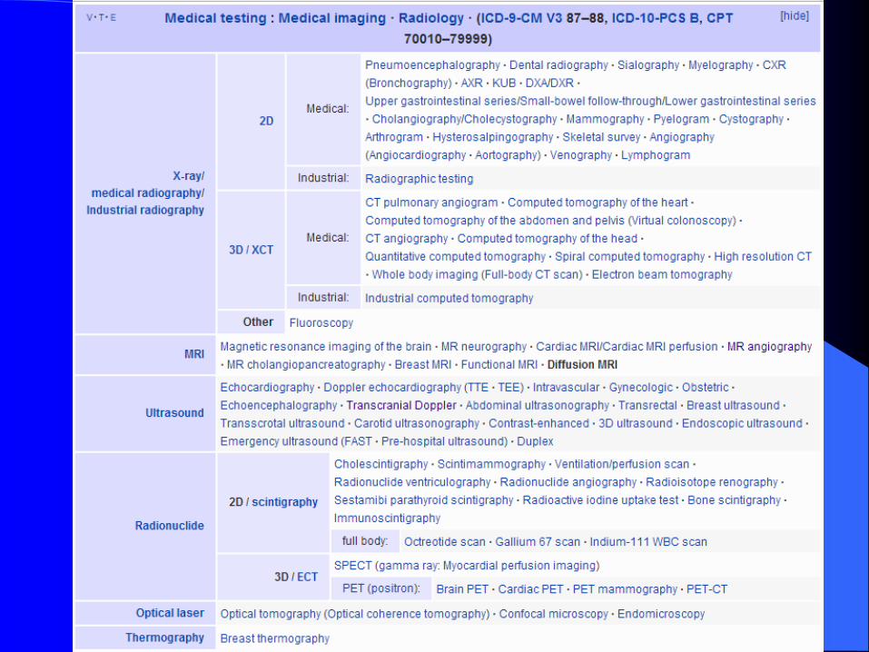
Special tests: MRI, CT, Bone Scan, fMRI, PET Scan,
DTI
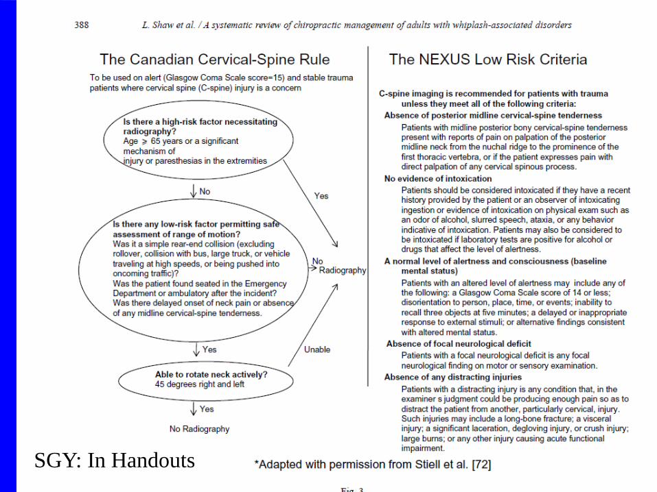
SGY: In Handouts
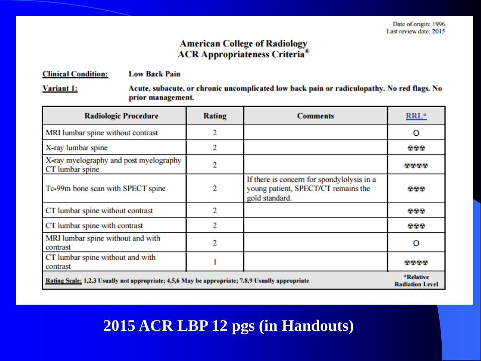
2015 ACR LBP 12 pgs (in Handouts)

1999 / 2012 ACR Cervical Spine 23pgs (in Handouts)

Published in the 2001 proceedings of the
Cervical Spine Research Society Annual Meeting
Whiplash Injury and Occult Vertebral Fracture: A Case Series of
SPECT Imaging of Patients with Persisting Pain Following a
Motor Vehicle Crash
Michael Freeman, Ph.D., D.C., M.P.H, Dan Sapir, M.D., Alex
Boutselis, M.D., John Gorup, M.D., Glen Tuckman, M.D., Arthur
Croft, D.C., M.P.H., M.S., Chris Centeno, M.D., Arnie Phillips, M.D.
BONE SCAN – SPECT Scan
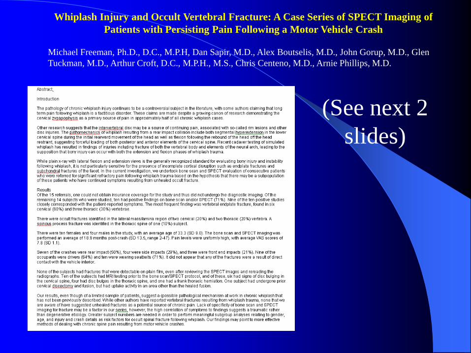
(See next 2
slides)
Whiplash Injury and Occult Vertebral Fracture: A Case Series of SPECT Imaging of
Patients with Persisting Pain Following a Motor Vehicle Crash
Michael Freeman, Ph.D., D.C., M.P.H, Dan Sapir, M.D., Alex Boutselis, M.D., John Gorup, M.D., Glen
Tuckman, M.D., Arthur Croft, D.C., M.P.H., M.S., Chris Centeno, M.D., Arnie Phillips, M.D.

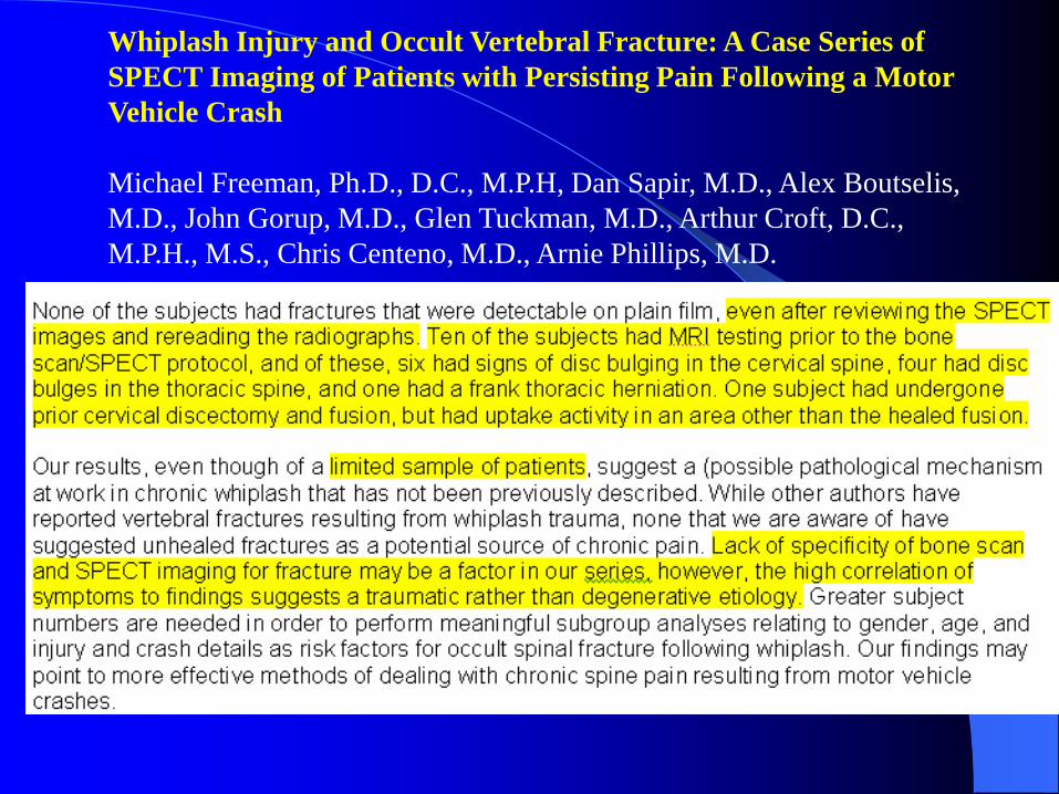
Whiplash Injury and Occult Vertebral Fracture: A Case
Series of SPECT Imaging of Patients with Persisting Pain
Following a Motor Vehicle Crash
Michael Freeman, Ph.D., D.C., M.P.H, Dan Sapir, M.D., Alex Boutselis,
M.D., John Gorup, M.D., Glen Tuckman, M.D., Arthur Croft, D.C.,
M.P.H., M.S., Chris Centeno, M.D., Arnie Phillips, M.D.

Whiplash Injury and Occult Vertebral Fracture: A Case Series of
SPECT Imaging of Patients with Persisting Pain Following a Motor
Vehicle Crash
Michael Freeman, Ph.D., D.C., M.P.H, Dan Sapir, M.D., Alex Boutselis,
M.D., John Gorup, M.D., Glen Tuckman, M.D., Arthur Croft, D.C.,
M.P.H., M.S., Chris Centeno, M.D., Arnie Phillips, M.D.

MRI: Alar & Transverse Ligaments
STUDY:
Magnetic Resonance Imaging of the Alar and
Transverse Ligaments in Acute Whiplash-
Associated Disorders 1 and 2: A Cross-Sectional
Controlled Study.
– Spine Volume 36(6) pgs. 417-496,E373-E453 March
15, 2011; DOI: 10.1097/BRS.0b013e3181da21a9
– Vetti, Nils MD *,+; Krakenes, Jostein MD, PhD *,+; Damsgaard,
Eivind MD, ++; Rorvik, Jarle MD, PhD *,+; Gilhus, Nils Erik
MD, PhD [S],[P]; Espeland, Ansgar MD, PhD *,+ Miscellaneous
Article

MRI: Alar & Transverse Ligaments (Study)
AB Study Design.
– Cross-sectional.
Objective.
– To describe alar- and transverse-ligament magnetic resonance imaging
(MRI) high-signal changes in acute whiplash-associated disorders (WAD)
grades 1 and 2 in relation to the severity and mechanics of trauma, and to
compare them with controls.
Summary of Background Data.
– The alar and transverse ligaments are important stabilizers at the
craniovertebral junction. Acute injury of these ligaments should be
detected as high-signal changes on high-resolution MRI.

MRI: Alar & Transverse Ligaments (Study)
Methods.
– In the study, 114 consecutive acute WAD I-II patients and 157 non-
injured controls underwent upper-neck high-resolution MRI, using
proton-weighted sequences and Short Tau Inversion Recovery (STIR).
– Two blinded radiologists independently graded high-signal changes 0
to 3 on proton images and assessed ligament high-signal intensity on
STIR.
– Image quality was evaluated as good, reduced, or poor (not
interpretable).
– Multiple logistic regression was used for both within- and between-
groups analyses.
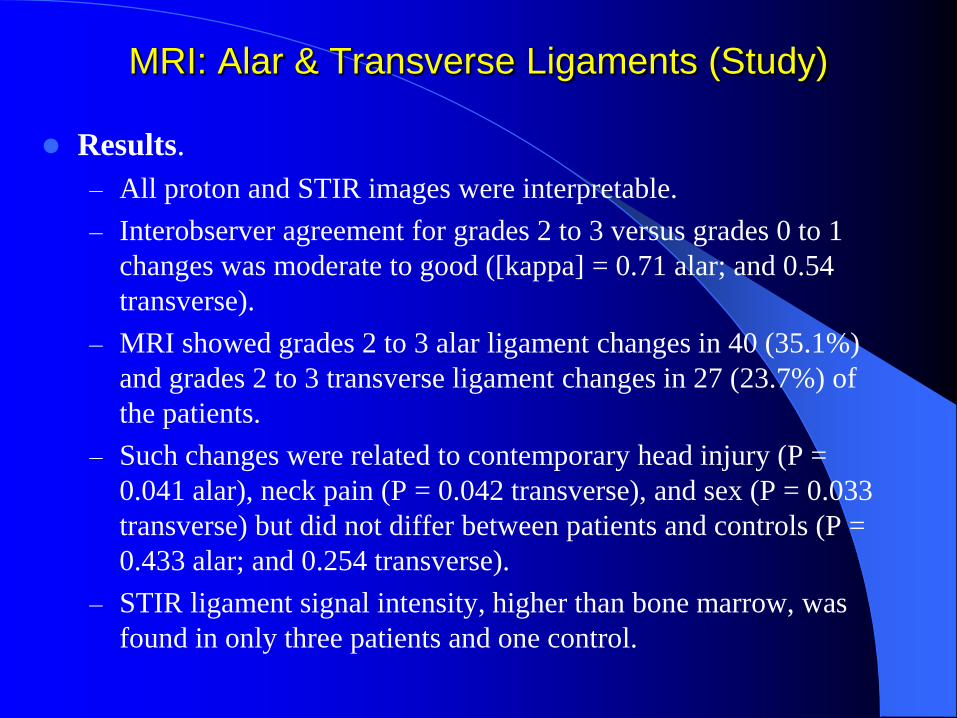
MRI: Alar & Transverse Ligaments (Study)
Results.
– All proton and STIR images were interpretable.
– Interobserver agreement for grades 2 to 3 versus grades 0 to 1
changes was moderate to good ([kappa] = 0.71 alar; and 0.54
transverse).
– MRI showed grades 2 to 3 alar ligament changes in 40 (35.1%)
and grades 2 to 3 transverse ligament changes in 27 (23.7%) of
the patients.
– Such changes were related to contemporary head injury (P =
0.041 alar), neck pain (P = 0.042 transverse), and sex (P = 0.033
transverse) but did not differ between patients and controls (P =
0.433 alar; and 0.254 transverse).
– STIR ligament signal intensity, higher than bone marrow, was
found in only three patients and one control.

MRI: Alar & Transverse Ligaments (Study)
Conclusion.
– This first study on high-resolution MRI of craniovertebral
ligaments in acute WAD 1-2 indicates that such trauma
does not induce high-signal changes.
– Follow-up studies are needed to find out whether pre-
traumatic high-signal changes imply reduced ligament
strength and can predict chronic WAD.


SGY: Note the dif. betw recumb vs. upright in
trauma group only!


See Handouts!

See Handouts!

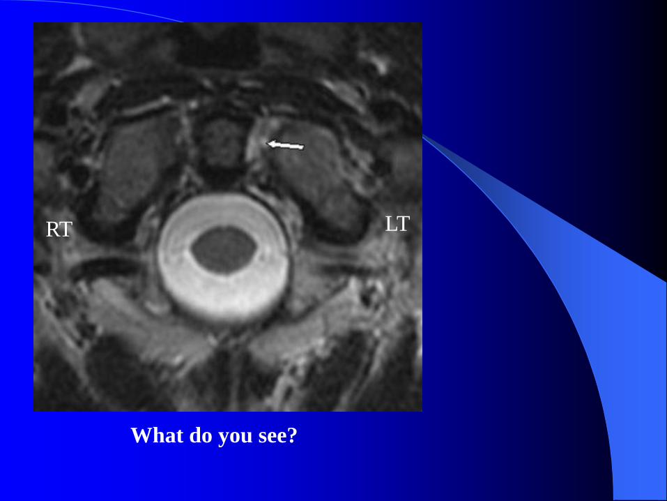
What do you see?

What do you see?
RT LT

LTRT

LT
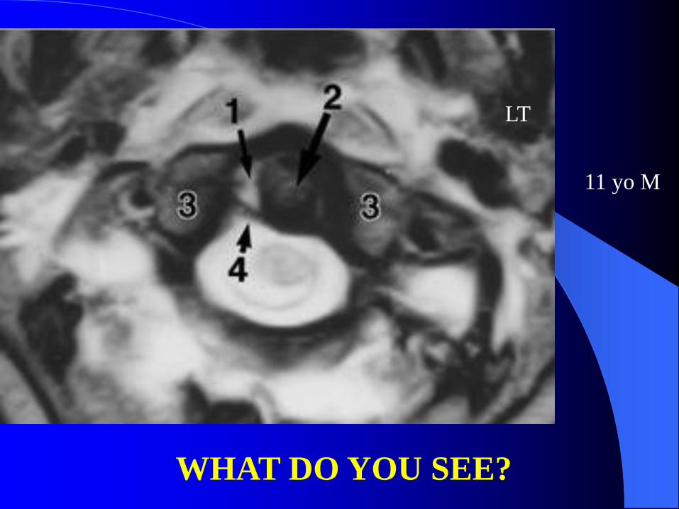
What do you see?

Note the LT lateral
translation of C1 in
reference to C2 and
the increased signal
intensity (T2
weighted image)LT

What do you see?

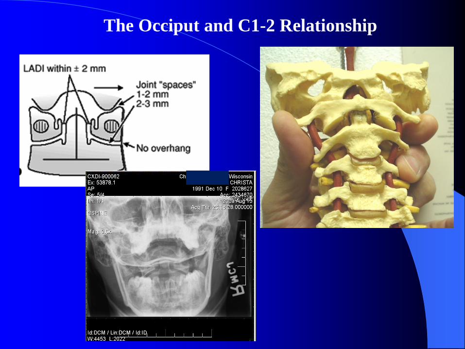
The Occiput and C1-2 Relationship

http://www.ajronline
.org/content/175/3/6
61.long
Am. J. of
Roentgenology
AJR:175, Sept. 2000
SEE Handouts!
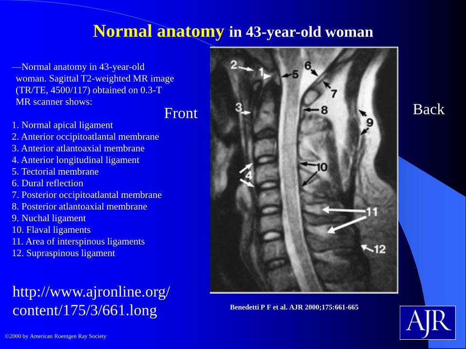
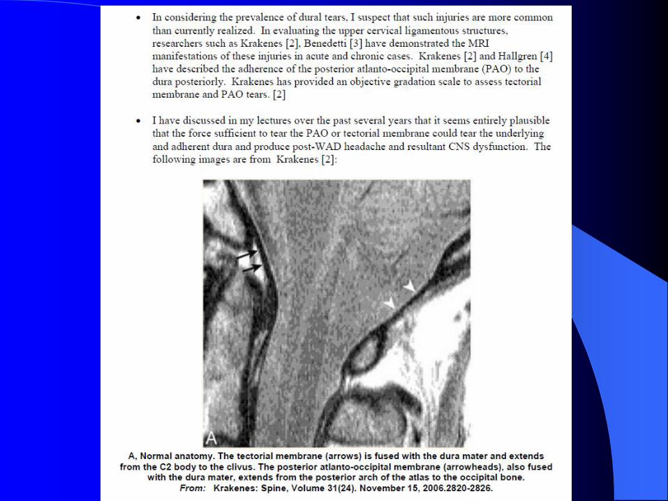
Normal anatomy in 43-year-old woman
Benedetti P F et al. AJR 2000;175:661-665
©2000 by American Roentgen Ray Society
—Normal anatomy in 43-year-old
woman. Sagittal T2-weighted MR image
(TR/TE, 4500/117) obtained on 0.3-T
MR scanner shows:
1. Normal apical ligament
2. Anterior occipitoatlantal membrane
3. Anterior atlantoaxial membrane
4. Anterior longitudinal ligament
5. Tectorial membrane
6. Dural reflection
7. Posterior occipitoatlantal membrane
8. Posterior atlantoaxial membrane
9. Nuchal ligament
10. Flaval ligaments
11. Area of interspinous ligaments
12. Supraspinous ligament
http://www.ajronline.org/
content/175/3/661.long
Front Back

—Normal anatomy in 21-year-old man.
Benedetti P F et al. AJR 2000;175:661-665
©2000 by American Roentgen Ray Society
—Normal anatomy in 21-
year-old man. Sagittal T1-
weighted MR image (TR/TE,
510/25) obtained on 0.3-T
scanner shows:
1. Normal apical ligament
2. Anterior atlantoaxial
membrane
http://www.ajronline.org/con
tent/175/3/661.long
SIDE VIEW
FRONT
C2C2
Back

—Normal anatomy in
a 38-year-old man.
Benedetti P F et al. AJR
2000;175:661-665
—Normal anatomy in 38-year-old man. Axial gradient echo or fast low-angle shot
MR image (TR/TE, 420/18; flip angle, 30°) obtained on 1.0-T MR scanner shows
1. Dens
2. Presumed anterior atlantodental ligaments
3. Alar ligaments
4. Transverse ligament
5. Lateral masses of C1
Coronal View
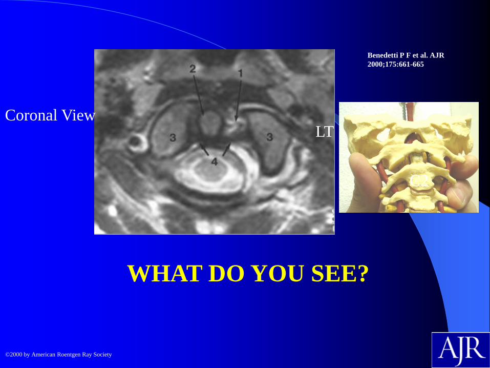
WHAT DO YOU SEE?
Benedetti P F et al. AJR
2000;175:661-665
©2000 by American Roentgen Ray Society
C2
Coronal ViewLT

—Left alar ligament tear in 19-year-old woman with severe neck pain after fall on her
head while snowboarding.
Benedetti P F et al. AJR
2000;175:661-665
©2000 by American Roentgen Ray Society
—Left alar ligament tear in 19-year-old woman with severe neck pain after fall on her head while
snowboarding. Fixed deviation of dens to right was seen on radiograph (not shown). C1-2 rotatory
subluxation was suspected. Axial T2-weighted MR image (TR/TE, 4000/90) obtained on 1.0-T MR
scanner shows isolated tear of left alar ligament (1) and deviation of dens (2) toward right with respect
to lateral masses of C2 (3). Transverse ligament (4) is intact. Sagittal images (not shown) depict normal
alignment of occipital condyles with C2, thus no rotatory subluxation is present. CT performed before
MR imaging was negative for fracture and fixed rotatory subluxation. These results allowed confident
symptomatic treatment that led to full recovery.
C2
Coronal ViewLT
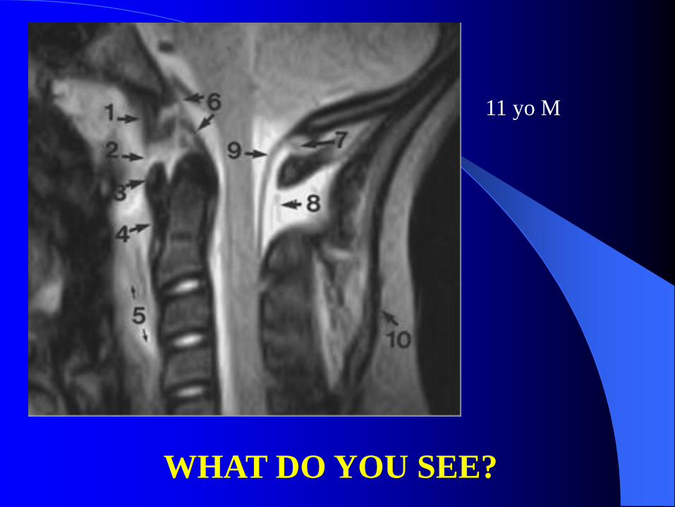
11 yo M
WHAT DO YOU SEE?

11 yo M
11 yo M
WHAT DO YOU SEE?
LT

11 yo M
LT
SAGITTAL PLANE STRESS XR
• Flexion / Extension Lateral Cervical

http://health-7.com/Handbook%20of%20Fractures/9%20-%20Cervical%20Spine
Ligaments, Capsules, Discs
and Muscles

Sagittal C-Spine Instability Panjabi & White: Clinical Checklist of
Instability (discussed in):
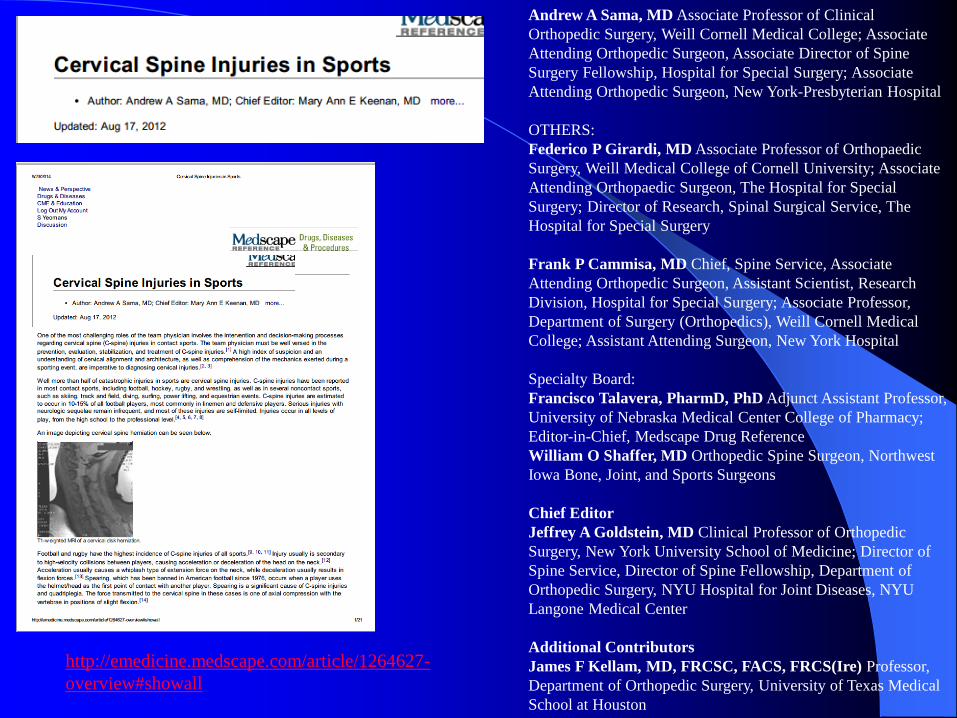
http://emedicine.medscape.com/article/1264627-overview#showall
Cervical Spine Injuries in Sports
Author: Andrew A Sama, MD; Chief Editor:
Mary Ann E Keenan, MD more...
http://emedicine.medscape.com/article/1264627-
overview#showall
Andrew A Sama, MD Associate Professor of Clinical
Orthopedic Surgery, Weill Cornell Medical College; Associate
Attending Orthopedic Surgeon, Associate Director of Spine
Surgery Fellowship, Hospital for Special Surgery; Associate
Attending Orthopedic Surgeon, New York-Presbyterian Hospital
OTHERS:
Federico P Girardi, MD Associate Professor of Orthopaedic
Surgery, Weill Medical College of Cornell University; Associate
Attending Orthopaedic Surgeon, The Hospital for Special
Surgery; Director of Research, Spinal Surgical Service, The
Hospital for Special Surgery
Frank P Cammisa, MD Chief, Spine Service, Associate
Attending Orthopedic Surgeon, Assistant Scientist, Research
Division, Hospital for Special Surgery; Associate Professor,
Department of Surgery (Orthopedics), Weill Cornell Medical
College; Assistant Attending Surgeon, New York Hospital
Specialty Board:
Francisco Talavera, PharmD, PhD Adjunct Assistant Professor,
University of Nebraska Medical Center College of Pharmacy;
Editor-in-Chief, Medscape Drug Reference
William O Shaffer, MD Orthopedic Spine Surgeon, Northwest
Iowa Bone, Joint, and Sports Surgeons
Chief Editor
Jeffrey A Goldstein, MD Clinical Professor of Orthopedic
Surgery, New York University School of Medicine; Director of
Spine Service, Director of Spine Fellowship, Department of
Orthopedic Surgery, NYU Hospital for Joint Diseases, NYU
Langone Medical Center
Additional Contributors
James F Kellam, MD, FRCSC, FACS, FRCS(Ire) Professor,
Department of Orthopedic Surgery, University of Texas Medical
School at Houston

A TOTAL of 5
or more =
UNSTABLE
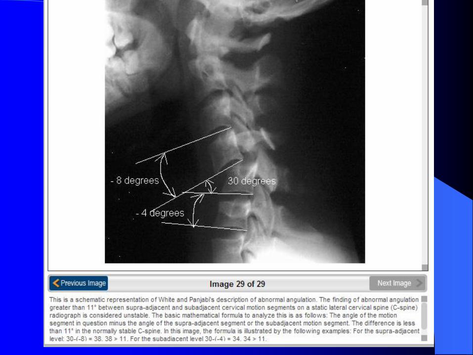
http://emedicine.medscape.com/article/1264627-overview#showall
SGY: Note the
inconsistency with
the AMA Guides
(11° in flexion vs.
here 20° in flexion;
11° in neutral)

http://emedic
ine.medscap
e.com/article
/1264627-
overview#sh
owall
SGY: Note the high
“false-positive” rate
& the recommended
cord diameter / canal
size option

C-Instability/AOMSI:
1) > 20% translation
anterior OR posterior
on FL or Ext XR
2) > 11° on FL only XR
3) Fusion/disc
arthroplasty
pg578 In: 6th Edition:
AMA Guides to the
Evaluation of
Permanent
Impairment, 2008

6th Edition AMA
Guides, 2008
6th Edition AMA Guides, 2008
NOTE: L-spine is different! FL – EXT (vs. FL only XR in C-spine
Method 1

6th Edition AMA Guides, 2008
Method 2
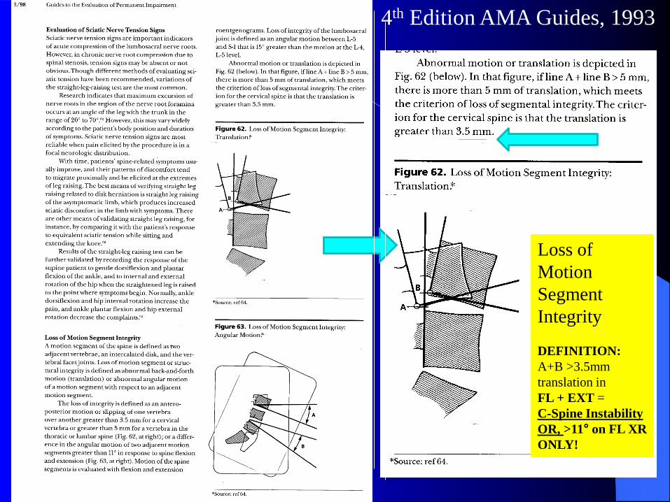
Loss of
Motion
Segment
Integrity
DEFINITION:
A+B >3.5mm
translation in
FL + EXT =
C-Spine Instability
OR, >11° on FL XR
ONLY!
4th Edition AMA Guides, 1993

CASE STUDIES

CASE 1: Name: L A; DOB: 8/14/1986, Age: 27;Sex: Female; X-
ray #: 10356; Complaint: Mid-back, low back and neck pain of a
chronic history; XR taken 2-19-2014
C5: 14°
C6: 1°
Net: 13°
13° > 11°
(neutral XR)EXT
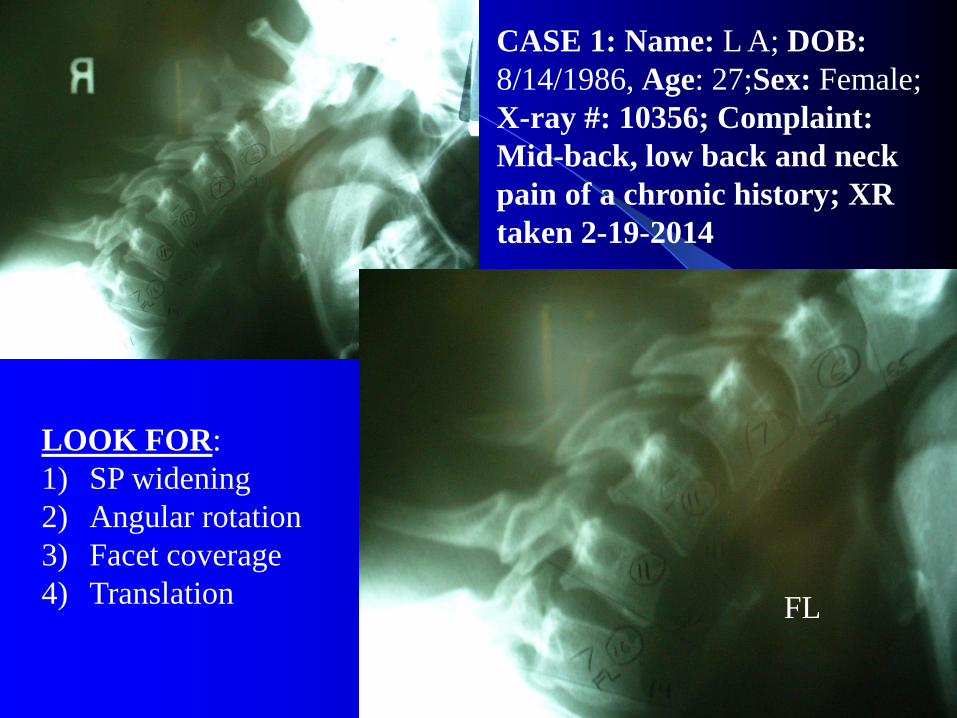
CASE 1: Name: L A; DOB:
8/14/1986, Age: 27;Sex: Female;
X-ray #: 10356; Complaint:
Mid-back, low back and neck
pain of a chronic history; XR
taken 2-19-2014
LOOK FOR:
1) SP widening
2) Angular rotation
3) Facet coverage
4) Translation FL
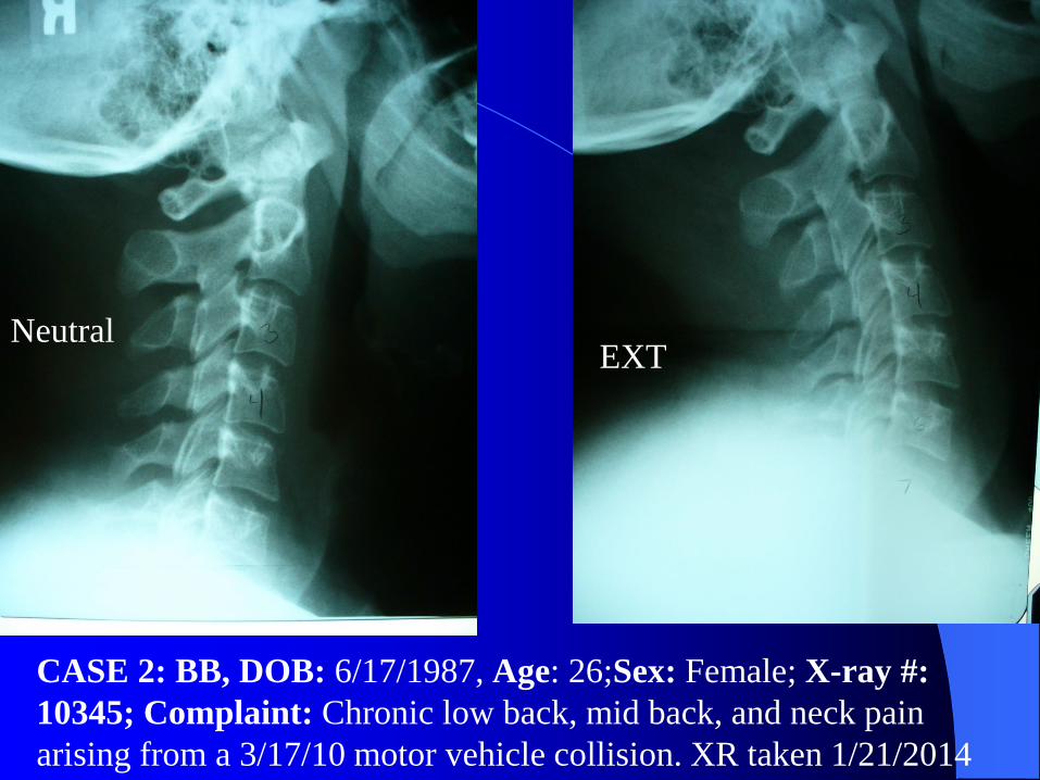
CASE 2: BB, DOB: 6/17/1987, Age: 26;Sex: Female; X-ray #:
10345; Complaint: Chronic low back, mid back, and neck pain
arising from a 3/17/10 motor vehicle collision. XR taken 1/21/2014
EXTNeutral

BB, DOB: 6/17/1987, Age: 26;Sex: Female; X-ray #: 10345;
Complaint: Chronic low back and mid back pain arising from a
3/17/10 motor vehicle collision.
C3/4= 12°
FL
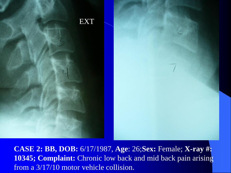
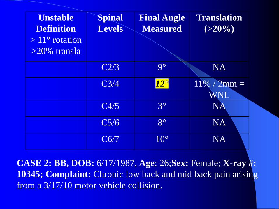
CASE 2: BB, DOB: 6/17/1987, Age: 26;Sex: Female; X-ray #:
10345; Complaint: Chronic low back and mid back pain arising
from a 3/17/10 motor vehicle collision.
EXT
CASE 2: BB, DOB: 6/17/1987, Age: 26;Sex: Female; X-ray #:
10345; Complaint: Chronic low back and mid back pain arising
from a 3/17/10 motor vehicle collision.
Unstable
Definition
> 11° rotation
>20% transla
Spinal
Levels
Final Angle
Measured
Translation
(>20%)
C2/3 9° NA
C3/4 12° 11% / 2mm =
WNL
C4/5 3° NA
C5/6 8° NA
C6/7 10° NA

FRONTAL PLANE STRESS XR
• RT & LT Lateral Flexion APOM
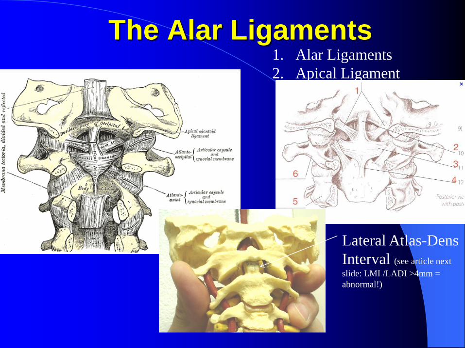
The Alar Ligaments1. Alar Ligaments
2. Apical Ligament
Lateral Atlas-Dens
Interval (see article next
slide: LMI /LADI >4mm =
abnormal!)

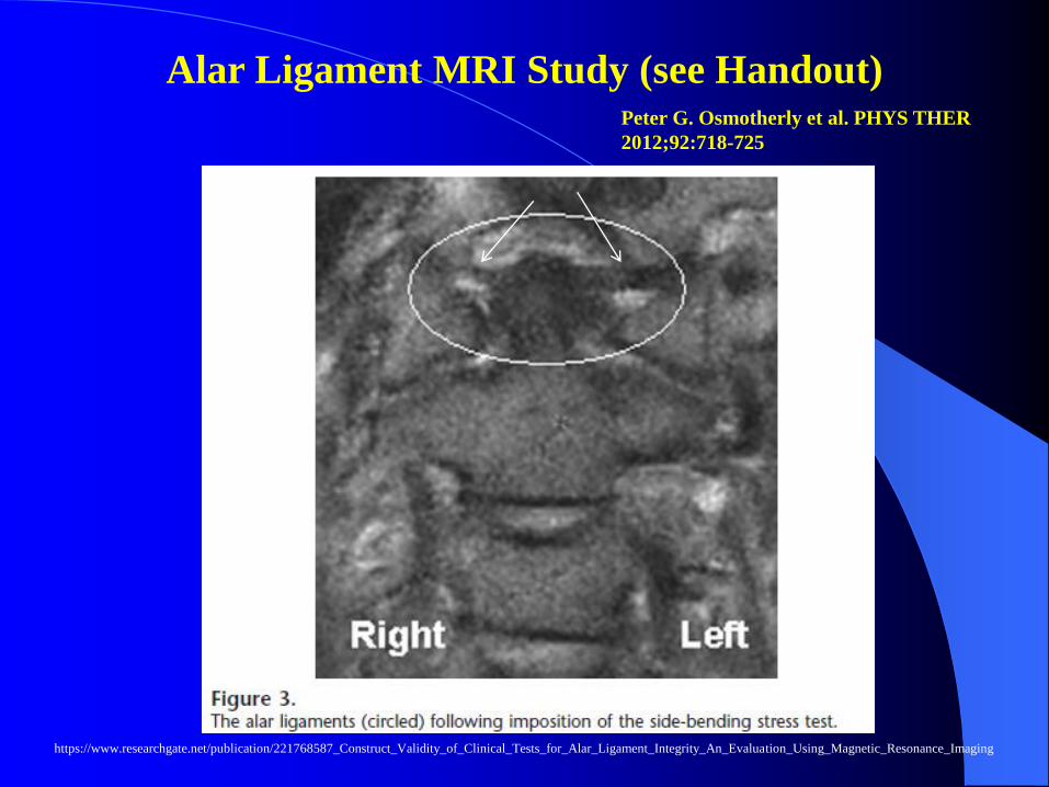
Alar Ligament MRI Study (see Handout)
Peter G. Osmotherly et al. PHYS THER
2012;92:718-725
https://www.researchgate.net/publication/221768587_Construct_Validity_of_Clinical_Tests_for_Alar_Ligament_Integrity_An_Evaluation_Using_Magnetic_Resonance_Imaging

Alar Ligament MRI Study
ABSTRACT SUMMARY:
Peter G. Osmotherly et al. PHYS THER
2012;92:718-725
https://www.researchgate.net/publication/221768587_Construct_Validity_of_Clinical_Tests_for_Alar_Ligament_Integrity_An_Evaluation_Using_Magnetic_Resonance_Imaging
Alar Lig: Side-bending & Rotation Stress (n=16)
– Lat. Fl: 1.15 mm (mean from neutral distance)
– Rotation: 2.08 mm (mean from neutral distance)
Clinical texts state stress XR should be N, FL, Ext
This study supports LF & Rotation stress XR

Alar Ligament MRI Study (see Handout)Peter G. Osmotherly et al. PHYS THER
2012;92:718-725
https://www.researchgate.net/publication/221768587_Construct_Validity_of_Clinical_Tests_for_Alar_Ligament_Integrity_An_Evaluation_Using_Magnetic_Resonance_Imaging
Alar Ligament MRI Study (see Handout)Peter G. Osmotherly et al. PHYS THER
2012;92:718-725
https://www.researchgate.net/publication/221768587_Construct_Validity_of_Clinical_Tests_for_Alar_Ligament_Integrity_An_Evaluation_Using_Magnetic_Resonance_Imaging

Summary of Findings Following the Examination of
Alar Ligament Stress Testing.
Peter G. Osmotherly et al. PHYS THER 2012;92:718-725
© 2012 American Physical Therapy Association
https://www.researchgate.net/publication/221768587_Construct_Validity_of_Clinical_Tests_for_Alar_Ligament_Integrity_An_Evaluation_Using_Magnetic_Resonance_Imaging

Intraclass Correlation Coefficients (ICCs) for the Left-Right Difference in Alar Ligament Length
Measurements.
Peter G. Osmotherly et al. PHYS THER 2012;92:718-725
© 2012 American Physical Therapy Association

Frontal Plane C1/2 Left Lateral
Flexion Stress X-ray
**5mm LT and 3mm RT Lateral ADI (&
notice the C1 lateral mass translation)
CASE 3: CE
DOB: 12-10-91;DOI: 8-15-09 MVC
LLF

Frontal Plane C1/2 Right
Lateral Flexion Stress X-ray
**4mm LT and RT Lateral ADI
CASE 3: CE DOB: 12-10-91; DOI: 8-15-09 MVC
RLF
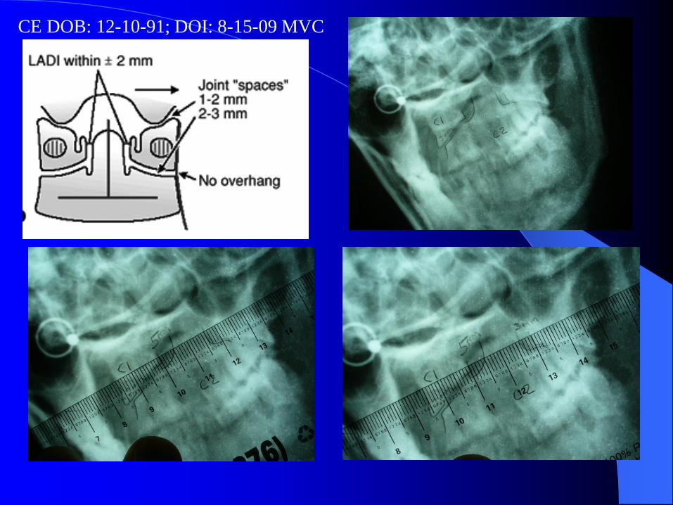
CE DOB: 12-10-91; DOI: 8-15-09 MVC
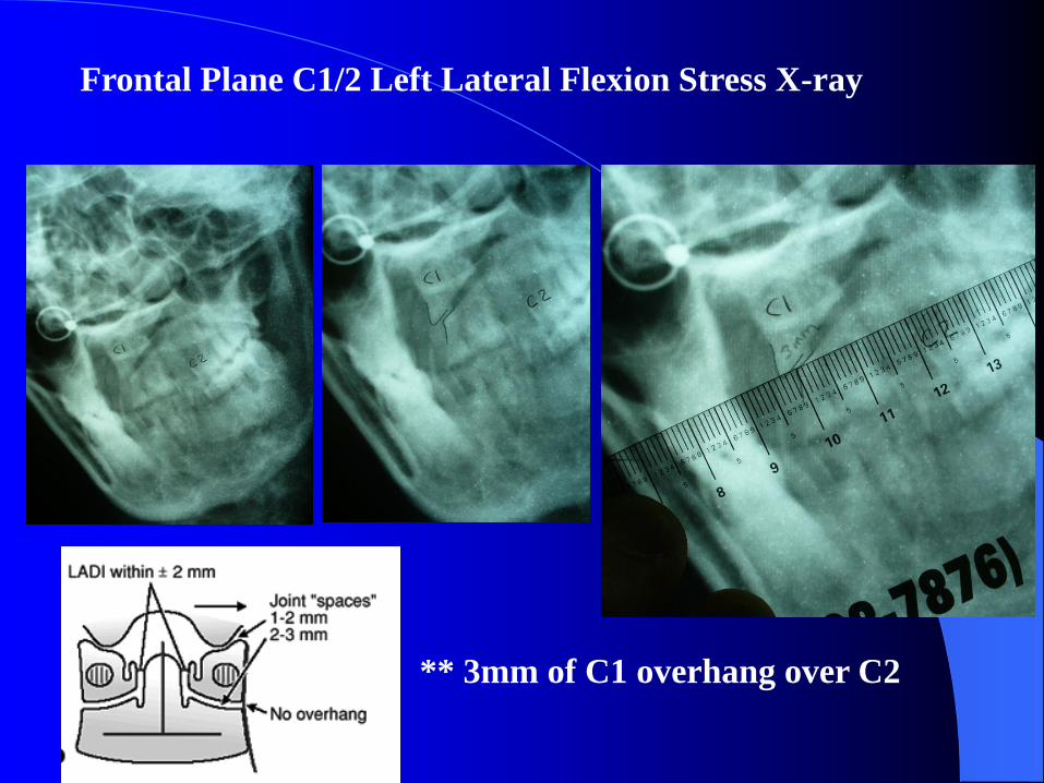
Frontal Plane C1/2 Left Lateral Flexion Stress X-ray
** 3mm of C1 overhang over C2

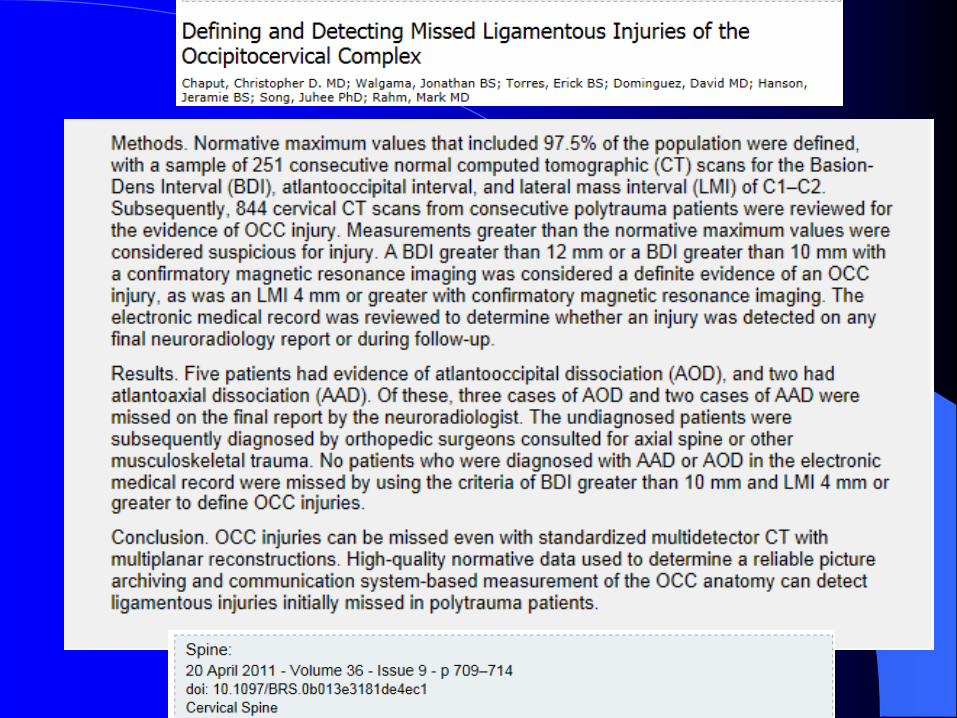

How many OCC injuries have
WE missed???

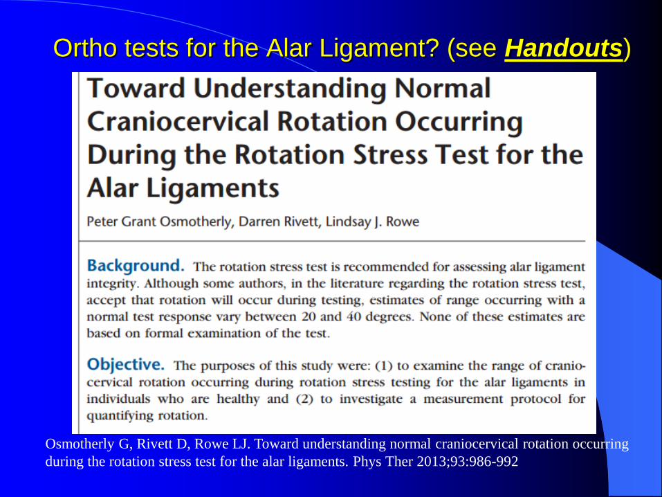
Ortho tests for the
Alar Ligament?
(see Handouts)
Osmotherly G, Rivett D, Rowe LJ. Toward understanding normal
craniocervical rotation occurring during the rotation stress test for
the alar ligaments. Phys Ther 2013;93:986-992
(SEE next 3 slides)
Ortho tests for the Alar Ligament? (see Handouts)
Osmotherly G, Rivett D, Rowe LJ. Toward understanding normal craniocervical rotation occurring
during the rotation stress test for the alar ligaments. Phys Ther 2013;93:986-992
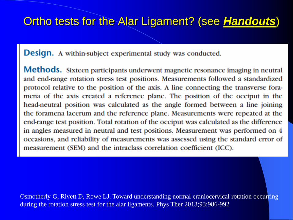
Ortho tests for the Alar Ligament? (see Handouts)
Osmotherly G, Rivett D, Rowe LJ. Toward understanding normal craniocervical rotation occurring
during the rotation stress test for the alar ligaments. Phys Ther 2013;93:986-992

Ortho tests for the Alar Ligament? (see Handouts)
Osmotherly G, Rivett D, Rowe LJ. Toward understanding normal craniocervical rotation occurring
during the rotation stress test for the alar ligaments. Phys Ther 2013;93:986-992

Ortho tests for the Alar Ligament? (see Handouts)
Fig. above from: Osmotherly PG, Rivett
D, Rowe LJ. Toward understanding
normal craniocervical rotation occurring
during the rotation stress test for the alar
ligaments. Phys Ther 2013;93:986-992
Fig. above from: Osmotherly PG, Rivett DA, Rowe LJ.
Construct validity of clinical tests for alar ligament
integrity: An evaluation using magnetic resonance
imaging. Phys Ther 2012;92:718-725.
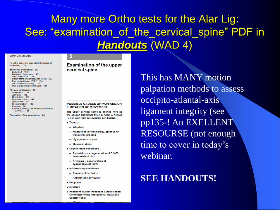
Many more Ortho tests for the Alar Lig:
See: “examination_of_the_cervical_spine” PDF in
Handouts (WAD 4)
This has MANY motion
palpation methods to assess
occipito-atlantal-axis
ligament integrity (see
pp135-! An EXELLENT
RESOURSE (not enough
time to cover in today’s
webinar.
SEE HANDOUTS!

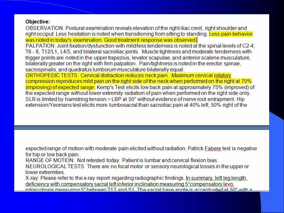
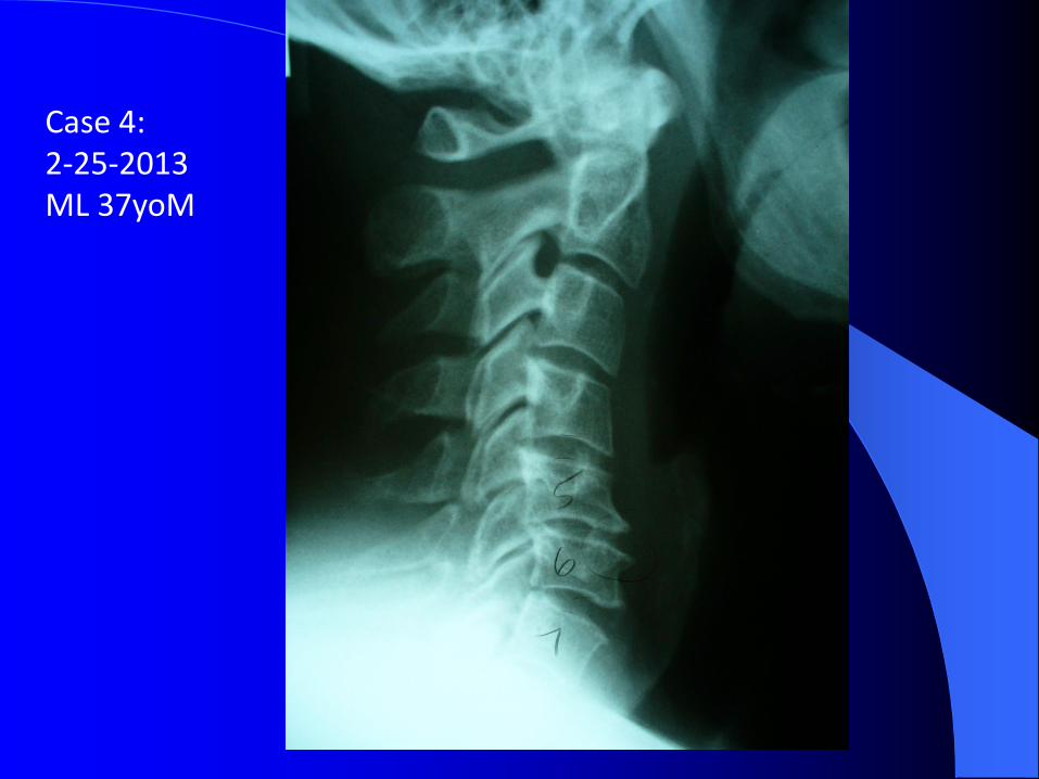
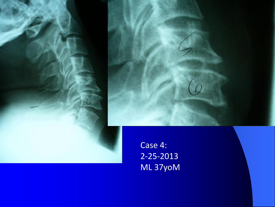
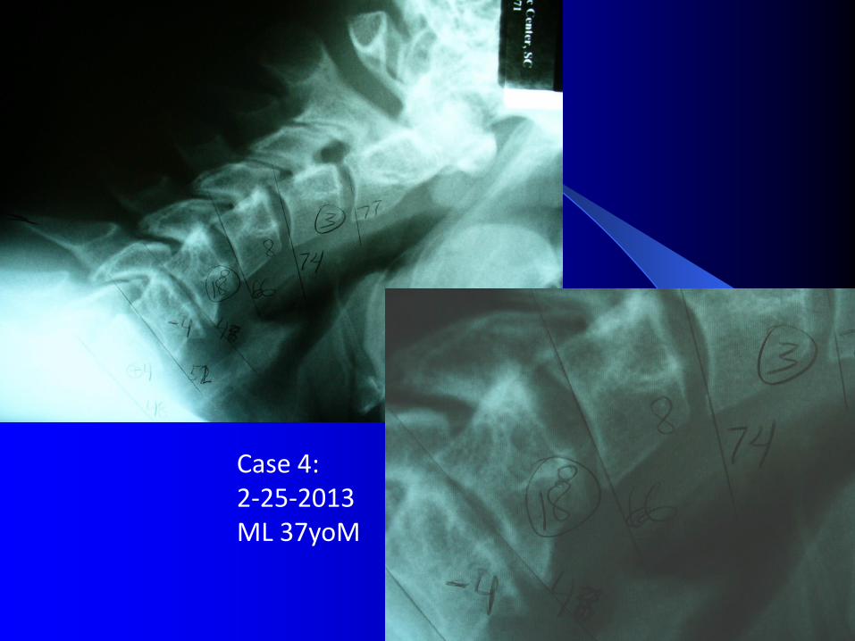
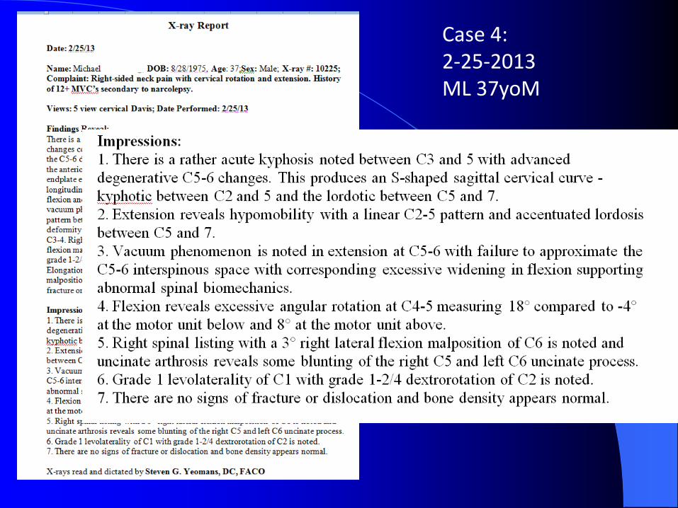
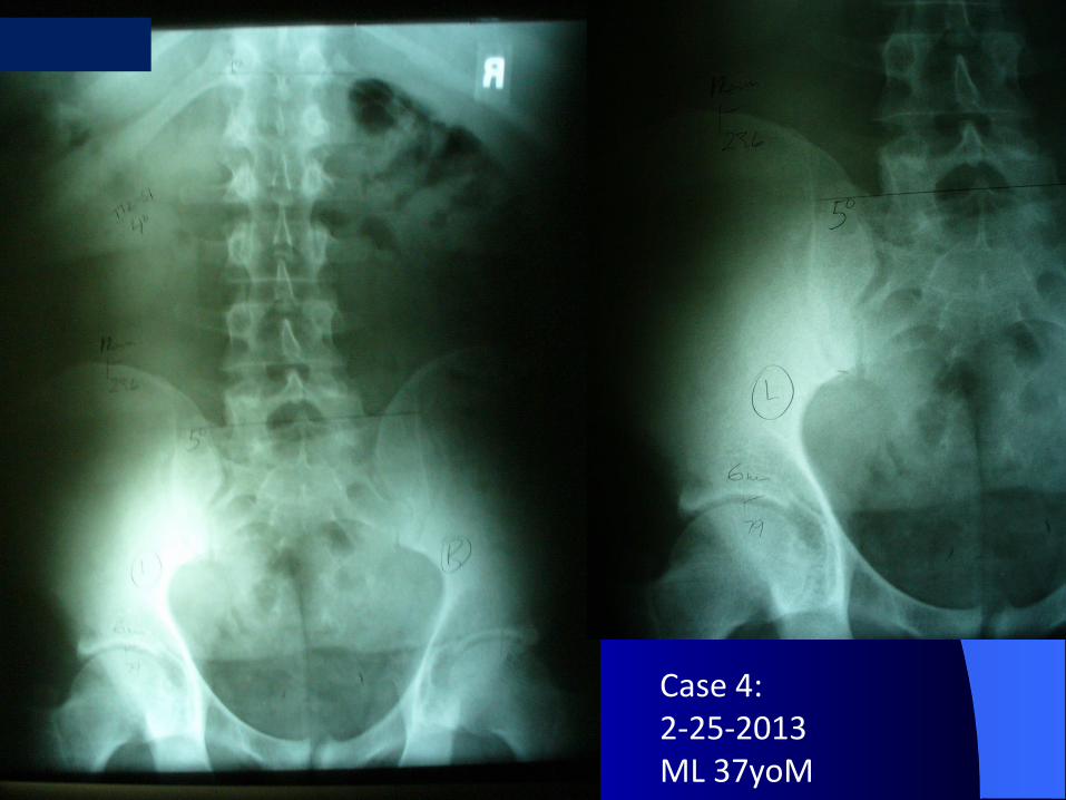
Case 4: Michael L. 37yo M
WAD Injury

INITIAL EXAMINATION 2-25-13


(LAST VISIT)
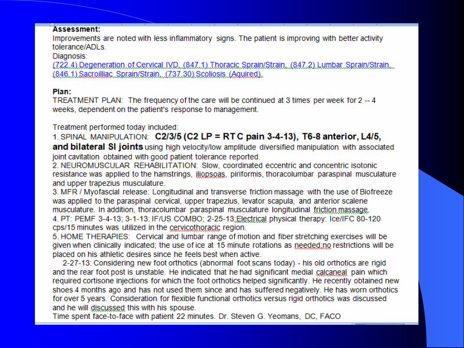
Case 4:2-25-2013ML 37yoM
Case 4: 2-25-2013ML 37yoM
Case 4:2-25-2013ML 37yoM

Case 4:2-25-2013ML 37yoM
Case 4:2-25-2013ML 37yoM
Case 4: 2-25-2013ML 37yoM


2-25-2013 ML 37yoMSUMMARY
Significant past Hx: 12 MVC’s (all “quick” recoveries
– within 3 days); martial artist
NOT severe: Pain: 1-3/10; C-BQ: 20%; YFQ: 13%
(<45 = low risk of prolonged recovery)
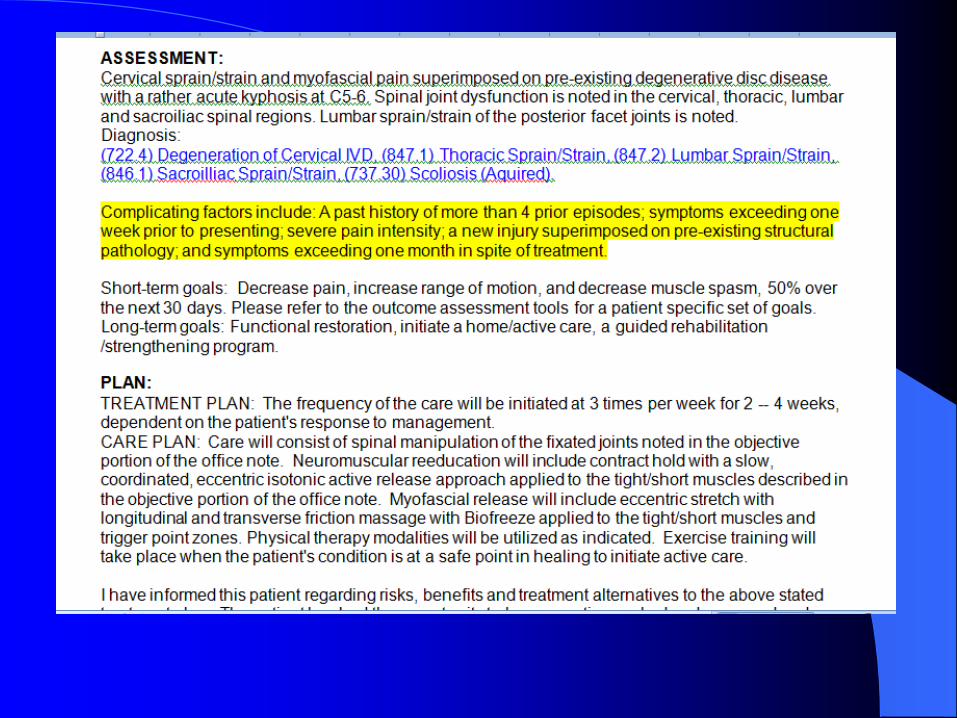
Complicating factors: Significant
XR Findings: Significant
Treatment Outcome: RESOLVED (only 4 visits in <2
weeks)
UNUSUAL CASE!!!
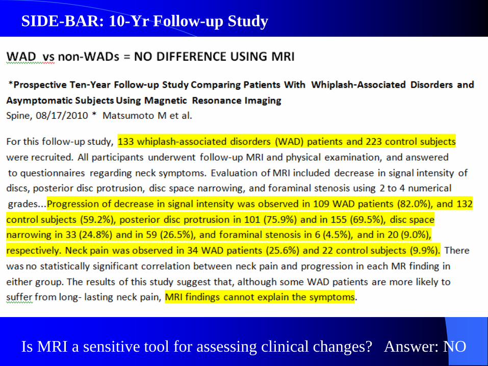
Is MRI a sensitive tool for assessing clinical changes? Answer: NO
SIDE-BAR: 10-Yr Follow-up Study

SIDE BAR:
NEW STUDY re: NDI!
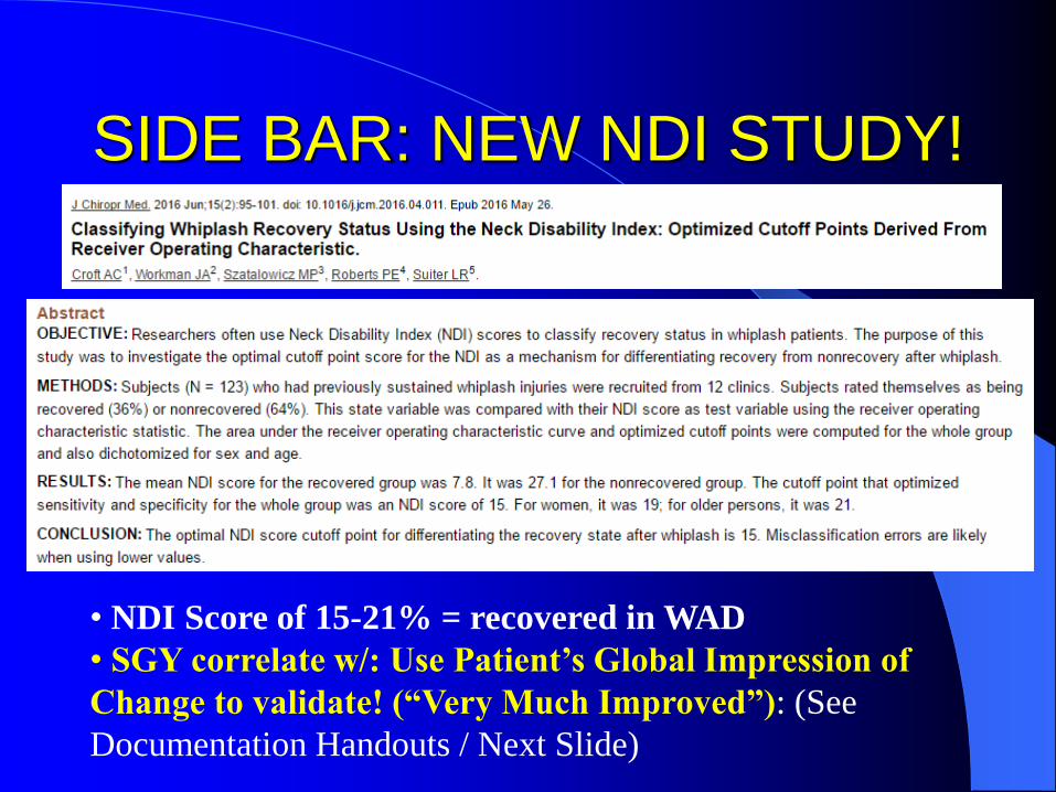
SIDE BAR: NEW NDI STUDY!
• NDI Score of 15-21% = recovered in WAD
• SGY correlate w/: Use Patient’s Global Impression of
Change to validate! (“Very Much Improved”): (See
Documentation Handouts / Next Slide)
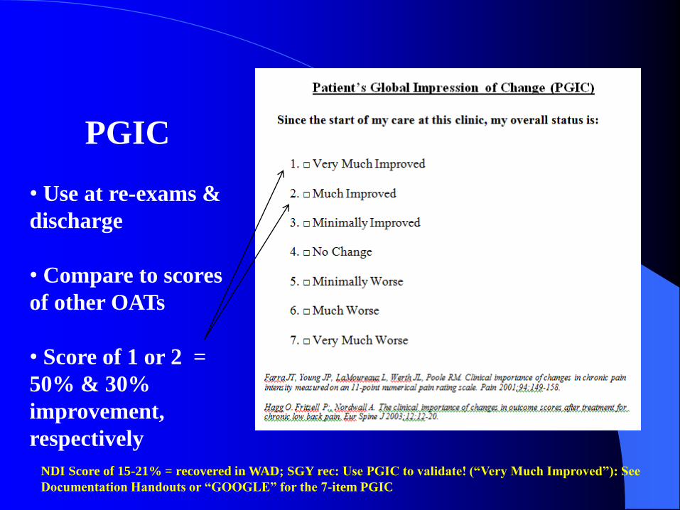
PGIC
• Use at re-exams &
discharge
• Compare to scores
of other OATs
• Score of 1 or 2 =
50% & 30%
improvement,
respectively
NDI Score of 15-21% = recovered in WAD; SGY rec: Use PGIC to validate! (“Very Much Improved”): See
Documentation Handouts or “GOOGLE” for the 7-item PGIC