what the neurologist expects from the ultrasound … filewhat the neurologist expects from the...
TRANSCRIPT

WHAT THE NEUROLOGIST EXPECTS
FROM THE ULTRASOUND OF THE PERIPHERAL NERVOUS SYSTEM
MARIA TRAVASAROU
NEUROLOGIST POLUMED “ΗΧΟΔΙΑΓΝΩΣΤΙΚΗ”

PRESENTATION OUTLINE
INDICATIONS FOR THE USE OF U/S IN THE PNS
ADVANTAGES
LIMITATIONS
COMPARISON WITH MRI
FUTURE PROSPECTS

INDICATIONS
ENTRAPMENT SYNDROMES
TRAUMATIC NERVE INJURIES
MISCELLANEOUS DISORDERS CHARCOT MARIE TOOTH LEPROSY
CARPAL TUNNEL SYNDROME
SPIRAL GROOVE SYNDROME
CUBITAL TUNNEL SYNDROME
PERONEAL NEUROPATHY
TRACTION PENETRATING TRAUMA IATROGENIC TUMORS-TUMOR LIKE
SYNDROMES NEUROFIBROMA, SCHWANNOMA HEMANGIOMA,LYMPHOMA,GANGLION CYST, SARCOMA INVASION,METASTASIS
CONGENITAL DISORDERS HNPP
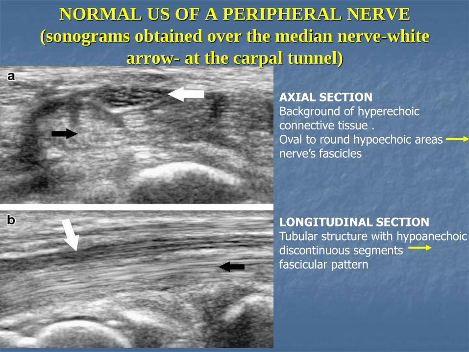
AXIAL SECTION Background of hyperechoic connective tissue . Oval to round hypoechoic areas nerve’s fascicles
NORMAL US OF A PERIPHERAL NERVE
(sonograms obtained over the median nerve-white
arrow- at the carpal tunnel)
LONGITUDINAL SECTION Tubular structure with hypoanechoic discontinuous segments fascicular pattern

CARPAL TUNNEL SYNDROME
(Axial a and longitudinal b sonograms over the
median nerves-whitte arrows- at the carpal tunnel)
Enlarged nerve compressed by a hypertrophied synovian tendon sheath (asterisks)
Nerve swollen proximally to the tunnel but thinned and hypoechoic in the canal

CUBITAL TUNNNEL SYNDROME
AXIAL Swollen hypoechoic nerve (whitte arrows)
LONGITUDINAL normal internal pattern of the nerve portion located proximal to the tunnel and irregular pattern of the nerve inside the tunnel, at the level of the medial epycondile (asterisk).

ULNAR NERVE TRAUMA

TIBIAL NERVE SCHWANNOMA
AXIAL COLOR DOPLER SONOGRAM Hypoechoic mass close to the tibialis posterior artery and veins
LONSITUDINAL Tumor clearly located Inside the nerve

ADVANTAGES
Excellent spatial resolution
No contraindications
Dynamic investigations possible
Investigation of a large part of the nerve path
Interventional ultrasound with diagnostic and therapeutic procedures
Easily available
Low cost

LIMITATIONS
Operator dependent
Relatively long learning curve
Poor contrast resolution
Limitations in some deep or difficult to assess, anatomical areas

ADVANTAGES AND DISADVANTAGES OF MRI
TO EXAMINE THE PERIPHERAL NERVOUS
SYSTEM
excellent contrast resolution
very good spatial resolution
Three dimensional planes
no limitation in examining deep
structures
good investigation of muscle mass
Research and development potential
Investigation is less operator dependent
Advantages Disadvantages
Contraindications to mri
Availability
High cost
static investigation occasionally
performed in non physiological positions
Long acquisition time

Developments and future
prospects
CONTRSAST ENHANCED ULTRASOUND
An animal study has shown that contrast enhanced ultrasound is feasible in the peripheral nervous imaging, enabling quantative measurements of PNS perfusion.

LITERATURE
ULTRASOUND IS THE FIRST CHOICE FOR PERIPHERAL NERVE IMAGING? Luka PADUA ET AL NEUROLOGY 2013;80;1627-7
ULTRASOUND OF THE PERIPHERAL NERVE BIANCHI ET AL JOINTBONE SPINE75 (2008) 643-649
MUSKULOSKELETAL ULTRASOUND FOR PERIPHERAL NERNE LESIONS KARE et al EUR J PHYS REHABIL MED 2012 48 665-74
CURRENT AND FUTURE IMAGING OF THE PERIPHERAL NERVOUS SYSTEM OHARA ET AL DIAGNOSTIC AND INTERVENTIONAL IMAGING 2014 95,17-26
HIGH RESOLUTION ULTRASONOGRAPHY IN EVALUATING PERIPHERAL NERVE ENTRAPMENT AND TRAUMA KOENIG et al NEUROSURG FOCUS 26 (2) 2009
INTRAOPERATIVE HIGH SOLUTION ULTRASOUND;A NEW TECHNIQUE IN THE MANAGEMENT OF PNS DISORDERS J NEUROSURG 2011 114 514-521