western trauma association critical decisions in trauma
TRANSCRIPT

Western Trauma Association Critical Decisions in Trauma:Evaluation and management of peripheral vascular injury, Part II
David V. Feliciano, MD, Ernest E. Moore, MD, Michael A. West, MD, Frederick A. Moore, MD,James W. Davis, MD, Christine S. Cocanour, MD, Thomas M. Scalea, MD,
and Robert C. McIntyre, Jr., MD, Indianapolis, Indiana
ABSTRACT: This is a position article frommembers of theWestern Trauma Association (WTA). Because there are no prospective randomized trials onthe evaluation and management of peripheral vascular trauma, the algorithm is based on the expert opinion of the WTA members andpublished observational studies. It may not be applicable at all hospitals caring for injured patients. The algorithm contains letters thatcorrespond to lettered text that is intentionally concise. This Part II algorithm focuses on operative techniques, while the Part I algorithm(J Trauma 2011;70: 1551Y1556) emphasized evaluation, diagnosis, and need for operation versus a therapeutic procedure performed inan interventional suite. (J Trauma Acute Care Surg. 2013;75: 391Y397. Copyright * 2013 by Lippincott Williams & Wilkins)
KEY WORDS: Peripheral vascular injury.
This is a recommended management algorithm from theWestern Trauma Association (WTA) addressing the man-
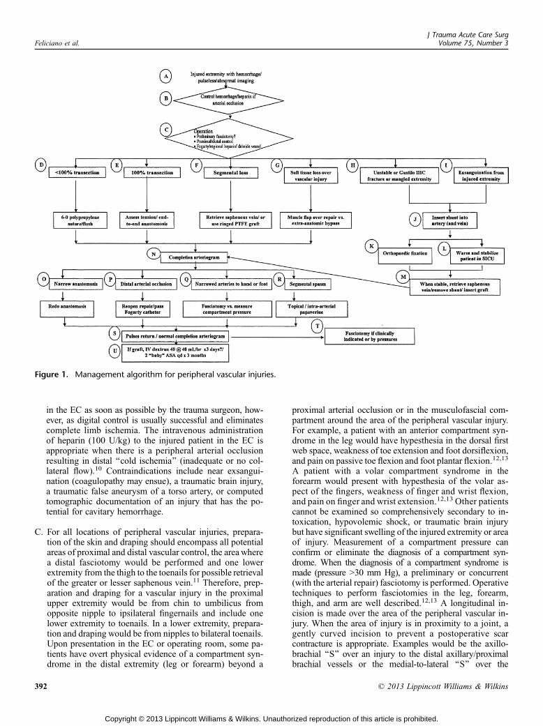
agement of peripheral vascular injuries. Because there is apaucity of published prospective randomized clinical trials thathave generated Class I data, the recommendations herein arebased primarily on published observational studies and theexpert opinion of WTA members. The algorithm (Fig. 1) andaccompanying comments represent a safe and sensible ap-proach that could be followed at most trauma centers. Werecognize that there will be patient, personnel, institutional, andsituational factors that may warrant or require deviation fromthe recommended algorithm and encourage institutions to usethis guideline to formulate their own local protocols.
The algorithm contains letters at decision points, and thecorresponding paragraphs in the text elaborate on the thoughtprocess and cite the pertinent literature. The annotated algo-rithm is intended to (a) serve as a quick bedside referencefor clinicians, (b) foster more detailed patient care protocolsthat will allow for prospective data collection and analysisto identify best practices, and (c) generate research projectsto answer specific questions concerning decision making inthe management of peripheral vascular injuries. The Part I
algorithm emphasized evaluation, diagnosis, and need for ope-ration versus a therapeutic procedure performed in an interven-tional suite, while this Part II algorithm will focus on operativetechniques.1
OPERATIVE INSTRUMENTATION ANDANCILLARY EQUIPMENT
Repair of an injured peripheral vessel is performed withthe surgeon and assistants wearing loupe magnification andbattery-powered head lamps. Standard vascular instrumentsincluding shallow and deep retractors, sharp Metzenbaumscissors, DeBakey forceps, Gerald forceps, jeweler’s forceps,fine tipped needle holders, and so on, should be a part of theinstrument tray. In addition, vascular adjuncts including vesselloops, Fogarty balloon catheters with stopcocks, intraluminalshunts, unfractionated heparin solution, and a contrast agent fora completion arteriogram should be in the operating room.
A. As described in Part I, indications for operation in patientswith suspected or documented peripheral vascular injuriesare hemorrhage, loss of major distal arterial pulse(s),or abnormal imaging results. While uncommon, a periph-eral arteriovenous fistula documented on physical ex-amination would be another indication if the insertion of anendovascular stent not be appropriate.2Y4
B. In the field or in the emergency center (EC), hemorrhagefrom the injured peripheral vessel is controlled by directpressure on the wound using a finger or pressure dressing orcompression of the proximal artery (‘‘pressure point’’).Application of a proximal tourniquet on the injured ex-tremity to control hemorrhage is now widely practiced incurrent military conflicts. Depending on local prejudices,experience, and transit times, it is likely that the use oftourniquets by civilian emergency medical services willincrease in the future.5Y9 Tourniquets should be removed
WTA ALGORITHM
J Trauma Acute Care SurgVolume 75, Number 3 391
Submitted: May 21, 2012, Revised: March 28, 2013, Accepted: March 28, 2013,Published online: August 9, 2013.
From the Denver Health Medical Center/University of Colorado (E.E.M.), Denver,Colorado; San Francisco General Hospital/University of California (M.A.W.),San Francisco, California; University of Florida (F.A.M.), Gainesville, Florida;University of California (J.W.D.), San Francisco–Fresno, California; Universityof California–Davis Medical Center (C.C.), Sacramento, Califonia; R AdamsCowley Shock Trauma Center/University of Maryland (T.M.S.), Baltimore,Maryland; and University of Colorado (R.C.M.), Denver, Colorado.
This study was presented at the 41st annual meeting of the Western Trauma Asso-ciation, February 27 to March 4, 2011, in Big Sky, Montana.
Address for reprints: David V. Feliciano, MD, Division of General Surgery, IndianaUniversity Medical Center, 545 Barnhill Dr, EH 509, Indianapolis, IN, 46202;email: [email protected].
DOI: 10.1097/TA.0b013e3182994b48
Copyright © 2013 Lippincott Williams & Wilkins. Unauthorized reproduction of this article is prohibited.
in the EC as soon as possible by the trauma surgeon, how-ever, as digital control is usually successful and eliminatescomplete limb ischemia. The intravenous administrationof heparin (100 U/kg) to the injured patient in the EC isappropriate when there is a peripheral arterial occlusionresulting in distal ‘‘cold ischemia’’ (inadequate or no col-lateral flow).10 Contraindications include near exsangui-nation (coagulopathy may ensue), a traumatic brain injury,a traumatic false aneurysm of a torso artery, or computedtomographic documentation of an injury that has the po-tential for cavitary hemorrhage.
C. For all locations of peripheral vascular injuries, prepara-tion of the skin and draping should encompass all potentialareas of proximal and distal vascular control, the area wherea distal fasciotomy would be performed and one lowerextremity from the thigh to the toenails for possible retrievalof the greater or lesser saphenous vein.11 Therefore, prep-aration and draping for a vascular injury in the proximalupper extremity would be from chin to umbilicus fromopposite nipple to ipsilateral fingernails and include onelower extremity to toenails. In a lower extremity, prepara-tion and draping would be from nipples to bilateral toenails.Upon presentation in the EC or operating room, some pa-tients have overt physical evidence of a compartment syn-drome in the distal extremity (leg or forearm) beyond a
proximal arterial occlusion or in the musculofascial com-partment around the area of the peripheral vascular injury.For example, a patient with an anterior compartment syn-drome in the leg would have hypesthesia in the dorsal firstweb space, weakness of toe extension and foot dorsiflexion,and pain on passive toe flexion and foot plantar flexion.12,13
A patient with a volar compartment syndrome in theforearm would present with hypesthesia of the volar as-pect of the fingers, weakness of finger and wrist flexion,and pain on finger and wrist extension.12,13 Other patientscannot be examined so comprehensively secondary to in-toxication, hypovolemic shock, or traumatic brain injurybut have significant swelling of the injured extremity or areaof injury. Measurement of a compartment pressure canconfirm or eliminate the diagnosis of a compartment syn-drome. When the diagnosis of a compartment syndrome ismade (pressure 930 mm Hg), a preliminary or concurrent(with the arterial repair) fasciotomy is performed. Operativetechniques to perform fasciotomies in the leg, forearm,thigh, and arm are well described.12,13 A longitudinal in-cision is made over the area of the peripheral vascular in-jury. When the area of injury is in proximity to a joint, agently curved incision to prevent a postoperative scarcontracture is appropriate. Examples would be the axillo-brachial ‘‘S’’ over an injury to the distal axillary/proximalbrachial vessels or the medial-to-lateral ‘‘S’’ over the
Figure 1. Management algorithm for peripheral vascular injuries.
J Trauma Acute Care SurgVolume 75, Number 3Feliciano et al.
392 * 2013 Lippincott Williams & Wilkins
Copyright © 2013 Lippincott Williams & Wilkins. Unauthorized reproduction of this article is prohibited.

antecubital area of the upper extremity. Proximal and distalcontrol around a peripheral vascular injury is appropriatewhen arterial occlusion is present or when compression or aproximal tourniquet prevents further bleeding from the area ofinjury. When hemorrhage cannot be controlled or a large he-matoma is present, it is appropriate to enter the area of injuryand apply vascular clamps directly around the perforation inthe peripheral artery and/or vein. Adequate suction devicesand appropriate retractors are mandatory to limit blood lossduring this approach. After proximal and distal vascularcontrol has been attained with the use of small DeBakeyvascular clamps, bulldog clamps, or vessel loops, the mag-nitude of the vascular injury (see D, E, F later) is assessed.A laceration encompassing greater than 25% of the cir-cumference of the artery increases the risk of distal embo-lization of local clot. With this injury or when there hasbeen a delay in treatment, Fogarty balloon catheters arepassed proximally and distally through the area of injury.Appropriate sizes of the Fogarty catheters would includethe following: #6 for the common and external iliac arteries,#4 to #5 for the common femoral artery, #4 for the superfi-cial femoral artery, #3 to #4 for the popliteal artery, and #3for the arteries in the leg. Overdistension of the Fogartyballoon should be avoided because this may injure normalintima. The goal is to confirm proximal arterial inflow andhave no thrombus on return of two consecutive distal passesof the balloon catheter. It is helpful to remember that pas-sage of a Fogarty balloon catheter into the leg will result inentrance into the peroneal artery approximately 90% of thetime.14 One option to overcome this anatomic issue is to firstinflate a Fogarty balloon in the proximal peroneal artery.Then, Fogarty balloon catheters are likely to be passed intothe anterior and posterior tibial arteries. In contrast, ballooncatheters are never passed into venous injuries because theywill disrupt valves. If heparin was not given before thistime, it should be administered intravenously at an appro-priate dosage. In addition, 20 mL to 25 mL of heparinizedsaline (50 U/mL) can be injected into the proximal and distalartery (40Y50 mL or another 2,000Y2,500 U) after passageof a Fogarty catheter. After a passage of Fogarty ballooncatheters and administration of heparin, a laceration of anartery from a knife or piece of glass is debrided minimallyback to healthy intima. Many arterial injuries from gunshotwounds or blunt trauma, however, have extensive intimalor wall injuries, and segmental resection may be necessary(see F later).
D. An injury to a peripheral artery or vein that does not resultin complete transection and in which there is minimal loss oftissue can be repaired, depending on luminal diameter, withan interrupted or continuous technique using 6-0 or 7-0 poly-propylene suture. No matter which technique is used, thelast one or two interrupted sutures are left untied, or thelast several loops of a continuous suture are left loose untilproximal and distal flushing is performed. With an arterialrepair, the proximal clamp is then reapplied, and distal back-flow flushes air out under the repair as the last knot is tied.Excessive tension should be avoided when tying the lastknot to avoid constricting the anastomosis. When repairing
a vein, the proximal clamp (toward the heart) is left off toevacuate intraluminal air.
E. An injury to a peripheral artery or vein that results incomplete transection is first managed with minimal debride-ment back to healthy intima at both ends. The amount oftension necessary to perform an end-to-end anastomosis isthen assessed. Should the tension be excessive, a decisionmust be reached on whether to divide and ligate branches ofthe vessel on both ends versus inserting an interpositiongraft. Because of the elasticity of arteries, sacrifice ofproximal and distal branches will often gain up to 3-cmtotal advancement of both ends; however, sacrifice of thegeniculate collaterals from the popliteal artery should beperformed with caution. This is because they furnish im-portant collateral flow to the leg should atheroscleroticocclusion of the superficial femoral artery occur later in life.Spatulated end-to-end anastomoses using 6-0 or 7-0 poly-propylene suture are recommended for arteries that are lessthan 1 cm in diameter distal to the axillary in the upperextremity and distal to the common femoral in the lowerextremity. The ends of a vessel with a diameter greaterthan 1 cm can be anastomosed using an interrupted or acontinuous suture technique with stay sutures 120 degreesor 180 degrees apart.15,16 An end-to-end anastomosis per-formed under tension will result in an ‘‘hourglass’’ narrow-ing and bleeding from suture holes. When an end-to-endanastomosis cannot be performed, an interposition graft isinserted.
F. When debridement of an injured vessel or the injury itselfresults in significant segmental loss, an interposition graftof reversed autogenous saphenous vein or ringed polytetra-fluoroethylene (PTFE or Teflon) is inserted.17,18 The saphe-nous vein in the groin or at the ankle is preferred becauseit is an endothelium-lined conduit, is readily available, is areasonable size match for most peripheral arteries and someveins, and, especially, has excellent long-term patency.17 Whenthe greater saphenous vein is absent, is diseased (multiplestenoses), has a small diameter, or is the only venous out-flow for an injured lower extremity, other options are avail-able. The lesser saphenous vein on the posterior aspect ofthe leg is an ideal replacement because it has a muscularwall similar to that of the greater saphenous vein. Anotheroption is to use the cephalic or basilic vein in the upperextremity as an interposition graft in the brachial artery. Asthese thin-walled veins dilate significantly when placed in thearterial system, ligation of branches during retrieval shouldbe further from the vein than when the greater saphenousvein is retrieved. The time required to create a panel orspiral vein graft is usually excessive in the trauma patient,and these grafts are used rarely to replace injured peripheralvessels. Another option is to insert a PTFE graft, which isreadily available off the shelf in appropriate sizes, is easy tosew, has a satisfactory patency when grafts with a diametergreater than 6 mm are used, and is remarkably resistant toinfection in the absence of exposure or adjacent osteomye-litis.18 A newer version of an expanded PTFE graft hasheparin molecules bonded directly to the luminal surface
J Trauma Acute Care SurgVolume 75, Number 3 Feliciano et al.
* 2013 Lippincott Williams & Wilkins 393
Copyright © 2013 Lippincott Williams & Wilkins. Unauthorized reproduction of this article is prohibited.

of the graft to reduce thrombosis, but there are not data onthe use of these grafts in trauma vascular repairs.19 Thepresumptive value of using a ringed PTFE graft is in thesupport available when the surrounding reaction to the for-eign body becomes a scar in the later postoperative period.All of the techniques described for an end-to-end anasto-mosis in E (heparinization, use of small vascular clamps,bulldog clamps, or vessel loops, passage of Fogarty ballooncatheters, lack of tension on the anastomoses, a fine suturetechnique with 6-0 or 7-0 polypropylene, and flushing se-quence) are used to complete the two anastomoses. It ishelpful to complete the distal anastomosis first in a difficultanatomic location such as the distal tibioperoneal trunk nearits bifurcation. This allows for better visualization of theposterior suture line and prevents narrowing of the orificesof the posterior tibial and peroneal arteries. The saphenousvein may not dilate enough to be a satisfactory interpositiongraft in the popliteal, femoral, common femoral, or axillaryveins. With the concerns noted previously for panel or spiralvein grafts, an appropriately sized PTFE graft with externalrings should be inserted and will have satisfactory short-term patency.20
G. A peripheral vascular repair should never be left exposed atthe completion of operation. Exposure of a lateral repair, end-to-end anastomosis, or saphenous vein graft will result incontamination leading to infection. In the hemodynamicallystable patient, a decision must be made before the arterialrepair is performed on whether there is enough healthymuscle available locally to cover the subsequent repair. Ifthere is not, then a decision must be made in conjunctionwith the plastic surgery service on whether there is healthymuscle close enough to be transposed or rotated to cover thearterial repair.21 If neither of these approaches is possible andthe arterial repair is likely to require segmental resection andan end-to-end anastomosis or interposition graft, an extra-anatomic bypass graft is inserted in the hemodynamicallystable patient.22 The approach separates injuries to the boneand soft tissue from the arterial repair. The operative tech-nique includes the following: (1) debridement of soft tissue/injured artery back to healthy tissue; (2) insertion of an extra-anatomic saphenous vein or PTFE graft with both anasto-moses and the graft covered with healthy soft tissue; and(3) open packing or a vacuum-assisted device applied to thedefect in soft tissue. In the hemodynamically unstable pa-tient, a lateral arteriorrhaphy is covered with a porcine xeno-graft placed under mesh gauze soaked in a saline-antibioticsolution.23 When segmental resection of the arterial injurywill be required, a temporary intraluminal shunt is inserted(see H), and the porcine xenograft-gauze combination isused to cover the area. Plastic surgery consultation is thenobtained to determine which of a large number of optionsfor coverage of the wound would be appropriate at the re-operation for removal of the shunt and insertion of an in-terposition graft.24,25
H. Mangled extremities with a combination of injuries to theartery, bone, soft tissue, tendon, and nerve resulted inamputation rates of 42% to 78% in older series.26Y28 While
amputation rates have decreased in recent years, long-termfunctional outcomes in patients with either limb salvage oramputation have been disappointing.29 In the absence ofabsolute criteria for amputation such as disruption of theposterior tibial nerve in the adult or a crush injury withwarm ischemia greater than 6 hours, limb salvage may beconsidered.30,31
I. In the patient with near exsanguination from an injury toa peripheral artery or vein, there is a temptation to ligatethe vessel and potentially sacrifice the limb if the patientsurvives. Because of advances such as ‘‘damage-controlresuscitation’’ and improvements in critical care, this ap-proach is no longer considered to be valid for major arterialinjuries.32
J. The first step in limb salvage in the patient with a mangledextremity or near exsanguination is to restore arterial in-flow and, if needed, venous outflow by the insertion oftemporary intraluminal shunts.33 In addition to intrave-nous tubing, a number of commercially available carotidartery-type shunts are available, and a rigid 14 Fr size hasbeen the most commonly inserted in recent years.33 Whilethe Pruitt-Inahara shunt is only 8 Fr in size, the addedT-piece will allow for infusion of vasodilators or antico-agulants. The patency of an intraluminal arterial shunt isconfirmed by the presence of palpable pulses or Dopplersignals at the ankle or wrist. Thoracostomy tubes in the 16 Frto 24 Fr size range are used as shunts in large peripheral veinssuch as the popliteal or femoral.
K. Depending on the patient’s hemodynamic status and pres-ence of other potentially life-threatening injuries, the or-thopedic surgery service would then perform some type offixation to stabilize the mangled extremity.
L. The patient with near exsanguination and patent shuntsis moved to the intensive care unit for damage controltransfusion/correction of any coagulopathy and rewarming.
M. Removal of temporary intraluminal shunts and insertionof vascular interposition grafts are appropriate after or-thopedic fixation at the first operation in the hemody-namically stable patient who is normothermic and has noother major injuries requiring immediate diagnostic ortherapeutic intervention. In contrast, the patient with pre-vious near exsanguination and insertion of temporaryintraluminal shunts is returned to the operating room whenhemodynamically stable and normothermic. The longestdwell time for an arterial intraluminal shunt in a recent largecivilian series was 52 hours,33 but there is on report of apatient with a 10-day dwell time in the right axillary ar-tery.34 There has been a recent report of increased in-fections in PTFE grafts placed at a reoperation after atemporary intraluminal shunt has been removed.35 If thesedata are noted in other reports, it would be recommendedthat an extra-anatomic bypass or insertion of an autoge-nous saphenous vein graft be performed after the shuntis removed.
J Trauma Acute Care SurgVolume 75, Number 3Feliciano et al.
394 * 2013 Lippincott Williams & Wilkins
Copyright © 2013 Lippincott Williams & Wilkins. Unauthorized reproduction of this article is prohibited.

N. Completion arteriograms are routinely performed in he-modynamically stable patients after peripheral arterial re-pairs under the following circumstances: (1) when therehas been a delay in diagnosis and/or treatment; (2) whenpalpable pulses are not present at the ankle or wrist afterrepair; (3) when there has been a complex repair such assegmental resection and an end-to-end anastomosis orinsertion of an interposition graft; or (4) when there isconcern about narrowing of an anastomosis.36 Aftercompletion of the arterial repair, metal clips are placed onadjacent soft tissue to mark the level of the single orproximal/distal anastomoses. A standard completion arte-riogram is then performed.
O. Narrowing of either anastomosis on the completion arterio-gram mandates reheparinization as indicated, reapplicationof proximal and distal vascular clamps or vessel loops, andcutting out the polypropylene sutures. Frayed tissue at theend of the vessel is then debrided, especially the adventitia.Should the end(s) of the artery appear to be in spasm, a Garrettcoronary artery dilator or Bakes biliary dilator is used togently dilate the end of the vessel.36 If a continuous suturetechnique was used on the first anastomosis, an interruptedsuture technique is used on the redo.
P. A distal occlusion of the superficial femoral or poplitealartery or tibioperoneal trunk beyond the arterial repair onthe completion arteriogram mandates reheparinization asindicated, reapplication of proximal and distal vascularclamps or vessel loops, and performing a 2-mm to 3-mmtransverse arteriotomy distal to the arterial repair. Pas-sage of appropriately sized Fogarty balloon catheters isperformed until two successive passes have no return ofthrombus. A second arteriogram using the catheter describedpreviously is then performed through the arteriotomy site. Ifan interposition graft has been inserted, the 2-mm to 3-mmtransverse arteriotomy is placed 1 cm proximal to thedistal anastomosis. An arteriotomy site in the injured arteryor in an interposition graft is closed with interrupted 6-0 or7-0 polypropylene sutures.When the distal arterial occlusionis present in either the anterior or the posterior tibial artery, itis unlikely that the transfemoral or transpopliteal passageof the Fogarty balloon catheter will enter the desired vesselunless the proximal peroneal artery is occluded with a sep-arate Fogarty balloon as previously described.14 If one ar-tery in the leg is occluded but the foot has at least onepalpable pulse and is clearly viable, it is acceptable to leavethe one vessel occluded without further intervention. Occlu-sion of two vessels mandates transpopliteal or transtibiopero-neal embolectomy, especially in patients with significant blunttrauma to the lower extremity (shearing of collaterals). Anembolectomy distal to the knee adds 1.5 hours to the opera-tive procedure and is performed through a longitudinal inci-sion 1 cm posterior to the posterior border of the tibia. Thecrural fascia is incised, the medial head of the gastrocnemiusmuscle is retracted posteriorly, and the tibial attachments of thesoleus muscle are divided.37 Full exposure of the distalpopliteal artery and entire tibioperoneal trunk usuallymandates division and ligation of the anterior tibial vein. All
three vessels of the leg require catheter embolectomy througha distal popliteal transverse arteriotomy, as long as each ves-sel can be occluded with a bulldog clamp or vessel loop toallow for passage of the catheter into the desired vessel. Ifonly the peroneal and posterior tibial arteries need to havean embolectomy, the transverse arteriotomy is made in thedistal tibioperoneal trunk.
Q. When the completion arteriogram documents narrowing or a‘‘beaded’’ appearance of the vessels in the leg, either a com-partment syndrome or a spasm is present. If the surgeon isconvinced that a compartment syndrome is present basedon the history (delay in treatment), physical examination(significant swelling of the leg or forearm; neuromuscularfindingsVsee C), or operative findings (need for simulta-neous arterial and venous clamping, venous ligation), a fas-ciotomy is performed.13 When the presentation is less clearto the surgeon, a compartment pressure is measured. A com-partment pressure of 30 mm Hg in the patient with anabnormal postrepair arteriogram would prompt a fasciotomyon most trauma services.
R. Spasm of vessels distal to an arterial repair is common inyoung patients but usually does not threaten the viability ofthe distal extremity. Restoration of a normal hemodynamicstate, reversal of hypothermia, and topical warming of theinjured extremity will reverse arterial spasm in 6 hours to8 hours in most patients. In rare patients, severe limb-threatening arterial spasm has been treated with a proximalintra-arterial bolus injection of 60-mg papaverine followedby an infusion of 30 mg/h to 60 mg/h in the past.38 Anotheroption used on rare occasions has been a proximal intra-arterial infusion of a solution of 1,000-mL normal saline,1,000-U heparin, and 500-mg tolazoline at a rate of 30 mL/hto 60 mL/h.39,40 Currently used vasodilators in angiographysuites include intra-arterial nitroglycerin (50Y100 mg) or nife-dipine (10 mg per os or sublingual).
S. Restoration of normal distal palpable pulses is reassuringafter the interventions described in O, P, Q, and R, but acompletion arteriogram is performed again in these patientsto verify that no other abnormalities are present at the suturelines or distally.
T. In high-risk patients (see Q) compartment pressure shouldbe measured distal to the arterial repair. The goal is to avoidthe 20% return to the operating room for delayed fascioto-mies as noted in the past.41
U. When a complex (segmental resection with end-to-endanastomosis or insertion of interposition graft) or compli-cated (need for postarteriogram intervention) arterial repairhas been performed, certain centers will infuse low-molecular-weight dextran at 40 mL/h for the first threepostoperative days. There are only modest data to supportthe use of dextran, and none of it is recent.42 All patientswith an end-to-end anastomosis, interposition graft,or when there is concern about distal runoff shouldbe started on aspirin 81 mg or 162 mg orally daily at
J Trauma Acute Care SurgVolume 75, Number 3 Feliciano et al.
* 2013 Lippincott Williams & Wilkins 395
Copyright © 2013 Lippincott Williams & Wilkins. Unauthorized reproduction of this article is prohibited.

convenience. This recommendation is based on the long-term results when aspirin is administered after aorto-coronary artery bypass with saphenous vein grafts.43,44
Instructions to the patient after arterial repair include adaily walking program and cessation of smoking.
DISCLAIMER
The WTA develops algorithms to provide guidance and recommenda-tions for particular practice areas, but does not establish the standard ofcare. TheWTA develops algorithms based on the evidence available in theliterature and the expert opinion of the task force in the recent timeframeof the publication. The WTA considers use of the algorithm to be vol-untary. The ultimate determination regarding its application is to bemadeby the treating physician and health care professionals with full consid-eration of the individual patient’s clinical status as well as available insti-tutional resources. Also, it is not intended to take the place of health careproviders judgment in diagnosing and treating particular patients.
DISCLOSURE
The authors declare no conflicts of interest.
REFERENCES1. Feliciano DV, Moore FA, Moore EE, West MA, Davis JW, Cocanour CS,
Kozar RA, McIntyre RC, Jr. Evaluation and management of peripheralvascular injury: part I. Western Trauma Association/Critical Decisions inTrauma. J Trauma. 2011;70:1551Y1556.
2. Lonn L, Delle M, Karlstrom L, Risberg B. Should blunt arterial trauma tothe extremities be treated with endovascular techniques? J Trauma. 2005;59:1224Y1227.
3. White R, Krajcer Z, Johnson M, et al. Results of a multicenter trial forthe treatment of traumatic vascular injury with a covered stent. J Trauma.2006;60:1189Y1196.
4. Reuben BC, Whitten MG, Sarfati M, Kraiss LW. Increasing use of endo-vascular therapy in acute arterial injuries: analysis of the national traumadata bank. J Vasc Surg. 2007;46:1222Y1226.
5. Rocko JM, Tischler C, Swan KG. Exsanguination in public: a preventabledeath. J Trauma. 1982;22:635.
6. DorlacWC, DeBakeyME, Holcomb JB, et al.Mortality from isolated civilianpenetrating extremity injury. J Trauma. 2005;59:217Y222.
7. Welling DR, Burris DG, Hutton JE, et al. A balanced approach to tour-niquet use: lessons learned and relearned. J Am Coll Surg. 2006;203:106Y115.
8. Beekley AC, Sebasta JA, Blackbourne LH, et al. Prehospital tourniquetuse in Operation Iraqi Freedom: effect on hemorrhage control and out-comes. J Trauma. 2008;64:S28YS37.
9. Swan KG, Jr, Wright DS, Barbagiovanni SS, et al. Tourniquets revisited.J Trauma. 2009;66:672Y675.
10. Daugherty MF, Sachatello CR, Ernst CB. Improved treatment of popli-teal artery injuries. Using anticoagulation and extra-anatomic reconstruc-tion. Arch Surg. 1978;113:1317Y1321.
11. Feliciano DV. Vascular injuries. In Maull KI, Cleveland HC, Strauch GO,Wolferth CC, eds. Advances in Trauma. Vol 2. Chicago, IL: Year BookMedical Publishers; 1987:179Y206.
12. Matsen FA, III. Diagnosis of compartmental syndromes. In Matsen FA,III, ed. Compartmental Syndromes. New York, NY: Grune & Stratton;1980:85Y100.
13. Dente CJ, Wyrzykowski AD, Feliciano DV. Fasciotomy. Curr Prob Surg.2009;46:769Y840.
14. Short D, Vaughan GD, III, Jachimczyk J, et al. The anatomic basis for theoccasional failure of transfemoral balloon catheter thromboembolectomy.Ann Surg. 1979;190:555Y556.
15. Carrel A. The surgery of blood vessels, etc. Johns Hopkins Hosp Bull.1907;18:18Y28.
16. Ball CG, Feliciano DV. A simple and rapid vascular anastomosis foremergency surgery: a technical case report. World J Emerg Surg. 2009;4:30Y32.
17. Mitchell FL, III, Thal ER. Results of venous interposition grafts in arterialinjuries. J Trauma. 1990;30:336Y339.
18. Feliciano DV, Mattox KL, Graham JM, BitondoCG. Five-year experiencewith PTFE grafts in vascular wounds. J Trauma. 1985;25:71Y82.
19. Dorigo W, Dicarlo F, Frois, N, et al. Lower limb revascularization with anew bioactive prosthetic graft: early and late results. Ann Vasc Surg.2008;22:79Y87
20. Parry NG, Feliciano DV, Burke RM, et al. Management and short-term patency of lower extremity venous injuries with various repairs.Am J Surg. 2003;186:631Y635.
21. Strinden WD, Dibbell DG, Sr, Turnipspeed WD, et al. Coverage of acutevascular injuries of the axilla and groin with transposition muscle flaps:case reports. J Trauma. 1989;29:512Y516.
22. Feliciano DV. Heroic procedures in vascular injury management. Therole of extra-anatomic bypasses. Surg Clin North Am. 2002;82:115Y124.
23. Ledgerwood AM, Lucas CE. Biological dressings for exposed vasculargrafts: a reasonable alternative. J Trauma. 1975;15:567Y574.
24. Attinger C. Soft-tissue coverage for lower-extremity trauma. Orthop ClinNorth Am. 1995;26:295Y334.
25. Haddock NT, Weichman KE, Reformat DD, et al. Lower extremityarterial patterns and reconstructive outcomes on patients with severelower extremity trauma: a 26-year review. J Am Coll Surg. 2010;210:66Y72.
26. Gustilo RB, Mendoza RM, Williams DN. Problems in the management oftype III (severe) open fractures: a new classification of type III openfractures. J Trauma. 1984;24:742Y746.
27. Caudle RJ, Stern RJ. Severe open fractures of the tibia. J Bone Joint SurgAm. 1987;69:801Y807.
28. Scalea TM, DuBose J, Moore EE, et al. Western Trauma AssociationCritical Decisions in Trauma: management of the mangled extremity.J Trauma Acute Care Surg. 2012;72:86Y93.
29. MacKenzie EJ, Bosse MJ. Factors influencing outcome following limb-threatening lower limb trauma: lessons learned from the lower extrem-ity assessment project (LEAP). J Am Acad Orthop Surg. 2006;14:S205YS210.
30. Lange RH, Bach AW, Hansen ST, Jr, Johansen KH. Open tibial fractureswith associated vascular injuries: prognosis for limb salvage. J Trauma.1985;25:203Y208.
31. Hansen ST, Jr. The type IIIC tibial fracture. Salvage or amputation. J BoneJoint Surg Am. 1987;69:799Y800.
32. Cotton BA, Gunter OL, Isbell J, et al. Damage control hematology: theimpact of a trauma exsanguination protocol on survival and blood productutilization. J Trauma. 2008;64:1177Y1183.
33. Subramanian A, Vercruysse G, Dente C, et al. A decade’s experiencewith temporary intravascular shunts at a civilian Level I trauma center.J Trauma. 2008;65:316Y326.
34. Feliciano DV, Accola KD, Burch JM, Spjut-Patrinely V. Extranatomicbypass for peripheral arterial injuries. Am J Surg. 1989;158:506Y510.
35. Wyrzykowski AD, Ball CG, Rozycki GS, Feliciano DV. Infected prostheticgrafts in vascular traumaVa rare occurrence. J Trauma. [in revision].
36. Feliciano DV. Managing peripheral vascular trauma. Infect Surg. 1986;5:659Y662, 682.
37. Wind GG, Valentine RJ. Popliteal artery. In Wind GG, Valentine RJ, eds.Anatomic Exposures in Vascular Surgery. Baltimore, MD: Williams &Wilkins; 1991:373Y410.
38. Ingegno MD, Williams RA. Pentoxifylline, vasodilators, and metabolicagents. In Veith FJ, Hobson RW, Williams RA, Wilson SE, eds. VascularSurgery. Principles and Practice. 2nd ed. New York, NY: McGraw-Hill,Inc; 1994:242Y245.
J Trauma Acute Care SurgVolume 75, Number 3Feliciano et al.
396 * 2013 Lippincott Williams & Wilkins
Copyright © 2013 Lippincott Williams & Wilkins. Unauthorized reproduction of this article is prohibited.

39. Dickerman RM, Gewertz BL, Foley DW, et al. Selective intra-arterialtolazoline infusion in peripheral arterial trauma. Surgery. 1977;81:605Y609.
40. Peck JJ, Fitzgibbons TJ, GasparMR. Devastating distal arterial trauma andcontinuous infusion of tolazoline. Am J Surg. 1983;145:562Y566.
41. Feliciano DV, Cruse PA, Spjut-Patrinely V, et al. Fasciotomy after traumato the extremities. Am J Surg. 1988;156:533Y536.
42. Atik M. The use of dextran in surgery: a current evaluation. Surgery.1969;65:548Y562.
43. Puskas JD, WilliamsWH, Mahoney EM, et al. Off-pump vs. conventionalcoronary artery bypass grafting: early and 1-year graft patency, cost, andquality-of-life outcomes. J AMA. 2004;291:1841Y1849.
44. Patrono C, Garcia-Rodriguez LA, Landolfi R, Baigent C. Low-dose aspirinfor the prevention of atherothrombosis. N Engl J Med. 2005;353:2376Y2383.
J Trauma Acute Care SurgVolume 75, Number 3 Feliciano et al.
* 2013 Lippincott Williams & Wilkins 397
Copyright © 2013 Lippincott Williams & Wilkins. Unauthorized reproduction of this article is prohibited.