wednesday slide conference 2008-2009 conference 21 - … · the armed forces institute of pathology...
TRANSCRIPT

The Armed Forces Institute of PathologyDepartment of Veterinary Pathology
WEDNESDAY SLIDE CONFERENCE 2008-2009
Conference Coordinator:Todd M. Bell, DVM
Conference 21
*Sponsored by the American Veterinary Medical Association, the American College of Veterinary Pathologists, and the C. L. Davis
Foundation.
1 April 2009
Conference Moderator:
Dr. Steven Weisbrode, VMD, DACVP, PhD
CASE I – 07-1829 (AFIP 3106281)
Signalment: 6-week-old female mixed breed horse
History: Five day history of lameness of right rear leg. Radiographic findings were interpreted to represent septic arthritis of the right coxofemoral joint.
Gross Pathology: The foal was in good post mortem condition and had a body condition score of 3/5. Approximately 50% of the cartilage of the right femoral head was ulcerated. On cross section, the subchondral bone underlying the ulcerated articular cartilage was white for a distance of up to 2 mm from the surface. The dorsolateral aspect of the right femoral head had a large cartilage flap that was attached at the margins of the articular surface, and the ventrolateral aspect of the right femoral head had a 4 cm long fissure within articular cartilage that remained attached to subchondral bone. The dorsal region of the acetabular cartilage was irregular with exposure of subchondral bone (ulceration). The right medial trochlear ridge had 2 cm focus of articular cartilage that extended 1 cm into subchondral bone (found on cutting in a horizontal plane using a band saw). The contralateral coxofemoral
joint, stifle joint, and both humeral joints were examined and found to be within normal limits.
Laboratory Results: None
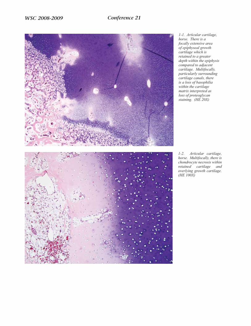
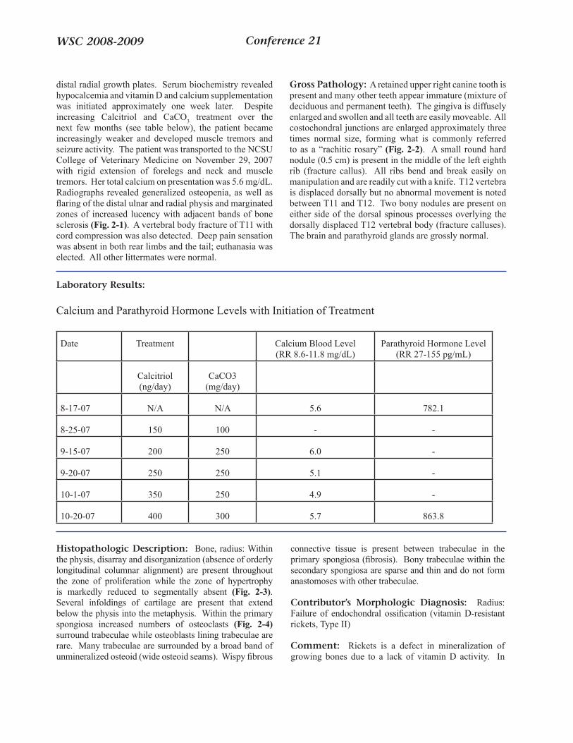
Histopathologic Description: Medial trochlear ridge of the right femur: There is a rectangular area of epiphyseal growth cartilage (AE complex) which extends deeper into the epiphysis (retained cartilage) compared with the adjacent cartilage (Fig. 1-1). The deep edge of this retained cartilage has cartilage cores present indicating active cartilage mineralization and vascular invasion as seen in endochondral ossification. The chondrocytes in this retained cartilage and the chondrocytes contiguous within its overlying growth cartilage have eosinophilic chondrocytes interpreted as chondrocyte coagulation necrosis compared with chondrocytes not associated with the retained region. In the overlying epiphyseal cartilage, in addition to chondrocyte coagulation necrosis, is a focus of loss of basophilia of the cartilage matrix (loss of proteoglycan staining). The blood vessels in the cartilage canals within this region of proteoglycan loss appear normal, but the contents of some of the cartilage canals in the contiguous growth cartilage with dead chondrocytes appear necrotic compared with contents of cartilage canals in adjacent cartilage with viable chondrocytes
WSC 2008-2009 Conference 21
1-1. Articular cartilage, horse. There is a focally extensive area of epiphyseal growth cartilage which is retained to a greater depth within the epiphysis compared to adjacent cartilage. Multifocally, particularly surrounding cartilage canals, there is a loss of basophilia within the cartilage matrix interpreted as loss of proteoglycan staining. (HE 20X)
1-2. Articular cartilage, horse. Multifocally, there is chondrocyte necrosis within retained cartilage and overlying growth cartilage. (HE 100X)

WSC 2008-2009 Conference 21
(Fig. 1-2). The primary trabeculae deep to the area of retained cartilage are course and occasionally fractured. In most of these trabeculae the bone is dead (karyolytic osteocytes) often with overlying viable woven bone. There is mild to moderate reactive fibrosis in the marrow in this region.
Contributor’s Morphologic Diagnosis: Focal chondronecrosis and retention (delayed endochondral ossification) of growth cartilage of the articular epiphyseal complex with osteonecrosis and infraction of subjacent trabeculae and associated marrow fibrosis.
Contributor’s Comment: This lesion represents articular epiphyseal complex dysplasia of osteochondrosis. This lesion likely was not clinically significant. The clinical signs in this case were attributed to the lesions in the femoral head which, after microscopic examination, were interpreted to be sequelae to suppurative osteomyelitis. The lesions in the distal femur were found on gross examination of sawed sections to check for foci of osteochondrosis.
Using terminology suggested in recent literature, the lesion in the distal femur is a good example of chondrocyte coagulation necrosis in both a focus of osteochondrosis latens and osteochondrosis manifesta.3 Often the chondrocyte coagulation necrosis is limited to the focus of osteochondrosis latens and it is hypothesized that this necrosis is secondary to ischemia and might be the initiating lesion of osteochondrosis. In the foal as in other species, it has been suggested that this cartilage necrosis is secondary to necrosis of blood vessels in the cartilage canals of growth cartilage.1 Interesting in this lesion is the presence of cartilage cores at the deep margin of the retained cartilage. This indicates cartilage mineralization and vascular invasion are taking place. This is not usually found in lesions of osteochondrosis manifesta. Osteochondrosis manifesta lesions are known to be able to resolve 2; therefore active endochondral ossification might represent attempts to resolve the lesion. The abnormal modeling, fractures and marrow fibrosis are presumed secondary to the altered endochondral ossification secondary to the osteochondrosis. The cause of the bone necrosis is uncertain but might be secondary to ischemia in this region due to fibrosis and fractures.
AFIP Diagnosis: Bone: Articular epiphyseal complex dysplasia, chronic, focally extensive, severe with osteonecrosis and infraction of subjacent trabeculae and marrow fibrosis Conference Comment: Osteochondrosis is an extremely important joint disorder of multiple species
including pigs, horses, large breed dogs, cattle, sheep, and deer.2 By definition, osteochondrosis is a focal disturbance of endochondral ossification.3 The exact pathogenesis of osteochondrosis is unclear, but the most likely explanation is focal ischemic necrosis of growth cartilage precipitated by necrosis of cartilage canal blood vessels.3 Synonyms for osteochondrosis include osteochondrosis dissecans and osteochondritis dissecans.2 In domestic animals osteochondrosis often leads to degenerative joint disease and lameness in affected animals.2
As mentioned by the contributor, a recent article has suggested new terminology adding modifiers to osteochondrosis. Osteochondrosis latens is defined as a lesion confined to the epiphyseal cartilage; osteochondrosis manifesta is a lesion resulting in delayed endochondral ossification that is visible on macroscopic and radiographic examination; osteochondrosis dissecans is the name of the lesion formed when a crack or fissure forms in an area of necrotic cartilage that extends up to the articular cartilage.3
Contributing Institution: Department of Veterinary Biosciences, The Ohio State University, Columbus, Ohio 43220, http://vet.osu.edu/biosciences.htm
References: 1. Olstad K, Ytrehus B, Ekman S, Carlson CS, Dolvik NI: Early lesions of osteochondrosis in the distal tibia of foals. J Orthop Res. 25(8):1094-105, 20072. Thompson K. Diseases of Bones and Joints. In: Pathology of Domestic Animals, vol. 1, ed. Maxie G, 5th ed., pp. 136-145. WB Saunders, Edinburgh, Scotland, 20073. Ytrehus B, Carlson CS, Ekman S: Etiology and pathogenesis of osteochondrosis. Vet Pathol 44:429-448, 2007
CASE II – APO7-2888 (AFIP 3103608).
Signalment: Seven-month-old, intact female Pomeranian, canine (Canis familiaris) History: The patient presented to the referring veterinarian at two months of age with generalized non-pruritic alopecia and tiring during play with littermates. Skin scrape was negative and the alopecia cleared with no treatment by five months of age. At that time a physical exam revealed bowing of the forelegs and thickened
WSC 2008-2009 Conference 21
distal radial growth plates. Serum biochemistry revealed hypocalcemia and vitamin D and calcium supplementation was initiated approximately one week later. Despite increasing Calcitriol and CaCO
3 treatment over the
next few months (see table below), the patient became increasingly weaker and developed muscle tremors and seizure activity. The patient was transported to the NCSU College of Veterinary Medicine on November 29, 2007 with rigid extension of forelegs and neck and muscle tremors. Her total calcium on presentation was 5.6 mg/dL. Radiographs revealed generalized osteopenia, as well as flaring of the distal ulnar and radial physis and marginated zones of increased lucency with adjacent bands of bone sclerosis (Fig. 2-1). A vertebral body fracture of T11 with cord compression was also detected. Deep pain sensation was absent in both rear limbs and the tail; euthanasia was elected. All other littermates were normal.
Gross Pathology: A retained upper right canine tooth is present and many other teeth appear immature (mixture of deciduous and permanent teeth). The gingiva is diffusely enlarged and swollen and all teeth are easily moveable. All costochondral junctions are enlarged approximately three times normal size, forming what is commonly referred to as a “rachitic rosary” (Fig. 2-2). A small round hard nodule (0.5 cm) is present in the middle of the left eighth rib (fracture callus). All ribs bend and break easily on manipulation and are readily cut with a knife. T12 vertebra is displaced dorsally but no abnormal movement is noted between T11 and T12. Two bony nodules are present on either side of the dorsal spinous processes overlying the dorsally displaced T12 vertebral body (fracture calluses). The brain and parathyroid glands are grossly normal.
Laboratory Results:
Calcium and Parathyroid Hormone Levels with Initiation of Treatment
Date Treatment Calcium Blood Level(RR 8.6-11.8 mg/dL)
Parathyroid Hormone Level (RR 27-155 pg/mL)
Calcitriol (ng/day)
CaCO3 (mg/day)
8-17-07 N/A N/A 5.6 782.1
8-25-07 150 100 - -
9-15-07 200 250 6.0 -
9-20-07 250 250 5.1 -
10-1-07 350 250 4.9 -
10-20-07 400 300 5.7 863.8
Histopathologic Description: Bone, radius: Within the physis, disarray and disorganization (absence of orderly longitudinal columnar alignment) are present throughout the zone of proliferation while the zone of hypertrophy is markedly reduced to segmentally absent (Fig. 2-3). Several infoldings of cartilage are present that extend below the physis into the metaphysis. Within the primary spongiosa increased numbers of osteoclasts (Fig. 2-4) surround trabeculae while osteoblasts lining trabeculae are rare. Many trabeculae are surrounded by a broad band of unmineralized osteoid (wide osteoid seams). Wispy fibrous
connective tissue is present between trabeculae in the primary spongiosa (fibrosis). Bony trabeculae within the secondary spongiosa are sparse and thin and do not form anastomoses with other trabeculae.
Contributor’s Morphologic Diagnosis: Radius: Failure of endochondral ossification (vitamin D-resistant rickets, Type II)
Comment: Rickets is a defect in mineralization of growing bones due to a lack of vitamin D activity. In

WSC 2008-2009 Conference 21
2-1. Distal radius and ulna, dog. There is flaring of the distal ulnar and radial physes and zones of increased epiphyseal lucency with adjacent bands of sclerosis. Radiograph courtesy of NCSU College of Veterinary Medicine, 4700 Hillsborough Street, Raleigh, NC 27606.
2-2. Thorax, dog. Diffusely, costochondral junctions are enlarged up to three times normal. Photograph courtesy of NCSU College of Veterinary Medicine, 4700 Hillsborough Street, Raleigh, NC 27606.

WSC 2008-2009 Conference 20
most species vitamin D is obtained primarily through the diet and metabolized in the liver. It is converted into its active form in the kidney. Low vitamin D activity leads to hypocalcemia and secondary hyperparathyroidism. This hyperparathyroidism causes mineral loss, especially calcium from bone. Rickets occurs when these changes take place during growth.
Two main forms of vitamin D-resistant rickets are characterized in humans. Type I is an inborn error in conversion of 25-(OH)D
3 to 1,25(OH)
2D
3 due to a
deficiency of the renal 1-hydroxylase enzyme. This condition responds to large doses of vitamin D
2 and D
3.
Vitamin D-resistant rickets Type II (VDRR II) in humans is an end organ resistance to 1,25(OH)
2D
3 due to an
autosomal recessive congenital defect in the vitamin D receptor (VDR) or a site distal to it. This type of rickets has been reported in a few cats, 4,5,6 but has never before been reported in a canine. In one of the feline cases the cat had signs of hypocalcemia, including muscle tremors, similar to the clinical signs in this canine patient.5 Two other cats had similar radiologic changes of the radius and ulna, as in the present case, as well as similar costochondral junction changes.4,6 Similarly these cases had no response to high levels of vitamin D or calcium supplementation; 4,6 however, in two of the cases, the cats became normal after physeal closure.4,5
Three main intracellular defects have been identified in human cases of VDRR II:
Hormone binding defects including decreased 1.
number of sites, decreased binding affinity, or complete absence of binding.Deficient nuclear localization – in these cases 2. there is normal binding affinity and capacity, but unmeasurable localization to the nucleus. A post-receptor defect characterized by normal 3. receptors but deficiency in the induction of the 25-(OH)D-24 hydroxylase enzyme in response to 1,25(OH)
2D.3
Defects can be detected using in vitro assessment of VDR binding or the subsequent cellular response to VDR binding by 1,25 dihydroxycholecalciferol (1,25-(OH)
2D) in cells,
typically fibroblasts cultured from skin biopsies, derived from individuals affected by clinical signs of VDRR II and compared with normal controls.4 A diagnosis of VDRR II was made in the present case based on clinical signs, radiographic findings, biochemistry, parathyroid levels, and the inability of fibroblasts from the skin of the dog to bind 1,25-(OH)
2D. A vitamin D receptor defect was
verified through genetic testing on cultured fibroblasts from this dog at Stanford University, confirming the diagnosis of VDRR II in this patient.
This patient also presented with generalized alopecia, as is the case in approximately 50% of human cases of VDRR II.3 Keratinocytes contain vitamin D receptors and can respond to the 1,25(OH)
2D
3 produced. The alopecia found
in VDR deficient patients suggests a biologic role for the VDR in the epidermis, particularly in the hair follicle.8 In VDR knock out mice models, the mice are fully haired and grossly normal after birth until approximately three
2-3. Bone, dog. Multifocally within the physis, there is a lack of regimentation and organization of chrondrocytes within the zone of proliferation, and the zone of hypertrophy is either absent or reduced. (HE 200X)
2-4. Bone, dog. Multifocally within the primary spongiosa, there are moderately increased numbers of osteoclasts, and there is moderate fibrosis between the trabeculae. (HE 400X)

WSC 2008-2009 Conference 20
months of age, progressing to generalized alopecia by eight months of age.8 These findings indicate that the prenatal hair growth and development of the epidermis and first hair growth cycle does not require VDR, but is important in the onset of the second hair growth cycle.8
Examination of the skin in this canine patient revealed large cystic follicles filled with keratin which corresponds to dermal changes noted in human cases and in rodent models of the disease.8
Humans with VDRR II, as well as the patient in this case, are born with normal hair, but become alopecic by six months of age and have rachitic changes that are resistant to all forms of vitamin D therapy.1 Alopecia generally does not improve in human patients,1 but normal pelage returned in our canine patient after several months. It is not clear if the alopecia is a genetically linked abnormality or related to the effect of 1,25-(OH)
2D
3 on the hair
follicle.1 In humans, alopecia seems to be a marker of a more severe form of the disease as judged by the earlier age at presentation, marked clinical aberrations and poor response to therapy.3
Therapy in humans begins with very large doses of vitamin D and oral calcium supplements but has had limited success. Refractory cases need long term nocturnal intravenous calcium infusions and these have successfully “healed” rickets and promoted mineralization in these patients;2 however, the therapy is cost prohibitive in veterinary cases.
AFIP Diagnosis: Bone: Physeal dysplasia characterized by disordered columns of chondrocytes with marrow fibrosis
Conference Comment: The contributor did a magnificent job of describing the physiology behind vitamin D-resistant rickets. A lack of dietary vitamin D, inadequate absorption of vitamin D from the gastrointestinal system, or a lack of adequate photobiosynthesis of vitamin D can also cause rickets.
Hypophosphatemia can lead to rickets, and is known as hypophosphatemic vitamin D-resistant rickets (renal hypophosphatemic rickets).7 Hypophosphatemia, normo-calcemia, and decreased renal tubular reabsorption of phosphate are characteristic of hypophosphatemic vitamin D-resistant rickets. Hypophosphatemia is the sequela of inadequate absorption of phosphorus from the gastrointestinal system or decreased/inadequate reabsorption of phosphorous from the renal system.7
Gross lesions of rickets are most striking at sites of rapid growth. The metaphyseal and epiphyseal regions of long
bones and the costochondral junctions are commonly affected, producing the classic “rachitic rosary” in affected animals.7
The classic histologic appearance of rickets is the disorganization of columns of chondrocytes and persistence of hypertrophic chondrocytes at sites of endochondral ossification, both at the physes and beneath the articular cartilage.7 The underlying trabecular bone is often disrupted, and irregular tongues of cartilage are often seen in the metaphyses due to disorganized and disrupted endochondral ossification.
Contributing Institution: North Carolina State University, College of Veterinary Medicine, 4700 Hillsborough Street, Raleigh, NC 27606http://www.cvm.ncsu.edu
References: 1. Al-Khenaizan S, Vitale P: Vitamin D-dependent rickets type II with alopecia: two case reports and review of the literature. Int J Dermatol 42:682-685, 20032. Avioli LV and Krane SM: Metabolic Bone Disease and Clinically Related Disorders, 3rd ed., pp. 221, 767-777, Academic Press, San Diego, CA, 19983. Favus, MJ: Primer on the Metabolic Bone Diseases and Disorders of Mineral Metabolism, 3rd ed., pp. 311-316, Lippincott-Raven, Philadelphia, PA, 19964. Godfrey DR, Anderson RM, Barber PJ, Hewison M: Vitamin D-dependent rickets type II in a cat. J Small Anim Pract 46:440-444, 20055. Schreiner CA, Nagode LA: Vitamin D-dependent rickets type 2 in a four-month old cat. JAVMA 222:337-339, 20036. Tanner E, Langley-Hobbs SJ: Vitamin D-dependent rickets type 2 with characteristic radiographic changes in a 4-month-old kitten. J Feline Med Surg 7:307-311, 20057. Thompson K: Diseases of bones and joints. In: Jubb, Kennedy, and Palmer’s Pathology of Domestic Animals, ed. Maxie MG, 5th ed., vol. 1, pp. 75-82. WB Saunders, Edinburgh, Scotland, 20078. Xie Z, Komuves L, Yu Q-C, Elalieh H, Ng DC, Leary C, Chang S, Crumrine D, Yoshizawa T, Kato S, Bikle DD: Lack of the vitamin D receptor is associated with reduced epidermal differentiation and hair follicle growth. J Invest Dermatol 118:11-16, 2002

WSC 2008-2009 Conference 21
CASE III – 07-5491 (AFIP 3067227)
Signalment: Late term fetus, unknown gender, Red Angus, (Bos taurus), bovine
History: Four abortions have occurred in this herd. Some of the aborted calves have had a short lower jaw.
Gross Pathology: The mandible was between 2 and 3 cm shorter than the maxilla. Both the upper and lower molars were impacted. On cut surface, the marrow cavities of the bones at the base of the skull were filled with excessive bone. The marrow cavity of the tibia and femur was filled with bone, leaving no marrow cavity. The cortex of the tibia and femur is thin.
Laboratory Results: Immunohistochemistry negative for BVD virus
Histopathologic Description: There is lack of resorption of the primary spongiosa in the distal metaphysis, with retention of cartilage deep into the distal metaphysic (Fig. 3-1). Surfaces of primary spongiosa lack osteoclasts.
Contributor’s Morphologic Diagnosis: Metaphysis
- persistence of primary spongiosa Contributor’s Comment: Osteopetrosis, also referred to as marble bone disease, occurs in multiple species of animals, and may have an inherited basis, or may have an acquired cause.5 The osteopetroses are a heterogeneous group of bone remodeling disorders characterized by an increase in bone density due to a defect in osteoclastic bone resorption.1 In cattle, osteopetrosis occurs in black and red Angus cattle in North America, and is assumed to be inherited as an autosomal recessive trait.2 Affected calves are small, premature (251-276 days of gestation), and usually stillborn. Clinically, they show brachygnathia inferior, impacted molar teeth and protruding tongue. The long bones are shorter than normal, and easily fractured. Radiographically, the medullary cavities are dense, without clear differentiation between the cortex and medulla. Vertebrae are shortened, frontal and parietal bones are thick, and bones of the cranial base are thick and dense. On cut surface, the metaphyses and diaphyses of long bones are filled with dense, unresorbed cones of primary spongiosa extending from the metaphysis to the center of the diaphysis. The bones are more fragile than normal and may be fractured. As a result of the skull abnormalities, the cerebral hemispheres are rectangular with flattened dorsal surfaces, the cerebellum is partially herniated through the foramen magnum, and the optic
3-1. Bone, ox. Diffusely, there is persistence of primary spongiosa to the level of the metaphysis with retention of cartilage cores and markedly reduced numbers of osteoclasts. (HE 100X)
WSC 2008-2009 Conference 21
nerves are hypoplastic.
Osteopetrosis also occurs in Hereford and Simmental cattle, resembling that of Angus calves.4,5 BVD virus infection can cause osteopetrosis in cattle.4 The osteopetrotic lesions are believed to be caused by transitory virus-induced osteoclast depletion. The gross lesions of BVD virus-induced osteopetrosis are different from the inherited variety in cattle.
Osteopetrosis-like lesions with metaphyseal sclerosis may occur in association with canine distemper virus infection in pups and in lead poisoning.5
In humans two forms of osteopetrosis are recognized: a severe, recessively inherited, lethal (malignant) form, with lesions present at birth, and a dominant (benign) form which becomes manifest later in life. In animals, most descriptions of osteopetrosis appear analogous to the malignant form and autosomal recessive inheritance is suspected in most cases.5
Histologically, growth plates are essentially normal but metaphyses are relatively avascular.5 Dense chondro-osseous tissue, consisting of cartilage matrix lined by a thick layer of woven bone, occupies the medulla. Osteoclasts are rare and when present they appear to be inactive. Failure to replace the primary spongiosa and its associated woven bone with thicker trabeculae of mature lamellar bone presumably accounts for the increased fragility of osteopetrotic bones.
The lack of osteoclastic resorption of bone can occur as a deficiency in numbers of osteoclasts, or dysfunction in osteoclasts that may be present in normal numbers. Osteoclasts are capable of breaking down both the inorganic and organic matrix of bone.1
AFIP Diagnosis: Bone: Osteosclerosis, diffuse, severe, with retention of cartilage cores
Conference Comment: Osteopetrosis has also been reported in four Peruvian Paso foals and one Appaloosa. The disease in horses has the same clinical and pathologic manifestations of the disease seen in the severe lethal form in Angus calves. Histologically, the only major difference is the presence of normal to increased numbers of osteoclasts. Ultrastructurally, osteoclasts in these foals do not have a ruffled border suggesting a functional defect in osteoclasts.5
Osteopetrosis has also been reported in white-tailed deer, Dachshund puppies, in an Australian Shepherd and a
Pekingese dog, and in cats. In cats, osteopetrosis has been linked to vitamin D toxicosis and feline leukemia virus.5
Contributing Institution: Dept Diagnostic Medicine/Pathobiology, Mosier Hall, 1800 Denison Ave., Kansas State University, Manhattan, KS 66506; http://www.vet.k-state.edu/depts/dmp/
References: 1. Balemans W, VanWesenbeeck L, VanHul W: A clinical and molecular overview of the human osteopetroses. Calcif Tissue Int 77:263-274, 20052. Greene HJ, Leipold HW, Hibbs CM, Kirkbride CA: Congenital osteopetrosis in Angus calves. J Am Vet Med Assoc 164:389-395, 19743. Ojo SA, Leipold HJ, Cho DY, Guffy MM: Osteopetrosis in two Hereford calves. J Am Vet Med Assoc 166:781-783, 19754. Scruggs DW, Fleming SA, Waslin WR, Grace AW: Osteopetrosis, anemia, thrombocytopenia, and marrow necrosis in beef calves naturally infected with bovine virus diarrhea virus. J Vet Diagn Invest 7:555-559, 19955. Thompson K: Diseases of bones and joints. In: Pathology of Domestic Animals, ed. Maxie G, 5th ed., vol. 1, pp 38-40. WB Saunders, Edinburgh, Scotland, 2007
CASE IV – 0120-08 (AFIP 3103199)
Signalment: 9-year-old, gelding, Swedish Warmblood riding horse (SWH), Equine
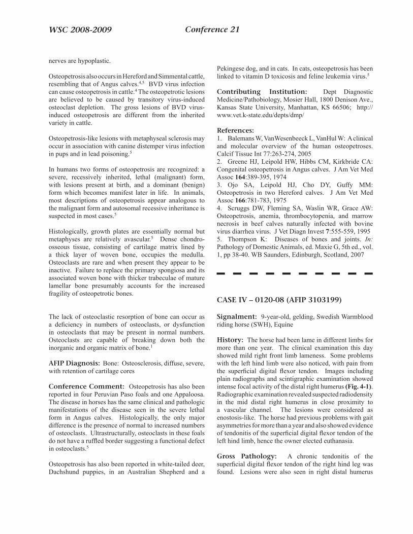
History: The horse had been lame in different limbs for more than one year. The clinical examination this day showed mild right front limb lameness. Some problems with the left hind limb were also noticed, with pain from the superficial digital flexor tendon. Images including plain radiographs and scintigraphic examination showed intense focal activity of the distal right humerus (Fig. 4-1).Radiographic examination revealed suspected radiodensity in the mid distal right humerus in close proximity to a vascular channel. The lesions were considered as enostosis-like. The horse had previous problems with gait asymmetries for more than a year and also showed evidence of tendonitis of the superficial digital flexor tendon of the left hind limb, hence the owner elected euthanasia.
Gross Pathology: A chronic tendonitis of the superficial digital flexor tendon of the right hind leg was found. Lesions were also seen in right distal humerus
WSC 2008-2009 Conference 21
macroscopically after the bone had been sawed (Figs. 4-2, 4-3). Bone tissue in trabecular and dense structures could be seen in the medullary cavity of the distal third (metadiaphyseal) humerus
Laboratory Results: None
Histopathologic Description: Sections from the distal area of humerus show a normal compacta with lamellar bone. In the medullary cavity intermingeled with adipose tissue, bone growth of trabecular bone tissue is found (Fig. 4-4). The bone trabeculae are lined by several osteoblasts and are composed of woven bone but often lined by lamellar bone structure. The larger trabecular are
4-1. Bone, horse. There is a focal area of intense scintigraphic activity within the medullary cavity. Scintograph courtesy of SLU, department of BVF, division of Pathology, Pharmacology & Toxicology, Box 7028, SE-
4-2, 4-3. Bone, horse. Within the medullary cavity and effacing bony trabeculae, there is a focally extensive proliferation of dense bony tissue. Photographs courtesy of SLU, department of BVF, division of Pathology, Pharmacology & Toxicology, Box 7028, SE-750 07 Uppsala, Sweden.
predominately of a lamellar pattern. A few osteoclasts can be seen adjacent to bone trabeculae. This type of idiopathic nonneoplastic bone growth in the medullary cavity would suggest enostosis-like lesions. The numerous osteoblasts explain the increased radiopharmaceutical uptake seen on scintigraphic examination.
Contributor’s Morphologic Diagnosis: Intra-medullary trabecular bone growth with osteoblastic activity compatible with enostosis-like lesions, right distal humerus, equine
Contributor’s Comment: In man2, enostosis is described as a relatively common lesion (14% in

WSC 2008-2009 Conference 21
cadaveric material). Enostosis has been defined as “a mass of proliferating bone within a bone.” The lesions are often referred to “bone islands, calcified medullary defects, cortical defects, enosteomas.” Enostoses of man are mostly found in the pelvis, spine, ribs and metaphyseal and epiphyseal parts of femur. Scintigraphy often shows absence of radiopharmaceutical uptake, indicating chronic lesions without osteoblast activity.
Canine panosteitis is described as an idiopathic disease, characterized by bone sclerosis of the diaphyses and metaphyses of long bones of large breeds. The dogs may be showing signs of illness with fever. Lameness is often presented as intermittent involving more than one limb. Multifocal areas of radio densities, often in association with nutrient foramen can be seen on radiographs. The bone changes will disappear and the dog recover completely.
In equine, enostosis-like lesions were first described as multifocal sclerotic areas within the medullary cavity of long bones and were reported in six Thoroughbreds, three Standardbreds and one Appoloosa.1 Most of the horses had a history of chronic lameness. The bone sclerotic areas seen on radiographs and as abnormal increased radioisotope uptake at scintigraphic examination were localized to the diaphyseal areas of the long bones. The lesions were endosteal and/or medullary. These lesions were named “enostosis-like” in order to differentiate them
from enostosis (bone islands seen in man) and panosteitis seen in dog. Later, Ramzan 4 also described enostosis-like lesions in 12 Thoroughbreds. Some of these lesions appeared related to lameness, but in many horses no association could be made. The author concluded that enostosis-like lesions are transient phenomena, with scintigraphic and radiographic resolution occurring over months. Multiple enostosis-like lesions has been reported in a racing Thoroughbred.3 As treatment of the disease, box rest and controlled exercise during 1-6 months have been suggested. Most of the horses recover and return to working or racing.
The etiology and pathogenesis of theses lesions are not known. Often the bone lesions have been found in the diaphyseal area close to the nutrient foramen, hence bone infarcts has been discussed as a cause. Stress fractures are also mentioned, but never proven to be associated with the radiodense areas. Microscopic examinations of the enostosis-like lesions in horses are not presented in the literature, hence a comparison of the present case and the reported enostosis-like lesions can not be made; however, the radiographic and scintigraphic findings are similar.
The present microscopic picture is that of a non-neoplastic non-specific bone growth with osteoblast activity, within the medullary cavities, very similar to changes seen microscopically in canine panosteitis.
4-4. Bone, horse. Within the medullary cavity, often contiguous with normal bony trabeculae, there is a focally extensive proliferation of woven bone. (HE 200X)

WSC 2008-2009 Conference 21
This horse had had a long history of gait asymmetries also involving tendonitis and the owner elected euthanasia. The enostosis-like lesions probably would have resolved and was not the reason for euthanasia.
AFIP Diagnosis: Bone: Intramedullary woven bone formation (enostosis), focal, moderate
Conference Comment: The contributor briefly mentioned canine panosteitis, a disease of unknown cause often seen in young large and giant breed dogs. Clinically, shifting leg lameness is often seen in affected animals. Thickening of the bone can occur on the periosteal and endosteal surfaces.5
Clinically, an increase in radiodensity in the marrow is initially seen and is caused by rapidly expanding areas of fibrovascular tissue that is quickly remodeled and converted to woven bone. Because of this rapid cyclical process of formation and resorption of bone, both osteoblasts and osteoclasts are often observed in the same microscopic field. Resting and reversal lines can also be present in relatively close proximity within medullary trabecular bone.5
Inflammation is usually not a feature of this disease, and
the lesions resolve spontaneously over time. The cause is currently unknown, but canine distemper virus has been suggested as a potential suspect.5
Contributing Institution: SLU, department of BVF, division of Pathology, Pharmacology & Toxicology, Box 7028, SE-750 07 Uppsala, Sweden, www.slu.se References: 1. Bassage LH, Ross MW: Enostosis-like lesions in the long bones of 10 horses: scintigraphic and radiographic features. Equine Vet J 30:35-42, 19982. Gould CF, Ly JQ, Lattin Jr GE, Beall DP, Sutcliffe JB: Bone tumor mimics: avoiding misdiagnosis. Curr Probl Diagn Radiol 36:124-141, 20073. Jones E, McDiarmid A: Case report. Multiple enostosis-like lesions in a racing Thoroughbred. Equine Vet Education. 17:92-95, 20054. Ramzan PHL: Equine enostosis-like lesions: 12 cases. Equine Vet J. 14:143-148, 20025. Thompson K: Diseases of bones and joints. In: Pathology of Domestic Animals, ed. Maxie G, 5th ed., pp.104-105, vol 1. WB Saunders, Edinburgh, Scotland, 2007