drharrisbiology.weebly.comdrharrisbiology.weebly.com/uploads/6/0/7/8/60782423/as... · web viewin...
TRANSCRIPT

AS- Biology Student GuideTopic 1 Proteins and Enzymes
Name:
Teacher:
Form:

Effective Learning 1-Excellent; 2-Good; 3-Needs to improve; 4-Cause for concern
Independent Learning• Do you work outside of formal learning sessions? • Are you able to demonstrate independent learning?
Questioning • Do you interrogate material/resources • Ask your LT / peers sensible, in depth questions • Question sources of information
Planning • Do you read additional material in preparation for the session
Assessment:Written test Date of test ___________________Target Attainment Grade:How will I achieve this?Evaluation:Self assessed learning grade ____Comments – (What can I do differently next time I do this?)Teacher assessed learning grade ____Comments:

• Plan your work in advance • Use assessment criteria to plan your tasks
Review/Amend • Do you look back at notes/tasks and review them regularly • Amend tasks having spoke/discussed with others or read more widely • Amend/review after advice from LTs
Active listening • Listen to understand • Be proactive and contribute in sessions/activities • Listen to other students in the group
Collaboration • Work effectively with others in the group • Take part in group work to ensure that everyone is involved • Take a range of roles within group work to encourage effective collaboration
Biological moleculesAll life on Earth shares a common chemistry. This provides indirect evidence for evolution.Despite their great variety, the cells of all living organisms contain only a few groups of carbon-based compounds that interact in similar ways.Proteins form many cell structures. They are also important as enzymes, chemical messengers and components of the blood.SPECIFICATION
Assessable Learning Outcomes
Covered
How did I review this
3.1.4.1 properties of Proteins
Amino acids are the monomers from which proteins are made. Thegeneral structure of an amino acid as:
NH2 represents an amine group,COOH represents a carboxyl group R represents a carbon-containing side chain.
The twenty amino acids that are common in all organisms differ only in their side group.
A condensation reaction between two amino acids forms a peptide bond.•• Dipeptides are formed by the

condensation of two amino acids.•• Polypeptides are formed by the condensation of many amino acids.
A functional protein may contain one or more polypeptides.
The role of hydrogen bonds, ionic bonds and disulfide bridges in the structure of proteins.
Proteins have a variety of functions within all living organisms. Therelationship between primary, secondary, tertiary and quaternary structure, and protein function.
The biuret test for proteins.Many proteins are enzymesEach enzyme lowers the activation energy of the reaction it catalyses.
The induced-fit model of enzyme action.
The properties of an enzyme relate to the tertiary structure of its active site and its ability to combine with complementary substrate(s) to form an enzyme-substrate complex.
•• The specificity of enzymes•• The effects of the following factors on the rate of enzyme controlledreactions – enzyme concentration, substrateconcentration, concentration of competitive and of noncompetitiveinhibitors, pH and temperature.
Students should be able to:•• appreciate how models of enzyme action have changed over time•• appreciate that enzymes catalyse a wide range of intracellular andextracellular reactions that determine structures and functionsfrom cellular to whole-organism level.
Students could be given thehydrogen ion concentrationof a solution in order to

calculate its pH, using theformula:pH = −log10 H+
PracticalRequired practical 1: Investigation into the effect of a namedvariable on the rate of an enzyme-controlled reaction.

KEY WORDS
keyword definition
Globular Protein
Poly Peptide
Peptide Bond
Amino Acid
R group
Amino group
Carboxyl group
Hydrogen bond
Alpha helix
Beta pleated sheet
Primary Structure
Secondary Structure
Tertiary Structure
Quarternary Structure

Disulfide Bridge
Ionic Bonds
Hydrogen bonds
Activation Energy
Substrate
Enzyme Substrate Complex
Lock and Key
Induced Fit hypothesis
Kinetic Energy
V max
Competitive inhibitor
Non competitive inhibitor
Negative feedback
Describe the test for protein.

Resources and suggested Reading: find this document on www.drharrisbiology.weebly.com to click the links.

Copies of Biological Science review can be found in the library : see the MLE for the index of past articles
http://www.biologymad.com/
http://www.mrothery.co.uk/ecology/Mod5Notestrimmed.htm
http://www.freeeschool.blogspot.co.uk/
The awesome Bozeman Biology YouTube site: Proteinshttps://www.youtube.com/watch?v=2Jgb_DpaQhM
Practice Questions
Answer these practice questions and self assess them against the mark schemes published on www.drharrisbiology.weebly.com
Q2. The diagram shows the structure of the amino acid serine.

(a) (i) Draw a box on the diagram around the R group of serine and label the box with the letter R.
(1)
(ii) Draw a circle around each of the parts of the serine molecule which would be removed when two other amino acid molecules join directly to it.
(1)
(b) (i) Which two substances are formed when two amino acid molecules join together?
1 ..........................................................................................................
2 ..........................................................................................................(1)
(ii) Name the type of bond formed between the joined pair of amino acid molecules.
.............................................................................................................(1)
(c) Explain how a change in the primary structure of a globular protein may result in a different three-dimensional structure.......................................................................................................................
......................................................................................................................
......................................................................................................................
......................................................................................................................
......................................................................................................................(3)
(Total 7 marks)
Q3. The diagram represents an enzyme molecule and three other molecules that

could combine with it.
(a) Which molecule is the substrate for the enzyme? Give a reason for your answer.
......................................................................................................................
......................................................................................................................(1)
(b) Use the diagram to explain how a non-competitive inhibitor would decrease the rate of the reaction catalysed by this enzyme.
......................................................................................................................
......................................................................................................................
......................................................................................................................
......................................................................................................................(3)
(c) Lysozyme is an enzyme. A molecule of lysozyme is made up of 129 amino acid molecules joined together. In the formation of its active site, the two amino acids that are at positions 35 and 52 in the amino acid sequence need to be close together.
(i) Name the bonds that join amino acids in the primary structure.
.............................................................................................................(1)
(ii) Suggest how the amino acids at positions 35 and 52 are held close together to form the active site.
.............................................................................................................
.............................................................................................................
.............................................................................................................(2)
(Total 7 marks)

Q4. Read the following passage.
Job’s Tears is a cereal plant which grows in the tropics. An unusual protein has been found in its grains. This protein is unusual because it has two functions. It acts as both an enzymeinhibitor and as an enzyme. As an inhibitor, the protein reduces the activity of starch-digesting enzymes. The protein acts as an enzyme by breaking down chitin, a polysaccharide found in the walls of many fungi, to its monomers. Because of the resulting more negative water potential in the cytoplasm of the fungus, this effectively leads to “death by osmosis” of anyfungus attacking the grain.
Our knowledge of the relationship between protein structure and function has led to thedevelopment of the new technology of protein engineering. This involves changing the amino acid sequence of a protein and altering its tertiary structure. Altering the tertiary structure changes the protein’s properties. So far, we have been unable to produce a protein with more than one function such as that found in Job’s Tears. We have had success, though, in making some enzymes more stable and less prone to heat denaturation. We have done this by substituting amino acids and allowing the formation of additional chemical bonds.
Use information from the passage and your own knowledge to answer the following questions.
(a) (i) The protein found in Job’s Tears breaks down chitin (line 4). What type of chemical reaction is involved in breaking down chitin?
.............................................................................................................(1)
(ii) Breakdown of chitin leads to “death by osmosis” of fungi attacking the grain (lines 6 - 7). Explain how.
.............................................................................................................
.............................................................................................................
.............................................................................................................
.............................................................................................................(2)
(iii) This protein does not break down the cell walls of the Job’s Tears plant.Explain why.
.............................................................................................................
.............................................................................................................(1)

(b) Explain what is meant by the tertiary structure of a protein (line 10).
.............................................................................................................
.............................................................................................................(1)
(c) (i) Explain how heating an enzyme leads to it being denatured.
.............................................................................................................
.............................................................................................................
.............................................................................................................
.............................................................................................................(2)
(ii) How can protein engineering make enzymes more stable and less prone to heat denaturation (line 13)?
.............................................................................................................
.............................................................................................................
.............................................................................................................
.............................................................................................................(2)
(d) Describe how the sequence of amino acids in part of the protein from Job’s Tears could enable this protein to act as an enzyme inhibitor.
......................................................................................................................
......................................................................................................................
......................................................................................................................
......................................................................................................................
......................................................................................................................
......................................................................................................................
......................................................................................................................(6)
(Total 15 marks)

Q5. Uric acid is produced in the body. One of the reactions involved in the production of uric acid is catalysed by xanthine oxidase.
xanthine oxidase
xanthine uric acid
(a) A sample of xanthine oxidase was tested by mixing with biuret reagent. Describe and explain the result of this test.
......................................................................................................................
......................................................................................................................
......................................................................................................................(2)
(b) Explain why xanthine oxidase is able to catalyse this reaction but it is not able to catalyse other reactions.
......................................................................................................................
......................................................................................................................
......................................................................................................................
......................................................................................................................(2)
(c) Gout is a painful condition caused by uric acid crystals in the joints. It is often treated with a drug that inhibits xanthine oxidase. The diagram shows a molecule of xanthine and a molecule of this drug.
Xanthine Drug usedto treat gout
Use the diagram to explain why this drug is effective in the treatment of gout.
......................................................................................................................
......................................................................................................................
......................................................................................................................
......................................................................................................................
...................................................................................................................... (3)
(Total 7 marks)

Q6. In an investigation into carbohydrase activity, the contents from part of the gut of a small animal were collected. The contents were added to starch solution at pH 7 and kept in a water bath at 25°C. At one-minute intervals, samples were removed and added to different test tubes containing dilute iodine solution. The colour intensity of each sample was determined. The graph shows the results.
(a) Explain the change in colour intensity.
......................................................................................................................
......................................................................................................................
......................................................................................................................
......................................................................................................................(2)
(b) Draw clearly labelled curves on the graph to show the expected result if the experiment was repeated
(i) at 35 °C;
(ii) at pH 2.(2)
(c) Explain how
(i) raising the temperature to 35 °C affects carbohydrase activity;
.............................................................................................................
.............................................................................................................
.............................................................................................................
.............................................................................................................
.............................................................................................................

.............................................................................................................
(ii) decreasing the pH affects carbohydrase activity.
.............................................................................................................
.............................................................................................................
.............................................................................................................
.............................................................................................................
.............................................................................................................
.............................................................................................................
.............................................................................................................
.............................................................................................................
.............................................................................................................
.............................................................................................................(7)
(Total 11 mark
Q7. (a) The diagrams represent an enzyme, its substrate and two other molecules, A and B.
The addition of a non-competitive inhibitor will prevent the formation of an enzyme-substrate complex. Draw a labelled diagram based on relevant molecules selected from the diagram above to explain how this occurs.
(2)

(b) A decrease in temperature decreases the kinetic energy of molecules in a solution. Explain how a decrease in temperature decreases the rate of an enzyme-controlled reaction.
......................................................................................................................
......................................................................................................................
......................................................................................................................
......................................................................................................................(2)
(c) Urea breaks hydrogen bonds. Explain how the addition of urea would affect the rate of an enzyme-controlled reaction.
......................................................................................................................
......................................................................................................................
......................................................................................................................
......................................................................................................................
......................................................................................................................
......................................................................................................................(3)
(Total 7 marks)
Y13 Essay Practice (hand written 2 sides of A4 lined please!)
You should write your essay in continuous prose.
Your essay will be marked for its scientific accuracy.
It will also be marked for your selection of relevant material from different parts of the specification and for the quality of your written communication.
The maximum number of marks that can be awarded is ScientificBreadth of knowledgeRelevanceQuality of written communication
16333
Write an essay on the following topic:
How the structure of proteins is related to their functions.(Total 25 marks)


M1. General Principles for marking the Essay:
Four skill areas will be marked: scientific content, breadth of knowledge, relevance and quality of language. The following descriptors will form a basis for marking.
Scientific Content (maximum 16 marks)
Category Mark Descriptor
Good
16
14
12
Most of the material reflects a comprehensive understanding of the principles involved and a knowledge of factual detail fully in keeping with a programme of A-level study. Some material, however, may be a little superficial. Material is accurate and free from fundamental errors but there may be minor errors which detract from the overall accuracy.
Average
10 8 6
Some of the content is of an appropriate depth, reflecting the depth of treatment expected from a programme of A-level study. Generally accurate with few, if any, fundamental errors. Shows a sound understanding of the key principles involved.
Poor
4 2
0
Material presented is largely superficial and fails to reflect the depth of treatment expected from a programme of A-level study. If greater depth of knowledge is demonstrated, then there are many fundamental errors.
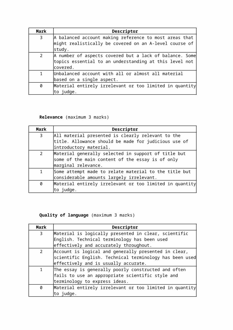
Breadth of Knowledge (maximum 3 marks)
Mark Descriptor3 A balanced account making reference to most areas that might realistically
be covered on an A-level course of study.2 A number of aspects covered but a lack of balance. Some topics essential
to an understanding at this level not covered.1 Unbalanced account with all or almost all material based on a single
aspect.0 Material entirely irrelevant or too limited in quantity to judge.

Relevance (maximum 3 marks)
Mark Descriptor3 All material presented is clearly relevant to the title. Allowance should be
made for judicious use of introductory material.2 Material generally selected in support of title but some of the main content
of the essay is of only marginal relevance.1 Some attempt made to relate material to the title but considerable
amounts largely irrelevant.0 Material entirely irrelevant or too limited in quantity to judge.
Quality of language (maximum 3 marks)
Mark Descriptor3 Material is logically presented in clear, scientific English. Technical
terminology has been used effectively and accurately throughout.2 Account is logical and generally presented in clear, scientific English.
Technical terminology has been used effectively and is usually accurate.1 The essay is generally poorly constructed and often fails to use an
appropriate scientific style and terminology to express ideas.0 Material entirely irrelevant or too limited in quantity to judge.
[25]
Additional guidance for assessing Scientific Content and Breadth of Knowledge in Essays
The following provides guidance about topics which might be included in the essays. It is not an exclusive list; the assessment of scientific content does not place restrictions on topics that candidates might refer to, provided they are
• relevant;
• at an appropriate depth for A level and
• accurate.
It is not expected that candidates would refer to all, or even most, of the topics to gain a top mark; the list represents the variety of approaches commonly encountered in the assessment to the essays.
In both essays, topics either from the option modules or beyond the scope of the specification were also given credit where appropriate.
How the structure of proteins in relation to their functions.

1. Structure (S)primary structure – peptide bondsecondary structuretertiary structure. Globular - bonds between R groups give spherical shape – shape determines function – active sites and receptor sites(allow quaternary structure – haemoglobin incorporates ions for oxygen transport)
2. Structural proteins (ST)fibrous – regular pattern of hydrogen bonds – coiling, (e.g. keratin coils twist together to form rope-like structures – flexible and strong)(e.g. collagen – coils more tightly bound – more rigid)
3. Transport (T)channel – complementary shape – charges – gatedcarrier – complementary shape – can change shapeactive transport – phosphate group attached by energy from ATP – can change shape
4. Enzymes (E)active site, enzyme-substrate complexactivation energy reduction - explanation e.g. brings molecules closer
5. Receptors (R)synapseinsulin / glucagonADHrhodopsin
6. Muscle (M)actin thin – binding sitemyosin thick - cross bridgestropomyosin – block binding sites
Breadth of knowledge 3 marks Four or more of the above 6 areas2 marks Three of the above 6 areas1 mark Two of the above 6 areas

M2. (a) (i) box drawn around R group (i.e. CH2OH group)(allow circle if labelled R);
1
(ii) circle drawn around either of the Hs on NH2 group and circle drawnaround the OH;
1
(b) (i) (di)peptide and water;1
(ii) peptide;1
(c) sequence of amino acids changes;tertiary structure changes / folds in a different way;bonds form in different places;(Reject peptide bonds)
3[7]
M3. (a) A and structure(of A) is complementary to that of the active site;1
(b) idea that non-competitive inhibitor(C) binds at a site not the activesite; binding causes a change in the shape of the active site;substrate is no longer able to bind to the active site;
3
(c) (i) peptide;1
(ii) idea that amino acid chain folds / tertiary structure;named bond holding tertiary structure e.g. ionic disulphide hydrogen;
{reject peptide)2
[7]

M4. (a) (i) Hydrolysis;1
(ii) Water enters fungus (by osmosis) which increases pressure inside fungus;Cell wall no longer strong enough / present so cannot withstand this;
2
(iii) Cell wall (of plant) not made of chitin / made of cellulose;Enzyme is specific to chitin / will not break down cellulose;
1
(b) Way in which the whole protein / polypeptide is folded / shape adopted by whole protein molecule / further folding of 2° structure;
Do not credit unqualified reference to three-dimensional shape.Reject third level / third sort.
1
(c) (i) More (kinetic) energy;Bonds / specified bonds (holding tertiary structure) break;
2
(ii) Change amino acids;Allowing formation of more hydrogen bonds / disulphide bridges;
2
(d) 1. Sequence of amino acids gives shape;2. This is tertiary structure;3. Has similar shape to substrate;4. Fits / competes for active site;5. Fits at site other than active site;6. Distorting active site;7. Therefore substrate will not fit (active site);
max 6[15]
M5. (a) Lilac / purple / mauve / violet;

Xanthine oxidase is a protein;Reject pink or blue as the resulting colour with biuret.
2
(b) Substrate has specific shape;
Allows binding / fitting / forms ES complex with active site;
Or
Active site has specific shape;
Allows binding / fitting / forms ES complex with substrate;Accept structure ≡ shape
2
(c) Xanthine similar shape to drug;
Drug fits active site / competes for active site / is a competitive inhibitor;
Less / no uric acid formed;3
[7]
M6. (a) colour results from starch-iodine reaction;decrease due to breakdown of starch by carbohydrase / enzyme;
2
(b) (i) curve drawn below curve on graph and starting at same point;1
(ii) curve drawn above curve on graph and starting at same point butfinishing above;
(allow curve or horizontal line)(allow alternative curve for pH if explanation in (ii) is consistent)
1
(c) (i) 1. increase in temperature increases kinetic energy;2. increases collisions (between enzyme / active site and substrate) / increases formation of enzyme / substrate

complexes;3. increases rate of breakdown of starch / rate of reaction / carbohydrase activity;
(ii) 4. (decrease in pH) increases H+ ions / protons which attach / attracted to amino acids;5. hydrogen / ionic bonds disrupted / broken which denatures enzyme / changes tertiary structure;6. changes shape / charge of active site so active site / enzyme unable to combine / fit with starch / enzyme-substrate complex no longer able to form;7. decreases rate of breakdown of starch / rate of reaction / carbohydrase activity;
(allow alternative explanation for pH if consistent with line drawn in (ii))
7[11]
M7. (a) diagram showing molecule A fitting in inhibition site; distortionof active site;
2
(b) molecules moving less / slower; reduces chance of collision(between enzyme and substrate) / of enzyme-substratecomplexes being formed; (reject converse)
2
(c) these bonds hold / maintain tertiary / globular structure (of enzyme);enzyme denatured / tertiary structures destroyed; (shape of) active sitedistorted / changes;substrate no longer fits / enzyme-substrate complex not formed;
3 max[7]
forming enzyme-substrate complex;2
[7]