water flux through mycelium of serpula lacrimans
TRANSCRIPT
Trans . Br . mycol. Soc. 84 (4), 601--608 (1985)
[ 601 ]
Printed in Great Britain
WATER FLUX THROUGH MYCELIUM OF SERPULALACRIMANS
By WENDY THOMPSON, D. EAMUS AND D . H. JENNINGSBotany Department, The University, P .O. Box 147, Liverpool L69 lBX
Values have been obtained for water flux and the velocity offlow through mycelium of Serpulalacrimans growing over Perspex. The values are 1-23 times greater than previous measure-ments of the velocity of solute translocation using isotopic tracers. Addition of 10 JLg cm ?
oligomycin to the food source inhibited both linear extension and also water uptake. However,2 '0 mol 1-1 glucose affected only water uptake. Changes in the water, solute and turgorpotentials of hyphae were studied for a 72 h period after the addition of glucose and werein keeping with the observed effects on growth and water uptake. The results are discussedin relation to the mechanisms of translocation and the ability of the mycelium to bufferalterations of the solute potential of the medium external to those hyphae at the food source.
The dry rot fungus Serpula lacrimans (Wulf. : Fr.)Schrot. can grow for considerable distances overnutritionally inert substrata. Clarke, Jennings &Coggins (1980) have provided evidence that wateris translocated through such growing mycelium andthere is now cons iderable support for the view thattranslocation of nutrients is brought about by bulkflow of water through mycelium, the flow beinggenerated osmotically (Coggins, Jennings &Clarke, 1980; Brownlee & Jennings, 1981a,1982 a). The velocities of movement of individualsolutesareverysimilar,around25 ern h-1.However,there is no information about the rate of waterabsorption by the mycelium at the food source orabout the volume of water moving through unitcross-sectional area of mycelium per unit time(flux). This paper provides such information,obtained as mycelium grew over inert andnon-absorbant substrata.
With respectto elucidating the processes involvedin the movement of water through the mycelium,the steady-state can be less revealing than whenthere is a change brought about by an externally-induced change in water uptake. To this end, wehave studied the effects on water flux of themetabolic inhibitor oligomycin and of a low solutepotential, generated by 2'0 mol 1-1 glucose. For thelatter, we also determined the water, solute andturgor potentials of the mycelium before and afterthe imposed treatment. Turgor pressure is knownto be the driving force for plant cell growth (Ray,Green & Cleland, 1972). Consequently, if theformer is also true for fungi, changes in growth maybe expected to be reflected in turgor changes andvice-versa. In addition, if there is to be bulk flowof water, there must be a turgor gradient in themycelium from the food source to the growing
front. The data reported here demonstrate thepresence of such a gradient under conditions whentranslocation is . known to be occurring anddisappearance of the gradient when translocation isknown to be inhibited. It is also shown that positiveturgor can still exist in the mycelial front even whenthe turgor gradient disappears.
MATERIALS AND METHODS
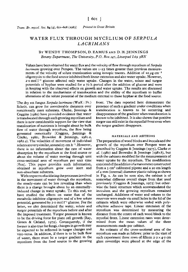
The preparation of wood blocks and inocula and thegrowth of the mycelium over Perspex were asdescribed by Coggins & Jennings (1975), Clarke etal. (1980) and Brownlee & Jennings (1981b), butwith the cabinets modified for the measurements ofwater uptake by the mycelium. The modificationconsisted ofthe addition of amanometer constructedfrom a 5 ern" calibrated pipette and a 20 em lengthof 4 mm (internal) diameter plastic tubing as shownin Fig . 1. As can be seen also, the cabinet is ofsomewhat different overall shape from that usedpreviously (Coggins & Jennings, 1975) but other-wise the basic structure which accommodated theinoculum and the growing mycelium remainedunchanged. Additions of water and solutes to thereservoir were made via small holes in the lid of thecabinets which were otherwise sealed with poly-ethylene adhesive tape. L inear extension of themycelium was determined by measuring thedistance from the centre of each wood block to themycelial front . Linear extension rate s were deter-mined from the mean values of the threemeasurements made per cabinet.
An estimate of the cross-sectional area of themycelium was made as follows : prior to the start ofeach experiment three rows of fifteen 2"2 x 4'0 emglass coverslips were placed at the edge of the
602 Water flux in Serpula lacrimans
~L
"",:"",: <.~G x
A
1M
TI iii
B
1c
1
Fig. 1. Perspex cabinet used for measuring water uptake and linear extension rate of Serpula lacrimansmycelium and effects of z mol I-I glucose and 10 ftg em'? oligomycin thereon. A, B, C, positions of rows ofglass coverslips used in estimations of hyphal cross-sectional area. A, °cm from inoculum; B, 10 em frominoculum; C, 20 cm from inoculum; F, filter paper; G, graduated pipette, attached slanting upwards to theside of the cabinet; H, hole for addition of solutions; L, Perspex lid; M, mycelium; P, colonized pine blocks;5, sterile water; T, plastic tubing; X, Perspex platform.
Experimental details
Uncolonized wood blocksColonized wood blocksColonized wood blocks; fungus treated
with formaldehyde after 10 dColonized wood blocks; after 456 h
50 ern" sterile water used to replacewater present in well initially
Colonized wood blocks; after 456 h50 ern" z·o mol I-I glucose used toreplace water in well initially
As z and 3Colonized wood blocks; 50 ern" of
0·1 % (v/v) ethanol used to replaceafter 472 h water present in wellinitially
Colonized wood blocks; 50 ern! ofro rzgcm! oligomycin in 0-1% (v/v)ethanol used to replace after 47z hwater present in well initially
Experimental treatment (3)
Experimental treatment (3)
Control for evaporation (z)Experimental control (3)
Experimental control (3)
4
Experiment
Table 1. Experimental details for measurement of water uptake and linear extension rate by mycelium ofSerpula lacrimans and effects of different treatments thereon
Treatment (number ofreplicates in parentheses)
Control for evaporation (1)Experimental treatment (3)Control for evaporation (z)
Mean water uptake/loss values were obtained by deducting values for controls for evaporation from values forcontrols and treatments.
Perspex platform nearest the inocula and 10 and20 cm away from the edge. The short axes of thecover slips were in the direction of mycelial growth.At the end of each experiment, the cover-slips, bythen covered with mycelium, were removedcarefully from the platform and placed face up onslides, which were stored in Petri dishes when
necessary at 2 °C prior to observation. Individualhyphae were then measured and counted under alight microscope. This was done by counting andmeasuring, for every fifth field, all hyphae whichcrossed a transect line extending across the entiremycelial system. Hyphae were grouped into thefollowing diameter classes: 2-4, 4-10, 10-20 and

Wendy Thompson, D. Eamus and D. H. Jennings
* Measurements made in first 456 h.t Measurements made in first 472 h.
Table 2. Mean linear extension rate (±S.E.M.) formycelium of Serpula lacrimans with pure water inwells of cabinets
(For further details see Table 1)
mounting were lowered into position and pressedfirmly down into the silicon rubber such that theyformed the roof of the well. A locking screw wasused to hold the thermocouple in position. Allmeasurements were made in the dew point mode,after calibration with a series of sodium chloridesolutions of known water potential.
RESUL TS
Mean linear extension rates for Serpula lacrimansgrowing over Perspex with pure water present inthe reservoir containing the wood inocula are givenin Table 2 and Fig. 2. The values are somewhatlower than reported previously (Jennings, 1982) forgrowth over Perspex under similar conditions butwithout water present in the reservoir. The lowerlinear extension rates might be due to the depletionof oxygen around that part of the mycelium on thewood which is submerged. Figure 3 shows thataddition of 2'0 mol 1-1 glucose had no significanteffect on linear extension rate, although addition ofwater to control and experimental cabinets causedsmall decreases and then increases in linearextension rate. On the other hand, 10 pg cm?
oligomycin clearly inhibited growth, althoughrecovery occurred within 30 h (Fig. 4).
The rate of water uptake as a function of time isgiven in Figs 2, sand 6. The rate of uptake was notconstant but fluctuated in an irregular manner.
It was not possible to detect any relationshipbetween changing rate of uptake and those changeswhich took place in linear extension rate seen inFigs 2-4. N0 significant correlation (P:::;; 0'1 )
between linear extension rate and water uptakewas found in any experiment when the data were
4'45±qq8±o'62'56±l 'O
Cabinetsused for
experimentaltreatment
Mean linear extension rate(mm day " )
Cabinetsused ascontrols
l '79±O'52'74 ±1 '2
Experiment
1
2*
4t
> 20 pm. A measure of total cross-sectional areawas obtained by summing the number of hyphae ineach class and multiplying by the mean diameter ofthe class . These values were corrected for the totalnumber of fields per transect. Total cross-sectionalareas per class were then added to give the totalcross-sectional area per transect line . In this way,the cross-sectional area of hyphae at the threepositions in the cabinet as indicated in Table 3 weredetermined.
Table 1 gives details of four experiments used todetermine water uptake and linear extension ratesof S. lacrimans. Any resulting differences betweenexperimental and control cabinets were analysedstatistically using the least significant differencemethod (P :::;; o-oy) for two-tailed tests.
Thermocouple psychometry was used to deter-mine water, solute and turgor potential of mycelialsamples from the cabinets . The samples ofapproximately 0'01 g were picked up from thePerspex with forceps and within 30 s sealed into thesample chambers. In the case of the mycelial front,the mycelium was cut with a razor o'S cm behindand the sample picked up at the cut. For samplesfrom the rear of the mycelia, 1 cm x 1 cm squareswere cut with a razor; three squares being for eachpsychometric determination. The same sampleswere used to determine both the water and solutepotential. After the former had been measured, thesamples were frozen rapidly in liquid nitrogen,thawed and the solute potential then measuredfrom the equation
'I'w = 'I' s+ 'I' p'
where 'I' w is the water potential, 'I' s the solutepotential and 'I' p the turgor potential. The latter isreduced to zero after freezing, due to rupturing ofthe hyphae as a result of ice-formation in the cell.Consequently after freeze thawing in liquidnitrogen, the 'I' w which is determined is equal to 'I'ssince 'I' p is zero. For the intact hyphae, 'I' wand 'I'scan be inserted in the above equation and 'I' p
calculated. Water and solute potentials weremeasured with a Wescor MR-33T microvoltmetercoupled either to a C-S2 or L-Sl sample chamber.The latter was modified to function as a C-S2chamber by producing a silicon rubber base withcentre well as follows (C . Berryman, pers. comm.).A 1 X 1 ern square of silicon rubber of 1 mmthickness was produced and a 3 mm diameter holepunched out of the centre. A square of similar sizewithout the hole was produced and when still stickybonded to the former square. When dry, the wellwas filled with mycelium and inserted into the L-S1body such that the well and mycelia lay directlybelow the thermocouple. The thermocouple and

604 Water flux in Serpula lacrimans
r 0-4 0·04..cE~s 0·3 ie 0'03 ..c::e :g0.;;;c .... ~x 0-.. 0·2 0·02... ::l
'" ..... ~:::.::i '"~
0·1 0·01
100 200 300 400 500 600 700 800
Time (h)Fig . 2. Linear extension rate and water uptake as a function of time by mycelium of Serpula lacrimans growingfrom wood blocks over Perspex (Expt 1). • , water uptake; 0, linear extension rate; bars represent standarderrors.
The estimates for the total hyphal cross-sectionalarea at the three different regions of the myceliumin each cabinet are given in Table 3. It can be seenthat, in spite of considerable changes in the
100 200 300 400 500 600
Time (h)
Fig. 4. Linear extension rate of mycelium of Serpulalacrimans before and after addition of 50 em' 10 ".g em"?oligomycin to well containing colonized wood blocks . • ,experimental cabinets; 0, control cabinets. Arrowindicates the time of addition of oligomycin (experimentalcabinets) or 0 '1 % (vI v) ethanol (control cabinets). Starindicates experimental and control values significantlydifferent (P ,;; 0 '05) .
r 0·5..c 1E~ 0·4Be 0·3::.9a" 0·2><"...'" O·lw::
:.::ia
600500200100o
0·2
300 400Time (h)
Fig . 3. Linear extension rate of myceliu m of Serpulalacrimans before and after addition of 50 em' 2 mol 1-1glucose to wells cont aining colonized wood blocks . • ,experimental cabinets; 0, control cabinets. Arrowindicates the time of addition of glucose (experimentalcabinets) or water (control cabinets).
subjected to linear, logarithmic and power regres-sion analyses. Both 2 '0 mol 1-1 glucose and10 pg ern'? oligomycin brought about water lossfrom the mycelium. The effect of glucose appearedto be almost immediate; with oligomycin at least8 h had elapsed before water uptake differedsignificantly (P ~ 0'05) from controls. The effect ofthe two solutes was relatively short-lived; recoveryto equivalent control levels being apparent within20-45 h for glucose and 4 h for oligomycin.
~eco.;;;::~ 0·1
"l;;"::
:.::i
r..cE.5 0·3

Wendy Thompson, D. Eamus and D. H. Jennings 605
Table 3. Cross-sectional area of different regions of mycelium of Serpula lacrimans growing from wood blockinocula over Perspex
Experiment Distance from inoculum (ern)and
cab inet 0 10 20 Mean ( ± S,E.) (mm')
Expt 1 0 '°47 0 '063 0 '044 0 '051 ± 5'9 x 10- 3
Expt 2A 0 '061 0 '°59 0' 063 0 '061 ± 1'1 x 10⁻³
B 0'°70 0-055 0'080 0,068 ± 7'2 x 10⁻³
C 0 '083 0-078 0 '055 0 '°72 ± 8,6 x 10⁻³
Expt 3A 0'090 0 '115 0 '125 O'110±O'OlB 0'065 0 -062 0'082 O'070±6-2 X10⁻³
C 0'°92 0 '°73 0'060 0-075 ± 9 '2 x 10⁻³
Expt 4A 0'°77 0 '09 0 0 '085 o'084±3'7 x 10⁻³
B 0'100 0 '07 1 0' 069 0'080 ± 0-001C 0 '108 0 -095 0-092 0'098 ± 4'9 x 10⁻³
Mean 0'079 0 '076 0'075S.E, ±5 '9 x 10⁻³ ± 5'9 x 10⁻³ ±7'2 X10⁻³
Table 4. Values for flux and velocity of waterthrough mycelium of Serpula lacrimans growing overPerspex
(Both were calculated using the values forthe cross-sectional area of mycelium given inTable 3.)
Experimentand Flux Velocity
cabinet (em" cm ? S- I) (cm h" )
Expt 1 5'22 X 10- ' 188
Expt 2A 3,69 X 10- ' 133B 4'42 x 10- ' 159C 1-32 x 10- 1 475
Expt 3A 2'63 X 10- ' 95B 2'83 x 10-' 102
C 3'39 X 10-' 122
Expt 4A 6'51 X 10- 3 23B 2,80 x 10-' 101
C 2'24 X10-' 81
Mean 4'10 x 10- ' 148
S.E. ±1'08±10- ' ± 39
morphology of the mycelium, accompanying theformation of strands or syrotia with age, thecross-sectional areas differ very little at the threeregions. Using the means ofthc values for the threeregions in each cabinet and values for volumes ofwater taken up over a 440 h period, which has tobe equated with flow of water through mycelium,
the velocity (ern S-I) of water movement has beencalculated (Table 4).
Table 5 gives values for the water, solute andturgor potentials of the mycelium before and atintervals after the addition of z-o mol 1-1 glucose tothe wells containing the inocula. It can be seen thatwith the medium water potential at zero, there wasa water potential gradient along the mycelium fromthe base to the growing mycelial front and furtherthat there was a gradient of turgor. The gradientcan be said to be positive, in that turgor was higherat the base than at the front. At the firstdetermination, after the addition of 2 '0 mol 1-1
glucose to the bathing medium, at 4'5 h, while thedirection of the water potential gradient remainedthe same the turgor gradient was reversed. Thereversed gradient increased in magnitude to a valueof 0·81 MPa at 24 h, subsequently to decline suchthat after 72 h a positive gradient was re-established.Recovery of the gradient appeared to be mediatedvia a continuing decrease in the solute potential.Throughout the whole period, the hyphae at themycelial front exhibited a positive turgor potential.
DISCUSSION
The primary aim of this investigation has been todetermine the velocity of water flow through themycelium of S. lacrimans as it extends over anon-nutrient, non-absorptive surface, The valuesobtained were in the range 23-475 ern h- 1 • Itshould be noted that because of the way the velocityhas been determined, i.e. by dividing the presumedvolume of water moving by the cross-sectional area

606 Water flux in Serpula lacrimans
Table 5. Water, solute and turgor potentials of mycelium of Serpula lacrimans growing from wood blocksover Perspex prior to and after exposure of mycelium at the blocks to z-o mot t» glucose
(Samples were taken from mycelium close to the blocks (base) and at the growing front .)
Prior tothe additionof glucose 4'5
Time after addition of glucose (h)
Z4 7z
Base Front Base Front Base Front Base Front Base Front
Water potentialSolute potentialTurgor potentialTurgor gradient
-Z'o - z'7-2'37 -z'94
0'37 0 '24+0'13
- Z'5 -4'46-z'70 -4'78
0 '19 0 '3z- 0 '13
(MPa)
- z '98 - 5'85-3'17 - 6'85
0'19 1'0- 0,81
- 3'85 -7'30- 4' 15 -7'40
0'30 0·60-0'3
- 4'Z1 -6,80-4'8z -7 'Zl
0 ·61 0 '41+ o·z
and no account was taken of the thickness of thewalls of the hyphae when measuring their diameter.Additionally, it has been assumed that all hyphaeare involved in water flow through the mycelium.While it is possible that some water may manifestitself as mycelial growth on the wood block inocu-lum and absorption into the wood, the amount ofwater absorbed in this way is likely to be small .
Brownlee & Jennings (198za) found that thevelocities of translocation of [14C]glucose, [14C]3-O-methyl glucose, [14C]aspartate, [3ZP]orthophos-phate and [UK] were all in the region of 20-30 ernh-1 • Thus water flow through the myceliumappears to be 1-23 times faster. This is of courseequating uptake of water into the mycelium withmovement through it. The apparent faster flow ofwater than solutes may be due to (i) evaporationfrom hyphae, (ii) movement of water from thetranslocating hyphae into those which are nottranslocating in the mycelium lying between theinoculum and first radioactive detector (Brownlee& Jennings, 198za), (iii) the presence of hyphaewhich allow translocation of water but not solutes,whose translocation is known to be restricted tocertain hyphae and hyphal systems (Brownlee &Jennings, 198zb) or (iv) the cross sectional area ofhyphae being greater in vivo than their diametersmeasured after dry storage at 2° conditions underwhich collapse and shrinkage is to be expected.
There are few other determinations of the velo-city or flux of water through mycelia. Duddridge,Malibari & Read (1980) obtained a value of27 cm h- 1 for the velocity of [3H]H zO alongrhizomorphs of Suitlus booinus, while Plunkett(1956) obtained transpiration fluxes of 0'13 x 10- 3
-3'5 X 10- 3 ern" crn" h- 1 from sporophores ofPolyporus brumalis subjected respectively to 96and 75 % relative humidity at 20°.
The experiments involving addition of oligo-mycin to the mycelium on the wood inoculum
*
1·2
1·0
o-s
r 0·6..c
],0·4..,
..>0:
'"i5. 0·2::>..s'" 0~
- 0·2
-o·gFig. 5. Water uptake by mycelium of Serpula lacrimansbefore and after addition of 50 em" Z mol 1- 1 glucose to
wells containing colonized wood blocks. • , experimentalcabinets ; 0, control cabinets. Arrow indicates time ofaddition of glucose (experimental cabinets) or water(control cabinets). Stars indicate experimental andcontrol values significantly different (P ~ 0'05 ).
- 0·6
of the pathway, it is in this instance synonymouswith flux (em" cm" h-1) . It is possible to comparethese values for the velocity ofwater flow with thosefor the movementalong the myceliumof radiotracersdetermined previously (Brownlee & Jennings,198za). However, it should be pointed out that thevalues for the velocity of water movement are likelyto be under-estimates since the calculation of thecross-sectional area of the mycel ium was madeassuming that all hyphae counted were functional
-0,4

Wendy Thompson, D. Eamus and D . H. Jennings
10·08
0·06
0·04r..c::
§ 0·02
"~ 0Q.;:I
- 0·02..."'CO~ - 0·04
- 0·06
- 0·08
-0·141-0,2836
- 1·4375 •
Fig . 6. Water uptake by mycelium of Serpula lacrimans before and after addition of 50 em' 10 Jig em- aoligomycin to wells containing colon ized wood blocks.•, experimental cab inets; 0, control cabinets. Arrowindicates time of addition of oligomycin (experimental cabinets) or 0 '1 % (v/v) ethanol (control cabinets).Star indicates experimental and control values significantly different (P ~ 0 '05 ).
indicate that respiration is necessary both forcontinued linear extension of the mycelium andwater uptake into it. Brownlee & Jennings (1981a)showed that 18 fLg 1-1 oligomycin inhibited dropletvolume increase and extension of hyphae ofS. lacrimans. However, in the investigationsreported here, there was no significant correlationbetween linear extension rate of the mycelial frontand the volume of water absorbed. The independ-ence of extension rate from water absorption wasconfirmed by those studies involving high concen-trations of glucose in which water uptake but notlinear extension rate was inhibited. Thus we mustpresume that the inhibition of growth broughtabout by oligomycin in the experiment describedabove was due to the inhibitor interfering withmetabolic processes at the hyphal tip .. In contrast with what is reported here, Clarke,Jennings & Coggins (1980) showed that the rate ofgrowth of S . lacrimans over relatively dry plasterwas changed by altering the osmotic conditions atthe wood inoculum. The difference between whatthe se authors found and the present results is likelyto be due to the effect of the substratum as a sinkfor water. Clarke et al . (1980) demonstrated theconsiderable flow of water into the plaster substrate
20
such that its water potential was raised from around- 100 MPa to close to zero. In the experimentsabove, the substratum is of course non-absorptive.Thus one concludes that under the presentexperimental conditions the mycelium was betterable to buffer alterations in the solute potential ofthe environment of the food source. The results inTable 5 support this view because they show that,over a 72 h period following the addition of 2'0
mol l" glucose to the medium external to themycelium at the wood inocula positive turgor wasmaintained at the growing mycelial front. If it isturgor that drives growth, it is clear that the lowwater potential of the medium brought about by theaddition of glucose has no effect on the driving forcefor growth. Since the water potential of 2 '0 mol l'?glucose is -6,8 MPa which is clearly less than thewater potential of the basal mycelium, water shouldmove out of it into the medium; this was observedin the determinations of water uptake. Indeed, thisinitial water loss as shown in Fig. 5 can, with someconfidence, be said to be the cause of the decreasedturgor found in the basal part of the mycelium. Themaintenance of positive turgor at the mycelial frontin spite of this efflux of water reinforces in a morequantitative manner, the view expressed above that
M YC 84
608 Water flux in Serpula lacrimans
the mycelium, particularly at the growing front, isbuffered against quite considerable changes inwater potential at the food source to which thegrowing mycelial front is connected.
Under normal growing conditions, there is agradient of turgor along the mycelium. The resultsin this paper show that presentation of 2'0 mol 1-1glucose solution to the mycelium at the food source,a treatment which is known to cause almostimmediate cessation of translocation (Brownlee &Jennings, 1982a), reversed the gradient of turgorwithin the same time-scale, as can be determinedwithin the limits of the experimental procedureused. However we cannot at present say whether ornot the turgor gradient under normal conditions isof sufficient magnitude to maintain translocationalong the mycelium without further informationabout the resistance to flow within the mycelium.Nevertheless, the measurement of this gradientclearly supports the view that translocation inS. lacrimans occurs by means of turgor-drivenbulk flow of solution.
We are grateful to S.E.R.C. for support of thisinvestigation. We wish to thank Dr A. M. Mortimerfor help with programming the computer for thestatistical treatment of the data in this study.
REFERENCES
BROWNLEE, C. & JENNINGS, D. H. (1981a). Furtherobservations on tear or drop formation by mycelium ofSerpulalacrimans. TransactionsoftheBritishMycologicalSociety 77, 33-40.
BROWNLEE, C. & JENNINGS, D. H. (1981b). The contentof soluble carbohydrates and their translocation inmycelium of Serpula lacrimans. Transactions of theBritish Mycological Society 77, 615-619.
BROWNLEE, C. & JENNINGS, D. H. (1982a). Long distancetranslocation in Serpula lacrimans: velocity estimatesand the continuous monitoring of induced perturba-tions. Transactions of the British Mycological Society 79,143-148.
BROWNLEE, C. & JENNINGS, D. H. (1982b). The pathwayof translocation in Serpula lacrimans. Transactions of theBritish Mycological Society 79, 401-407.
CLARKE, R. W., JENNINGS, D. H. & COGGINS, C. R.(198o). Growth of Serpula lacrimans in relation to waterpotential of substrate. Transactions of the BritishMycological Society 75, 271-280.
COGGINS, C. R. & JENNINGS, D. H. (1975). A cabinetdesigned for fungal growth studies and an example ofits use in investigating the sensitivity of Serpulalacrimans to zinc oxychloride. International Biode-terioration Bulletin 11, 64-66.
COGGINS, C. R., JENNINGS, D. H. & CLARKE, R. W.(198o). Tear or drop formation by mycelium of Serpulalacrimans. Transactions of the British MycologicalSociety 75, 63-67·
DUDDRIDGE, J. A., MALIBARI, A. & READ, D. J. (1980).Structure and function of mycorrhizal rhizomorphswith special reference to their role in water transport.Nature, London 287, 834-836.
JENNINGS, D. H. (1982). The movement of Serpulalacrimans from substrate to substrate over nutritionallyinert surfaces. In Decomposer Basidiomycetes (ed.J. C. Frankland, J. Hedger & M. J. Swift), pp. 91- 108.Cambridge: Cambridge University Press.
PLUNKETT, B. E. (1956). The influence of factors of theaeration complex and light upon fruit-body form inpure cultures of an agaric and a polypore. Annals ofBotany 20, 563-586.
RAY, P. M., GREEN, P. B. & CLELAND, R. G. (1972). Roleof turgor in plant cell growth. Nature, London 239,163-164.
(Received for publication 26 July 1984)