wassmer, sc; taylor, te; rathod, pk; mishra, sk;...
TRANSCRIPT
Wassmer, SC; Taylor, TE; Rathod, PK; Mishra, SK; Mohanty, S;Arevalo-Herrera, M; Duraisingh, MT; Smith, JD (2015) Investigatingthe Pathogenesis of Severe Malaria: A Multidisciplinary and Cross-Geographical Approach. The American journal of tropical medicineand hygiene, 93 (3 Supp). pp. 42-56. ISSN 0002-9637 DOI: https://doi.org/10.4269/ajtmh.14-0841
Downloaded from: http://researchonline.lshtm.ac.uk/2548713/
DOI: 10.4269/ajtmh.14-0841
Usage Guidelines
Please refer to usage guidelines at http://researchonline.lshtm.ac.uk/policies.html or alterna-tively contact [email protected].
Available under license: http://creativecommons.org/licenses/by/2.5/
Am. J. Trop. Med. Hyg., 93(Suppl 3), 2015, pp. 42–56doi:10.4269/ajtmh.14-0841Copyright © 2015 by The American Society of Tropical Medicine and Hygiene
Investigating the Pathogenesis of Severe Malaria: A Multidisciplinaryand Cross-Geographical Approach
Samuel C. Wassmer, Terrie E. Taylor, Pradipsinh K. Rathod, Saroj K. Mishra, Sanjib Mohanty, Myriam Arevalo-Herrera,Manoj T. Duraisingh, and Joseph D. Smith*
Division of Parasitology, Department of Microbiology, New York University School of Medicine, New York, New York; Department of Pathology,Sydney Medical School, The University of Sydney, Sydney, Australia; Department of Osteopathic Medical Specialties, College of Osteopathic
Medicine, Michigan State University, East Lansing, Michigan; Blantyre Malaria Project, University of Malawi College of Medicine,Blantyre, Malawi; Departments of Chemistry and Global Health, University of Washington, Seattle, Washington; Department of Internal Medicine,Ispat General Hospital, Orissa, India; Caucaseco Scientific Research Center, Cali, Colombia; Department of Immunology and Infectious Diseases,
Harvard School of Public Health, Boston, Massachusetts; Seattle Biomedical Research Institute, Seattle, Washington;Department of Global Health, University of Washington, Seattle, Washington
Abstract. More than a century after the discovery of Plasmodium spp. parasites, the pathogenesis of severe malaria isstill not well understood. The majority of malaria cases are caused by Plasmodium falciparum and Plasmodium vivax,which differ in virulence, red blood cell tropism, cytoadhesion of infected erythrocytes, and dormant liver hypnozoitestages. Cerebral malaria coma is one of the most severe manifestations of P. falciparum infection. Insights into its complexpathophysiology are emerging through a combination of autopsy, neuroimaging, parasite binding, and endothelial charac-terizations. Nevertheless, important questions remain regarding why some patients develop life-threatening conditionswhile the majority of P. falciparum-infected individuals do not, and why clinical presentations differ between children andadults. For P. vivax, there is renewed recognition of severe malaria, but an understanding of the factors influencingdisease severity is limited and remains an important research topic. Shedding light on the underlying disease mechanismswill be necessary to implement effective diagnostic tools for identifying and classifying severe malaria syndromes anddeveloping new therapeutic approaches for severe disease. This review highlights progress and outstanding questionsin severe malaria pathophysiology and summarizes key areas of pathogenesis research within the International Centersof Excellence for Malaria Research program.
INTRODUCTION
Malaria is a major global infectious disease caused by para-sitic protozoans of the genus Plasmodium. Of the five Plasmo-dium species that infect humans, Plasmodium falciparum andPlasmodium vivax cause themajority of cases, andP. falciparumis the most virulent and responsible for the majority of deaths.1
Despite recent reductions in the overall malaria case incidence,malaria remains a leading cause of morbidity and mortalityin the developing world. In 2012, there were an estimated207 million cases of malaria and over 600,000 deaths.1 Themajority of malaria deaths (90%) occur in children in Africa,where falciparum malaria accounts for as many as one in sixchildhood deaths and is the biggest killer of African childrenbetween the ages of 1 and 4 years.2,3 Outside Africa, thereare a variety of transmission settings where P. falciparum,P. vivax, or both are present. In lower transmission settings inSouth America, India, and southeast Asia, adult populationsare at higher risk for severe malaria.Malaria is a complex disease, and the spectrum of disease
manifestations differs between children and adults.4 Symptomscan range from none, in individuals with asymptomatic para-sitemia, to mild, in patients with undifferentiated fever, tosevere, in patients with life-threatening anemia, metabolic aci-dosis, cerebral malaria (CM), and multiorgan system involve-ment.5 Only a small minority of infections, less than 1–2%, leadsto severe malaria.6 Because pathogenetic mechanisms are com-plex and poorly understood, current treatment primarily relieson antimalarial drugs and supportive care. Here we focus on
recent advancements in understanding the molecular patho-genesis of CM and the variable presentations between childrenand adults.Several pathogenetic mechanisms have been proposed for
CM including mechanical microvascular obstruction by seques-tered infected erythrocytes (IEs),7 activation of immune cellsand release of pro-inflammatory cytokines,8,9 endothelial dys-function,10 dysregulation of coagulation pathways,11,12 blood–brain barrier (BBB) permeability,13 and brain swelling.14
Furthermore, autopsy studies have subdivided pediatric casesinto two different groups based on histopathological patterns.The CM1 group has sequestration only, while CM2 group hassequestration plus vascular pathology (ring hemorrhages,fibrin-platelet thrombi, and monocytes).15,16 Ring hemor-rhages and cerebral thrombosis are also described in a pro-portion of adult cases,17 but whether there is an equivalentCM1/CM2 dichotomy in adults is less clear. Recent find-ings implicate a specific subset of parasites that adhere toendothelial protein C receptor (EPCR) in severe childhoodmalaria.18 As EPCR plays a key role in regulating coagula-tion and endothelial cytoprotective and barrier properties,19
this raises the possibility there may be linkages betweenIE cytoadhesion and microvascular complications in CM.20
However, the precise molecular processes that account forthe pathophysiological differences between CM1, CM2, andadult CM are poorly understood. Elucidating key pathogeneticmechanisms in CM and severe malaria may suggest new treat-ment options to improve patient outcomes.Unlike P. falciparum, P. vivax rarely causes severe disease in
healthy travelers and is a less deadly parasite.21 Factors thatmay contribute to the lower virulence are that P. vivax onlyinfects reticulocytes and the absence of the cytoadhesion proteinfamily responsible for sequestration in P. falciparum infec-tions.21,22 These differences limit the blood-stage parasite
*Address correspondence to Joseph D. Smith, Seattle BiomedicalResearch Institute, 307 Westlake Avenue N, Suite 500, Seattle, WA98109. E-mail: [email protected]
42
burden and spectrum of cytoadhesion-based complications.Another distinction is thatP. vivax has dormant liver hypnozoitestages, which can reactivate and lead to blood-stage relapses.Relapses contribute to vivax morbidity, but the mechanismsleading to severe vivax disease remain to be elucidated. Thisreview covers recent findings on the pathological pathways inpediatric and adult CM, as well as severe malaria cases in low-transmission settings in South America and India because ofP. vivax infections, highlighting progress and outstanding ques-tions in severe malaria pathophysiology in the context of thepathogenesis research activities within the International Centersof Excellence forMalariaResearch (ICEMR) program.
SEVERE FALCIPARUM MALARIA INCHILDREN AND ADULTS
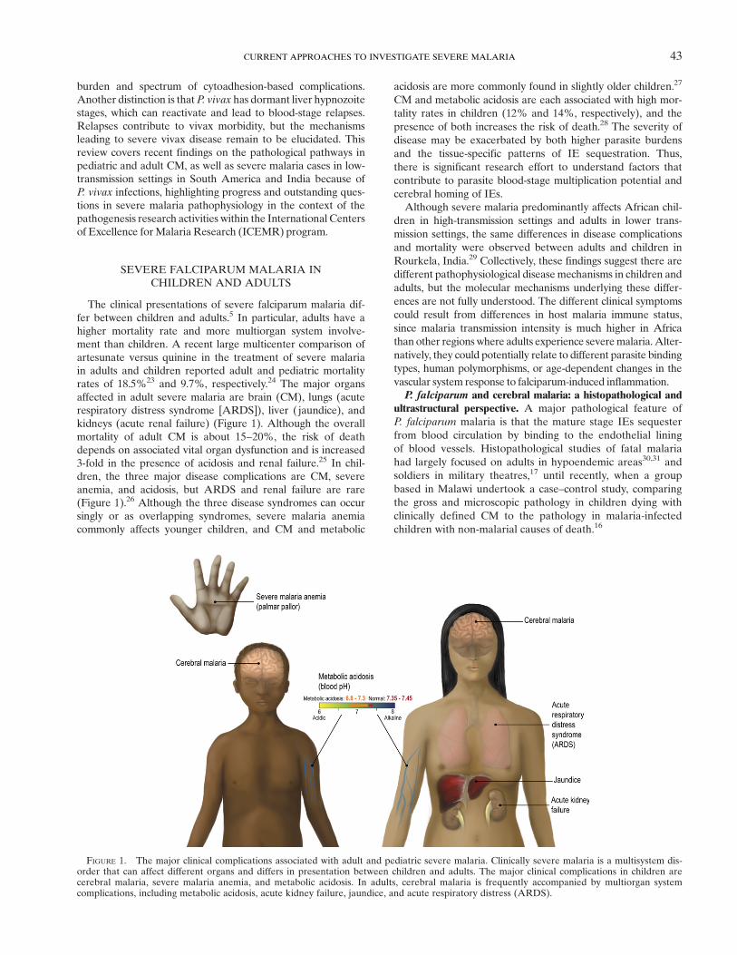
The clinical presentations of severe falciparum malaria dif-fer between children and adults.5 In particular, adults have ahigher mortality rate and more multiorgan system involve-ment than children. A recent large multicenter comparison ofartesunate versus quinine in the treatment of severe malariain adults and children reported adult and pediatric mortalityrates of 18.5%23 and 9.7%, respectively.24 The major organsaffected in adult severe malaria are brain (CM), lungs (acuterespiratory distress syndrome [ARDS]), liver ( jaundice), andkidneys (acute renal failure) (Figure 1). Although the overallmortality of adult CM is about 15–20%, the risk of deathdepends on associated vital organ dysfunction and is increased3-fold in the presence of acidosis and renal failure.25 In chil-dren, the three major disease complications are CM, severeanemia, and acidosis, but ARDS and renal failure are rare(Figure 1).26 Although the three disease syndromes can occursingly or as overlapping syndromes, severe malaria anemiacommonly affects younger children, and CM and metabolic
acidosis are more commonly found in slightly older children.27
CM and metabolic acidosis are each associated with high mor-tality rates in children (12% and 14%, respectively), and thepresence of both increases the risk of death.28 The severity ofdisease may be exacerbated by both higher parasite burdensand the tissue-specific patterns of IE sequestration. Thus,there is significant research effort to understand factors thatcontribute to parasite blood-stage multiplication potential andcerebral homing of IEs.Although severe malaria predominantly affects African chil-
dren in high-transmission settings and adults in lower trans-mission settings, the same differences in disease complicationsand mortality were observed between adults and children inRourkela, India.29 Collectively, these findings suggest there aredifferent pathophysiological diseasemechanisms in children andadults, but the molecular mechanisms underlying these differ-ences are not fully understood. The different clinical symptomscould result from differences in host malaria immune status,since malaria transmission intensity is much higher in Africathan other regionswhere adults experience severemalaria.Alter-natively, they could potentially relate to different parasite bindingtypes, human polymorphisms, or age-dependent changes in thevascular system response to falciparum-induced inflammation.P. falciparum and cerebral malaria: a histopathological and
ultrastructural perspective. A major pathological feature ofP. falciparum malaria is that the mature stage IEs sequesterfrom blood circulation by binding to the endothelial liningof blood vessels. Histopathological studies of fatal malariahad largely focused on adults in hypoendemic areas30,31 andsoldiers in military theatres,17 until recently, when a groupbased in Malawi undertook a case–control study, comparingthe gross and microscopic pathology in children dying withclinically defined CM to the pathology in malaria-infectedchildren with non-malarial causes of death.16
FIGURE 1. The major clinical complications associated with adult and pediatric severe malaria. Clinically severe malaria is a multisystem dis-order that can affect different organs and differs in presentation between children and adults. The major clinical complications in children arecerebral malaria, severe malaria anemia, and metabolic acidosis. In adults, cerebral malaria is frequently accompanied by multiorgan systemcomplications, including metabolic acidosis, acute kidney failure, jaundice, and acute respiratory distress (ARDS).
43CURRENT APPROACHES TO INVESTIGATE SEVERE MALARIA
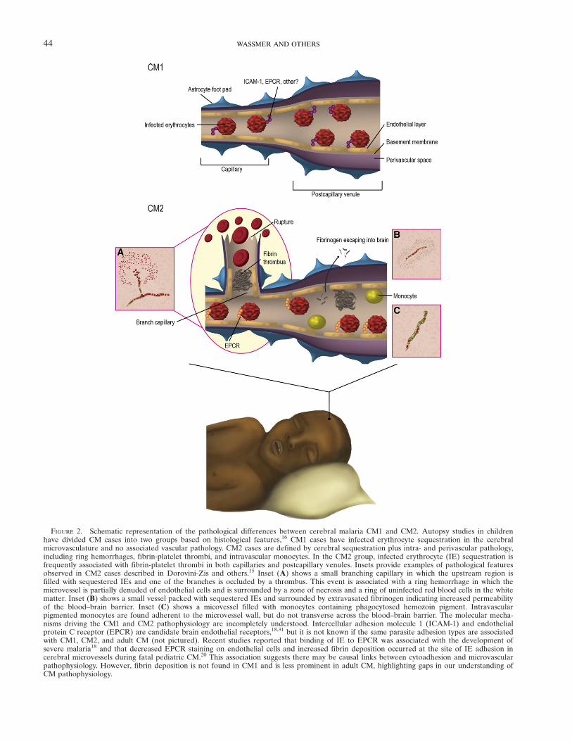
FIGURE 2. Schematic representation of the pathological differences between cerebral malaria CM1 and CM2. Autopsy studies in childrenhave divided CM cases into two groups based on histological features,16 CM1 cases have infected erythrocyte sequestration in the cerebralmicrovasculature and no associated vascular pathology. CM2 cases are defined by cerebral sequestration plus intra- and perivascular pathology,including ring hemorrhages, fibrin-platelet thrombi, and intravascular monocytes. In the CM2 group, infected erythrocyte (IE) sequestration isfrequently associated with fibrin-platelet thrombi in both capillaries and postcapillary venules. Insets provide examples of pathological featuresobserved in CM2 cases described in Dorovini-Zis and others.15 Inset (A) shows a small branching capillary in which the upstream region isfilled with sequestered IEs and one of the branches is occluded by a thrombus. This event is associated with a ring hemorrhage in which themicrovessel is partially denuded of endothelial cells and is surrounded by a zone of necrosis and a ring of uninfected red blood cells in the whitematter. Inset (B) shows a small vessel packed with sequestered IEs and surrounded by extravasated fibrinogen indicating increased permeabilityof the blood–brain barrier. Inset (C) shows a micovessel filled with monocytes containing phagocytosed hemozoin pigment. Intravascularpigmented monocytes are found adherent to the microvessel wall, but do not transverse across the blood–brain barrier. The molecular mecha-nisms driving the CM1 and CM2 pathophysiology are incompletely understood. Intercellular adhesion molecule 1 (ICAM-1) and endothelialprotein C receptor (EPCR) are candidate brain endothelial receptors,18,31 but it is not known if the same parasite adhesion types are associatedwith CM1, CM2, and adult CM (not pictured). Recent studies reported that binding of IE to EPCR was associated with the development ofsevere malaria18 and that decreased EPCR staining on endothelial cells and increased fibrin deposition occurred at the site of IE adhesion incerebral microvessels during fatal pediatric CM.20 This association suggests there may be causal links between cytoadhesion and microvascularpathophysiology. However, fibrin deposition is not found in CM1 and is less prominent in adult CM, highlighting gaps in our understanding ofCM pathophysiology.
44 WASSMER AND OTHERS
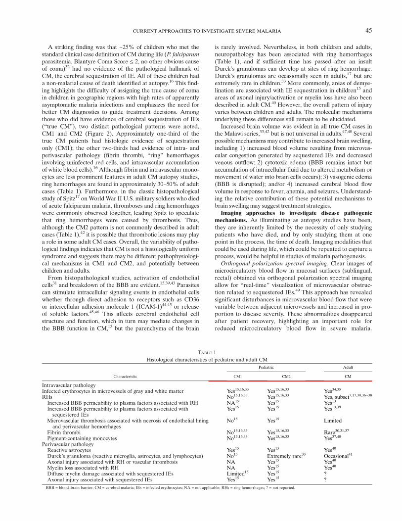
A striking finding was that ~25% of children who met thestandard clinical case definition of CM during life (P. falciparumparasitemia, Blantyre Coma Score ≤ 2, no other obvious causeof coma)32 had no evidence of the pathological hallmark ofCM, the cerebral sequestration of IE. All of these children hada non-malarial cause of death identified at autopsy.16 This find-ing highlights the difficulty of assigning the true cause of comain children in geographic regions with high rates of apparentlyasymptomatic malaria infections and emphasizes the need forbetter CM diagnostics to guide treatment decisions. Amongthose who did have evidence of cerebral sequestration of IEs(“true CM”), two distinct pathological patterns were noted,CM1 and CM2 (Figure 2). Approximately one-third of thetrue CM patients had histologic evidence of sequestrationonly (CM1); the other two-thirds had evidence of intra- andperivascular pathology (fibrin thrombi, “ring” hemorrhagesinvolving uninfected red cells, and intravascular accumulationof white blood cells).16 Although fibrin and intravascular mono-cytes are less prominent features in adult CM autopsy studies,ring hemorrhages are found in approximately 30–50% of adultcases (Table 1). Furthermore, in the classic histopathologicalstudy of Spitz17 onWorldWar II U.S. military soldiers who diedof acute falciparum malaria, thromboses and ring hemorrhageswere commonly observed together, leading Spitz to speculatethat ring hemorrhages were caused by thrombosis. Thus,although the CM2 pattern is not commonly described in adultcases (Table 1),42 it is possible that thrombotic lesions may playa role in some adult CM cases. Overall, the variability of patho-logical findings indicates that CM is not a histologically uniformsyndrome and suggests there may be different pathophysiologi-cal mechanisms in CM1 and CM2, and potentially betweenchildren and adults.From histopathological studies, activation of endothelial
cells31 and breakdown of the BBB are evident.15,39,43 Parasitescan stimulate intracellular signaling events in endothelial cellswhether through direct adhesion to receptors such as CD36or intercellular adhesion molecule 1 (ICAM-1)44,45 or releaseof soluble factors.45,46 This affects cerebral endothelial cellstructure and function, which in turn may mediate changes inthe BBB function in CM,13 but the parenchyma of the brain
is rarely involved. Nevertheless, in both children and adults,neuropathology has been associated with ring hemorrhages(Table 1), and if sufficient time has passed after an insultDurck’s granulomas can develop at sites of ring hemorrhage.Durck’s granulomas are occasionally seen in adults,17 but areextremely rare in children.33 More commonly, areas of demye-lination are associated with IE sequestration in children15 andareas of axonal injury/activation or myelin loss have also beendescribed in adult CM.40 However, the overall pattern of injuryvaries between children and adults. The molecular mechanismsunderlying these differences still remain to be elucidated.Increased brain volume was evident in all true CM cases in
the Malawi series,33,42 but is not universal in adults.47,48 Severalpossiblemechanismsmay contribute to increased brain swelling,including 1) increased blood volume resulting from microvas-cular congestion generated by sequestered IEs and decreasedvenous outflow; 2) cytotoxic edema (BBB remains intact butaccumulation of intracellular fluid due to altered metabolism ormovement of water into brain cells occurs); 3) vasogenic edema(BBB is disrupted); and/or 4) increased cerebral blood flowvolume in response to fever, anemia, and seizures. Understand-ing the relative contribution of these potential mechanisms tobrain swellingmay suggest treatment strategies.Imaging approaches to investigate disease pathogenic
mechanisms. As illuminating as autopsy studies have been,they are inherently limited by the necessity of only studyingpatients who have died, and by only studying them at onepoint in the process, the time of death. Imaging modalities thatcould be used during life, which could be repeated to capture aprocess, would be helpful in studies of malaria pathogenesis.Orthogonal polarization spectral imaging. Clear images of
microcirculatory blood flow in mucosal surfaces (sublingual,rectal) obtained via orthogonal polarization spectral imagingallow for “real-time” visualization of microvascular obstruc-tion related to sequestered IEs.49 This approach has revealedsignificant disturbances in microvascular blood flow that werevariable between adjacent microvessels and increased in pro-portion to disease severity. These abnormalities disappearedafter patient recovery, highlighting an important role forreduced microcirculatory blood flow in severe malaria.
TABLE 1Histological characteristics of pediatric and adult CM
Characteristic
Pediatric Adult
CM1 CM2 CM
Intravascular pathologyInfected erythrocytes in microvessels of gray and white matter Yes15,16,33 Yes15,16,33 Yes34,35
RHs No15,16,33 Yes15,16,33 Yes, subset7,17,30,36–38
Increased BBB permeability to plasma factors associated with RH NA15 Yes15 Yes13
Increased BBB permeability to plasma factors associated withsequestered IEs
Yes15 Yes15 Yes13,39
Microvascular thrombosis associated with necrosis of endothelial liningand perivascular hemorrhages
No15 Yes15 Limited
Fibrin thrombi No15,16,33 Yes15,16,33 Rare30,31,37
Pigment-containing monocytes No15,16,33 Yes15,16,33 Yes37,40
Perivascular pathologyReactive astrocytes Yes15 Yes15 Yes40
Durck’s granuloma (reactive microglia, astrocytes, and lymphocytes) No15 Extremely rare33 Occasional41
Axonal injury associated with RH or vascular thrombosis NA Yes15 Yes40
Myelin loss associated with RH NA Yes15 Yes40
Diffuse myelin damage associated with sequestered IEs Limited15 Yes15 ?Axonal injury associated with sequestered IEs Yes15 Yes15 ?BBB = blood–brain barrier; CM = cerebral malaria; IEs = infected erythrocytes; NA = not applicable; RHs = ring hemorrhages; ? = not reported.
45CURRENT APPROACHES TO INVESTIGATE SEVERE MALARIA
However, because the expression of surface receptors variesbetween organs,50 what is seen in accessible areas may notreflect what is happening in the brain.Ocular funduscopy. The eye and the brain have similar
embryologic origins, and themicrovasculatures of the two organsystems share important features.51 In addition, the optic funduscan be readily observed and studied during life in patients withsevere malaria. In conjunction with the Malawi autopsy study,ophthalmologists described a unique malarial retinopathy con-sisting of white-centered hemorrhages, vessel color changes,and peri- and extramacular whitening.52 At least one of thesefindings was present in all cases of true CM (i.e., patients withevidence of cerebral sequestration of IEs at autopsy), andalthough recognition of the retinopathy requires a trainedobserver with relatively expensive equipment (direct and indi-rect ophthalmoscopes), it has created the opportunity, exploitedby the ICEMRprogram, to use amore specific clinical case defi-nition of CM. Retinal hemorrhages correlate, numerically, withthe ring hemorrhages seen in fatal cases of pediatric CM.53
Vessel color changes reflect the presence of sequestered, para-sitized, and de-hemoglobinized red cells,54 while the whiteningrepresents areas of impaired perfusion.55
Ophthalmologic observations on adults with severe malariaare relatively sparse, but they are consistent with the reportedpediatric findings in that approximately one-third of adultsmeeting the standard clinical case definition of CM have noevidence of malarial retinopathy.56 Retinal hemorrhages arecommonly observed,57 but vessel color changes, seen in ~32%of children with CM,58 are only rarely seen in adults.56 Theseverity of malaria retinopathy is strongly associated withmalaria mortality in both adults and children.51,56
Neuroimaging. Neuroimaging neatly addresses the two pri-mary deficiencies of the autopsy approach: survivors can beimaged and serial studies can be carried out throughout thecourse of the acute illness. However, the worldwide distribu-tion of sophisticated radiological capacity does not include
malaria-endemic areas, so most descriptions of neuroimagingfindings in malaria patients have been single case reports frompatients hospitalized in more developed countries.14
Computed tomography scan technology is relatively uncom-plicated and affordable, and the process itself is quick. Thisapproach was the first used to illuminate disease pathogenesisin malaria patients, and highlighted the importance of increasedbrain volume.59–61 Most of these studies were done before theimportance of malarial retinopathy was recognized, though thepossibility of classification errors complicates interpretation ofthese findings.Individual case reports of magnetic resonance imaging (MRI)
findings in patients with CM (as reviewed in reference 59) havedescribed a variety of findings, all of which have been corrobo-rated by larger, systematic studies in Thai adults62 andMalawianchildren.63 Increased brain volume is strongly associated witha fatal outcome in children.64 Cortical involvement (oftenrestricted to specific lobes), and changes in the periventricularwhite matter, the corpus callosum, and the thalami are commonin childrenwith retinopathy-positive CM.Both of the larger studies were limited by the strength of the
magnet (0.2 tesla [T] in Thailand, 0.35 T inMalawi).A collabora-tive effort between two independent ICEMR projects (Table 2)will address this problem while simultaneously addressing dis-parities between the clinical manifestations of severe disease inadults and children. The joint effort is currently being carriedout between two hospitals, one located in Malawi and one inIndia, both of which have MRI facilities. Adults and pediatricpatients with severe malaria in India (retinopathy-positive CM,with and without other organ system involvement) will undergoMRI on a 1.5 T machine, and their findings will be comparedwith those in retinopathy-positive CM pediatric patients inMalawi. The clinical protocol has been standardized betweenthe two field sites, and four MRI sequences will be common toboth projects, as their magnet strengths are different. To ensurethe accurate interpretation and comparison of MRI findings in
TABLE 2ICEMR activities related to severe malaria
ICEMR Research activities related to pathogenesis of malaria
Southeast Asia Collecting descriptive data on malaria patients attending local hospitals at sentinel sites,including data on disease manifestation
South Asia (India) Investigating the molecular and cellular basis of severe Plasmodium falciparum and severePlasmodium vivax infections in hospital patients recruited at multiple locations in India
India Assessing the role of interindividual variations in endothelial responsiveness to TNF in thedevelopment of cerebral malaria
Investigating the pathology of cerebral malaria in India patients using novel MRI techniquesCollaborating with the southern Africa ICEMR (Malawi) on MRI findings in adults and
children with severe malariaEast Africa (Uganda) Investigating the role of prompt and effective therapy for minimizing the risk of severe malaria
in cohorts of children living in high-endemic settingsCollecting descriptive data on characteristics and outcomes of children admitted with severe
malaria at six public hospitals in UgandaSouthern African (Zambia/Zimbabwe) Collecting descriptive data on clinical diagnoses for persons seeking care at rural health centersSouthern Africa (Malawi) Collecting hospital-based data on febrile illnesses (malarial and non-malarial)
Collaborating with south Asia ICEMR (India) on MRI findings in adults and childAmazonia Observational, hospital-based observations of severe P. vivax malaria; 16S rRNA molecular
and blood culture analysis of severe malaria casesLatin America (outside Amazonia) Clinical profile of malaria in different epidemiological settings in Colombia, and their
association with parasite and host immunological statusDetermine the effects of immune status, nutritional factors, and helminth coinfection on
complicated malaria cases in ColombiaSouthwest Pacific Collecting data on childhood severe malaria admissions to major hospital serving
Madang ProvinceICEMR = International Centers of Excellence for Malaria Research; MRI = magnetic resonance imaging; TNF = tumor necrosis factor.
46 WASSMER AND OTHERS
these sequences, all the images will be scored and sharedbetween the radiologists, via a web-based platform to enhancestandardization.65 This study will permit, for the first time, theclinical characterization of pediatric and adult CM by neuroim-aging and a precise comparison of carefully clinically definedcohorts of CM patients of different ages and from differentcontinents. Such extensive MRI techniques have never beenapplied systematically to patients with acute malaria and repre-sent a promising approach to investigating the relationshipbetween brain swelling and the onset of CM.Vascular activation/dysfunction and coagulation pathways
in severe malaria. The brain swelling observed during CMbothin Indian adults and Malawian children might be the conse-quence of disruption of the BBB associated with the pathoge-netic processes of CM, resulting in vasogenic edema. Thishypothesis is currently being investigated as part of a Malawi–India inter-ICEMR initiative (Table 2) and is in line with theemergence of the endothelial cell as a central player in the path-ophysiology of the neurologic syndrome. Although its involve-ment as a substrate for IE sequestration in the brain wasidentified very early on,66 results published over the past decadehave highlighted the complex role of cerebral endothelial cellsin the development of CM. One of the main goals of the IndiaICEMR is to investigate parameters inherent in the host endo-thelium that may result in an increased susceptibility to severemalaria in Indian adults infected with P. falciparum, an axis ofresearch that is divided into threemain approaches.Variations and heritability of the host endothelial respon-
siveness to tumor necrosis factor alpha. A central component
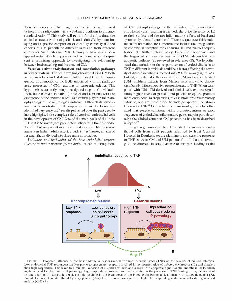
of CM pathophysiology is the activation of microvascularendothelial cells, resulting from both the cytoadherence of IEto their surface and the pro-inflammatory effects of local andsystemically released cytokines.67 The consequences of this endo-thelial inflammation are numerous and include the upregulationof endothelial receptors for enhancing IE and platelet seques-tration; the further release of cytokines and chemokines andthe trigger of a tumor necrosis factor (TNF)–dependent pro-apoptotic pathway (as reviewed in reference 68). We hypothe-sized that variation in the responsiveness of endothelial cells toTNF in different individuals could be a factor affecting the sever-ity of disease in patients infected with P. falciparum (Figure 3A).Indeed, endothelial cells derived from CM and uncomplicated(UM) children patients from Malawi were shown to displaysignificantly different ex vivo responsiveness to TNF.When com-pared with UM, CM-derived endothelial cells express signifi-cantly higher levels of parasite and platelet receptors, producemore endothelial microparticles, release more pro-inflammatorycytokine, and are more prone to undergo apoptosis on stimu-lation with TNF.69 On the basis of these results, it was hypothe-sized that genetic variations within promoter, intron, or exonsequences of endothelial inflammatory genes may, in part, deter-mine the clinical course in CM patients, as has been describedin sepsis.70
Using a large number of freshly isolated microvascular endo-thelial cells from adult patients admitted to Ispat GeneralHospital in Rourkela, we are planning to compare the responseto TNF between CM and UM patients from India and investi-gate the different factors, extrinsic or intrinsic, leading to the
FIGURE 3. Proposed influence of the host endothelial responsiveness to tumor necrosis factor (TNF) on the severity of malaria infection.Low endothelial TNF responders are less prone to upregulate receptors involved in the sequestration of infected erythrocyte (IE) and plateletsthan high responders. This leads to a minimal adhesion of IE and host cells and a lower pro-apoptotic signal for the endothelial cells, whichmight account for the absence of pathology. High responders, however, are over-activated in the presence of TNF, leading to high adhesion ofIE and a strong pro-apoptotic signal, possibly resulting in the breakdown of the blood–brain barrier and, ultimately, to vasogenic edema (A).Potential clinical benefits offered by angiopoietin (Ang)-1 as a quiescence agent for high TNF-responding endothelial cells during cerebralmalaria (CM) (B).
47CURRENT APPROACHES TO INVESTIGATE SEVERE MALARIA
interindividual differential activation of the endotheliumbetweenthe two patient categories. The comparative analysis of thevariation in transcripts between the two high and low TNF-responding groups of endothelial cells will give us insights intothe pathways involved in the acute activation observed in CMpatients, and will be compared with the results obtained inMalawian children. This project is carried out not only with aview to understanding the molecular basis of disease but alsoto identifying patients at risk by analyzing specific single nucle-otide polymorphisms associated with high and low responders.It will also assess if there are age-specific differences in endo-thelial responsiveness. Understanding the mechanistic basisof vascular dysfunction in severe malaria may suggest newtreatment options.Reversibility of the systematic endothelial activation in CM
patients. The presence of TNF as a trigger of inflammation inmalaria led to the assessment of a TNF-blocking approachin CM. Although in vitro treatments produced favorableresults, anti-TNF clinical trials failed to reduce mortality inthese patients.71,72 The use of a targeted compound block-ing the downstream endothelial activation signaling cascaderesulted in a reduction of endothelial inflammation in vitro.73
However, this effect was only observed when the compoundwas administered simultaneously with the cytokine, whichwould be effectively impossible in vivo. Since most of thepatients admitted to the ward have already high levels ofTNF, an acute therapy might work by dampening the existingendothelial inflammatory response in CM patients. Angio-poietin (Ang)-1 has recently become a topic of increasing inter-est in endothelial cell quiescence and survival,74 and plasmaAng-2/Ang-1 ratio has been shown not only to be crucial for theendothelial activation but also to discriminate UM and CM.Indeed, high levels of Ang-2 are associated with mortality inpatients with CM, whereas high levels of Ang-1 are associatedwith UM (as reviewed in reference 75). Since the use of Ang-1offers clinical benefits as a quiescence agent for endothelial cellsin an elegant model of sepsis,76 it is conceivable that restoringthe Ang-2/Ang-1 balance in favor of Ang-1 would block andpotentially reverse the ongoing inflammatory processes in CMpatients at the time of admission (Figure 3B).The potential clinical benefits of Ang-1 are currently being
evaluated as part of the ongoing project on primary endothe-lial cells at Ispat General Hospital. Using the endothelial cellbanks isolated from CM patients, the effects of Ang-1 onTNF-stimulated endothelium will be measured, with a view todevelop new adjunct therapies and improve disease outcomein CM.The role of EPCR in adult CM. Recent studies reported
that binding of IE to EPCR was associated with the develop-ment of severe malaria18 and that decreased EPCR stainingon endothelial cells and increased fibrin deposition occurredat the site of IE adhesion in cerebral microvessels during fatalpediatric CM.20 A causal relationship between cytoadhesionand coagulopathy was therefore suggested for the first time,and the pivotal role of EPCR in the organ specificity of thesyndrome was proposed.77,78 One of the major aims of theIndia ICEMRs is to further investigate the role of EPCR inthe development of CM in Indian adults, as fibrin deposition isa far less prominent pathological feature in southeast Asianadults than African children who succumb to CM (Table 1).20,30
Since endothelial cell cultured from subcutaneous fat resemblecerebral vascular endothelial cell and represent a useful ex vivo
model for examining brain endothelial alteration in the con-text of CM,69 this approach is being carried out by performingphenotypical analyses of primary subcutaneous endothelialcells isolated from patients admitted at Ispat General Hospital,followed by targeted gene expression profiling (RNA andmiRNA) and genetic analyses of genes selected for their rele-vance in the protein C pathway. The results will 1) contributeto a better understanding of the pathogenic mechanisms forchildhood and adult disease, 2) assess the overall importanceof EPCR in mediating the cytoprotective effects of activatedprotein C (APC) in the brain, and 3) evaluate new avenues oftranslational research. A collaborative protocol is currentlybeing developed between the India ICEMR and the clinicalteam to extend these analyses to endothelial cells isolatedfrom postmortem brain biopsies samples of fatal CM.Parasite biomass and severe malaria. It is difficult to mea-
sure the total parasite biomass of P. falciparum (circulating andsequestered) from blood sampling because of the “hidden”sequestered component. To overcome this challenge, a newapproach has been introduced byDondorp and others79 in whichthe plasma concentration of a soluble parasite molecule servesas a surrogate for the total parasite biomass. Plasmodiumfalciparum histidine-rich protein-2 (HRP-2) is a water-solubleprotein produced throughout the parasite life cycle and releasedlargely (but not exclusively) at the time of schizont rupture.79,80
It has a long half-life and persists in the plasma for up to 21 days,even after successful treatment81; HRP-2 detection (present/absent) is the basis of many rapid diagnostic tests, but quantita-tive measures of HRP-2 can discriminate between retinopathy-positive and retinopathy-negative CM,82 can predict whichchildren with uncomplicated malaria are more likely to deteri-orate,83 and can distinguish between patients with complicatedmalaria, mild malaria, asymptomatic parasitemia, and non-malarial fevers.84 A model, based on plasma half-life of HRP-2in vivo¸ production rates of HRP-2 in vitro, and parasite multi-plication rates suggests that HRP-2 concentrations reflect totalbody parasite burden (sequestered and circulating parasites).79
In general, the associations between HRP-2 concentration anddisease severity support the hypothesis that parasite biomassis a major determinant of malaria pathogenesis. However, arecent longitudinal birth cohort study of Tanzanian childrenfollowed from birth to 2–4 years of age indicated that whileparasite burden was higher on average in severe malaria epi-sodes, high parasite burden was insufficient to cause severedisease.85 Thus, high parasite burden appears to be an impor-tant determinant in severe malaria, but other factors may act inconcert to precipitate severe malaria episodes.Parasite invasion pathways and malaria severity. Higher
parasite biomass is a risk factor for severe malaria and may bedriving increased systematic inflammation, endothelial activationmarkers, and metabolic acidosis by microvascular obstruction.The circumstances leading to higher parasite burdens in severemalaria are likely multifactorial and incompletely understood.However, potential parasite factors are red blood cell (RBC)invasion efficiency and the cytoadhesion efficiency of infectedRBCs. Mathematical modeling approaches suggest that inva-sion efficiency can be a significant driver of peak parasite den-sity during an infection and concomitant pathogenesis.86 Rodentmalaria parasites can shift from a nonlethal to a lethal formfollowing a change in preference from reticulocytes to oldernormocytes resulting in huge increases in parasite biomass andpathology.87 In humans, there is evidence that the efficiency
48 WASSMER AND OTHERS
of the invasion process can be a virulence determinant inP. falciparum parasites.88 Clearly, this can be influenced bygenetic polymorphisms within both the host and the parasite, aswell as acquired immunity. In addition, the ability of parasites toinvade RBCs using alternative receptors, known as invasion path-ways, can facilitate immune evasion and persistence of malariainfections89 and ultimately contribute to malaria pathogenesis.Anemia may result from chronic low-burden infections.90
Invasion potential has been measured in two ways: by para-site multiplication rate and by selectivity of RBCs. Both havebeen shown to be strongly associated with the severity ofP. falciparum malaria in one population in southeast Asia,88
suggesting the existence of parasite molecular factors that medi-ate pathogenesis through increased proliferation. However, asimilar study was carried out with parasite isolates from Africaand no association was found between invasion efficiency, selec-tivity, and disease severity.91 It is not clear whether this is due toregional differences in parasites or in host factors, such as thelevel of acquired antimalarial immunity.Previous work carried out in several varied geographical
areas have shown that natural P. falciparum isolates are capa-ble of using multiple ligand–receptor invasion pathways, andexhibit variation in pathway usage, suggesting mechanisms bywhich invasion efficiency could be altered via parasite-basedmechanisms. These studies have shown that both sialic acid–dependent and sialic acid–independent invasion pathways arecommonly used by parasites collected directly from infectedhumans, and a few isolates have been shown to be able toswitch between the use of sialic acid–dependent and sialicacid–independent pathways. Switching of one isolate was asso-ciated with reduced invasion efficiency.89
With the genome sequenced, Plasmodium parasites havebeen found to possess a diverse number of ligands for invasion.Two superfamilies of invasion ligands, the reticulocyte-binding-protein-like (RBL) and the erythrocyte-binding-protein-like(EBL) have been identified.92 Much data from studies withP. falciparum suggests that each parasite ligand has a single cog-nate receptor, defining alternative invasion pathways and thatthere is a hierarchy of different ligand–receptor interactions.Further, variation can exist at the level of sequence and expres-sion changes for these invasion ligands, suggesting a molecularbasis for switching between the use of different invasion path-ways, either for immune evasion, to change the parasite multi-plication rate, and/or RBC selectivity. To better understand themolecularmechanisms driving higher parasite burdens in severemalaria, an ICEMR group in India is addressing the interplaybetween parasite invasion efficiency and IE cytoadhesion phe-notypes in disease severity.Parasite adhesion and severe malaria. As described above,
cytoadhesion of IEs is a major virulence determinant for CMcomplications. Furthermore, high parasite burdens and themassive sequestration of IEs in different tissue beds andresulting microvascular obstruction may lead to metabolic aci-dosis.4 The majority of falciparum infections are not severe,which suggests that the parasite is relatively well adapted tosequester in microvessels without killing the host. Cytoadhesionof IEs is predominantly mediated through the var gene/P. falciparum erythrocyte membrane protein 1 (PfEMP1) fam-ily of adhesion proteins.93–95 PfEMP1 proteins are anchoredat parasite-induced, knob-like protrusions on the erythrocytemembrane,93 exposing them to host antibodies. Clonal anti-genic variation of var genes enables P. falciparum to evade anti-
body destruction and to bind to different host receptors.96 Eachparasite encodes approximately 60 different var copies97 withlimited overlap of var gene repertoires between parasite haplo-types.98 The vast intra- and interstrain diversity in PfEMP1repertoires enables parasites to establish chronic infectionsand repeatedly infect hosts with different parasite genotypes.A fundamental question for pathogenesis is whether specificPfEMP1 and host-receptor interactions have a causal role insevere malaria.Despite extensive sequence diversity, the majority of var
genes can be classified into threemain subfamilies (A, B, andC)on the basis of upstream gene sequence and chromosomal loca-tion.99 Interstrain sequence comparisons have also identifiedthree unusual strain-transcendent var genes (var1csa, var2csa,type 3 var).100–102 Each PfEMP1 protein encodes multiple adhe-sion domains called Duffy binding-like (DBL) and cysteine-richinterdomain region domains.95 PfEMP1 adhesion domains areclassified into different types (α, β, γ, δ, etc.) and subtypes basedon sequence similarity.101,103 Using adhesion domain classifica-tion, interstrain sequence comparisons have revealed a smallnumber of tandem domain arrangements of 2–4 domains, calleddomain cassettes (DC), which are unusually conserved betweenparasite genotypes.101
The prototypical example of a specific PfEMP1 and diseaseis malaria in pregnancy. In this case, the strain-transcendentVAR2CSA mediates placental binding.104,105 It has been morechallenging to determine if a specific PfEMP1 subset is associ-ated with CM because of the difficulty of studying the brain.Analysis of var gene expression in patients has suggested thatmost infections contain a heterogeneous population of para-sites expressing a mixture of A, B, or C var genes. In hostswith limited malaria immunity and severe pediatric malaria,the ratio of PfEMP1 variants appears to be skewed towardhigher group A expression.106–108 These findings suggest thatgroup A PfEMP1 encode adhesion traits that facilitate para-site multiplication in malaria naive hosts and may includebinding properties that predispose to severe malaria. As indi-viduals acquire anti-PfEMP1 antibodies through repeated infec-tions, the proportion of group B and C variants appear toincrease.108,109 However, even in pregnant African women whohave acquired considerable antimalarial immunity, there washigh var2csa expression from parasites recovered from placenta,but mixed var2csa and A, B, C var expression from parasitescirculating in the blood.110 Thus, the parasite strategy of havinga heterogeneous population appears to persist even after indi-viduals have acquired substantial antimalarial immunity.106–108
More recently, it was shown that parasites expressingPfEMP1 proteins encoding DC8 or DC13 are strongly selectedon human brain microvascular endothelial cells in vitro111,112
and are highly expressed in children with severe malaria orCM.113 The DC8 is found in an unusual chimeric gene betweengroups B and A and the DC13 is restricted to group A vari-ants. Both DC8 and DC13 proteins, as well as a subset of othergroup A variants, were found to encode a novel binding prop-erty for EPCR,18 the receptor for APC. As the APC–EPCRpathway plays a key role in regulating blood coagulation andendothelial barrier properties,19,114 this has raised the possibil-ity that there may be a linkage between IE binding and CMpathogenesis. However, given the different clinical presenta-tion and autopsy findings in children and adults (Table 1),5 animportant question is whether different PfEMP1 variants areassociated with CM1, CM2, and adult CM.
49CURRENT APPROACHES TO INVESTIGATE SEVERE MALARIA
As discussed above, one possibility is that host polymor-phisms or age-specific differences in endothelial responses maycontribute to pathophysiological differences. Alternatively, dif-ferent parasite binding variants may be associated with CM1,CM2, and adult CM. For instance, ICAM1 has also been pro-posed to be a cerebral sequestration receptor.31 Therefore, onepossibility is that ICAM1+, EPCR− binding variants play a morepredominant role in CM1 where fibrin-platelet clots and ringhemorrhages are absent, whereas EPCR+ binding variants arepredominant in CM2 (Figure 2). To evaluate if parasite bindingphenotype influences disease pathogenesis, more information isneeded on the binding specificity of DC8, DC13, and othergroup A–expressing parasites for ICAM1 and EPCR.111,112
In addition, multiple domains in DC8 PfEMP1 bind to brainendothelial cells.115 Therefore, this analysis should includedefining the other host receptors that act in concert with EPCRto mediate firm endothelial binding, as these adhesion traitsmay also influencemicrovascular pathology.Although considerable work has been done on var gene
expression in severe pediatric malaria,18,107,108,113,116 almost noinformation exists on DC8 or DC13 var gene expression inadult severe malaria. One of the aims of the India ICEMR isto investigate the expression of var genes in Indian adults.This question is also being evaluated as part of a collaborativeeffort between multiple independent ICEMRs using carefullyclinically defined cohorts, in which patients in India haveundergone MRI, fundoscopic examinations and have beenevaluated for endothelial responsiveness to TNF. By having aprecise comparison between MRI and fundoscopic findings,PfEMP1 expression, and host endothelial phenotypes, it maybe possible to distinguish if host or parasite factors contributeto different pathological manifestations.
SEVERE VIVAX MALARIA IN CHILDRENAND ADULTS
The other major Plasmodium species infecting humans isP. vivax. Although P. vivax infections are rare in most ofAfrica because of the high percentage of the human popula-tion with the Duffy blood group antigen–negative phenotypethat is highly resistant to RBC invasion,117,118 it is estimatedthat over 2.5 billion people are at risk of P. vivax transmission.Approximately 91% of the populations at risk of transmissionare in central and southeast Asia.117 Furthermore, in Brazil,P. falciparum cases are declining, and P. vivax has become thedominant parasite species in many endemic areas.119
Historically, P. vivax has been considered a relatively benignparasite, but recently there has been a renewed appreciationthat it carries a significant morbidity and mortality burden inendemic regions.21,120,121 Furthermore, a 5–15% mortality ratewas reported in the early neurosyphilis therapies of patientswith P. vivax.120 Part of the explanation for the “benign” repu-tation, despite the evidence for mortality, is that vivax parasitesare highly restricted to reticulocytes and therefore cannotachieve the same high parasite biomass as P. falciparum.4 Asecond difference is that P. vivax possesses relatively poor IEadhesive capacity compared with P. falciparum.122 Major ques-tions for vivax pathogenesis include how does a parasite that islimited to lower grade parasitemias cause severe malaria? Andis severe disease a consequence of vivax infection alone, therelapsing nature of P. vivax, or do other comorbidities influencedisease severity? Within the ICEMRs, work is being done to
better understand the prevalence and severity of P. vivax infec-tions in Latin America and Asia and to characterize factorsthat may contribute to disease severity.Clinically, vivax infections are associated with a chronic debil-
itating febrile illness that can be accompanied by chills,vomiting, malaise, and headache.21 On a per parasite basis,P. vivax is highly potent at inducing pro-inflammatory cytokines,such as TNF21,120,121,123,124 and has a much lower pyrogenicthreshold than P. falciparum (180 vivax parasites/μL comparedwith 1,000 falciparum parasites/μL).124,125 The most frequentsevere complications of vivax infection are severe anemia andacute respiratory distress.121 Cerebral malaria is a rare compli-cation of P. vivaxmono-infection, although it has been reportedin India.126 In general, even less is known about the pathoge-netic mechanisms in vivax malaria than P. falciparum, and it isnot known if P. vivax CM cases reflect a particular strain ofP. vivax, and/or a region-specific host susceptibility.Parasite adhesion and severe vivax malaria. Unlike
P. falciparum, P. vivax IEs become more deformable as theymature,127 and all parasite stages are visible in peripheral bloodsmears.21 However, late-stage schizont forms are underrepre-sented in peripheral blood,119 suggesting sequestration mayoccur. The lack of a continuous culture system has hinderedresearch into P. vivax cytoadhesion, but the mechanism is dis-tinct from P. falciparum becauseP. vivax IEs lack knob-like pro-trusions and do not encode var genes.22 Ex vivo studies haveshown that P. vivax IEs adhere to placental cryosections as wellas human lung—albeit at 10–15 times lower binding levels thanP. falciparum.128 A strong candidate for P. vivax cytoadhesionand rosetting functions is a family of variant sub-telomeric genesnamed vir.129 On the basis of the sequence analysis, VIR pro-teins are classified into different groups, which have been foundto have different subcellular localizations and functions.130 Tostudy the cellular trafficking and adhesive functions of VIR pro-teins, they have been transfected into a poorly cytoadhesiveP. falciparum strain (3D7), permitting gain of function studies.Two of three transfected VIR proteins were transported to theIE surface and one conferred ICAM1 binding activity.131
Whether cytoadhesion has a role in organ-specific disease com-plications is currently being investigated. There are few autopsyfindings from polymerase chain reaction–confirmed P. vivaxmono-infections. In one postmortem series from Brazil, ARDSand pulmonary edemawas associated with accumulation of neu-trophils in the interalveolar space, and scattered P. vivax IEswere present inside the pulmonary capillaries.132 A singleautopsy performed in India showedmonocyte infiltrates in alve-olar capillaries.133 It has been postulated thatP. vivax sequestra-tion in pulmonary microvessels may trigger the inflammatoryinflux,134 butmore work is needed to prove this hypothesis.Parasite invasion pathways and vivax malaria severity. In
contrast to the deadly P. falciparum, which is able to invadeRBCs of all age, it has been suggested that the lack of fatalitiesfrom P. vivax malaria is related to its unique restriction to inva-sion and growth in reticulocytes. TheDuffy blood group antigenon RBCs has a key role in invasion.118 This protein is recog-nized by the P. vivaxDuffy binding protein (DBP),135 a leadingvivax vaccine candidate. Although the identification of Duffy-dependent and Duffy-independent strains in Madagascar136
indicates that P. vivax can use alternative invasion pathways, itis unknown how extensively Duffy-independent strains are dis-tributed throughout the world. In addition, a single amino acidpolymorphism in the Duffy antigen Fy(a)/Fy(b) affects P. vivax
50 WASSMER AND OTHERS
invasion efficiency and the risk of clinical vivax in Brazil,137 butthe effect of this polymorphism has not been examined in otherparts of the world.Despite the strong preference of P. vivax for reticulocytes,
there is still a relatively poor understanding of why P. vivax isunable to invade normocytes or of the potential role of alter-native invasion pathways in disease severity. A reticulocyte-binding protein complex was identified (PvRBP-1 andPvRBP-2), which plays a key role in reticulocyte binding andinvasion.138 A related protein family was subsequently dis-covered in P. falciparum and named reticulocyte homologyor RBL proteins. Plasmodium vivax genome sequences indi-cate the presence of numerous RBL paralogs,22 and intrigu-ingly an additional DBP paralog,139 which might contributeto different modes of invasion, immune evasion, and patho-genesis. Within the India ICEMR, P. vivax in vitro invasionassays are being conducted to characterize the role of inva-sion pathways in disease severity.
CROSS-ICEMR COMPARISON OF RESEARCHACTIVITIES RELATED TO SEVERE MALARIA
The ICEMR program covers a wide range of malaria trans-mission intensities for P. falciparum and P. vivax. Within theICEMR program, nine ICEMRs based in south Asia, India,east and southern Africa, Amazonia, and southwest Pacific arecollecting descriptive data on the characteristics and outcomesof patients admitted with severe malaria (Table 2). This broadapproach can provide a better understanding of the relationshipbetween severe malaria outcomes across the endemicity spec-trum and may lend itself to meta-analysis to understand riskfactors for incidence of severe disease. In addition, individualICEMRs are investigating the role of prompt and effectivetherapies on minimizing severe malaria outcomes in Africanchildren and assessing the clinical profile and their associationwith the parasite and host immunological status and the roleof nutritional factors and helminth coinfections in complicatedmalaria cases in Colombia (Table 2).
CONCLUSIONS
Although the pathophysiology of CM is complex, pediatricautopsy studies have demonstrated twomajor patterns: cerebralmicrovessels with sequestered IEs alone (CM1) and cerebralmicrovessels with IE sequestration plus evidence of endothelialdysfunction and activation of coagulation (CM2). Ring hemor-rhages and cerebral thrombosis are also described in a pro-portion of adult cases, but whether there is an equivalentCM1/CM2 dichotomy in adults is unclear. Neuroimaging stud-ies have highlighted an important role for brain swelling inpediatric CM, which is less commonly observed in adult CM.A recent focus has been the microvascular interactionsbetween P. falciparum IEs and cerebral endothelial cells, andhow these binding interactions may contribute to disease pre-sentation. Furthermore, because of the inaccessibility of cere-bral microvessels, dermal biopsies provide a noninvasiveapproach to profile the endothelial reactivity of patients withsevere or non-severe malaria complications. It has been postu-lated that EPCR-binding parasites associated with severepediatric malaria may impair the protein C pathway in cerebralmicrovessels and thereby directly contribute to coagulopathyand endothelial barrier disruption. However, further work is
needed to understand to what extent parasite adhesion or endo-thelial phenotypes may contribute to the pathophysiologicaldifferences between CM1, CM2, and adult CM.By comparison to P. falciparum, the lower lethality of P.
vivax may relate to invasion and growth in reticulocytes andlower cytoadhesive properties. Nevertheless, despite its benignreputation, there has been a surge in reports on severe vivaxmalaria and a growing appreciation that P. vivax is not harm-less. Recent studies in Peru suggest that severe vivax can occurin monoendemic malaria regions.140 Although highly restrictedto reticuloctyes, genome projects have revealed a large expan-sion of invasion ligand gene families in P. vivax. Thus, it will beimportant to investigate if invasion pathways influence vivaxdisease severity. Within the ICEMR program, current researchefforts are focused on understanding disease mechanisms, asan important prerequisite to developing new tools to diagnoseand treat severe malaria.
Received December 29, 2014. Accepted for publication March 10, 2015.
Published online August 10, 2015.
Acknowledgments: We thank all the ICEMRs for the contributions ofthe information included in this journal supplement. We also thankAdriana Lippy for drawing the illustrations in Figures 1 and 2.
Financial Support: This work was supported by the following Coop-erative Agreements from the United States Public Health Service,National Institute of Allergy and Infectious Diseases: U19AI089672,U19AI089674, 5U19AI089676,U19AI089680,U19AI089681,U19AI089683,U19AI089686, U19AI089688, U19AI089696, and U19AI089702. Thecontent is solely the responsibility of the authors and does not neces-sarily represent the official views of the National Institutes of Health.
Authors’ addresses: Samuel C. Wassmer, Department of Microbiol-ogy, New York University School of Medicine, New York, NY, andDepartment of Pathology, Sydney Medical School, The Universityof Sydney, Sydney, Australia, E-mail: [email protected] E. Taylor, Department of Osteopathic Medical Specialties,College of Osteopathic Medicine, Michigan State University, EastLansing, MI, and Blantyre Malaria Project, University of MalawiCollege of Medicine, Blantyre, Malawi, E-mail: [email protected] K. Rathod, Departments of Chemistry and GlobalHealth, University of Washington, Seattle, WA, E-mail: [email protected]. Saroj K. Mishra and Sanjib Mohanty, Department ofInternal Medicine, Ispat General Hospital, Orissa, India, E-mails:[email protected] and [email protected], Caucaseco Scientific Research Center, Cali, Colombia, E-mail:[email protected]. Manoj T. Duraisingh, Department of Immunol-ogy and Infectious Diseases, Harvard School of Public Health, Boston,MA, E-mail: [email protected]. Joseph D. Smith, Seattle Bio-medical Research Institute, Seattle, WA, and Department of GlobalHealth, University of Washington, Seattle, WA, E-mail: [email protected].
This is an open-access article distributed under the terms of theCreative Commons Attribution License, which permits unrestricteduse, distribution, and reproduction in any medium, provided theoriginal author and source are credited.
REFERENCES
1. World Health Organization, 2013. World Malaria Report. Avail-able at: http://www.who.int/malaria/publications/world_malaria_report_2013/report/en/w.
2. Murray CJ, Ortblad KF, Guinovart C, Lim SS, Wolock TM,Roberts DA, Dansereau EA, Graetz N, Barber RM, BrownJC, Wang H, Duber HC, Naghavi M, Dicker D, Dandona L,Salomon JA, Heuton KR, Foreman K, Phillips DE, FlemingTD, Flaxman AD, Phillips BK, Johnson EK, Coggeshall MS,Abd-Allah F, Abera SF, Abraham JP, Abubakar I, Abu-Raddad LJ, Abu-Rmeileh NM, Achoki T, Adeyemo AO, AdouAK, Adsuar JC, Agardh EE, Akena D, Al Kahbouri MJ,Alasfoor D, Albittar MI, Alcala-Cerra G, Alegretti MA, Alemu
51CURRENT APPROACHES TO INVESTIGATE SEVERE MALARIA
ZA, Alfonso-Cristancho R, Alhabib S, Ali R, Alla F, AllenPJ, Alsharif U, Alvarez E, Alvis-Guzman N, Amankwaa AA,Amare AT, Amini H, Ammar W, Anderson BO, Antonio CA,Anwari P, Arnlov J, Arsenijevic VS, Artaman A, AsgharRJ, Assadi R, Atkins LS, Badawi A, Balakrishnan K, BanerjeeA, Basu S, Beardsley J, Bekele T, Bell ML, Bernabe E,Beyene TJ, Bhala N, Bhalla A, Bhutta ZA, AbdulhakAB, Binagwaho A, Blore JD, Bose D, Brainin M, BreitbordeN, Castaneda-Orjuela CA, Catala-Lopez F, Chadha VK, ChangJC, Chiang PP, Chuang TW, Colomar M, Cooper LT, CooperC, Courville KJ, Cowie BC, Criqui MH, Dandona R, DayamaA, De LD Degenhardt L, Del Pozo-Cruz B, Deribe K, DesJarlais DC, Dessalegn M, Dharmaratne SD, Dilmen U, DingEL, Driscoll TR, Durrani AM, Ellenbogen RG, ErmakovSP, Esteghamati A, Faraon EJ, Farzadfar F, FereshtehnejadSM, Fijabi DO, Forouzanfar MH, Fra Paleo U, Gaffikin L,Gamkrelidze A, Gankpé FG, Geleijnse JM, Gessner BD,Gibney KB, Ginawi IA, Glaser EL, Gona P, Goto A, GoudaHN, Gugnani HC, Gupta R, Gupta R, Hafezi-Nejad N,Hamadeh RR, Hammami M, Hankey GJ, Harb HL, HaroJM, Havmoeller R, Hay SI, Hedayati MT, Pi IB, Hoek HW,Hornberger JC, Hosgood HD, Hotez PJ, Hoy DG, HuangJJ, Iburg KM, Idrisov BT, Innos K, Jacobsen KH, JeemonP, Jensen PN, Jha V, Jiang G, Jonas JB, Juel K, Kan H,Kankindi I, Karam NE, Karch A, Karema CK, Kaul A,Kawakami N, Kazi DS, Kemp AH, Kengne AP, KerenA, Kereselidze M, Khader YS, Khalifa SE, Khan EA, KhangYH, Khonelidze I, Kinfu Y, Kinge JM, Knibbs L, Kokubo Y,Kosen S, Defo BK, Kulkarni VS, Kulkarni C, Kumar K,Kumar RB, Kumar GA, Kwan GF, Lai T, Balaji AL, LamH, Lan Q, Lansingh VC, Larson HJ, Larsson A, Lee JT,Leigh J, Leinsalu M, Leung R, Li Y, Li Y, De Lima GM, LinHH, Lipshultz SE, Liu S, Liu Y, Lloyd BK, Lotufo PA,Machado VM, Maclachlan JH, Magis-Rodriguez C, MajdanM, Mapoma CC, Marcenes W, Marzan MB, Masci JR,Mashal MT, Mason-Jones AJ, Mayosi BM, Mazorodze TT,Mckay AC, Meaney PA, Mehndiratta MM, Mejia-RodriguezF, Melaku YA, Memish ZA, Mendoza W, Miller TR, MillsEJ, Mohammad KA, Mokdad AH, Mola GL, Monasta L,Montico M, Moore AR, Mori R, Moturi WN, MukaigawaraM, Murthy KS, Naheed A, Naidoo KS, Naldi L, Nangia V,Narayan KM, Nash D, Nejjari C, Nelson RG, NeupaneSP, Newton CR, Ng M, Nisar MI, Nolte S, Norheim OF,Nowaseb V, Nyakarahuka L, Oh IH, Ohkubo T, Olusanya BO,Omer SB, Opio JN, Orisakwe OE, Pandian JD, PapachristouC, Caicedo AJ, Patten SB, Paul VK, Pavlin BI, Pearce N,Pereira DM, Pervaiz A, Pesudovs K, Petzold M, PourmalekF, Qato D, Quezada AD, Quistberg DA, Rafay A, Rahimi K,Rahimi-Movaghar V, Ur Rahman S, Raju M, Rana SM, RazaviH, Reilly RQ, Remuzzi G, Richardus JH, Ronfani L, Roy N,Sabin N, Saeedi MY, Sahraian MA, Samonte GM, Sawhney M,Schneider IJ, Schwebel DC, Seedat S, Sepanlou SG, Servan-Mori EE, Sheikhbahaei S, Shibuya K, Shin HH, Shiue I,Shivakoti R, Sigfusdottir ID, Silberberg DH, Silva AP,Simard EP, Singh JA, Skirbekk V, Sliwa K, Soneji S, SoshnikovSS, Sreeramareddy CT, Stathopoulou VK, Stroumpoulis K,Swaminathan S, Sykes BL, Tabb KM, Talongwa RT, TenkorangEY, Terkawi AS, Thomson AJ, Thorne-Lyman AL, TowbinJA, Traebert J, Tran BX, Dimbuene ZT, Tsilimbaris M,Uchendu US, Ukwaja KN, Uzun SB, Vallely AJ, VasankariTJ, Venketasubramanian N, Violante FS, Vlassov VV, VollsetSE, Waller S, Wallin MT, Wang L, Wang X, Wang Y,Weichenthal S, Weiderpass E, Weintraub RG, WestermanR, White RA, Wilkinson JD, Williams TN, WoldeyohannesSM, Wong JQ, Xu G, Yang YC, Yano Y, Yentur GK, Yip P,Yonemoto N, Yoon SJ, Younis M, Yu C, Jin KY, El SayedZaki M, Zhao Y, Zheng Y, Zhou M, Zhu J, Zou XN, LopezAD, Vos T, 2014. Global, regional, and national incidenceand mortality for HIV, tuberculosis, and malaria during1990–2013: a systematic analysis for the Global Burden ofDisease Study 2013. Lancet 384: 1005–1070.
3. Murray CJ, Rosenfeld LC, Lim SS, Andrews KG, Foreman KJ,Haring D, Fullman N, Naghavi M, Lozano R, Lopez AD,2012. Global malaria mortality between 1980 and 2010: a sys-tematic analysis. Lancet 379: 413–431.
4. Miller LH, Baruch DI, Marsh K, Doumbo OK, 2002. The path-ogenic basis of malaria. Nature 415: 673–679.
5. Beales PF, Brabin B, Dorman E, Gilles HM, Loutain L, MarshK, Molyneux ME, Olliaro P, Schapira A, Touze JE, Hien TT,Warrell DA, White N, 2000. Severe falciparum malaria. TransR Soc Trop Med Hyg 94 (Suppl 1): S1–S90.
6. Marsh K, 1992. Malaria—a neglected disease? Parasitology 104(Suppl): S53–S69.
7. Marchiafava E, Bignami A, 1894. On summer-autumnal fevers.Charles TE, ed. Two Monographs on Malaria and the Para-sites of Malarial Fevers. London, United Kingdom: The NewSydenham Society, 1–393.
8. Clark IA, Rockett KA, 1994. The cytokine theory of humancerebral malaria. Parasitol Today 10: 410–412.
9. van der Heyde HC, Nolan J, Combes V, Gramaglia I, GrauGE, 2006. A unified hypothesis for the genesis of cerebralmalaria: sequestration, inflammation and hemostasis leadingto microcirculatory dysfunction. Trends Parasitol 22: 503–508.
10. Wassmer SC, Combes V, Grau GE, 2011. Platelets and micro-particles in cerebral malaria: the unusual suspects. DrugDiscov Today Dis Mech 8: e15–e23.
11. Francischetti IM, Seydel KB, Monteiro RQ, 2008. Blood coagu-lation, inflammation, and malaria. Microcirculation 15: 81–107.
12. Moxon CA, Heyderman RS, Wassmer SC, 2009. Dysregulationof coagulation in cerebral malaria. Mol Biochem Parasitol166: 99–108.
13. Medana IM, Turner GD, 2006. Human cerebral malaria andthe blood-brain barrier. Int J Parasitol 36: 555–568.
14. Mohanty S, Taylor TE, Kampondeni S, Potchen MJ, Panda P,Majhi M, Mishra SK, Wassmer SC, 2014. Magnetic resonanceimaging during life: the key to unlock cerebral malaria patho-genesis? Malar J 13: 276.
15. Dorovini-Zis K, Schmidt K, Huynh H, Fu W, Whitten RO,Milner D, Kamiza S, Molyneux M, Taylor TE, 2011. The neu-ropathology of fatal cerebral malaria in Malawian children.Am J Pathol 178: 2146–2158.
16. Taylor TE, Fu WJ, Carr RA, Whitten RO, Mueller JS, FosikoNG, Lewallen S, Liomba NG, Molyneux ME, 2004. Differenti-ating the pathologies of cerebral malaria by postmortem para-site counts. Nat Med 10: 143–145.
17. Spitz S, 1946. The pathology of acute falciparum malaria. MilSurg 99: 555–572.
18. Turner L, Lavstsen T, Berger SS, Wang CW, Petersen JE, AvrilM, Brazier AJ, Freeth J, Jespersen JS, Nielsen MA, MagistradoP, Lusingu J, Smith JD, Higgins MK, Theander TG, 2013.Severe malaria is associated with parasite binding to endothe-lial protein C receptor. Nature 498: 502–505.
19. Bouwens EA, Stavenuiter F, Mosnier LO, 2013. Mechanismsof anticoagulant and cytoprotective actions of the protein Cpathway. J Thromb Haemost 11 (Suppl 1): 242–253.
20. Moxon CA, Wassmer SC, Milner DA Jr, Chisala NV, Taylor TE,Seydel KB, Molyneux ME, Faragher B, Esmon CT, Downey C,Toh CH, Craig AG, Heyderman RS, 2013. Loss of endothelialprotein C receptors links coagulation and inflammation to para-site sequestration in cerebral malaria in African children. Blood122: 842–851.
21. Price RN, Tjitra E, Guerra CA, Yeung S, White NJ, AnsteyNM, 2007. Vivax malaria: neglected and not benign. Am J TropMed Hyg 77: 79–87.
22. Carlton JM, Adams JH, Silva JC, Bidwell SL, Lorenzi H,Caler E, Crabtree J, Angiuoli SV, Merino EF, Amedeo P,Cheng Q, Coulson RM, Crabb BS, del Portillo HA, EssienK, Feldblyum TV, Fernandez-Becerra C, Gilson PR, GueyeAH, Guo X, Kang’a S, Kooij TW, Korsinczky M, Meyer EV,Nene V, Paulsen I, White O, Ralph SA, Ren Q, SargeantTJ, Salzberg SL, Stoeckert CJ, Sullivan SA, Yamamoto MM,Hoffman SL, Wortman JR, Gardner MJ, Galinski MR,Barnwell JW, Fraser-Liggett CM, 2008. Comparative geno-mics of the neglected human malaria parasite Plasmodiumvivax. Nature 455: 757–763.
23. Dondorp A, Nosten F, Stepniewska K, Day N, White N, 2005.Artesunate versus quinine for treatment of severe falciparummalaria: a randomised trial. Lancet 366: 717–725.
24. Dondorp AM, Fanello CI, Hendriksen IC, Gomes E, SeniA, Chhaganlal KD, Bojang K, Olaosebikan R, Anunobi N,
52 WASSMER AND OTHERS
Maitland K, Kivaya E, Agbenyega T, Nguah SB, Evans J,Gesase S, Kahabuka C, Mtove G, Nadjm B, Deen J, Mwanga-Amumpaire J, Nansumba M, Karema C, Umulisa N, UwimanaA, Mokuolu OA, Adedoyin OT, Johnson WB, Tshefu AK,Onyamboko MA, Sakulthaew T, Ngum WP, Silamut K,Stepniewska K, Woodrow CJ, Bethell D, Wills B, OnekoM, Peto TE, von Seidlein L, Day NP, White NJ, 2010. Artesunateversus quinine in the treatment of severe falciparum malariain African children (AQUAMAT): an open-label, randomisedtrial. Lancet 376: 1647–1657.
25. Newton PN, Stepniewska K, Dondorp A, Silamut K, ChierakulW, Krishna S, Davis TM, Suputtamongkol Y, Angus B,Pukrittayakamee S, Ruangveerayuth R, Hanson J, Day NP,White NJ, 2013. Prognostic indicators in adults hospitalizedwith falciparum malaria in western Thailand. Malar J 12: 229.
26. White NJ, Pukrittayakamee S, Hien TT, Faiz MA, MokuoluOA, Dondorp AM, 2014. Malaria. Lancet 383: 723–735.
27. Marsh K, Snow RW, 1997. Host-parasite interaction and mor-bidity in malaria endemic areas. Philos Trans R Soc Lond BBiol Sci 352: 1385–1394.
28. Marsh K, Forster D, Waruiru C, Mwangi I, Winstanley M,Marsh V, Newton C, Winstanley P, Warn P, Peshu N, 1995.Indicators of life-threatening malaria in African children. NEngl J Med 332: 1399–1404.
29. Mohanty S,Mishra SK, Pati SS, Pattnaik J, Das BS, 2003. Compli-cations and mortality patterns due to Plasmodium falciparummalaria in hospitalized adults and children, Rourkela, Orissa,India.Trans R Soc TropMedHyg 97: 69–70.
30. MacPherson GG, Warrell MJ, White NJ, Looareesuwan S,Warrell DA, 1985. Human cerebral malaria. A quantitativeultrastructural analysis of parasitized erythrocyte sequestra-tion. Am J Pathol 119: 385–401.
31. Turner GD, Morrison H, Jones M, Davis TM, LooareesuwanS, Buley ID, Gatter KC, Newbold CI, Pukritayakamee S,Nagachinta B, White NJ, Berendt AR, 1994. An immunohisto-chemical study of the pathology of fatal malaria. Evidence forwidespread endothelial activation and a potential role for inter-cellular adhesion molecule-1 in cerebral sequestration. Am JPathol 145: 1057–1069.
32. World Health Organization, 2011. World Malaria Report 2011.Geneva, Switzerland: World Health Organization.
33. Milner DA Jr, Whitten RO, Kamiza S, Carr R, Liomba G,Dzamalala C, Seydel KB, Molyneux ME, Taylor TE, 2014.The systemic pathology of cerebral malaria in African children.Front Cell Infect Microbiol 4: 104.
34. Nagatake T, Hoang VT, Tegoshi T, Rabbege J, Ann TK,Aikawa M, 1992. Pathology of falciparum malaria in Vietnam.Am J Trop Med Hyg 47: 259–264.
35. Riganti M, Pongponratn E, Tegoshi T, Looareesuwan S,Punpoowong B, Aikawa M, 1990. Human cerebral malaria inThailand: a clinico-pathological correlation. Immunol Lett 25:199–205.
36. Toro G, Roman G, 1978. Cerebral malaria. A disseminatedvasculomyelinopathy. Arch Neurol 35: 271–275.
37. Oo MM, Aikawa M, Than T, Aye TM, Myint PT, Igarashi I,Schoene WC, 1987. Human cerebral malaria: a pathologicalstudy. J Neuropathol Exp Neurol 46: 223–231.
38. Maneerat Y, Viriyavejakul P, Punpoowong B, Jones M,Wilairatana P, Pongponratn E, Turner GD, Udomsangpetch R,2000. Inducible nitric oxide synthase expression is increasedin the brain in fatal cerebralmalaria.Histopathology 37: 269–277.
39. Brown H, Hien TT, Day N, Mai NT, Chuong LV, Chau TT,Loc PP, Phu NH, Bethell D, Farrar J, Gatter K, White N,Turner G, 1999. Evidence of blood-brain barrier dysfunctionin human cerebral malaria. Neuropathol Appl Neurobiol 25:331–340.
40. Medana IM, Day NP, Hien TT, Mai NT, Bethell D, Phu NH,Farrar J, Esiri MM, White NJ, Turner GD, 2002. Axonalinjury in cerebral malaria. Am J Pathol 160: 655–666.
41. Porta J, Carota A, Pizzolato GP, Wildi E, Widmer MC,Margairaz C, Grau GE, 1993. Immunopathological changes inhuman cerebral malaria. Clin Neuropathol 12: 142–146.
42. Haldar K, Murphy SC, Milner DA, Taylor TE, 2007. Malaria:mechanisms of erythrocytic infection and pathological corre-lates of severe disease. Annu Rev Pathol 2: 217–249.
43. Brown HC, Chau TT, Mai NT, Day NP, Sinh DX, White NJ,Hien TT, Farrar J, Turner GD, 2000. Blood-brain barrierfunction in cerebral malaria and CNS infections in Vietnam.Neurology 55: 104–111.
44. Jenkins N, Wu Y, Chakravorty S, Kai O, Marsh K, Craig A,2007. Plasmodium falciparum intercellular adhesion molecule-1-based cytoadherence-related signaling in human endothelialcells. J Infect Dis 196: 321–327.
45. Tripathi AK, Sullivan DJ, Stins MF, 2007. Plasmodiumfalciparum-infected erythrocytes decrease the integrity ofhuman blood-brain barrier endothelial cell monolayers. JInfect Dis 195: 942–950.
46. Gillrie MR, Lee K, Gowda DC, Davis SP, Monestier M, Cui L,Hien TT, Day NP, Ho M, 2012. Plasmodium falciparum his-tones induce endothelial proinflammatory response and bar-rier dysfunction. Am J Pathol 180: 1028–1039.
47. Medana IM, Day NP, Sachanonta N, Mai NT, Dondorp AM,Pongponratn E, Hien TT, White NJ, Turner GD, 2011. Comain fatal adult human malaria is not caused by cerebraloedema. Malar J 10: 267.
48. Ponsford MJ, Medana IM, Prapansilp P, Hien TT, Lee SJ,Dondorp AM, Esiri MM, Day NP, White NJ, Turner GD,2012. Sequestration and microvascular congestion are associ-ated with coma in human cerebral malaria. J Infect Dis 205:663–671.
49. Dondorp AM, Ince C, Charunwatthana P, Hanson J, vanKuijen A, Faiz MA, Rahman MR, Hasan M, Bin YE, GhoseA, Ruangveerayut R, Limmathurotsakul D, Mathura K,White NJ, Day NP, 2008. Direct in vivo assessment of micro-circulatory dysfunction in severe falciparum malaria. J InfectDis 197: 79–84.
50. Aird WC, 2012. Endothelial cell heterogeneity. Cold SpringHarb Perspect Med 2: a006429.
51. Maccormick IJ, Beare NA, Taylor TE, Barrera V, White VA,Hiscott P, Molyneux ME, Dhillon B, Harding SP, 2014. Cere-bral malaria in children: using the retina to study the brain.Brain 137: 2119–2142.
52. Beare NA, Lewallen S, Taylor TE, Molyneux ME, 2011.Redefining cerebral malaria by including malaria retinopathy.Future Microbiol 6: 349–355.
53. White VA, Lewallen S, Beare NA, Molyneux ME, TaylorTE, 2009. Retinal pathology of pediatric cerebral malaria inMalawi. PLoS One 4: e4317.
54. Lewallen S, White VA, Whitten RO, Gardiner J, Hoar B,Lindley J, Lochhead J, McCormick A, Wade K, Tembo M,Mwenechanyana J, Molyneux ME, Taylor TE, 2000. Clinical-histopathological correlation of the abnormal retinal vesselsin cerebral malaria. Arch Ophthalmol 118: 924–928.
55. Beare NA, Harding SP, Taylor TE, Lewallen S, Molyneux ME,2009. Perfusion abnormalities in children with cerebral malariaand malarial retinopathy. J Infect Dis 199: 263–271.
56. Maude RJ, Beare NA, Abu Sayeed A, Chang CC,Charunwatthana P, Faiz MA, Hossain A, Yunus EB, HoqueMG, Hasan MU, White NJ, Day NP, Dondorp AM, 2009.The spectrum of retinopathy in adults with Plasmodiumfalciparum malaria. Trans R Soc Trop Med Hyg 103: 665–671.
57. Abu Sayeed A, Maude RJ, Hasan MU, Mohammed N,Hoque MG, Dondorp AM, Faiz MA, 2011. Malarial reti-nopathy in Bangladeshi adults. Am J Trop Med Hyg 84:141–147.
58. Beare NA, Taylor TE, Harding SP, Lewallen S, Molyneux ME,2006. Malarial retinopathy: a newly established diagnostic signin severe malaria. Am J Trop Med Hyg 75: 790–797.
59. Mohanty S, Mishra SK, Patnaik R, Dutt AK, Pradhan S, DasB, Patnaik J, Mohanty AK, Lee SJ, Dondorp AM, 2011. Brainswelling and mannitol therapy in adult cerebral malaria: a ran-domized trial. Clin Infect Dis 53: 349–355.
60. Newton CR, Peshu N, Kendall B, Kirkham FJ, Sowunmi A,Waruiru C, Mwangi I, Murphy SA, Marsh K, 1994. Brainswelling and ischaemia in Kenyans with cerebral malaria.Arch Dis Child 70: 281–287.
61. Patankar TF, Karnad DR, Shetty PG, Desai AP, Prasad SR,2002. Adult cerebral malaria: prognostic importance of imagingfindings and correlation with postmortem findings. Radiology224: 811–816.
53CURRENT APPROACHES TO INVESTIGATE SEVERE MALARIA

62. Looareesuwan S, Wilairatana P, Krishna S, Kendall B,Vannaphan S, Viravan C, White NJ, 1995. Magnetic reso-nance imaging of the brain in patients with cerebral malaria.Clin Infect Dis 21: 300–309.
63. Potchen MJ, Kampondeni SD, Seydel KB, Birbeck GL,Hammond CA, Bradley WG, DeMarco JK, Glover SJ, UgorjiJO, Latourette MT, Siebert JE, Molyneux ME, Taylor TE,2012. Acute brain MRI findings in 120 Malawian childrenwith cerebral malaria: new insights into an ancient disease.AJNR 33: 1740–1746.
64. Seydel KB, Kampondeni SD, Valim C, Potchen MJ, MilnerDA, Muwalo FW, Birbeck GL, Bradley WG, Fox LL, GloverSJ, Hammond CA, Heyderman RS, Chilingulo CA, MolyneuxME, Taylor TE, 2015. Brain swelling and death in pediatriccerebral malaria. N Engl J Med 372: 1126–1137.
65. Potchen MJ, Kampondeni SD, Ibrahim K, Bonner J, SeydelKB, Taylor TE, Birbeck GL, 2013. NeuroInterp: a methodfor facilitating neuroimaging research on cerebral malaria.Neurology 81: 585–588.
66. Ho M, White NJ, 1999. Molecular mechanisms of cytoadherencein malaria. Am J Physiol 276: C1231–C1242.
67. Wassmer SC, Combes V, Grau GE, 2003. Pathophysiology ofcerebral malaria: role of host cells in the modulation ofcytoadhesion. Ann N Y Acad Sci 992: 30–38.
68. Combes V, Coltel N, Faille D, Wassmer SC, Grau GE, 2006.Cerebral malaria: role of microparticles and platelets in alter-ations of the blood-brain barrier. Int J Parasitol 36: 541–546.
69. Wassmer SC, Moxon CA, Taylor T, Grau GE, Molyneux ME,Craig AG, 2011. Vascular endothelial cells cultured frompatients with cerebral or uncomplicated malaria exhibit differ-ential reactivity to TNF. Cell Microbiol 13: 198–209.
70. Zehnbauer B, 2005. Population genetics in critical illness. CritCare Med 33: 242–243.
71. Di Perri G, Di Perri IG, Monteiro GB, Bonora S, Hennig C,Cassatella M, Micciolo R, Vento S, Dusi S, Bassetti D,Concia E, 1995. Pentoxifylline as a supportive agent in thetreatment of cerebral malaria in children. J Infect Dis 171:1317–1322.
72. van Hensbroek MB, Palmer A, Onyiorah E, Schneider G, JaffarS, Dolan G, Memming H, Frenkel J, Enwere G, Bennett S,Kwiatkowski D, Greenwood B, 1996. The effect of a mono-clonal antibody to tumor necrosis factor on survival from child-hood cerebral malaria. J Infect Dis 174: 1091–1097.
73. Wassmer SC, Cianciolo GJ, Combes V, Grau GE, 2005. Inhibi-tion of endothelial activation: a new way to treat cerebralmalaria? PLoS Med 2: e245.
74. Papapetropoulos A, Fulton D, Mahboubi K, Kalb RG, O’ConnorDS, Li F, Altieri DC, Sessa WC, 2000. Angiopoietin-1 inhibitsendothelial cell apoptosis via the Akt/survivin pathway. J BiolChem 275: 9102–9105.
75. Page AV, Liles WC, 2013. Biomarkers of endothelial activation/dysfunction in infectious diseases. Virulence 4: 507–516.
76. Mei SH, McCarter SD, Deng Y, Parker CH, Liles WC, StewartDJ, 2007. Prevention of LPS-induced acute lung injury in miceby mesenchymal stem cells overexpressing angiopoietin 1.PLoS Med 4: e269.
77. Aird WC, Mosnier LO, Fairhurst RM, 2014. Plasmodiumfalciparum picks (on) EPCR. Blood 123: 163–167.
78. Ho M, 2014. EPCR: holy grail of malaria cytoadhesion? Blood123: 157–159.
79. Dondorp AM, Desakorn V, Pongtavornpinyo W, SahassanandaD, Silamut K, Chotivanich K, Newton PN, Pitisuttithum P,Smithyman AM, White NJ, Day NP, 2005. Estimation of thetotal parasite biomass in acute falciparum malaria fromplasma PfHRP2. PLoS Med 2: e204.
80. Howard RJ, Uni S, Aikawa M, Aley SB, Leech JH, Lew AM,Wellems TE, Rener J, Taylor DW, 1986. Secretion of a malar-ial histidine-rich protein (Pf HRP II) from Plasmodiumfalciparum-infected erythrocytes. J Cell Biol 103: 1269–1277.
81. Mayxay M, Pukrittayakamee S, Chotivanich K, LooareesuwanS, White NJ, 2001. Persistence of Plasmodium falciparumHRP-2 in successfully treated acute falciparum malaria. TransR Soc Trop Med Hyg 95: 179–182.
82. Seydel KB, Fox LL, Glover SJ, Reeves MJ, Pensulo P, MuiruriA, Mpakiza A, Molyneux ME, Taylor TE, 2012. Plasma concen-
trations of parasite histidine-rich protein 2 distinguish betweenretinopathy-positive and retinopathy-negative cerebral malariain Malawian children. J Infect Dis 206: 309–318.
83. Fox LL, Taylor TE, Pensulo P, Liomba A, Mpakiza A, VarelaA, Glover SJ, Reeves MJ, Seydel KB, 2013. Histidine-richprotein 2 plasma levels predict progression to cerebral malariain Malawian children with Plasmodium falciparum infection.J Infect Dis 208: 500–503.
84. Hendriksen IC, Mwanga-Amumpaire J, von Seidlein L, MtoveG, White LJ, Olaosebikan R, Lee SJ, Tshefu AK, WoodrowC, Amos B, Karema C, Saiwaew S, Maitland K, Gomes E,Pan-Ngum W, Gesase S, Silamut K, Reyburn H, Joseph S,Chotivanich K, Fanello CI, Day NP, White NJ, Dondorp AM,2012. Diagnosing severe falciparum malaria in parasitaemicAfrican children: a prospective evaluation of plasma PfHRP2measurement. PLoS Med 9: e1001297.
85. Goncalves BP, Fried M, Duffy PE, 2014. Parasite burden andseverity ofmalaria in Tanzanian children.NEngl JMed 371: 482.
86. Lim C, Hansen E, DeSimone TM, Moreno Y, Junker K, Bei A,Brugnara C, Buckee CO, Duraisingh MT, 2013. Expansion ofhost cellular niche can drive adaptation of a zoonotic malariaparasite to humans. Nat Commun 4: 1638.
87. Iyer JK, Amaladoss A, Genesan S, Preiser PR, 2007. Variableexpression of the 235 kDa rhoptry protein of Plasmodiumyoelii mediate host cell adaptation and immune evasion. MolMicrobiol 65: 333–346.
88. Chotivanich K, Udomsangpetch R, Simpson JA, Newton P,Pukrittayakamee S, Looareesuwan S, White NJ, 2000. Para-site multiplication potential and the severity of falciparummalaria. J Infect Dis 181: 1206–1209.
89. Persson KE, McCallum FJ, Reiling L, Lister NA, Stubbs J,Cowman AF, Marsh K, Beeson JG, 2008. Variation in use oferythrocyte invasion pathways by Plasmodium falciparummediates evasion of human inhibitory antibodies. J Clin Invest118: 342–351.
90. Halliday KE, Karanja P, Turner EL, Okello G, Njagi K, DubeckMM, Allen E, Jukes MC, Brooker SJ, 2012. Plasmodiumfalciparum, anaemia and cognitive and educational perfor-mance among school children in an area of moderate malariatransmission: baseline results of a cluster randomized trial onthe coast of Kenya. Trop Med Int Health 17: 532–549.
91. Deans AM, Lyke KE, Thera MA, Plowe CV, Kone A, DoumboOK, Kai O, Marsh K, Mackinnon MJ, Raza A, Rowe JA, 2006.Low multiplication rates of African Plasmodium falciparum iso-lates and lack of association of multiplication rate and red bloodcell selectivity with malaria virulence. Am J Trop Med Hyg74: 554–563.
92. Cowman AF, Crabb BS, 2006. Invasion of red blood cells bymalaria parasites. Cell 124: 755–766.
93. Baruch DI, Pasloske BL, Singh HB, Bi X, Ma XC, Feldman M,Taraschi TF, Howard RJ, 1995. Cloning the P. falciparum geneencoding PfEMP1, a malarial variant antigen and adherencereceptor on the surface of parasitized human erythrocytes. Cell82: 77–87.
94. Smith JD, Chitnis CE, Craig AG, Roberts DJ, Hudson-TaylorDE, Peterson DS, Pinches R, Newbold CI, Miller LH, 1995.Switches in expression of Plasmodium falciparum var genescorrelate with changes in antigenic and cytoadherent pheno-types of infected erythrocytes. Cell 82: 101–110.
95. Su XZ, Heatwole VM, Wertheimer SP, Guinet F, Herrfeldt JA,Peterson DS, Ravetch JA, Wellems TE, 1995. The large diversegene family var encodes proteins involved in cytoadherence andantigenic variation of Plasmodium falciparum-infected erythro-cytes. Cell 82: 89–100.
96. Kraemer SM, Smith JD, 2006. A family affair: var genes,PfEMP1 binding, and malaria disease. Curr Opin Microbiol 9:374–380.
97. Gardner MJ, 1999. The genome of the malaria parasite. CurrOpin Genet Dev 9: 704–708.
98. Barry AE, Leliwa-Sytek A, Tavul L, Imrie H, Migot-Nabias F,Brown SM, McVean GAV, Day KP, 2007. Population genomicsof the immune evasion (var) genes of Plasmodium falciparum.PLoS Pathog 3: e34.
99. Lavstsen T, Salanti A, Jensen AT, Arnot DE, Theander TG,2003. Sub-grouping of Plasmodium falciparum 3D7 var genes
54 WASSMER AND OTHERS
based on sequence analysis of coding and non-coding regions.Malar J 2: 27.
100. Kraemer SM, Kyes SA, Aggarwal G, Springer AL, NelsonSO, Christodoulou Z, Smith LM, Wang W, Levin E,Newbold CI, Myler PJ, Smith JD, 2007. Patterns of generecombination shape var gene repertoires in Plasmodiumfalciparum: comparisons of geographically diverse isolates.BMC Genomics 8: 45.
101. Rask TS, Hansen DA, Theander TG, Gorm PA, Lavstsen T,2010. Plasmodium falciparum erythrocyte membrane protein 1diversity in seven genomes—divide and conquer. PLoSComput Biol 6: e1000933.
102. Trimnell AR, Kraemer SM, Mukherjee S, Phippard DJ, JanesJH, Flamoe E, Su XZ, Awadalla P, Smith JD, 2006. Globalgenetic diversity and evolution of var genes associated with pla-cental and severe childhood malaria. Mol Biochem Parasitol148: 169–180.
103. Smith JD, Subramanian G, Gamain B, Baruch DI, Miller LH,2000. Classification of adhesive domains in the Plasmodiumfalciparum erythrocyte membrane protein 1 family. Mol Bio-chem Parasitol 110: 293–310.
104. Fried M, Duffy PE, 1996. Adherence of Plasmodiumfalciparum to chondroitin sulfate A in the human placenta.Science 272: 1502–1504.
105. Salanti A, Staalsoe T, Lavstsen T, Jensen AT, Sowa MP, ArnotDE, Hviid L, Theander TG, 2003. Selective upregulation of asingle distinctly structured var gene in chondroitin sulphateA-adhering Plasmodium falciparum involved in pregnancy-associated malaria. Mol Microbiol 49: 179–191.
106. Jensen AT, Magistrado P, Sharp S, Joergensen L, Lavstsen T,Chiucchiuini A, Salanti A, Vestergaard LS, Lusingu JP,Hermsen R, Sauerwein R, Christensen J, Nielsen MA, HviidL, Sutherland C, Staalsoe T, Theander TG, 2004. Plasmodiumfalciparum associated with severe childhood malaria preferen-tially expresses PfEMP1 encoded by group A var genes. J ExpMed 199: 1179–1190.
107. Kyriacou HM, Stone GN, Challis RJ, Raza A, Lyke KE, TheraMA, Kone AK, Doumbo OK, Plowe CV, Rowe JA, 2006.Differential var gene transcription in Plasmodium falciparumisolates from patients with cerebral malaria compared tohyperparasitaemia. Mol Biochem Parasitol 150: 211–218.
108. Warimwe GM, Keane TM, Fegan G, Musyoki JN, Newton CR,Pain A, Berriman M, Marsh K, Bull PC, 2009. Plasmodiumfalciparum var gene expression is modified by host immunity.Proc Natl Acad Sci USA 106: 21801–21806.
109. Cham GK, Turner L, Lusingu J, Vestergaard L, Mmbando BP,Kurtis JD, Jensen AT, Salanti A, Lavstsen T, Theander TG,2009. Sequential, ordered acquisition of antibodies to Plasmo-dium falciparum erythrocyte membrane protein 1 domains.J Immunol 183: 3356–3363.
110. Rovira-Vallbona E, Dobano C, Bardaji A, Cistero P, RomagosaC, Serra-Casas E, Quinto L, Bassat Q, Sigauque B, AlonsoPL, Ordi J, Menendez C, Mayor A, 2011. Transcription ofvar genes other than var2csa in Plasmodium falciparum para-sites infecting Mozambican pregnant women. J Infect Dis 204:27–35.
111. Avril M, Tripathi AK, Brazier AJ, Andisi C, Janes JH, Soma VL,Sullivan DJ Jr, Bull PC, Stins MF, Smith JD, 2012. A restrictedsubset of var genes mediates adherence of Plasmodiumfalciparum-infected erythrocytes to brain endothelial cells.Proc Natl Acad Sci USA 109: E1782–E1790.
112. Claessens A, Adams Y, Ghumra A, Lindergard G, Buchan CC,Andisi C, Bull PC, Mok S, Gupta AP, Wang CW, Turner L,Arman M, Raza A, Bozdech Z, Rowe JA, 2012. A subset ofgroup A-like var genes encodes the malaria parasite ligandsfor binding to human brain endothelial cells. Proc Natl AcadSci USA 109: E1772–E1781.
113. Lavstsen T, Turner L, Saguti F, Magistrado P, Rask TS, JespersenJS, Wang CW, Berger SS, Baraka V, Marquard AM, Seguin-Orlando A, Willerslev E, Gilbert MT, Lusingu J, Theander TG,2012. Plasmodium falciparum erythrocyte membrane protein 1domain cassettes 8 and 13 are associated with severe malaria inchildren. Proc Natl Acad Sci USA 109: E1791–E1800.
114. Esmon CT, 2012. Protein C anticoagulant system—anti-inflammatory effects. Semin Immunopathol 34: 127–132.
115. Avril M, Brazier AJ, Melcher M, Sampath S, Smith JD, 2013.DC8 and DC13 var genes associated with severe malaria bindavidly to diverse endothelial cells. PLoS Pathog 9: e1003430.
116. Bertin GI, Lavstsen T, Guillonneau F, Doritchamou J, WangCW, Jespersen JS, Ezimegnon S, Fievet N, Alao MJ, Lalya F,Massougbodji A, Ndam NT, Theander TG, Deloron P, 2013.Expression of the domain cassette 8 Plasmodium falciparumerythrocyte membrane protein 1 is associated with cerebralmalaria in Benin. PLoS One 8: e68368.
117. Gething PW, Elyazar IR, Moyes CL, Smith DL, Battle KE,Guerra CA, Patil AP, Tatem AJ, Howes RE, Myers MF,George DB, Horby P, Wertheim HF, Price RN, Mueller I,Baird JK, Hay SI, 2012. A long neglected world malaria map:Plasmodium vivax endemicity in 2010. PLoS Negl Trop Dis6: e1814.
118. Miller LH, Mason SJ, Clyde DF, McGinniss MH, 1976. Theresistance factor to Plasmodium vivax in blacks. The Duffy-blood-group genotype, FyFy. N Engl J Med 295: 302–304.
119. Costa FT, Lopes SC, Albrecht L, Ataide R, Siqueira AM, SouzaRM, Russell B, Renia L, Marinho CR, Lacerda MV, 2012. Onthe pathogenesis of Plasmodium vivax malaria: perspectivesfrom the Brazilian field. Int J Parasitol 42: 1099–1105.
120. Baird JK, 2013. Evidence and implications of mortality associ-ated with acute Plasmodium vivax malaria. Clin MicrobiolRev 26: 36–57.
121. Naing C, Whittaker MA, Nyunt Wai V, Mak JW, 2014. IsPlasmodium vivax malaria a severe malaria? A systematicreview and meta-analysis. PLoS Negl Trop Dis 8: e3071.
122. Costa FT, Lopes SC, Ferrer M, Leite JA, Martin-Jaular L,Bernabeu M, Nogueira PA, Mourao MP, Fernandez-BecerraC, Lacerda MV, del Portillo H, 2011. On cytoadhesion ofPlasmodium vivax: raison d’etre? Mem Inst Oswaldo Cruz106 (Suppl 1): 79–84.
123. Karunaweera ND, Grau GE, Gamage P, Carter R, Mendis KN,1992. Dynamics of fever and serum levels of tumor necrosisfactor are closely associated during clinical paroxysms inPlasmodium vivax malaria. Proc Natl Acad Sci USA 89:3200–3203.
124. Luxemburger C, Thwai KL, White NJ, Webster HK, Kyle DE,Maelankirri L, Chongsuphajaisiddhi T, Nosten F, 1996. Theepidemiology of malaria in a Karen population on the westernborder of Thailand. Trans R Soc Trop Med Hyg 90: 105–111.
125. Collins WE, Jeffery GM, Roberts JM, 2004. A retrospectiveexamination of the effect of fever and microgametocyte counton mosquito infection on humans infected with Plasmodiumvivax. Am J Trop Med Hyg 70: 638–641.
126. Tanwar GS, Khatri PC, Sengar GS, Kochar A, Kochar SK,Middha S, Tanwar G, Khatri N, Pakalapati D, Garg S, DasA, Kochar DK, 2011. Clinical profiles of 13 children withPlasmodium vivax cerebral malaria. Ann Trop Paediatr 31:351–356.
127. Suwanarusk R, Cooke BM, Dondorp AM, Silamut K,Sattabongkot J, White NJ, Udomsangpetch R, 2004. Thedeformability of red blood cells parasitized by Plasmodiumfalciparum and P. vivax. J Infect Dis 189: 190–194.
128. Carvalho BO, Lopes SC, Nogueira PA, Orlandi PP, BargieriDY, Blanco YC, Mamoni R, Leite JA, Rodrigues MM, SoaresIS, Oliveira TR, Wunderlich G, Lacerda MV, del Portillo HA,Araujo MO, Russell B, Suwanarusk R, Snounou G, Renia L,Costa FT, 2010. On the cytoadhesion of Plasmodium vivax-infected erythrocytes. J Infect Dis 202: 638–647.
129. del Portillo HA, Fernandez-Becerra C, Bowman S, Oliver K,Preuss M, Sanchez CP, Schneider NK, Villalobos JM,Rajandream MA, Harris D, Pereira da Silva LH, Barrell B,Lanzer M, 2001. A superfamily of variant genes encodedin the subtelomeric region of Plasmodium vivax. Nature 410:839–842.
130. Fernandez-Becerra C, Pein O, de Oliveira TR, YamamotoMM, Cassola AC, Rocha C, Soares IS, de Braganca PereiraCA, del Portillo HA, 2005. Variant proteins of Plasmodiumvivax are not clonally expressed in natural infections. MolMicrobiol 58: 648–658.
131. Bernabeu M, Lopez FJ, Ferrer M, Martin-Jaular L, Razaname A,Corradin G, Maier AG, Del Portillo HA, Fernandez-Becerra C,2012. Functional analysis of Plasmodium vivax VIR proteins
55CURRENT APPROACHES TO INVESTIGATE SEVERE MALARIA
reveals different subcellular localizations and cytoadherence tothe ICAM-1 endothelial receptor. Cell Microbiol 14: 386–400.
132. Lacerda MV, Fragoso SC, Alecrim MG, Alexandre MA,Magalhaes BM, Siqueira AM, Ferreira LC, Araujo JR, MouraoMP, Ferrer M, Castillo P, Martin-Jaular L, Fernandez-BecerraC, del Portillo H, Ordi J, Alonso PL, Bassat Q, 2012. Post-mortem characterization of patients with clinical diagnosis ofPlasmodium vivax malaria: to what extent does this parasitekill? Clin Infect Dis 55: e67–e74.
133. Valecha N, Pinto RG, Turner GD, Kumar A, Rodrigues S,Dubhashi NG, Rodrigues E, Banaulikar SS, Singh R, DashAP, Baird JK, 2009. Histopathology of fatal respiratory dis-tress caused by Plasmodium vivax malaria. Am J Trop MedHyg 81: 758–762.
134. Anstey NM, Russell B, Yeo TW, Price RN, 2009. The patho-physiology of vivax malaria. Trends Parasitol 25: 220–227.
135. Adams JH, Hudson DE, Torii M, Ward GE, Wellems TE,Aikawa M, Miller LH, 1990. The Duffy receptor family ofPlasmodium knowlesi is located within the micronemes ofinvasive malaria merozoites. Cell 63: 141–153.
136. Menard D, Barnadas C, Bouchier C, Henry-Halldin C, GrayLR, Ratsimbasoa A, Thonier V, Carod JF, Domarle O, ColinY, Bertrand O, Picot J, King CL, Grimberg BT, Mercereau-
Puijalon O, Zimmerman PA, 2010. Plasmodium vivax clinicalmalaria is commonly observed in Duffy-negative Malagasypeople. Proc Natl Acad Sci USA 107: 5967–5971.
137. King CL, Adams JH, Xianli J, Grimberg BT, McHenry AM,Greenberg LJ, Siddiqui A, Howes RE, da Silva-Nunes M,Ferreira MU, Zimmerman PA, 2011. Fy(a)/Fy(b) antigenpolymorphism in human erythrocyte Duffy antigen affects sus-ceptibility to Plasmodium vivax malaria. Proc Natl Acad SciUSA 108: 20113–20118.
138. Galinski MR, Medina CC, Ingravallo P, Barnwell JW, 1992. Areticulocyte-binding protein complex of Plasmodium vivaxmerozoites. Cell 69: 1213–1226.
139. Menard D, Chan ER, Benedet C, Ratsimbasoa A, Kim S, ChimP, Do C, Witkowski B, Durand R, Thellier M, Severini C,Legrand E, Musset L, Nour BY, Mercereau-Puijalon O, SerreD, Zimmerman PA, 2013. Whole genome sequencing of fieldisolates reveals a common duplication of the Duffy bindingprotein gene in Malagasy Plasmodium vivax strains. PLoSNegl Trop Dis 7: e2489.
140. Quispe AM, Pozo E, Guerrero E, Durand S, Baldeviano GC,Edgel KA, Graf PC, Lescano AG, 2014. Plasmodium vivaxhospitalizations in a monoendemic malaria region: severevivax malaria? Am J Trop Med Hyg 91: 11–17.
56 WASSMER AND OTHERS