wan rohan! binti wan taib - connecting repositories · wan rohan! binti wan taib ... universiti...
TRANSCRIPT
MOLECULAR SCREENING USING NON RADIOACTIVE DIFFERENTIAL DISPLAY TECHNIQUE IN MALAY
KELANTANESE PATIENTS WITH PEPTIC DISEASES
WAN ROHAN! BINTI WAN TAIB
...
UNIVERSITI SAINS MALAYSIA
2005
MOLECULAR SCREENING USING NON RADIOACTIVE
DIFFERENTIAL DISPLAY TECHNIQUE IN MALAY KELANTANESE
PATIENTS WITH PEPTIC DISEASES
by
WAN ROHAN I BINTI WAN T AlB
Thesis submitted in fulfillment of the
requirements for the degree
of Master of Science
June 2005

ii
Dedication
Special thanks are dedicated to my loving parents, Hj Wan Taib Mohamad and Hjh Wan
'-Fatimah Wan Endut for their moral support and prayers. Not forgetting to my dearest
husband, Dr. Wan Zulkafli Wan Ibrahim and our three sons, Arif Firdaus, Amiru
Solihin and Alif Zulhakimi for their understanding, support and enriching love during
my endeavour. I love you all.
iii
ACKNOWLEDGEMENTS
My deepest appreciation is dedicated to my main supervisor, Associate Professor Dr
Abdul Hamid Mat Sain for his supervision, advice and support throughout my study. I
am also greatly indebted to Professor Dr. Mohd Nizam Isa, the former Director of The
Human Genome Center for his continuous assistance and guidance. I am very
thorougbly thankful to Associate Professor Mohamad Mabruk from The Advanced
Medical and Dental Institute, USM, Penang for his generosity in reviewing the final
write up of my thesis.
I would also like to express my utmost gratitude to the Human Genome Center staff and
post graduate students such as Mohd Ros Sidek, K.Siti Fatimah, Ida, Oyah, K.Khai, Ina,
Che Na, Along, Ja, Syahril, Aziz and others for their hospitality and help throughout my
work.
I would also like to acknowledge the Endoscopy Unit staffs in HUSM especially Dr.
Saiful, Dr. Kamal, Dr Amir Hakim and nurses who had been very cooperative and
,helpful in obtaining tissue samples.
Last but not least, my special thanks to Universiti Sains Malaysia for the conferment of
the Academic Staffs Teaching Scholarships (ASTS) in supporting my study and for
disbursing the USM Short Term Grant (304/PPSP/6131198) for my research work.
iv
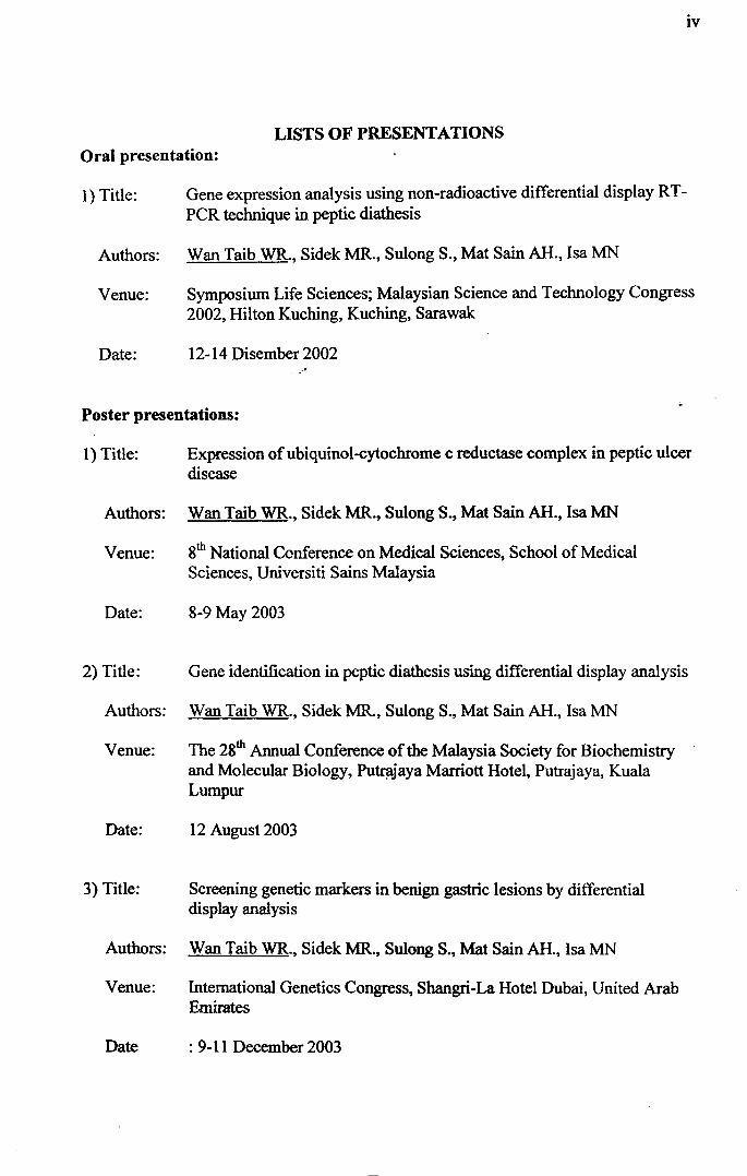
LISTS OF PRESENTATIONS Oral presentation:
1) Title: Gene expression analysis using non-radioactive differential display RTPCR technique in peptic diathesis
Authors: Wan Taib WR., Sidek MR., Sulong S., Mat Sain AH., Isa MN
Venue: Symposium Life Sciences; Malaysian Science and Technology Congress 2002, Hilton Kuching, Kuching, Sarawak
Date: 12-14 Disember 2002 .·''
Poster presentations:
1) Title: Expression ofubiquinol-cytochrome c reductase complex in peptic ulcer disease
Authors: Wan Taib WR., Sidek MR., Sulong S., Mat Sain AH., lsa MN
Venue: gth National Conference on Medical Sciences, School of Medical Sciences, Universiti Sains Malaysia
Date: 8-9 May 2003
2) Title: Gene identification in peptic diathesis using differential display analysis
Authors: Wan Taib WR., Sidek MR., Sulong S., Mat Sain AH., Isa MN
Venue: The 28th Annual Conference of the Malaysia Society for Biochemistry and Molecular Biology, Put:f!:ljaya Marriott Hotel, Putrajaya, Kuala Lumpur
Date: 12 August 2003
3) Title: Screening genetic markers in benign gastric lesions by differential display analysis
Authors: Wan Taib WR., Sidek MR., Sulong S., Mat Sain AH., Isa MN
Venue: International Genetics Congress, Shangri-La Hotel Dubai, United Arab Emirates
Date : 9-11 December 2003
v
Journal Publication:
1) Wan Rohani Wan Taib, Mohd Ros Sidek, Abdul Hamid Mat Sain, Mohd Nizam I sa A Known Gene in a Novel Location May Be Related to Gastric Carcinogenesis The Internet Journal of Gastroenterology. 2004. Volume I Number I (has been accepted for publication)
2) Wan Rohani Wan Taib, Mohd Ros Sidek, Abdul Hamid Mat Sain, Mohd Nizam I sa Is the Overexpression of Ubiquinol Cytochrome C Reductase in Erosive Gastritis Contributory to G:stric Carcinogenesis? Malaysian Journal of Biochemistry and Molecular Biology. 2004 Volume I 0 as Short communications -(has been accepted for publication)
Vl
LIST OF CONTENTS Contents Page
TITLE
DEDICATION ii
ACKNOWLEDGEMENT iii
LIST OF PRESENTATIONS iv
LIST OF CONTENTS vi
LIST OF TABLES X
LIST OF FIGURES xi
ABBREVIATIONS xiii
ABSTRACT XV
ABSTRAK xvii
CHAPTER 1 LITERATURE REVIEW l
1.1. Introduction
1.2. Peptic ulcer diathesis 4 12.1. Deftnition 4 12.2. Epidemiology 5 12.3. Classiftcations 7
1.2.3.1. Gastric ulcer 7 1.2.32. Duodenal ulcer 8 12.3.3. Gastritis 10 1.2.3.4. Duodenitis 11
1.2.4. Etiology 12 1.2.4.1. Diet factors 12 1.2.4.2. Chemical factors 14 1.2.4.3. Infectious agents 14
12.4.3 (a) Helicobacter pylori 15 1.2.4.3 (b) Herpes viruses 19
12.4.4 Genetic markers 19 · 1.2.4.4 (a) ABO blood group & Secretor status 22
12.4.4 (b) Family studies 24 1.2.4.4 (c) Twin studies 25 12.4.4 (d) Pepsinogen 26 1.2.4.4 (e) Other genetic markers 27
1.2.5 A genetic classiftcation 28
vii
1.2.6 Relationship between genetic alterations with gastric neoplasm/cancer 28
1.3. Differential Display Technique 31 1.3 .1 Gene expression 31 1.3.2 Differential display analysis 36 1.3.3 Principles of differential display 39
1.4. Aim of the study 45
1.5. Flowchartofthe study 46
CHAPTER 2 MATERIALS AND METHODS 48
2.1. Decontamination of apparatus 48 2.1.1 Reagents preparation 48
2.1.1.1 0.1% DEPC-treated water 48 2.1.1.1 0.5% Sodium Dodecyl Sulphate (SDS) 49 2.1.1.2 0.1% Sodium hydroxide (NaOH) 49 2.1.1.3 1 mM Ethylenediaminetetracetic Acid {EDT A) 49 2.1.1.4 3% Hydroxide peroxide (H202) 49 2.1.1.5 70% Ethanol 50
2.1.2 Elimination ofRNase from apparatus 50 2.1.2.1 Disposable plasticware 50 2.1.2.2 Non disposable plasticware 50 2.1.2.3 Glassware 50 2.1.2.4 Electrophoresis tanks 51 2.1.2.5 Solutions 51
22. Sample preparation 51 2.2.1 RNA extraction 54
22.1.1 Homogenization process 56 2.2. 1.2 RNA extraction procedure using a commercial kit 56
2.2.2 Quantification of RNA 57
2.2.3 Integrity of RNA 58 22.3.1 lOx TBE buffer 59 22.3.2 1x TBE buffer 60 2.2.3.3 2% Agarose gel preparation 60 2.2.3.4 2% Agarose gel electrophoresis &
Ethidium bromide staining 60
2.3 Differential display 61 2.3.1 Reverse transcription 63 2.3.2 Polymerase chain reaction 64
2.4 6% Denaturing Polyacrylamide gel electrophoresis (PAGE) 66
2.4.1 6% Denaturing PAGE preparation 2.4.1.1 25% Ammonium persulphate (APS) 2.4.12 Vertical electrophoresis set-up
2.4.2 Silver staining 2.4.2.1 Staining solutions preparation & procedure
2.4.2.1 (a) Fixative solution 2.4.2.1 (b) Staining solution 2.4.2.1 (c) Developer solution 2.4.2.1 (d) Stop solution
2.5. Band recovery and purification
2.6. Reamplification of eluted eDNA
2.7. Checking the size ofreamplified eDNA
2.8. eDNA purification
2.9. Sequencing of differentially expressed eDNA fragments
67 68 68
70 70 70 71 71 71
72
73
74
74
75
CHAPTER 3 RESULTS 76
·3.1. Total genomic RNA from tissues biopsies 76
3.2. Size separation of eDNA fragment on 6% denaturing polyacrylamide-urea gel electrophoresis (PAGE) 78
3.3. Checking for reamp1ified products 81
3 .4. Identification ofUbiquinol-Cytochrome C Reductase complex (Complex ill) gene 83 3.4.1 Screening analysis using differential display on
6% denaturing polyacrylamide-urea gel electrophoresis 83 3.42 Sequencing analysis 85
3.5. Identification of Ribosomal Protein I27a (RPL27a) gene 92 3.5 .1 Screening analysis using differential display on
6% denaturing polyacrylamide-urea gel electrophoresis 92 3.5 .2 Sequencing analysis 94
CHAPTER 4 DISCUSSION 101
4.1. Gene expression profile by differential display analysis 101
4.2. Non radioactive and non labeling DDRT -PCR analysis 106
viii
4.3. Identification ofUbiquinol-Cytochrome C Reductase complex (Complex lll) gene
4.4. Identification of Ribosomal Protein l27a
4.5. Future and further investigations 4.5.1 Confirmatory tests 4.5.2 mRNA quantification 4.5.3 Linkage analysis 4.5 .4 lmmWlOhistochemical analysis
CHAPTER 5 SUMMARY
REFERENCES
APPENDIX
110
116
119 120 120 121 123
124
126
136
IX
X
LIST OF TABLES
Tables Page
1.1 Relative risks with various associated genetic factors in peptic ulceration. Adapted from Rotter et al., 1992. 21
1.2 Proposed classification of peptic ulcer based on genetic features. Adapted from Rotter & Grossman, 1980. 30
1.3 Several techniques applicable in gene expression study 35 Adapted from Kozian &.Kirschbaum, 1999.
1.4 The principle of the differential display method. 42
2.1 Tissue biopsies samples collection. 53
2.2 Components ofRNAimage kit 1 (GenHunter, USA). 62
2.3 Reverse transcription reaction. 63
2.4 Reverse transcription conditions. 64
2.5 PCR mixture preparation. 65
2.6 PCR conditions. 65
2.7 Reamplification of cDNAs in a single PCR reaction. 73
3.1 Sequence of amplified eDNA. 86
3.2 Gene product derived from NCBI database. 89
3.3 Sequence of amplified eDNA. 95
3.4 Gene product derived from NCBI database. 98
.. Xl.
LIST OF FIGURES
Figures Page
l.l A model of the pathogenesis of peptic ulcer and the sequence of genetic predisposition combining with environmental factors to produce duodenal ulcers. Adapted from Porro et al. 1999. 9
1.2 The role of Helicobacter pylori infection in the development of gastric cancer. Adapted from Watters & Kiire 1995. 18
1.3 The schematic diagram shows a Central Dogma Adapted from crystaluah.edu/-carter/protein/ images/dogma.jpg. 32
lA Anchoring primers to produce eDNA pools for Differential Display (DD). Adapted from Weinzierl, 1999. 43
1.5 Experimental Details of the Differential Display Technique. Adapted from Weinzier~ 1999. 44
3.1 A schematic representation of the assessment of total RNA integrity from tissues (lane 1 to lane 5) using 1 % agarose gel electrophoresis. 77
3.2 Differential display of mRNAs on 6% Denaturing Polyacrylamide-Urea geL 80
3.3 The reamplified products were nm on 2 % agarose gel Electrophoresis. 82
3.4 6% Denaturing Polyacrylamide-Urea Gel Electrophoresis (PAGE) of amplified eDNA products obtained from gastritis (G) and normal tissue {N) with primer combination (H-Tt lC with H-AP8). 84
3.5 Identification of expressed gene. 87
3.6 Alignments for homology with ubiquinol-cytochrome c reductase using Blast 2 Sequence
(http://www.ncbi.nlm.nih.gov/BLAST/) 88
3.7 Schematic diagram showing the location ofubiquinol-cytochrome c reductase complex gene on chromosome 22q 123 90
3.8 Sequence analysis results for ubiquinol-cytochrome c reductase 91
..........,,,, _,
ABBREVIATIONS
PUD : Peptic ulcer disease
H. pylori : Helicobacter pylori
NSAIDs : Nonsteroidal anti-inflammatory drugs
NOC : N-nitroso compounds
ASA : Acetylsalicylic acid
• HSV : Herpes simplex virus
lgA : lmmWlOglobulin A
PG : Pepsinogen
HLA : Human leukocyte antigen
hTERT : Human telomerase catalytic subunit
mRNA : Messenger ribonucleic acid
tRNA : Transfer ribonucleic acid
rRNA : Ribosomal nbonucleic acid
eDNA : Complementary deoxyribonucleic acid
RDA : Representational difference analy~is
SAGE : Serial analysis of gene expression
DDRT-PCR : Differential display reverse transcription-polymerase chain reactiom
MMLV : Moloney murine leukemia virus
OGDS : OesophagoGastroDuodenoScope
PAGE : Polyacrylamide gel electropooresis
NUl : National Institute of Health
RLP27a
ZES
RFLP
kDa
:Ribosomal protein large 27a
: Zollinger-Ellison syndrome
: Restriction fragment length polymorphism
: Kilo Dalton
xiv
XV
ABSTRACT
Peptic diseases are the most common chronic diseases of adulthood and proven to have
a substantial multifactorial inherited components. Genetic influences play some role in
the predisposition to both forms of ulcers (gastric and duodenal ulcer). A small
proportion of chronic gas~c ulcers are susceptible to be transformed into malignancy.
The possible somatic mutatio~s that ~e place have not been extensively studied. The
discovery of some genetic changes at the vicinity of the chronic benign inflammatory
lesions is important in relation to the elucidation of the carcinogenesis of gastric
cancers. The general aims of this study were to screen for differentially expressed genes
in peptic diathetic patients and to apply a technique of non radioactive differential
display analysis (DDRT-PCR). DDRT-PCR has been shown to be highly effective in
identifying sequences that are differentially expressed in various cell types and this
technique makes it possible to obtain reproducible result and efficiently identify specific
mRNAs. Twenty tissue sample biopsies of gastric mucosa of the antrum were collected
from peptic diathetic patients at Endoscopy unit. Total RNAs were extracted by using
RNA extraction kit (RNeasy Mini Kit, Qiagen). The DDRT-PCR analysis was
performed by a 2- step method which were reverse transcription and polymerase chain
reactions (RNAimage Kit 1, GenHunter). Six percent denaturing Polyacrylamide Gel
Electrophoresis (PAGE) was carried out in order to obtain the size of separation of
eDNA fragments and visualized by silver staining. Once differentially expressed
mRNAs were identified, the corresponding cDNAs were eluted from the band of the gel
and reamplified. The sequence of cDNAs were determined using an ABI Prism DNA
XVI
Sequencer. The sequences were searched for its homology using GenBank. databases
provided by National Institutes of Health (NIH, USA). Two differentially expressed
genes were identified, namely, ubiquinol-cytochrome c reductase complex (Complex
III) gene and ribosomal protein L27a gene in gastritis tissue compared to normal gastric
tissue. The expressed genes can be analyzed to determine their involvement in the
pathogenesis of peptic diathesis. The determination of these genes will be used to study
whether similar genetic derangement occur in gastric cancers in the future. This
knowledge will enhance the understanding of carcinogenesis of chronic inflammatory
lesions.
PENYARINGAN MOLEKULAR MENGGUNAKAN TEKNIK PAPARAN
PERBANDINGAN BUKAN RADIOAKTIF DI KALANGAN PESAKIT
MELA YU DI KELANTAN BAGI PENY AKIT PEPTIK
ABSTRAK
xvii
Penyakit ulser peptik merupakan penyakit ~onik yang kerap berlaku pada golongan
dewasa dan telah terbukti disebabkan oleh komponen ~warisan pelbagai penyebab.
Pengaruh genetik memainkan peranan yang penting di dalam menyumbang kepada
kedua-dua jenis ulser (ulser gaster dan ulser duodenum). Sebahagian kecil ulser gaster
kronik lebih mudah terubah ke peringkat malignan. Kebarangkalian mutasi somatik
yang terlibat masih belum dikaji secara meluas. Penemuan sebarang perubahan genetik
pada peringkat lesi inflamasi benigna adalah penting di dalam hubungkait kepada
teijadinya fenomena karsinogenesis kanser gaster. Matlamat umwn kajian ini adalah
untuk mengenalpasti gen yang terekspresi yang berbeza bagi pesakit ulser peptik dan
untuk mengaplikasi teknik paparan perbandingan bukan radioaktif ("non-radioactive
differential display") atau lebih dikenali sebagai DDRT-PCR. Analisis DDRT-PCR
telah terbukti sangat berkesan di dalam pengenalpastian jujukan gen yang terekspresi di
dalam pelbagai jenis sel dan teknik ini dapat memberi keputusan yang senang untuk
dihasilkan dan pengenalpastian mRNA yang spesifik dengan berkesan. Dua puluh
sampel tisu biopsi dari bahagian antrum mukosa gaster dikumpul dari pesakit-pesakit
ulser peptik. RNA jumlah diekstrak menggunakan kit ekstrak RNA (RNeasy Mini Kit,
Qiagen, USA). Analisis DDRT-PCR merangkumi 2 peringkat teknik iaitu transkripsi
terbalik dan tindak balas rantai polimerase (RNAimage Kit 1, GenHunter, USA).

XVlll
Elektroforesis 6% gel poliakrilimida termusnah (PAGE) dilakukan bagi melihat basil
pemisahan saiz fragmen eDNA setelah diwarnakan oleh pewarnaan perak. Setelah
mRNA yang terekspres. dikenalpasti, eDNA tersebut dielut dari jalur yang dipotong
keluar dari gel dan direamplifikasi. Jujukan gen tersebut ditentukan oleh ABI Prism
DNA Sequencer. Jujukan tersebut dibandingkan dengan jujukan homologi yang
disediakan di dalam pangkalan GenBank oleh National Institute of Health (NIH, USA).
Kami betjaya mengenalpasti dua gen yang terekspresi iaitu gen ubiquinol-cytochr9me c
reductase (Komplek III) dan gen ribosomal protein L27a pada tisu gastritis berbanding
dengan tisu gastrik normal. Gen yang terekspres boleh dianalisa bagi menentukan ~ ~
penglibatannya di dalam patogenesis penyakit ulser peptik Penentuan jenis gen ini
berguna di dalam kajian lanjutan samada ketidakaturan genetik yang sama juga berlaku
di dalam kanser gaster. Pengetahuan ini akan mengukuhkan kefahaman proses
karsinogenesis bagi lesi inflamasi kronik.
CHAPTER1
LITERATURE REVIEW
1.1 INTRODUCTION
;,
Peptic ulcer disease (PUD) is a chronic, recurrent disorder that is characterized by
lesions in the upper gastrointestinal tract which appears as reddish and inflamed, or as
small depressions or excavations in the upper gastro-intestinal tract. An ulcer can form
at any area exposed to gastric acid and pepsin, a digestive enzyme instrumental in the
breakdown of protein and hence a derivation of a term "peptic ulcer". The areas most
commonly·affected are the upper part of the duodenum (duodenal ulcer), the stomach
itself (gastric ulcer) and less commonly, the esophagus (Greenberger & Thier, 1990).
Peptic ulcer diseases affect all age groups, but is rare in children. Men have twice the
risk for ulcers as women do. The risk for duodenal ulcers tends to occur first at around
age 25 and continues until age 75; gastric ulcers peak in people between the ages of 55
and 65 (Valle, et al. 1999).
A series of step-wise precancerous lesions, starting with chronic atrophic gastritis,
progressing to intestinal metaplasia, dysplasia, and finally becoming cancer were known
2
to occur in some cases; this sequence occurs over several decades (Ley, eta/. 2001). In
general, there are various contributory tendencies in carcinogenesis such as people
exposed to the risks involving the genetic and .environmental factors, prevalence of the
lesions in the population, morphological characters of the lesions and their potential
evolution from benign to neoplastic lesion. Evidences gathered thus far from several
scientific fields has led to the hypothesis that the clinical manifestations of most gastric
cancers are only a late event of a biologic phenomenon initiated many years previously
during the chronic inflammatory phase.
Clearly, further molecular analysis is needed to identify other alterations that may
contribute to gastric carcinogenesis and that may underlie the formation of premalignant
lesions of gastric cancer and, thus, may function as markers for an increased risk of
developing gastric cancer (Ebert, et al. 2000 & Boussioutas et a/. 2003). To date,
multiple genetic and molecular alterations in the multistage processes of gastric
carcinogenesis have been reported, including inactivated tumor suppressor genes such
as p53 and APC gene and activated oncogenes such as c-met and K-sam, which are
frequently amplified and overexpressed in gastric cancer. In addition, microsatellite
instability and alteration of adhesion molecule expression were demonstrated in gastric
cancer. Identification of more differentially expressed genes in gastric cancer may be
needed to elucidate the molecular mechanism of gastric carcinogenesis (Jung, et al.
2000).
Incorporation of epidemiological, clinical, histopathological, molecular genetics,
microbiological, occupational and behaviour assessments have had a major impact on
our understanding of gastric cancer today. In the future, collaboration of scientists from
different disciplines will be even more critical because it can lead to the identification of
3
previously unrecognized factors relevant to gastric carcinogenesis as well as to the
further development and subsequent implementation of a successful prevention program
(Christian, eta!. 1999).
Intense research during the past decade has resulted in several discoveries suggesting
not only that there are a number of genes that play a relatively minor role in
susceptibility of gastric cancer, such as the gene for blood group A, but also that there
may be genes that are able to make a major contribution to cancer susceptibility
(McConnell, 1983).
It has thus been clear for a number of years that genetic factors predispose to peptic
ulcer, but the mode of inheritance of this genetic predisposition has not been resolved.
For over a decade, the hypothesis of polygenic inheritance was used to explain the
genetics of peptic ulcer. Polygenic or multifactorial inheritance refers to the concept that
the hereditary component of a given disorder is due to the contribution of many genes
acting together (polygenic), resulting in a continuum of genetic predisposition toward
the disorder. Thus, clinical disease would exist when the presence of a sufficient
number of genes, perhaps in combination with environmental factors exceeds a
threshold level (Rotter & Grossman, 1980). The search for differentially expressed
genes in gastrie cancer and its premalignant lesions may help to define molecular
alterations in the gastric mucosa that may precede the development of gastric cancer.
Differential display technique presents a novel method for the identification of
aberrantly expressed genes in various biological states, such as carcinogenesis or
developmental process. Generally, this method has proven to be highly effective for the
4
identification of differentially expressed genes in the process of malignant
transformation. Furthermore, compared with other cloning methods, such as subtraction
hybridization, this method is advantageous because of its high reproducibility and the
identification of mRNAs with a low copy-number per cell (Ebert, eta/. 2000).
Gastroduodenal ulceration is still poorly understood and changes in gene expression
may provide new mechanistic insights (Szabo, eta/. 2001 ). Therefore, further molecular
analysis is needed to identify other molecular changes that may contribute to gastric
carcinogenesis and may function as markers of gastric cancer (Jung, eta/. 2000). We
used this method to search for differentially expressed genes in peptic ulcer diathesis.
1.2 PEPTIC ULCER DISEASE
1.2.1 DEFINITION
Peptic ulcer disease refers to breaks in the mucosa of the stomach and small intestine,
principally the proximal duodenum, that are produced by the action of gastric secretions
and also contributed by Helicobacter pylori infections in many cases. Although peptic
ulceration can occur as high as Barret esophagus and as low as Meckel diverticulum
with gastric heterotopia, for practical purposes, peptic ulcer disease essentially affects
the distal stomach and prOKimal duodenum (Rubin & Farber, 1999 & Shayne, 2002).
An ulcer is generally thought to occur when there is an imbalance between the
aggressiveness forces of acid and pepsin and the less well-defined defensive forces of
mucosal resistance and regeneration. The goal of ulcer therapy, both medical and
surgical, is to correct this imbalance to promote ulcer healing, relieve symptoms, and
prevent complications and recurrences. Peptic ulcer tends to be an episodic, chronic
disorder, characterized by symptomatic periods and pain-free intervals. The natural
history of ulcer may be differ as the different diseases leading to an ulcer are delineated
by clinical, physiologic and genetic studies (Rotter, et a!. 1992)
1.2.2 EPIDEMIOLOGY
Peptic ulcer is among the most common of the chronic diseases, occurring in 2% to 10%
of a world population, depending on such factors as geography, the specific population,
and level of health care. Both gastric and duodenal ulcer rates increase rapidly with age.
In the Danish studies the incidence of duodenal ulcer increased almost linearly with age,
reaching 0.3% in males over the age of 75 years. For gastric ulcer, the incidence was
low before age 40 in males and reached its peak for those aged 60 to 64 years, while in
women it increased with advancing age (Rotter, et a!. 1992). In the United States,
approximately 10% of Americans eventually develop peptic ulcer disease (PUD), and
about 10% of patients presenting with abdominal pain are diagnosed with peptic ulcer.
Prevalence has decreased in the US over the last 30 years. Frequency of PUD is
decreasing in the developed world but increasing in developing countries (Shayne,
2002).
Although the rate of incidence of gastric cancer has recently declined, gastric cancer is
still one of the most common malignancies worldwide and is the second most common
cause of cancer-related deaths (Tahara, et a/. 1996). The link between gastric ulcers and
gastric cancer come from epidemiologic observations in South East Asia, where Bonne
and co-workers in 1983 reported that Chinese immigrants had a high frequency of
u
gastric carcinoma and atrophic gastritis with "goblet cell metaplasia", whereas both
lesions were infrequently in native Malays (Correa, 1983).
H pylori prevalence is high in South East Asia including Malaysia. The background
prevalence of the entire Malaysian population is estimated to be around 40%. The
previous study in year 2000 carried out in Malaysia found that the incidence was higher
in Chinese and Indian communities than in the Malay community. The same trend was
also noted in ~eighbouring Singapore. The reason for this racial differences in incidence
is uncertain. The three races in Malaysia have been living in the same country for more
than two generations and are exposed to the same environment. Suggested explanations
for this fmding include genetic differences and transmission and perpetuation of
infection within the same ethnic group resulting from varied habits and socio-cultural
practices. Another contributory factor may be that the Chinese and Indians being
originally immigrants races may have brought the infection over from their home
countries. Amjad (2000) reported in his study that 43 (86%) of the 50 index patients had
family members sero-positive for H pylori infection. Of the index cases who were
Indian all the tested family members were positive, while 90% of the Chinese members
tested positive. The incidence of H pylori in the Malay family members was the lowest
at 71% (Amjad, 2000).
Kudva from Malaysia reported that peptic ulcer was seen in 21% of 1119 patients while
visual evidence of other gastric or duodenal mucosal lesions (gastritis or non-erosive
duodenitis) was in 20%. With increasing age, the prevalence of peptic ulcer steadily
increased and non-ulcer dyspepsia decreased (Kudva, 1990).
7
There are over half a million new cases diagnosed each year and up to 4 million people
have a flare up of the disease each year. About 1 out of every 1 0 people will at some
time in life have an ulcer.
1.2.3 CLASSIFICATIONS
1.2.3.1 Gastric Ulcer
An acute gastric ulcer .is a disease of abrupt or rapid onset and short duration. A focal
mucosal defect superficial to the muscularis mucosae heals by epithelial regeneration
without scar formation. In a deeper lesion, the amount of fibrosis produced is a
reflection of the depth and duration of the lesions. Most acute ulcers therefore heal
leaving little fibrotic reaction (Bouchier, eta/. 1993).
Benign chronic gastric ulcer is a common disorder. A study of self-reported peptic
ulcers in the United States found 4.3 million persons ever having had a gastric ulcer, 1.6
million of whom had their ulcers diagnosed in the year before the study. A familiar
study from Denmark found a lifetime prevalence of 1.2% for men and 0.6% for women.
The incidence and prevalence of gastric ulcers are determined primarily in association
with the major causes of the disease: Hpylori infection and nonsteroidal anti
inflammatory drug (NSAID) use. Other associated -risks, such as smoking, alcohol use
and socioeconomic status disappear when these primary causes are taken into account
(Yardley, 1990).

8
1.2.3.2 Duodeaal Ulcer
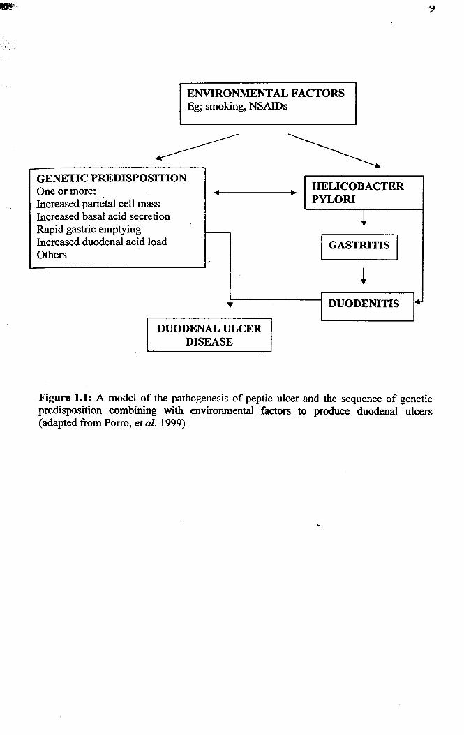
Duodenal ulcer can be defined as erosion in the lining of the duodenum (first part of the
small intestine, connecting to the stomach). Duodenal ulcers are commonly associated
with the presence of the bacteria Helicobacter pylori in the stomach. Risk factors are
aspirin and non steroidal anti inflammatory drugs (NSAID) use, cigarette smoking, and
older age. Figure l.l shows that several factors which contribute to duodenal ulcer
disease. A combination of environm<;ntal factors, genetic factors and H.pylori trigger
~ the clinical sequelae from inflammatory process to ulcer formation. Duodenal ulcer has
historically occurred more frequently in men, but more recent data suggest similar rates
in both men and women. The lifetime prevalence of a peptic ulcer is 5 to 10% and
approaches 10 to 20% m patients who are H. pylori positive
(http://www.nlm.nih.gov/medlineplus/ency/article/000206.htm).
ENVlRONMENTALFACTORS Eg; smoking, NSAIDs
GENETIC PREDISPOSITION One or more: ~
Increased parietal cell mass Increased basal acid secretion Rapid gastric emptying r--Increased duodenal acid load Others
DUODENAL ULCER DISEASE
HELICOBACTER PYLORI
+ I GASTRITIS I
~ DUODENITIS •
Figure 1.1: A model of the pathogenesis of peptic ulcer and the sequence of genetic predisposition combining with environmental factors to produce duodenal ulcers (adapted from Porro, eta!. 1999)
10
1.2.3.3 Gastritis
Gastritis includes a myriad of disorders that involve inflammatory changes in the gastric
mucosa, including erosive gastritis caused by a noxious irritant, reflux gastritis from
exposure to bile and pancreatic fluids, hemorrhagic gastritis, infectious gastritis and
gastric mucosal atrophy (Shayne, 2002).
Acute gastritis, acute ulcer and acute mucosal damage are considered together because
they represent the gastric mucosal responses to acute injury. Depending on the causes, ;.
they represent varying degrees of mucosal necrosis with subsequent inflammation.
Acute gastritis has well-established and consistent clinical associations such as a recent
history of drug ingestion, alcohol excess leading to haemorrhagic erosions, shock,
sepsis, multi organ failure and so on (Bouchier, eta/. 1993).
On the contrary, chronic gastritis is a heterogenous group of gastric mucosal disorders
characterized by wide spread injury, usually associated with a chronic, mixed acute or
chronic inflammatory response. Chronic gastritis can be defined as any diffuse chronic
inflammatory process involving the mucosal lining of the stomach. This definition
encompasses both specific and non specific subvariants of chronic gastritis. Specific
forms of chronic gastritis are associated with distinct disease processes and include
various entities such as granulomatous inflammation, eosinophilic infiltrative disorders,
Menetrier's disease and Zollinger-Ellison syndrome (Bouchier, et a/. 1993). Chronic
atrophic gastritis has been cited as one of the most important precursors of gastric
cancer (Nagayo, 1993).
II
There is currently a resurgence of interest in gastritis. Knowledge about gastritis
commenced when a link between the immune system and diffuse gastric mucosal
disease was established. The recent rediscovery ofthe Gram-negative spiral bacterium,
H. pylori, which exclusively colonizes epithelium of antrum part and the mounting
evidence of its causal relationship to the gastritis, has further emphasized the marked
heterogeneity ofthis disorder (Bouchier, eta/. 1993).
1.2.3.4 Duodenitis
Duodenitis is defined as an inflammatory condition of the proximal duodenum, usually
with maximal involvement of the bulb, and often but not invariably associated with
dyspeptic symptoms occurring in the absence of a chronic duodenal ulcer. The clinical
importance of duodenal inflammation in the absence of chronic ulceration remains
unclear. In all probability, H pylori infection is a major cause of chronic duodenitis.
Duodenal inflammation may also occur in specific conditions such as tuberculosis,
Crohn's disease, coeliac disease, septicaemia, giardiasis and ankylostomiasis (Bouchier,
eta/. 1993 ).
Inflammatory changes in the first part of the duodenum may occur alone or in
association with a peptic duodenal ulcer when the inflammation is most marked in the
immediately adjacent mucosa although it can be widespread within the duodenal bulb.
Whether non-specific duodenitis always represents a stage which can lead to ulceration
or alternatively may follow the healing of an ulcer, or whether it is a distinct entity has
not been resolved. The significance of duodenitis and its relationship to duodenal
ulceration continues to be debated. The reasons for this include the differing clinical,
endoscopic and histological criteria used to make the diagnosis, the variability of
symptoms experienced by patients with "duodenitis" and the usually patchy nature of
the inflammation (Cheli & Giacosa,, 1983).
1.2.4 AETIOLOGY
Several factors are suspected to _play a role in gastric carcinogenesis, including the
effects of diet, exogenous chemicals, intragastric ~ynthesis of carcinogens, genetic
factors, infectious agents and pathological conditions in the stomach, such as gastritis.
Recent molecular genetic studies have provided evidence that genetic alterations of the
human genome play important roles in the multistage process of gastric carcinogenesis
(Christian, eta/. 1999).
1.2.4.1 Diet factors
Coffee, both with and without caffeine, stimulates gastric acid secretion. The evidence
linking coffee drinking to ulcers is doubtful. In 1974, Friedman et a/. did not find any
association between alcohol and coffee consumption and the prevalence of peptic ulcer.
In contrast, Paffenbarger and co workers (1974) found in college students that ingestion
of coffee and other beverages (mainly colas) increased the risk of later development of
ulcers. Ingestion of milk decreased the risk. Cigarette smoking was correlated with
subsequent development of peptic ulcer. Doll and coworkers found that gastric ulcer
healed faster if smoking was stopped (Cooke, 1980). Alcohol (ethanol) also readily
causes erosive and hemorrhagic gastritis in both experimental animals and in man,
causing changes that are comparable to those seen with nonsteroidal anti-inflammatory

13
drugs (NSAIDs) and bile acids. Furthermore acute hemorrhagic lesions are frequently
found in chronic alcoholics. Even variations in types of alcoholic beverages consumed
may be important. For instance, because of low alcohol concentration in beer, inclusion
of beer drinkers in a study may reduce correlation between gastritis and alcohol
consumption (Yardley, 1990).
Most studies do not implicate type of food as causes of ulcer disease. Dietary treatment,
once in vogue, was based on the belief that small, bland meals might reduce the
secretion of acid and ~psin, buffer the acid secreted into the stomach, reduce the gastric
motor activity and maintain the resistance of the gastric mucosa. Interest has developed
in the study of a possible relationship between fiber in the diet and the development of
duodenal ulceration, as well as the possible therapeutic effect of high-fiber staple diets.
Fiber binds bile acids effectively and may therefore be of potential importance in
conditions in which bile reflux is thought to cause mucosal damage. Fiber-enriched
wheat bran changes the profile of the postprandial pH curve and reduces pepsin
concentrations (Rotter, et a/. 1992).
The intake of smoked and heavily salted, nitrated and carbohydrated food should be
avoided. High salt consumption can cause stomach irritation which can lead to the
development of atrophic gastritis. Salt also causes excessive cell replication and
increase the mutagenicity of nitrosated foods. Nitrates when reduced to nitrites can lead
to subsequent synthesis of carcinogenic N-nitroso compounds (NOC). Smoked fish
contain polycyclic aromatic hydrocarbons which when administered in an edible oil
vehicle induced forestomach cancer (Christian, eta/. 1999).
14
1.2.4.2 Chemical factors
Many non-steroidal anti-inflammatory drugs (NSAIDs) are important causes of acute
gastric injury, and of these aspirin (acetylsalicylic acid, ASA) is the best studied. After
exposure to aspirin the stomach rapidly develops an acute erosive. and hemorrhagic
gastritis, seen as multiple petechial hemorrhages and often concentrated in antrum
(Yardley, 1990). Chronic acetylsalicylic acid usage is nowwell established as a cause of
gastric ulcer. This_ is very well documented in the report from the Boston Drug
Surveillance Program. In that. study there was an association between hospital
admissions for newly diagnosed uncomplicated benign gastric ulcer and heavy long
term acetylsalicylic acid ingestion ( 4 or more days per week). No relationship was
found between acetylsalicylic acid usage (heavy or light) and duodenal ulcer. Anti
inflammatory agents such as acetylsalicylic acid, phenylbutazone, indometacin,
cortisone and adrenocorticotropin have also been shown to retard the healing of gastric
ulcers and favor perforation and hemorrhage making it vulnerable to normal gastric
secretions (Cooke, 1980 & Shayne, 2002).
1.2.4.3 Infectious agents
There are various infectious agents which contribute to the development of peptic ulcer.
Bacteria such as Helicobacter pylori, Helicobacter heilmannii and Mycoplasma can
tolerate with the acidic environment in stomach. Urease produced by bacteria can
neutralize gastric acidity and induce inflammation. Viruses such as Herpes simplex
virus and Epstein Barr Virus are thought to cause ulceration to the stomach and
duodenum.

1J
1.2.4.3 (a) Helicobacter pylori
Some discoveries showed that stomach and duodenal ulcers might be caused by a gram
negative spiral shaped microaerophilic bacterium named He/icobacter pylori. H.pylori
colonizes the antrum in 95% of patients; half of these will also have the organism in the
corpus (Bouchier, et al.1993 & Disotell, 2003). Helicobacter pylori has astonishing
ability to colonize the human gastric mucosa, an extraordinary hostile environment, and
to persist for decades, despite host inflammatory and immune responses~ It is present in
practically all human populations .• The _colonization is about 30-50% in developed
countries, while in developing countries it can exceed 80%. Initial colonization occurs
predominantly during childhood mainly from other family members. Chronic H. pylori
colonization is recognized as significant risk factor for gastritis, ulcers and cancers
(Matic, 2003 & Amjad, 2000).
H. pylori inhabits exclusively gastric-type epithelium, including gastric metaplasia in
the duodenum. The infected epithelium shows degenerative changes comprising
intracellular oedema, detachment from the basal lamina and cell necrosis. The
prevalence of H. pylori infection and chronic gastritis rises with age, in parallel with
the age-related increase in the prevalence of gastritis (Bouchier, et al.1993).
How H pylori infection produces gastric ulcers is still under intense investigation.
Unlike in duodenal ulcers, gastric acid hypersecretion does not occur in gastric ulcers.
Ulcerogenesis is caused by tissue damage and loss of normal protective factors. The
bacteria penetrates into mucosal layer and attaches to phospholipids, sialylated
glycoproteins and Lewis B antigens (in patients with blood group 0). Ammonia
16
generated by H. pylori urease damages the gastric mucosa, possibly by depleting a
ketoglutarate, an essential substrate in the tricarboxylic acid cycle.
Helicobacter pylori induces gastric inflammation in virtually all colonized individuals,
and such gastritis increases the risk for peptic ulcer disease and distal gastric
adenocarcinoma by expressing cytotoxic proteins such as cag A or vag A. However,
only a minority of persons carrying H.pylori develop clinical sequelae, suggesting that
particular bacterial products ~ay contribute to pathogenesis (Peek, et a/. 2000 & Leung,
eta/. 2002).
Hypothetical steps in ulcer formation by H. pylori is described. H. pylori uses urease to
protect it from acid during transit to the mucin layer. It colonizes the mucin layer and
may adhere to the gastric mucosa. Products of the bacteria provoke an inflammatory
response that ultimately damages the mucosa. Virulence factors are thought to be
involved at each stage (Salyers & Whitt, 1994). H. pylori tends to achieve its
pathogenetic role by triggering an intense leukocyte infiltration of the gastric mucosa,
and neutrophil activation provides a major source of reactive oxygen metabolites which
can cause tissue damage mainly in the absence of antioxidants. H. pylori virulence
factors promote release of a variety of chemoattractants/inflammatory mediators.
Longstanding H .pylori-associated gastritis predisposes to gastric carcinogenesis. It is
postulated that R pylori causes superficial gastritis which initiates a process that leads,
through atrophy, intestinal metaplasia and dysplasia to the development of gastric
cancer as shown in Figure 1.2 (Porro, et a/. 1999). Other interference comes from
nutrition defects, abnormalities in antibodies' function and gastrectomy. Various
17
regimens of reactive oxygen metabolite scavengers appear to have potentials as new
treatment strategies for upper gastrointestinal diseases (Kountouras, eta/. 2001).
It has been postulated that H pylori infection favors back diffusion of hydrogen ions
with subsequent breakdown of the mucosal barrier through alteration in the composition
of the glycoprotein of the mucus. It has also been postulated that the hyperacidity in
duodenal ulcer patients induces gastric metaplasia in the duodenal bulb, which becomes
a target for H pylori colonization and ultimately ulcer formation.
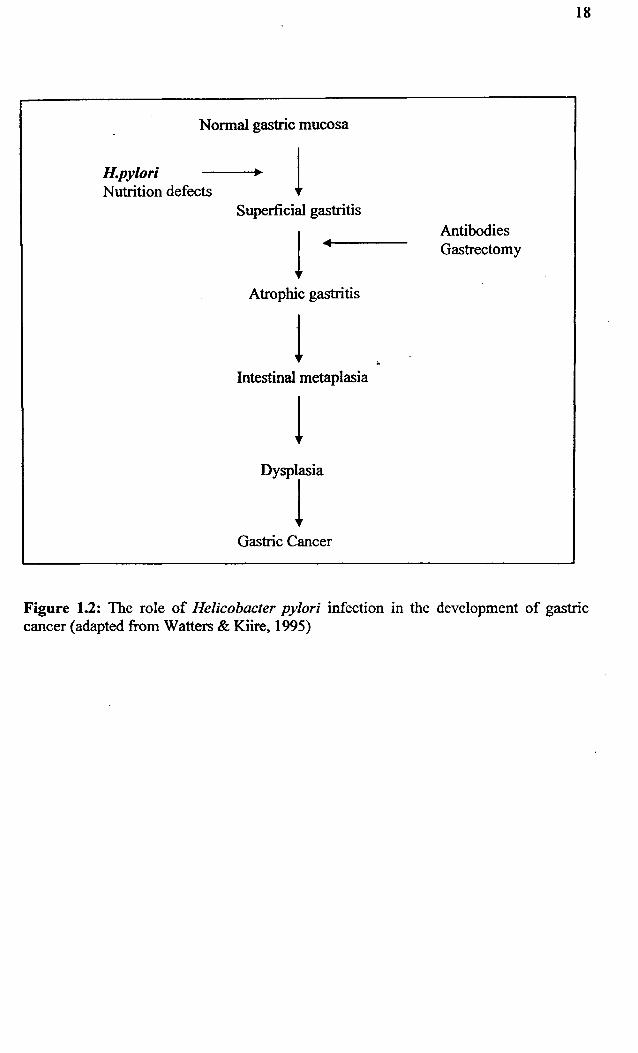
Normal gastric mucosa
H.pylori Nutrition defects 1
Superficial gastritis
1 Atrophic gastritis
1 Intestinal metaplasia
Gastric Cancer
Antibodies Gastrectomy
18
Figure 1.2: The role of Helicobacter pylori infection in the development of gastric cancer (adapted from Watters & Kiire, 1995)

19
1.2.4.3 (b) Herpes viruses
In 1967, Neuman and· Knyvett first suggested that herpes simplex virus was an etiologic
factor in peptic ulcer. It was subsequently suggested that chronic infection of a vagus
nerve by herpes simplex virus (HSV) could provide the mechanism leading to peptic
ulcer. Vestergaard and Rune (1980) reported that 94% of recurrent duodenal ulcer
patients were seropositive for HSV type 1 compared with 80% of controls. Saliva and
duodenal juice were tested for herpes simplex vi_rus type 1 Immunoglobulin A (IgA),
and higher levels were found in the ulcer group (Rotter, et ql. 1992). These findings
provide support for an association between active duodenal ulcer and herpes virus
infection.
1.2.4.4 Genetic markers
Peptic ulcer disease illustrates the importance of genetic factors and their interaction
with environmental mechanisms. The genetic predisposition varies from individual to
individuals; all persons are not equally susceptible to peptic ulcer (Grossman, et a/.
1981 ). Genes can be used as markers for cell recruitment, activation and mucosal
synthesis of immunoregulatory molecules (Dieckgraefe, et a/. 2000). Several scientists
have suggested the role of genetic factors in the pathogenesis of ulcer diseases. The
familial basis of duodenal ulcer and its mode of inheritance have thus far been an
enigma, resulting in the emergence of the polygenic hypothesis and the concept of
genetic heterogeneity (Habibullah, eta!. 1984).
20
The precancerous lesions of the gastrointestinal tract can be divided into those
determined by single genes and therefore in a more or less simple Mendelian manner
and those in which the genetic influence is more complex and due to genes at several
loci influencing susceptibility to environmental carcinogenic factors (McConnell, 1983).
In the present state of knowledge, it is probably best to assume that an interaction
between genetic and environmental factors underlies the great majority of
gastrointestinal cancers.
Subsequently, the genetics of chronic gastritis has been extensively investigated by the _ •
Helsinki group (1971) and it has been shown that severe atrophic gastritis is largely
genetically determined (Varis, 1971). The liability to severe atrophic fundic gastritis
was shown to be significantly higher in the first-degree relatives of patients with this
type of gastritis. This is a strong probability that this liability to fundic gastritis may be
due to a single genetic factor rather than to common environmental factors (McConnell,
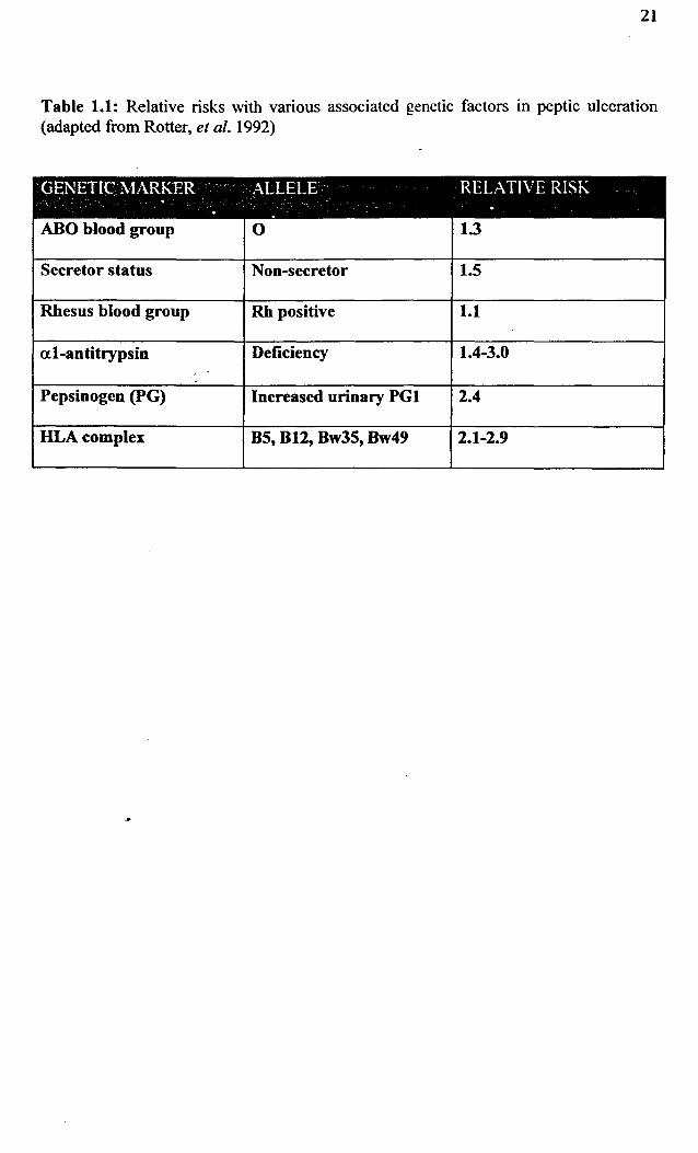
1983). Table 1.1 shows a relative risk of genetic factors in contributing to the formation
of peptic ulcer disease. Pepsinogen and HLA complex contribute a higher relative risk
in ulceration compared to other factors.
21
Table 1.1: Relative risks with various associated genetic factors in peptic ulceration (adapted from Rotter, eta/. 1992)
GENETIC MARKER - · ALLELE RELATIVE RISK . . . . ABO blood group 0 1.3
Secretor status Non-secretor 1.5
Rhesus blood group Rh positive 1.1
at-antitrypsin Deficiency 1.4-3.0 .•
Pepsinogen (PG) Increased urinary PGl 2.4
HLAcomplex 85, 812, 8w35, 8w49 2.1-2.9
I '··· ~ 'c
22
The Polygenic hypothesis
The genetics of peptic ulcer cannot be explained by a single, simple autosomal or sex-
linked, dominant or recessive defect. Thus, until recently peptic ulcer was considered a
polygenic disorder. Polygenic (multifactorial) disorders are thought to be caused by the
interaction of several genes with environmental factors. The hereditary component in
these illnesses reflects the combined contribution of many genes, resulting in a
continuum of genetic predisposition to illness - the more genes, the greater the
predisposition. The gene markers, blood group 0 and secretors status, provided some
direct support for the polygenic hypothesis because when they are present together the
risk is greater than when they are present separately. Although there may be a polygenic
contribution towards peptic ulcer, evidence indicates that the major genetic factors and
the differing physiologic observations can be best accounted for by the alternative
explanation such as genetic heterogeneity ( Grossman, et al. 1981 & Mueller & Young,
2001).
1.2.4.4 (a) ABO Blood Group and Secretor Status
The basic concept behind studying blood groups and other gene markers is that if a
disease is positively associated with traits that are shown to I>e inherited in a Mendelian
pattern, such as AB0 blood groups, then these genetic traits form a background of
predisposition for the disease (Grossman, et al. 1981). In a large number of studies
carried out there is an increased proportion of persons with peptic ulceration have been
found to have blood group 0 in a variety of different population groups. These findings
do not mean that all persons with blood group 0 will develop a duodenal ulcer but
merely that their risk of having a duodenal ulcer is 30% greater than in persons with
23
other blood groups. Gastric ulcer is also associated with blood group but the association
is not as strong as in the case of duodenal ulcer (Mueller & Young, 1995). The ABO
genes are therefore involved in determining liability to the disease and the ABO locus is
probably only one of several that play a part (McConnell, 1983). Interestingly, patients
with gastric ulcers do not exhibit a greater frequency of blood group 0. Associations
between certain histocompatibility antigens and peptic ulcers have been claimed but are
still debated (Rubin & Farber, 1999).
In Hong, Kong, Lam and Ong (1976) grouped their duodenal ulcer patients by age of
onset and found that their early-onset group (onset below age 20 years) has a
significantly stronger family history, had a frequency of blood group 0 similar to that of
controls, more frequently presented with gastrointestinal bleeding as the first
manifestation of the disease and rarely had complications such as perforation,
obstruction, intractable pain or secondary gastric ulcer. In contrast, their late-onset
group (onset after the age 20 years) had an infrequent family history of ulcer disease,
had an increased incidence of blood group 0, presented less frequently with
gastrointestinal bleeding and had an increased frequency of complications such as
perforation, pyloroduodenal stenosis, severe pain, virulent ulcer and secondary gastric
ulcer (Rotter & Grossman, 1980).
Peptic ulceration is also associated with the secretor status for the ABO blood system. It
has been found that duodenal ulcer and gastric ulcers to a lesser extent, are more
common in persons who are non-secretors than in persons who are secretors. In fact,
secretor status appears to be more important than a person's blood group in determining
the likelihood of developing a peptic ulcer, with persons who are non-secretors being
24
50% more likely to develop peptic ulceration than the general population. The two
factors together have a multiplicative effect, with persons who are non-secretors and
blood group 0 having 2.5 times the risk of developing peptic ulceration compared to the
general population (Mueller & Young, 1995 & Ming, 1992). ABH secretor genes
determine the ability of individuals to secrete these antigens since they are glycoprotein
constituents of gastric mucus. Their absence might alter the ability of mucus to protect
the mucosa (Samloff, 1980). These observations on blood group 0 and nonsecretors
have been confirmed by numerous investigators throughout the world and constituted
early important evidence for the role of genetic factors in peptic ulcer (Grossman, et a/.
1981 & Mueller & Young, 2001 ).
1.2.4.4 (b) Family Studies
The incidence of the disease in relatives of patients is compared with the incidence in
the general population to determine familial aggregation or grouping of a disorder. The
relatives of index patients share genes in common with the patients in direct proportion
to the closeness of their relationship. The multifactorial model thus predicts that such
relatives will share some of the disease-predisposition genes and hence will be shifted
toward the threshold for disease and have a higher disease frequency than the general
. population (Rotter & Grossman, 1980). Family studies have consistency shown peptic
ulcer disease to be two or three times as frequent in the first-degree relatives of peptic
ulcer patients as it is in relatives of control subjects. Because these differences persisted
across generations and social classes, genetic factors were presumed to explain these
findings (Grossman, eta/. 1981). First-degree relatives of patients with duodenal ulcers