walking assessment after lumbar puncture in normal ... · nph gait improvement after spinal tap...
TRANSCRIPT

clinical articleJ neurosurg 126:148–157, 2017
IdIopathIc normal-pressure hydrocephalus (iNPH) was first described in 1965.1 The syndrome is characterized by the clinical triad of gait disturbance, cognitive dys-
function, and urinary symptoms. The prevalence of iNPH reported in selected populations and in community-based studies on adults 18–79 years of age varies between 0.1% and 2.9%.16 It is one of the few neurological diseases in which a gait disorder and/or dementia can improve to a complete remission of symptoms. A disturbed gait is usu-ally the first sign and most disabling symptom.14 Typical
features are a diminished gait velocity, a reduced stride length due to the co-contractions of proximal muscles,40 a reduced foot-to-floor clearance (step height) during the swing phase of the gait cycle, and increased balance-relat-ed gait measurements such as step width and foot rotation angles.37,39 Such difficulties can improve after the removal of 30–50 ml of cerebrospinal fluid (CSF) via lumbar punc-ture (LP). Not only is this improvement important for es-tablishing the diagnosis, but it is also one of the few prog-nostic indicators of suitability for shunt implantation.23 In
abbreviations CSF = cerebrospinal fluid; DTS7 = dual-task walking with serial 7 subtractions; DTVF = dual-task walking with verbal fluency; EI = Evans Index; iNPH = idiopathic normal-pressure hydrocephalus; LP = lumbar puncture; MMSE = Mini-Mental State Examination; MoCA = Montréal Cognitive Assessment; rm = repeated mea-surement; STMS = maximal walking speed; STPS = preferred walking speed; TP = time point; VR = variation rate.sUbMitteD July 16, 2015. accePteD December 10, 2015.inclUDe when citing Published online March 18, 2016; DOI: 10.3171/2015.12.JNS151663.
Walking assessment after lumbar puncture in normal-pressure hydrocephalus: a delayed improvement over 3 daysroman schniepp, MD,1,2 raimund trabold, MD,3 alexander romagna, MD,3 Farhoud akrami,2 Kristin hesselbarth,2 Max wuehr, PhD,2 aurelia Peraud, MD,3 thomas brandt, MD,2,4 Marianne Dieterich, MD,1,2 and Klaus Jahn, MD2,5
1Department of Neurology, 2German Center for Vertigo and Balance Disorders, 3Department of Neurosurgery, 4Institute for Clinical Neuroscience, Ludwig-Maximilians University Munich; and 5Schoen Klinik, Bad Aibling, Bavaria, Germany
obJective The determination of gait improvement after lumbar puncture (LP) in idiopathic normal-pressure hydro-cephalus (iNPH) is crucial, but the best time for such an assessment is unclear. The authors determined the time course of improvement in walking after LP for single-task and dual-task walking in iNPH.MethoDs In patients with iNPH, sequential recordings of gait velocity were obtained prior to LP (time point [TP]0), 1–8 hours after LP (TP1), 24 hours after LP (TP2), 48 hours after LP (TP3), and 72 hours after LP (TP4). Gait analysis was performed using a pressure-sensitive carpet (GAITRite) under 4 conditions: walking at preferred velocity (STPS), walking at maximal velocity (STMS), walking while performing serial 7 subtractions (dual-task walking with serial 7 [DTS7]), and walking while performing verbal fluency tasks (dual-task walking with verbal fluency [DTVF]).resUlts Twenty-four patients with a mean age of 76.1 ± 7.8 years were included in this study. Objective responder status moderately coincided with the self-estimation of the patients with subjective high false-positive results (83%). The extent of improvement was greater for single-task walking than for dual-task walking (p < 0.05). Significant increases in walking speed were found at TP2 for STPS (p = 0.042) and DTVF (p = 0.046) and at TP3 for STPS (p = 0.035), DTS7 (p = 0.042), and DTVF (p = 0.044). Enlargement of the ventricles (Evans Index) positively correlated with early improve-ment. Gait improvement at TP3 correlated with the shunt response in 18 patients.conclUsions Quantitative gait assessment in iNPH is important due to the poor self-evaluation of the patients. The maximal increase in gait velocity can be observed 24–48 hours after the LP. This time point is also best to predict the response to shunting. For dual-task paradigms, maximal improvement appears to occur later (48 to 72 hours). Assess-ment of gait should be performed at Day 2 or 3 after LP.http://thejns.org/doi/abs/10.3171/2015.12.JNS151663Key worDs normal-pressure hydrocephalus; gait speed; improvement; lumbar puncture; functional neurosurgery
©AANS, 2017J neurosurg Volume 126 • January 2017148

nPh gait improvement after spinal tap test
J neurosurg Volume 126 • January 2017 149
patients who strongly respond to LP, their gait pattern of-ten dramatically improves after shunt placement.15
Given the significance of LP in establishing the di-agnosis of iNPH, the quantification of gait changes has become clinically important within the last decade. How-ever, many studies on gait changes after LP have used clinical gait scores rather than quantitative gait analysis procedures,8,33 resulting in a limited interpretation of their findings. One quantitative study of 10 NPH patients who had undergone LP showed that gait velocity and stride length were the most responsive gait parameters, whereas cadence and balance-related parameters remained unaf-fected.38 Authors of that study suggested that an increase in gait velocity by approximately 20% after LP could be considered a relevant improvement.
There is also indirect evidence that the type of quanti-tative gait examination strongly influences the outcome of the measurement. The importance of measuring dual mo-tor-cognition tasks in patients with mild cognitive impair-ment or dementia to assess mobility problems is well-es-tablished.3,6 This method was recently studied in an NPH cohort.4 The authors showed that patients with iNPH have reduced abilities to perform a motor-cognitive dual task. Moreover, assessing dual-task behavior was equivalent or superior to estimating the LP effect in patients with iNPH.
One pivotal requirement in assessing gait changes after LP is selecting the right time point to make such an as-sessment. Clearly, gait improvement after LP is a transient phenomenon, and the temporal characteristics for single- and dual-task walking can be different. Evaluating the gait performance of iNPH patients after LP at a time point of maximal improvement is desirable since it would allow an assessment with the highest sensitivity and best predictive value for shunt implantation.
However, there are few data about the time course of gait changes after LP in iNPH. Studies using nonquan-titative, examiner-based gait evaluation and quantitative measures have provided evidence of early improvement within the first 24 hours after LP.2,33,44 Other studies have supported the view that gait improvement can be detect-ed more than 24 hours after LP.4,38 Clinical standards of care usually include a follow-up assessment immediately after or the same day as the LP, although there is no con-sensus about the real time course of walking improve-ment after LP.
Until now, no study has investigated the temporal char-acteristics of gait improvement by using a sequential pro-tocol of gait assessments over several days after LP. Infor-mation about the gradual improvement of gait in the days after LP would allow us to adjust the post-LP measure-ment to a time of optimal sensitivity for estimating the LP effect and to avoid false-negative results. Thus, we investi-gated gait improvement after LP over a time span ranging from the day of LP (1–8 hours after LP, Day 1) to Days 2, 3, and 4 after LP. The temporal characteristics of gait im-provement after LP were then correlated with the response to shunt surgery and to repeated LP therapy. We further compared the time course of walking improvement while performing single-task walking and while performing dual-task walking in iNPH patients after LP.
MethodsParticipants
Patients in the departments of neurology and neuro-surgery at the University Hospital of the Ludwig-Max-imilians University Munich were recruited for this pro-spective study if they fulfilled the following clinical and radiological criteria for iNPH, as proposed by the German Neurological Society guidelines and adapted from Relkin and colleagues:34 1) at least 2 clinical signs of the Hakim triad including gait disorder and cognitive dysfunction, 2) enlargement of the lateral ventricles (assessed using the Evans Index [EI] > 0.3) on MRI or CT, 3) an open-ing CSF pressure < 15 cm H2O, and 4) exclusion of other differential diagnoses. All participants gave their written informed consent to take part in the study. The study was performed according to the guidelines laid down in the Declaration of Helsinki and its later amendments. The lo-cal ethics committee approved the study protocol.
All patients enrolled in the study underwent physical and neurological examination including testing for motor, sensory, and basal ganglia disorders. If necessary, addi-tional complementary technical diagnostic procedures were performed, for example, electrophysiological ex-aminations, neuroimaging of the brain or spinal cord, and vestibular testing. Cognition was assessed using either the Mini-Mental State Examination (MMSE) or the Montréal Cognitive Assessment (MoCA).30,43
lumbar Puncture ProceduresIf a suspected diagnosis of iNPH could not be rejected,
patients underwent an LP procedure, during which 30–50 ml of CSF was removed. Gait performance was evaluated before the LP (time point [TP]0), 1–8 hours after the LP (TP1), 24 hours after LP (TP2), 48 hours after LP (TP3), and 72 hours after LP (TP4).
gait assessmentGait performance was measured using a pressure-sen-
sitive carpet system (GAITRite, CIR Systems Inc.) 6.7 m in length with a sampling rate of 120 Hz. Relevant tem-poral and spatial gait cycle parameters were recorded, but only walking speed was analyzed. The gait analysis proce-dure involved a protocol of 4 different conditions: 2 speed conditions (single-task preferred walking speed [STPS] and maximal walking speed [STMS]), as well as 2 cog-nitive dual-task conditions (using working memory while counting backwards, that is, dual-task walking with serial 7 subtractions [DTS7]; and using semantic memory while enumerating animal names, that is, dual-task walking with verbal fluency [DTVF]). Each condition was tested twice. Each walk was started 1.5 m in front of the mat and contin-ued for 1.5 m beyond it to provide steady-state locomotion.
After each recording, patients were asked to evaluate their gait performance (“Do you think that your gait per-formance has improved after LP?”) by answering “yes” or “no.”
Data analysisWalking velocity was calculated as the mean of the 2

r. schniepp et al.
J neurosurg Volume 126 • January 2017150
recordings for each condition. Matlab (The Mathworks Inc.) and SPSS (SPSS Institute Inc.) were used for data analysis. To quantify the relative change in walking speed after LP, a variation rate (VR) was calculated using the following formula:
The effects of the dependent variable were analyzed using a 2-way repeated measurement (rm)ANOVA and a Bon-ferroni post hoc analysis, with time points (TP1, TP2, TP3, TP4) and gait conditions (STPS, STMS, DTS7, DTVF) as factors. Significant interaction effects were further broken down into simple main effects, and the covariates of age, sex, height, and leg length were included in the model. Correlations were calculated using Pearson’s and Kendall-Tau procedures. The results were considered significant at p < 0.05. Contingency between the self-evaluation of gait improvement and improvement according to objective gait measures was analyzed using chi-square tests.
resultsDemographic information and clinical Features
Twenty-four patients, 7 female and 17 male, were in-cluded in the study. Their mean age was 76.1 ± 7.8 years, and the mean duration of the gait disorder was 35 ± 26 months (Table 1). All patients showed gait impairments typical of iNPH and had cognitive dysfunction (score be-low 25 points on the MMSE or below 26 points on the MoCA). Twenty patients had bladder dysfunction. Com-puted tomography or MRI revealed a mean EI of 0.39 ± 0.05, and each patient had an EI > 0.3. The mean time pe-riod between LP and gait analysis was 3.2 ± 2.7 hours for TP1, 26.3 ± 4.8 hours for TP2, 53.7 ± 6.4 hours for TP3, and 74.3 ± 9.9 hours for TP4.
Measurement of the opening pressure after LP revealed that the intracranial pressure was below 15 cm H2O for each patient. A ventriculoperitoneal shunt was placed in 10 patients at 3–12 months after the LP. All of these pa-tients reported sustained improvement in walking perfor-mance at 3–12 months after surgery. Eight patients were treated with repeated LPs at intervals of 3–6 months. Six patients refused any further invasive treatment, for example, repeated LPs or shunt insertion. Four of those patients were treated pharmacologically with l-dopamine and memantine. Follow-up measurements of walking ca-pacity were performed in all patients; the follow-up period ranged from 3 months to a maximum of 12 months (2 per-sons without invasive treatment).
relationship between subjective and objective MeasuresThe rmANOVA model did not show significant differ-
ences in the walking characteristics between the groups of “subjective responders” (patients who answered “yes” to the question, “Do you think that your gait performance has improved after LP?”) and “subjective nonresponders” (patients who answered “no”).
A maximum of 18 “objective responders” to LP (pa-tients who had improvement in their gait after LP ac-
cording to objective measures [pressure-sensitive carpet system]) had ≥ 20% improvement in walking speed (in at least 1 condition, compared with baseline) in at least 1 post-LP measurement. Table 2 summarizes the contin-gency and diversity of the self-evaluation and technically based gait evaluation. Chi-square tests were not significant (p = 0.437, chi-square = 0.605). Thirty-three percent (6 of 18) of the patients with significant objectively measured gait improvements estimated that they had a (false) nega-tive result, whereas 83% (5 of 6) of the patients without significant gait improvement estimated that they had an improvement in gait (false-positive). Only 4 patients re-ported an immediate improvement at TP1. Fourteen pa-tients reported an improved gait at TP2, 17 patients at TP3, and 12 patients at TP4 (Table 3).
Nine of the 17 subjective responders underwent shunt implantation; 1 patient who did not subjectively improve (but showed an increased walking speed during gait anal-ysis) underwent surgery 10 months after LP.
table 1. Demographic and clinical characteristics of 24 patients with inPh
Variable No.
Basic demographic information Females/males 7:17 Mean age in yrs 76 ± 8 Mean height in m 1.74 ± 0.12 Mean duration of symptoms in yrs 35.4 ± 25.7Clinical feature Patients w/ gait disorder 24 Patients w/ bladder dysfunction 20 Patients w/ cognitive dysfunction 24 Mean MMSE score 22.4 ± 3.2 Mean MoCA score 21.5 ± 3.1 Mean ICP pressure in cm H2O at LP (range) 10.4 (7–14)Mean concomitant diseases (CCI) in points 12.3 ± 4.2Neuroimaging Patients w/ MRI 18 Patients w/ CT 6 Mean EI 0.39 ± 0.05Therapy Patients w/ shunt placement 10 Patients w/ repeated LP 8 Patients w/ no CSF therapy 6
CCI = Charlson Comorbidity Index; ICP = intracranial pressure.
table 2. contingency table for self-evaluation versus objective gait analysis
Objective Gait Assessment
Self-EvaluationResultsImproved Not Improved
Improved (≥20%) 12 6 FN 6/18, TP 12/18Not improved (<20%) 5 1 FP 5/6, TN 1/6
FN = false negative; FP = false positive; TN = true negative; TP = true positive.

nPh gait improvement after spinal tap test
J neurosurg Volume 126 • January 2017 151
temporal consistency of the gait MeasuresIndividual comparisons of the objective responder sta-
tus at each time point revealed a high inner-subject consis-tency of the data. Table 3 summarizes the absolute num-bers of objective responders and the patients who switched (called “converters”) from responder to nonresponder status or vice versa between the subsequent time points. Converters from nonresponder to responder status were mainly observed between TP1, TP2, and TP3, according to the idea of gradual gait improvement after LP. Convert-ers from responder to nonresponder status were mainly observed between TP3 and TP4, indicating a weakening of the LP effect after 48 hours. No patients switched the responder status more than 1 time within the 3 days after measurement. A maximum of 2 patients converted from responder to nonresponder status between TP2 and TP3 for the condition of STMS (only 1 of those also at the other conditions).
Dual-task PerformanceWalking speeds during DTS7 (F = 4.467, p = 0.024)
and DTVF (F = 4.239, p = 0.028) were significantly de-creased compared with those during STPS (at TP0). These differences remained at TP1 and TP2 but diminished at TP3 and TP4. The mean dual-task costs (that is, the per-centage decrease in walking speed during dual-task walk-ing) were -28.5% ± 18.9% for DTS7 and -28.1% ± 15.8% for DTVF at TP0.
There were no correlations between baseline walking performance and the individual dual-task costs, but there was a strong correlation between the individual dual-task costs for both dual tasks (R2 = 0.94, p < 0.001).
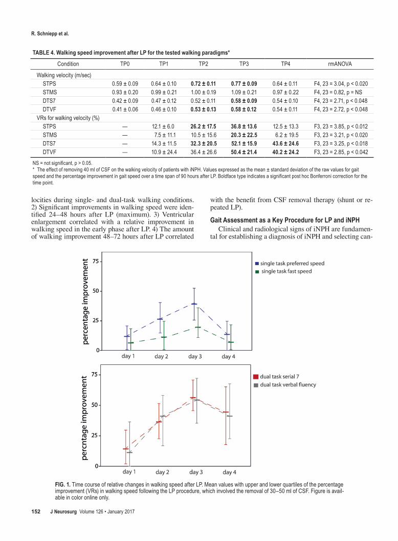
walking velocities after lPIn the rmANOVA model, there was a significant in-
crease in walking velocity after LP for the walking con-ditions of STPS, DTS7, and DTVF (Table 4). Bonferroni post hoc analysis of the interaction effect (walking condi-tion × time point) revealed a significant increase in walk-ing speed (compared with TP0) at TP2 for STPS (F = 3.96, p = 0.042) and for DTVF (F = 3.52, p = 0.046). At TP3, there were significant increases in walking speed (com-pared with TP0) for STPS (F = 4.11, p = 0.035), for DTS7 (F = 3.98, p = 0.042), and for DTVF (F = 3.76, p = 0.044).
The rmANOVA model of the VRs in walking speed revealed a significant increase after LP for all 4 walking conditions (Table 4 and Fig. 1).
evans index and gait Performance in inPhThe EIs of the iNPH patients were increased (mean of
0.39, range 0.32–0.45); thus, every patient met the crite-rion of ventricle enlargement (EI > 0.30). There was no significant correlation between the EIs and the absolute values of gait velocity at any time point of the gait mea-surements. Significant positive correlations for STPS and STMS were found for the VRs in walking speed. The EIs positively correlated with the VRs in walking speed at TP1 for STPS (R2 = 0.693, p = 0.0001; Fig. 2) and for STMS (R2 = 0.406, p = 0.049). At TP2, there was a posi-tive correlation between the VRs in walking speed and the EIs for STPS only (R2 = 0.612, p = 0.001; Table 5).
relationship between lP response and therapeutic outcome
The improvement in walking speed after therapy sig-nificantly depended on the therapeutic option chosen. The greatest improvement in walking speed was found for shunt surgery (for example, 34.7% ± 11.1% for STPS), followed by the repeated LP strategy (for example, 17.2% ± 18.8% for STPS). Patients who refused further CSF re-moval showed a gradual decrease in walking speed (for example, -28.1% ± 13.2% for STPS). The administration of dopamine and memantine did not completely reverse this decline (for example, -16.2% ± 6.2% for STPS). Parallel re-sults were present in the other walking conditions (Supple-mental Table 1). Post hoc analysis of the rmANOVA model showed that shunt surgery and repeated LPs led to sig-nificantly greater improvements in gait velocity compared with those in patients without CSF removal (p < 0.05). The response to LP correlated with improvement after shunt surgery and under repeated CSF removals. However, cor-relations between the VRs in walking speed improvement after LP and the VRs in walking speed improvement under CSF removal therapy (either shunt or repeated LPs) were only significant for delayed time points at TP3 (for STPS, for STMS, and for DTS7, all p < 0.05; Supplemental Table 2). No correlation was found for the EIs and gait improve-ments during follow-up.
DiscussionThis study provides the first evidence of a delayed
change in gait speed in patients with iNPH during the first 3 days after LP. Our main findings are as follows: 1) A reduction in gait velocity is a key feature of the iNPH gait disorder, and the removal of CSF improves walking ve-
table 3. time course of subjective and objective gait improvement after lP
Gait Improvement TP1 TP2 TP3 TP4
Subjective responder 4 14 17 12Objective responder Responder/converter (R/NR) Responder/converter (R/NR) Responder/converter (R/NR) Responder/converter (R/NR) STPS 8/(0/0) 16/(8/0) 16/(1/1) 7/(1/10) STMS 2/(0/0) 7/(7/2) 12/(5/0) 7/(2/7) DTS7 8/(0/0) 11/(4/1) 19/(8/0) 13/(1/7) DTVF 9/(0/0) 12/(4/1) 18/(7/1) 13/(1/6)
NR = nonresponder; R = responder. * Converter status mentioned as converted to responder and converted to nonresponder.
r. schniepp et al.
J neurosurg Volume 126 • January 2017152
locities during single- and dual-task walking conditions. 2) Significant improvements in walking speed were iden-tified 24–48 hours after LP (maximum). 3) Ventricular enlargement correlated with a relative improvement in walking speed in the early phase after LP. 4) The amount of walking improvement 48–72 hours after LP correlated
with the benefit from CSF removal therapy (shunt or re-peated LP).
gait assessment as a Key Procedure for lP and inPhClinical and radiological signs of iNPH are fundamen-
tal for establishing a diagnosis of iNPH and selecting can-
table 4. walking speed improvement after lP for the tested walking paradigms*
Condition TP0 TP1 TP2 TP3 TP4 rmANOVA
Walking velocity (m/sec) STPS 0.59 ± 0.09 0.64 ± 0.10 0.72 ± 0.11 0.77 ± 0.09 0.64 ± 0.11 F4, 23 = 3.04, p < 0.020 STMS 0.93 ± 0.20 0.99 ± 0.21 1.00 ± 0.19 1.09 ± 0.21 0.97 ± 0.22 F4, 23 = 0.82, p = NS DTS7 0.42 ± 0.09 0.47 ± 0.12 0.52 ± 0.11 0.58 ± 0.09 0.54 ± 0.10 F4, 23 = 2.71, p < 0.048 DTVF 0.41 ± 0.06 0.46 ± 0.10 0.53 ± 0.13 0.58 ± 0.12 0.54 ± 0.11 F4, 23 = 2.72, p < 0.048VRs for walking velocity (%) STPS — 12.1 ± 6.0 26.2 ± 17.5 36.8 ± 13.6 12.5 ± 13.3 F3, 23 = 3.85, p < 0.012 STMS — 7.5 ± 11.1 10.5 ± 15.6 20.3 ± 22.5 6.2 ± 19.5 F3, 23 = 3.21, p < 0.020 DTS7 — 14.3 ± 11.5 32.3 ± 20.5 52.1 ± 15.9 43.6 ± 24.6 F3, 23 = 3.25, p < 0.018 DTVF — 10.9 ± 24.4 36.4 ± 26.6 50.4 ± 21.4 40.2 ± 24.2 F3, 23 = 2.85, p < 0.042
NS = not significant, p > 0.05.* The effect of removing 40 ml of CSF on the walking velocity of patients with iNPH. Values expressed as the mean ± standard deviation of the raw values for gait speed and the percentage improvement in gait speed over a time span of 90 hours after LP. Boldface type indicates a significant post hoc Bonferroni correction for the time point.
Fig. 1. Time course of relative changes in walking speed after LP. Mean values with upper and lower quartiles of the percentage improvement (VRs) in walking speed following the LP procedure, which involved the removal of 30–50 ml of CSF. Figure is avail-able in color online only.

nPh gait improvement after spinal tap test
J neurosurg Volume 126 • January 2017 153
didates for shunt placement. A positive response to LP is the most common clinical sign used to verify the diagno-sis and to predict a beneficial outcome of shunt surgery.47 Quantitative gait assessment helps by providing standard-ized and comparable data for the clinician.38 The impor-tance of such quantitative measures for evaluating LP is further stressed by the low reliability of patient self-evalu-ations. Only 54% of the patients had subjective outcomes consistent with the objective gait measures. Obviously, there is a strong tendency to subjectively overestimate gait performance after LP. Increased levels of motivation and the anticipation of possible treatment options after a positive LP outcome could lead to such false-positive self-evaluations. Since a positive LP outcome serves as a strong argument for shunt insertion in iNPH, clinicians should assess gait in an objective way to avoid false-positive eval-uations of the LP procedure.
While the patient’s appraisal of an improvement in gait after LP is decisive in delivering treatment for iNPH, it is still subject to false results. Quantitative gait measures, however, provide objective measures of the clinical out-come that could be used to identify false-positive respond-ers and avoid exposing this subgroup to surgical risks.
walking speed reduction as a Key aspect of the inPh gait Disorder
It has been demonstrated that a reduced walking pace is a key feature of the gait disorder in patients with iNPH.38 This clinical hallmark can be easily assessed. In agree-ment with previous studies, the iNPH cohort in the current study showed a preferred walking pace that was approxi-mately 50% of the walking pace of healthy subjects.46 Re-duced walking speed is a key feature of mobility impair-ments and has been linked to a decreased quality of life, decreased mobility, and impaired everyday independence of elderly persons.27,28 Stolze et al. suggested that an im-provement in walking speed after LP by ≥ 20% could be considered a relevant improvement.38 This threshold was used in the present study to define the objective responder status, and the maximum average walking speed improve-ment was 37% at TP2 for STPS. During STMS, the per-centage of improvement was lower. Thus, patients with iNPH increased their self-selected walking pace, but their maximal speed capacity was only slightly improved.
Dual-task Performance of Patients with inPhCurrently, postural control is believed to share atten-
tional resources with cognition.17 Dual-task paradigms—that is, the simultaneous tasks of walking and cognition—have been used to shed light on motor and cognitive inter-ference during gait.48 The walking and cognitive functions of patients with iNPH are known to be independently improved after LP.37,42 In a recent study, spatiotemporal gait parameters assessed during dual-task walking were proven to be a good discriminator of responders versus nonresponders in patients with iNPH after LP.4
Our results support and extend these findings, show-ing that 22 of 24 patients with iNPH walked more slowly during the dual task than during the single task. The av-erage dual-task cost of approximately 28% is comparable to findings reported in a recent study5 and other clinical cohorts.10,19
The comparison of dual-task costs between the serial 7 task and the verbal fluency task shows a strong inter-task correlation; thus, the type of cognitive task is not relevant for the result of the dual-task gait assessment. Instead, impaired dual-task performance reflects a disturbance in higher-level processing domains (for example, attention-
table 5. evans indexes and walking performance in inPh patients*
Condition TP0 TP1 TP2 TP3 TP4
EI & walking speed STPS NS NS NS NS NS STMS NS NS NS NS NS DTS7 NS NS NS NS NS DTVF NS NS NS NS NSEI & VRs in walking speed STPS — 0.693, p = 0.000 0.612, p = 0.001 NS NS STMS — 0.406, p = 0.049 NS NS NS DTS7 — NS NS NS NS DTVF — NS NS NS NS
* Results of Pearson’s correlation procedures of individual EIs and gait velocities and the improvement in gait velocities (expressed in VRs).
Fig. 2. Pearson’s correlation of the EI and the VRs in walking speed im-provement on the day of LP (1–8 hours after LP). R2 = 0.693, p < 0.001.

r. schniepp et al.
J neurosurg Volume 126 • January 2017154
dividing problem)32 in iNPH that results in an inferior gait performance.
Studies on gait impairment due to small vessel disease of the brain revealed the impact of predominantly frontal white matter alterations and stressed the fundamental role of frontal brain regions in the cognitive control of motor performance (for example, the anterior thalamic radia-tion7,13,36). We found no relationship between the EI and the dual-task cost in our data, a result that suggests that ven-tricular enlargement alone cannot explain the dual-task impairments in iNPH. Metabolic, vascular, or other dis-turbances of deep white matter in the frontal brain regions may explain the dual-task problems of patients with iNPH; however, our study design does not allow us to speculate on any underlying pathophysiological mechanisms. Fur-ther studies using more sophisticated neuroimaging tech-niques are necessary to investigate brain structure and/or function relationships in iNPH.
Analysis of the improvement in walking speed after LP revealed greater relative improvements for dual-task walking than for single-task walking. However, this effect may be a result of the lower baseline values of gait speed at TP0. In contrast to the earlier findings of Allali et al.,4 our absolute improvements for single-task walking were higher than those for dual-task walking. In particular, for a clinical setup, when the calculation of small absolute dif-ferences in relative changes is difficult, it may be better to assess iNPH walking performance in a single-task proce-dure. If single-task walking is only slightly affected in pa-tients with iNPH, the addition of dual-task walking para-digms may help to avoid ceiling effects for the LP results.
temporal characteristics of gait improvement in inPh after lP
The improved walking of patients with iNPH after LP is a transient phenomenon. An earlier study provided evi-dence of improvement within the first 24 hours after LP;44 however, subsequent time points more than 24 hours after LP were not investigated. Our study is the first system-atic investigation of the time course of gait improvement within the first 72 hours after LP. The results of the single-task and dual-task walking assessments indicated that the maximum gait improvement can be detected 24–72 hours after LP. Nearly half of the objective responders accord-ing to our definition of a responder status would have been missed within the first 24 hours after LP. This find-ing indicates that assessment of gait speed changes within the first 24 hours after LP is susceptible to false-negative results. Apart from the gradual improvement over 48–72 hours, the inner-subject consistency for subsequent mea-surements was high. Only 2 patients had conflicting results over the time span measured. Thus the improvement in walking speed reflects a robust outcome measure of the LP effect.
There is evidence that the extent of gait improvement after LP corresponds to the extent of the benefit after shunt surgery.12,18 This association was present in all patients with iNPH who had undergone CSF removal therapies, ei-ther shunt surgery or repeated LPs. Significant correlations between the LP response and therapy outcomes, however, were only present for gait assessments at late time points
(that is, 48–72 hours after LP). This finding further indi-cates that the delayed improvement in gait velocity after a single LP could have higher predictive power than the re-sults of early assessments. Accordingly, clinicians should measure the effects on walking performance after LP at the time of maximum change to adequately estimate the capacity for improvement after therapy.
Pathophysiological models of the gait disorder in iNPH suggest a direct compression or deformation of the motor fibers in the medial portion of the corona radiata.35 The removal of CSF may inhibit the pathological high pressure gradients in the lateral and third ventricles and the sylvian fissures,22 which could lead to a decompression of frontal and/or mesencephalic fibers of motor or premotor areas. Other studies provide evidence for increases in regional blood flow in different brain areas of patients with iNPH after LP.9,11,20,31,45 These mechanisms would explain an im-mediate release of symptoms after CSF removal. In the current study, 8 patients showed slight gait improvements immediately after LP. Moreover, the observed significant correlation between the extent of lateral ventricle enlarge-ment (measured by the EI) and early gait improvement further supports the pressure-related symptom release in iNPH. However, the current study also has implications for delayed alternative and/or complementary pathophysi-ological processes, which could be involved in the LP re-sponse. Momjian et al. described the relationship between reduced cerebral blood flow in the deep white matter of patients with iNPH and impaired cerebral vascular auto-regulation.29 Patients with iNPH were shown to have dis-turbed autoregulation of cerebral arteries, especially near the lateral ventricles. Impaired autoregulation of the ar-teries could led to edema and local ischemia, which were thought to contribute to the emergence of the gait disor-der in iNPH. Lumbar puncture–associated changes in the CSF pressure gradient at the lateral ventricle walls and in the surrounding white matter were shown to lead to a shift in the watershed phenomenon of the regional blood flow, thus improving vascular autoregulation in the peri-ventricular areas.25,26 Moreover, changes in the interstitial fluid pressure by CSF removal could decrease edema in the periventricular tissue,24,41 thereby improving the clear-ance of vasoactive and/or neurotoxic metabolites. Both the improved vascular autoregulation and the enhanced capacity for metabolic clearance may be the pathophysi-ological substrates of delayed walking improvement in the first days after LP.21
However, the current study was not designed to ade-quately address the relationship between brain structure and function in iNPH. Future investigations using neuro-imaging and neurophysiological measurements in parallel are required to directly address these pathophysiological mechanisms.
recommendations for a clinical gait assessment During the lP Procedure
Subjective estimations of gait improvement by patients with iNPH are prone to false-negative and false-positive ratings. Quantitative gait measurements can help to im-prove the sensitivity and predictive value of the LP pro-cedure: Gait velocity (and stride length) can be easily

nPh gait improvement after spinal tap test
J neurosurg Volume 126 • January 2017 155
assessed using a 35-ft (8-m) walking test, during which the physician records the ambulation time and the total steps. To assess walking speed capacity and the individual utilization of preferred speed, single-task walking with preferred speed and with maximally fast speed should be measured. We recommend performing a dual-task walk-ing examination by adding an executive task (serial 7) and/or verbal fluency tasks (naming words) for patients with only minor impairments during single-task walking. Moreover, these conditions help to identify the extent of attentional resources spent on walking and simulate every-day mobility tasks.
The evaluation of post-LP measurements should be done not only 2–6 hours after the LP (to assess direct im-provement) but also between 24 and 48 hours after the LP to avoid false-negative results and to estimate the real ex-tent of walking improvement (Fig. 3).
limitations of the studyThis study has several limitations. The study cohort
of 24 patients with iNPH is relatively small for estimat-ing correlations between the follow-up outcomes and the LP response. With the 18 patients who were treated with either shunt surgery or repeated LPs, we have reached a sufficient sample size for the Pearson’s correlations. For correlations with other outcome variables, such as qual-ity of life, mobility measures, or independency, a further systematic study is necessary.
Moreover, the time points of follow-up measurements differed from 3 to 12 months. With respect to a possible dynamic disease course, this heterogeneity in follow-up times may lead to deficient interpretation of the follow-up results; however, only 2 patients (both with a non–CSF removal therapy) had a late follow-up measurement.
The analysis of associations between LP results and gait outcomes after shunt insertion or repeated LPs was per-formed with a very homogeneous follow-up period of 3–6 months after therapy.
conclusionsThe evaluation of post-LP measurements in iNPH
should be done not only 2–6 hours after the LP (to assess direct improvement) but also between 24 and 48 hours af-ter the LP to avoid false-negative results and to estimate the real extent of walking improvement.
acknowledgmentsThis work was supported by the German research founda-
tion (Deutsche Forschungsgemeinschaft, DFG JA 1087/1-1), the German Hertie Foundation, and the German Federal Ministry for Education and Science (BMBF, Nr. 01EO0901).
We thank Judy Benson for copyediting the manuscript.
references 1. Adams RD, Fisher CM, Hakim S, Ojemann RG, Sweet WH:
Symptomatic occult hydrocephalus with “normal” cerebro-spinal-fluid pressure: a treatable syndrome. N Engl J Med 273:117–126, 1965
2. Agostini V, Lanotte M, Carlone M, Campagnoli M, Azzolin I, Scarafia R, et al: Instrumented gait analysis for an objective pre-/postassessment of tap test in normal pressure hydro-cephalus. Arch Phys Med Rehabil 96:1235–1241, 2015
3. Allali G, Kressig RW, Assal F, Herrmann FR, Dubost V, Beauchet O: Changes in gait while backward counting in demented older adults with frontal lobe dysfunction. Gait Posture 26:572–576, 2007
4. Allali G, Laidet M, Beauchet O, Herrmann FR, Assal F, Ar-mand S: Dual-task related gait changes after CSF tapping: a
Fig. 3. Schematic of the time course of gait improvement after CSF removal (removal of 30–50 ml CSF) in patients with iNPH. Schematic of gait velocity improvement after LP. Gait velocity can be easily assessed on a 25-ft/8-m walking test. Single-task walking assessment should include walking with preferred speed and with fast speed to obtain information about the overall walk-ing capacity and the self-selected utilization of this capacity. The performance of dual tasks with executive (serial 7) and verbal flu-ency tasks (naming words) helps to identify attentional resources during walking and to simulate everyday mobility tasks. Walking performance in iNPH improves directly after the LP: there is a gradual improvement for 24–48 hours (for single task). Single-task walking improvement appears earlier than dual-task walking improvement.

r. schniepp et al.
J neurosurg Volume 126 • January 2017156
new way to identify idiopathic normal pressure hydrocepha-lus. J Neuroeng Rehabil 10:117, 2013
5. Armand S, Allet L, Landis T, Beauchet O, Assal F, Allali G: Interest of dual-task-related gait changes in idiopathic normal pressure hydrocephalus. Eur J Neurol 18:1081–1084, 2011
6. Beauchet O, Freiberger E, Annweiler C, Kressig RW, Her-rmann FR, Allali G: Test-retest reliability of stride time variability while dual tasking in healthy and demented adults with frontotemporal degeneration. J Neuroeng Rehabil 8:37, 2011
7. Benson RR, Guttmann CR, Wei X, Warfield SK, Hall C, Schmidt JA, et al: Older people with impaired mobility have specific loci of periventricular abnormality on MRI. Neurol-ogy 58:48–55, 2002
8. Bugalho P, Guimarães J: Gait disturbance in normal pressure hydrocephalus: a clinical study. Parkinsonism Relat Disord 13:434–437, 2007
9. Capel C, Kasprowicz M, Czosnyka M, Baledent O, Smielews-ki P, Pickard JD, et al: Cerebrovascular time constant in pa-tients suffering from hydrocephalus. Neurol Res 36:255–261, 2014
10. Coelho FG, Stella F, de Andrade LP, Barbieri FA, Santos-Galduróz RF, Gobbi S, et al: Gait and risk of falls associated with frontal cognitive functions at different stages of Al-zheimer’s disease. Neuropsychol Dev Cogn B Aging Neu-ropsychol Cogn 19:644–656, 2012
11. Czosnyka ZH, Czosnyka M, Whitfield PC, Donovan T, Pick-ard JD: Cerebral autoregulation among patients with symp-toms of hydrocephalus. Neurosurgery 50:526–533, 2002
12. Damasceno BP, Carelli EF, Honorato DC, Facure JJ: The pre-dictive value of cerebrospinal fluid tap-test in normal pressure hydrocephalus. Arq Neuropsiquiatr 55:179–185, 1997
13. de Laat KF, van Norden AG, Gons RA, van Oudheusden LJ, van Uden IW, Bloem BR, et al: Gait in elderly with cerebral small vessel disease. Stroke 41:1652–1658, 2010
14. Graff-Radford NR, Godersky JC: Normal-pressure hydro-cephalus. Onset of gait abnormality before dementia predicts good surgical outcome. Arch Neurol 43:940–942, 1986
15. Ishikawa M, Hashimoto M, Kuwana N, Mori E, Miyake H, Wachi A, et al: Guidelines for management of idiopathic nor-mal pressure hydrocephalus. Neurol Med Chir (Tokyo) 48 (Suppl):S1–S23, 2008
16. Jaraj D, Rabiei K, Marlow T, Jensen C, Skoog I, Wikkelsø C: Prevalence of idiopathic normal-pressure hydrocephalus. Neurology 82:1449–1454, 2014
17. Kerr B, Condon SM, McDonald LA: Cognitive spatial pro-cessing and the regulation of posture. J Exp Psychol Hum Percept Perform 11:617–622, 1985
18. Klinge P, Hellström P, Tans J, Wikkelsø C: One-year outcome in the European multicentre study on iNPH. Acta Neurol Scand 126:145–153, 2012
19. Kressig RW, Herrmann FR, Grandjean R, Michel JP, Beauchet O: Gait variability while dual-tasking: fall predic-tor in older inpatients? Aging Clin Exp Res 20:123–130, 2008
20. Kristensen B, Malm J, Fagerland M, Hietala SO, Johansson B, Ekstedt J, et al: Regional cerebral blood flow, white mat-ter abnormalities, and cerebrospinal fluid hydrodynamics in patients with idiopathic adult hydrocephalus syndrome. J Neurol Neurosurg Psychiatry 60:282–288, 1996
21. Lundin F, Tisell A, Leijon G, Leinhard OD, Davidsson L, Grönqvist A, et al: Preoperative and postoperative 1H-MR spectroscopy changes in frontal deep white matter and the thalamus in idiopathic normal pressure hydrocephalus. J Neurol Neurosurg Psychiatry 84:188–193, 2013
22. Malm J, Kristensen B, Fagerlund M, Koskinen LO, Ekstedt J: Cerebrospinal fluid shunt dynamics in patients with idio-pathic adult hydrocephalus syndrome. J Neurol Neurosurg Psychiatry 58:715–723, 1995
23. Marmarou A, Bergsneider M, Klinge P, Relkin N, Black PM: The value of supplemental prognostic tests for the preopera-tive assessment of idiopathic normal-pressure hydrocephalus. Neurosurgery 57 (3 Suppl):S17–S28, ii–v, 2005
24. Marmarou A, Takagi H, Shulman K: Biomechanics of brain edema and effects on local cerebral blood flow. Adv Neurol 28:345–358, 1980
25. Mathew NT, Hartmann A, Meyer JS: The use of regional cerebral blood flow measured with the gamma camera in neurological diagnosis. Int J Neurol 11:194–205, 1977
26. Meyer JS, Tachibana H, Hardenberg JP, Dowell RE Jr, Kitagawa Y, Mortel KF: Normal pressure hydrocephalus. Influences on cerebral hemodynamic and cerebrospinal fluid pressure—chemical autoregulation. Surg Neurol 21:195–203, 1984
27. Middleton A, Fritz SL, Lusardi M: Walking speed: the func-tional vital sign. J Aging Phys Act 23:314–322, 2015
28. Middleton A, Fulk GD, Beets MW, Herter TM, Fritz SL: Self-selected walking speed is predictive of daily ambulatory activity in older adults. J Aging Phys Act [epub ahead of print], 2015
29. Momjian S, Owler BK, Czosnyka Z, Czosnyka M, Pena A, Pickard JD: Pattern of white matter regional cerebral blood flow and autoregulation in normal pressure hydrocephalus. Brain 127:965–972, 2004
30. Nasreddine ZS, Phillips NA, Bédirian V, Charbonneau S, Whitehead V, Collin I, et al: The Montreal Cognitive As-sessment, MoCA: a brief screening tool for mild cognitive impairment. J Am Geriatr Soc 53:695–699, 2005
31. Owler BK, Pena A, Momjian S, Czosnyka Z, Czosnyka M, Harris NG, et al: Changes in cerebral blood flow during cere-brospinal fluid pressure manipulation in patients with normal pressure hydrocephalus: a methodological study. J Cereb Blood Flow Metab 24:579–587, 2004
32. Pashler H: Dual-task interference in simple tasks: data and theory. Psychol Bull 116:220–244, 1994
33. Ravdin LD, Katzen HL, Jackson AE, Tsakanikas D, Assuras S, Relkin NR: Features of gait most responsive to tap test in normal pressure hydrocephalus. Clin Neurol Neurosurg 110:455–461, 2008
34. Relkin N, Marmarou A, Klinge P, Bergsneider M, Black PM: Diagnosing idiopathic normal-pressure hydrocephalus. Neu-rosurgery 57 (3 Suppl):S4–S16, ii–v, 2005
35. Rogers A, Bichsel D, Momjian S: Deformation of the corona radiata and internal capsule in normal pressure hydrocepha-lus. Neurochirurgie 60:216–221, 2014
36. Srikanth V, Phan TG, Chen J, Beare R, Stapleton JM, Re-utens DC: The location of white matter lesions and gait—a voxel-based study. Ann Neurol 67:265–269, 2010
37. Stolze H, Drücke H, Kuhtz-Buschbeck J Jöhnk K, Illert M, Deuschl G: Gait analysis in normal pressure hydrocepha-lus—which parameters respond to the CSF-tap test? Elec-troencephalogr Clin Neurophysiol 106 (Suppl 1):61, 1998 (Abstract PS7-1)
38. Stolze H, Kuhtz-Buschbeck JP, Drücke H, Jöhnk K, Diercks C, Palmié S, et al: Gait analysis in idiopathic normal pressure hydrocephalus—which parameters respond to the CSF tap test? Clin Neurophysiol 111:1678–1686, 2000
39. Stolze H, Kuhtz-Buschbeck JP, Drücke H, Jöhnk K, Illert M, Deuschl G: Comparative analysis of the gait disorder of normal pressure hydrocephalus and Parkinson’s disease. J Neurol Neurosurg Psychiatry 70:289–297, 2001
40. Sudarsky L, Simon S: Gait disorder in late-life hydrocepha-lus. Arch Neurol 44:263–267, 1987
41. Tamaki N, Shirakuni T, Ehara K, Matsumoto S: Character-ization of periventricular edema in normal-pressure hydro-cephalus by measurement of water proton relaxation times. J Neurosurg 73:864–870, 1990
42. Thomas G, McGirt MJ, Woodworth G, Heidler J, Rigam-

nPh gait improvement after spinal tap test
J neurosurg Volume 126 • January 2017 157
onti D, Hillis AE, et al: Baseline neuropsychological profile and cognitive response to cerebrospinal fluid shunting for idiopathic normal pressure hydrocephalus. Dement Geriatr Cogn Disord 20:163–168, 2005
43. Tombaugh TN, McIntyre NJ: The mini-mental state examina-tion: a comprehensive review. J Am Geriatr Soc 40:922–935, 1992
44. Virhammar J, Cesarini KG, Laurell K: The CSF tap test in normal pressure hydrocephalus: evaluation time, reliability and the influence of pain. Eur J Neurol 19:271–276, 2012
45. Virhammar J, Laurell K, Ahlgren A, Cesarini KG, Larsson EM: Idiopathic normal pressure hydrocephalus: cerebral perfusion measured with pCASL before and repeatedly after CSF removal. J Cereb Blood Flow Metab 34:1771–1778, 2014
46. Wikkelsø C, Andersson H, Blomstrand C, Lindqvist G: The clinical effect of lumbar puncture in normal pressure hydro-cephalus. J Neurol Neurosurg Psychiatry 45:64–69, 1982
47. Wikkelsö C, Andersson H, Blomstrand C, Lindqvist G, Svendsen P: Normal pressure hydrocephalus. Predictive value of the cerebrospinal fluid tap-test. Acta Neurol Scand 73:566–573, 1986
48. Woollacott M, Shumway-Cook A: Attention and the control of posture and gait: a review of an emerging area of research. Gait Posture 16:1–14, 2002
DisclosuresThe authors report no conflict of interest concerning the materi-
als or methods used in this study or the findings specified in this paper.
author contributionsConception and design: Schniepp, Trabold, Wuehr, Peraud, Brandt, Dieterich, Jahn. Acquisition of data: Schniepp, Trabold, Romagna, Akrami, Hesselbarth. Analysis and interpretation of data: all authors. Drafting the article: Schniepp, Trabold, Akrami, Hesselbarth, Wuehr, Peraud, Brandt, Dieterich, Jahn. Critically revising the article: Schniepp, Trabold, Akrami, Wuehr, Peraud, Brandt, Dieterich, Jahn. Reviewed submitted version of manu-script: Schniepp, Trabold. Approved the final version of the manuscript on behalf of all authors: Schniepp. Statistical analysis: Schniepp, Akrami, Hesselbarth, Wuehr, Jahn. Administrative/technical/material support: Schniepp, Romagna, Hesselbarth, Wuehr, Brandt, Dieterich, Jahn. Study supervision: Schniepp, Trabold, Romagna, Wuehr, Peraud, Brandt, Dieterich, Jahn.
supplemental information Online-Only ContentSupplemental material is available with the online version of the article.
Supplemental Tables. http://thejns.org/doi/suppl/10.3171/ 2015.12.JNS151663.
correspondenceRoman Schniepp, Department of Neurology, University of Munich, Marchioninistrasse 15, Munich 81377, Germany. email: [email protected].