von willebrand disease in the netherlands eva .marie.pdf · von willebrand disease in the...
TRANSCRIPT

Von Willebrand Disease in the
NetherlandsEva Maria de Wee
Von Willebrand D
isease in the Netherlands
Eva M
aria de Wee

Von Willebrand Disease in the
Netherlands

Von Willebrand Disease in the Netherlands © Eva Maria de Wee ISBN nr: 978-90-9026169-0 Graphic design Havéka Printing Havéka The studies described in this thesis were financially supported by Stichting Haemophilia, CSL Behring (unrestricted grant) Printing of this thesis was financially supported by J.C. van den Tol stichting, Baxter, Bayer, CSL Behring, Erasmus MC 2011 Rotterdam

VON WILLEBRAND DISEASE IN THE NETHERLANDS
De ziekte van von Willebrand in Nederland
Proefschrift
ter verkrijging van de graad van doctor aan de Erasmus Universiteit Rotterdam
op gezag van de rector magnificus
Prof.dr. H.G. Schmidt
en volgens besluit van het College voor Promoties.
De openbare verdediging zal plaatsvinden op woensdag 26 oktober 2011 om 11.30 uur
door
Eva Maria de Wee
geboren te Schiedam

PROMOTIECOMMISSIE
Promotor Prof.dr. F.W.G. Leebeek
Overige leden Prof.dr. H. ten Cate
Prof.dr. J.L.C.M. van Saase
Prof.dr. P. Sonneveld

Je mist meer dan je meemaakt Helemaal niet erg
Martin Bril
voor mijn ouders


Index
Chapter 1 General introduction and aim of the thesis …………………………. 9 Chapter 2 Diagnosis and management of Von Willebrand Disease in the
Netherlands …………………………………………….……………….. 17 Chapter 3 Von Willebrand Disease type 3: an update ….…….………………. 31 Chapter 4 Determinants of bleeding phenotype in adult patients with
moderate or severe Von Willebrand Disease …………………….. 43 Chapter 5 Effect of fibrinolysis on bleeding phenotype in moderate and severe Von Willebrand Disease …………….………………………. 63 Chapter 6 Gynaecological and obstetric bleeding in moderate and severe Von Willebrand Disease …......................................……………....... 77 Chapter 7 Health related quality of life among adult patients with moderate
and severe Von Willebrand Disease ………………………………… 91 Chapter 8 Impact of Von Willebrand Disease on health related quality of life in a pediatric population ………………………………………… 105
General discussion ………………................................................. 121
Summary / samenvatting ………………………………….………… 129
Dankwoord………………………………………………………........... 137 Publications……………………………………………………......…… 139 Curriculum vitae………………………………………………………… 140 Abbreviations…………………………………………………………… 141 PhD portfolio……………………………………………………………. 142


Chapter 1
General introduction and aim of the thesis

Chapter 1
10
General introduction and aim of this thesis General introduction In 1926 Erik von Willebrand, a Finnish medical doctor, published his first findings of a bleeding disorder he described as pseudohemophilia1. His index case was a five year old girl named Hjördis. She lived on the Åland islands in the Gulf of Bothnia between Sweden and Finland. Both her parents had troublesome nose bleeds2. Of the eleven children eight of them including Hjördis had bleeding symptoms. Four of her sisters had died from uncontrolled bleeding at an early age, and Hjördis herself died at the age of 13 during her fourth menstruation3. Later this disease would be known as Von Willebrand Disease (VWD). VWD is the most common inherited bleeding disorder, with a prevalence of 0.5 -1%4. However, in the general population approximately 1 in 10.000 individuals has VWD with clinically relevant severe bleeding, for which treatment is needed5. VWD is caused by either a deficiency or abnormality of Von Willebrand Factor (VWF)5. Von Willebrand factor Von Willebrand Factor (VWF) plays an important role in primary hemostasis6. Primary hemostasis is the process of platelet plug formation after disrupting of a vessel wall. VWF binds platelets to the exposed subendothelium, and mediates platelet-platelet binding7. In addition VWF is the carrier protein of FVIII and prevents degradation of FVIII, thereby determining the half life of FVIII8. The VWF gene is located at chromosome 12. It contains 52 exons and 180 kb. VWF is formed as a pre-propeptide; a signal peptide of 22 amino acids, a propeptide of 741 amino acids and a mature subunit of 2050 amino acids. The mature subunits have several structural domains. VWF is synthesized by endothelial cells and megakaryocytes9. Von Willebrand Disease VWD is an inherited bleeding disorder, but may be acquired in rare cases. The hereditary VWD types are subdivided in type 1, 2, and 3. Type 1 VWD is a quantitative defect, characterized by a partial deficiency of VWF. This is the most common type of VWD (70%). Type 2 VWD is a qualitative defect, due to the synthesis of an abnormal VWF molecule. Based on the current SSC-ISTH criteria four subtypes are distinguished: 2A, 2B, 2M and 2N10. In type 2A there is an abnormal synthesis or increased proteolysis of VWF multimers resulting in the loss of high molecular weight multimers. It is characterized by a disproportionally low ristocetin co-factor activity compared to von Willebrand antigen. Type 2B is characterized by a "gain of function" mutation of binding to GPIb, leading to spontaneous binding to platelets and subsequent rapid clearance of the platelets and large VWF multimers from the circulation. A mild thrombocytopenia may occur in type 2B patients. In type 2M a “loss of function” mutation of binding to GPIb is present, which is associated with reduced binding of VWF to platelets. The multimer pattern is normal in type 2M patients. In type 2N VWD a reduced binding of VWF to factor VIII is observed. Patients suffering from this type of VWD have normal VWF levels but have low factor VIII levels. Type 3 is the most severe form of VWD, defined as no detectable VWF levels in plasma, and is associated with strongly reduced factor VIII levels. Type 3 patients have the most severe bleeding phenotype11-12.

General introduction and aim of the thesis
11
Von Willebrand Disease in the Netherlands Based on previous epidemiologic studies it is estimated that in the Netherlands the referral based prevalence of moderate to severe von Willebrand disease (VWD) is approximately 1 in 10,000 (1650 patients)5. This does not include patients with mild type 1 disease (VWF levels 30-50 U/dL), or individuals with borderline VWF levels with a mild bleeding phenotype, of which the prevalence is higher and may even reach 1:100 individuals. Despite the frequency of the disease only a limited number of studies have been performed on clinical presentation, determinants of bleeding phenotype and Quality of Life (QoL). Therefore we have initiated a nationwide study on moderate and severe VWD in the Netherlands, the Willebrand in the Netherlands study, the WiN study. Bleeding and bleeding variability in Von Willebrand Disease VWD is a heterogeneous disorder with a large variability in bleeding frequency and severity between VWD patients. Patients with VWD have frequent bleeding episodes, mostly of mucocutaneous origin, varying from gum bleeds and epistaxis to intestinal bleeding. VWF and FVIII levels largely determine the bleeding tendency; however the variation in bleeding tendency between individuals with VWD is not completely related to VWF levels13. Some patients bleed excessively, whereas others with similar VWF levels in plasma have no or only mild bleeding problems. Bleeding Score Within a large European study (the MCMDM-1VWD study) Tosetto et. al. have developed a bleeding score to quantify the number and severity of bleeding symptoms13, in order to discriminate between subjects with type 1 VWD and individuals without VWD. This score has been validated in other patient groups with type 1 VWD14-15. So far only limited data on the bleeding score in patients with type 2 and 3 VWD is available, and it is unknown whether it can be used in these more severely affected patients to assess the bleeding severity. Furthermore the association between laboratory parameters of VWF, FVIII and the bleeding score is known for patients with type 1 VWD, but not for VWD type 2 and 3. Besides FVIII and VWF also other coagulation parameters can affect bleeding phenotype. A recent study demonstrated that thrombin generation capacity may influence the bleeding phenotype of VWD patients16. Another factor that may determine the variability in clinical expression of VWD is the rate of fibrinolysis. Fibrinolysis is the process of degradation of a fibrin clot. The effect of fibrinolysis on the bleeding tendency in VWD patients has not yet been investigated. The fibrinolytic potential in healthy individuals is highly variable. This variability may also influence the bleeding phenotype of VWD individuals. Patients with disorders of fibrinolysis predominantly present with mucocutaneous bleeding17-19, such as menorrhagia, epistaxis and gum bleeding. These bleeding symptoms are also frequently observed in patients with VWD20. Therefore, differences in fibrinolytic capacity may influence the bleeding tendency among VWD patients, i.e. enhanced fibrinolysis may result in a more severe bleeding phenotype. Women with Von Willebrand Disease Theoretically, men and women are equally likely to be affected with VWD, but in women the disorder is more often clinically manifest because of the bleeding challenges that are associated with menstruation and childbirth20.

Chapter 1
12
Most of the studies performed so far addressed these bleeding problems only in mild type 1 disease rather than the more severe VWD types, and most of the latter studies are small case series. In these studies women with VWD frequently have menorrhagia, with reported prevalence ranging from 74 to 92%21-23. This may lead to impaired quality of life (QoL)24. One study reported that women with VWD more often underwent hysterectomy than women without VWD25. The above mentioned studies may suffer from selection bias given the fact that patients seeking medical attention for bleeding and menorrhagia have predominantly been included. Quality of Life of patients with Von Willebrands Disease Bleeding episodes may not only affect physical functioning of patients with VWD, but may have an impact on emotional and psychosocial well being as well. For instance planned activities can be interrupted by an acute bleeding episode and severe bleeding episodes need interventions and consultations at the hospital. In patients with type 3 VWD muscle and joint bleedings may result in arthropathy and disabilities. It is well known from haemophilia A and B that this will affect daily life activities and functioning. The effect of VWD on daily life can be evaluated by measuring health-related quality of life (QoL), a multidimensional construct that quantifies patient-perceived well-being and functioning in terms of physical, emotional, mental and social components26. Despite the impact that frequent and severe bleeding11, 20, 27-28 may have on QoL, thus far only two small studies have addressed QoL in adults. No studies on QoL in children with VWD have been performed so far. Aim and outline of the thesis The aim of this thesis is to investigate the clinical presentation and impact of moderate and severe VWD in the Netherlands. Therefore we have initiated a nation- wide study, the Willebrand in the Netherlands (WiN) study. The objective of the WiN study is to assess understanding of the clinical presentation, the treatment and the complications of treatment in moderate and severe von Willebrand disease. Another aim is to obtain insight in the influence of von Willebrand disease on quality of life. In chapter 2 we will provide an overview how patients with VWD are diagnosed and managed in the Netherlands. Most patients with moderate or severe VWD, who are treated with coagulation factor concentrates in case of bleeding or interventions, are treated and followed in one of the 13 Hemophilia Treatment Centers. A recently updated Dutch consensus guideline of hemophilia and allied bleeding disorders provides guidance on the current optimal diagnostic strategy and treatment of VWD29. Type 3 is the most severe form of VWD, characterized by no detectable VWF present in the circulation. Bleeding complications, genetic background, and treatment of type 3 VWD will be reviewed in more detail in chapter 3. In chapter 4 we will study the pattern of bleeding symptoms in moderate and severe VWD patients with low VWF levels (≤ 30 U/dL), and assess the association with type of VWD and VWF/FVIII levels. To determine the bleeding phenotype we use the recently developed and revised Tosetto Bleeding Score13. As has been mentioned before, bleeding phenotype is highly variable in VWD patients. We hypothesize that enhanced fibrinolysis may result in a more severe bleeding phenotype. Therefore we evaluate in chapter 5 the fibrinolytic potential by measuring the plasma clot lysis time in patients with moderate or severe VWD and study the association with bleeding severity. In chapter 6 we assess gynaecological and obstetrical symptoms in a large unselected cohort of women with moderate or severe VWD, and will investigate

General introduction and aim of the thesis
13
whether health-related quality of life (QoL) is affected due to gynaecological problems, including menorrhagia, pregnancy related problems, early fetal loss and postpartum bleeding. Furthermore QoL will be studied in a large group of over 500 adult patients included in the WiN study population with moderate to severe VWD in chapter 7 and in children in chapter 8. We will compare QoL of VWD patients with reference populations, and assess whether HR-QoL is associated with type of VWD and with bleeding severity. Finally the findings will be summarized, discussed and recommendations for further study will be presented in chapter 9.
Chapter 1
14
References 1. Von Willebrand E. Hereditär pseudohemofili. Fin Läkaresällsk Handl. 1926;68:87–112. 2. Berntorp E. Erik von Willebrand. Thrombosis research. 2007;120 Suppl 1:S3-4. 3. Nyman D, Eriksson AW, Blomback M, Frants RR, Wahlberg P. Recent investigations of the
first bleeder family in Aland (Finland) described by von Willebrand. Thrombosis and haemostasis. Feb 23 1981;45(1):73-76.
4. Rodeghiero F, Castaman G, Dini E. Epidemiological investigation of the prevalence of von Willebrand's disease. Blood. Feb 1987;69(2):454-459.
5. Sadler JE, Mannucci PM, Berntorp E, et al. Impact, diagnosis and treatment of von Willebrand disease. Thrombosis and haemostasis. Aug 2000;84(2):160-174.
6. Ruggeri ZM. The role of von Willebrand factor in thrombus formation. Thrombosis research. 2007;120 Suppl 1:S5-9.
7. Rangarajan S. Von Willebrand factor - two sides and the edge of a coin. Haemophilia. Jan 2011;17(1):61-64.
8. Jacquemin M. Factor VIII-von Willebrand factor binding defects in autosomal recessive von Willebrand disease type Normandy and in mild hemophilia A. New insights into factor VIII-von Willebrand factor interactions. Acta Haematol. 2009;121(2-3):102-105.
9. Goodeve AC. The genetic basis of von Willebrand disease. Blood Rev. May 2010;24(3):123-134.
10. Sadler JE, Budde U, Eikenboom JC, et al. Update on the pathophysiology and classification of von Willebrand disease: a report of the Subcommittee on von Willebrand Factor. J Thromb Haemost. Oct 2006;4(10):2103-2114.
11. Eikenboom JC. Congenital von Willebrand disease type 3: clinical manifestations, pathophysiology and molecular biology. Best practice & research. Jun 2001;14(2):365-379.
12. Metjian AD, Wang C, Sood SL, et al. Bleeding symptoms and laboratory correlation in patients with severe von Willebrand disease. Haemophilia. Apr 6 2009.
13. Tosetto A, Rodeghiero F, Castaman G, et al. A quantitative analysis of bleeding symptoms in type 1 von Willebrand disease: results from a multicenter European study (MCMDM-1 VWD). J Thromb Haemost. Apr 2006;4(4):766-773.
14. Bowman M, Mundell G, Grabell J, et al. Generation and validation of the Condensed MCMDM-1VWD Bleeding Questionnaire for von Willebrand disease. J Thromb Haemost. Dec 2008;6(12):2062-2066.
15. Marcus PD, Nire KG, Grooms L, Klima J, O'Brien SH. The power of a standardized bleeding score in diagnosing paediatric type 1 von Willebrand's disease and platelet function defects. Haemophilia. Sep 22 2010.
16. Rugeri L, Beguin S, Hemker C, et al. Thrombin-generating capacity in patients with von Willebrand's disease. Haematologica. Dec 2007;92(12):1639-1646.
17. Carpenter SL, Mathew P. Alpha2-antiplasmin and its deficiency: fibrinolysis out of balance. Haemophilia. Nov 2008;14(6):1250-1254.
18. Leebeek FW, Stibbe J, Knot EA, Kluft C, Gomes MJ, Beudeker M. Mild haemostatic problems associated with congenital heterozygous alpha 2-antiplasmin deficiency. Thromb Haemost. Feb 25 1988;59(1):96-100.
19. Mehta R, Shapiro AD. Plasminogen activator inhibitor type 1 deficiency. Haemophilia. Nov 2008;14(6):1255-1260.
20. Silwer J. von Willebrand's disease in Sweden. Acta paediatrica Scandinavica. 1973;238:1-159.
21. Kadir RA, Economides DL, Sabin CA, Pollard D, Lee CA. Assessment of menstrual blood loss and gynaecological problems in patients with inherited bleeding disorders. Haemophilia. Jan 1999;5(1):40-48.
22. Kouides PA, Phatak PD, Burkart P, et al. Gynaecological and obstetrical morbidity in women with type I von Willebrand disease: results of a patient survey. Haemophilia. Nov 2000;6(6):643-648.
23. Ragni MV, Bontempo FA, Hassett AC. von Willebrand disease and bleeding in women. Haemophilia. Sep 1999;5(5):313-317.
24. Shankar M, Chi C, Kadir RA. Review of quality of life: menorrhagia in women with or without inherited bleeding disorders. Haemophilia. Jan 2008;14(1):15-20.
25. Kirtava A, Drews C, Lally C, Dilley A, Evatt B. Medical, reproductive and psychosocial experiences of women diagnosed with von Willebrand's disease receiving care in haemophilia treatment centres: a case-control study. Haemophilia. May 2003;9(3):292-297.

General introduction and aim of the thesis
15
26. Guyatt GH, Feeny DH, Patrick DL. Measuring health-related quality of life. Annals of internal medicine. Apr 15 1993;118(8):622-629.
27. Fressinaud E, Meyer D. International survey of patients with von Willebrand disease and angiodysplasia. Thrombosis and haemostasis. Sep 1 1993;70(3):546.
28. Lak M, Peyvandi F, Mannucci PM. Clinical manifestations and complications of childbirth and replacement therapy in 385 Iranian patients with type 3 von Willebrand disease. British journal of haematology. 2000;111(4):1236-1239.
29. Eikenboom J, Fijnvandraat K. Behandeling van de ziekte van von Willebrand. In: Leebeek FW, Mauser-Bunschoten EP, eds. Richtlijn: Diagnostiek en behandeling van hemofilie en aanverwante hemostasestoornissen2009:115-126; ISBN 978-190-8523-8195-8520.


Chapter 2
Diagnosis and management of Von Willebrand
Disease in the Netherlands
Eva M. de Wee
Frank W.G. Leebeek
Jeroen C.J. Eikenboom
Semin Thromb Hemost. 2011;37:480-487

Chapter 2
18
Abstract In the Netherlands specialized care for patients with a bleeding disorder, including hemophilia, von Willebrand Disease (VWD) and allied disorders is concentrated in thirteen Hemophilia Treatment Centers. The Dutch Hemophilia Treaters Society, the Dutch Hemophilia Nurses’ Society, and the Netherlands Hemophilia Patients Society collaborate to optimize management of patients with a bleeding disorder.
A recently updated consensus guideline of hemophilia and allied bleeding disorders provide guidance on the current optimal diagnostic strategy and treatment of VWD. Genetic testing is not routinely performed in the Netherlands.
DDAVP is the choice of treatment in VWD patients responsive to DDAVP, as is determined by a test infusion. Coagulation factor concentrates are used in non-responsive individuals, in case of a contra-indication for DDAVP, or in type 2B and type 3 VWD. These concentrates are available for all patients in the Netherlands; however, these may only be administered in a Hemophilia Treatment Center or under responsibility of a Hemophilia Treatment Center.
Recently a study on moderate and severe VWD in the Netherlands (the WiN study) was initiated to obtain more insight on VWD diagnosis, treatment and the burden of the disease.

Diagnosis and management of Von Willebrand Disease in the Netherlands
19
Introduction The Netherlands is the 61st most populated country in the world with a population of 16.5 million inhabitants, who are living on an land area of 33.883 km²; with a population density of 487 per km²1. Based on previous epidemiologic studies it is estimated that in the Netherlands the referral based prevalence of moderate to severe von Willebrand disease (VWD) is approximately 1650 patients (1 in 10,000)2. This does not include patients with mild type 1 disease (VWF levels 30-50 U/dL), or individuals with borderline VWF levels with a mild bleeding phenotype, of which the prevalence is higher and may reach 1:100 individuals. In comparison we have an estimated 1600 individuals with hemophilia A or B in the Netherlands. VWD is the most common inherited bleeding disorder that affects both sexes and is characterised by mucocutaneous bleeding episodes, or bleeding after surgery or trauma. The disease is caused by a deficiency or abnormality of von Willebrand Factor (VWF), resulting in reduced VWF activity. Care for patients with a bleeding disorder in the Netherlands In 2000 a hemophilia management policy was written by the Ministry of Health in collaboration with the Dutch hemophilia treaters society and the patients society, which stated that care for patients with a bleeding disorder (hemophilia and allied disorders, including VWD) should be concentrated in Hemophilia Treatment Centers (HTCs). Currently there are thirteen HTCs, of which six are also appointed for the care of children with bleeding disorders. These centers are geographically distributed over the Netherlands, see figure 13. This includes the seven academic (university) hospitals and six other large municipal hospitals. Figure 1: Distribution of Hemophilia Treatment Centers in the Netherlands

Chapter 2
20
In the hemophilia management policy it is stated that all patients with a bleeding disorder with the need of coagulation factor concentrate replacement therapy should be treated in a HTC or under responsibility of a HTC. These patients are regularly seen in a HTC at least once a year and all patients have a personal treatment protocol in which the disease, type and severity, and treatment plan for mild, severe or life-threatening bleeding is recorded. Care for patients with a bleeding disorder consists of guidance of the patients to prevent, and if necessary treat, bleeding episodes with desmopressin or FVIII/VWF factor concentrates for VWD. In addition the HTC coordinates treatment with coagulation factor concentrate prior to, during and after surgery or dental care, and after trauma. Specific expertise is necessary to ensure that patients have access to the full range of clinical specialties and appropriate laboratory services. In HTCs a multi-disciplinary team is present, consisting of a hematologist (internist) and a pediatrician, a hemophilia nurse, a physical therapist, an orthopedic surgeon, and a social worker. For counseling of patients a clinical geneticist is part of the multi-disciplinary team in most academic HTCs. All HTCs have a specialized hematostasis laboratory with 24 hour facilities to measure Factor VIII, Factor IX, VWF, and factor inhibitors. Several organizations are involved in the care of patients with bleeding disorders in the Netherlands. The Dutch Hemophilia Treaters Society (NVHB): One or two representatives of all HTCs, hematologist and pediatricians who are responsible for hemophilia care in the HTCs, are organized in the Dutch Hemophilia Treaters Society (Nederlandse Vereniging van Hemofilie Behandelaren, NVHB). All members of the NVHB meet four times a year to discuss new developments in the organization and management of hemophilia, VWD and allied disorders, new scientific research projects, as well as discussing case histories. In addition complications of treatment are registered and discussed in order to improve safety of treatment. Furthermore the NVHB publishes consensus guidelines regarding diagnosis and management of hemophilia, VWD, and allied disorders4. The Dutch Hemophilia Nurses’ Society (NVHV): In all Dutch HTCs specialized hemophilia nurses are employed, who are dedicated to take care of patients with bleeding disorders. They are united in the Hemophilia Nurses’ Society (Nederlandse Vereniging van Hemofilie Verpleegkundigen, NVHV). They meet at least three times a year. All nurses have been trained in programs and specialized courses on hemophilia and other bleeding disorders. Protocols and guidelines are developed by these nurses to optimize care for patients with a bleeding disorder. The nurses have a central role in the care of patients, not only in the multidisciplinary team, but also towards the patients as they are often the first to contact. If patients or parents need to be trained to administer factor concentrates for on demand treatment at home or prophylaxis, this is done by the hemophilia nurses under supervision of the hematologists or pediatricians. The Netherlands Hemophilia Patients Society (NVHP): Patients with a bleeding disorder in the Netherlands are organized in a well functioning patient’s organization, the Netherlands Hemophilia Patients Society (Nederlandse Vereniging van Hemofilie Patiënten, NVHP). The NVHP acts on behalf of patients with hemophilia and allied bleeding disorders such as VWD and Glanzmann’s thrombasthenia. This society has approximately 1600 members. There is an excellent collaboration with the Dutch

Diagnosis and management of Von Willebrand Disease in the Netherlands
21
Hemophilia Treaters Society, the NVHB. One of the aims of the NVHP is to optimize treatment of patients with a bleeding disorder. The NVHP regularly organizes meetings for her members, where information is presented on new developments in the treatment of bleeding disorders and complications of treatment by experts in the field. The NVHP publishes the periodical called Faktor four times a year. Informing patients and family members is an important goal of the NVHP. In collaboration with the NVHB brochures about various subjects are written. The NVHP also organizes summer camps and sailing camps for its members. This allows patients and family members to get acquainted and share information with each other. Willebrand in the Netherlands study In the Netherlands several research projects on hemophilia have been initiated during the last decades. Medical and social developments of hemophilia treatment in the Netherlands have been investigated in the Hemophilia in the Netherlands (HIN) studies. Since 1972, 5 cross-sectional national surveys among all hemophilia patients in the Netherlands were performed, the latest in 20015.
Hardly any information on clinical aspects, burden of disease, and quality of life of VWD patients was available in the Netherlands. Therefore in 2007 the Willebrand in the Netherlands (WiN) study was initiated6.
Figure 2: WiN study population The objectives of this study are to obtain more insight and understanding in the
clinical presentation and impact of the disease in VWD patients in the Netherlands. Also current treatment and the complications of treatment in moderate and severe VWD are studied. Another goal is to investigate the influence of VWD on quality of life. Nearly all patients with moderate or severe VWD in the Netherlands are registered at one of the thirteen HTCs, because of the previously mentioned management policy that coagulation factor concentrate treatment can only be administered in or under

Chapter 2
22
sex males (n,%) 325 40%
females (n,%) 481 60%
age males (median, range) 37 0-84
females (median, range) 44 0-87
VWD severity severe VWD 201 40%
moderate VWD 605 60%
Moderate VWD: VWF levels 10-30 U/dL
Severe VWD: VWF levels <10 U/dL
total
n=806
supervision of a HTC. All patients with moderate or severe VWD were invited to participate in the WiN study. We used a strict definition for moderate VWD, i.e. VWF antigen (VWF:Ag), VWF ristocetin cofactor activity (VWF:RCo) or VWF collagen binding activity (VWF:CB) 11-30 U/dL and/or and Factor VIII coagulant activity (FVIII:C) levels ≤ 40 U/dL, and severe VWD, i.e. VWF:Ag, VWF:RCo or VWF:CB ≤ 10 U/dL. The study population completed an extensive questionnaire; we obtained information about patient characteristics, bleeding symptoms, current treatment or treatment received in the past, and quality of life. A blood sample was drawn to obtain plasma, which is stored in a plasma bank, and to isolate DNA, stored in a DNA bank. This study includes most moderate-severe patients with VWD in the Netherlands, because 76% of all eligible individuals who were diagnosed with moderate or severe VWD in any of the thirteen Dutch Hemophilia Treatment Centers participated in the study (figure 2). Characteristics of the WiN study cohort are shown in table 1. Table 1: Patients’ characteristics Prevalence of VWD in the Netherlands The exact prevalence of VWD in the Netherlands in not known. With a referral based prevalence of 100 per million and the distribution of subtypes of 70%-25%-5% for type 1, 2 and 3 respectively, it is expected that around 1150 patients have type 1 VWD, 400 type 2 VWD, and 80 type 3 VWD. Currently no national registry of patients with bleeding disorders is established in the Netherlands.
Based on the identified and included patients of the WiN study we obtained the first data on type and severity of VWD in patients with moderate or severe VWD in the Netherlands. No data are available about mild VWD and platelet-type VWD. The exact prevalence of platelet-type VWD in the Netherlands is unknown. Table 2 shows the prevalence of type of VWD of the identified and included WiN patients with moderate or severe VWD in the Netherlands in absolute number and per million inhabitants.

Diagnosis and management of Von Willebrand Disease in the Netherlands
23
Type (n=806)Identified in the
Netherlands * (n=1131)
Number per 106 †
inhabitants
Included in WiN study.
Absolute number (%) *
(n=806)
Mild/possible type 1 VWD NA NA NA
Moderate to severe VWD 1 (n,%) 670 40.6 460 (57%)
2 (n,%) 377 22.8 293 (36%)
2A 182 11 139
2B 94 5.7 70
2M 41 2.5 32
2N 21 1.3 16
2 not specified 39 2.4 36
3 (n,%) 46 2.8 37 (5%)
not specified 38 2.3 16 (2%)
Platelet-type VWD NA NA NA
* data obtained through the WiN study, NA: data not available
† 16.5 million inhabitants in the Netherlands
Table 2: Prevalence of type of VWD in moderate-severe VWD patients in the Netherlands
Diagnostic strategy Figure 3 illustrates the diagnostic strategy that is followed in the Netherlands for VWD testing4,7. First the patient has to have a clear bleeding diathesis, mainly mucocutaneous bleeding. If the patient’s history or the family history is suspicious of VWD, screening coagulation tests are performed (Activated Partial Thromboplastin Time (APTT), Prothrombin Time (PT), a screening test for primary hemostasis for instance bleeding time (BT) or Platelet Function Analyzer-100® (PFA), and platelet count). The next step is measurement of VWF:Ag, VWF:RCo and FVIII:C levels. Some centers also measure VWF:CB. The Dutch guideline states that if levels are normal, testing should be performed at least three times. If the patient’s results are suggestive of the diagnosis VWD, multimer analysis and a ristocetin induced platelet agglutination (RIPA) test is performed to classify the type of VWD, according to the current ISTH guidelines8. If a patient is diagnosed with VWD, a DDAVP (desmopressin; 1-deamino-8-D-arginine vasopressin) infusion test will be performed in all types of VWD, except for type 3 and type 2B, and in case of a DDAVP contra-indication. This test determines the level of VWF increase after infusion of DDAVP, the duration of response and potential side effects. In some cases the DDAVP response may help to confirm the subtype of VWD, as after rise of VWF the ratio between VWF:RCo and VWF:Ag may become more clear. However, this is not done as a routine diagnostic procedure. Finally, after completion of the diagnostic strategy and the DDAVP testing, a personalized treatment protocol is made, summarizing the exact diagnosis, subtype, kind of treatment, prophylactic or on demand treatment, and dosage according to minor, major, or life-threatening bleeding. Genetic testing Genetic testing for VWD is not performed routinely in the Netherlands. If type 2 VWD is suspected or diagnosed, genotyping is sometimes performed to distinguish between types 2A and 2B, and between type 2B and platelet-type VWD. This is especially performed in those individuals who do not have all typical characteristics of type

Chapter 2
24
2B9,10. Another reason for genetic testing is to differentiate between hemophilia A and VWD type 2N. In 16% (128 of 806) patients included in the WiN study molecular analysis has been performed. In some cases this was done for research purposes and not related to patient care or diagnosis. However, this was not done according to standardized methods11. Sometimes only a part of the VWD gene was screened, e.g. sequencing of exon 28 only if a patient was suspected for type 2B disease. In the WiN cohort 70 patients with type 2B were included, of whom in 30 (43%) molecular analysis was performed. In the Netherlands genetic testing is only advised in type 2 VWD4. In rare cases genetic testing is performed because of genetic counseling, for instance if a couple has a first child with type 3 VWD. Figure 3: Diagnostic algorithm of VWD testing in the Netherlands Treatment strategy For treatment of VWD patients the following medication is used in the Netherlands: DDAVP, FVIII/VWF concentrate, tranexamic acid and oral contraceptives. The choice of medication depends on type of VWD, severity of a bleeding, or the type of surgical or dental intervention. If it is possible to treat a VWD patient with DDAVP this is the product of first choice12. DDAVP DDAVP is registered for intravenous (Minrin® (Ferring, Hoofddorp, Netherlands)) and intranasal (Octostim® (Ferring)) use. It is recommended to perform a DDAVP infusion test in all VWD patients, except type 2B and type 3, to test whether desmopressin is

Diagnosis and management of Von Willebrand Disease in the Netherlands
25
Indication Dose in IU
FVIII/kg*
Frequency of
Infusions
Target
Mild mucocutaneous
bleeding (epistaxis/oral
cavity)
20 Usually single dose
Spontaneous or traumatic
bleeding
20-40 Usually single dose
Dental extraction 20-40 Single dose plus
tranexamic acid
FVIII:C and VWF:RCo >0.50
IU/ml
Surgery Prior to surgery and 36 hours
post surgery FVIII:C and
VWF:RCo >0.80 IU/ml
Major surgery 50 Twice daily 25 IU
FVIII/kg, based on
FVIII:C levels
FVIII:C >0.50 IU/ml for 7-10
days
Minor surgery 30-50 Twice daily 15-25 IU
FVIII/kg, based on
FVIII:C levels
FVIII:C >0.50 IU/ml for 3 days
and >0.30 IU/ml for additional
4-7 days
*Dose is irrespective of patients' own FVIII:C levels and based on usage of Haemate-P®.
N.B. dosage based on FVIII will differ according to the concentrate used.
efficacious. Blood samples are obtained before and 1 and 4 hours after administration of DDAVP. Some hemophilia treatment centers obtain more blood samples even 24 hours after administering DDAVP. It is recommended to use intravenous DDAVP for the test, although if intranasal DDAVP will be used in the future, also intranasal DDAVP can be used for the test. In type 2B VWD use of DDAVP is considered contra-indicated in the Netherlands, because of the possibility of inducing severe thrombocytopenia. DDAVP is hardly ever used in children < 3 year, or in pregnancy. Coagulation factor concentrates In the Netherlands the most regularly used FVIII/VWF concentrate to treat VWD is Haemate-P® (CSL Behring, Marburg, Germany), although also Wilate® (Octapharma, Vienna, Austria) and Wilfactin® (LFB, Les Ulis, France) have recently been registered. In the past also Immunate® (Baxter, Vienna, Austria) was registered. However, in a clinical study with Immunate performed in the Netherlands, there was insufficient haemostatic response in patients with VWD, and this product is currently not used anymore for VWD patients in the Netherlands13. The relationship between FVIII and VWF:RCo concentration is known for the coagulation factor concentrates used in the Netherlands. The dosage of coagulation factor concentrates is based on international units per kilogram of body weight FVIII activity, because historically concentrates were labeled solely in terms of this moiety. Furthermore, it is laborious to perform VWF:RCo, so levels are not readily available and monitoring of treatment based on VWF:RCo is arduous. Guidelines for substitution with coagulation factor concentrate in the Netherlands are shown in table 312. These guidelines are specified for Haemate-P® but may differ according to concentrate used. Table 3: Guidelines for substitution with VWF/FVIII concentrates in VWD

Chapter 2
26
Coagulation factor concentrates are available for all patients in the Netherlands, but are only reimbursed by the insurance companies when administered in a HTC or when administered in another hospital using a predefined treatment plan made by the specialist in a HTC. Experiences in the past with viral infections in patients with bleeding disorders due to contaminated coagulation factor concentrates or unexpected high rate of inhibitor development after infusion of factor concentrate illustrates the importance of a strict registration of administered coagulation factor concentrates14. Therefore, HTCs register all coagulation factor concentrates (type, brand, dose and batch of the product), that are administered to each individual patient, as is regulated by law (Statute blood products April 1999). Hardly any data are available on viral infections contracted by coagulation factor concentrates in patients with VWD in the past. Of the 806 patients included in the WiN cohort 12 are known to be infected with HIV. In total 23 patients report to be infected with hepatitis C, of whom seven patients spontaneously cleared the virus without treatment and 16 patients have chronic hepatitis C infection. Tranexamic acid In the Netherlands the antifibrinolytic drug tranexemic acid is registered under the name Cyklokapron® (Meda Pharma, Amstelveen, Netherlands). Epsilon-aminocaproic acid is not available in the Netherlands. Tranexamic acid is often given as treatment for mucocutaneous bleedings, if necessary in combination with DDAVP or coagulation factor concentrates. It is also given before and after surgical or dental procedures. The recommended duration of treatment with tranexamic acid is 7-10 days. Dosage in patients > 40kg is 1 gram 3 or 4 times daily, in patients < 40kg the dosing is 25-50 mg/kg 3 or 4 times a day. It is recommended to prescribe tranexamic acid in women with menorrhagia. Treatment of VWD during pregnancy During pregnancy and delivery treatment of women with type 1 VWD it is usually not necessary, because of the rise of VWF levels during pregnancy. In the Netherlands it is advised to measure VWF parameters and FVIII:C at 30 weeks of gestation, in order to decide whether VWF parameters are sufficient for normal hemostasis during delivery. It is recommended that FVIII:C and VWF: RCo levels are > 50 U/dl. If not, adequate treatment measures should be taken, such as infusion of FVIII/VWF concentrate. DDAVP is considered contra-indicated during pregnancy in the Netherlands, because it can cause hypotension, hyponatriaemia and preliminary contractions. Post-delivery, after clamping of the umbilical cord, DDAVP can be used. After delivery also tranexamic acid can be used in order to prevent bleeding in the post-partum days, when FVIII:C and VWF levels return to their base-line values. Quality of life of Dutch VWD patients The WiN study measured quality of life in a large cohort of Dutch moderate and severe VWD patients. Data were available for both adults and children. In children and adults Health Related Quality of Life (HR-QoL) was affected in VWD patients compared to a reference population in the Netherlands. Children with moderate or severe VWD had lower HR-QoL scores for the scales general health and parental impact, compared to the reference population. This is most prominent in type 3 VWD15. Adults with moderate or severe VWD had lower HR-QoL scores for the domains vitality compared to the reference population and also for general health in females only. A more severe bleeding phenotype, measured with the Tosetto bleeding

Diagnosis and management of Von Willebrand Disease in the Netherlands
27
score, was associated with lower HR-QoL both in adults and in children16. In children more frequent or more severe bleeding had impact on parents and family life. Conclusion In the Netherlands care for patients with a bleeding disorder is concentrated in thirteen HTCs. The clinical care of patients is of high quality and treatment with coagulation factor concentrates is available for all VWD patients. The recent WiN study has provided new insights in the demographics of VWD in the Netherlands and will definitely yield a lot of relevant phenotypic and genotypic information in the near future.
Chapter 2
28
References 1. CIA - The World Factbook - Netherlands 2010;ISSN 1553-8133. 2. Sadler JE, Mannucci PM, Berntorp E, et al. Impact, diagnosis and treatment of von Willebrand
disease. Thrombosis and haemostasis. Aug 2000;84(2):160-174. 3. Borst-Eijlers E. Beleidsvisie hemofilie (Nr.CSZ/ZT-9820982). Staatscourant.
1999;161:8(www.minvws.nl). 4. Leebeek FW, Mauser-Bunschoten EP, Editors. Nederlandse Vereniging
van Hemofiliebehandelaars. Richtlijn diagnostiek en behandeling van hemofilie en aanverwante hemostasestoornissen. 2009(Alphen aan de Rijn: Van Zuiden Communications B.V.).
5. Plug I, Peters M, Mauser-Bunschoten EP, et al. Social participation of patients with hemophilia in the Netherlands. Blood. Feb 15 2008;111(4):1811-1815.
6. De Wee EM, Mauser-Bunschoten EP, Van der Bom JG, et al. Willebrand in the Netherlands:the WiN study. Background and study design. Haemophilia. 2008;14 (suppl.2):114-114.
7. Vademecum Hematologie. Afdeling hematologie Erasmus MC. 2008. 8. Sadler JE, Budde U, Eikenboom JC, et al. Update on the pathophysiology and classification of
von Willebrand disease: a report of the Subcommittee on von Willebrand Factor. J Thromb Haemost. Oct 2006;4(10):2103-2114.
9. Gomez Garcia EB, Brouwers GJ, Kappers-Klunne MC, Leebeek FW, van Vliet HH. [Intermittent thrombocytopenia as a manifestation of Von Willebrand's disease]. Ned Tijdschr Geneeskd. Jun 22 2002;146(25):1192-1195.
10. Gomez Garcia EB, Brouwers GJ, Leebeek FW. [From gene to disease; from mutations in the Von Willebrand factor gene to hemorrhagic diathesis and thrombocytopenia]. Ned Tijdschr Geneeskd. Jun 22 2002;146(25):1180-1182.
11. Keeney S, Bowen D, Cumming A, Enayat S, Goodeve A, Hill M. The molecular analysis of von Willebrand disease: a guideline from the UK Haemophilia Centre Doctors' Organisation Haemophilia Genetics Laboratory Network. Haemophilia. Sep 2008;14(5):1099-1111.
12. Eikenboom J, Fijnvandraat K. Behandeling van de ziekte van von Willebrand. In: Leebeek FW, Mauser-Bunschoten EP, eds. Richtlijn: Diagnostiek en behandeling van hemofilie en aanverwante hemostasestoornissen2009:115-126; ISBN 978-190-8523-8195-8520.
13. Ver Elst KM, van Vliet HD, Kappers-Klunne MC, Leebeek FW. In vitro studies, pharmacokinetic studies and clinical use of a high purity double virus inactivated FVIII/VWF concentrate (Immunate) in the treatment of von Willebrand disease. Thromb Haemost. Jul 2004;92(1):67-74.
14. Rosendaal FR, Nieuwenhuis HK, van den Berg HM, et al. A sudden increase in factor VIII inhibitor development in multitransfused hemophilia A patients in The Netherlands. Dutch Hemophilia Study Group. Blood. Apr 15 1993;81(8):2180-2186.
15. Leebeek F, De Wee E. Von Willebrands disease type 3: un update. Hematology education, the education program for the annual congress of the European Hematology Association. 2010(4):74-78.
16. De Wee EM, Mauser-Bunschoten EP, van der Bom JG, et al. Health related quality of life among adult patients with moderate and severe Von Willebrand disease. J Thromb Haemost. Mar 23 2010.


Chapter 3
Von Willebrand Disease type 3:
an update
Frank W.G. Leebeek
Eva M. de Wee
Hematology Education EHA, 2010, 74-78

Chapter 3
32
Summary Von Willebrand disease type 3 is a rare autosomal recessive inherited bleeding disorder characterized by the total absence of von Willebrand factor in plasma. Patients with type 3 disease experience frequent and severe bleeding episodes, including muco-cutaneous bleeds, such as mennorhagia, epistaxis, gastro-intestinal bleeding and bleeding post-delivery. In addition haemophilia-like bleeding symptoms, including joint and muscle bleeding, may occur due to the low levels of factor VIII in these individuals. Recent studies have indicated that health related quality of life is decreased in individuals with type 3 VWD compared to healthy individuals. Current management strategies predominantly involve infusion of plasma-derived FVIII/Von Willebrand factor concentrates in case of bleeding or surgical procedures. Recently, a study has been initiated to study the value of prophylaxis in patients with VWD with recurrent bleeding episodes. Several evidence based guidelines on diagnosis and management of VWD have been published in the last few years. In this article we will give an update on various diagnostic and management issues of type 3 VWD.
Von Willebrand Disease type 3: an update
33
Introduction Von Willebrand disease (VWD) is the most frequently occurring inherited bleeding disorder and is characterized by reduced von Willebrand factor (VWF) levels in the circulation1. VWF has two roles in blood coagulation: first it is involved in adhesion of platelets to the subendothelium after tissue injury by interacting with the GPIb receptor on platelets, second it serves as a carrier protein for FVIII. Reduced levels of VWF will therefore not only lead to changes in primary hemostasis, i.e. reduced platelet adhesion and aggregation, but also in secondary hemostasis due to reduced FVIII levels. Various types of VWD can be distinguished. Type 1, the most frequently occurring type of VWD with a frequency 75 to 80%, is a quantitative defect in which both VWF antigen and activity levels are equally reduced. Type 2 VWD, which occurs in 20% of VWD patients, is caused by a qualitative defect of VWD and is associated with relatively low VWF activity levels compared to VWF antigen levels. It is caused by the synthesis of an abnormal VWF molecule. Type 3 VWD, occurs in less than 5% of the VWD patients, and is characterized by a total absence of VWF and is associated with a severe bleeding tendency1. The incidence of type 3 VWD is ranging from 0.5 - 6.0 per million. Because of the absence VWF, the half-life of FVIII is strongly reduced, resulting in very low levels of FVIII. The first patient with type 3 disease was reported in 1926 by Erik von Willebrand2. As a medical doctor he was consulted because of severe bleeding after a minor trauma in a 5 year old girl of the Aland island in the Botnic Gulf. She had eleven brothers and sisters, of whom ten had similar complaints. Several of the children had died due to uncontrolled bleeding, and the girl eventually also died at the age of 14 of severe bleeding during her fourth menstruation. She is considered the first patient to be described with VWD type 3. In the last few years several new studies and guidelines have been published on VWD and this review summarizes the latest findings on symptoms, diagnosis, genetic background and treatment of type 3 VWD. Signs and symptom Type 3 VWD patients have a severe bleeding phenotype. Due to the absence of VWF they experience severe mucosa-associated bleeding, including nose bleeds, mennorhagia and sometimes life-threatening gastrointestinal bleeding. Because of the strongly reduced FVIII levels, they also have hemophilia-like bleeding symptoms, including bleeding in joints and muscles. In a recent large observational study, the Willebrand in the Netherlands (WiN-study), we assessed symptoms of VWD patients in a large cohort of moderate-severe and severe VWD patients, including 36 type 3 VWD patients. The type 3 VWD patients reported various frequently occurring and severe bleeding episodes. The findings are listed in table 1 and compared with the bleeding frequencies in the general population3-4. All women in the reproductive period reported mennorhagia and therefore were treated with antifibrinolytics or oral contraceptives. Eight women needed blood transfusion, replacement therapy with coagulation factor concentrate or underwent hysterectomy. In our cohort 67% of the type 3 VWD patients reported the occurrence of hemarthrosis. These findings are comparable with findings from an Iranian cohort and an Italian cohort that have been reported previously5-6. VWD type 3 and quality of life Despite the frequency and severity of bleeding episodes occurring in VWD type 3 patients, the impact on health related quality of life (HR-QoL) has hardly been

Chapter 3
34
type 3 VWD (n=36) normals (n=500)
Epistaxis 92 5
Cutaneous 94 12
Bleeding from minor wounds 86 0.2
Oral Cavity 89 7
GI Bleeding 31 1
Tooth extraction 67 5
Post-surgical bleeding 39 1
Menorrhagia 100* 25
Post-partum hemorrhage 50** 19
Muscle hematomas 53 not reported
Hemarthrosis 67 0
CNS Bleeding 0 0
* 10 women gave birth, 5 had post-partum hemorrhage
** 13 menstruating women, all have treatment for menorrhagia
Table 1: Incidence (%) of bleeding symptoms in patients with type 3 VWD included in the WiN study and without VWD (adapted from Silwer, 1973) 4 studied. HR-QoL is a multidimensional construct for quantifying patient-perceived well-being and functioning in terms of physical, emotional, mental and social components. Two small studies on health-related quality of life (HR-QoL) in VWD with a limited number of patients with type 3 have been reported so far. Barr et al found differences between VWD patients and the general population for emotion, cognition and pain7. Solovieva et al. reported a lower morbidity burden in 47 type 2 and type 3 VWD patients8. The average HR-QoL of VWD patients was better than that of patients with haemophilia, although patients with VWD reported lower scores for the vitality domain compared to the hemophilia patients. In the previously mentioned WiN-study we studied HR-QoL using the SF-36 questionairre in over 500 patients aged 16 to 87 years. Compared to the general population, HR-QoL in VWD patients was lower in the vitality domain. Patients with the most severe bleeding phenotype, measured by the Tosetto bleeding score, had lower HR-QoL in 8 domains than patients with less severe bleeding type, especially on the domains of physical functioning, role limitations due to physical functioning, bodily pain, general health, social functioning, and physical component summary. Twenty-one of the included patients had type 3 VWD, and they had a statistically significantly lower score than patients with type 1 VWD and type 2 VWD for the domains of physical functioning, role limitations due to physical functioning, bodily pain, and physical component summary3. Diagnosis The diagnosis of VWD is based on the trias of a personal bleeding history, a family history of bleeding, and laboratory abnormalities, including reduced levels of VWF in plasma. In contrast to mild type 1 VWD, which is sometimes difficult to diagnose since laboratory parameters can vary over time, type 3 VWD is easy to diagnose. Bleeding time is strongly prolonged, and the closure time measured by the PFA100® is usually > 300 seconds. The definition of type 3 VWD disease varies in the literature. Strictly defined no VWF should be present in plasma, however some define

Von Willebrand Disease type 3: an update
35
type 3 VWD as VWF antigen and activity below 3%9. VWF is also not detectable in platelets of patients with type 3 VWD. No multimers can be detected using SDS-protein electrophoresis1. Usually FVIII:C levels are strongly reduced, and vary between 1 and 9%5. Genetic background Type 3 VWD has an autosomal recessive inheritance pattern10. Elegant studies on obligate carriers of type 3 VWD disease revealed that they frequently have a mild VWF deficiency, which may often be subclinical11. Mostly the phenotype is caused by compound heterozygosity for two different gene defects resulting in a null phenotype. Seldom patients with type 3 VWD are homozygous. In some regions consanguinity is more common as is type 3 VWD. For example in Iran type 3 VWD occurs more frequent and in a recent study about 10 type 3 patients were homozygous for their mutations12.
Currently 109 mutations are reported in type 3 VWD (International Society on Thrombosis and Hemostasis [ISTH] SSC VWF database). Amongst these are large gene deletions, nonsense mutations, frameshift, splice site mutations, small insertions and missense mutations. These mutations are distributed throughout the VWF gene and have been found in 35 of the 52 exons. Only a few mutations have been found repeatedly. Recently a novel deletion mutation of VWF exons 4 and 5 has been described in 6 unrelated British families13-14. It has been debated whether testing for VWD mutations is necessary, although it is generally accepted for genetic counseling.15 In 2008, a detailed guideline has been published on the molecular analysis of von Willebrand disease from the UKHCDO16. They reported that genetic testing may be valuable in type 3 disease, because family studies may be facilitated and counselling is possible for future family planning. It is advised to perform nucleotide sequence analysis following PCR amplification of the essential regions, defined as the promoter region, exons 1-52, together with their splice junctions and flanking sequences, and the 3’ polyadenylation signal region. If no mutations are found or if a patient appears to be homozygous for a given mutation the possibility of an insertion, rearrangement or whole or partial gene deletion on the other VWF allele should be considered. Treatment In recent years several guidelines have been published on the management of VWD, from the UK (UKHCDO, 2004), USA (NHLBI,2008), Italy (AICE,2009) and the Netherlands (NVHB,2009).17 9, 18-19 Although limited prospective management studies have been performed in VWD, these guidelines are evidenced-based and provide the basis for current treatment strategies of patients with VWD in case of bleeding, in patients undergoing surgical procedures and in women during pregnancy and delivery. Because no VWF is synthesized or stored in endothelial cells in patients with type 3 VWD treatment with DDVAP is not possible20. Therefore these patients are always treated with FVIII/VWF concentrate in case of bleeding or before surgery and delivery. FVIII/VWF concentrates are plasma derived and were mostly developed in the past for treatment of hemophilia A patients. These concentrates contain different amounts of VWF depending on the purification and virus inactivating procedures. Factor VIII products that contain limited VWF should not be used to treat VWD.21 It is of utmost importance to know the FVIII and VWF content of a concentrates in order to treat VWD patients. Some concentrates have a VWF:FVIII ratio of 2.2, others of 1.0, or even lower21-22. It is advised only to use concentrates

Chapter 3
36
that contain at least as much VWF as FVIII ( VWF:FVIII ratio >1.0). International valid guidelines for the use of concentrates are hard to give, since various factor concentrates are registered in different countries, and some intermediate purity FVIII products are used off-label for VWD. In recent years VWF concentrates have been developed that do not contain FVIII23. It should be noted that, when using these concentrates in emergency situations, FVIII concentrate should be infused in addition to correct for the FVIII deficiency in type 3 patients24. Recently the first phase 1 studies have been initiated with recombinant VWF. It is expected that it will take some years of study before the concentrates will become available25.
An important additional measure in treating patients with VWD is the use of fibrinolysis inhibitors, such as tranexamic acid or epsilon amino caproic acid. Especially in muco-cutaneous bleeding, including menorraghia, nosebleeds, or gastrointestinal bleeding antifibrinolytics are useful. Also in case of surgery inhibition of fibrinolysis may result in less bleeding. Tranexamic acid, which inhibits fibrinolysis by interfering with the lysine binding site of plasminogen, thereby reducing the binding of plasminogen to fibrin, can be administered at an oral dose of 1 gram four times daily. In the USA, epsilon aminocaproic acid is used more frequently. In some patients infusion of platelet concentrates can be beneficial because they contain VWF, in contrast to the patients endogenous platelets26. Dosing of FVIII/VWF concentrate Historically dosing of FVIII/VWF concentrate in type 3 patients has been based on FVIII content of the vials. FVIII:C levels rise with 2 IU/dl per U/kg infused and VWF:ristocetin cofactor activity (VWF:RCo) levels rise with 1.5 IU/dl per U/kg infused. Nowadays many manufacturers label the factor concentrates with both FVIII and VWF:RCo units. In the Netherlands treatment guidelines are still based on FVIII units, in the USA it is recommended to dose on VWF:RCo units9,19. Both strategies have their limitations. FVIII:C levels do not necessarily reflect VWF:RCo levels, since FVIII has a different half-life than VWF:RCo. On the other hand VWF:RCo levels are not readily available and therefore treatment cannot be guided by VWF:RCo levels. For major surgery or bleeding it is recommended to give a loading dose of 40-60 U/kg, aiming at a FVIII and VWF level of around 100 IU/dl (100%). A maintenance dose should be given every 8 to 24 hours in order to maintain through levels of FVIII and VWF:RCo of at least 50 IU/dl for at least 7 to 14 days. For minor surgery or in case of minor bleeding 30-60 U/kg should be administered, followed by a maintenance dose of 20-40 U/kg every 12-48 hours aiming at a trough level of VWF:RCo and FVIII of >50 IU/dl for 3 to 5 days (see table 2). Monitoring of treatment Besides careful clinical observation of the VWD patient, treatment should also be monitored using laboratory techniques. Bleeding time cannot be used to determine the efficacy of treatment with FVIII/VWF concentrate, because this does not normalize despite adequate replacement therapy27-28. Also other global primary hemostasis function tests, including PFA-100® are not useful, because these tests do not correct after infusion of even high dose concentrate28. This may be related to the lack of VWF in platelets in these individuals. FVIII:C levels are frequently used to monitor treatment, because they can be measured easily and fast, whereas functional test for VWF are elaborate and time-consuming. It is evident however that FVIII:C levels do not always reflect VWF levels and may overestimate VWF:RCo

Von Willebrand Disease type 3: an update
37
Indication for FVIII/VWF dose* Duration of Therapeutic goal levels treatment FVIII/VWF dose* treatment Therapeutic goal levels
Major surgery / Initial dose 40-60 U/kg 7-14 days At time of surgery VWF:RCo and
bleeding Maintenance 20-40 U/kg FVIII:C 100 IU/dL; maintenance
every 8-24 hours > 50 IU/dL trough levels
Minor surgery / Initial dose 30-60 U/kg 3-5 days At time of surgery VWF:RCo and
bleeding Maintenance 20-40 U/kg FVIII:C 50-100 IU/dL; maintenance every 12-48 hours >50 IU/dL trough levels
Delivery Initial dose 30-40 U/kg 3-5 days At time of delivery** and during Maintenance 20-24 U/kg maintenance VWF:RCo and every 12-48 hours FVIII:C > 50 IU/dL
* based on VWF:RCo levels labelled on the concentrate. ** In case of caesarean section see major surgery VWF:RCo = VWF ristocetin cofactor activity
activity18. It is therefore recommended that both VWF:RCo and FVIII levels be measured daily to guide FVIII/VWF concentrate dosing. Table 2: Current treatment and management guidelines for patients with type 3 von Willebrand disease Side effects of treatment In the early days of plasma concentrates patients with type 3 VWD were treated with single donor plasma products, including cryoprecipitate. Unfortunately several VWD patients have been infected by HIV or hepatitis C virus. Although the frequency of viral transmissions have been lower than in patients with haemophilia, the number of infected individuals is considerable29. While plasma VWF products are still the only concentrates available, the currently used products are very safe due to virus inactivating steps during procedure, nevertheless infections with other pathogens may still be a threat30.
A rare, but severe side effect only seen in type 3 VWD patients is the development of allo-antibodies to VWF after FVIII/VWF concentrate infusion. This has been reported in 2 to 9.5 % of patients with type 3 VWD, but the true incidence is still unknown. Antibody formation may be related to large gene deletions in the VWF gene31. Unfortunately inhibitor development can be associated with life-threatening anaphylactic reactions during infusion of the concentrate5,32. Type 3 VWD individuals with inhibitory antibodies may be treated with frequently administered, or continuous infusion of, recombinant FVIII concentrate or recombinant FVIIa.18,33-34. In recent years venous thrombotic complications have been described in VWD type 3 patients, who were treated with FVIII/VWF factor concentrate peri-operatively. This may be related to the high FVIII levels obtained due to normalisation of VWF levels, resulting in a rise of endogenous FVIII on top of the infused FVIII in the concentrate. This can be avoided by antithrombotic prophylaxis during the period of coagulation factor infusion peri-operatively and close monitoring of FVIII:C levels. FVIII:C levels above 150 % (1.50 IU/dl) should be avoided35-36.

Chapter 3
38
Prophylactic treatment Because bleeding symptoms may be very severe, i.e. severe nose bleeds, recurrent joint bleeds or gastrointestinal bleeding, some patients are treated prophylactically with FVIII/VWF concentrate. These individual receive FVIII/VWF concentrate once to three times per week to prevent or reduce bleeding episodes. In several reports the experience with prophylaxis in type 3 VWD has been described, although data are still limited37-38. Berntorp reported 28 patients with type 3 disease who were treated with a mean of 24 U FVIII/kg body weight give one to three times a week37. The most frequent reason for prophylaxis was recurrent intractable nose or mouth bleeds in children and joint bleeds, mennorhagia and gastro-intestinal bleeding for adults. The number of bleeds decreased dramatically after start of prophylaxis. Currently no generally accepted prophylaxis treatment regimens are available. Therefore a world-wide study has been initiated to prospectively study prophylaxis in patients with recurrent severe bleeding in VWD by an international consortium, the Von Willebrand Disease Prophylaxis Network. Currently this Network is conducting the first prospective study, the VWD International Prophylaxis (VIP-) study, in VWD patients who are eligible for prophylaxis because of recurrent bleeding. They will receive a standard dose of once weekly FVIII/VWF concentrate, that is increased to twice weekly or three times weekly, according to predefined dose-escalation criteria in case of insufficient response. In addition retrospective data are collected of patients who are currently on prophylaxis or have been on prophylaxis in the past39. This study will reveal important information on the optimal treatment regimen for long-term prophylaxis in severe VWD patients. Future Because VWD type 3 is a rare disorder, large cohort studies are scarce. In our WiN study we will study the relationship between several coagulation parameters, including FVIII levels, on the bleeding severity in type 3 disease. This should however be extended to other, larger study populations of type 3 VWD patients in order to make firm conclusions. Also the incidence of inhibitors, the laboratory measurement of antibodies to VWF, the risk factors for antibody development and the treatment of patients with antibodies should be addressed in large cohort studies. Objectives for a large study on VWD type 3 have been formulated in the past by Federici31. Gene therapy is a potential treatment for type 3 VWD. Considering the prevalence, the symptoms and the currently available treatment of VWD, it does not have a high priority.
Von Willebrand Disease type 3: an update
39
References 1. Sadler JE, Budde U, Eikenboom JC, et al. Update on the pathophysiology and classification of
von Willebrand disease: a report of the Subcommittee on von Willebrand Factor. J Thromb Haemost. Oct 2006;4(10):2103-2114.
2. Willebrand EAV. Hereditar pseudohemophili. Fin Lakar-esallsk Handl. 1926;68:87-112. 3. de Wee EM M-BE, van der Bom JC, Degenaar-Dujardin EL, Eikenboom JCJ, Fijnvandraat K,
de Goede-Bolder A, Laros-van Gorkom B, Meijer K, Raat H, Leebeek FWG. Bleeding severity and quality of life in von Willebrand disease. Journal of Thrombosis and Haemostasis 2009;5(suppl 2):PP-MO-633.
4. Silwer J. von Willebrand's disease in Sweden. Acta paediatrica Scandinavica. 1973;238:1-159.
5. Lak M, Peyvandi F, Mannucci PM. Clinical manifestations and complications of childbirth and replacement therapy in 385 Iranian patients with type 3 von Willebrand disease. British journal of haematology. Dec 2000;111(4):1236-1239.
6. Federici AB. Clinical diagnosis of von Willebrand disease. Haemophilia. Oct 2004;10 Suppl 4:169-176.
7. Barr RD, Sek J, Horsman J, et al. Health status and health-related quality of life associated with von Willebrand disease. American journal of hematology. Jun 2003;73(2):108-114.
8. Solovieva S. Clinical severity of disease, functional disability and health-related quality of life. Three-year follow-up study of 150 Finnish patients with coagulation disorders. Haemophilia. Jan 2001;7(1):53-63.
9. Nichols WL, Hultin MB, James AH, et al. von Willebrand disease (VWD): evidence-based diagnosis and management guidelines, the National Heart, Lung, and Blood Institute (NHLBI) Expert Panel report (USA). Haemophilia. Mar 2008;14(2):171-232.
10. Lillicrap D. Genotype/phenotype association in von Willebrand disease: is the glass half full or empty? J Thromb Haemost. Jul 2009;7 Suppl 1:65-70.
11. Castaman G, Rodeghiero F, Tosetto A, et al. Hemorrhagic symptoms and bleeding risk in obligatory carriers of type 3 von Willebrand disease: an international, multicenter study. J Thromb Haemost. Oct 2006;4(10):2164-2169.
12. Shahbazi S, Mahdian R, Ala FA, Lavergne JM, Denis CV, Christophe OD. Molecular characterization of Iranian patients with type 3 von Willebrand disease. Haemophilia. Sep 2009;15(5):1058-1064.
13. Sutherland MS, Keeney S, Bolton-Maggs PH, Hay CR, Will A, Cumming AM. The mutation spectrum associated with type 3 von Willebrand disease in a cohort of patients from the north west of England. Haemophilia. Sep 2009;15(5):1048-1057.
14. Sutherland MS, Cumming AM, Bowman M, et al. A novel deletion mutation is recurrent in von Willebrand disease types 1 and 3. Blood. Jul 30 2009;114(5):1091-1098.
15. Peake IR, Goodeve AC. Genetic testing for von Willebrand disease: the case for. J Thromb Haemost. Oct 26 2009.
16. Keeney S, Bowen D, Cumming A, Enayat S, Goodeve A, Hill M. The molecular analysis of von Willebrand disease: a guideline from the UK Haemophilia Centre Doctors' Organisation Haemophilia Genetics Laboratory Network. Haemophilia. Sep 2008;14(5):1099-1111.
17. Pasi KJ, Collins PW, Keeling DM, et al. Management of von Willebrand disease: a guideline from the UK Haemophilia Centre Doctors' Organization. Haemophilia. May 2004;10(3):218-231.
18. Mannucci PM, Franchini M, Castaman G, Federici AB. Evidence-based recommendations on the treatment of von Willebrand disease in Italy. Blood transfusion = Trasfusione del sangue. Apr 2009;7(2):117-126.
19. Eikenboom J FK. Behandeling van de ziekte van von Willebrand. In: Leebeek FW, Mauser-Bunschoten EP, eds. Richtlijn diagnostieke en behandeling van hemofilie en aanverwante hemostasestoornissen. 2009:115-126.
20. Castaman G, Lattuada A, Mannucci PM, Rodeghiero F. Factor VIII:C increases after desmopressin in a subgroup of patients with autosomal recessive severe von Willebrand disease. British journal of haematology. Jan 1995;89(1):147-151.
21. Ver Elst KM, van Vliet HD, Kappers-Klunne MC, Leebeek FW. In vitro studies, pharmacokinetic studies and clinical use of a high purity double virus inactivated FVIII/VWF concentrate (Immunate) in the treatment of von Willebrand disease. Thrombosis and haemostasis. Jul 2004;92(1):67-74.

Chapter 3
40
22. Lethagen S, Carlson M, Hillarp A. A comparative in vitro evaluation of six von Willebrand factor concentrates. Haemophilia. May 2004;10(3):243-249.
23. Goudemand J, Scharrer I, Berntorp E, et al. Pharmacokinetic studies on Wilfactin, a von Willebrand factor concentrate with a low factor VIII content treated with three virus-inactivation/removal methods. J Thromb Haemost. Oct 2005;3(10):2219-2227.
24. Borel-Derlon A, Federici AB, Roussel-Robert V, et al. Treatment of severe von Willebrand disease with a high-purity von Willebrand factor concentrate (Wilfactin): a prospective study of 50 patients. J Thromb Haemost. Jun 2007;5(6):1115-1124.
25. Turecek PL, Mitterer A, Matthiessen HP, et al. Development of a plasma- and albumin-free recombinant von Willebrand factor. Hamostaseologie. Oct 2009;29 Suppl 1:S32-38.
26. Castillo R, Monteagudo J, Escolar G, Ordinas A, Magallon M, Martin Villar J. Hemostatic effect of normal platelet transfusion in severe von Willebrand disease patients. Blood. May 1 1991;77(9):1901-1905.
27. Foster PA. A perspective on the use of FVIII concentrates and cryoprecipitate prophylactically in surgery or therapeutically in severe bleeds in patients with von Willebrand disease unresponsive to DDAVP: results of an international survey. On behalf of the Subcommittee on von Willebrand Factor of the Scientific and Standardization Committee of the ISTH. Thrombosis and haemostasis. Nov 1995;74(5):1370-1378.
28. van Vliet HH, Kappers-Klunne MC, Leebeek FW, Michiels JJ. PFA-100 monitoring of von Willebrand factor (VWF) responses to desmopressin (DDAVP) and factor VIII/VWF concentrate substitution in von Willebrand disease type 1 and 2. Thrombosis and haemostasis. Sep 2008;100(3):462-468.
29. Federici AB, Santagostino E, Rumi MG, et al. The natural history of hepatitis C virus infection in Italian patients with von Willebrand's disease: a cohort study. Haematologica. Apr 2006;91(4):503-508.
30. Berntorp E, Archey W, Auerswald G, et al. A systematic overview of the first pasteurised VWF/FVIII medicinal product, Haemate P/ Humate -P: history and clinical performance. Eur J Haematol Suppl. May 2008(70):3-35.
31. Federici AB. Clinical and molecular markers of inherited von Willebrand disease type 3: are deletions of the VWF gene associated with alloantibodies to VWF? J Thromb Haemost. Oct 2008;6(10):1726-1728.
32. Mannucci PM, Federici AB. Antibodies to von Willebrand factor in von Willebrand disease. Advances in experimental medicine and biology. 1995;386:87-92.
33. Ciavarella N, Schiavoni M, Valenzano E, Mangini F, Inchingolo F. Use of recombinant factor VIIa (NovoSeven) in the treatment of two patients with type III von Willebrand's disease and an inhibitor against von Willebrand factor. Haemostasis. 1996;26 Suppl 1:150-154.
34. Franchini M, Gandini G, Giuffrida A, De Gironcoli M, Federici AB. Treatment for patients with type 3 von Willebrand disease and alloantibodies: a case report. Haemophilia. May 2008;14(3):645-646.
35. Mannucci PM. Venous thromboembolism in von Willebrand disease. Thrombosis and haemostasis. Sep 2002;88(3):378-379.
36. Makris M, Colvin B, Gupta V, Shields ML, Smith MP. Venous thrombosis following the use of intermediate purity FVIII concentrate to treat patients with von Willebrand's disease. Thrombosis and haemostasis. Sep 2002;88(3):387-388.
37. Berntorp E. Prophylaxis and treatment of bleeding complications in von Willebrand disease type 3. Seminars in thrombosis and hemostasis. Sep 2006;32(6):621-625.
38. Federici AB. Prophylaxis of bleeding episodes in patients with von Willebrand's disease. Blood transfusion = Trasfusione del sangue. Sep 2008;6 Suppl 2:s26-32.
39. Berntorp E, Abshire T. The von Willebrand disease prophylaxis network: exploring a treatment concept. J Thromb Haemost. Nov 2006;4(11):2511-2512.


Chapter 4
Determinants of Bleeding Phenotype in Adult
Patients with Moderate or Severe Von
Willebrand Disease
Eva M. de Wee
Eveline P. Mauser-Bunschoten
Johanna G. van der Bom
Manon E.L. Degenaar-Dujardin
Jeroen C.J. Eikenboom
Arja de Goede-Bolder
Britta A.P. Laros-van Gorkom
Karina Meijer
Karly Hamulyák
Marten R. Nijziel
Karin Fijnvandraat
Frank L.G. Leebeek
for the WiN study group
Submitted

Chapter 4
44
Abstract Background Von Willebrand disease (VWD) is characterized by a large variability in bleeding. So far only a few studies have investigated determinants of bleeding in VWD. Objectives To evaluate the pattern and severity of bleeding in VWD patients with VWF antigen or activity levels ≤ 30 U/dL and to assess determinants of bleeding severity. Methods Bleeding severity was assessed using all items listed in the Tosetto Bleeding Score, using a self-administrated questionnaire. VWF and FVIII levels were measured in a central laboratory. Results 666 Dutch adult VWD patients were included. Oral-cavity bleeding (100%), menorrhagia (85%), cutaneous bleeding (77%) and bleeding from minor wounds (77%) occurred most frequently. Higher age was associated with a higher bleeding score. A 10-year increase was associated with 0.8 points (0.4-1.1) higher BS. Females had higher BS than males (median 12 versus 10, p=0.007). BS differed significantly between VWD type 1, 2, and 3: median 10 (range -1-31), 13 (0-33) and 19.5 (2-35) respectively (p ≤0.001). Within the type 2 subgroup, patients with type 2B had the highest BS 17 (4-31). BS was associated with VWF/FVIII levels: individuals with VWF:Ag levels <10 IU/dL had 4.5 point (95%CI 2.6-6.5) higher BS and those with FVIII <10 IU/dL had 8.7 point (5.6-11.7) higher BS than those with levels >10 IU/dL. In type 3 patients 1% FVIII decrease was associated with 0.4 point (0.1-0.7) BS increase (p=0.018). Conclusion Age, sex, (sub) type of VWD, and VWF and FVIII levels, are strong determinants of bleeding in VWD.

Determinants of Bleeding Phenotype in Adult Patients with Moderate or Severe Von Willebrand Disease
45
Introduction Von Willebrand Disease (VWD) is the most common inherited bleeding disorder1 caused by a quantitative or qualitative defect in Von Willebrand Factor (VWF). VWF plays a major role in hemostasis by promoting platelet adhesion and aggregation. In addition VWF is the carrier protein of FVIII2. Patients with VWD regularly suffer from bleeding episodes, varying from gum bleeds, epistaxis, gastro-intestinal bleeding, menorrhagia, to bleeding after surgical intervention3-5. Type 1 VWD is characterized by a partial quantitative deficiency of VWF, whereas qualitative abnormal variants of VWF are classified as type 2 VWD. Type 3 VWD is characterized by a total deficiency of VWF6.
Clinical expression of VWD is very heterogeneous with a large variability in bleeding frequency and severity between patients and within one patient over time. Tosetto et al. have developed a bleeding score (BS) to quantify the number and severity of bleeding symptoms7, in order to discriminate between subjects with type 1 VWD and individuals without VWD. This European study included mainly type 1 patients who had mildly decreased levels (<50 IU/dL). So far only limited data about the BS in patients with VWF lower than 30 IU/dL, including severe type 1, type 2 and 3 VWD are available. It is yet unknown whether the Tosetto bleeding score can be used to assess the bleeding pattern and severity in these more severely affected patients. Bowman et al. determined a condensed BS in 42 subjects in whom VWD was previously diagnosed, including 16 type 1, 14 type 2, and 12 type 3 patients, and found strong differences in scores8. Similarly within the Zimmerman Project 35 patients with type 2 VWD and 28 with type 3 VWD have been investigated using the BS9. Federici et al. reported on bleeding in type 2B patients and included the BS in their analysis10. Although previous studies have investigated the association between laboratory parameters of VWF, FVIII and the BS in type 1 VWD, This has never been assessed in VWD type 2 and 3. Therefore it is of utmost interest to study the determinants of bleeding in a large group of adult patients with various types of VWD11.
The aim of our study is to evaluate the bleeding phenotype and pattern of bleeding symptoms in a large unselected cohort of adult patients with moderate or severe VWD defined as VWF levels ≤30 U/dL, and to assess which factors influence bleeding pattern and severity. The study was conducted in a unique large nationwide cohort of patients with VWD in the Netherlands (WiN- study).
Patients and Methods Subjects In 2007 we initiated a nation-wide cross-sectional study among patients with VWD in the Netherlands, the “Willebrand in the Netherlands” (WiN) study. Patients were recruited between October 2007 and October 2009 at all 13 Hemophilia Treatment Centers in the Netherlands. We included patients diagnosed with type 1, type 2 and type 3 VWD who fulfilled both of the following inclusion criteria: 1) hemorrhagic symptoms or a family history of von Willebrand disease; 2) historically lowest levels of VWF antigen (VWF:Ag) ≤ 30 U/dL and/or VWF activity (VWF ristocetin cofactor activity (VWF:RCo) ≤ 30 U/dL and/or factor (F)VIII:C ≤ 40 U/dL, determined at the local Hemophilia Treatment Center. Patients were excluded if other disorders of hemostasis resulting in a hemorrhagic diathesis were known.
Chapter 4
46
For the current analysis we only selected adult patients (≥ 16 years), because the Tosetto Bleeding Score is not yet validated for use in children. The Medical Ethical Committees at all participating centers approved this study, and written informed consent was obtained from all study participants. Definitions Determination of type of VWD was based on the current ISTH guidelines6, 11, using centrally measured plasma concentrations of VWF:Ag, VWF:Act, and FVIII:C, performed in Jan-March 2010.
An index case was defined as a patient who was referred to a Hemophilia Treatment Center because of bleeding problems. An affected family member was defined as a patient who was screened for VWD because VWD was diagnosed previously in a family member.
Co-morbidity was defined as any medical condition other than VWD which required medical attention of a general practitioner or specialist. Assessment methods Participants were asked to complete an extensive self-administered questionnaire, containing questions on bleeding episodes, treatment of VWD, side effects of treatment, concomitant disease, Quality of Life, and social aspects12-13. The Tosetto Bleeding Score was incorporated into this questionnaire. The questionnaire was sent by postal mail to all participants, followed by 2 reminders if necessary.
For the present investigation we used a condensed version of the Tosetto Bleeding Score7, retaining in the questionnaire only those questions relevant to compute the BS, as was previously described by Bowman8. The BS was computed using the BS study criteria7. The BS systematically evaluates the number and severity of twelve different bleeding symptoms, and scored these on a scale ranging from -1 to 4 points. Higher scores reflect more severe or frequent bleeding. The total for all 12 items results in a Bleeding Score. Minimum score for males and females was -3, maximum score for males was 37, whereas for females it was 45.
In our study a self-administered version of the condensed Tosetto Bleeding Score was used, whereas the original Tosetto Bleeding Score was developed as an expert-administered Bleeding Score. To validate the use of our method we randomly selected 25 VWD patients from our cohort and obtained both the self-completed BS and an expert-administered BS by detailed questioning by a well-trained physician. This revealed that the completed BS was comparable with the expert-administered BS (median 13 (range 4-28) versus median 14 range (-2-25), p= 0.06).The results of this comparison is also depicted in the supplemental figure.
A possible bias in our BS analysis is the use of short term (prophylactic) treatment with factor concentrates or DDAVP in patients undergoing surgery. In the Tosseto BS these individuals would score 4 points (use of DDAVP or factor concentrates), whereas this does not necessarily reflect a more severe bleeding phenotype. Therefore we did not score 4 points if patients received prophylactic factor concentrates or DDAVP before they underwent surgery, dental extraction, or gave birth to eliminate a possible “prophylaxis-bias”, as has been suggested before by Tosetto14. Laboratory measurements in patients with VWD Historic measured VWF and FVIII levels in their own Hemophilia Treatment Centers were used as inclusion criteria for the WiN study. Patients with at least one

Determinants of Bleeding Phenotype in Adult Patients with Moderate or Severe Von Willebrand Disease
47
measurement of VWF ≤30 U/dl or FVIII ≤40 U/dL (for type 2N) were included. Because we also wanted to study type 2N VWD, VWD patients with a level of FVIII ≤40 U/dL but VWF levels above 30 U/dl were included. The historically lowest VWF parameters and FVIII levels are in U/dL or in %, according to the local laboratory. Most adult participants agreed to draw blood for measuring von Willebrand parameters in a central laboratory at inclusion in the study.
Peripheral venous blood was collected in tubes containing 3.2% (0.105 M) sodium citrate. Subsequently, the tubes were centrifuged twice at 2200 x g for 10 minutes at room temperature and finally the citrated platelet-poor plasma (PPP) was aliquoted and stored at -80°C.
Plasma levels of VWF:Ag, VWF Collagen Binding (VWF:CB), VWF:Act and FVIII:C were measured centrally (Erasmus University Medical Center, Rotterdam, The Netherlands). VWF:Ag level was measured with an in-house ELISA using a polyclonal rabbit anti-human VWF antibody (DakoCytomation, Glostrup, Denmark) for capturing and a HRP-conjugated anti-human VWF antibody (DakoCytomation, Glostrup, Denmark) for detecting. VWF:CB level was measured with an in-house ELISA using collagen type 1 (Sigma-Aldrich, St Louis, USA) for capturing and a HRP-conjugated anti-human VWF antibody (DakoCytomation, Glostrup, Denmark) for detecting. To assess VWF activity we used a VWF:Act assay that measures the ability of VWF to bind GpIbα. The VWF:Act assay uses latex particles coated with a monoclonal murine antibody direct against the GpIbα binding domain of VWF (Instrumentation Laboratory B.V, Breda, The Netherlands). These latex particles were incubated with the patient plasma and agglutination of the particles, proportionally to the GpIbα binding activity of VWF, was measured15. In the Erasmus university Medical Center Rotterdam we have previously validated the VWF:Act test in plasma samples that were sent to our laboratory for diagnostic purposes (n=122) and studied the association with our previously used VWF:RCo activity test. We obtained a Spearman correlation coefficient of 0.942 (p<0.0001). FVIII:C was measured in an one-stage clotting assay (TriniCLOT, Biomerieux, Marcy l'Etoile, France) with FVIII-deficient plasma (Biopool, Umea, Sweden). All assays used commercial reference plasma (Normal reference plasma, Precision biologic, Kordia, Leiden, Netherlands) which was standardized against the WHO standard by the manufacturer. The new centrally measured VWF parameters and FVIII levels are expressed in IU/dL. Multimeric pattern was evaluated by low resolution 0.9% agarose (Bio-Rad Laboratories, Hercules, CA, USA) gel electrophoresis followed by capillary Western blotting16. VWF multimer patterns were evaluated by two independent reviewers (HCJE and FWGL). VWF multimers were classified as either abnormal, normal or absent by comparison with the commercial reference plasma (Normal reference plasma, Precision biologic, Kordia, Leiden, Netherlands). Abnormal multimers were defined as a deviation from a normal distribution according to the MCMDM-1VWD study17. Determination of type of VWD into type 1, type 2 and type 3 VWD and subclassification was based on the centrally determined VWF and FVIII parameters, according to ISTH guidelines6, 11. In short, type 3 was defined as both a VWF:Ag and VWF:Act level of < 5 U/dL, irrespective of FVIII:C level. Type 2N patients had a FVIII/VWF:Ag ratio < 0.70. Type 1 patients were defined as a VWF:Act/VWF:Ag ratio ≥ 0.70, whereas type 2 patients had a ratio < 0.70. If type 2 patients had normal multimers they were classified as 2M. If patients had abnormal multimers they were classified as 2A or 2B patients. For logistic reasons we used locally performed ristocetin-induced platelet aggregation (RIPA) tests and if available mutation analysis of the patient or a family member performed in the patients own

Chapter 4
48
total n=666
sex males (n,%) 243 36%
females (n,%) 423 64%
age males (median, range) 44 16-85
females (median, range) 46 16-83
type* 1 (n,%) 345 59%
2 (n,%) 216 37%
2A 140
2B 37
2M 23
2N 16
3 (n,%) 26 4%
blood group* O (n,%) 357 61%
non-O (n,%) 223 38%
missing (n) 7 1%
index/AFM index (n,%) 335 50%
AFM (n,%) 304 46%
unknown (n,%) 27 4%
*n=587 based on patients of whom plasma was available
AFM: affected family member
Hemophilia Treatment Centers in order to classify type 2B patients. Phenotypic blood group was determined by mixing plasma of patients with red blood cells of donors with known blood group, as has been described previously18. Statistical methods The continuous variables, such as age, and VWF and FVIII levels were expressed as medians (ranges). BS presented a skewed distribution to the right. Due to a non-normal distribution, Mann-Whitney U or Kruskal Wallis tests were used to test statistical significance of differences in bleeding scores between groups. VWF and FVIII levels were categorized into four groups (0-10, 11-20, 21-30, and >30 IU/dL). We used linear regression to model the association of bleeding score with age, VWF levels, and FVIII levels in a univariate model. Next, in multivariate models we adjusted for age and sex. A p-value of ≤ 0.05 was considered statistically significant. Results Enrolled subjects and laboratory data In the 13 Hemophilia Treatment Centers in the Netherlands 1067 VWD patients were identified who fulfilled the inclusion criteria of the WiN study, based on the historically lowest measured plasma level of VWF and FVIII. All these individuals received an invitation to participate in the study of whom 806 patients (76% response) were included. The questionnaire was completed by all individuals, including 666 adults and 140 children (<16 years). All adult patients were included for the present analyses. Plasma was obtained of 587 (88%) patients. Table 1: Characteristics of the WiN study population
Patient characteristics are summarized in table 1. The majority (64%) was
female. The median age of males was 44 years and of females 46 years. Based on the current ISTH criteria, the majority of patients had VWD type 1 (n=345, 59%),

Determinants of Bleeding Phenotype in Adult Patients with Moderate or Severe Von Willebrand Disease
49
Lowest level
total total type 1 type 2 type 3
n=666 n=587* n=336† n=210† n=23† p for trend‡VWF:Ag median IU/dL (IQR) 30 (21-41) 30 (19-46) 39 (25-54) 25 (16-35) 0 (0-3) <0.001
VWF:CB median IU/dL (IQR) 21 (13-31) 25 (8-53) 45 (26-68) 8 (6-16) 0 (0-3) <0.001
VWF:Act median IU/dL (IQR) 16 (9-26) 25 (9-56) 48 (26-72) 8 (4-17) 0 (0-1) <0.001
FVIII:C median IU/dL (IQR) 44 (32-59) 53 (35-75) 68 (52-89) 37 (27-49) 2 (1-12) <0.001
*n=587 based on patients of whom plasma was available. IQR: inter quartile range
† pregnant patients and patients who used desmopressin/clotting factors in the past 72 hours were excluded
‡ p for trend between type 1, type 2 and type 3 VWD
Lowest level= the historically lowest values measured previously in the patients own hemophilia treatment center
New values= centrally measured values in blood drawn at inclusion in the study
New values
whereas 37% (n=216) had type 2 VWD and 4% (n=26) had type 3 VWD. Fifty percent were index patients, in whom VWD was diagnosed because of bleeding and 46% were affected family members. In 4% this was unknown. In table 2 the historically lowest VWF and FVIII levels measured in the patients own hemophilia treatment center, which were only used as inclusion criteria for the study as well as the centrally measured VWF and FVIII levels of blood drawn at inclusion in the study are shown. Obviously the levels measured centrally at inclusion were somewhat higher than the lowest historical levels (Table 2). This may partly be explained by aging of the individuals after the diagnosis of VWD was made. Median (interquartile range (IQR)) VWF levels and FVIII levels measured centrally were: VWF:Ag median 30 IU/dL (IQR 19-46), VWF:CB median 25 IU/dL (IQR 8-53), VWF:Act median 25 IU/dL (IQR 9-56), and FVIII median 53 IU/dL (IQR 35-75). Table 2: VWF/FVIII parameters of the WiN study population Bleeding score according to age and gender A histogram of the Bleeding Score is depicted in figure 1. Gender was strongly associated with bleeding severity. Females reported more bleeding symptoms than males (bleeding score median 12 (range -1 to 35) versus median 10 (range -1 to 31), p=0.007), see figure 2. Index cases reported higher bleeding scores than affected family members: median 13 (range -1 to 35) versus median 11 (range -1 to 31), p=0.001. Using linear regression, BS steadily prolonged with age, a 10 year increase of age was associated with 0.8 point increase in BS (95% CI 0.4 to 1.1). In females every 10 year increase of age was associated with 1.0 point increase in BS (95% CI 0.6 to 1.5). In males this age effect was not observed. Bleeding symptoms and need of treatment in patients with VWD Figure 3A shows the proportion of VWD patients with a BS ≥1 for each of the bleeding symptoms of the Tosetto Bleeding Score, which indicates that the patient has experienced this bleeding symptom in the past. Most frequently occurring bleeding symptoms were oral cavity bleeding (100%), menorrhagia (85% of women who have been menstruating), cutaneous bleeding (77%) and prolonged bleeding from minor wounds (77%). Central nervous system (CNS) bleeding occurred very rarely (1%).

Chapter 4
50
Figure 1: Histogram of the Bleeding Score for adults with VWD Bars represent median Bleeding Score for males (M) and females (F). On the y-axis is the number of patients with a certain score depicted. On the x-axis the BS is depicted Figure 2: Bleeding Scores for different age groups and per sex * difference in BS between males and females for this age group, Mann Whitney U test p< 0.05. For the age group 51-60 years p=0.052

Determinants of Bleeding Phenotype in Adult Patients with Moderate or Severe Von Willebrand Disease
51
Figure 3: Prevalence of bleeding symptoms and treatment
A) proportion of VWD patients with score ≥1 per Bleeding Score item, indicating that this bleeding symptom occurred. B) proportion of patients with score ≥1 for which a blood transfusion, DDAVP, or FVIII/VWF factor concentrate was given because of bleeding. X=not applicable. * frequencies are only of women who have been menstruating, or who gave birth. GI: gastrointestinal, PPH: postpartum hemorrhage, CNS: central nervous system
In order to study the severity of the bleeding episodes we studied the
proportion of VWD patients who experienced bleeding episodes for which treatment, including blood transfusion, DDAVP, or coagulation-factor concentrate was necessary (figure 3B). This type of treatment was most frequently initiated for CNS

Chapter 4
52
bleeding (100%, meaning that all patients who experienced a CNS bleeding were given treatment), gastro-intestinal bleeding (74%), postpartum hemorrhage (62%), hemarthrosis (52%) and muscle hematoma (47%). GI bleeding for which treatment with DDAVP or factor concentrate was given occurred in 6% of type 1 patients, 17% of type 2 patients and 27% of type 3 VWD patients. As expected, hemarthrosis for which treatment with DDAVP or factor concentrate was given, occurred mostly in type 3 patients (58%) and less often in type 2 (12%) and type 1 (10%).
Bleeding score according to type of VWD Bleeding score varied according to the type of VWD diagnosed with a median BS of 10 (range -1 to 31) in VWD type 1, 13 (range 0 to 33) in type 2 VWD, and 19.5 (range 2 to 35) in type 3 (figure 4A). The BS differed significantly between the types of VWD (p ≤0.001). Bleeding scores of the various subtypes of type 2 VWD also differed significantly, as depicted in figure 4B. Of the patients with type 2, patients with subtype 2B had the highest median BS of 17 (range 4 to 31), whereas patients with subtype 2M had the lowest median BS of 9 (range 0 to 22) (p ≤0.001). The two most occurring mutations in the 24 genotyped type 2B patients in the Dutch WiN population were R1306W and R1308C. These individuals had a median BS of 17.5 (range 11-26, n=10) and median 16 (range 12-31, n=10), respectively. However because mutation analysis was not performed in a large part of the WiN study, we cannot compare these data with other type 2B phenotypes. Figure 4: Bleeding Score according to type of VWD A) Bleeding Score according to type of VWD B) Bleeding Score according to subtype in patients with VWD type 2 Median Bleeding Score is represented by the line in the box-plot ** p<0.001; * p<0.01 Association between Bleeding Score and VWF and FVIII levels The BS was inversely associated with centrally measured levels of VWF and FVIII, i.e. patients with the lowest VWF and FVIII levels had the highest BS. After adjustment for age and gender, the bleeding score among subjects with VWF:Ag, VWF:Act or FVIII levels <10 IU/dL was respectively 4.5 (2.6-6.5), 3.1 (1.8-4.4), and 8.7 (5.6-11.7) points higher compared to scores of subjects with levels ≥10 IU/dL.

Determinants of Bleeding Phenotype in Adult Patients with Moderate or Severe Von Willebrand Disease
53
Figure 5 shows the bleeding score according to centrally measured VWF:Ag, VWF:CB, VWF:Act and FVIII. The regression coefficients are depicted in table 3, the BS is 5.0 points higher in VWD patients with VWF:Ag levels 0-10 IU/dL compared to patients with VWF:Ag levels >30 IU/dL (adjusted for age and gender, p <0.001). Individuals with levels of VWF:CB, VWF:Act and FVIII <10 IU/dL have BSs that are 4.8, 4.0, and 9.4 points higher than individuals with VWF:CB, VWF:Act and FVIII levels >30 IU/dL (adjusted for age and gender, p <0.001 for all). Figure 5: Bleeding Score according to VWF/FVIII levels Association between bleeding score and von Willebrand factor / FVIII:C levels. For each level of VWF:Ag, vWF:CB, VWF:Act and FVIII:C the boxes span from the 25th to the 75th percentile, the line in the box-plots represents the median value of the Bleeding Score In type 3 patients we found a strong inverse association between FVIII level and BS: a 1% decrease in FVIII was associated with 0.4 points (0.1 to 0.7) increase in BS (p=0.018). Of VWD patients with FVIII level between 0-5 IU/dL, 6-10 IU/dL and 11-15 IU/dL, respectively 71% (10/14), 43% (3/7) and 36% (5/14) suffered a hemarthrosis in the past. Bleeding score and VWF/FVIII levels according to blood group The frequency of blood group O in type 1 patients was higher (69%) compared to the general Dutch population (47%). In type 2 and type 3 VWD the frequency of blood group O was 51% and 56%, respectively. In type 1 VWD patients VWF:Act levels differed significantly between those with blood group O and blood group non-O (53 versus 38 IU/dL, p=0.024), see table 4. BS did not differ in the total group of VWD patients with blood group O (median 11, range -1 to 30) and non-O (median 12, range -1 to 35), p=0.557. Also in type 1, type 2 and type 3 patients separately no statistically significant differences in BS were found between blood group O and non-O. Also if in type 1 patients we adjusted for VWF:Act levels, no statistically significant differences in BS were found between blood group O and non-O (p=0.412).

Chapter 4
54
crude adjusted*
n difference† difference†
VWF:Ag 0-10 IU/dL 56 4.3 (2.3-6.4) 5.0 (3.0-7.0)
11-20 IU/dL 107 -0.7 (-2.2-0.9) 0.1 -1.5-1.6)
21-30 IU/dL 135 -0.2 (-1.6-1.3) 0.1 (-1.3-1.5)
>30 IU/dL 289 1 (ref) 1 (ref)
VWF:CB 0-10 IU/dL 177 4.2 (2.9-5.5) 4.8 (3.5-6.1)
11-20 IU/dL 93 0.2 (-1.5-1.8) 0.4 (-1.2-2.0)
21-30 IU/dL 61 -0.4 (-2.4-1.5) -0.4 (-2.2-1.5)
>30 IU/dL 256 1 (ref) 1 (ref)
VWF:Act 0-10 IU/dL 154 3.3 (1.9-4.8) 4.0 (2.6-5.4)
11-20 IU/dL 105 1.2 (-0.4-2.8) 1.3 (-0.3-2.9)
21-30 IU/dL 72 -0.1 (-2.0-1.7) -0.2 (-2.0-1.6)
>30 IU/dL 251 1 (ref) 1 (ref)
FVIII 0-10 IU/dL 21 8.9 (5.8-12.0) 9.4 (6.4-12.4)
11-20 IU/dL 31 1.6 (-1.0-4.1) 2.2 (-0.3-4.7)
21-30 IU/dL 61 1.0 (-0.8-2.9) 1.5 (-0.3-3.4)
>30 IU/dL 474 1 (ref) 1 (ref)
* corrected for age and gender. † difference reflects the increase in BS (95%CI)
compared to the group with levels >30 IU/dL
New centrally measured levels of VWF and FVIII were used
blood group n (%) median BS (range) p VWF:Ag* VWF:CB* VWF:Act* FVIII*
Type 1 O 234 (69%) 10 (-1 to 30) 0.404 41 52† 53† 67
non O 107 (31%) 9 (-1 to 31) 34 35 38 68
Type 2 O 109 (51%) 12 (0 to 30) 0.404 22† 9† 8 36†
non O 105 (49%) 13 (0 to 33) 28 7 11 40
Type 3 O 14 (56%) 23 (2 to 30) 0.112 0 0 0 2non O 11 (44%) 17 (8 to 35) 1 0 0 4
* mean levels in IU/dL, pregnant patients and patients who used desmopressin/clotting factors previous 72 hours before
blood sampling were excluded
† significant difference in level between blood group O and non-O for this type of VWD (MWU, p<0.05)
New centrally measured levels of VWF and FVIII were used
Table 3: Crude and adjusted differences in bleeding score according to VWF and FVIII levels Table 4: Bleeding Score, blood group, and VWF/FVIII level per type of VWD Discussion We investigated the bleeding phenotype and determinants of bleeding in a large cohort of 666 patients with moderate or severe VWD, with lowest historically measured VWF levels of ≤30 U/dL. We found a strong association between more severe bleeding, measured by the Tosetto bleeding score, and increasing age, female sex, circulating plasma VWF and FVIII levels and type of VWD. Type 3 patients had the highest BS and type 1 patients the lowest. Within the group of type 2 patients the bleeding score was significantly higher in subtype 2B compared to subtype 2A, 2M and 2N. Blood group was not a determinant of the bleeding phenotype in our study. The Bleeding Score (BS), which was developed by Tosetto et.al.7 as a diagnostic tool for type 1 VWD, was used to study bleeding phenotype in a cohort of

Determinants of Bleeding Phenotype in Adult Patients with Moderate or Severe Von Willebrand Disease
55
patients with moderate or severe VWD, with VWF levels ≤30 IU/dL. In our cohort the BS in women increased with age, as was also observed in the original study of Tosetto7. An explanation for the increasing BS with age is that older patients are more exposed to risk of bleeding because of surgery and dental procedures. However, males in our cohort reached a BS plateau with increasing age, whereas women tend to increase their BS. An explanaition might be that women may also suffer from menorrhagia, postpartum bleeding, or dysfunctional uterine bleeding during the perimenopausal period19-20. Our study revealed higher BS for type 1 patients (median 10) than the cohort of Tosetto et.al. in which a median BS of 9 was found in index cases and median BS of 4 in affected family members. However, in the WiN study we have only included type 1 patients with low VWF levels (≤30 IU/dL) and have therefore a different assembled cohort than studied in the European Type 1 study7.
In our study the data were gathered using a self-completed questionnaire, whereas the Tosetto Bleeding Score was originally designed as an expert-administered questionnaire. In a pilot study in 25 patients we compared the self-reported Tosetto Bleeding Score with the BS obtained by a physician using the condensed Tosetto Bleeding Score 7. We found some individual differences but the median score of these 25 patients was comparable. In previous analyses we showed that the BS was also strongly associated with Quality of Life12-13. This clearly shows that a self-administered BS represents the burden of the bleeding phenotype experienced by the patients. The observed differences in BS between patients with type 3 VWD and those with type 1 VWD patients in our study suggests that the self-administration of the questionnaire provides reliable results7, 21. Furthermore the BS differences between types of VWD are comparable to other studies8-9. Recently, several new tools for measuring bleeding phenotype have been developed, including the ISTH/BAT score22-23, and the Bleeding History Phenotyping Initiative at Rockefeller University by Coller et al. (oral communication SSC meeting ISTH 2011). In the future these new tools should be used in comparative studies in order to further explore and quantify bleeding symptoms in patients with VWD to assess their respective value. Bleeding phenotype was strongly dependent on type of VWD, which is found both in our large cohort as well as in other studies8-9. Anecdotal reports suggested that within type 2 VWD patients bleeding may be more severe in subtype 2B VWD, in which not only a deficiency of VWF is present but also thrombocytopenia may exist24-
26. In our study we observed a significantly higher BS in subtype 2B patients compared to all other subtypes 2 VWD. Because platelet count was not available we could not demonstrate an association between bleeding and platelet number. However this association has already previously been demonstrated by Federici et.al.27. In their study lower BS were observed in type 2B patients than in our cohort10. This may be caused by the kind of the mutation in the VWF gene causing type 2B. The two most occurring mutations in the 22 genotyped type 2B patients in the Dutch WiN population were R1306W and R1308C. Because mutation analysis was not performed in a large part of the WiN study, we cannot compare these data with other type 2B phenotypes. In the study of Federici however these two mutations were associated with a higher BS compared to other type 2B mutations27. Severe bleeding complications like hemarthrosis, gastro-intestinal bleeding and central nervous system (CNS) bleeding occurred rarely in type 1 and 2 VWD. However, if these bleeding symptoms occurred they were frequently treated with a blood transfusion or factor concentrates. Gastro-intestinal bleeding is a bleeding
Chapter 4
56
symptom causing severe morbidity, frequently associated with agiodysplasia, but occurs also without a visible lesion in the gastrointestinal tract. Several patients with GI tract bleeding need frequent treatment with factor concentrates every day or every other day28-29. In our study especially patients with type 2A, type 2B and type 3 repeated to suffer from these burdensome bleeding symptoms. Indeed a previous study stated that gastro-intestinal bleeding is occurring almost exclusively in subtypes of the disease which are associated with a reduction in high-molecular-weight multimers of VWF29.
In our cohort the severe VWD patients might have experienced a ceiling effect, meaning that they could have fairly easy reached a maximum plateau of the BS. Patients who had a single gastrointestinal bleeding which was treated with factor concentrate have the same score as patients with several gastrointestinal bleeding episodes requiring multiple transfusions. This is a limitation of using the Tosetto Bleeding Score for assessing severity of the disease.
It has been previously reported that in type 1 VWD patients the bleeding phenotype was dependent on VWF and FVIII levels7. Also in our study the clinical severity of the disease was associated with VWF:Ag, VWF:CB, VWF:Act and FVIII levels (table 3). In type 3 patients, who all have VWF:Ag and VWF:Act levels <5 IU/dL, FVIII was a strong determinant of bleeding, especially for hemarthrosis which was mainly seen in individuals with FVIII levels < 5 IU/dL. Metjian et.al. also found that lower FVIII levels predicted a higher risk of joint bleeding in VWD patients30. In our study hemarthrosis occurred more frequently than in the type 3 VWD study of Lak et.al.31. An explanation might be that our study cohort is older than in the other studies31. Blood group is a strong determinant of VWF levels32-33, however we showed that blood group was not a determinant of bleeding phenotype in patients with low VWF levels. Blood group O was overrepresented in type 1 VWD patients in our study compared to the general Dutch population, as has been shown in other cohorts also34-36. VWF and FVIII levels were part of the inclusion criteria of our study, which explains that we did not observe lower levels in VWD patients with blood group O. An important issue in the diagnosis of VWD is whether ABO-specific normal ranges should be used37-39. Our study suggests that this should not be done, because the bleeding phenotype is clearly associated with actual VWF levels but not with blood group. This study has some limitations. A possible disadvantage of a self-completed questionnaire is the fact that it is unknown whether the respondent understood the questions properly. To overcome this we conducted a pilot study in which respondents filled in the questionnaire in the presence of the investigator using the think aloud method40. This resulted in rephrasing of some questions. The Tosetto Bleeding Score was originally developed for diagnostic purposes to distinguish between adult type 1 VWD patients and patients without VWD7, 21. As others have successfully used the Bleeding Score to differentiate subgroups in other types of VWD9-10, its use seems to be justified in our study. Another limitation was that we included patients based on historical VWF levels measured in their own hemophilia treatment center, which do not use similar VWF assays. To overcome inter-laboratory differences we obtained new plasma of nearly 90% of all patients and measured in a central laboratory all VWF related parameters. The VWF levels tended to be higher in the central measurement which may be due to the naturally occurring variation in levels of VWF and FVIII and due to the difference in age at the time of sampling.

Determinants of Bleeding Phenotype in Adult Patients with Moderate or Severe Von Willebrand Disease
57
The strength of our study is the large number of unselected patients with VWD included, with a response rate of 76%. Our study covers almost all patients with VWD in the Netherlands. The large majority of all individuals diagnosed with moderate or severe VWD are treated in one of the 13 Dutch Hemophilia Treatment Centers, because the Dutch guidelines for treatment of hemophilia and allied disorders indicate that all patients with a bleeding disorder who are treated with factor concentrate replacement therapy should be treated in a HTC or under responsibility of a HTC.
In conclusion, in patients with VWD increasing age, female sex, (sub)type of VWD and low VWF and FVIII levels (<10 IU/dL) are associated with a more severe bleeding phenotype. In type 3 VWD bleeding phenotype is strongly dependent upon FVIII levels.
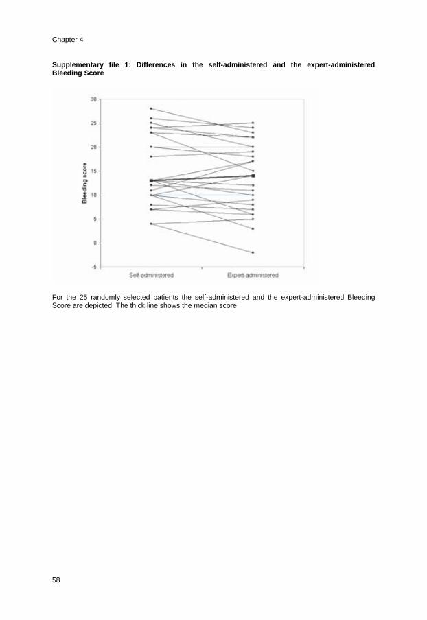
Chapter 4
58
Supplementary file 1: Differences in the self-administered and the expert-administered Bleeding Score For the 25 randomly selected patients the self-administered and the expert-administered Bleeding Score are depicted. The thick line shows the median score

Determinants of Bleeding Phenotype in Adult Patients with Moderate or Severe Von Willebrand Disease
59
References
1. Rodeghiero F, Castaman G, Dini E. Epidemiological investigation of the prevalence of von Willebrand's disease. Blood. Feb 1987;69(2):454-459.
2. Ruggeri ZM. Structure of von Willebrand factor and its function in platelet adhesion and thrombus formation. Best Pract Res Clin Haematol. Jun 2001;14(2):257-279.
3. James AH. More than menorrhagia: a review of the obstetric and gynaecological manifestations of von Willebrand disease. Thrombosis research. 2007;120 Suppl 1:S17-20.
4. Kadir RA, Chi C. Women and von Willebrand disease: controversies in diagnosis and management. Seminars in thrombosis and hemostasis. Sep 2006;32(6):605-615.
5. Kouides PA. Current understanding of von Willebrand's disease in women - some answers, more questions. Haemophilia. Jul 2006;12 Suppl 3:143-151.
6. Sadler JE, Budde U, Eikenboom JC, et al. Update on the pathophysiology and classification of von Willebrand disease: a report of the Subcommittee on von Willebrand Factor. J Thromb Haemost. Oct 2006;4(10):2103-2114.
7. Tosetto A, Rodeghiero F, Castaman G, et al. A quantitative analysis of bleeding symptoms in type 1 von Willebrand disease: results from a multicenter European study (MCMDM-1 VWD). J Thromb Haemost. Apr 2006;4(4):766-773.
8. Bowman M, Mundell G, Grabell J, et al. Generation and validation of the Condensed MCMDM-1VWD Bleeding Questionnaire for von Willebrand disease. J Thromb Haemost. 2008;6(12):2062-2066.
9. Gill JC, Christopherson PA, Flood VH, Friedman KD, Montgomery RR. The Zimmerman Program Investigators. Bleeding Scores in Von Willebrand Disease (VWD) Re-Visited: Analysis of the TS Zimmerman Program for the Molecular and Clinical Biology of VWD. ASH Annual Meeting Abstracts. 2008;112: 425 (abstract).
10. Federici AB, Mannucci PM, Castaman G, et al. Clinical and molecular predictors of thrombocytopenia and risk of bleeding in patients with von Willebrand disease type 2B: a cohort study of 67 patients. Blood. Jan 15 2009;113(3):526-534.
11. Nichols WL, Hultin MB, James AH, et al. von Willebrand disease (VWD): evidence-based diagnosis and management guidelines, the National Heart, Lung, and Blood Institute (NHLBI) Expert Panel report (USA). Haemophilia. 2008;14(2):171-232.
12. de Wee EM, Fijnvandraat K, de Goede-Bolder A, et al. Impact of von Willebrand disease on health related quality of life in a pediatric population. J Thromb Haemost. 2010.
13. de Wee EM, Mauser-Bunschoten EP, Van Der Bom JG, et al. Health-related quality of life among adult patients with moderate and severe von Willebrand disease. J Thromb Haemost. 2010;8(7):1492-1499.
14. Tosetto A, Castaman G, Rodeghiero F. Bleeding scores in inherited bleeding disorders: clinical or research tools? Haemophilia. May 2008;14(3):415-422.
15. Salem RO, Van Cott EM. A new automated screening assay for the diagnosis of von Willebrand disease. Am J Clin Pathol. May 2007;127(5):730-735.
16. Smith DR, Murphy D. Capillary blotting of agarose gels. Methods Mol Biol. 1996;58:23-25. 17. Budde U, Schneppenheim R, Eikenboom J, et al. Detailed von Willebrand factor multimer
analysis in patients with von Willebrand disease in the European study, molecular and clinical markers for the diagnosis and management of type 1 von Willebrand disease (MCMDM-1VWD). J Thromb Haemost. 2008;6(5):762-771.
18. Landsteiner K. On agglutination of normal human blood. Transfusion. 1961;1:5-8. 19. Astrup K, Olivarius Nde F, Moller S, Gottschau A, Karlslund W. Menstrual bleeding patterns in
pre- and perimenopausal women: a population-based prospective diary study. Acta Obstet Gynecol Scand. 2004;83(2):197-202.
20. Duckitt K. Managing perimenopausal menorrhagia. Maturitas. 2010;66(3):251-256. 21. Rodeghiero F, Castaman G, Tosetto A, et al. The discriminant power of bleeding history for
the diagnosis of type 1 von Willebrand disease: an international, multicenter study. J Thromb Haemost. 2005;3(12):2619-2626.
22. Tosetto A, Castaman G, Plug I, Rodeghiero F, Eikenboom J. Prospective evaluation of the clinical utility of quantitative bleeding severity assessment in patients referred for hemostatic evaluation. J Thromb Haemost. Jun 2011;9(6):1143-1148.
23. Rodeghiero F, Tosetto A, Abshire T, et al. ISTH/SSC bleeding assessment tool: a standardized questionnaire and a proposal for a new bleeding score for inherited bleeding disorders. J Thromb Haemost. Sep 2010;8(9):2063-2065.

Chapter 4
60
24. Mathew P, Greist A, Maahs JA, Lichtenberg EC, Shapiro AD. Type 2B vWD: the varied clinical manifestations in two kindreds. Haemophilia. 2003;9(1):137-144.
25. Casonato A, Sartorello F, Pontara E, et al. A novel von Willebrand factor mutation (I1372S) associated with type 2B-like von Willebrand disease: an elusive phenotype and a difficult diagnosis. Thrombosis and haemostasis. 2007;98(6):1182-1187.
26. Roland K, Rapson D, Lillicrap D, James P. The value of genetic testing for type 2B Von Willebrand disease. Clin Lab Haematol. 2006;28(1):17-21.
27. Mannucci PM, Franchini M, Castaman G, Federici AB. Evidence-based recommendations on the treatment of von Willebrand disease in Italy. Blood Transfus. Apr 2009;7(2):117-126.
28. Federici AB. Prophylaxis of bleeding episodes in patients with von Willebrand's disease. Blood Transfus. 2008;6 Suppl 2:s26-32.
29. Makris M. Gastrointestinal bleeding in von Willebrand disease. Thrombosis research. 2006;118 Suppl 1:S13-17.
30. Metjian AD, Wang C, Sood SL, et al. Bleeding symptoms and laboratory correlation in patients with severe von Willebrand disease. Haemophilia. Apr 6 2009.
31. Lak M, Peyvandi F, Mannucci PM. Clinical manifestations and complications of childbirth and replacement therapy in 385 Iranian patients with type 3 von Willebrand disease. British journal of haematology. Dec 2000;111(4):1236-1239.
32. Jenkins PV, O'Donnell JS. ABO blood group determines plasma von Willebrand factor levels: a biologic function after all? Transfusion. 2006;46(10):1836-1844.
33. Gallinaro L, Cattini MG, Sztukowska M, et al. A shorter von Willebrand factor survival in O blood group subjects explains how ABO determinants influence plasma von Willebrand factor. Blood. 2008;111(7):3540-3545.
34. Goodeve A, Eikenboom J, Castaman G, et al. Phenotype and genotype of a cohort of families historically diagnosed with type 1 von Willebrand disease in the European study, Molecular and Clinical Markers for the Diagnosis and Management of Type 1 von Willebrand Disease (MCMDM-1VWD). Blood. 2007;109(1):112-121.
35. James PD, Notley C, Hegadorn C, et al. The mutational spectrum of type 1 von Willebrand disease: Results from a Canadian cohort study. Blood. 2007;109(1):145-154.
36. Cumming A, Grundy P, Keeney S, et al. An investigation of the von Willebrand factor genotype in UK patients diagnosed to have type 1 von Willebrand disease. Thrombosis and haemostasis. 2006;96(5):630-641.
37. Collins PW, Cumming AM, Goodeve AC, Lillicrap D. Type 1 von Willebrand disease: application of emerging data to clinical practice. Haemophilia. 2008;14(4):685-696.
38. Klarmann D, Eggert C, Geisen C, et al. Association of ABO(H) and I blood group system development with von Willebrand factor and Factor VIII plasma levels in children and adolescents. Transfusion. 2010;50(7):1571-1580.
39. Favaloro EJ, Soltani S, McDonald J, Grezchnik E, Easton L, Favaloro JW. Reassessment of ABO blood group, sex, and age on laboratory parameters used to diagnose von Willebrand disorder: potential influence on the diagnosis vs the potential association with risk of thrombosis. Am J Clin Pathol. Dec 2005;124(6):910-917.
40. Boren MT, Ramey J. Thinking aloud : Reconciling theory and practice. IEEE transactions on professional communication 2000;43(3):261-278.


Chapter 5
Effect of fibrinolysis on bleeding phenotype in
moderate and severe
Von Willebrand Disease
Eva M. de Wee
Kevin Klaij
Jeroen C.J. Eikenboom
Johanna G. van der Bom
Manon E.L. Degenaar-Dujardin
Karin Fijnvandraat
Arja de Goede-Bolder
Britta A.P. Laros-van Gorkom
Eveline P. Mauser-Bunschoten
Karina Meijer
Gerard Goverde
Peter-Willem G. van der Linden
Dingeman C. Rijken
Frank W.G. Leebeek
for the WiN study group
Haemophilia, accepted

Chapter 5
64
Abstract Background Patients with Von Willebrand Disease (VWD), the most common inherited bleeding disorder, display large variation in bleeding tendency, which is not completely related to VWF levels. The cause of variability in clinical expression is largely unknown. The effect of plasma fibrinolytic capacity on bleeding tendency in VWD patients has not been investigated. Objectives We hypothesized that enhanced fibrinolysis may result in a more severe bleeding phenotype. Therefore, we measured the fibrinolytic potential in patients with moderate or severe VWD to investigate the contribution of fibrinolysis to the bleeding tendency. Patients and methods Fibrinolytic potential was measured as plasma clot lysis time (CLT) with and without addition of potato carboxypeptidase inhibitor (PCI) in 638 patients with moderate or severe VWD who participated in a nationwide multicenter cross-sectional study. Bleeding severity was measured using the Bleeding Score (BS). Results CLTs were significantly longer, indicative of hypofibrinolysis, in men compared to women with VWD (106.2 (IQR 95.7-118.1) vs. 101.9 (IQR 92.8-114.0) minutes). CLTs prolonged with increasing age. No association was found between VWF or FVIII levels and CLT, nor between VWF or FVIII levels and CLT+PCI. No association was observed for BS in a model with 10log-transformed CLT, adjusted for age, sex, VWF:Act and FVIII (b= 6.5 (95%CI -0.3 - 13.4)). Conclusion Our study showed that the plasma fibrinolytic potential does not influence bleeding tendency in VWD patients and therefore does not explain the variability in bleeding phenotype in VWD.

Effect of fibrinolysis on bleeding phenotype in moderate and severe Von Willebrand Disease
65
Introduction Von Willebrand Disease (VWD) is the most common inherited bleeding disorder, caused by defects in or reduced levels of Von Willebrand Factor (VWF)1. VWF plays a major role in primary hemostasis by facilitating adhesion of platelets to the endothelium, thereby initiating aggregation of platelets to form a platelet plug. In addition, VWF is the carrier protein of factor VIII (FVIII)2.
In patients with VWD, VWF and FVIII levels largely determine the bleeding tendency, however the variation in bleeding tendency between individuals with VWD is not completely related to VWF levels3. Some patients bleed excessively, whereas others with similar VWF levels in plasma have only mild bleeding problems. Also within families with similar mutations, large differences in bleeding phenotype are observed3. The cause of this variability in clinical expression of VWD is largely unknown. It has recently been shown that variability in thrombin generation may lead to differences in bleeding phenotype in VWD4.
Another factor that may determine the variability in clinical expression of VWD is the rate of fibrinolysis. The effect of fibrinolysis on the bleeding tendency in VWD patients has not yet been investigated. The fibrinolytic system converts plasminogen into plasmin, which degrades the insoluble fibrin clot into soluble fibrin degradation products. Thrombin activatable fibrinolysis inhibitor (TAFI) connects the coagulation cascade with the fibrinolytic system. Upon high concentrations of thrombin, TAFI is activated by thrombin and effectively inhibits fibrinolysis by removing carboxyterminal lysine residues from partially degraded fibrin, thereby diminishing the cofactor function of fibrin in plasminogen activation5.
Recent studies have indicated that the fibrinolytic potential, measured by a clot lysis assay, show considerable inter-individual variation6. This variability may also influence the bleeding phenotype of VWD individuals. In addition, in severe VWD patients the fibrinolytic potential may be altered by strongly reduced FVIII levels, leading to impaired thrombin generation via the intrinsic feedback-loop and less TAFI activation. Therefore, reduced FVIII levels in VWD not only have an effect on coagulation but may also enhance fibrinolysis. Previous studies have already indicated that TAFI levels and activation correlate with the fibrinolytic potential in healthy individuals7-8. Patients with enhanced fibrinolysis predominantly present with mucocutaneous bleeding9-11, such as menorrhagia, epistaxis and gum bleeding. These bleeding symptoms are also frequently observed in patients with VWD12.
We hypothesised that differences in fibrinolytic capacity may influence the bleeding tendency among VWD patients, i.e. enhanced fibrinolysis may result in a more severe bleeding phenotype. Therefore, we measured the fibrinolytic potential in a large cohort of patients with moderate or severe VWD to investigate the role of fibrinolysis in bleeding phenotype of VWD patients.
Methods Willebrand in Netherlands study We performed a nation-wide cross-sectional study among patients with moderate and severe VWD in the Netherlands, the “Willebrand in the Netherlands” (WiN) study. Patients were recruited at all 13 Haemophilia Treatment Centres in the Netherlands.
We included patients diagnosed with type 1, type 2 and type 3 VWD who fulfilled both of the following inclusion criteria: 1) hemorrhagic symptoms or a family history of von Willebrand disease; 2) historically lowest levels of VWF antigen (VWF:Ag) ≤ 30 U/dL and/or VWF activity (VWF ristocetin cofactor activity (VWF:RCo)

Chapter 5
66
≤ 30 U/dL and/or factor (F)VIII:C ≤ 40 U/dL, determined at the local Hemophilia Treatment Center. Patients were excluded if other disorders of hemostasis resulting in a hemorrhagic diathesis were known. Patients were excluded if other congenital disorders of haemostasis resulting in a hemorrhagic diathesis were present.
For the current analysis we included all patients, both children and adults. Data were obtained between October 2007 and October 2009. All participants, or their parents, completed a questionnaire and a blood sample was obtained. The Medical Ethical Committee decided that in children blood could only be obtained if for clinical purposes a blood sample was needed. The Medical Ethical Committees at all participating centres approved this study, and written informed consent was obtained from all study participants and/or their parents. Definitions Determination of type of VWD was based on the current ISTH guidelines13-14, using the laboratory parameters determined at the local Hemophilia Treatment Center. The severity of VWD was defined as proposed by Federici15. Severe VWD was defined as the presence of at least one of the following laboratory abnormalities: VWF:Ag <10 U/dL, and/or VWF:CB <10 U/dL, and/or VWF:RCo <10 U/dL, and/or FVIII <20 U/dL. Moderate VWD was defined as VWF:Ag 10-30 U/dL, and/or VWF:CB 10-30 U/dL, and/or VWF:RCo 10-30 U/dL, and/or FVIII 20-40 U/dL15. Children were defined as study participants aged 0-16 years. Laboratory measurements in VWD Historic measured VWF and FVIII levels in the Hemophilia Treatment Centers were used as inclusion criteria for the WiN study, and patients with at least one measurement of VWF below 30 U/dl or FVIII below 40 U/dL (for type 2N), respectively were included. All participants were asked to give blood for re-evaluation of von Willebrand parameters.
Peripheral venous blood was collected in tubes containing 3.2% (0.109 M) sodium citrate. Subsequently, the tubes were centrifuged twice at 2200 x g for 10 minutes at room temperature and finally the citrated platelet-poor plasma (PPP) was aliquoted and stored at -80°C.
Plasma levels of VWF:Ag, VWF Collagen Binding (VWF:CB), VWF activity (VWF:Act) and FVIII were measured centrally (Erasmus University Medical Center, Rotterdam, Netherlands). Briefly, VWF:Ag level was measured with an in-house ELISA using a polyclonal rabbit anti-human VWF antibody (DakoCytomation, Glostrup, Denmark) for capturing and HRP-conjugated anti-human VWF antibody (DakoCytomation, Glostrup, Denmark) for detecting. VWF:CB level was measured with an in-house ELISA using collagen type 1 for capturing and HRP-conjugated anti-human VWF antibody for detecting. The VWF:Act assay uses latex particles coated with a monoclonal murine antibody direct against the GpIbα binding domain of VWF. These latex particles were incubated with the patient plasma (Instrumentation Laboratory B.V, Breda, The Netherlands) and agglutination of the particles, proportionally to the GpIbα binding activity of VWF, was measured (Sysmex CA-1500, TOA Medical Electronics). FVIII:C was measured in a one-stage clotting assay (TriniCLOT, Biomerieux, Marcy l'Etoile, France) with FVIII-deficient plasma (Biopool, Umea, Sweden).

Effect of fibrinolysis on bleeding phenotype in moderate and severe Von Willebrand Disease
67
Figure 1: A representative graph of the output generated by the plasma fibrinolytic potential assay Clot-lysis time (CLT) was calculated as the difference between clotting and lysis time Plasma fibrinolytic potential The fibrinolytic potential of VWD patients was studied using a plasma fibrinolytic potential assay7, 16-17. Lysis of a tissue factor-induced clot by exogeneous tissue-type plasminogen activator (t-PA) was studied by monitoring changes in turbidity during clot formation and subsequent lysis as described previously7. Briefly, 70 µL of citrated PPP was added to 96 wells plate (Falcon) followed by 49 µL of assay buffer (25 mM HEPES; 137 mM NaCl; 3.5 mM KCl; 1% BSA; pH 7.4). After mixing, 85 µL of the diluted plasma was added to another 96 wells plate containing 15 µL assay mixture. The assay mixture included: assay buffer, tissue factor (Innovin, Dade Behring, Marburg, Germany), CaCl2, tPA (Actilyse, Boehringer Ingelheim, Ingelheim am Rhein, Germany), phospholipids (Rossix, Mölndal, Sweden). Plasma samples were measured both in absence and presence of potato carboxypeptidase inhibitor (PCI), a potent inhibitor of activated TAFI18. The inhibitory effect of TAFI on the fibrinolytic potential is excluded by adding PCI. The final concentrations in the plasma clot were 1000x diluted tissue factor (Innovin), 17mM CaCl2, 30 ng/mL tPA, 10 µM phospholipids and 2x diluted plasma. In the measurements with PCI, the final concentration of PCI in the plasma clot was 30 µg/mL. Finally, after mixing the diluted plasma with the assay mixture (1400 rpm on a plate shaker for 10 seconds), 50 µL paraffin oil (Merck, Darmstadt, Germany) was added to the wells. The plate was then immediately inserted into a 37° C preheated microplat e reader (Biotek, Winooski, Vermont, USA). Optical density was measured at 405 nm every minute for 300 minutes. The time of the midpoint from lowest to highest optical density was used as a measure for clotting time, while the time of the midpoint from highest to lowest optical density was used as a measure for lysis time. The clot-lysis time (CLT) was calculated as the difference between clotting and lysis time. A representative graph of the fibrinolytic potential assay (optical density plotted against time) is shown in figure 1. The intra- and inter-assay variation coefficients were 3.5% and 6.5%, respectively.
In patients with type 3 VWD, low FVIII levels due to an increased FVIII clearance, will theoretically lead to less thrombin generation and subsequently to less TAFI activation and more enhancement of fibrinolysis (shortened CLT). Clot lysis time ratio (CLT ratio) was defined as CLT divided by CLT+PCI, thus CLT with TAFI

Chapter 5
68
activity divided by CLT without TAFI activity. CLT ratio was measured to study the effect of TAFI per type of VWD.
Bleeding severity Bleeding severity was assessed by using the Tosetto Bleeding Score (BS)3, 19. The BS systematically evaluates bleeding symptoms, and accounts for both the number and severity of the bleeding symptoms. The 12 bleeding items are scored on a scale ranging from -1 to 4 points. Higher scores reflect more severe/frequent bleeding. The total for all 12 items results in a Bleeding Score that can range from -3 (no bleeding) to 45 (severe bleeding).
For additional analysis, we also used a mucocutaneous bleeding score (BS-mucocutaneous), which accounts for 6 mucocutaneous bleeding symptoms (epistaxis, bleeding from minor wounds, oral cavity bleeding, gastrointestinal bleeding, bleeding after tooth extraction and menorrhagia). Statistical analysis Data were analyzed with two objectives. First, determinants of CLT were assessed. CLT presented a skewed distribution to the right. Due to non-normal distribution, the Mann-Whitney U and Kruskal-Wallis tests were used to compare CLT between groups. We used linear regression to model the associations between CLT and its potential determinants. Because the distribution of CLT is skewed, CLT was 10 log-transformed. Second, we investigated the association between CLT and BS. Three linear regression models were built with BS or BS-mucocutaneous as dependent variable and 10 log-CLT, age, sex, FVIII, and VWF:Act as the predictor variables. A p-value of ≤ 0.05 was considered statistically significant. Figure 2: Flow chart of the WiN study population
Results Population WiN study In the Willebrand in the Netherlands (WiN) study, 806 patients with moderate to severe VWD were included. Plasma was available for 687 VWD patients. Eighteen

Effect of fibrinolysis on bleeding phenotype in moderate and severe Von Willebrand Disease
69
total n=638
sex females (n,%) 389 61%
age median, IQR 45 30 to 58
VWD type 1 (n,%) 372 58%
2 (n,%) 222 35%
2A 92
2B 57
2M 30
2N 15
2 not specified 28
3 (n,%) 28 4%
unspecified (n,%) 16 3%
VWF:Ag median U/dL, IQR 29 18 to 44
VWF:CB median U/dL, IQR 22 7 to 49
VWF:ACT median U/dL, IQR 22 8 to 52
FVIII:C median U/dL, IQR 50 32 to 72
Blood group O n,% 382 60%
VWD severity severe VWD (n,%) 253 40%
moderate VWD (n,%) 385 60%
Bleeding Score median, range 10 -1 to 35
CLT median (minutes), IQR 102.5 92.3 to 114.0
CLT + PCI median (minutes), IQR 72.1 65.4 to 80.9
IQR: interquartile range
PCI: potato carboxypeptidase inhibitor
VWD patients were excluded because of either pregnancy or treatment with desmopressin or FVIII/VWF concentrate three days before blood samples were drawn. In 31 VWD patients no CLT could be determined as the plasma clot did not reach the midpoint from maximum turbidity to clear transition within 300 minutes. These patients were excluded from subsequent analyses. A flow chart of the study population is shown in figure 2.
Table 1 shows the characteristics of the WiN study population. In total, for 638 patients both data on bleeding phenotype and CLT were available; of these 61% were female. The majority of patients were diagnosed as VWD type 1 (n=372, 58%), type 2 was diagnosed in 35% (n=222), type 3 was diagnosed in 4% (n=28) of the included patients, and 3% (n=16) of the patients remained non-classified. VWF:Ag, VWF:CB, VWF:Act and FVIII levels were strongly reduced in our cohort with VWD patients, see table 1. Median CLT of the study population was 102.5 minutes (inter-quartile range (IQR) 92.3-114.0 minutes), median CLT+PCI was 72.1 minutes (IQR 65.4-80.9 minutes). Table 1: Characteristics of the WiN study population
An association between VWF or FVIII level and Bleeding Score (BS) was found. The median BS was 11 (95% CI 10-11) in (n= 564) patients with VWF:Ag levels >10 IU/dL and 15 (13-17) in (n= 74) patients with VWF:Ag level ≤10 IU/dL. Patients with VWF:Act levels >10 IU/dL (n= 473) or FVIII:C levels >10 IU/dL (n= 605) had a median BS of 10 (10-11) and 11 (10-12) respectively. A median BS of 14 (13-15) and 18 (15-21) was observed in patients with VWF:Act levels ≤10 IU/dL (n= 186) or FVIII:C levels ≤10 IU/dL (n= 33).

Chapter 5
70
levels* CLT (IQR) p for trend† CLT+PCI (IQR) p for trend†
VWF:Ag 0-10 100.3 (90.4-112.5) 0.122 72.5 (65.6-79.1) 0.222
10-20 99.5 (90.8-109.7) 71.0 (63.3-78.7)20-30 101.5 (91.0-114.2) 71.0 (64.6-79.8)>30 103.8 (94.7-117.6) 73.3 (66.3-82.8)
VWF:CB 0-10 102.4 (91.3-111.6) 0.316 72.2 (65.0-80.9) 0.822
10-20 103.9 (93.5-111.2) 72.4 (67.2-79.7)20-30 96.6 (89.4-112.6) 71.0 (64.4-78.5)>30 103.0 (93.6-117.7) 72.1 (65.2-82.2)
VWF:Act 0-10 100.4 (90.3-110.5) 0.121 71.0 (64.9-78.5) 0.421
10-20 103.2 (91.8-110.6) 72.5 (66.7-80.0)
20-30 103.5 (92.9-115.5) 72.0 (64.5-81.8)
>30 103.3 (93.4-117.9) 72.6 (65.9-82.8)
FVIII:C 0-10 101.9 (90.2-112.2) 0.097 74.6 (67.9-78.9) 0.132
10-20 102.5 (90.3-111.3) 71.7 (65.4-78.4)
20-30 98.5 (90.9-107.4) 68.7 (64.9-78.7)
>30 103.4 (92.9-115.0) 72.2 (65.3-82.1)
* in IU/dL
†Clot lysis time and clot lysis time+PCI were 10 log-transformed
N median CLT IQR *p-value median CLT+PCI
IQR *p-value
sex children boys 35 90.8 85.3 - 98.0 0.600 66.4 62.2 - 74.4 0.771
girls 26 90.4 84.8 - 103.0 68.4 60.4 - 74.1
adults males 214 106.2 95.7 - 118.1 0.008 71.7 65.7 - 81.5 0.379
females 363 101.9 92.8 - 114.0 72.8 66.6 - 81.8
age 0-16 61 90.6 85.3 - 100.5 <0.001 67.5 61.0 - 74.1 <0.001
16-40 200 99.4 90.9 - 108.5 70.0 63.6 - 78.1
41-60 244 105.1 93.1 - 122.4 73.3 65.4 - 83.6
61-86 133 107.0 99.0 - 117.3 75.4 68.4 - 83.7
type VWD type 1 372 101.0 91.5 - 112.6 0.259 70.4 63.9 - 78.9 <0.001
type 2 221 104.2 94.2 - 114.5 74.5 68.3 - 82.0
type 3 28 103.2 89.6 - 112.0 76.2 69.5 - 87.4
blood group O 382 101.2 91.9 - 111.8 0.085 71.5 65.0 - 80.3 0.100
non-O 247 104.0 93.0 - 115.0 73.4 65.9 - 81.6
IQR: interquartile range
* Mann Whitney U or Kruskal-Wallis test for differences between subgroups
Association between VWF and FVIII levels and CLT No associations were found between VWF or FVIII level and CLT, see table 2. Table 2: Association between CLT and VWF/FVIII levels Patients in a subgroup with higher FVIII level (>30 IU/dL) had slightly higher CLTs, although this difference was not statistically significant, p for trend 0.097. No associations were found between VWF or FVIII level and CLT+PCI, measuring CLT in the absence of TAFI activity, see table 3.
Table 3: Clot lysis time in subgroups of VWD patients

Effect of fibrinolysis on bleeding phenotype in moderate and severe Von Willebrand Disease
71
Determinants of CLT in VWD patients CLTs were prolonged in men compared to women with VWD (106.2 (IQR 95.7-118.1) vs. 101.9 (IQR 92.8-114.0) minutes, p= 0.008), see table 2. In children no sex difference was found (90.8 (IQR 85.3-98.0) vs. 90.4 (IQR 84.8-103.0) minutes, p= 0.600), for boys and girls respectively, see figure 3. CLT steadily prolonged with age, an increase in age of 10 years was associated with an increase in CLT of 3.3 minutes (95% CI 2.3 to 4.4). CLTs of the children were significantly shorter compared to adult VWD patients (90.6 (IQR 85.3-100.5) vs. 103.7 (IQR 93.2-115.0) minutes, p <0.001). No difference in CLT was found between patients with type 1, 2 and 3 VWD. However, a significant difference in CLT+PCI (to exclude the effect of TAFI) between the various types of VWD was observed, see table 2. Patients with type 3 VWD had prolonged CLTs compared to VWD type 1 and type 2. Within the group of patients with type 2 VWD, patients with subtype 2A had the highest CLT (data not shown). Figure 3: Clot lysis time per gender and age-group Children had significantly lower CLTs compared to all other age groups (Mann-Whitney test p<0.001). Only in patients aged 41-60 years a significantly different CLT was found between males and females (Mann-Whitney test p=0.006). Both for males and females CLTs increased significantly with age (Kruskal-Wallis test p<0.001). Error bars: 95% CI Association between CLT measured in presence and absence of PCI Correlation between CLT and CLT+PCI was highly significant (r= 0.857, p <0.001), indicating that VWD patients with a high overall CLT also had a high basal, TAFI-independent CLT. To study the contribution of TAFI and TAFI activation on lysis time we measured CLT ratios. In figure 4 CLT ratios per type of VWD are depicted. A significant higher CLT ratio was observed in type 1 compared to type 2 and type 3 VWD patients (median CLT ratio in type 1 VWD 1.45 (IQR 1.37-1.52), type 2 VWD 1.39 (IQR 1.32-1.47), type 3 VWD 1.36 (1.27-1.43), p <0.001).

Chapter 5
72
BS BS mucocutaneous
n=638 b (95% CI) b (95% CI)
CLT
Model I 9.6 (2.8 - 16.4) 7.9 (3.3 - 12.4)
Model II 5.8 (-1.2 - 12.8) 5.4 (0.7 - 10.1)
Model III 6.5 (-0.3 - 13.4) 5.3 (0.7 - 10.0)
Model I: independent variable 10log-CLTs
Model II: independent variables 10log-CLTs, age and sex
Model III: independent variables 10log-CLTs, age, sex, VWF:Act and FVIII
b represents the increase in BS or BSmucocutaneous per minute increase of 10log-CLT
Figure 4: Clot lysis time ratios in different types of VWD CLT ratio is defined as CLT (with TAFI activity) divided by CLT+PCI (without TAFI activity). * p <0.05. Bars represent the median CLT:CLT+PCI ratio Effect of fibrinolysis on bleeding score For both BS and BS-mucocutaneous three models were built in order to adjust for other factors that could potentially affect the association between BS and CLT, including age, sex, VWF:Act and FVIII. The results are summarized in table 4. An association was found between both BS and BS-mucocutaneous with 10log-transformed CLT (b= 9.6 (95% CI 2.8 - 16.4), and b= 7.9 (95% CI 3.3 – 12.4) respectively). However, for BS and CLT no significant association was found after adjustment for age, sex, VWF:Act and FVIII (b= 6.5, 95%CI -0.3 - 13.4), for BS-mucocutaneous a small regression coefficient was found (b= 5.3, 95% CI 0.72 - 10.0). After additional stratification for type of VWD this association disappeared (data not shown). Table 4: Linear regression models representing the effect of clot lysis time on the Bleeding Score
Effect of fibrinolysis on bleeding phenotype in moderate and severe Von Willebrand Disease
73
Discussion We did not observe an association between the fibrinolytic potential, measured by a plasma-based clot lysis assay, and bleeding tendency in our cohort of moderate to severe VWD patients. Therefore our hypothesis that a higher fibrinolytic potential may increase the bleeding tendency in VWD patients could not be confirmed in this study.
In patients with VWD, a large variation in fibrinolytic capacity was observed, which was irrespective of FVIII and VWF level. The fibrinolysis assay we used is an overall measure of the plasma fibrinolytic potential in which the total balance of plasma proteins involved in clot degradation determines the outcome, and a single factor is only of limited importance7. The large variability in CLT was also observed earlier in healthy individuals6, 16, 20.
We hypothesized that enhanced fibrinolysis might result in a more severe bleeding tendency, leading to an inverse relationship between CLT and bleeding phenotype, assessed by the Tosetto Bleeding Score. We studied mucocutaneous bleeding separately, because of the high fibrinolytic activity in mucosal tissue and the high frequency of mucosal bleeding in patients with fibrinolysis abnormalities21. We found a weak, positive association between CLT and BS, however in adjusted models this association disappeared. This is in line with a previous study in hemophilia patients, in whom an association between clot lysis and bleeding phenotype could not be demonstrated22.
An explanation for the fact that no association between BS and CLT was found, might be that our cohort was strictly defined and only patients with moderate or severe VWD were included who have a relatively high bleeding score. The selection of the study-group, not including mild VWD patients may have masked the effect of fibrinolysis on bleeding tendency. Mild VWD patients have a larger variability in bleeding phenotype and may be better suited to study the effect of fibrinolysis on bleeding variability. Furthermore, we studied bleeding phenotype by determining the BS in our study3. We used the BS because it is a validated method to determine bleeding phenotype in VWD patients. The BS was originally developed by Tosetto et.al. to distinguish between type 1 VWD patients and patients without VWD. This bleeding score has not been used to quantify bleeding symptoms in patients with disorders of fibrinolysis.
CLT was also measured in presence of PCI, an inhibitor of activated TAFI, to study specifically the role of TAFI. We hypothesized that very low FVIII levels in severe VWD patients might influence the fibrinolytic system because of less TAFI activation, which may in turn lead to differences in bleeding phenotype. The fibrinolytic potential measured in the presence of TAFI inhibitor (CLT+PCI) was indeed increased (shortened CLT+PCI). The anticipated effect of low FVIII activity and concomitant reduced TAFI activation on CLT is not reflected in the clot lysis assay in our study (table 3). Previous studies showed that FVIII levels were not associated with CLTs in healthy individuals7. Furthermore, it was shown that addition of only 0.01 U/dL FVIII in severe hemophilia A patients, who are completely lacking FVIII, already resulted in maximal prolongation of CLT, suggesting the effect of FVIII at fibrinolysis is maximal at low concentration23.
Although we did not find an association between the global plasma fibrinolysis and bleeding tendency, future studies should investigate whether the variation in bleeding tendency in VWD might be partly related to individual components of the fibrinolytic system. In the global fibrinolysis assay that we used in this study, exogenous tPA was added, therefore effects of tPA or other fibrinolysis protein

Chapter 5
74
variations might be masked. Other global fibrinolysis assays or measurement of individual fibrinolysis proteins including α2-antiplasmin, PAI-1 or TAFI levels may still be relevant in the understanding the variation in bleeding tendency in VWD. In conclusion, our study showed that the plasma fibrinolytic potential does not influence bleeding tendency in VWD patients and therefore does not explain the variability in bleeding phenotype in VWD.

Effect of fibrinolysis on bleeding phenotype in moderate and severe Von Willebrand Disease
75
References 1. Sadler JE, Mannucci PM, Berntorp E, et al. Impact, diagnosis and treatment of von Willebrand
disease. Thrombosis and haemostasis. 2000;84(2):160-174. 2. Ruggeri ZM. Structure of von Willebrand factor and its function in platelet adhesion and
thrombus formation. Best Pract Res Clin Haematol. Jun 2001;14(2):257-279. 3. Tosetto A, Rodeghiero F, Castaman G, et al. A quantitative analysis of bleeding symptoms in
type 1 von Willebrand disease: results from a multicenter European study (MCMDM-1 VWD). J Thromb Haemost. Apr 2006;4(4):766-773.
4. Rugeri L, Beguin S, Hemker C, et al. Thrombin-generating capacity in patients with von Willebrand's disease. Haematologica. Dec 2007;92(12):1639-1646.
5. Rijken DC, Lijnen HR. New insights into the molecular mechanisms of the fibrinolytic system. J Thromb Haemost. Jan 2009;7(1):4-13.
6. Mosnier LO, von dem Borne PA, Meijers JC, Bouma BN. Plasma TAFI levels influence the clot lysis time in healthy individuals in the presence of an intact intrinsic pathway of coagulation. Thrombosis and haemostasis. Nov 1998;80(5):829-835.
7. Lisman T, de Groot PG, Meijers JC, Rosendaal FR. Reduced plasma fibrinolytic potential is a risk factor for venous thrombosis. Blood. Feb 1 2005;105(3):1102-1105.
8. Meltzer ME, Lisman T, de Groot PG, et al. Venous thrombosis risk associated with plasma hypofibrinolysis is explained by elevated plasma levels of TAFI and PAI-1. Blood. Jul 8 2010;116(1):113-121.
9. Carpenter SL, Mathew P. Alpha2-antiplasmin and its deficiency: fibrinolysis out of balance. Haemophilia. Nov 2008;14(6):1250-1254.
10. Mehta R, Shapiro AD. Plasminogen activator inhibitor type 1 deficiency. Haemophilia. Nov 2008;14(6):1255-1260.
11. Leebeek FW, Stibbe J, Knot EA, Kluft C, Gomes MJ, Beudeker M. Mild haemostatic problems associated with congenital heterozygous alpha 2-antiplasmin deficiency. Thrombosis and haemostasis. Feb 25 1988;59(1):96-100.
12. Silwer J. von Willebrand's disease in Sweden. Acta paediatrica Scandinavica. 1973;238:1-159.
13. Sadler JE, Budde U, Eikenboom JC, et al. Update on the pathophysiology and classification of von Willebrand disease: a report of the Subcommittee on von Willebrand Factor. J Thromb Haemost. Oct 2006;4(10):2103-2114.
14. Nichols WL, Hultin MB, James AH, et al. von Willebrand disease (VWD): evidence-based diagnosis and management guidelines, the National Heart, Lung, and Blood Institute (NHLBI) Expert Panel report (USA). Haemophilia. 2008;14(2):171-232.
15. Federici AB. Clinical diagnosis of von Willebrand disease. Haemophilia. Oct 2004;10 Suppl 4:169-176.
16. Guimaraes AH, de Bruijne EL, Lisman T, et al. Hypofibrinolysis is a risk factor for arterial thrombosis at young age. Br J Haematol. Apr 2009;145(1):115-120.
17. Hoekstra J, Guimaraes AH, Leebeek FW, et al. Impaired fibrinolysis as a risk factor for Budd-Chiari syndrome. Blood. Jan 14 2010;115(2):388-395.
18. Leebeek FW, Goor MP, Guimaraes AH, et al. High functional levels of thrombin-activatable fibrinolysis inhibitor are associated with an increased risk of first ischemic stroke. J Thromb Haemost. Oct 2005;3(10):2211-2218.
19. Bowman M, Riddel J, Rand ML, Tosetto A, Silva M, James PD. Evaluation of the diagnostic utility for von Willebrand disease of a pediatric bleeding questionnaire. J Thromb Haemost. Aug 2009;7(8):1418-1421.
20. Hoekstra J, Guimaraes AH, Leebeek FW, et al. Impaired fibrinolysis as a risk factor for Budd-Chiari syndrome. Blood. Jan 14 2010;115(2):388-395.
21. Agren A, Wiman B, Stiller V, et al. Evaluation of low PAI-1 activity as a risk factor for hemorrhagic diathesis. J Thromb Haemost. Jan 2006;4(1):201-208.
22. van Dijk K, van der Bom JG, Fischer K, de Groot PG, van den Berg HM. Phenotype of severe hemophilia A and plasma levels of risk factors for thrombosis. J Thromb Haemost. May 2007;5(5):1062-1064.
23. Mosnier LO, Lisman T, van den Berg HM, Nieuwenhuis HK, Meijers JC, Bouma BN. The defective down regulation of fibrinolysis in haemophilia A can be restored by increasing the TAFI plasma concentration. Thrombosis and haemostasis. Oct 2001;86(4):1035-1039.

Chapter 6
Gynaecological and obstetric bleeding in
moderate and severe
Von Willebrand Disease
Eva M. de Wee
H. Marieke Knol
Eveline P. Mauser-Bunschoten
Johanna G. van der Bom
Jeroen C.J. Eikenboom
Karin Fijnvandraat
Arja de Goede-Bolder
Britta Laros-van Gorkom
Paula F. Ypma
Sonja Zweegman
Karina Meijer
Frank W.G. Leebeek
for the WiN study group
Thrombosis and Haemostasis, accepted

Chapter 6
78
Abstract A nation-wide cross-sectional study was initiated to assess gynaecological and obstetrical symptoms in an unselected cohort of women with moderate and severe VWD in the Netherlands. 423 women aged ≥16 years were included. Bleeding severity was measured using the Tosetto Bleeding Score (BS). Menorrhagia, defined as occurrence of ≥2 menorrhagia symptoms, was reported by 81%. Of all VWD women, 78% received any kind of treatment for menorrhagia and 20% underwent a hysterectomy predominantly because of severe menstrual bleeding. Over half of the women reported more blood loss than can be expected with a normal delivery. In 52% of reported pregnancy losses curettage was needed because of bleeding. Mean number of live births was 1.9, which is comparable with the general Dutch population. In conclusion, women with moderate or severe VWD frequently have menorrhagia in need of treatment and 20% of the VWD women underwent a hysterectomy. Bleeding complications occurred in over 50% of the women after childbirth or pregnancy loss. Progeny seems not to be affected in women with moderate or severe VWD.

Gynaecological and obstetric bleeding in moderate and severe Von Willebrand Disease
79
Introduction Von Willebrand Disease (VWD) is caused by defects in or reduced levels of Von Willebrand Factor (VWF). It is the most common inherited bleeding disorder and affects 0.5-1% of the population, although not all patients with low VWF levels have clinically relevant bleeding episodes1-2.
Patients with VWD frequently have bleeding episodes, varying from gum bleeds and epistaxis to intestinal bleeding. In theory, men and women are equally likely to be affected, but in women VWD is more often clinically manifest because of the bleeding challenges that are associated with menstruation and childbirth 1. Tosetto et al. have developed a bleeding score (BS) to quantify the number and severity of bleeding symptoms2.Two of the 12 items of the BS include menorrhagia and postpartum haemorrhage. We used the BS, a validated and commonly used instrument, to determine the severity of menorrhagia and postpartum haemorrhage.
The majority of published studies investigating the prevalence of gynaecological symptoms in women with VWD are case series of a relatively small number of women3-7. In addition these women had predominantly type 1 or mild VWD. In these studies women with VWD frequently have menorrhagia with reported prevalence ranging from 74-92%, which may impair quality of life (QoL)4-5, 8. Also increased absence from school or work during menstruation is reported (5-6, 10-13). One study reported that women with VWD more often underwent hysterectomy than women without VWD9. The above mentioned studies may suffer from selection bias given the fact that patients seeking medical attention for bleeding and menorrhagia have predominantly been included.
Therefore the aim of our study was to assess gynaecological and obstetrical symptoms in a large unselected cohort of women with moderate or severe VWD who participated in a nation-wide study. Methods Participants We performed a nation-wide cross-sectional study among patients with moderate and severe VWD in the Netherlands, the “Willebrand in the Netherlands” (WiN) study. Data on gynaecological and obstetric bleeding were obtained retrospectively. Patients were recruited at all 13 Hemophilia Treatment Centers (HTCs) in the Netherlands. We included patients diagnosed with type 1, type 2 and type 3 VWD who fulfilled both of the following inclusion criteria: 1) hemorrhagic symptoms or a family history of von Willebrand disease; 2) historic levels of VWF antigen (VWF:Ag) ≤ 30 U/dL and/or VWF activity (VWF ristocetin cofactor activity (VWF:RCo) and/or VWF collagen binding assay (VWF:CB)) ≤ 30 U/dL and/or FVIII:C ≤ 40 U/dL at least once. Classification of VWD into type 1, 2 and 3 was based on VWF parameters measured in laboratories of the various HTCs and according to classification guidelines10-11. Patients with mild VWD were excluded, as were patients with other congenital disorders of hemostasis resulting in a hemorrhagic diathesis.
For the present analyses we selected all women aged 16 years and older. Data were obtained between October 2007 and October 2009. The Medical Ethical Committees at all participating HTCs approved this study, and written informed consent was obtained from all study participants.

Chapter 6
80
A) Menorrhagia: ≥2 symptoms at the time of study or in the past
▪ subjective excessive menstrual bleeding
▪ loss of blood clots during menstrual bleeding
▪ requirement of oral iron therapy or blood transfusion
▪ heavy menstrual flow that interferes with daily life
▪ menstrual period that lasts longer than 7 days
B) Severity of bleeding symptoms*
Severity of menorrhagia (BSmenorrhagia)
Score 0: No menorrhagia
Score 1: Consultation only
Score 2: Antifibrinolytics or pill use
Score 3: Dilatation and curettage or iron therapy
Score 4: Blood transfusion, FVIII/VWF concentrate, desmopressin or hysterectomy
Severity of PPH (BS-PPH)*
Score -1: No bleeding in at least two deliveries
Score 0: No deliveries or no bleeding in one delivery
Score 1: Consultation only
Score 2: Dilatation and curettage, iron therapy, antifibrinolytics
Score 3: Blood transfusion or FVIII/VWF concentrate or desmopressin
Score 4: Hysterectomy
* BS= Bleeding Score, scores are derived from the Tosetto Bleeding Score
PPH= postpartum hemorrhage
Assessment methods All participants completed an extensive questionnaire, which contained questions on bleeding episodes, treatment of VWD, side effects of treatment, concomitant disease, and employment (17). The Bleeding Score was incorporated into this questionnaire.
The Bleeding Score was used as previously described for bleeding severity in type 1 VWD by Tosetto et al2. It systematically evaluates bleeding symptoms, and accounts for both the number and severity of the bleeding symptoms. The severity and frequency of 12 items are scored on a scale ranging from -1 to 4 points. Higher scores reflect more severe/frequent bleeding. The total for all 12 items results in a Bleeding Score (range 3 to 45). Definitions Severe VWD was defined as the presence of at least one of the following laboratory abnormalities: VWF:Ag ≤ 10 U/dL, and/or VWF:RCo ≤ 10 U/dL, and/or FVIII:C ≤ 20 U/dL. Moderate VWD was defined as VWF:Ag 10-30 U/dL, and/or VWF:RCo 10-30 U/dL, and/or FVIII:C 20-40 U/dL12.
Menorrhagia was defined as the occurrence of at least two of the symptoms listed in table 1A13-15. Severity of menorrhagia was determined according to the menorrhagia items of the Tosetto Bleeding Score (BSmenorrhagia)2. The score for this item ranges from 0 to 4 (table 1B). Table 1: A) Definition of menorrhagia and B) classification of severity of menorrhagia and postpartum haemorrhage, according to the Tosetto Bleeding Score

Gynaecological and obstetric bleeding in moderate and severe Von Willebrand Disease
81
Severity of bleeding complications following childbirth was determined according to the postpartum haemorrhage (PPH) item of the Tosetto Bleeding Score (BS-PPH)2. The score for this item ranges from -1 to 4 (table 1B).
Foetal loss was defined as spontaneous miscarriages, foetal death and intrauterine death.
Laboratory measurements of VWD Historic measured VWF and FVIII levels in the Hemophilia Treatment Centers were used as inclusion criteria for the WiN study, and patients with at least one measurement of VWF or FVIII below 30 U/dL or 40 U/dL respectively were included.
Peripheral venous blood was collected at inclusion of the study. Plasma levels of VWF antigen (VWF:Ag), VWF Collagen Binding (VWF:CB), VWF activity (VWF:Act) and FVIII activity (FVIII:C) were measured centrally in the Erasmus university Medical Center, Rotterdam, The Netherlands. VWF:Ag level was measured with an in-house ELISA using a polyclonal rabbit anti-human VWF antibody (DakoCytomation, Glostrop, Denmark) for capturing and a HRP-conjugated anti-human VWF antibody (DakoCytomation, Glostrup, Denmark) for detecting. VWF:CB level was measured with an in-house ELISA using collagen type 1 (Sigma-Aldrich, St Louis, USA) for capturing and a HRP-conjugated anti-human VWF antibody (DakoCytomation, Glostrup, Denmark) for detecting. To assess VWF activity we have used an VWF:Act assay that measures the ability of VWF to bind GpIbα. The VWF:Act assay uses latex particles coated with a monoclonal murine antibody direct against the GpIbα binding domain of VWF (Instrumentation Laboratory B.V, Breda, The Netherlands). These latex particles were incubated with the patient plasma and agglutination of the particles, proportionally to the GpIbα binding activity of VWF, was measured 16. In the Erasmus university Medical Center Rotterdam we have validated this test and compared it with the VWF:RCo activity test. We obtained a Spearman correlation coefficient of 0.942 FVIII:C was measured in a one-stage clotting assay (TriniCLOT, Biomerieux, Marcy l'Etoile, France) with FVIII-deficient plasma (Biopool, Umea, Sweden). Multimeric pattern was evaluated by low resolution 0.9% agarose (Bio-Rad Laboratories, Hercules, CA, USA) gel electrophoresis followed by capillary Western blotting17. VWF multimer patterns were evaluated by two independent reviewers (HCJE and FWGL). VWF multimers were classified as either abnormal, normal or absent by comparison with the commercial reference plasma (Normal reference plasma, Precision biologic, Kordia, Leiden, Netherlands). Abnormal multimers were defined as a deviation from a normal distribution; according to the MCMDM-1VWD study18.
Determination of type of VWD into type 1, type 2 and type 3 VWD and subclassification was based on the centrally determined VWF and FVIII parameters, according to ISTH guidelines10-11, 19. Statistical methods Continuous variables, expressed as medians (ranges) were used for age of menarche, duration of period, number of days of heavy menstrual bleeding, and data on deliveries and PPH. The chi-square test was performed to analyse differences between the prevalence of symptoms and bleeding scores between subgroups. ANOVA test was used to analyse differences in age and duration of menstrual bleeding, and for data on deliveries and PPH. Significant differences were defined as a p-value ≤0.05.

Chapter 6
82
n=423
age median, range 46 16-83
VWD type* 1 (n,%) 242 64%
2 (n,%) 120 32%
2A 83
2B 15
2M 14
2N 8
3 (n,%) 16 4%
VWF:Ag* median U/dL, IQR 33 21 to 47
VWF:CB* median U/dL, IQR 28 11 to 57
VWF:Rco* median U/dL, IQR 28 12 to 59
FVIII:C* median U/dL, IQR 56 37 to 80
VWD severity* severe VWD 121 32%
moderate VWD 257 68%
Bleeding Score median, range 12 -1 to 35
*n=378 based on patients of whom plasma was available
IQR: interquartile range
Results A total of 423 women were included in the study, see figure 1. Table 2 represents the patient characteristics. Median age was 46 (range 16-83) years. A total of 242 (64%) women had type 1 VWD, 120 (32%) had type 2 VWD, and 16 (4%) had type 3 VWD. Figure 1: Flow chart of study inclusion Table 2: Patient characteristics of the women included in the study

Gynaecological and obstetric bleeding in moderate and severe Von Willebrand Disease
83
Menorrhagia in women with moderate or severe VWD Median age of menarche was 13 years (interquartile range 12-14), table 3. The median duration of menstrual bleeding of the women included in this study was 7 days. Menorrhagia, defined as occurrence of ≥2 menorrhagia symptoms, was reported by 81% of the women (table 3). The two most frequent symptoms were excessive menstrual bleeding (82%) and loss of blood clots (80%). No differences were observed for type 1, type 2 and type 3 VWD.
Eighty-five percent of all VWD women had consulted their general practitioner or gynaecologist because of menorrhagia, including 24 women who did not qualify as menorrhagia according to our strict definition. Treatment of menorrhagia in patients with moderate or severe VWD Nearly all women with menorrhagia (99%) had used some treatment for menorrhagia. Supplementary file 2 shows which treatment women with moderate or severe VWD received because of menorrhagia. Most women with menorrhagia use or have used hormonal contraceptives (hormone therapy, oral contraceptives, or levonorgestrel intrauterine device) to control menstrual blood loss (68%). In 10% of the women desmopressin was given because of menorrhagia. Eleven percent of the women received a blood transfusion at least once because of anaemia due to menorrhagia. Fifty-six percent of the women with type 3 VWD received a blood transfusion, FVIII/VWF concentrate, or underwent a hysterectomy because of menorrhagia.
Figure 2: Treatment of menorrhagia in women with moderate or severe VWD (n=342) * hormone therapy, oral contraceptives, hormone releasing IUD patients may have used more than one treatment option

Chapter 6
84
p*
age of menarche median, IQR 13 12-14 13 12-14 13 12-14 13 12-14 0.63
duration period median, IQR 7 6-8 7 5-8 7 6-8 7 7-10 0.76
number of days heavy menstrual bleeding median, IQR 4 3-5 4 3-6 4 3-5 4 3-6.5 0.41
Menorrhagia symptoms median, IQR 3 2-4 3 2-4 3 2-4 3 1-4 0.52
excessive menstrual bleeding n, % 347 82% 202 83% 98 82% 13 81% 0.19
loss of blood clots n, % 338 80% 196 81% 100 83% 10 63% 0.48
requirement of iron or blood transfusion n, % 184 44% 105 43% 60 50% 10 63% 0.08
heavy menstrual flow that interferes with daily life n, % 192 45% 110 45% 58 48% 7 44% 0.77
menstrual period that lasts > 7 days n, % 150 36% 81 33% 51 43% 6 38% 0.16
menorrhagia ≥ 2 symptoms present n, % 342 81% 196 81% 99 83% 12 75% 0.76
Menorrhagia severity median, IQR 3 2-4 3 2-4 3 2-4 3 2-4 0.10
0: no menorrhagia n, % 64 15% 41 17% 13 11% 1 6%
1: consultation only n, % 29 7% 19 8% 5 4% 1 6%
2: antifibrinolytics or pill use n, % 98 23% 46 19% 31 26% 5 31%
3: dilatation and currettage or iron therapy n, % 121 29% 74 31% 37 31% 3 19%
4: blood transfusion, FVIII/VWF concentrate, n, % 111 26% 62 26% 34 28% 9 56%
desmopressin or hysterectomy
* Chi square or ANOVA test for differences between subgroups
IQR: interquartile range
n=423 n=242 n=120 n=16
total type 1 type 2 type 3
Table 3: Menorrhagia in women with moderate or severe VWD
Of all included women with VWD, 20% (n=84) underwent a hysterectomy. In the group of women >40 years 28% underwent a hysterectomy. Of the women with type 1, type 2, and type 3 VWD respectively 24%, 14% and 13% underwent a hysterectomy. In 31 (37%) of the women VWD was diagnosed after the hysterectomy, in 29 (35%) before hysterectomy, whereas in the other women this was unknown. The median age at the time of hysterectomy was 37 years (range 26-54). Median age at the time of hysterectomy did not differ for women who were diagnosed before (38, range 27-51) or after (37, range 26-53) the hysterectomy, p= 0.358. Data on surgery-related bleeding was available in 50 hysterectomies, of which 29 were complicated by a bleeding (58%). A hysterectomy was more often complicated by bleeding if VWD was not yet diagnosed before the hysterectomy. A hysterectomy was performed in 68% of the women because of menorrhagia. In the other women it is unknown whether bleeding was the cause of the hysterectomy. Two women underwent endometrial ablation. Pregnancies and bleeding in patients with moderate or severe VWD Of the total cohort of 423 women 314 (74%) had ever been pregnant. The mean number of live births per woman with moderate or severe VWD above the age of 40 years is 1.9. The 314 women had 691 deliveries. Of the 314 women 159 (51%) reported more blood loss than can be expected with a normal delivery, see table 4. This was not different in women who gave birth recently or decades ago. In women aged 16-40 years, 41 to 60 years and >60 years a PPH occurred in 46%, 51% and 55% respectively (p=0.610). In 77 (11%) of the 691 deliveries a blood transfusion was given because of postpartum haemorrhage (PPH). Twenty-seven percent of the primary PPHs (within 24 hours after childbirth) occurred in women who received prophylactic FVIII/VWF concentrate or desmopressin. The severity of PPH is reflected in Tosetto bleeding score on PPH (BS-PPH). Of the 314 women who have been pregnant, 101 (33%) had a BS-PPH of ≥2, indicating that they were treated for PPH. A blood transfusion, FVIII/VWF

Gynaecological and obstetric bleeding in moderate and severe Von Willebrand Disease
85
p-value
Number of deliveries n 691 513 116 0.006
Number of deliveries with postpartum hemorrhage n, % 258 37% 181 35% 51 44% 0.168
Number of women with postpartum hemorrhage n, % 159 51% 107 48% 35 56% 0.263
Tosetto bleeding score postpartum hemorrhage 0.046
-1: no bleeding in at least two deliveries n, % 121 39% 93 42% 18 29%
0: no deliveries or no bleeding in one delivery n, % 43 14% 28 13% 8 13%
1: consultation only n, % 49 16% 38 17% 7 11%
2: dilatation and curettage, iron therapy, antifibrinolytics n, % 8 3% 5 2% 1 2%
3: blood transfusion or FVIII/VWF concentrate or desmopressin n, % 90 29% 54 24% 28 45% 4: hysterectomy n, % 3 1% 3 1% 0 0%
* Chi square or ANOVA test for differences between subgroups
*n=283 based on patients of whom plasma was available
total moderate VWD severeVWD
n=314 n=221* n=62*
concentrate or desmopressin was needed in 90/314 (29%) women, and 3 women (1%) underwent a hysterectomy because of a massive PPH. Table 4: Bleeding complications during deliveries in women with VWD who have been pregnant at least once BS-PPH was significantly different between women with moderate and severe VWD: women with moderate VWD had a median BS-PPH of 0 (interquartile range -1 to 4), whereas women with severe VWD had a median BS-PPH of 1 (range -1 to 3) (p=0.046). We found an association between VWF levels, FVIII level and BS-PPH. Patients with the lowest VWF and FVIII levels had the highest BS for the item PPH. Women with VWF:Ag, VWF:Act or FVIII levels <10 IU/dL compared to women with levels ≥10 IU/dL, had a 1.0 (0.3-1.7), 0.5 (0.0-0.9), and 1.8 (0.6-3.0) point BS-PPH increase respectively. Spontaneous abortion, foetal death and intrauterine death in moderate or severe VWD We collected data on elective abortions, spontaneous miscarriages and foetal deaths. Twenty women did not fill in this part of the questionnaire, therefore data were available for 294 of the 314 women who have been pregnant. Of these, 115 women (39%) had a total of 201 pregnancy losses (elective abortions, spontaneous miscarriages and foetal deaths). In 52% of the pregnancy losses curettage was needed because of bleeding. Discussion In this nationwide study of a large unselected cohort of 423 women with moderate to severe VWD in the Netherlands, we have demonstrated a very high prevalence of menorrhagia. Nearly 80% of all the VWD women had used medication or underwent an intervention because of menorrhagia. Of all included women with VWD, 20% underwent a hysterectomy. Moreover, a high incidence of bleeding complications associated with pregnancies was observed. Thirty-nine percent of these women had a pregnancy loss, half of the women needed curettage because of bleeding after pregnancy losses. Progeny was comparable with the general Dutch population. In this largest cohort of women with VWD described so far, including the majority of all women with moderate to severe VWD in the Netherlands, 81%

Chapter 6
86
reported the occurrence of 2 or more symptoms of menorrhagia and 85% sought medical attention for menorrhagia. This is comparable with other, smaller studies about menorrhagia in women with VWD4-5, 8, 20. These are very high numbers in comparison to the general population in which 5-10% of women in reproductive age has sought medical attention for menorrhagia21. There is a discrepancy between the percentage of women consulting a physician for menorrhagia (85%) and the percentage of women with menorrhagia (81%). Probably most of the women have menorrhagia, but did not fulfil the strict definition of menorrhagia we used in our study. In addition, women may subjectively perceive their menses as normal when mothers or sisters also have menorrhagia. The high number of hysterectomies (20%) in our VWD cohort is of particular concern. The proportion of hysterectomies is nearly twice as high as previously reported in a Dutch study on women with chronic menorrhagia. In this study 11% of the women underwent a hysterectomy at a median age of 4222. The women in our study underwent the hysterectomy at a younger age (median 38 years), which is comparable with other studies on women with VWD4, 6, 8, 20. A hysterectomy was complicated by bleeding more often if VWD was not yet diagnosed. It is therefore of utmost importance that gynaecologists consider inherited bleeding abnormalities including VWD, because in these women other treatment options, i.e. intranasal desmopressin and/or tranexamic acid, might have resulted in less menstrual blood loss. In case surgery is still needed, desmopressin or FVIII/VWF concentrate can be given perioperative to prevent bleeding complications. Fortunately the high proportion of hysterectomies does not seem to reduce the progeny, as the number of children is similar to the general population. Despite the increased awareness of bleeding problems in women with VWD in the last decades13, 23-24, postpartum haemorrhage is still a major concern in these women. Treatment options like FVIII/VWF concentrate became available, nevertheless we observed no reduction in postpartum haemorrhage over the last decades. A blood transfusion after delivery was more often needed in VWD women (11% of all deliveries) compared to the general population in which the incidence of blood transfusion after vaginal delivery and caesarean section is 1% and 1-7%, respectively25. In our study we used retrospective data, therefore it is impossible to definitely confirm the diagnosis of postpartum haemorrhage, furthermore it is known that surveys on postpartum haemorrhage show higher numbers than discharge summaries26. Remarkable, improvement of care and guidelines has not decreased the frequency of postpartum haemorrhage. The cause of postpartum haemorrhage in women who received prophylactic treatment was unknown. Prospective studies are needed in order to improve outcome and to optimize current treatment guidelines27. A main concern of many women with VWD is whether they have lower rates of conception or a higher chance of miscarriages or spontaneous abortions. Our study revealed a mean number of live births of 1.9 per woman above the age of 40, which is comparable with the general Dutch population (1.8)28. Therefore, we concluded that having VWD does not result in fewer children. Our questionnaire did not distinguish between early and late foetal loss, therefore it is not possible to draw firm conclusions about the prevalence of foetal loss in our cohort. A very high number of women (52%) needed curettage because of bleeding after pregnancy losses. This is in line with a previous study, in which also a high incidence of post-abortal bleeding was observed29. In the general population the number of curettage after pregnancy loss was only 2-20%30. The high number of bleeding can partly be explained by the
Gynaecological and obstetric bleeding in moderate and severe Von Willebrand Disease
87
low FVIII and VWF levels in VWD, which do not rise significantly until the second trimester by which stage many foetal losses have already occurred31-32.
Our study has some limitations. First the study design, we performed a retrospective study in which data were gathered using a self-completed questionnaire, and we have only self-reported data about menorrhagia and postpartum haemorrhage. Recall bias may be a potential problem. We determined the severity of menorrhagia and PPH using the Tosetto Bleeding Score and did not quantify blood loss for instance with the pictorial blood loss assessment chart (PBAC) score. However data on the need of blood transfusion seem to be reliable and showed an increase in the VWD women compared to the general population. We defined menorrhagia as the presence of ≥ 2 symptoms (see table 1), based on available literature and recommendation of an international expert panel13-15. Second, the Tosetto Bleeding Score used in this study was designed as a physician-administrated questionnaire and not for self-administration. However, patients with severe VWD and type 3 had higher bleeding scores, compared to patients with type 1 VWD and moderate VWD, suggesting that this self-completed questionnaire revealed reliable results. A third limitation is that the Tosetto Bleeding Score was originally developed only to distinguish between adult type 1 VWD patients and patients without VWD2, 33. However recently also others have successfully used the Bleeding Score in type 2 and 3 VWD34-35, and we think that this also reflects severity of bleeding phenotype in our cohort of patients. The strength of our study is the large number of unselected women included. Our study covers almost all women with moderate and severe VWD in the Netherlands, since the large majority of all individuals who were diagnosed with moderate or severe VWD in any of the 13 Dutch Haemophilia Centres participated in the study. Therefore referral bias is limited, especially since our cross-sectional study included all women, regardless of the presence of menorrhagia. Finally, central laboratory testing of VWF and FVIII levels was performed, excluding bias by inter-laboratory differences. In conclusion, women with moderate or severe VWD frequently have menorrhagia in need of treatment and a large proportion of the VWD women underwent a hysterectomy. Bleeding complications occur in over half of the women after childbirth or pregnancy loss. Progeny seems not to be affected in women with moderate or severe VWD.

Chapter 6
88
References 1. Silwer J. von Willebrand's disease in Sweden. Acta paediatrica Scandinavica. 1973;238:1-
159. 2. Tosetto A, Rodeghiero F, Castaman G, et al. A quantitative analysis of bleeding symptoms in
type 1 von Willebrand disease: results from a multicenter European study (MCMDM-1 VWD). J Thromb Haemost. Apr 2006;4(4):766-773.
3. Kadir RA, Sabin CA, Pollard D, Lee CA, Economides DL. Quality of life during menstruation in patients with inherited bleeding disorders. Haemophilia. Nov 1998;4(6):836-841.
4. Kouides PA, Phatak PD, Burkart P, et al. Gynaecological and obstetrical morbidity in women with type I von Willebrand disease: results of a patient survey. Haemophilia. Nov 2000;6(6):643-648.
5. Ragni MV, Bontempo FA, Hassett AC. von Willebrand disease and bleeding in women. Haemophilia. Sep 1999;5(5):313-317.
6. Kirtava A, Crudder S, Dilley A, Lally C, Evatt B. Trends in clinical management of women with von Willebrand disease: a survey of 75 women enrolled in haemophilia treatment centres in the United States. Haemophilia. Mar 2004;10(2):158-161.
7. Lethagen S, Hillarp A, Ekholm C, Mattson E, Hallden C, Friberg B. Distribution of von Willebrand factor levels in young women with and without bleeding symptoms: influence of ABO blood group and promoter haplotypes. Thrombosis and haemostasis. Jun 2008;99(6):1013-1018.
8. Kadir RA, Economides DL, Sabin CA, Pollard D, Lee CA. Assessment of menstrual blood loss and gynaecological problems in patients with inherited bleeding disorders. Haemophilia. Jan 1999;5(1):40-48.
9. Kirtava A, Drews C, Lally C, Dilley A, Evatt B. Medical, reproductive and psychosocial experiences of women diagnosed with von Willebrand's disease receiving care in haemophilia treatment centres: a case-control study. Haemophilia. May 2003;9(3):292-297.
10. Sadler JE, Budde U, Eikenboom JC, et al. Update on the pathophysiology and classification of von Willebrand disease: a report of the Subcommittee on von Willebrand Factor. J Thromb Haemost. Oct 2006;4(10):2103-2114.
11. Nichols WL, Hultin MB, James AH, et al. von Willebrand disease (VWD): evidence-based diagnosis and management guidelines, the National Heart, Lung, and Blood Institute (NHLBI) Expert Panel report (USA). Haemophilia. Mar 2008;14(2):171-232.
12. Federici AB. Clinical diagnosis of von Willebrand disease. Haemophilia. Oct 2004;10 Suppl 4:169-176.
13. James AH, Kouides PA, Abdul-Kadir R, et al. Von Willebrand disease and other bleeding disorders in women: consensus on diagnosis and management from an international expert panel. Am J Obstet Gynecol. Jul 2009;201(1):12 e11-18.
14. Warner PE, Critchley HO, Lumsden MA, Campbell-Brown M, Douglas A, Murray GD. Menorrhagia I: measured blood loss, clinical features, and outcome in women with heavy periods: a survey with follow-up data. Am J Obstet Gynecol. May 2004;190(5):1216-1223.
15. ACOG practice bulletin: management of anovulatory bleeding. Int J Gynaecol Obstet. Mar 2001;72(3):263-271.
16. Salem RO, Van Cott EM. A new automated screening assay for the diagnosis of von Willebrand disease. Am J Clin Pathol. May 2007;127(5):730-735.
17. Smith DR, Murphy D. Capillary blotting of agarose gels. Methods Mol Biol. 1996;58:23-25. 18. Budde U, Schneppenheim R, Eikenboom J, et al. Detailed von Willebrand factor multimer
analysis in patients with von Willebrand disease in the European study, molecular and clinical markers for the diagnosis and management of type 1 von Willebrand disease (MCMDM-1VWD). J Thromb Haemost. May 2008;6(5):762-771.
19. Sadler JE. A revised classification of von Willebrand disease. For the Subcommittee on von Willebrand Factor of the Scientific and Standardization Committee of the International Society on Thrombosis and Haemostasis. Thrombosis and haemostasis. 1994;71(4):520-525.
20. Foster PA. The reproductive health of women with von Willebrand Disease unresponsive to DDAVP: results of an international survey. On behalf of the Subcommittee on von Willebrand Factor of the Scientific and Standardization Committee of the ISTH. Thrombosis and haemostasis. Aug 1995;74(2):784-790.
21. Oehler MK, Rees MC. Menorrhagia: an update. Acta Obstet Gynecol Scand. May 2003;82(5):405-422.

Gynaecological and obstetric bleeding in moderate and severe Von Willebrand Disease
89
22. Knol HM, Bogchelman DH, Kluin-Nelemans HC, van der Zee AG, van der Meer J, Meijer K. Routine evaluation and treatment of unexplained menorrhagia: do we consider haemostatic disorders? Eur J Obstet Gynecol Reprod Biol. Jun 22 2010.
23. Kadir RA, Chi C. Women and von Willebrand disease: controversies in diagnosis and management. Seminars in thrombosis and hemostasis. Sep 2006;32(6):605-615.
24. Kouides PA. Current understanding of von Willebrand's disease in women - some answers, more questions. Haemophilia. Jul 2006;12 Suppl 3:143-151.
25. Jansen AJ, van Rhenen DJ, Steegers EA, Duvekot JJ. Postpartum hemorrhage and transfusion of blood and blood components. Obstet Gynecol Surv. Oct 2005;60(10):663-671.
26. James AH, Jamison MG. Bleeding events and other complications during pregnancy and childbirth in women with von Willebrand disease. J Thromb Haemost. Jun 2007;5(6):1165-1169.
27. Eikenboom J, Fijnvandraat K. Behandeling van de ziekte van von Willebrand. In: Leebeek FW, Mauser-Bunschoten EP, eds. Richtlijn: Diagnostiek en behandeling van hemofilie en aanverwante hemostasestoornissen2009:115-126; ISBN 978-190-8523-8195-8520.
28. CBS. Central bureau of statistics. Statistics Netherlands. 2010;Available at www.cbs.nl/en-GB:Accessed August 24, 2010.
29. Kadir RA, Lee CA, Sabin CA, Pollard D, Economides DL. Pregnancy in women with von Willebrand's disease or factor XI deficiency. Br J Obstet Gynaecol. Mar 1998;105(3):314-321.
30. Hamerlynck JV, Wieringa-de Waard M, Middledorp S. [From the Cochrane Library: both expectant management and curettage are suitable options in case of miscarriage]. Ned Tijdschr Geneeskd. Dec 16 2006;150(50):2750-2752.
31. Franchini M. Haemostasis and pregnancy. Thrombosis and haemostasis. Mar 2006;95(3):401-413.
32. Molvarec A, Rigo J, Jr., Boze T, et al. Increased plasma von Willebrand factor antigen levels but normal von Willebrand factor cleaving protease (ADAMTS13) activity in preeclampsia. Thrombosis and haemostasis. Feb 2009;101(2):305-311.
33. Rodeghiero F, Castaman G, Tosetto A, et al. The discriminant power of bleeding history for the diagnosis of type 1 von Willebrand disease: an international, multicenter study. J Thromb Haemost. Dec 2005;3(12):2619-2626.
34. Gill JC, Christopherson PA, Flood VH, Friedman KD, Montgomery RR. The Zimmerman Program Investigators. Bleeding Scores in Von Willebrand Disease (VWD) Re-Visited: Analysis of the TS Zimmerman Program for the Molecular and Clinical Biology of VWD. ASH Annual Meeting Abstracts. 2008;112: 425 (abstract).
35. Federici AB, Mannucci PM, Castaman G, et al. Clinical and molecular predictors of thrombocytopenia and risk of bleeding in patients with von Willebrand disease type 2B: a cohort study of 67 patients. Blood. Jan 15 2009;113(3):526-534.


Chapter 7
Health related quality of life among adult
patients with moderate and severe Von
Willebrand Disease
Eva M. de Wee
Eveline P. Mauser-Bunschoten
Johanna G. van der Bom
Manon E.L. Degenaar-Dujardin
Jeroen C.J. Eikenboom
Karin Fijnvandraat
Arja de Goede-Bolder
Britta Laros-van Gorkom
Karina Meijer
Hein Raat
Frank W.G. Leebeek
for the WiN study group
J Thromb Haemost. 2010 Jul;8(7):1492-9

Chapter 7
92
Abstract
Background: Von Willebrand Disease (VWD) is the most frequent inherited bleeding disorder. It is unknown how this disorder affects quality of life. Objectives: This nationwide multicenter cross-sectional study determined health-related quality of life (HR-QoL) in adult patients with moderate or severe VWD, and assessed whether bleeding severity and type of VWD are associated with HR-QoL. Methods: HR-QoL was assessed using the SF-36, and bleeding severity was measured using the Bleeding Score (BS). Results: 509 patients participated; 192 males and 317 females, median age and range 45 (16-87) and 47 (16-84) respectively. Compared to the general population, HR-QoL in VWD patients was lower in the vitality domain (61 vs 66 p<0.001 for females, 67 vs 72 p<0.001 for males). Patients with the most severe bleeding phenotype (highest quartile BS, BS > 17) had lower HR-QoL in 8 domains than patients with less severe bleeding type (lowest quartile BS, BS < 7) in the univariate analysis. After adjustment for age, sex, co-morbidity, and employment/educational status, a more severe bleeding phenotype was associated with lower scores on the domains of physical functioning, role limitations due to physical functioning, bodily pain, general health, social functioning, and physical component summary. Conclusions: HR-QoL is lower in VWD patients compared to the general population. HR-QoL is strongly associated with bleeding phenotype.

Health related quality of life among adult patients with moderate and severe Von Willebrand Disease
93
Introduction Von Willebrand Disease (VWD) is the most common autosomal inherited bleeding disorder and affects up to one percent of the population, although not all patients with low VWF levels have clinically relevant bleeding episodes1-2. It is a heterogeneous bleeding disorder caused by defects or reduced plasma concentrations of Von Willebrand Factor (VWF).
VWF plays a major role in primary hemostasis by binding platelets to the endothelium (adhesion), thereby initiating aggregation of platelets to form a platelet plug. In addition VWF is the carrier protein of factor VIII3. Whereas type 1 VWD is characterized by a partial quantitative deficiency of VWF, qualitatively abnormal variants of VWF are classified as type 2 VWD. Type 3 VWD is characterized by a total deficiency of VWF4.
The diagnosis of VWD is based on the presence of mucocutaneous bleeding symptoms, reduced circulating VWF levels, and an autosomal dominant or recessive inheritance1, 5. Patients with VWD frequently have bleeding episodes, varying from gum bleeds and epistaxis to heavy intestinal bleeding, and menorrhagia in women6-8. Treatment of VWD consists of replacement of VWF and FVIII in case of bleeding, after trauma, or prior to invasive procedures. This can be achieved by desmopressin, which induces secretion of autologous VWF and FVIII into plasma, or by administering the deficient factors using plasma concentrates containing VWF and FVIII9.
HR-QoL is a multidimensional construct for quantifying patient-perceived well-being and functioning in terms of physical, emotional, mental and social components10. Despite the frequency and severity of bleeding, especially in patients with low VWF levels11-15, there are only two small studies about the impact of VWD on health-related quality of life (HR-QoL)16-17. A study by Barr et al. used the Health Utility Index 2 and 3 and included only a limited number of patients (n=28), most of whom had type 1 disease16. Solovieva et al. reported HR-QoL of 47 type 2 and type 3 VWD patients17. HR-QoL was measured using the SF-36, but scores of patients were not compared with the general population.
The aim of this study was to assess HR-QoL in adult patients with moderate and severe VWD, and to study whether HR-QoL is associated with type of VWD and with bleeding severity using the Tosetto Bleeding Score (BS)18. Methods
Participants We performed a nation-wide cross-sectional study among patients with moderate and severe VWD in the Netherlands, the “Willebrand in the Netherlands” (WiN) study. Patients were recruited at all 13 Hemophilia Treatment Centers in the Netherlands. We included patients diagnosed with type 1, type 2 and type 3 VWD who fulfilled both of the following inclusion criteria: 1) hemorrhagic symptoms or a family history of von Willebrand disease; 2) historic levels of VWF antigen (VWF:Ag) ≤ 30 U/dL and/or VWF activity (VWF ristocetin cofactor activity (VWF:RCo) and/or VWF collagen binding assay (VWF:CB)) ≤ 30 U/dL and/or factor (F)VIII:C ≤ 40 U/dL. Classification of VWD into type 1, 2 and 3 was based on VWF measurements obtained in laboratories of the various centers and has been described elsewhere4. Patients were excluded if other congenital disorders of hemostasis resulting in a hemorrhagic diathesis were present.
Chapter 7
94
Domain Explanation
Physical functioning limitations in daily activities (e.g. walking, dressing)
Role physical difficulties with work or daily activities due to physical health problems
Bodily pain presence of pain and its limitations due to pain
General health subjective evaluation of general health status
Social functioning limitations in social activities (e.g. meeting friends)
Vitality Loss of energy or presence of fatigue
Role emotional difficulties with work or daily activities due to emotional problems
Mental health presence of depressive feelings or nervousness
PCS physical component summary
MCS mental component summary
For these analyses we selected all patients aged 16 years and older. Data were obtained between October 2007 and February 2009. The Medical Ethical Committees at all participating centers approved this study, and written informed consent was obtained from all study participants.
Definitions Severe VWD was defined as the presence of at least one of the following laboratory abnormalities: VWF:Ag ≤ 10 U/dL, and/or VWF:RCo ≤ 10 U/dL, and/or FVIII:C ≤ 20 U/dL. Moderate VWD was defined as VWF:Ag 10-30 U/dL, and/or VWF:RCo 10-30 U/dL, and/or FVIII:C 20-40 U/dL19.
An index case was defined as a patient who was referred to a Hemophilia Treatment Center because of bleeding problems. An affected family member was defined as a patient who was screened for VWD because VWD was diagnosed previously in a family member.
Co-morbidity was defined as any condition other than VWD present in the patients of the WiN study cohort. Assessment methods Participants were asked to complete a questionnaire. The questionnaire contained questions on bleeding episodes, treatment of VWD, side effects of treatment, concomitant disease, and employment. The Bleeding Score and the validated generic QoL assessment tool, the SF-36 were incorporated into this questionnaire. This self-administrated questionnaire was sent by post to all participants, followed by 2 reminders if necessary.
The Bleeding Score was used as previously described for bleeding severity in type 1 VWD by Tosetto et al18. It systematically evaluates bleeding symptoms, and accounts for both the number and severity of the bleeding symptoms. The severity and frequency of 12 items are scored on a scale ranging from -1 to 4 points. Higher scores reflect more severe/frequent bleeding. The total for all 12 items result in a Bleeding Score (range –3 to 45).
HR-QoL was assessed using the Dutch version of the SF-36 questionnaire20-
22. This questionnaire contains 36 items representing 8 domains of HR-QoL: physical functioning, role limitations due to physical health problems, bodily pain, general health, vitality, social functioning, role limitations due to emotional problems, and mental health (table 1).
Table 1: SF-36 domains and summary scores
Health related quality of life among adult patients with moderate and severe Von Willebrand Disease
95
The first 4 domains concern physical properties, the second 4 domains represent mental/emotional properties. All raw scores are linearly converted to a 0 to 100 scale, with higher scores indicating a better HR-QoL. In addition, the Physical Component Summary (PCS) and Mental health Component Summary (MCS) were calculated using standard algorithms. Component Summary scores of 50 represent the mean in the US reference population; 10 points above/below 50 reflect one standard deviation difference in either direction23. Only the component summary scores are normed, the domain scores are not normed. This instrument has been shown to be both valid and reliable across various patient groups and cultures24-25. Furthermore the SF-36 is used in previous studies in other bleeding disorders, including hemophilia and VWD. This enabled us to compare our findings with previous studies. Normative data were derived from a nationwide, population-based Dutch health status survey26. For this analysis we used data of the general population (n=1625) and WiN study patients (n=496) aged 16-75 year. The 3 men and 10 women aged 76 years or above were excluded from this comparison. We decided to exclude 13 persons because the normative data are stratified and in the oldest strata for age the WiN cohort has only a limited number of participants.
Statistical methods T-tests and Mann Whitney U tests or ANOVA and Kruskal Wallis tests were carried out to test whether the mean scores of the SF-36 differed between patients and the general population and between different patient subgroups. Nonparametric tests were also used, because the domains of the SF-36 were not normally distributed. However, there were no differences found between the parametric and nonparametric tests. In order to indicate the clinical significance of statistically significant differences, effect sizes were estimated, relating differences in mean scores between subgroups to the dispersion of the scores ((Mean (subgroup1)-Mean (subgroup2))/standard deviation (largest of the two). Effect sizes (d) were defined following Cohen’s guidelines: small effect 0.2 ≤ d < 0.5; moderate effect 0.5 ≤ d < 0.8; large effect d ≥ 0.827. A p-value of ≤0.05 was considered significant. Multivariate linear regression models were used to access the association between bleeding phenotype (measured with the Bleeding Score) and HR-QoL. We adjust for age, sex, co-morbidity, employment status and education status. The regression coefficients represent the change in outcome (i.e. scores on domains of HR-QoL) if the variable is present relative to the change if it is absent. Results Of the eligible subjects (n=653), 509 participated (response rate 78%). The most frequent reasons not to participate were lack of time and no interest. Table 2 represents the patients’ characteristics according to Bleeding Score quartiles as a measurement of bleeding phenotype. The majority of the patients were female (62%). Median age of the males and females was respectively 45 (interquartile range (IQR) 29) and 47 (IQR 23) years. A total of 377 (74%) patients had moderate VWD, 132 (26%) had severe VWD.

Chapter 7
96
Table 2: Characteristics of participating VWD patients according to Bleeding Score quartiles as a measurement of bleeding phenotype
HR-QoL in patients with VWD compared to the general population To investigate the association between VWD and HR-QoL, the mean SF-36 scores of patients with moderate and severe VWD were compared with scores of the general population (table 3). Females with VWD scored statistically significantly lower for general health (difference 5 [95% Confidence Interval (CI) 2,8]), vitality (difference 4 [CI 2,7]), and physical component summary (difference 2 [CI 1,3]). Male VWD patients only scored statistically significant lower for vitality (difference 5 [CI 2,8]). However these effect sizes were only small, considering Cohen’s guidelines27.
total quartile 1 quartile 2 quartile 3 quartile 4
score -1 to 7 8 to 11 12 to 17 18 to 34
n=509 n=131 n=148 n=103 n=127
sex male (n,%) 192 (38%) 55 (42%) 58 (39%) 32 (31%) 47 (37%)
female (n,%) 317 (62%) 76 (58%) 90 (61%) 71 (69%) 80 (63%)
age male (median, range) 45 (16-87) 42 (16-87) 43 (17-80) 46 (18-72) 47 (18-73)
female (median, range) 47 (16-84) 42 (17-81) 47 (17-73) 47 (19-80) 52 (16-84)
VWD severity severe VWD 132 (26%) 20 (15%) 40 (27%) 27 (26%) 45 (35%)
moderate VWD 377 (74%) 111 (85%) 108 (73%) 76 (74%) 82 (65%)
type 1 (n,%) 282 (55%) 86 (63%) 84 (57%) 60 (58%) 55 (43%)
2 (n,%) 196 (39%) 46 (35%) 59 (40%) 38 (37%) 53 (42%)
2A 101 27 32 15 27
2B 43 5 6 12 20
2M 23 7 9 5 2
2N 14 3 7 3 1
2 not specified 15 4 5 3 0
3 (n,%) 21 (4%) 0 1 (1%) 2 (2%) 18 (14%)
unspecified (n,%) 10 (2%) 2 (2%) 4 (3%) 3 (3%) 1 (1%)
index/AFM index patient (n,%) 237 (47%) 45 (34%) 58 (39%) 53 (52%) 81 (64%)
AFM (n,%) 234 (46%) 74 (57%) 72 (49%) 44 (43%) 44 (35%)
unknown (n,%) 38 (7%) 12 (9%) 18 (12%) 6 (6%) 2 (2%)
employment 100% employed (n,%) 345 (68%) 101 (77%) 96 (65%) 77 (75%) 71 (56%)
<25% unemployed (n,%) 88 (17%) 16 (12%) 26 (18%) 12 (12%) 34 (27%)
25-49% unemployed (n,%) 10 (2%) 2 (2%) 4 (3%) 0 4 (3%)
50-79% unemployed (n,%) 10 (2%) 0 5 (3%) 1 (1%) 4 (3%)
>80% unemployed (n,%) 26 (5%) 4 (3%) 5 (3%) 6 (6%) 11 (9%)
unknown (n,%) 30 (6%) 8 (6%) 12 (8%) 7 (7%) 3 (2%)
highest educational level elementary school (n,%) 17 (3%) 6 (5%) 3 (2%) 3 (3%) 6 (5%)
secondary school (n, %) 249 (49%) 67 (51%) 75 (51%) 57 (55%) 51 (40%)
higher education/university (n,%) 134 (26%) 35 (27%) 38 (26%) 30 (29%) 33 (26%)
unknown (n,%) 109 (21%) 23 (18%) 32 (22%) 13 (13%) 37 (29%)
co-morbidity yes (n,%) 235 (46%) 56 (43%) 56 (38%) 49 (48%) 74 (58%)
no (n, %) 261 (51%) 73 (56%) 86 (58%) 53 (52%) 49 (39%)
unknown (n,%) 13 (3%) 2 (2%) 6 (4%) 1 (1%) 4 (3%)
AFM: affected family member
Bleeding Score quartiles

Health related quality of life among adult patients with moderate and severe Von Willebrand Disease
97
Table 3: HR-QoL in VWD patients compared to the general population Relation between Bleeding Score and HR-QoL Figure 1 depicts the relation between the Bleeding Score quartiles and the physical domains of the SF-36. If patients had a more severe bleeding phenotype (higher Bleeding Score) HR-QoL scores were lower. ANOVA for trend analyses in the five HR-QoL domains depicted in figure 1 resulted in p values <0.001. Table 4 shows the relation between bleeding phenotype, measured by the Bleeding Score, and HR-QoL. Patients in the quartile with the highest Bleeding Score, indicating a more severe bleeding phenotype, were compared with patients in the quartile with the lowest Bleeding Score. In the univariate analysis a higher Bleeding Score was associated with lowerscores on all domains but the mental health and mental component summary. After adjustment for age, sex, co-morbidity, employment status and educational status, a higher Bleeding Score was still associated with a statistically significant lower scores for physical functioning (difference -7 [CI -13,-2]), role limitations due to physical functioning (difference -12 [CI -21,-2), bodily pain (difference -11 [CI -17,-5]), general health (difference -8 [CI -14,-3]), social functioning (difference -6 [CI -11,0]) and physical component summary (difference -5 [CI -7,-2]). Effect sizes were small or moderate.
difference (95% effect size
SF-36 mean sd mean sd confidence interval) p-value (d)
Physical functioning (PF) 84 15.9 82 23.7 1 (-2,4) 0.504 0.07
Role limitations due to physical functioning (RP) 77 29.5 77 37.4 0 (-5,5) 1.000 0.00
Bodily pain (BP) 73 17.8 74 26.4 -1 (-4,2) 0.546 -0.03
General health perceptions (GH) 72 14.9 66 22.2 5 (2,8) <0.001 0.25
Vitality (VI) 66 14.4 61 19.4 4 (2,7) <0.001 0.24
Social functioning (SF) 84 19.0 82 22.4 2 (-1,5) 0.174 0.07
Role limitations due to emotional problems (RE) 80 29.3 84 33.3 -4 (-8,0) 0.074 -0.12
General mental health (MH) 75 14.4 74 16.8 0 (-2,2) 0.667 0.03
Physical component summary (PCS) 50 6.1 48 11.0 2 (1,3) 0.004 0.14
Mental component summary (MCS) 51 7.1 51 10.2 0 (-1,1) 1.000 -0.05
difference (95% effect size
mean sd mean sd confidence interval) p-value (d)
Physical functioning (PF) 86 12.7 89 18.1 -3 (-6,0) 0.033 -0.14
Role limitations due to physical functioning (RP) 80 22.7 85 32.0 -5 (-10,0) 0.046 -0.17
Bodily pain (BP) 77 15.2 83 25.0 -6 (-10,-2) 0.002 -0.22
General health perceptions (GH) 72 13.1 71 21.4 1 (-2,4) 0.538 0.05
Vitality (VI) 72 12.1 67 18.5 5 (2,8) <0.001 0.28
Social functioning (SF) 86 15.4 86 20.4 0 (-3,3) 1.000 0.00
Role limitations due to emotional problems (RE) 86 21.8 87 30.1 -1 (-5,3) 0.671 -0.04
General mental health (MH) 79 11.4 79 14.1 0 (-2,3) 1.000 0.03
Physical component summary (PCS) 50 4.8 52 9.3 -2 (-3,0) 0.006 -0.23
Mental component summary (MCS) 53 5.2 52 8.4 1 (0,3) 0.131 0.12
* t-test was used to compare HR-QOL scores of VWD patients with published normative data 21
normdata females
n=694 n=307
WiN females
n=931 n=189
normdata males WiN males

Chapter 7
98
β BS lowest quartile vs effect β BS lowest quartile vs effect
highest quartile (95% CI) size highest quartile (95% CI) size
SF-36 unadjusted p-value (d) adjusted* p-value (d)
Physical functioning (PF) -16 (-22,-10) <0.001 0.57 -7 (-13,-2) 0.009 0.34
Role limitations due to physical functioning (RP) -22 (-31,-12) <0.001 0.52 -12 (-21,-2) 0.018 0.34
Bodily pain (BP) -20 (-26,-13) <0.001 0.68 -11 (-17,-5) <0.001 0.47
General health perceptions (GH) -13 (-18,-7) <0.001 0.54 -8 (-14,-3) 0.004 0.39
Vitality (VI) -6 (-11,-1) 0.017 0.24 -2 (-8,3) 0.457 0.1
Social functioning (SF) -10 (-16,-5) <0.001 0.45 -6 (-11,0) 0.049 0.28
Role limitations due to emotional problems (RE) -12 (-21,-3) 0.009 0.29 -7 (-18,3) 0.152 0.21
General mental health (MH) -3 (-7,2) 0.221 0.12 1 (-3,6) 0.579 -0.06
Physical component summary (PCS) -8 (-11,-6) <0.001 0.63 -5 (-7,-2) <0.001 0.53
Mental component summary (MCS) -1 (-4,2) 0.502 0.09 0 (-2,3) 0.765 -0.09
Values are regression coefficients (95% confidence interval); *adjusted for age, sex, comorbidity, educational status and
employment status. Interpretation: the crude score on physical functioning in patients in the quartile with the highest Bleeding Score
was 16 points lower than that in patients in the quartile with the lowest Bleeding Score. After adjustment the score for physical
functioning was 7 points lower.
Figure 1: Relation between bleeding phenotype (Bleeding Score (BS) quartiles) and physical domains of HR-QoL Physical functioning (PF), role limitations due to physical health problems (RP), bodily pain (BP), general health (GH), physical component summary (PCS). Bars represent 95% confidence intervals. ‡: ANOVA for trend p-value <0.001 HR-QoL compared between different types of VWD Subgroup analyses showed that type of VWD is associated with HR-QoL (supplementary file 1), corrected for age and sex. Patients with type 3 VWD had a statistically significantly lower score than patients with type 1 VWD and type 2 VWD for the domains of physical functioning, role limitations due to physical functioning, bodily pain, and physical component summary. If patients with moderate VWD were Table 4: Crude and adjusted affects of bleeding phenotype on the domains of the SF-36, patients in the quartile with the highest Bleeding Score were compared with patients in the quartile with the lowest Bleeding Score

Health related quality of life among adult patients with moderate and severe Von Willebrand Disease
99
compared with patients with severe VWD no statistically significant differences in HR-QoL were found, corrected for age and sex (data not shown). HR-QoL of index cases and affected family members Index cases compared to affected family members, corrected for age and sex, had statistically significant lower scores for physical functioning (difference 5 [CI 1,9]), bodily pain (difference 7 [CI 2,12]), general health (difference 5 [CI 2,9]), and physical component summary (difference 2 [CI 0,4]) (data not shown). Discussion In this nationwide study in over 500 adult patients with VWD, we found a lower HR-QoL score on the vitality domain as compared to the general population. A more severe bleeding phenotype was related to a lower level of HR-QoL on almost all domains. Also type of VWD was associated with HR-QoL on the physical domains.
This is the first study on HR-QoL in a large cohort of VWD patients. So far only two articles have been published on VWD and HR-QoL16-17. A study by Barr et al. used the Health Utility Index 2 and 3 and included only a limited number of type 1 VWD patients (n=28)16. They found differences between patients and the general population for emotion, cognition and pain, whereas in our study only vitality was negatively affected. Solovieva et al. reported a lower morbidity burden in 47 type 2 and type 3 VWD patients17. HR-QoL was, like in our study, measured using the SF-36. They found that the average HR-QoL of VWD patients was better than that of patients with hemophilia, although patients with VWD reported lower scores for the vitality domain compared to hemophilia patients. Solovieva did not compare HR-QoL scores of patients with a coagulation disorder with the general population. If we compared the data of our study with the data of Solieva, it seemed that the HR-QoL scores of the severe VWD patients in their cohort were lower. Due to the low number of severe VWD patients in their study (n=10) this reached the level of significance only for the physical component summary (p=0.04). Nevertheless for most domains the severe VWD patients of their cohort have a 8 point lower score than the severe patients in this study. An explanation could be that a severe patient was defined differently in the studies. In our cohort a severe patient had VWF levels < 10 U/dL, in their cohort only type 3 VWD patients were considered.
Our study showed a difference in HR-QoL between patients with VWD and the general population. Vitality is the domain that is lower in both sexes compared to the general population. This is the scale that measured energy and fatigue. In females vitality may be affected by menorrhagia, a frequently occurring complaint in VWD, which may also lead to anaemia and therefore to fatigue. In individuals with type 3 disease or severe bleeding type 1 and 2 patients frequent bleeding may also result in reduced energy and fatigue, especially in case of recurrent joint bleeding or gastrointestinal bleeding. In women the general health domain was also reduced, which may be due to menorrhagia or pregnancy related bleedings. For the other domains our results showed similarity of HR-QoL of VWD patients and the general population. Apparently most patients with VWD have relatively good HR-QoL scores, even the patients in our cohort with moderate and severe VWD who all have VWF levels ≤ 30 U/dL. In VWD it is the acute bleeding which affects HR-QoL, in contrast to hemophilia where long term effects, such as arthropathy and hepatitis C, mainly determine HR-QoL. If the acute bleeding is treated, it seems HR-QoL of VWD patients is comparable with the general population. Our study was performed in the

Chapter 7
100
Netherlands, where a high treatment standard of hemophilia and related bleeding disorders is achieved28-29. Most patients are treated with desmopressin or VWF containing clotting factor concentrates in case of bleeding, and during dental and surgical procedures28. This may explain the limited differences between VWD patients and the general population.
Although previously only a limited number of studies have been performed in VWD, quality of life research in hemophilia patients has been conducted for the last twenty years. Not only general questionnaires were used in hemophilia patients, but also disease specific instruments, in both children and adults30. Patients with severe and moderate hemophilia have a higher burden of disease compared to patients with VWD. They may suffer from hemarthrosis and severe arthropathy and need regular treatment with coagulation factor concentrates. The majority of hemophilia patients treated before the introduction of viral inactivation were infected with hepatitis C and/or HIV31-33. Previous studies found that HR-QoL is mainly dependent on severity of hemophilia, age, orthopaedic status, hepatitis C infection, and comorbidities34-37. We compared the outcome of HR-QoL in hemophilia patients with the findings of our study Miners et al. studied 249 patients with mild, moderate and severe hemophilia. HR-QoL was measured with the SF-36 and the EuroQoL. It seemed that in general VWD had less impact and a lower disease burden. However, when we compared the outcome of HR-QoL in severe hemophilia patients of the study of Miners et al. with type 3 VWD patients in our study, severe hemophilia patients scored only statistically significantly lower on the domains of physical functioning, general health, and physical component summary38. HR-QoL in VWD seemed to be mostly associated with bleeding phenotype. In contrast to hemophilia patients, only a limited number of patients with VWD had chronic hepatitis C infection or arthopathy39.
Using the Bleeding Score recently developed by Tosetto et al, the severity of the bleeding phenotype is a strong predictor of HR-QoL in patients with moderate and severe VWD. Not only in univariate analyses but also in multivariate analyses corrected for age, sex, co-morbidity, employment status and education status, Bleeding Score was still strongly associated with HR-QoL. Frequent bleeding may lead to absence of school or work as has previously been reported for hemophilia. For women with VWD this may be related to excessive menstrual blood loss. Therefore we think that adjustment for sociodemographic factors may be of importance. In our study all physical HR-QoL domains and the social functioning domain were affected. We demonstrated that more frequent and/or more severe bleedings were associated with lower HR-QoL. The physical domains were more affected compared to the domains on emotional and mental topics.
Treatment given to reduce the severity and frequency of bleeding episodes may improve HR-QoL. Whether in patients with a high bleeding score, for instance type 3 VWD patients, prophylactic treatment can improve HR-QoL should be addressed in prospective studies. Furthermore to measure more specific HR-QoL matters in VWD a disease specific QoL questionnaire for VWD should be developed. This tool could also be used to longitudinally follow patients and measure the impact of (new) treatment regimes including prophylaxis in severe VWD. This is also addressed in the currently ongoing VIP study of the VWD prophylaxis network which has also HR-QoL as one of the (secondary) endpoints40. To our knowledge no studies have been performed on cost-effectiveness of treatment in VWD. One of the aspects of such studies could be the improvement of HR-QoL by certain treatment strategies.
Health related quality of life among adult patients with moderate and severe Von Willebrand Disease
101
This study has some limitations. First, only 4% of the included patients had type 3 VWD (n=21). Although these patients had lower HR-QoL than the other VWD patients in two domains, this may be underestimated and/or imprecise due to the low number. A second limitation is that the information about bleeding was generated from a self-administrated questionnaire. The Bleeding Score used in this study was designed as a physician-administrated questionnaire. The fact that patients with severe VWD compared to patients with moderate VWD, and patients with type 3 VWD compared to type 1 VWD patients had higher Bleeding Scores suggests that this self-administrated questionnaire revealed reliable results. The third limitation is that although the Bleeding Score is validated in a large European multicenter study (the Molecular and Clinical Markers for the Diagnosis and Management of Type 1 von Willebrand Disease (MCMDM-1 VWD)), it was originally developed to distinguish between type 1 VWD patients and patients without VWD18, 41. Recently however also others have used the Bleeding Score in other types of VWD42. Finally we used a postal survey. Advantages of a postal survey are that it is standardised and therefore a reliable method of research. Disadvantages are the fact that it is unknown whether the respondent understood the questions properly. Furthermore do the questions asked mean the same to all respondents? To overcome these disadvantages we conducted a pilot study in which respondents filled in the questionnaire in presence of the investigator using the think aloud method43. This resulted in rephrasing of some questions.
The strength of our study is the large number of patients included. In addition our study covers almost all patients with VWD in the Netherlands, since 78% of all individuals who were diagnosed with moderate or severe VWD in any of the 13 Dutch Haemophilia Centres participated in the study. Furthermore it was possible to compare the data of VWD patients with a large set of normative data in the Netherlands.
In conclusion, our study showed that HR-QoL is lower in VWD patients and bleeding phenotype is a major determinant of HR-QoL in VWD patients.

Chapter 7
102
References 1. Sadler JE, Mannucci PM, Berntorp E, et al. Impact, diagnosis and treatment of von Willebrand
disease. Thrombosis and haemostasis. Aug 2000;84(2):160-174. 2. Rodeghiero F, Castaman G, Dini E. Epidemiological investigation of the prevalence of von
Willebrand's disease. Blood. Feb 1987;69(2):454-459. 3. Ruggeri ZM. Structure of von Willebrand factor and its function in platelet adhesion and
thrombus formation. Best Pract Res Clin Haematol. Jun 2001;14(2):257-279. 4. Sadler JE, Budde U, Eikenboom JC, et al. Update on the pathophysiology and classification of
von Willebrand disease: a report of the Subcommittee on von Willebrand Factor. J Thromb Haemost. Oct 2006;4(10):2103-2114.
5. Kessler CM. Diagnosis and treatment of von Willebrand disease: new perspectives and nuances. Haemophilia. Dec 2007;13 Suppl 5:3-14.
6. James AH. More than menorrhagia: a review of the obstetric and gynaecological manifestations of von Willebrand disease. Thrombosis research. 2007;120 Suppl 1:S17-20.
7. Kouides PA. Current understanding of von Willebrand's disease in women - some answers, more questions. Haemophilia. Jul 2006;12 Suppl 3:143-151.
8. Kadir RA, Chi C. Women and von Willebrand disease: controversies in diagnosis and management. Seminars in thrombosis and hemostasis. Sep 2006;32(6):605-615.
9. Mannucci PM. Treatment of von Willebrand's Disease. The New England journal of medicine. Aug 12 2004;351(7):683-694.
10. Guyatt GH, Feeny DH, Patrick DL. Measuring health-related quality of life. Annals of internal medicine. Apr 15 1993;118(8):622-629.
11. Foster PA. The reproductive health of women with von Willebrand Disease unresponsive to DDAVP: results of an international survey. On behalf of the Subcommittee on von Willebrand Factor of the Scientific and Standardization Committee of the ISTH. Thrombosis and haemostasis. Aug 1995;74(2):784-790.
12. Fressinaud E, Meyer D. International survey of patients with von Willebrand disease and angiodysplasia. Thrombosis and haemostasis. Sep 1 1993;70(3):546.
13. Lak M, Peyvandi F, Mannucci PM. Clinical manifestations and complications of childbirth and replacement therapy in 385 Iranian patients with type 3 von Willebrand disease. British journal of haematology. 2000;111(4):1236-1239.
14. Silwer J. von Willebrand's disease in Sweden. Acta paediatrica Scandinavica. 1973;238:1-159.
15. Eikenboom JC. Congenital von Willebrand disease type 3: clinical manifestations, pathophysiology and molecular biology. Best practice & research. Jun 2001;14(2):365-379.
16. Barr RD, Sek J, Horsman J, et al. Health status and health-related quality of life associated with von Willebrand disease. American journal of hematology. Jun 2003;73(2):108-114.
17. Solovieva S. Clinical severity of disease, functional disability and health-related quality of life. Three-year follow-up study of 150 Finnish patients with coagulation disorders. Haemophilia. Jan 2001;7(1):53-63.
18. Tosetto A, Rodeghiero F, Castaman G, et al. A quantitative analysis of bleeding symptoms in type 1 von Willebrand disease: results from a multicenter European study (MCMDM-1 VWD). J Thromb Haemost. Apr 2006;4(4):766-773.
19. Federici AB. Clinical diagnosis of von Willebrand disease. Haemophilia. Oct 2004;10 Suppl 4:169-176.
20. Hays RD, Morales LS. The RAND-36 measure of health-related quality of life. Annals of medicine. Jul 2001;33(5):350-357.
21. Ware JE, Jr., Sherbourne CD. The MOS 36-item short-form health survey (SF-36). I. Conceptual framework and item selection. Medical care. Jun 1992;30(6):473-483.
22. Van der Zee KI, Sanderman R. Het meten van de algemene gezondheidstoestand met de RAND-36: een handleiding. Groningen, The Netherlands: Noordelijk Centrum voor Gezondheidsvraagstukken1993.
23. Ware JE, Jr., Kosinski M, Keller SD. SF-36® Physical and Mental Health Summary Scales: A User's Manual. Boston, MA: The Health Institute; 1994.
24. McHorney CA, Ware JE, Jr., Lu JF, Sherbourne CD. The MOS 36-item Short-Form Health Survey (SF-36): III. Tests of data quality, scaling assumptions, and reliability across diverse patient groups. Medical care. Jan 1994;32(1):40-66.

Health related quality of life among adult patients with moderate and severe Von Willebrand Disease
103
25. McHorney CA, Ware JE, Jr., Raczek AE. The MOS 36-Item Short-Form Health Survey (SF-36): II. Psychometric and clinical tests of validity in measuring physical and mental health constructs. Medical care. Mar 1993;31(3):247-263.
26. Aaronson NK, Muller M, Cohen PD, et al. Translation, validation, and norming of the Dutch language version of the SF-36 Health Survey in community and chronic disease populations. Journal of clinical epidemiology. Nov 1998;51(11):1055-1068.
27. Cohen J. Statistical power analysis for the behavioral sciences. New York: Acedemic Press; 1977.
28. Eikenboom J, Fijnvandraat K. Behandeling van de ziekte van von Willebrand. In: Leebeek FW, Mauser-Bunschoten EP, eds. Richtlijn: Diagnostiek en behandeling van hemofilie en aanverwante hemostasestoornissen2009:115-126; ISBN 978-190-8523-8195-8520.
29. Ver Elst KM, van Vliet HD, Kappers-Klunne MC, Leebeek FW. In vitro studies, pharmacokinetic studies and clinical use of a high purity double virus inactivated FVIII/VWF concentrate (Immunate) in the treatment of von Willebrand disease. Thrombosis and haemostasis. Jul 2004;92(1):67-74.
30. Remor E, Young NL, Von Mackensen S, Lopatina EG. Disease-specific quality-of-life measurement tools for haemophilia patients. Haemophilia. Oct 2004;10 Suppl 4:30-34.
31. Jansen NW, Roosendaal G, Lafeber FP. Understanding haemophilic arthropathy: an exploration of current open issues. British journal of haematology. Dec 2008;143(5):632-640.
32. Darby SC, Ewart DW, Giangrande PL, et al. Mortality from liver cancer and liver disease in haemophilic men and boys in UK given blood products contaminated with hepatitis C. UK Haemophilia Centre Directors' Organisation. Lancet. Nov 15 1997;350(9089):1425-1431.
33. Makris M, Preston FE, Triger DR, et al. Hepatitis C antibody and chronic liver disease in haemophilia. Lancet. May 12 1990;335(8698):1117-1119.
34. Posthouwer D, Plug I, van der Bom JG, Fischer K, Rosendaal FR, Mauser-Bunschoten EP. Hepatitis C and health-related quality of life among patients with hemophilia. Haematologica. Jun 2005;90(6):846-850.
35. Barr RD, Saleh M, Furlong W, et al. Health status and health-related quality of life associated with hemophilia. American journal of hematology. Nov 2002;71(3):152-160.
36. Molho P, Rolland N, Lebrun T, et al. Epidemiological survey of the orthopaedic status of severe haemophilia A and B patients in France. The French Study Group Haemophilia. Jan 2000;6(1):23-32.
37. Trippoli S, Vaiani M, Linari S, Longo G, Morfini M, Messori A. Multivariate analysis of factors influencing quality of life and utility in patients with haemophilia. Haematologica. Jul 2001;86(7):722-728.
38. Miners AH, Sabin CA, Tolley KH, Jenkinson C, Kind P, Lee CA. Assessing health-related quality-of-life in individuals with haemophilia. Haemophilia. Nov 1999;5(6):378-385.
39. Metjian AD, Wang C, Sood SL, et al. Bleeding symptoms and laboratory correlation in patients with severe von Willebrand disease. Haemophilia. Jul 2009;15(4):918-925.
40. Berntorp E, Abshire T. The von Willebrand disease prophylaxis network: exploring a treatment concept. J Thromb Haemost. Nov 2006;4(11):2511-2512.
41. Rodeghiero F, Castaman G, Tosetto A, et al. The discriminant power of bleeding history for the diagnosis of type 1 von Willebrand disease: an international, multicenter study. J Thromb Haemost. Dec 2005;3(12):2619-2626.
42. Gill JC, Christopherson PA, Flood VH, Friedman KD, Montgomery RR. The Zimmerman Program Investigators. Bleeding Scores in Von Willebrand Disease (VWD) Re-Visited: Analysis of the TS Zimmerman Program for the Molecular and Clinical Biology of VWD. ASH Annual Meeting Abstracts. 2008;112: 425 (abstract).
43. Boren M, Ramey J. Thinking aloud: reconciling theory and practice. IEEE Trans Prof Commun 2005; 43: 261–278.


Chapter 8
Impact of Von Willebrand Disease on health
related Quality of Life
in a pediatric population
Eva M. de Wee
Karin Fijnvandraat
Arja de Goede-Bolder
Eveline P. Mauser-Bunschoten
Jeroen C.J. Eikenboom
Paul P. Brons
Frans J. Smiers
Rienk Tamminga
Rianne Oostenbrink
Hein Raat
Johanna G. van der Bom
Frank W.G. Leebeek
for the WiN study group
J Thromb Haemost. 2011 Mar;9(3):502-9

Chapter 8
106
Abstract Background: Von Willebrand Disease (VWD) is the most frequent inherited bleeding disorder. Whether VWD is associated with health-related quality of life (HR-QoL) in children is unknown. Objectives: This nationwide cross-sectional study measured HR-QoL in children with moderate or severe VWD. Our primary aim was to compare HR-QoL of VWD patients with reference populations. Additionally we studied the impact of bleeding phenotype and VWD type on HR-QoL. Methods: HR-QoL was assessed using the Infant/Toddler QoL Questionnaire (ITQOL; 0-5 years) and Child Health Questionnaire (CHQ; 6-15 years) and compared with reference population scores. Using multivariate analysis, the influence of type of VWD and bleeding phenotype on HR-QoL scores was evaluated. Results: Compared to reference populations, preschool children (0–5 years, n=46) with VWD had lower HR-QoL scores for general health perceptions and parental time. School children (6-15 years, n=87) with VWD had lower scores for physical functioning, role functioning: emotional-behavioural, general health perceptions, and physical summary. Type of VWD was associated with HR-QoL in school children for bodily pain, general health perceptions, parental emotion, family activities and physical summary. Scores of children with type 3 VWD were on average 15 points lower than the reference population on above mentioned scales. A more severe bleeding phenotype was associated with a lower score on 11/15 physical, emotional and social scales. Conclusion: HR-QoL is lower in VWD children compared with reference populations, in particular in school children. The negative impact of VWD is sensitive to type of VWD and bleeding phenotype, besides physical scales also emotional and social scales are affected.

Impact of Von Willebrand Disease on health related Quality of Life in a pediatric population
107
Introduction Von Willebrand Disease (VWD) is the most common hereditary bleeding disorder worldwide, affecting up to 0.5-1% of the population1. VWD is caused by defects in or reduced levels of Von Willebrand Factor (VWF). VWF plays a major role in primary haemostasis by facilitating adhesion of platelets to the endothelium, thereby initiating aggregation of platelets to form a platelet plug. In addition VWF is the carrier protein of factor VIII2. VWD is characterised by the presence of mucocutaneous bleeding symptoms, reduced circulating VWF levels, and autosomal dominant or recessive inheritance1, 3. Whereas type 1 VWD is characterized by a partial quantitative deficiency of VWF, qualitatively abnormal variants of VWF are classified as type 2 VWD. Type 3 VWD is characterized by a total deficiency of VWF4.
Patients with VWD have frequent bleeding episodes, varying from bouts of epistaxis to severe bleeding, including bleeding after trauma or surgery, recurrent intestinal bleeding and, in women, menorrhagia5-6. In children the most common bleedings are epistaxis and easy bruising7. Treatment of VWD consists of increasing VWF and FVIII levels in case of bleeding, after trauma, or prior to invasive procedures. This can be achieved by desmopressin, which induces secretion of autologous VWF and FVIII into plasma, or by administering the deficient factors using plasma concentrates containing both VWF and FVIII8.
Bleeding episodes may not only affect physical functioning of patients with VWD, but they have an impact on emotional and psychosocial wellbeing as well as was shown in adult patients9. The bleeding episodes are likely to affect the daily life of the child and the family as planned activities can be interrupted by an acute bleeding episode and severe bleeding episodes may need interventions requiring a trip to the hospital. Parents may be overprotective to their child participating in normal childhood activities10 as excessive bleeding may occur after small injuries. The effect of VWD on daily life can be evaluated by measuring health-related quality of life (HR-QoL), a multidimensional construct that quantifies patient-perceived well-being and functioning in terms of physical, emotional, mental and social components11. Despite the impact that frequent and severe bleeding may have on HR-QoL, thus far no studies have addressed HR-QoL in children with VWD.
The aim of this study was to measure HR-QoL in paediatric patients with moderate or severe VWD, and to study the impact of type of VWD and bleeding severity on HR-QoL.
Methods Participating children We performed a nation-wide cross-sectional study among patients with moderate or severe VWD in the Netherlands, the “Willebrand in the Netherlands” (WiN) study. Patients were recruited at all 13 Haemophilia Treatment Centres in the Netherlands. We included patients diagnosed with type 1, type 2 and type 3 VWD who fulfilled both of the following inclusion criteria: 1) hemorrhagic symptoms or a family history of von Willebrand disease; 2) historic levels of VWF antigen (VWF:Ag) ≤ 30 U/dL and/or VWF activity (VWF ristocetin cofactor activity (VWF:RCo)) ≤ 30 U/dL and/or factor (F)VIII:C ≤ 40 U/dL. Classification of VWD into type 1, 2 and 3 was based on VWF measurements obtained in laboratories of the participating centres and according to the official classification guidelines4. Patients were excluded if other congenital disorders of haemostasis resulting in a hemorrhagic diathesis were present.

Chapter 8
108
For the current analysis we included all children under the age of 16 years. We identified in total 182 eligible children in the HTCs with VWF levels ≤ 30 U/dL. These 182 eligible subjects received an invitation for the study, 133 participated (response 73%). Data were obtained between October 2007 and October 2009. The Medical Ethical Committees at all participating centres approved this study, and written informed consent was obtained from all study participants and/or their parents.
Outcome measurements Primary outcome measurement was to study HR-QoL in children with VWD compared to reference populations. Secondary outcome measurements were the association of type of VWD and bleeding phenotype with HR-QoL. Definitions Co-morbidity was defined as any medical condition other than VWD which required medical attention of a general practitioner or paediatrician, we used it as a dichotomous variable: present or not present.
Assessment methods Children and parents were asked to complete a questionnaire, which contained questions on bleeding episodes, treatment of VWD, side effects of treatment and concomitant disease. Also a generic HR-QoL measurement and a questionnaire on bleeding severity were incorporated. We compiled a questionnaire for two age groups; preschool children 0-5 years old and school children 6-15 years old. The questionnaires were different in the content of the generic measurement only (preschool children ITQOL, school children CHQ). For all children proxy reports were obtained for HR-QoL, in addition self-reported data were collected for children aged 10 to 16 years old. The questionnaire was sent by regular mail to all children and their parents, followed by two reminders if necessary. HR-QoL measurements In preschool children HR-QoL was assessed using the validated Infant and Toddler Quality of Life Questionnaire (ITQOL)12. The ITQOL is to be completed by parents, and contains 97 items divided over nine multi-item scales and two single-item scales.
For school children we used the validated Child Health Questionnaire Parent Form 50 (CHQ-PF50)13, one of the most widely applied paediatric HR-QoL measures14. The CHQ-PF50 is to be completed by parents and contains 50 items divided over 11 multi-item scales and two single-item scales.
For children aged 10 to 16 years old we also used the Child Health Questionnaire Child Form 87 (CHQ-CF87)15. The CHQ-CF87 self-report form has 87 items divided over 10 multi-item scales and two single-item scales.
These generic HR-QoL measurements have a similar structure and methodological approach, but were designed for different age groups. In supplementary file 1 all scales, number of items, and the score interpretation of the ITQOL, CHQ-PF50 and CHQ-CF87 are given. Per scale, the items are summarized and transformed to a 0-100 scale, with higher scores indicating better HR-QoL16-17. In accordance with established scoring procedures, if a respondent missed 50% or more of a multi-item scale, a score was not calculated16-17. Only the CHQ-PF50 “Physical” and “Psychosocial” summary scores were normed, based on a factor analytical model of a US child population sample. Summary scores of 50 represent

Impact of Von Willebrand Disease on health related Quality of Life in a pediatric population
109
the mean in the US reference population; a 10 points difference from 50 reflects one standard deviation16.
Reference data were derived from nationwide, population-based Dutch health status surveys of 410 Dutch infants/toddlers12 (ITQOL) and 353 Dutch school children (CHQ-PF50)13. There were no reference data available for the ‘change in health’ scale in school children. Bleeding severity Bleeding severity was assessed by using the Tosetto Bleeding Score (BS)18-19. The BS systematically evaluates bleeding symptoms, and accounts for both the number and severity of the bleeding symptoms. The 12 bleeding items are scored on a scale ranging from -1 to 4 points. Higher scores reflect more severe/frequent bleeding. The total for all 12 items results in a Bleeding Score that can range from -3 (no bleeding) to 45 (severe bleeding). Statistical methods Differences in mean HR-QoL scores of patients and reference populations were tested using t-tests. Differences in child-reported and parent-reported data were tested using the paired t-test. Subgroup differences for HR-QoL scores between types of VWD were tested using ANOVA. If a HR-QoL scale was associated with type of VWD, multiple linear regression models were used to assess the association between HR-QoL scores and type of VWD treated as a categorical variable. HR-QoL scales were used as dependent variable and age, sex, co-morbidity and dummies for type of VWD as independent variables. The regression coefficients represent the change in outcome (i.e. scores on scales of HR-QoL), per type of VWD compared to the reference population. Also multiple linear regression models were used to assess the association between HR-QoL and bleeding phenotype (measured with the Bleeding Score, divided in quartiles, treated as ordinal variable), adjusted for age, sex, and co-morbidity. For associated scales also the lowest and the highest Bleeding Score quartile were compared with ANOVA, adjusted for age, sex, and co-morbidity. A p-value of ≤ 0.05 was considered significant. In order to examine the clinical significance of statistically significant differences, effect sizes were estimated, relating differences in mean scores between subgroups to the dispersion of the scores ((Mean (subgroup1)-Mean (subgroup2))/standard deviation (largest of the two). Effect sizes (d) were defined following Cohen’s guidelines: small effect 0.2 ≤ d < 0.5; moderate effect 0.5 ≤ d < 0.8; large effect d ≥ 0.820. In general, d=0.50 is considered the threshold for a minimally important difference21. Our study population of 46 preschool children is sensitive to detect differences of 6.5 points or higher with adequate power (80%), the population of 87 school children is sensitive to detect significant differences of 5.0 points or higher. In addition, to examine the strength of concordance between ratings of parents and children we calculated intraclass correlation coefficients (ICCs), which take into account the individual variability between parent and children pairs. ICCs less than 0.4 were considered to reflect poor to fair, between 0.4 and 0.6 moderate, between 0.6 and 0.8 good, and greater than 0.8 excellent agreement 22.

Chapter 8
110
sex boys (n,%) 28 61% 50 58%
girls (n,%) 18 39% 37 42%
age (months)* boys (median, range) 35 4-80 131 71-202
girls (median, range) 41 5-69 140 79-199
type 1 (n,%) 17 37% 52 60%
2 (n,%) 24 52% 25 28%
2A 12 12
2B 8 3
2M 1 4
2N 0 1
2 not specified 3 5
3 (n,%) 5 11% 10 12%
index/AFM index patient (n,%) 7 15% 28 32%
AFM (n,%) 35 76% 54 62%
unknown (n,%) 4 9% 5 6%
co-morbidity yes (n,%) 13 28% 16 18%
no (n, %) 32 70% 71 82%
unknown (n,%) 1 2% 0 0%
bleeding score quartile 1 (score) 1 to 2 1 to 4
quartile 2 (score) 3 to 5 5 to 7
quartile 3 (score) 6 to 8 8 to 11
quartile 4 (score) 9 to 14 12 to 29
AFM: affected family member
* parents of one 5 year old boy completed the questionnaire for school children, and some 5 year old
children received a correct questionnaire but when completed the children had become 6 years old
0-5 years
n=46
6-15 years
n=87
Results A total of 133 children were included in the study. Table 1 represents the patients’ characteristics according to age subgroup. Table 1: Characteristics of participating VWD patients according to age subgroup HR-QoL in children with VWD compared to the reference population Parents of preschool children with VWD reported lower HR-QoL scores compared to the reference population on the general health perceptions scale and parental time impact scale (moderate and small effect size respectively), and higher scores for ‘change in health’ (large effect size), see table 2. Parents of school children with VWD reported lower HR-QoL scores compared to the reference population on the scales physical functioning, role functioning: emotional/behavioural, general health perceptions, and the physical summary (small effect sizes). For both age groups no differences in HR-QoL scores were found between boys and girls (data not shown). Impact of type of VWD on HR-QoL Figure 1 depicts HR-QoL scores of children with type 1, type 2 and type 3 VWD for preschool and school children separately.

Impact of Von Willebrand Disease on health related Quality of Life in a pediatric population
111
In preschool children no significant differences were observed by regression analysis comparing HR-QoL scales for type 1, type 2 and type 3 VWD. In school children a significant difference was observed between type of VWD for the following CHQ scales: bodily pain, general health perceptions, parental impact: emotional, family activities, and physical summary. This difference was mainly caused by children with type 3 VWD, who reported approximately 15 point lower HR-QoL scores Table 2: HR-QoL in children with VWD compared to the reference population
difference effect size
Preschool children§
mean sd mean sd (95% CI) (d)
Physical functioning (PF) 97 9.8 98 5.1 0 (-3,3)
Growth and development (GD) 87 10.6 84 11.7 -3 (-6,0)
Bodily pain (BP) 84 16.8 80 17.4 -4 (-9,1)
Temperament and moods (TM) 77 10.5 77 11.5 0 (-3,3)
General behavior (BE) 73 12.7 73 13.0 0 (-4,4)
Getting along (GA) 71 8.8 72 10.2 0 (-3,3)
General health perceptions (GH) 79 14.5 70 15.4 -9 (-13,-5) 0.58
Parental impact: emotional (PE) 92 10.5 91 10.4 -1 (-5,2)
Parental impact: time (PT) 93 11.0 83 27.0 -10 (-14,-6) 0.37
Family cohesion (FC) 75 18.8 78 20.2 2 (-4,8)
Change in health (CH)†
56 18.4 79 17.5 23 (17,28) -1.25
difference effect size
School children‡
mean sd mean sd (95% CI) (d)
Physical functioning (PF) 99 4.3 96 13.4 -3 (-5,-1) 0.22
Role functioning: Emotional/Behavioral (REB) 98 7.2 95 15.1 -2 (-5,0) 0.20
Role functioning: Physical (RP) 96 15.6 95 15.2 -1 (-4,3)
Bodily pain (BP) 86 17.2 82 21.6 -4 (-8,0)
General behavior (BE) 79 13.1 80 13.8 2 (-2,5)
Mental health (MH) 81 12.1 80 13.0 -1 (-4,2)
Self esteem (SE) 79 11.0 80 11.9 1 (-1,4)
General health perceptions (GH) 83 13.4 76 19.5 -7 (-10,-3) 0.36
Parental Impact: Emotional (PE) 86 15.2 85 16.7 -2 (-5,2)
Parental Impact: Time (PT) 94 13.0 94 17.4 0 (-4,3)
Family activities (FA) 92 11.9 89 15.4 -3 (-5,0)
Family cohesion (FC) 72 19.4 73 20.0 0 (-4,5)
Physical summary (PhS) 56 5.7 54 8.4 -3 (-4,-1) 0.24
Psychosocial summary (PsS) 53 6.4 53 7.3 0 (-1,2)
§ ITQOL, ‡ CHQ-PF50
* t test was used to compare HR-QOL scores of children with VWD with published normative data (18,19)
† A score of 50 indicates a similar perceived health as 1 year ago, scores higher and lower than 50 indicate
a better or worse percieved health respectively
reference VWD patients
reference
n=410
VWD patients
n=46
n=353 n=87
of the above mentioned scales compared to the general population and type 1 and 2 VWD patients (see supplementary file 2). Impact of bleeding phenotype on HR-QoL To study the association between bleeding phenotype and HR-QoL, we used multiple linear regression with Bleeding Score as independent variable divided in quartiles. In

Chapter 8
112
preschool children we found an association between bleeding phenotype and ‘change in health’, children with a more severe bleeding phenotype had lower HR-QoL scores (data not shown). In school children, measured with the CHQ-PF50, a more severe bleeding phenotype had a strong negative effect on HR-QoL in 11/15 scales, on physical, emotional and social scales of HR-QoL (table 3). The scales most associated were bodily pain (difference per quartile increase -6 [CI -10 to -2]) and general health perceptions (per quartile increase -7 [CI -11 to -4]). On average a higher bleeding score quartile resulted in a 4 point lower HR-QoL score. This difference was mainly caused by children in the highest bleeding score quartile. For the following scales a moderate effect size was found between children in the highest and lowest bleeding score quartile: family activities and psychosocial summary; and large effect size: bodily pain, general health perceptions, parental emotion, parental time, and physical summary (table 3). Figure 1: HR-QoL scores of patients with different type of VWD Preschool children (ITQOL) and school children (CHQ) (range 0–100); scores of children with type 1, type 2 and type 3 VWD are projected on top of each other. BE, general behaviour perception; BP, bodily pain/discomfort; CH, change in health; FA, limitations on family activities; FC, family cohesion; GH, general health perceptions; MH, mental health; PE, emotional impact on the parent; PF, physical functioning; PhS, physical summary; PsS, psychosocial summary; PT, impact on the parent’s personal time; REB, role functioning: emotional/behavioural limitations; RP, role functioning: physical limitations; SE, self-esteem * ANOVA, corrected for age and gender Comparison of child-reported and parent-reported HR-QoL For 50 children aged 10 to 16 years (median 13 years) both parent-reported and child-reported data were available. Children reported similar HR-QoL scores compared to their parents for all scales except role functioning: physical, general behaviour and general health perceptions, for these scales children had significantly higher scores compared to their parents however with small effect sizes (figure 2). Intraclass correlation coefficients between parent and children’s ratings ranged from moderate (1 scale) to good (8 scales) and excellent (1 scale), indicating a low variability between parental and child reports.

Impact of Von Willebrand Disease on health related Quality of Life in a pediatric population
113
Table 3: Relation between bleeding phenotype and HR-QoL in school children for all Bleeding Score quartiles and the highest versus the lowest Bleeding Score quartile (parent reported data)
β BS in quartiles β BS lowest quartile (n=20) vs effect
(95% CI) highest quartile (n=24) sizeSchool children (CHQ-PF50) adjusted* (95% CI) adjusted
§(d)
Physical functioning (PF) -3 (-6,-1) -10 (-21,0)
Role functioning: Emotional/Behavioral (REB) -3 (-6,-1) -10 (-21,2)
Role functioning: Physical (RP) -4 (-7,-1) -12 (-24,0)
Bodily pain (BP) -6 (-10,-2) -21 (-34,-8) 1.05
General behavior (BE) -2 (-4,1)
Mental health (MH) -2 (-4,1)
Self esteem (SE) -3 (-5,0) -7 (-15,0)
General health perceptions (GH) -7 (-11,-4) -23 (-35,-11) 1.20
Parental Impact: Emotional (PE) -5 (-8,-2) -16 (-26,-6) 0.99
Parental Impact: Time (PT) -5 (-8,-2) -16 (-27,-5) 0.89
Family activities (FA) -4 (-7,-1) -11 (-20,-2) 0.79
Family cohesion (FC) 1 (-3,4)
Change in health (CH) 2 (-1,5)
Physical summary (PhS) -3 (-5,-2) -11 (-17,-5) 1.22Psychosocial summary (PsS) -2 (-3,0) -5 (-10,0) 0.67
Values are regression coefficients (95% confidence interval); *adjusted for age, gender, and comorbidity.
Interpretation: the crude score on physical functioning in school children was 3 points lower in the next bleeding score quartile
§ ANOVA, corrected for age, gender, and co-morbidity Figure 2: Child self-reported and parent-reported HR-QoL scores Paired t-test was used to compare CHQ-CF87 and CHQ-PF50 scores. * p <0.05 PF, physical functioning; RP, role functioning: physical limitations; BP, bodily pain/discomfort; BE, general behaviour perception; MH, mental health; SE, self-esteem; GH, general health perceptions; FA, limitations on family activities; FC, family cohesion; CH, change in health

Chapter 8
114
Discussion In this nationwide study of 133 children with moderate or severe VWD HR-QoL scores were reduced compared to the reference population. To our knowledge this is the first study on HR-QoL in children with VWD, whereas only a limited number of studies have been performed on HR-QoL in adults with VWD9, 23-24. Children with VWD had lower scores for the general health perceptions scale compared to the reference population. The general health perceptions scale measures how parents evaluate their child’s health at the moment and in the future. This scale may be sensitive to the severity of bleeding episodes that children have experienced in the past. We observed that this scale was more associated in preschool children than in school children. This might reflect parental uncertainties about the future; e.g. bleedings that may occur while children are changing teeth, playing sports, or in girls who start menstruating. Also lower scores for physical functioning were found, suggesting that bleeding episodes may have a negative effect on physical activities. More research is necessary with a validated VWD specific QoL instrument to gain insight what impairs physical functioning in school children with VWD. Beyond these physical limitations, VWD had impact on general functioning, illustrated by lower scores on role functioning: emotional/behavioural in school children, indicating that children with VWD are also faced with emotional or behavioural problems in school work or playing with friends. This might relate to worries about bleeds occurring during play or school activities or bleeding episodes that already have occurred. In contrast, a previous study about HR-QoL in adults with VWD showed no impairment of emotional scales9. The observed higher ‘change in health’ scores, which means that the child’s health is considered better than a year ago in the VWF patients compared to the reference population, may reflect improved HR-QoL due to treatment and support compared to one year ago. Treatment and support of patients with VWD in Haemophilia Treatment Centers play an important role. Besides treatment also education and insight in the disease is given by the multidisciplinary team of these centers, which might result in improvement of coping abilities. It may also reflect increased confidence of the parents in their child as it grows older without having excessive bleeding symptoms. Increased ‘change in health’ as seen in VWD is typical in chronic diseases, as in asthma, with better control of exacerbations by treatment25.
We demonstrated that more frequent and/or more severe bleedings were associated with lower HR-QoL. Frequent bleeding may lead to absence of school as has been reported for haemophilia26. Limitations in family activities and the lower scores for the parental impact scales could be caused by hospital visits and rescheduled planned activities. Previous research on HR-QoL in haemophilia patients demonstrated that only physical domains are constantly associated27-28, whereas our study on VWD also showed reduced QoL scores for emotional and social scales. Joint bleeding is mostly seen in hemophiliacs, whereas bleeding in VWD is mainly of mucocutaneous origin. It might be that anxiety and inconvenience due to epistaxis and social impairment as a result of menorrhagia, which obviously only occurs in VWD, account for the differences in emotional scales between patients with hemophilia and VWD. Because we found that HR-QoL is strongly associated with bleeding phenotype it is of utmost interest how we can prevent bleeding and improve treatment. Recently a study has been initiated in VWD patients with frequent spontaneous bleeding to investigate whether prophylactic treatment with FVIII/VWF factor concentrates can improve HR-QoL29. A VWD specific HR-QoL questionnaire is currently being developed30-31.

Impact of Von Willebrand Disease on health related Quality of Life in a pediatric population
115
An association between HR-QoL and type of VWD was only found in school children. In pre-school children type 3 patients had lower scores than type 1 and 2 patients, as was seen in school children. However, probably due to the low number of type 3 patients (n=5) no statistically significant differences were observed.
Children with VWD rated their HR-QoL higher than their parents for three scales. These results suggest children and parents may not necessarily share similar views about the overall impact of VWD. Therefore the reported emotional and behavioural impacts in children with VWD compared to the reference population probably reflect worries of the parents, as child-reported HR-QoL scores for the emotional and behavioural scales were higher than parent-reported scores. Child self-reported scores better reflect scores of adults with VWD than parent reported scores, as in our cohort in adults with VWD compared to the reference population only vitality was affected in both sexes9. It is well known that proxy-reports are not equivalent to patient self-reports, especially for emotional scales32-34. Furthermore, not only in our study but in general, parents of children with chronic diseases tend to underestimate their children’s HR-QoL in comparison with child self-report35-36. Our study has some limitations. First, the Tosetto Bleeding Score that we used in our study to quantify severity of bleeding and study the association with HR-QoL, was originally developed to distinguish between adult type 1 VWD patients and patients without VWD19, 37. However recently also others have successfully used the Bleeding Score in other types of VWD38-39. This score has not been developed for children, however the pediatric modified Tosetto Bleeding Score has been validated for some populations, indicating that the BS can be used in children18, 40-41. The BS used in our study was designed as a expert- administered questionnaire. The fact that patients with severe VWD compared with patients with moderate VWD, and patients with type 3 VWD compared with type 1 VWD patients had higher BSs suggests that the self-administrated questionnaire we used in our study revealed reliable results. Second, we used a postal survey. Advantages of a postal survey are that it is standardized and, therefore, a reliable method of research. A possible disadvantage is the fact that it is unknown whether the respondent understood the questions properly. To overcome this we conducted a pilot study in which respondents filled in the questionnaire in the presence of the investigator using the think aloud method42. This resulted in rephrasing of some questions. Finally, we studied HR-QoL in children with moderate or severe VWD who attended Haemophilia Treatment Centres. They may represent a more severe subset of the population than patients recruited from primary care centers. Children with mild type 1 VWD are not included in our analysis, results are not applicable to children with mild VWD. A further limitation is that we only used generic HR-QoL questionnaires, because no VWD specific HR-QoL score is available at the moment.
The strength of our study is the large number of children included. In addition, our study is representative for all children with moderate or severe VWD in the Netherlands, since 73% of all children known in any of the 13 Dutch Haemophilia Treatment Centres participated in the study. Furthermore it was possible to compare the data of VWD patients with a large set of reference data of the general population in the Netherlands.
In conclusion, we show for the first time that children with moderate or severe VWD have lower HR-QoL scores than children of the general population. The negative impact of VWD is sensitive to type of VWD and bleeding phenotype, not only physical scales are affected but also emotional and social scales.

Cha
pter
8
116
Sca
leIT
QO
LC
HQ
-PF
50C
HQ
-CF
87D
escr
iptio
n lo
w s
core
Des
crip
tion
high
sco
re
Phy
sica
l fun
ctio
ning
(P
F)
106
9C
hild
is li
mite
d a
lot i
n pe
rfor
min
g al
l phy
sica
l act
iviti
es d
ue to
hea
lthC
hild
per
form
s al
l typ
es o
f phy
sica
l act
iviti
es w
ithou
t lim
itatio
ns d
ue to
heal
th
Gro
wth
and
dev
elop
men
t (G
D)
10-
-P
aren
t is
very
dis
satis
fied
with
dev
elop
men
t (ph
ysic
al g
row
th, m
otor
,
lang
uage
, cog
nitiv
e), h
abits
(ea
ting,
feed
ing,
sle
epin
g) a
nd o
vera
ll
tem
pera
men
t
Par
ent i
s ve
ry s
atis
fied
with
dev
elop
men
t (ph
ysic
al g
row
th, m
otor
,
lang
uage
, cog
nitiv
e), h
abits
(ea
ting,
feed
ing,
sle
epin
g) a
nd o
vera
ll
tem
pera
men
tR
ole
func
tioni
ng:
emot
iona
l (R
E)
-3
Chi
ld is
lim
ited
a lo
t in
scho
ol w
ork
or a
ctiv
ities
with
frie
nds
as a
res
ult
of e
mot
iona
l pro
blem
s
Chi
ld h
as n
o lim
itatio
ns in
sch
oolw
ork
or a
ctiv
ities
with
frie
nds
as a
resu
lt of
em
otio
nal p
robl
ems
Rol
e fu
nctio
ning
: be
havi
oura
l (R
B)
-3
Chi
ld is
lim
ited
a lo
t in
scho
ol w
ork
or a
ctiv
ities
with
frie
nds
as a
res
ult
of b
ehav
iour
pro
blem
s
Chi
ld h
as n
o lim
itatio
ns in
sch
oolw
ork
or a
ctiv
ities
with
frie
nds
as a
resu
lt of
beh
avio
ur p
robl
ems
Rol
e fu
nctio
ning
: phy
sica
l (R
P)
-2
3C
hild
is li
mite
d a
lot i
n sc
hool
wor
k or
act
iviti
es w
ith fr
iend
s as
a r
esul
t
of p
hysi
cal h
ealth
Chi
ld h
as n
o lim
itatio
ns in
sch
oolw
ork
or a
ctiv
ities
with
frie
nds
as a
resu
lt of
phy
sica
l hea
lth
Bod
ily p
ain
(BP
)3
22
Chi
ld h
as e
xtre
mel
y se
vere
, fre
quen
t and
lim
iting
bod
ily
pain
/dis
com
fort
Chi
ld h
as n
o pa
in o
r lim
itatio
ns d
ue to
pai
n/di
scom
fort
Tem
pera
men
t and
moo
ds (
TM
)18
--
Chi
ld v
ery
ofte
n ha
s ce
rtai
n m
oods
and
tem
pera
men
ts, s
uch
as
slee
ping
/eat
ing
diffi
culti
es, c
rank
ines
s, fu
ssin
ess
unre
spon
sive
ness
and
lack
of p
layf
ulne
ss a
nd a
lert
ness
Chi
ld n
ever
has
cer
tain
moo
ds a
nd te
mpe
ram
ents
, suc
h as
slee
ping
/eat
ing
diffi
culti
es, c
rank
ines
s, fu
ssin
ess
unre
spon
sive
ness
and
lack
of p
layf
ulne
ss a
nd a
lert
ness
Gen
eral
beh
avio
r (B
E)
136
16P
aren
t bel
ieve
s ch
ild’s
beh
avio
r is
poo
r an
d lik
ely
to g
et w
orse
Par
ent b
elie
ves
child
’s b
ehav
ior
is e
xcel
lent
and
will
con
tinue
as
such
Get
ting
alon
g (G
A)
15-
-C
hild
ver
y of
ten
exhi
bits
beh
avio
ral p
robl
ems,
suc
h as
not
follo
win
g
dire
ctio
ns, h
ittin
g, b
iting
oth
ers,
thro
win
g ta
ntru
ms,
and
bei
ng e
asily
dist
ract
ed, w
hile
pos
itive
beh
avio
r, s
uch
as a
bilit
y to
coo
pera
te, t
o
appe
ar s
orry
, and
to a
djus
t to
new
situ
atio
ns is
sel
dom
sho
wn
Chi
ld n
ever
exh
ibits
beh
avio
ral p
robl
ems,
suc
h as
not
follo
win
g
dire
ctio
ns, h
ittin
g, b
iting
oth
ers,
thro
win
g ta
ntru
ms,
and
bei
ng e
asily
dist
ract
ed, w
hile
pos
itive
beh
avio
r, s
uch
as a
bilit
y to
coo
pera
te, t
o
appe
ar s
orry
, and
to a
djus
t to
new
situ
atio
ns is
freq
uent
ly s
how
n
Men
tal h
ealth
(M
H)
-5
16C
hild
has
feel
ings
of a
nxie
ty a
nd d
epre
ssio
n al
l of t
he ti
me
Chi
ld fe
els
peac
eful
, hap
py, a
nd c
alm
all
of th
e tim
e
Sel
f est
eem
(S
E)
-6
14C
hild
is v
ery
diss
atis
fied
with
abi
litie
s, lo
oks,
fam
ily/p
eer
rela
tions
hips
and
life
over
all
Chi
ld is
ver
y sa
tisfie
d w
ith a
bilit
ies,
look
s, fa
mily
/pee
r re
latio
nshi
ps
and
life
over
all
Gen
eral
hea
lth p
erce
ptio
ns (
GH
)12
613
Par
ent b
elie
ves
child
’s h
ealth
is p
oor
and
likel
y to
get
wor
seP
aren
t bel
ieve
s ch
ild’s
hea
lth is
exc
elle
nt a
nd w
ill c
ontin
ue to
be
so
Par
enta
l im
pact
: em
otio
nal (
PE
)7
3-
Par
ent e
xper
ienc
es a
gre
at d
eal o
f em
otio
nal w
orry
/con
cern
as
a
resu
lt of
chi
ld’s
phy
sica
l and
/or
psyc
hoso
cial
hea
lth a
nd/o
r gr
owth
and
deve
lopm
ent
Par
ent d
oesn
’t ex
perie
nce
feel
ings
of e
mot
iona
l wor
ry/c
once
rn a
s a
resu
lt of
chi
ld’s
phy
sica
l and
/or
psyc
hoso
cial
hea
lth a
nd/o
r gr
owth
and
deve
lopm
ent
Par
enta
l im
pact
: tim
e (P
T)
73
-P
aren
t exp
erie
nces
a lo
t of l
imita
tions
in ti
me
avai
labl
e fo
r pe
rson
al
need
s be
caus
e of
chi
ld’s
phy
sica
l and
/or
psyc
hoso
cial
hea
lth a
nd/o
r
grow
th a
nd d
evel
opm
ent
Par
ent d
oesn
’t ex
perie
nce
limita
tions
in ti
me
avai
labl
e fo
r pe
rson
al
need
s be
caus
e of
chi
ld’s
phy
sica
l and
/or
psyc
hoso
cial
hea
lth a
nd/o
r
grow
th a
nd d
evel
opm
ent
Fam
ily a
ctiv
ities
(F
A)
66
6T
he c
hild
’s h
ealth
ver
y of
ten
limits
and
inte
rrup
ts fa
mily
act
iviti
es o
r is
a so
urce
of f
amily
tens
ion
The
chi
ld’s
hea
lth n
ever
lim
its o
r in
terr
upts
fam
ily a
ctiv
ities
or
is a
sour
ce o
f fam
ily te
nsio
n
Fam
ily c
ohes
ion
(FC
)1
11
Fam
ily’s
abi
lity
to g
et a
long
is r
ated
‘‘po
or’’
Fam
ily’s
abi
lity
to g
et a
long
is r
ated
‘‘ex
celle
nt’’
Cha
nge
in h
ealth
(C
H)
11
1C
hild
’s h
ealth
is m
uch
wor
se n
ow th
an o
ne y
ear
ago
Chi
ld’s
hea
lth is
muc
h be
tter
now
than
one
yea
r ag
o
Num
ber
of it
ems
103
5087
- th
is s
cale
is n
ot p
rese
nt in
this
HR
-QoL
que
stio
nnai
re
ITQ
OL:
Infa
nt a
ndT
oddl
er Q
ualit
y of
Life
Que
stio
nnai
re
CH
Q-P
F50
: Chi
ld H
ealth
Que
stio
nnai
re P
aren
t For
m 5
0
CH
Q-C
F87
: Chi
ld H
ealth
Que
stio
nnai
re C
hild
For
m 8
7
} 3
Num
ber
of it
ems
Sup
plem
enta
ry fi
le 1
: Sca
les,
num
ber
of it
ems
per
scal
e, a
nd s
core
inte
rpre
tatio
n of
the
ITQ
OL
17, C
HQ
-PF
5018
, and
CH
Q

Impact of Von Willebrand Disease on health related Quality of Life in a pediatric population
117
type 1 type 2 type 3 β type 1 β type 2 β type 3
n=17 n=24 n=5 ANOVA* (95% CI) (95% CI) (95% CI)
Preschool children§
mean (sd) mean (sd) mean (sd) p for trend adjusted†
adjusted†
adjusted†
Physical functioning (PF) 97 (5.7) 98 (4.0) 97 (7.5) 0.653
Growth and development (GD) 82 (11.7) 86 (12.0) 77 (8.2) 0.353
Bodily pain (BP) 80 (15.9) 81 (14.7) 75 (33.3) 0.808
Temperament and moods (TM) 79 (12.0) 76 (10.5) 76 (15.1) 0.551
General behavior (BE) 73 (10.5) 74 (15.9) 68 (6.5) 0.731
Getting along (GA) 70 (10.5) 72 (9.6) 77 (11.5) 0.42
General health perceptions (GH) 73 (17.0) 70 (14.3) 61 (14.1) 0.346
Parental impact: emotional (PE) 93 (9.7) 90 (11.0) 87 (10.9) 0.369
Parental impact: time (PT) 80 (29.0) 88 (21.6) 73(43.5) 0.531
Family cohesion (FC) 73 (18.5) 79 (21.7) 86 (18.8) 0.516
Change in health (CH) 81 (20.8) 80 (12.7) 65 (22.4) 0.191
type1 type 2 type 3 β type 1 β type 2 β type 3
n=52 n=25 n=10 ANOVA* (95% CI) (95% CI) (95% CI)
School children‡
mean (sd) mean (sd) mean (sd) p for trend adjusted†
adjusted†
adjusted†
Physical functioning (PF) 97 (8.5) 95 (20.0) 92 (14.9) 0.488
Role functioning: Emotional/Behavioral (REB) 97 (9.9) 93 (22.7) 93 (15.0) 0.541
Role functioning: Physical (RP) 98 (7.4) 92 (22.6) 90 (21.1) 0.17
Bodily pain (BP) 85 (22.4) 82 (16.3) 65 (23.7) 0.028 -1 (-7,4) -4 (-11,4) -20 (-32,-9)
General behavior (BE) 79 (13.0) 82 (14.0) 80 (17.9) 0.675
Mental health (MH) 81 (12.7) 82 (12.6) 75 (15.5) 0.356
Self esteem (SE) 82 (10.9) 78 (16.1) 77 (14.7) 0.309
General health perceptions (GH) 79 (17.2) 78 (16.1) 54 (26.1) 0.001 -3 (-8,1) -5 (-11,1) -28 (-37,-19)
Parental Impact: Emotional (PE) 86 (14.9) 86 (19.8) 71 (12.6) 0.035 1 (-4,5) 1 (-6,7) -13 (-23,-3)
Parental Impact: Time (PT) 95 (14.5) 92 (23.0) 90 (16.1) 0.6
Family activities (FA) 90(15.1) 91 (12.7) 77 (20.2) 0.036 0 (-4,3) 0 (-6,5) -13 (-21,-5)
Family cohesion (FC) 71 (19.5) 73 (22.2) 80 (16.2) 0.472
Change in healthb (CH) 52 (13.0) 61 (19.5) 56 (16.7) 0.043
Physical summary (PhS) 55 (6.5) 53 (9.9) 47 (11.0) 0.013 -1 (-3,1) -4 (-6,-1) -9 (-13,-5)
Psychosocial summary (PsS) 54 (6.8) 54 (7.9) 51 (8.2) 0.456
§ ITQOL, ‡ CHQ-PF50
*ANOVA, corrected for age and gender
† Values are regression coefficients (95% confidence interval) compared to the reference population; adjusted for age and gender
Interpretation: the crude score on bodily pain in school children with type 1 VWD was 1 points lower compared to the reference population
Supplementary file 2: Impact of type of VWD on HR-QoL in children with VWD

Chapter 8
118
References 1. Sadler JE, Mannucci PM, Berntorp E, et al. Impact, diagnosis and treatment of von Willebrand
disease. Thrombosis and haemostasis. Aug 2000;84(2):160-174. 2. Ruggeri ZM. Structure of von Willebrand factor and its function in platelet adhesion and
thrombus formation. Best Pract Res Clin Haematol. Jun 2001;14(2):257-279. 3. Kessler CM. Diagnosis and treatment of von Willebrand disease: new perspectives and
nuances. Haemophilia. Dec 2007;13 Suppl 5:3-14. 4. Sadler JE, Budde U, Eikenboom JC, et al. Update on the pathophysiology and classification of
von Willebrand disease: a report of the Subcommittee on von Willebrand Factor. J Thromb Haemost. Oct 2006;4(10):2103-2114.
5. Kouides PA. Current understanding of von Willebrand's disease in women - some answers, more questions. Haemophilia. Jul 2006;12 Suppl 3:143-151.
6. Kadir RA, Chi C. Women and von Willebrand disease: controversies in diagnosis and management. Seminars in thrombosis and hemostasis. Sep 2006;32(6):605-615.
7. Nosek-Cenkowska B, Cheang MS, Pizzi NJ, Israels ED, Gerrard JM. Bleeding/bruising symptomatology in children with and without bleeding disorders. Thrombosis and haemostasis. Mar 4 1991;65(3):237-241.
8. Mannucci PM. Treatment of von Willebrand's Disease. The New England journal of medicine. Aug 12 2004;351(7):683-694.
9. De Wee EM, Mauser-Bunschoten EP, van der Bom JG, et al. Health related quality of life among adult patients with moderate and severe Von Willebrand disease. J Thromb Haemost. Mar 23 2010.
10. Colletti CJ, Wolfe-Christensen C, Carpentier MY, et al. The relationship of parental overprotection, perceived vulnerability, and parenting stress to behavioral, emotional, and social adjustment in children with cancer. Pediatr Blood Cancer. Aug 2008;51(2):269-274.
11. Guyatt GH, Feeny DH, Patrick DL. Measuring health-related quality of life. Annals of internal medicine. Apr 15 1993;118(8):622-629.
12. Raat H, Landgraf JM, Oostenbrink R, Moll HA, Essink-Bot ML. Reliability and validity of the Infant and Toddler Quality of Life Questionnaire (ITQOL) in a general population and respiratory disease sample. Qual Life Res. Apr 2007;16(3):445-460.
13. Raat H, Bonsel GJ, Essink-Bot ML, Landgraf JM, Gemke RJ. Reliability and validity of comprehensive health status measures in children: The Child Health Questionnaire in relation to the Health Utilities Index. Journal of clinical epidemiology. Jan 2002;55(1):67-76.
14. Raat H, Mohangoo AD, Grootenhuis MA. Pediatric health-related quality of life questionnaires in clinical trials. Curr Opin Allergy Clin Immunol. Jun 2006;6(3):180-185.
15. Raat H, Landgraf JM, Bonsel GJ, Gemke RJ, Essink-Bot ML. Reliability and validity of the child health questionnaire-child form (CHQ-CF87) in a Dutch adolescent population. Qual Life Res. Sep 2002;11(6):575-581.
16. Landgraf JM, Abetz L, Ware JE Jr. Child Health Questionnaire. A User’s Manual. Boston: The Health Institute, New England Medical Center. 1996.
17. Landgraf JM. The Infant/Toddler Child Health Questionnaire: Conceptual framework, Logic Content, and Preliminary Psychometric Results. Boston, MA: Health Act. 1994.
18. Bowman M, Riddel J, Rand ML, Tosetto A, Silva M, James PD. Evaluation of the diagnostic utility for von Willebrand disease of a pediatric bleeding questionnaire. J Thromb Haemost. Aug 2009;7(8):1418-1421.
19. Tosetto A, Rodeghiero F, Castaman G, et al. A quantitative analysis of bleeding symptoms in type 1 von Willebrand disease: results from a multicenter European study (MCMDM-1 VWD). J Thromb Haemost. Apr 2006;4(4):766-773.
20. Cohen J. Statistical power analysis for the behavioral sciences. New York: Acedemic Press; 1977.
21. Norman GR, Sloan JA, Wyrwich KW. Interpretation of changes in health-related quality of life: the remarkable universality of half a standard deviation. Medical care. May 2003;41(5):582-592.
22. Landis JR, Koch GG. The measurement of observer agreement for categorical data. Biometrics. Mar 1977;33(1):159-174.
23. Barr RD, Sek J, Horsman J, et al. Health status and health-related quality of life associated with von Willebrand disease. American journal of hematology. Jun 2003;73(2):108-114.

Impact of Von Willebrand Disease on health related Quality of Life in a pediatric population
119
24. Solovieva S. Clinical severity of disease, functional disability and health-related quality of life. Three-year follow-up study of 150 Finnish patients with coagulation disorders. Haemophilia. Jan 2001;7(1):53-63.
25. Oostenbrink R, Jansingh-Piepers EM, Raat H, et al. Health-related quality of life of pre-school children with wheezing illness. Pediatr Pulmonol. Oct 2006;41(10):993-1000.
26. Plug I, van der Bom JG, Peters M, et al. Thirty years of hemophilia treatment in the Netherlands, 1972-2001. Blood. Dec 1 2004;104(12):3494-3500.
27. Miners AH, Sabin CA, Tolley KH, Jenkinson C, Kind P, Lee CA. Assessing health-related quality-of-life in individuals with haemophilia. Haemophilia. Nov 1999;5(6):378-385.
28. Plug I, Peters M, Mauser-Bunschoten EP, et al. Social participation of patients with hemophilia in the Netherlands. Blood. Feb 15 2008;111(4):1811-1815.
29. Berntorp E, Abshire T. The von Willebrand disease prophylaxis network: exploring a treatment concept. J Thromb Haemost. Nov 2006;4(11):2511-2512.
30. Von Mackensen S. Assesment of health-related quality of life in VWD patients - results of the VWD-QoL pilot-study. Haemophilia Meeting Abstracts. 2010;16(suppl. 4):139.
31. Moorthi C, Bade A, Von Mackensen S, Overberg D, Auerwald G. Clinical situation in patients with von Willebrand disease in combination with a newly developed disease-specific quality of life questionnaire (Wilqol) for Germany. Haemophilia Meeting Abstracts. 2010;16(suppl. 4):151.
32. Achenbach TM, McConaughy SH, Howell CT. Child/adolescent behavioral and emotional problems: implications of cross-informant correlations for situational specificity. Psychol Bull. Mar 1987;101(2):213-232.
33. Chang PC, Yeh CH. Agreement between child self-report and parent proxy-report to evaluate quality of life in children with cancer. Psychooncology. Feb 2005;14(2):125-134.
34. Varni JW, Limbers CA, Burwinkle TM. Parent proxy-report of their children's health-related quality of life: an analysis of 13,878 parents' reliability and validity across age subgroups using the PedsQL 4.0 Generic Core Scales. Health Qual Life Outcomes. 2007;5:2.
35. Eiser C, Morse R. Can parents rate their child's health-related quality of life? Results of a systematic review. Qual Life Res. 2001;10(4):347-357.
36. Baca CB, Vickrey BG, Hays RD, Vassar SD, Berg AT. Differences in Child versus Parent Reports of the Child's Health-Related Quality of Life in Children with Epilepsy and Healthy Siblings. Value Health. Apr 30 2010.
37. Rodeghiero F, Castaman G, Tosetto A, et al. The discriminant power of bleeding history for the diagnosis of type 1 von Willebrand disease: an international, multicenter study. J Thromb Haemost. Dec 2005;3(12):2619-2626.
38. Gill JC, Christopherson PA, Flood VH, Friedman KD, Montgomery RR. The Zimmerman Program Investigators. Bleeding Scores in Von Willebrand Disease (VWD) Re-Visited: Analysis of the TS Zimmerman Program for the Molecular and Clinical Biology of VWD. ASH Annual Meeting Abstracts. 2008;112: 425 (abstract).
39. Federici AB, Mannucci PM, Castaman G, et al. Clinical and molecular predictors of thrombocytopenia and risk of bleeding in patients with von Willebrand disease type 2B: a cohort study of 67 patients. Blood. Jan 15 2009;113(3):526-534.
40. Bowman M, Hopman WM, Rapson D, Lillicrap D, Silva M, James P. A prospective evaluation of the prevalence of symptomatic von Willebrand disease (VWD) in a pediatric primary care population. Pediatr Blood Cancer. Mar 8 2010.
41. Biss TT, Blanchette VS, Clark DS, et al. Quantitation of bleeding symptoms in children with von Willebrand disease: use of a standardized pediatric bleeding questionnaire. J Thromb Haemost. Feb 2 2010.
42. Boren MT, Ramey J. Thinking aloud : Reconciling theory and practice. IEEE transactions on professional communication 2000;43(3):261-278.


General discussion

122
The Willebrand in the Netherlands (WiN) study is the first large nation-wide study which specifically focuses on patients with moderate and severe VWD, with VWF levels <30 IU/dL and aims to study the clinical impact and treatment of this patient group. Previous cohort studies mainly focused on mild type 1 VWD, for instance the European MCMDM-1VWD study1. Other groups only reported retrospective data based on large registries of patients with VWD, lacking centralized laboratory monitoring and without the use of validated bleeding scores to assess the bleeding phenotype2-3. Bleeding phenotype of patients with moderate and severe VWD The WiN study is the first study on bleeding phenotype of patients with moderate to severe VWD measured with the Tosetto Bleeding Score. We found that age, female sex, type of VWD and VWF/FVIII levels are the main determinants of bleeding. Type 3 patients who by definition all have VWF levels < 5 IU/dL, had frequent joint and muscle bleeds, which are also frequently observed in hemophilia patients. In VWD type 3 patients FVIII levels were strongly associated with the occurrence of hemarthros. Although the long-term outcome with respect to the development of arthropathy due to recurrent bleedings in VWD is yet unknown, this may implicate that in analogy of hemophilia, type 3 VWD patients might benefit of prophylactic substitution with coagulation factor concentrate4-6. This is now prospectively evaluated by an international study group (the von Willebrand Prophylaxis Network) in a dose-escalation study using prophylactically different dosages of FVIII/VWF concentrate in order to reduce the number of bleeds (VIP study)7. ABO-blood group is a strong determinant of VWF levels8-9. It is still unknown however, whether blood group also determines bleeding in VWD patients. In the WiN study we showed that blood group was not a determinant of bleeding phenotype in patients with moderate and severe VWD with low VWF levels (< 30IU/dl). An important issue regarding the diagnosis of VWD is whether ABO-specific normal ranges should be used10-12. Our study suggested that this should not be done, because the bleeding phenotype was clearly associated with actual VWF levels but not with blood group. Women with moderate and severe VWD We observed a very high incidence of bleeding symptoms in women with VWD, including menorrhagia and bleeding after child birth. In addition we showed novel data on number of live births, and fetal loss and subsequent bleeding in women with Von Willebrand Disease. Having VWD has severe consequences for women with low VWF levels as was observed in our study cohort. Most women experience heavy menstrual blood loss, and almost one third of the women over 40 years of age underwent a hysterectomy, predominantly because of excessive bleeding. It is therefore of utmost importance that gynaecologists consider inherited bleeding abnormalities including VWD, because in these women other treatment options, i.e. intranasal desmopressin and/or tranexamic acid, might have resulted in less menstrual blood loss. In addition the women with VWD have been at a higher risk of per-operative bleeding during hysterectomy if the diagnosis was not made before surgery.
A main concern of many women with VWD is whether they have lower rates of conception or a higher chance of miscarriages or spontaneous abortions. Our study revealed a mean number of live births in women with VWD which is comparable with the general Dutch population13. Therefore the high proportion of hysterectomies does not seem to reduce the progeny. Our questionnaire could not distinguish between

General discussion
123
early and late foetal loss, therefore it is not possible to draw firm conclusions about the prevalence of foetal loss in our cohort. A very high number of women (52%) needed curettage because of bleeding after pregnancy losses. The high number of bleeding can partly be explained by the low FVIII and VWF levels in VWD, which do not rise significantly until the second trimester by which stage many fetal losses have already occurred. We observed also a high frequency of postpartum hemorrhage. Our study is a self-reported and retrospective study, which may introduce recall bias. However these women reported not only more severe bleeding, this was also substantiated by a high number of blood transfusions. This was found in women who gave child-birth decades ago, but also in those who have recently been pregnant. This may indicate that despite increasing awareness of bleeding disorders in women and despite recent guidelines, our peri-partum strategies should be improved in order to reduce bleeding complications. Prospective studies are needed to address this issue. Quality of Life in patients with moderate and severe VWD This is the first study that shows that Quality of Life (QoL) is affected in patients with VWD, both in children and adults. Previous studies in hemophilia patients already showed that QoL is affected, but only physical domains are constantly affected14-15, whereas our study in VWD also showed reduced QoL scores for emotional and social domains. Using data from the literature we compared children with VWD and hemophilia patients using HAEMO-QoL results16. Boys with severe hemophilia reported lower QoL than our total cohort of moderate-severe VWD patients. Children with type 3 VWD and boys with severe hemophilia had similar QoL scores, suggesting that QoL is equally impaired in type 3 VWD patients as in boys with (clinically) severe hemophilia. It is still disputed whether the HAEMO-QoL questionnaire can be used for a population of VWD patients, because of the differences of both diseases. VWD affect both sexes and specific questions for mucosal bleeding, which are more prevalent in VWD, are lacking in the HAEMO-QoL. Therefore a VWD specific HR-QoL questionnaire is currently being developed by Dr. Von Mackensen, Hamburg Germany17. This disease specific questionnaire can be used to follow patients longitudinally and measure the impact of new treatment regimes including prophylaxis in severe VWD. Because we found that QoL is strongly associated with bleeding phenotype in both children and adults, it is of utmost interest how we can prevent bleeding and improve treatment. Recently a study has been initiated in VWD patients with frequent spontaneous bleeding to investigate whether prophylactic treatment with FVIII/VWF factor concentrates can improve HR-QoL18. Furthermore VWD specific HR-QoL questionnaires are needed and currently being developed17, 19.
Interestingly, children with VWD rated their HR-QoL higher than their parents. These results suggest children and parents may not necessarily share similar views about the overall impact of VWD. Therefore the reported emotional and behavioural impacts in children with VWD compared to the reference population probably reflect worries of the parents, as child-reported HR-QoL scores for the emotional and behavioural scales were higher than parent-reported scores. Child self-reported scores are more comparable with scores of adult VWD patients than parent reported scores, as in our cohort in adults with VWD compared to the reference population only vitality was affected in both sexes20. It is well known that proxy-reports are not equivalent to that reported by the patient, in particular on emotional scales21-23.

124
Furthermore, in general parents of children with chronic diseases tend to underestimate their children’s HR-QoL in comparison with child self-report24-25. Future perspectives In the studies described in this thesis, we have mainly focused on bleeding complications of VWD and the impact on quality of life. Several research questions however remain unanswered and should be addressed in future studies using the WiN study database.
It has been hypothesized that patients with VWD are protected against arterial thrombosis such as myocardial infarction and ischemic stroke, because patients with inherited clotting factor deficiencies have a lifelong state of hypocoagulability. This has previously been observed for hemophilia patients, carriers of hemophilia and patients with Factor XI deficiency26-28. It is yet unknown whether patients with VWD are also protected against arterial thrombotic disease. Also the optimal treatment of arterial thrombosis is still an unresolved issue in VWD patients. The use of platelet inhibitory agents may result in more severe bleeding. On the other hand, the use of coagulation factor concentrates during percutaneous coronary interventions may result in thrombotic complications29. The data on comorbidity, including arterial thrombosis, obtained in the WiN study have to be analyzed in order to obtain more insight in this important issue
The WiN study collected plasma and DNA of participating patients with moderate or severe VWD. Mutations or polymorphisms in the Von Willebrand Factor (VWF) gene affect VWF antigen and activity levels. In the Netherlands only a minority of VWD patients have been extensively screened for mutations in the VWF gene. Therefore, mutation analysis of all included individuals should be performed in the future. It is expected that new mutations causative for VWD will be found. Identifying new mutations in the VWF gene would reveal considerable insight in the pathophysiology of VWD. In addition is would be valuable to study the association between the bleeding phenotype in patients with moderate to severe VWD included in the WiN study and underlying mutations. To obtain more insight in the variation of bleeding phenotype within VWD families with several affected family members, the difference in clinical presentation between probands (index patients) and affected family members should be studied. Recently also genetic variations in genes besides the VWF gene have been shown to determine VWF and FVIII levels in plasma30. These genetic variants, for instance in the SNARE proteins, have been shown to influence VWF levels not only in healthy individuals, but also in patients with thrombotic disorders31. It would be of interest to study the association between these genetic variants and VWF levels in the VWD patients included in the WiN study, and investigate whether these variations influence bleeding tendency in these individuals.
The WiN study comprises a large cohort of VWD patients. Subgroup analysis of various age groups may reveal information on age-specific problems that are encountered. We included a large cohort of 140 children with moderate to severe VWD. In children bleeding phenotype can be measured using the Pediatric Bleeding Questionnaire, which contains the same questions as the Bleeding Score and some additional child-specific questions32. Within the WiN questionnaire both the Tosetto and the new pediatric bleeding questionnaire can be compared and validated.

General discussion
125
References 1. Eikenboom J, Van Marion V, Putter H, et al. Linkage analysis in families diagnosed with type 1
von Willebrand disease in the European study, molecular and clinical markers for the diagnosis and management of type 1 VWD. J Thromb Haemost. Apr 2006;4(4):774-782.
2. Metjian AD, Wang C, Sood SL, et al. Bleeding symptoms and laboratory correlation in patients with severe von Willebrand disease. Haemophilia. Jul 2009;15(4):918-925.
3. Federici AB, Barillari G, Zanon E, et al. Efficacy and safety of highly purified, doubly virus-inactivated VWF/FVIII concentrates in inherited von Willebrand's disease: results of an Italian cohort study on 120 patients characterized by bleeding severity score. Haemophilia. Jan 2010;16(1):101-110.
4. Federici AB. Clinical diagnosis of von Willebrand disease. Haemophilia. Oct 2004;10 Suppl 4:169-176.
5. Lak M, Peyvandi F, Mannucci PM. Clinical manifestations and complications of childbirth and replacement therapy in 385 Iranian patients with type 3 von Willebrand disease. British journal of haematology. 2000;111(4):1236-1239.
6. Berntorp E, Petrini P. Long-term prophylaxis in von Willebrand disease. Blood Coagul Fibrinolysis. Apr 2005;16 Suppl 1:S23-26.
7. Berntorp E, de Moerloose P, Ljung RC. The role of prophylaxis in bleeding disorders. Haemophilia. Jul 2010;16 Suppl 5:189-193.
8. Jenkins PV, O'Donnell JS. ABO blood group determines plasma von Willebrand factor levels: a biologic function after all? Transfusion. 2006;46(10):1836-1844.
9. Gallinaro L, Cattini MG, Sztukowska M, et al. A shorter von Willebrand factor survival in O blood group subjects explains how ABO determinants influence plasma von Willebrand factor. Blood. 2008;111(7):3540-3545.
10. Collins PW, Cumming AM, Goodeve AC, Lillicrap D. Type 1 von Willebrand disease: application of emerging data to clinical practice. Haemophilia. 2008;14(4):685-696.
11. Klarmann D, Eggert C, Geisen C, et al. Association of ABO(H) and I blood group system development with von Willebrand factor and Factor VIII plasma levels in children and adolescents. Transfusion. 2010;50(7):1571-1580.
12. Favaloro EJ, Soltani S, McDonald J, Grezchnik E, Easton L, Favaloro JW. Reassessment of ABO blood group, sex, and age on laboratory parameters used to diagnose von Willebrand disorder: potential influence on the diagnosis vs the potential association with risk of thrombosis. Am J Clin Pathol. Dec 2005;124(6):910-917.
13. CBS. Central bureau of statistics. Statistics Netherlands. 2010;Available at www.cbs.nl/en-GB:Accessed August 24, 2010.
14. Miners AH, Sabin CA, Tolley KH, Jenkinson C, Kind P, Lee CA. Assessing health-related quality-of-life in individuals with haemophilia. Haemophilia. Nov 1999;5(6):378-385.
15. Plug I, Peters M, Mauser-Bunschoten EP, et al. Social participation of patients with hemophilia in the Netherlands. Blood. Feb 15 2008;111(4):1811-1815.
16. Pollak E, Muhlan H, S VONM, Bullinger M. The Haemo-QoL Index: developing a short measure for health-related quality of life assessment in children and adolescents with haemophilia. Haemophilia. Jul 2006;12(4):384-392.
17. Von Mackensen S. Assesment of health-related quality of life in VWD patients - results of the VWD-QoL pilot-study. Haemophilia Meeting Abstracts. 2010;16(suppl. 4):139.
18. Berntorp E, Abshire T. The von Willebrand disease prophylaxis network: exploring a treatment concept. J Thromb Haemost. Nov 2006;4(11):2511-2512.
19. Moorthi C, Bade A, Von Mackensen S, Overberg D, Auerwald G. Clinical situation in patients with von Willebrand disease in combination with a newly developed disease-specific quality of life questionnaire (Wilqol) for Germany. Haemophilia Meeting Abstracts. 2010;16(suppl. 4):151.
20. De Wee EM, Mauser-Bunschoten EP, van der Bom JG, et al. Health related quality of life among adult patients with moderate and severe Von Willebrand disease. J Thromb Haemost. Mar 23 2010.
21. Achenbach TM, McConaughy SH, Howell CT. Child/adolescent behavioral and emotional problems: implications of cross-informant correlations for situational specificity. Psychol Bull. Mar 1987;101(2):213-232.
22. Chang PC, Yeh CH. Agreement between child self-report and parent proxy-report to evaluate quality of life in children with cancer. Psychooncology. Feb 2005;14(2):125-134.

126
23. Varni JW, Limbers CA, Burwinkle TM. Parent proxy-report of their children's health-related quality of life: an analysis of 13,878 parents' reliability and validity across age subgroups using the PedsQL 4.0 Generic Core Scales. Health Qual Life Outcomes. 2007;5:2.
24. Eiser C, Morse R. Can parents rate their child's health-related quality of life? Results of a systematic review. Qual Life Res. 2001;10(4):347-357.
25. Baca CB, Vickrey BG, Hays RD, Vassar SD, Berg AT. Differences in Child versus Parent Reports of the Child's Health-Related Quality of Life in Children with Epilepsy and Healthy Siblings. Value Health. Apr 30 2010.
26. Salomon O, Steinberg DM, Koren-Morag N, Tanne D, Seligsohn U. Reduced incidence of ischemic stroke in patients with severe factor XI deficiency. Blood. Apr 15 2008;111(8):4113-4117.
27. Biere-Rafi S, Baarslag MA, Peters M, et al. Cardiovascular risk assessment in haemophilia patients. Thrombosis and haemostasis. Feb 1 2011;105(2):274-278.
28. Sramek A, Kriek M, Rosendaal FR. Decreased mortality of ischaemic heart disease among carriers of haemophilia. Lancet. Aug 2 2003;362(9381):351-354.
29. Coppola A, Tagliaferri A, Franchini M. The management of cardiovascular diseases in patients with hemophilia. Seminars in thrombosis and hemostasis. Feb 2010;36(1):91-102.
30. Smith NL, Chen MH, Dehghan A, et al. Novel associations of multiple genetic loci with plasma levels of factor VII, factor VIII, and von Willebrand factor: The CHARGE (Cohorts for Heart and Aging Research in Genome Epidemiology) Consortium. Circulation. Mar 30 2010;121(12):1382-1392.
31. van Loon JE, Leebeek FW, Deckers JW, et al. Effect of genetic variations in syntaxin-binding protein-5 and syntaxin-2 on von Willebrand factor concentration and cardiovascular risk. Circ Cardiovasc Genet. Dec 1 2010;3(6):507-512.
32. Biss TT, Blanchette VS, Clark DS, et al. Quantitation of bleeding symptoms in children with von Willebrand disease: use of a standardized pediatric bleeding questionnaire. J Thromb Haemost. May 2010;8(5):950-956.


Summary / samenvatting

130
The main objective of this thesis was to study the clinical presentation, determinants of bleeding phenotype and health-related Quality of Life (QoL) in patients with Von Willebrand Disease (VWD). Therefore we have initiated a nation-wide study on moderate and severe VWD in the Netherlands, the Willebrand in the Netherlands (WiN) study. The diagnosis and management of VWD in the Netherlands is described in Chapter 2. In the Netherlands specialized care for patients with a bleeding disorder, including hemophilia, von Willebrand Disease and allied bleeding disorders is concentrated in thirteen Hemophilia Treatment Centers. The exact prevalence of VWD in the Netherlands in not known. With a referral based prevalence of 100 per million and the distribution of subtypes of 70%-25%-5% for type 1, 2 and 3 respectively, it is expected that around 1150 patients have type 1 VWD, 400 type 2 VWD patients, and 80 type 3 VWD patients. The Dutch Hemophilia Treaters Society, the Dutch Hemophilia Nurses’ Society, and the Netherlands Hemophilia Patients Society collaborate to optimize management of patients with a bleeding disorder. A recently updated consensus guideline of hemophilia and allied bleeding disorders provide guidance on the current optimal diagnostic strategy and treatment of VWD 190. DDAVP is the choice of treatment in VWD patients responsive to DDAVP, as is determined by a test infusion. Coagulation factor concentrates are used in non-responsive individuals, in case of a contra-indication for DDAVP, or in type 2B and type 3 VWD. These concentrates are available for all patients in the Netherlands; however, these may only be administered in a Hemophilia Treatment Center or under responsibility of a Hemophilia Treatment Center. Type 3 VWD is the most severe form of VWD and is a rare autosomal recessive inherited bleeding disorder, characterized by the absence of von Willebrand factor in the circulation. In Chapter 3 an update on various diagnostic and management issues of type 3 VWD is given. Patients with type 3 VWD experience frequent and severe bleeding episodes. In the WiN study we demonstrated that not only mucocutaneous bleeding was present, but also haemophilia-like bleeding symptoms are frequently encountered. Joint and muscle bleeding occurred in 67% and 53% of type 3 patients, possibly related to the low levels of factor VIII in these individuals 154. QoL was decreased in individuals with type 3 VWD compared to healthy individuals. Current management strategies predominantly involve infusion of plasma-derived FVIII/Von Willebrand Factor concentrates in case of bleeding or before surgical or dental procedures. In Chapter 4 bleeding symptoms in 666 adult patients with moderate and severe VWD were evaluated. We assessed which clinical and laboratory determinants influence bleeding severity, measured with the Tosetto Bleeding Score (BS). These self-reported data were obtained with a questionnaire. It was shown that the most frequent bleeding symptoms were oral-cavity bleeding (100%), menorrhagia (85% of women), cutaneous bleeding (77%) and minor wounds (77%). Severe bleeding complications like hemarthrosis, GI tract bleeding and central nervous system (CNS) bleeding occur rarely in type 1 and 2 VWD. In case these bleeding episodes do occur they are frequently treated with blood transfusion, desmopressin or factor concentrates. Age was a strong determinant of the bleeding phenotype determined by the BS. Ten year age increase was associated with 1.0 points (0.6-1.5) higher BS in females. Females had higher BS than males (median 12 versus 10, p=0.007). BS

Summary / samenvatting
131
differed significantly between the different types of VWD. The BS was lowest in patients with type 1, intermediate in patients with type 2 and highest patients with type 3. BS in subtype 2B patients was significantly higher than in the other subgroups of type 2 VWD patients. BS was, as expected, dependent upon VWF and FVIII levels. Individuals with VWF:Ag levels <10 IU/dL had higher BS than those with levels >10 IU/dl. Similar findings were found for FVIII levels. In type 3 patients FVIII was a strong determinant of bleeding, since every percent FVIII decrease was associated with a BS increase of 0.4 points. These results indicate that a more severe bleeding phenotype, reflected in a higher bleeding score, is dependent upon increasing age, type of VWD and VWF and FVIII levels (<10 IU/dL). In type 3 VWD bleeding phenotype is more severe in patients with the lowest FVIII levels. Patients with Von Willebrand Disease (VWD) display large variation in bleeding tendency, which is not completely related to VWF levels. Previous studies have shown that variability in thrombin generation may lead to differences in bleeding phenotype in VWD. We hypothesized that enhanced fibrinolysis may result in a more severe bleeding phenotype. To address this question we measured the fibrinolytic potential in 638 patients with moderate or severe VWD, and investigated the association between fibrinolysis and bleeding tendency (Chapter 5 ). A large variability of CLT was found in the individuals with VWD as has previously been described in healthy individuals. The fibrinolytic potential was strongly dependent upon age and sex. However, no association was found between VWF or FVIII levels and the fibrinolytic potential, nor between bleeding phenotype and the fibrinolytic potential. These results allowed us to conclude that the plasma fibrinolytic potential does not influence bleeding tendency in this cohort with moderate and severe VWD patients, and therefore does not explain the variability in bleeding phenotype in there patients. VWD is an autosomal disorder, therefore men and women are equally likely to be affected. In women however VWD is more often clinically manifest, because of the bleeding challenges that are associated with menstruation and childbirth. In Chapter 6 the gynaecological and obstetrical symptoms of women with moderate or severe VWD were studied. This study included over 420 women with moderate or severe VWD. Most of these women frequently have menorrhagia for which nearly all use or have used hormonal contraceptives (hormone therapy, oral contraceptives, or levonorgestrel intrauterine device) to control menstrual blood loss. An unexpected high percentage of VWD women (20%) underwent a hysterectomy, predominantly because of severe menstrual bleeding. A hysterectomy was complicated by bleeding more often if VWD was not yet diagnosed before the surgery. It is therefore of utmost importance that gynaecologists consider inherited bleeding abnormalities including VWD as a cause of excessive menstrual blood loss. Especially, because in these women other treatment options i.e. intranasal desmopressin and/or tranexamic acid, might have resulted in less menstrual blood loss. Over half of the women reported excessive bleeding after child birth, defined as more blood loss than can be expected with a normal delivery. Furthermore, a blood transfusion after delivery was more often needed in VWD women compared to the general population. In 52% of the reported pregnancy losses curettage was needed because of bleeding. Our study revealed a mean number of live births of 1.9 per woman, which is comparable with the general Dutch population (1.8). This suggests that having VWD does not affect progeny

132
Despite the frequency and severity of bleeding, especially in patients with low VWF levels, there is very little known about the impact of VWD on health related quality of life (QoL). In Chapter 7 we extensively studied QoL in a large group of 509 adult patients with various types of VWD that were included in the WiN study. Compared to the general population, QoL in VWD patients was significantly lower on the vitality domain. Patients with the most severe bleeding phenotype (highest quartile BS) had lower QoL on 8 domains compared to patients with less severe bleeding type (lowest quartile BS). A more severe bleeding phenotype was associated with lower scores on the domains of physical functioning, role limitations due to physical functioning, bodily pain, general health, social functioning, and physical component summary. In Chapter 8 we studied for the first time QoL in 133 children with moderate or severe VWD. Compared to reference populations, 46 preschool children (0-5 years) with VWD had lower QoL scores for general health perceptions and parental time. School children (6-15 years, n=87) with VWD had lower scores for physical functioning, role functioning: emotional-behavioural, general health perceptions, and physical summary. Type of VWD was strongly associated with QoL in school children. Scores of children with type 3 VWD were on average 15 points lower than the reference population on above mentioned scales. A more severe bleeding phenotype was associated with a lower score on 11/15 physical, emotional and social scales. The results of Chapter 7 and Chapter 8 allowed us to conclude that both children and adults with moderate or severe VWD have lower QoL scores than the general population. The negative impact of VWD is dependent of type of VWD and bleeding phenotype. Not only physical scales are affected but also emotional and social scales.
Summary / samenvatting
133
Het belangrijkste doel van dit proefschrift was om de klinische presentatie, determinanten van bloedingsfenotype en kwaliteit van leven (QoL) te bestuderen in patiënten met de ziekte van Von Willebrand (VWD). Daarom zijn wij een landelijke onderzoek naar matige-ernstige en ernstige ziekte van Von Willebrand in Nederland geïnitieerd, het Willebrand in Nederland (WIN) onderzoek. Diagnose en behandeling van VWD in Nederland is beschreven in hoofdstuk 2 . In Nederland is gespecialiseerde zorg voor patiënten met een bloedingsziekte, zoals hemofilie, ziekte van Von Willebrand en aanverwante bloedingsziekten, geconcentreerd in dertien hemofilie behandelcentra. De exacte prevalentie van VWD in Nederland is niet bekend. Op basis van een verwijzings gebaseerde prevalentie van 100 per miljoen en een verdeling van de subtypen van 70% -25% -5% voor type 1, 2 en 3, zijn er ongeveer 1150 patiënten met type 1 VWD, 400 type 2 VWD patiënten, en hebben 80 patiënten type 3 VWD. De Nederlandse Vereniging van Hemofilie-behandelaars, de Nederlandse Vereniging van Hemofilie Verpleegkundigen en de Nederlandse Vereniging van Hemofilie-Patiënten werken samen om een optimale behandeling van patiënten met een bloedingsziekte te bewerkstelligen. Recent is een nieuwe richtlijn voor hemofilie en aanverwante bloedingsziekten opgesteld om voorwaarden te creëren voor een optimale diagnostische strategie en behandeling van hemofilie, VWD en aanverwante bloedingsziekten [1]. DDAVP is de eerste keuze van behandeling bij patiënten met VWD die hierop goed reageren, zoals wordt getest bij een DDAVP-test. Stollingsfactor concentraten (FVIII/VWF concentraat) worden gebruikt in individuen die niet reageren op DDAVP, of als er een contra-indicatie is voor DDAVP, zoals bij type 2B en type 3 VWD patiënten. Stollingsfactor concentraten zijn beschikbaar voor alle patiënten in Nederland, en worden meestal toegediend in een hemofilie behandelcentrum of onder verantwoordelijkheid van een hemofilie behandelcentrum. VWD type 3 is de meest ernstige vorm van VWD en is een zeldzame autosomaal recessief erfelijke aandoening, gekenmerkt door de afwezigheid van von Willebrand Factor (VWF) in de circulatie. In hoofdstuk 3 zijn verschillende diagnostische en management vraagstukken van VWD type 3 patiënten beschreven. Patiënten met VWD type 3 hebben frequente en ernstige bloedingen. In de WiN studie hebben we aangetoond dat niet alleen slijmvliesbloedingen aanwezig zijn, maar dat ook hemofilie-achtige bloedingssymptomen vaak voorkomen. Gewricht- en spierenbloedingen deden zich voor in respectivelijk 67% en 53% van de type 3 VWD patiënten, waarschijnlijk is dit gerelateerd aan de lage factor VIII (FVIII) levels bij deze personen [2]. QoL was lager in personen met VWD type 3 in vergelijking met gezonde individuen. Huidige behandeling van type 3 VWD patiënten betreft voornamelijk infusie van FVIII/VWF concentraat in het geval van bloedingen of rondom chirurgische of tandheelkundige ingrepen. In hoofdstuk 4 zijn bloedingssymptomen van 666 volwassenen met matig-ernstige en ernstige VWD geëvalueerd. We bestudeerden welke klinische en VWF parameters van invloed zijn op het bloedingsfenotype, gemeten met de Tosetto Bloedings Score (BS), gebaseerd op een door de patiënten zelf ingevulde gestandaardiseerde vragenlijst. De meest voorkomende bloedingssymptomen waren mondbloedingen (100%), menorragie (85% van de vrouwen), huid bloedingen (77%) en langdurig bloeden van kleine wondjes (77%). Ernstige bloedingen zoals gewrichtsbloedingen, maag-darm bloedingen en bloedingen van het centrale

134
zenuwstelsel komen zelden voor bij patiënten met type 1 en 2 VWD. Als een ernstige bloeding optreedt, worden ze vaak behandeld met bloedtransfusies, DDAVP of stollingsfactor concentraten. Leeftijd was een belangrijke determinant van het bloedingsfenotype, gemeten met de BS. Een toename in leeftijd van tien jaar was geassocieerd met een toename van de BS van 1,0 punt (0,6-1,5) bij vrouwen. Vrouwen hadden een hogere BS dan mannen (mediaan 12 versus 10, p = 0,007). De BS was significant verschillend tussen de verschillende typen VWD. De BS was het laagst in patiënten met type 1 VWD, hoger in patiënten met type 2 VWD en het hoogst in patiënten met type 3 VWD. BS van subtype 2B patiënten was significant hoger dan in de andere subgroepen van type 2-patiënten met VWD. De BS was afhankelijk van VWF en FVIII levels. Patiënten met een VWF:Ag level <10 IU/dL hadden een hogere BS dan patiënten met een VWF:Ag level ≥10 IU/dL. Vergelijkbare bevindingen werden gevonden voor FVIII levels. Bij type 3 patiënten was FVIII level een sterke determinant van het bloedingsfenotype. Elke procent FVIII daling ging gepaard met een BS stijging van 0,4 punten. Deze resultaten geven aan dat een ernstiger bloedingsfenotype, weergegeven door een hogere Bloedings Score, afhankelijk is van leeftijd, type VWD, en VWF en FVIII levels (<10 IU/dL). In type 3 VWD hebben patiënten met de laagste FVIII levels een ernstiger bloedingsfenotype. In patiënten met VWD is er een grote variatie in bloedingsneiging, die niet volledig is gerelateerd aan VWF levels. Eerdere studies hebben aangetoond dat variabiliteit in de vorming van trombine (trombine-generatie) kan leiden tot verschillen in bloedingsfenotype in patiënten met VWD. Onze hypothese was, dat een verhoogde fibrinolyse kan leiden tot een ernstiger bloedingsfenotype. Om deze vraag te beantwoorden hebben we de fibrinolytische potentiaal (clot lysis tijd, CLT) van 638 patiënten met matig-ernstige of ernstige VWD gemeten, en de associatie tussen fibrinolyse en bloedingsneiging (hoofdstuk 5 ) onderzocht. Een grote variabiliteit van CLT werd gevonden in personen met VWD, zoals eerder beschreven is bij gezonde individuen. De fibrinolytische potentiaal was sterk afhankelijk van leeftijd en geslacht. Er werd geen associatie gevonden tussen VWF of FVIII levels en de fibrinolytische potentiaal, noch tussen bloedingsfenotype en fibrinolyse. Derhalve hebben we geconcludeerd dat de plasma-fibrinolytische potentiaal geen invloed heeft op het bloedingsfenotype in dit cohort met patiënten met matig-ernstige en ernstige VWD, en dat de variabiliteit in bloedingfenotype niet verklaard kan worden door verschillen in fibrinolytische capaciteit. VWD is een autosomale aandoening, dit betekent dat mannen en vrouwen in gelijke mate zijn aangedaan. Echter, bij vrouwen is VWD vaker klinisch manifest, door vrouw-specifieke bloedingen zoals overvloedige menstruatie en bloedingen bij de bevalling. In hoofdstuk 6 zijn de gynaecologische en obstetrische klachten van vrouwen met een matig-ernstige of ernstige VWD bestudeerd. Deze studie omvatte meer dan 420 vrouwen met matige of ernstige ziekte van von Willebrand. De meeste van deze vrouwen hebben menorragie, waarvoor bijna alle vrouwen hormonale anticonceptiva gebruiken of hebben gebruikt (hormoon therapie, de pil, of Mirena spiraal) om menstrueel bloedverlies te verminderen. Een onverwacht hoog percentage (20%) van vrouwen met VWD onderging een hysterectomie, voornamelijk vanwege hevige menstrueel bloedverlies. Een hysterectomie werd vaker gecompliceerd door een bloeding als VWD nog niet was gediagnosticeerd vóór de operatie. Het is daarom van het allergrootste belang dat gynaecologen denken

Summary / samenvatting
135
aan erfelijke bloedingsziekten afwijkingen, inclusief VWD, als oorzaak van overmatig menstrueel bloedverlies. Vooral, omdat in deze vrouwen andere behandelings opties zoals intranasale DDAVP en/of tranexaminezuur, mogelijk zouden hebben geresulteerd in minder menstrueel bloedverlies. Meer dan de helft van de vrouwen heeft aangegeven overmatig te bloeden na de geboorte van een kind, gedefinieerd als meer bloedverlies dan verwacht kan worden bij een normale bevalling. Bovendien was een bloedtransfusie na de bevalling vaker nodig in vrouwen met VWD in vergelijking met de algemene bevolking. In 52% van zwangerschapsverlies was curettage noodzakelijk vanwege bloedingen. Onze studie toonde een gemiddeld aantal levendgeborenen van 1,9 per vrouw, die vergelijkbaar is met de algemene Nederlandse bevolking (1,8). Dit suggereert dat het hebben van VWD niet het aantal nakomelingen beinvloedt. Ondanks de frequentie en de ernst van bloedingen, vooral bij patiënten met lage VWF levels, is er weinig bekend over de invloed van VWD op de gezondheid gerelateerde kwaliteit van leven (QoL). In hoofdstuk 7 hebben we QoL uitgebreid bestudeerd in een grote groep van 509 volwassen met verschillende typen VWD die deelnamen aan de WIN studie. Vergeleken met de algemene bevolking, was QoL van patiënten met VWD significant lager voor het domein vitaliteit. Patiënten met het meest ernstige bloedingsfenotype (hoogste kwartiel BS) hadden een lagere QoL voor 8 domeinen in vergelijking met patiënten met een minder ernstig bloedingsfenotype (laagste kwartiel BS). Een ernstiger bloedingsfenotype was geassocieerd met lagere scores op de domeinen fysiek functioneren, rol beperkingen als gevolg van fysiek functioneren, lichamelijke pijn, algemene gezondheid, sociaal functioneren en fysieke samenvatting. In hoofdstuk 8 onderzochten we voor het eerst QoL in 133 kinderen met matig-ernstige of ernstige VWD. Ouders van jonge kinderen (0-5 jaar, n=46) met VWD achtten de gezondheid van hun kinderen slechter dan ouders van kinderen uit de algemene bevolking, ook hadden ouders van deze jonge kinderen minder tijd voor zichzelf in vergelijking met de algemene bevolking. Schoolgaande kinderen (6-15 jaar, n = 87) met VWD hadden lagere scores voor fysiek functioneren, emotionele functioneren, algemene gezondheid perceptie, en fysieke samenvatting. Het type VWD was sterk geassocieerd met kwaliteit van leven bij schoolgaande kinderen. Scores van kinderen met VWD type 3 waren gemiddeld 15 punten lager dan de algemene bevolking. Een ernstiger bloedingsfenotype was geassocieerd met een lagere score op 11/15 fysieke, emotionele en sociale schalen. Uit de resultaten van hoofdstuk 7 en hoofdstuk 8 concluderen we dat zowel kinderen als volwassenen met matig-ernstige of ernstige VWD lagere QoL scores hebben dan de algemene bevolking. De negatieve gevolgen van VWD zijn afhankelijk van het type VWD en het bloedingsfenotype. Niet alleen fysieke schalen zijn aangetast, maar ook emotionele en sociale schalen.

137
DANKWOORD
In oktober 2006 ben ik begonnen aan dit promotie traject en nu, vijf jaar later ligt hier het resultaat. Dit zou niet gelukt zijn zonder de hulp van anderen. Prof.dr. F.W.G. Leebeek, beste Frank, toen ik bij je begon was je mijn co-promotor en nu ben je mijn promotor. Ik ben zeer tevreden over onze samenwerking, jij hopelijk ook? Je gaf me de ruimte om zelf dingen uit te zoeken en op mijn manier in te vullen. Je maakte iedere week in je drukke agenda tijd voor me vrij, om als klankbord te fungeren en me bij te sturen. Je kon me motiveren als ik niet in de gaten had wat ik in een jaar had geleerd of had verricht, zodat ik met hernieuwd enthousiasme weer verder ging. Het WiN onderzoek heeft een uitgebreide stuurgroep waarin verschillende disciplines zijn vertegenwoordigd. Anske, Karin, Jeroen, Karina, Arja, Eveline, Britta, Manon en in een eerder stadium ook Irena, Iris en José; ik ben jullie dank verschuldigd. Ik heb gebruik mogen maken en kunnen leren van jullie expertise en kennis. Het was onwaarschijnlijk hoe snel ik reactie kreeg van de gehele stuurgroep op mijn vragen en manuscripten. Jullie wisten een artikel altijd beter te laten lopen en in minder woorden meer informatie te geven. De inclusie van het WiN onderzoek is om trots op te zijn. Dit is te danken aan alle enthousiaste hemofilieverpleegkundigen/hemostase-medewerkers. Ik heb heel veel hulp van jullie gehad en iedereen was altijd zo enthousiast! Het Rotterdamse hemofilie behandelteam, jullie hebben me de ziekte van von Willebrand in de praktijk laten zien bij volwassenen en kinderen en hebben al die jaren met me mee gedacht. Ik kon altijd priksetjes komen brengen en speciale VWD spreekuren zijn opgezet. Dank hiervoor. De stollingsgroep: Moniek, bij jou mocht ik altijd aankloppen met vragen over statistiek en het was erg gemakkelijk om even de gang over te steken om van je spss-kennis gebruik te maken of een kop thee te drinken. Dick, dank voor je hulp met de fibrinolytische metingen en het manuscript. Marieke, jij wist op het stolrapport altijd iedereen te betrekken bij het probleem, dankzij jou heb ik inzicht gekregen in de stolling in bredere zin. Emile, Ana, Goran, Tamara, Elim, Simone, Joyce en Janine jullie hebben me wegwijs gemaakt binnen het stollingsclubje en op de 13e. Dank voor de gezellige tijd. Sorry als ik weer eens in een vlaag van opruimingswoede met goede bedoelingen te rigoureus oplossingen had weggegooid… Tijdens mijn promotie hebben Jacqueline, Kevin en Hassan vele metingen uitgevoerd op de WiN-samples, bijgestaan door Sjef. Heel hartelijk dank voor jullie hulp bij de metingen van de VWF parameters, clot lysis tijd en alle multimeren. Zonder jullie was al het vele werk nooit afgerond. Jullie zijn mijn helden! Marianne en Jasper, jullie waren mijn mede-AIO’s in crime. Marianne mijn vaste roommate op congressen en Jasper mijn vaste lunchpartner. Ik heb het ontzettend leuk gehad met jullie! Zorg dat jullie ook snel klaar zijn met deze promotie-molensteen. Ik weet zeker dat jullie een fantastische internist en MDL-arts zullen worden. Yvonne, jij gaat over het vervolg traject van de WiN studie. Jammer dat we elkaar maar zo kort hebben meegemaakt, want ik mag je graag. Fijn om de WiN in jouw handen achter te laten.

138
Data’s en RV-ers: mijn kamergenoten en naburige chique koffie drinkers. Het was gezellig (en soms ook druk) bij jullie op de kamer. Dank voor het luisterend oor dat jullie waren en voor jullie levendigheid. We praten in de DDHK zo nu en dan even bij. Ik heb de hematologie verruild voor de radiotherapie. Misschien niet direct een logische keuze, maar één die me uitstekend bevalt. Mede dankzij de gezellige club assistenten bij de radiotherapie waar ik me heel snel thuis voelde. Lieve vrienden (Niels en Chiara, Ron en Hien, Marieke en Harm-Jan en Akkie en Florien). Van ons clubje van tien ben ik de zesde die een dankwoord schrijft, er is niets nieuws meer om op te schrijven! Het was en is een feest om met elkaar het leven door te gaan. Dank voor alle jaren samen waarin we veel leuke, dwaze en soms verdrietige dingen hebben meegemaakt. Marieke, fijn dat je nu mijn paranimf wilt zijn, 7 jaar later! Dank voor je vriendschap, je mening, en de wandelingen. Claudia, mijn lieve vriendin en paranimf to be. We kennen elkaar ondertussen 20 jaar. Op de middelbare school waren we onafscheidelijk, nu zijn we beiden volwassen, ik kan nog altijd veel met je delen. Ik had je graag aan mijn zijde gehad tijdens mijn promotie! Beste Lucas en Anneke, ook jullie wil ik bedanken voor alle steun die jullie hebben gegeven. Dank voor jullie interesse, enthousiasme en fijne weekenden in het Barchemse. Vroeger was het altijd koud daar in het oosten van het land, tegenwoordig verblijven we in jullie buitenhuis met de gouden randjes. Ik voel me welkom en vrij bij jullie. Lieve pap en mam, aan jullie heb ik veel te danken. Jullie staan veel voor ons klaar en zijn een (soms kritisch) luisterend oor. Jullie hebben me gevormd tot wie ik ben. Door jullie heb ik geleerd om me af te vragen waarom de dingen zijn zoals ze zijn. Doorvragen en verder kijken, dat is me bijgebracht. Verder moest dat serieuze meisje van vroeger altijd horen dat ze wat vaker moest lachen en wat makkelijker contact moest maken, het is gelukt pap en mam! Wat fijn toch dat we allemaal in Maasland wonen. Dank voor alle uurtjes die Fien en Wietse bij jullie kunnen doorbrengen. Steef, mijn broertje, geweldig dat je mijn paranimf wilt zijn. Je was altijd zo´n stoere kerel, maar sinds de komst van Neil ben je echt een lieve papa. Onze levens hebben parallel gelopen en beginnen elkaar nu wat meer te kruisen. Tot slot mijn lief, Cor. Met jou lukt het om dit proefschrift voor elkaar te krijgen. Het is heel fijn om iemand naast je te hebben die in je geloofd, die je terug fluit als je te veel wilt en je opbeurt als je even niet weet hoe het moet. Soms zie ik niet bestaande beren en jij weet me dat feilloos duidelijk te maken. Samen met Fien en Wietse zijn we een mooi gezinnetje. Jij schreef het al: “Ik kijk uit naar de rust die we gaan krijgen, genietend van onze twee hummel de bummels: een mooier span is er simpelweg niet…”

139
LIST OF PUBLICATIONS Jorda MA, Rayman N, Valk P, De Wee E, Delwel R. Identification, characterization, and function of a novel oncogene: the peripheral cannabinoid receptor Cb2. Ann N Y Acad Sci. 2003 May;996:10-6. De Wee EM, Leebeek FW. Ziekte van von Willebrand in Nederland, het WiN-onderzoek. Ned Tijdschr Hematol 2007;4:229-31 De Wee EM, Ikram MK, Dippel DW, Leebeek FW. Transient focal cerebral ischaemia and bilateral pulmonary embolism after desmopressin treatment for von Willebrand's disease. Haemophilia. 2008 Sep;14(5):1133-4. Leebeek FW, De Wee EM. Von Willebrand Disease type 3: an update. Hematology Education EHA, 2010, 74-78. De Wee EM, Mauser-Bunschoten EP, Van der Bom JG, Degenaar-Dujardin ME, Eikenboom HC, Fijnvandraat K, De Goede-Bolder A, Laros-Van Gorkom BA, Meijer K, Raat H, Leebeek FW; Win Study Group. Health-related quality of life among adult patients with moderate and severe von Willebrand disease. J Thromb Haemost. 2010 Jul;8(7):1492-9. De Wee EM, Fijnvandraat K, De Goede-Bolder A, Mauser-Bunschoten EP, Eikenboom JC, Brons PP, Smiers FJ, Tamminga R, Oostenbrink R, Raat H, Van der Bom JG, Leebeek FW; WiN Study Group. Impact of von Willebrand disease on health-related quality of life in a pediatric population. J Thromb Haemost. 2011 Mar;9(3):502-9. De Wee EM, Leebeek FW, Eikenboom HC. Diagnosis and management of Von Willebrand Disease in the Netherlands. Semin Thromb Hemost. 2011;37:480-487 De Wee EM, Knol HM, Mauser-Bunschoten EP, Van der Bom JG, Eikenboom JC, Fijnvandraat K, De Goede-Bolder A, Laros-van Gorkom BA, Ypma PF, Zweegman S, Meijer K, Leebeek FW; WiN Study Group. Gynaecological and obstetric bleeding in moderate and severe Von Willebrand Disease, Thrombosis and Haemostasis 2011, in press. De Wee EM, Klaij K , Eikenboom JC, Van der Bom JG, Fijnvandraat K, Laros-van Gorkom BA, Mauser-Bunschoten EP, Meijer K, Goverde G, Van der Linden PW, Rijken DC, Leebeek FW; WiN Study Group. Effect of fibrinolysis on bleeding phenotype in moderate and severe Von Willebrand Disease, in press. De Wee EM, Mauser-Bunschoten EP, Van der Bom JG, Degenaar-Dujardin MEL, Eikenboom JC, De Goede-Bolder A, Laros-van Gorkom BA, Meijer K, Hamulyák K, Nijziel MR, Fijnvandraat K, Leebeek FW; WiN Study Group. Determinants of Bleeding Phenotype in Adult Patients with Moderate or Severe Von Willebrand Disease, submitted.
140
CURRICULUM VITAE Eva M. de Wee is op 9 januari 1979 geboren te Schiedam. Na het behalen van het atheneum diploma aan het Christelijk Lyceum in Delft, ging zij in 1997 naar Nijmegen om daar scheikunde te studeren na uitgeloot te zijn voor geneeskunde. In 1998 behaalde zij haar propedeuse scheikunde. In 1998 begon zij haar studie geneeskunde aan de Erasmus Universiteit in Rotterdam, waarna zij in 2005 het artsexamen haalde. Daarna werkte zij als arts-assistent in het Zuider ziekenhuis (nu Maasstad ziekenhuis) te Rotterdam. Van oktober 2006 tot en met februari 2011 was zij werkzaam op de afdeling hematologie van het Erasmus MC te Rotterdam als onderzoeker in opleiding. Gedurende deze periode werden onderleiding van Prof.dr. F.W.G. Leebeek de studies beschreven in dit proefschrift uitgevoerd. Sinds maart 2011 is zij werkzaam bij de afdeling radiotherapie in het Erasmus Medisch Centrum te Rotterdam, in juni 2011 startte zij in dit ziekenhuis met de opleiding tot radiotherapeut (opleider Prof.dr. P.C. Levendag). Zij is getrouwd met Cor van der Leest en zij hebben twee prachtige kinderen: Fien en Wietse.

141
LIST OF ABBREVIATIONS BS Bleeding score
CLT Clot lysis time
FVIII:C Factor VIII clotting activity
HR-QoL Health-related Quality of Life
HTC Hemophilia treatment center
NVHB Dutch Hemophilia Treaters Society
NVHP The Netherlands Hemophilia Patients Society
NVHV Dutch Hemophilia Nurses’ Society
QoL Quality of Life
TAFI Thrombin activatable fibrinolysis inhibitor
VWD Von Willebrand Disease
VWF Von Willebrand Factor
VWF:Act Von Willebrand Factor activiteit
VWF:Ag Von Willebrand Factor antigen
VWF:CB Von Willebrand Factor collagen binding
VWF:RCo Von Willebrand Factor ristocetin cofactor activity
WiN Willebrand in the Netherlands
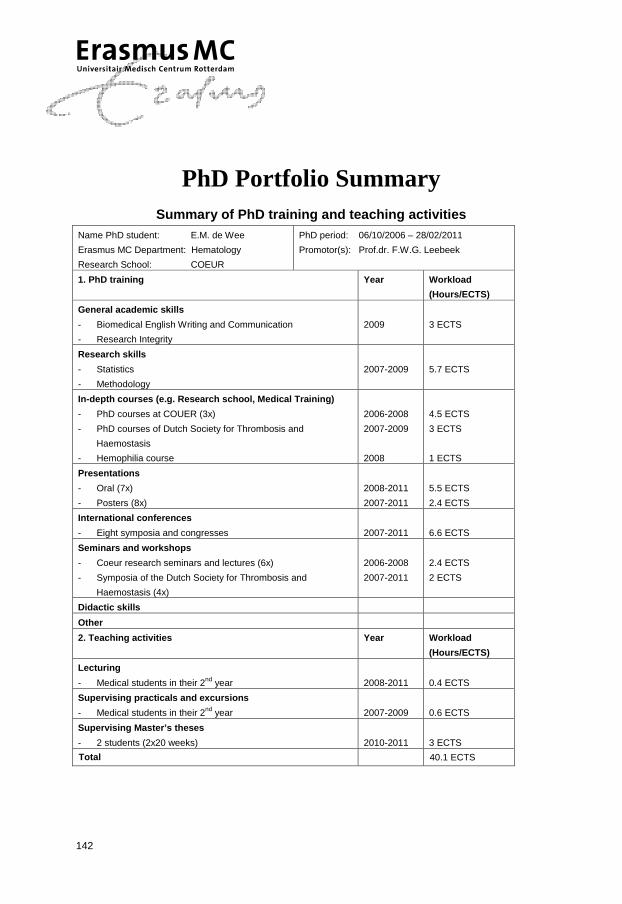
142
PhD Portfolio Summary Summary of PhD training and teaching activities
Name PhD student: E.M. de Wee
Erasmus MC Department: Hematology
Research School: COEUR
PhD period: 06/10/2006 – 28/02/2011
Promotor(s): Prof.dr. F.W.G. Leebeek
1. PhD training Year Workload
(Hours/ECTS)
General academic skills
- Biomedical English Writing and Communication
- Research Integrity
2009
3 ECTS
Research skills
- Statistics
- Methodology
2007-2009
5.7 ECTS
In-depth courses (e.g. Research school, Medical Training)
- PhD courses at COUER (3x)
- PhD courses of Dutch Society for Thrombosis and
Haemostasis
- Hemophilia course
2006-2008
2007-2009
2008
4.5 ECTS
3 ECTS
1 ECTS
Presentations
- Oral (7x)
- Posters (8x)
2008-2011
2007-2011
5.5 ECTS
2.4 ECTS
International conferences
- Eight symposia and congresses
2007-2011
6.6 ECTS
Seminars and workshops
- Coeur research seminars and lectures (6x)
- Symposia of the Dutch Society for Thrombosis and
Haemostasis (4x)
2006-2008
2007-2011
2.4 ECTS
2 ECTS
Didactic skills
Other
2. Teaching activities Year Workload
(Hours/ECTS)
Lecturing
- Medical students in their 2nd year
2008-2011
0.4 ECTS
Supervising practicals and excursions
- Medical students in their 2nd year
2007-2009
0.6 ECTS
Supervising Master’s theses
- 2 students (2x20 weeks)
2010-2011
3 ECTS
Total 40.1 ECTS

Von Willebrand Disease in the
NetherlandsEva Maria de Wee
Von Willebrand D
isease in the Netherlands
Eva M
aria de Wee