volume rendering reconstructions of the anatomy of small ... · hexamethyldisilazane and air drying...
TRANSCRIPT
Volume rendering reconstructions of the anatomy of small aquatic beetles (Insecta: Coleoptera)
scanned with the Skyscan 1172 high resolution micro-CT
J. Alba-Tercedor & C.E. Sáinz-Cantero Caparrós
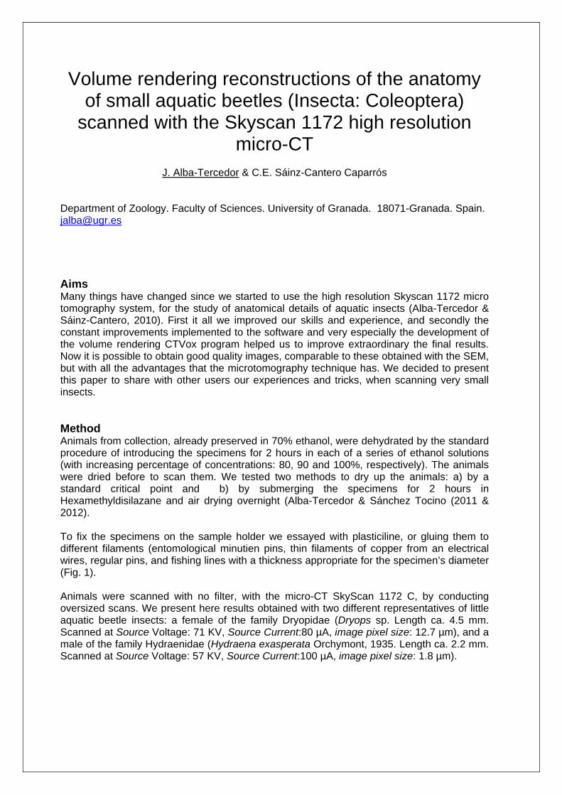
Department of Zoology. Faculty of Sciences. University of Granada. 18071-Granada. Spain. [email protected] Aims Many things have changed since we started to use the high resolution Skyscan 1172 micro tomography system, for the study of anatomical details of aquatic insects (Alba-Tercedor & Sáinz-Cantero, 2010). First it all we improved our skills and experience, and secondly the constant improvements implemented to the software and very especially the development of the volume rendering CTVox program helped us to improve extraordinary the final results. Now it is possible to obtain good quality images, comparable to these obtained with the SEM, but with all the advantages that the microtomography technique has. We decided to present this paper to share with other users our experiences and tricks, when scanning very small insects. Method Animals from collection, already preserved in 70% ethanol, were dehydrated by the standard procedure of introducing the specimens for 2 hours in each of a series of ethanol solutions (with increasing percentage of concentrations: 80, 90 and 100%, respectively). The animals were dried before to scan them. We tested two methods to dry up the animals: a) by a standard critical point and b) by submerging the specimens for 2 hours in Hexamethyldisilazane and air drying overnight (Alba-Tercedor & Sánchez Tocino (2011 & 2012). To fix the specimens on the sample holder we essayed with plasticiline, or gluing them to different filaments (entomological minutien pins, thin filaments of copper from an electrical wires, regular pins, and fishing lines with a thickness appropriate for the specimen’s diameter (Fig. 1). Animals were scanned with no filter, with the micro-CT SkyScan 1172 C, by conducting oversized scans. We present here results obtained with two different representatives of little aquatic beetle insects: a female of the family Dryopidae (Dryops sp. Length ca. 4.5 mm. Scanned at Source Voltage: 71 KV, Source Current:80 µA, image pixel size: 12.7 µm), and a male of the family Hydraenidae (Hydraena exasperata Orchymont, 1935. Length ca. 2.2 mm. Scanned at Source Voltage: 57 KV, Source Current:100 µA, image pixel size: 1.8 µm).

Figure 1: To fix the specimens on the sample holder we essayed with plasticiline, or gluing them to different filaments (a: entomological minutien pins, b & d: thin filaments of copper from electrical wires, c: fishing lines with a thickness appropriate for the specimen’s diameter, and d: regular pins).
Figure 2: Scheme of the procedure followed by using the CTAn tasks to eliminate the outer wall of the body (e): 1.- Thresholding, followed by Shrink-wrap (a & b), obtaining an adjusted region of interest (ROI) (c); 2.- Morphological operations, eroding the ROI; thus a new thinner ROI was obtained (d); 3.- Reload the image, and 4.- Save bitmaps (f). When studying the internal anatomy of insects it helps a lot to eliminate the outer wall of the body (exoskeleton of insects), by “stripping” the specimens (see Fig. 6). To get these results
a b c
d e f
a b c d e

we with run the following tasks in CTAn: 1.- Thresholding, followed by Shrink-wrap (Fig. 2: a & b), obtaining an adjusted region of interest (ROI) (Fig. 2c); 2.- Morphological operations, eroding the ROI (before we measured the thickness of the outer wall in pixel, selecting 25), thus a new thinner ROI was obtained Fig. 2d; 3.- Reload the image, and 4.- Save bitmaps, applying it to the “image inside ROI”. So finally it was obtained a new dataset with images without the external body wall (Fig. 2e). Results and discussion At the beginning we were using a critical point procedure to dry up the animals, obtaining good results (Figs.:3-6). However, later, we experimented that we could obtain totally similar results with the Hexamethyldisilazane (HDMDS), (Alba-Tercedor & Sánchez-Tocino, 2010, 2012) resulting easier and cheaper. So this is the method that nowadays we use as a routine. In fact there are a lot of references dealing with the totally comparable results obtained by using a critical point in comparison with HDMDS (see for example Bray, 1993). With respect the two general methods used for the fixation and positioning specimens on the sample holder to scan, it is important to know that either the plasticiline, or the selected filament, in most cases must be removed later from the images, by using the different tasks options of the CTan software (what we knows as “cleaning”). Thus, the use of plasticiline resulted adequate for specimens opaque enough to X-ray to be able to eliminate it from images by running a thresholding (segmenting) task (and this was applied for the Figs.: 3-6). When animals has a similar opacity as the plasticiline, still is possible to eliminate it from the images by drawing by hand the appropriate regions of interest (ROI) when using CTAn, and saving the selected ROI as a new data set. For this purpose, and in general for accuracy during ROI drawings, a graphics tablet facilitates the task. For very tiny specimens (ca. 2 mm), as occurs with the selected specimens of the genus Hydraena (Fig. 7), using plasticiline may result quite tricky when mounting them in an appropriate centered and vertical position (necessary to scan at the highest magnification), even making a small hole in the plasticiline, and to avoid that successive corrections don’t damage the specimens. Thus we have been testing to fix them onto different filaments (Fig. 1) by gluing them either with “Loctite” or wood glue. First one has the advantage of the fast fixation, but it has the problem that once the specimen is fixed it is not possible to correct its position. Moreover in many cases results a problem for its elimination. This is why in most cases the wood glue gave better results, because is easier to correct the position before it dries completely (or even to remove it for the specimens by submerging them again in water), and as is less opaque it can be easily eliminated during the cleaning tasks when running CTAn. A problem still existing is the automatic drawing of scales onto the pictures. Up to now, this is possible by selecting the option to “insert scale bar “in the Save bitmaps task of CTAn. However this produces a long bar with a scale drawn on the tip. Thus when the volume render image is visualized and rotated either with CTVox or with the volume option of CTAn, the scale vision (and the number with the magnitude) disappears at certain positions. Thus to be able to drawn an appropriate scale we used the DataViewer measure option, measuring a reference distance. So later, it is not difficult to draw a line on the reconstructed images reproducing the scale line, as we did for Fig. 7.
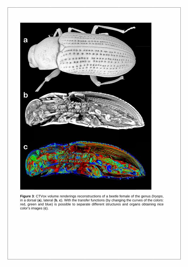
Figure 3in a dorsared, greecolor’s im
b
c
a
: CTVox voal (a), lateraen and bluemages (c).
olume rendeal (b, c). Wie) is possib
erings reconth the trans
ble to separ
nstructions sfer functionrate differen
of a beetle ns (by channt structure
female of tging the cus and orga
the genus Drves of the
ans obtainin
Dryops, colors:
ng nice

Figure 4: CTVox volume renderings reconstructions from a ventral view (a: external, b: internal) of a beetle female of the genus Dryops.
b
a

Figure 5Progressmiddle (b
a
b
c
: CTVox voive cuts pe
b), and vent
olume rendeermit to obtral (c).
erings reconbserve the
nstructions internal st
of a beetle ructures at
female of tdifferent l
the genus Devels: dors
Dryops. sal (a),

Figure 6: CTVox volume renderings reconstructions of a beetle female of the genus Dryops to which the outer wall of the body (exoskeleton) has been removed (“stripping”) by following the procedure schematized in figure 2. (a: dorsal, b: outermost ventral, and c: removing more internally to point out the ganglia nervous central chain).
a
b
c

Figure 7: CTVox volume renderings reconstructions of a tiny male species of the aquatic beetle (Hydraena exasperata): external body general appearance (a), and internal views of the anatomy (b: dorsal, c: lateral). With the transfer functions (by changing the curves of the colors: red, green and blue) is possible to separate different structures and organ, obtaining nice colors’ images.
a
b
c
Conclusions The procedure to obtain good volume rendering reconstruction is easier than it can appear at a first sight. It starts with an adequate scanning. For this, we recommend adjusting the microtomograph to the actual conditions by obtaining flat corrections quite often and why not each day before to start a new scanning session. The fixation of specimens on the sample holder can be done either directly with plasticiline of by gluing the animals at the tip of a filament, and punch it to a small piece of plasticiline (adhered on the sample holder). For this purpose the wood glue gives to us quite good results, permitting an easy thresholding and to unglue later the specimens, either to correct their position and/or to keep them again in the original collection from where they come from. During the reconstructions, with NRecon it is possible not only to enhance the contrasts, rotate the sample, and to select a region of interest (ROI) to reconstruct. Also the software permits also to make some sort of thresholding to eliminate the glue. Later a more accurate “cleaning” of the raw images with CTAn eliminates the noise, and making possible to select an appropriate ROI (or VOI), permitting to remove the outer wall of the body (in our case the exoskeleton of insects). We must confess that the CTVox program and their posterior and continue improvements, has represented a key stone for our research on the study of the anatomy of little aquatic insects. It has improved enormously the volume rendering images, giving to the results obtained of “our” little insects a completely new life, permitting to get images of the external parts comparable to those of the scanning electron microscopy, with advantage of the observation of the internal anatomy. And, moreover the most important: once the data sets have been obtained and processed the specimens can be studied as many times as the researcher wishes at different positions, changing the light conditions, opacity, shadow, color, etc.. So it becomes a very versatile tool not only for research but also for teaching purposes permitting to get images of the external parts comparable to those of the scanning electron microscopy, with advantage of the observation of the internal anatomy. Moreover the possibility of play with the transfer functions, by changing the curves of the colors (red, green and blue) helps to separate different structures and organs with nice color’s images (Fig. 3c and Fig. 7: c & d). However, still we miss the possibility to get an automatic more versatile option to draw a scale bar, that the one already existing in the Save bitmaps task of the software CTAn. Acknowledgements To the Skyscan company people who kindly gave us fast and effective support. We are especially indebted and we like to express our recognition to: Alexander Sasov, Stephan Boons, Xuan Liu, Phil Salmon, and Jeroen Hostens; for their patience, effectiveness and kindness answering our constant bombing of queries and suggestions, and for their constant improvements to the software implementing new options. This work was supported by the Spanish “Ministerio de Educación y Ciencia” (CGL2007 – 61856/BOS), and the “Junta de Andalucía” (RNM-02654). References:
1. Alba-Tercedor, J., Sáinz-Cantero, C. “Studying Aquatic Insects Anatomy with the Skyscan 1172 high-resolution micro-CT “. Skyscan Users Meeting 2010 vol. 2: 8-11, 2010 (http://www.skyscan.be/company/UM2010/abstract_08.pdf)
2. Alba-Tercedor, J, Sánchez-Tocino, L. “The use of the SkyScan 1172 high-resolution micro-CT to elucidate if the spicules of the “sea slugs” (Mollusca: Nudibranchia,
Opisthobranchia) have a structural or a defensive function”. Skyscan Users Meeting 2011, 2011 (http://www.skyscan.be/company/UM2011/abstract_22.pdf)
3. Alba-Tercedor, J, Sánchez-Tocino, L. “High-Resolution Micro-CT of the Anatomy of the Sea Slug Polycera quadrilineata”. Microscopy and Analysis, Volume 26 (1): 17-19, 2012
4. Bray, D. F., Bagu, J., Koegler, P. “Comparison of hexamethyldisilazane (HDMDS), Peldri II, and critical-point drying methods for scanning electron microscopy of biological specimens”. Microscopy Research and Technique, 26: 489–495. doi: 10.1002/jemt.1070260603, 1993