volume 8, no. 1 issn 2194-0479 altex - eusaat congress
TRANSCRIPT
ALTEXVolume 8, No. 1
ISSN 2194-0479(2019)
Linz 2019 ‒ EUSAAT 2019
ProceedingsEthical & legal issues Free communicationsImplementing EU Dir 63/2010 – update In silico modelsInitiative for implementing serum free culture mediaIn vitro techniques for CNS toxicity and disease studiesReduction & refinement Replacement – advanced technologiesSpecific endpoints of toxicityStem cell models and technology (hIPS, ES, mES, mIPS…)How to account for uncertainties of reference methods & data?Animal experimentation: Working towards a paradigm change'Young Scientists' session
3D Models & multi-organ- chips (MOC),
human-organ-chips (HOC)3R Centers in Europe &
international – national and local centers
3Rs in education and academia
Advanced safety testing of cosmetics & consumer
products and alternatives to animal testing in
food safety, nutrition and efficacy
An integrated interdisciplinary approach
to animal-free nano- material and chemical
safety assessment: Results of the in3 project
Biological barriers Disease models using
human cells, tissues and organs
EcotoxicologyEfficacy and safety
testing of drugs, medical devices & biopharmaceutics

EpiDerm™ Skin Corrosion, Skin Irritation, Phototoxicity, Genotoxicity Micronucleus and Comet assay, Medical devices, Skin Inflammation, Skin metabolism, Radiation Damage, Percutaneous Absorption …
EpiOcular™ Ocular Irritation, “Sub-Draize” Ocular Irritation Testing (Mild, Milder, Mildest), Specific Ocular Irritation of Surfactants, Cosmetics and Consumer Products …
EpiIntestinal™ Intestinal Toxicity, Drug Delivery, Inflammation, Fibrosis, Infection, Epithelial Restitution …
KEY FEATURES OF OUR PRODUCTS
Metabolically active, human cell-derived, 3D reconstructed tissue models Objective, quantifiable endpoints Excellent in vitro / in vivo correlation Guaranteed long-term reproducibilityAvailability of partial and full thickness tissue modelsAvailability of different tissue formats including high throughput screening plate Price convenient testing kits as well as single tissues
EXCELLENT CUSTOMER SERVICEAND PROFESSIONAL SCIENTIFIC SUPPORT
Prompt reply to customer´s requests and needs Free scientific and technical consulting before purchasing of productMonitoring of every single shipmentDedicated trainings sessions in our facilities or on-siteInternational support provided by EU, US, Turkey and Japan representativesMore than 20 years of tissue quality control data summarized in open databases - the most data of any tissue engineering company !Since 2016 ISO 9001:2008 certified
INTERNATIONAL VALIDATIONS AND PROJECTS
EpiDerm™ECVAM Skin Corrosion, Validated Assay - OECD TG 431ECVAM Skin Irritation, Validated Assay - OECD TG 439ECVAM Pre-Validated and ICH Accepted Phototoxicity Assay Cosmetics Europe Validation Project on Genotoxicity AssaysGerman Skin Penetration Validation Study for Surfactants and FormulationsIrritation Potency of Extracts from Medical Devices Study (ISO 10993-10)
EpiOcular™ECVAM/Cosmetics Europe Eye Irritation, Validated Assay - OECD TG 492US EPA Accepted for Antimicrobial Products with Cleaning Claims (AMCPs)COLGATE/IIVS Eye Irritation Validation StudyCon4Eye Project on Eye Irritation Testing Strategies
EpiVaginal™NIH Funded HIV Research CONRAD Microbicides Study
AFFILIATIONS AND MEMBERSHIP IN PROFESSIONAL OR-GANISATIONS AND CONSORTIA IN THE EU
The world leader in innovative 3D reconstructed human tissue modelsISO 9001 CERTIFIED
MatTek In Vitro Life Science Laboratories Mlynské Nivy 73, 821 05 Bratislava, Slovakia, EUPhone: +421-2-3260-7408Fax: +421-2-3260-7404E-mail: [email protected]: www.mattek.com
TISSUE MODELS AVAILABLE FROM SLOVAKIA AND USA
EpiDermFT™ Anti-Aging, Skin Longevity, Skin Hydration, UV Protection, Percutaneous Absorption …
EpiCorneal™ Drug Delivery, Infection, Inflammation, Ophthalmic Product Testing …
EpiOral™ and EpiGingival™ Mucosal and Oral Irritation, Transmucosal and Buccal Drug Delivery, Gum Disease, Oral Cancer, Smokeless Tobacco Effects, Oral Epithelial Proliferation, Antimicrobial Barrier Function …
EpiAirway™ Drug Delivery, Respiratory Infection and Toxicology, Tobacco Smoke Toxicology, Nano-particle Toxicology, Gene Expression Analysis, RNAi/siRNA Therapeutic Drug …
MelanoDerm™ Skin Lightening-Darkening, Skin Pigmentation Modulation, Keratinocyte-Melanocyte Interactions …
Psoriasis™ Anti-Psoriatic Drug Screening, Basic Psoriasis Research ...
EpiVaginal™ Feminine Product Irritation, Microbicide Testing, Immuno-competent, HIV-1 Infection Sexually Transmitted Infection, Inflammation, Vaginal Drug Delivery …
Melanoma™ Tumor Invasion, Anti-Melanoma Drug Screening ...
TISSUE MODELS AVAILABLE FROM USA
MatTek Corporation 200 Homer Avenue, Ashland, MA 01721, USAPhone: +1-508-881-6771Fax: +1-508-879-1532 E-mail: [email protected]: www.mattek.com
Your trustworthy partner in the world of in vitro science for the past 30 years
PRIMARY CELLS & MEDIA FROM USAPrimary human cells derived from human tissue for highthroughput screening

ALTEX Proceedings 8, 1/2019 I
Dear friends and colleagues,
on behalf of EUSAAT, the European Society for Alternatives to Animal Testing, we welcome you to the 22nd European 3Rs Congress and the 19th EUSAAT Congress in Linz/Austria on October 10-13, 2019.
Since 1991 the “Linz-Congress” has emerged in Europe as the major scientific event in the field of the 3Rs and in 2019 the EUSAAT 2019 3Rs Congress Linz will be the largest international 3Rs congress. We are particularly pleased that our international partners from Japan, JSAAE, the Japanese Society for Alternatives to Animal Experiment, and from China, CCARE, the Chinese Center for Alternatives Research and Evaluation, will report on their activities in our sessions on “3R Centers in Europe & international”.
EUSAAT 2019 is hosting oral and poster presentations to facilitate discussions and the exchange of ideas to actively promote the most advanced non-animal methods in the life sciences. The Scientific Committee has identified the subjects that are currently in the focus of 3Rs research in Europe as well as internationally. In 2019 the EUSAAT Congress features sessions on Refinement and Reduction (e.g. animal welfare of experimental animals, academia and education, ethical and legal issues, drug development and toxicity studies) as well as on Replacement (e.g. disease models based on human cells and tissues, non-animal tools for basic biomedical research, stem cells, bioprinting, ’omics technologies, human 3D tissue culture methods and human multi-organ-chips (MOCs)). Since at EUSAAT 2018 the sessions on establishing 3Rs networks in Europe were most popular, we will have several sessions on this topic as well as a Round Table Discussion.
For the first time, we are holding the YOU EUSAAT 2019 events aimed at young and early career scientists (up to the age of 35) working or planning to work in the 3Rs field. We want to encourage the dialogue among young scientists and with experienced mentors from the 3Rs area to facilitate opportunities for establishing professional networks. In particular, YOU EUSAAT 2019 will provide a free and open atmosphere for exchanging ideas and career experiences and networking with each other!
We are proud that four distinguished colleagues have accepted our invitation to serve as keynote speakers. In addition, we are honored that both the Bjorn Ekwall Memorial Fund (BEMF) will hold the 2019 BEMF Award Ceremony and Lecture at EUSAAT 2019 and also that the editor of ALTEX will present the award for the best article published in the ALTEX journal in 2018 at EUSAAT 2019.
This year we will again host two Round Table Discussions on topics that are of importance for implementing the 3Rs at the international level. The first will cover the “International Impact of Closing Research Animal Facilities in Europe” and the second will be on “Establishing an International 3Rs Centers Network”.
EUSAAT is very happy to continue the “Young Scientists Travel Award (YSTA)” program in 2019 and we want to thank the SET Foundation for its generous funding. The YSTA program will provide 20 young scientists with the opportunity to share their ideas on the implementation of the 3Rs in education and novel test systems to reduce the suffering as well as the number of animals in research.
The EUSAAT Board is pleased that the number of sponsors of the EUSAAT congresses has continuously increased, since due to their financial support we can keep the scientific standard high and the congress fee low. This is important to attract young scientists from Europe and abroad. Thus, on behalf of the participants, the EUSAAT Board and the Scientific Committee are grateful to our sponsors.
Moreover, we are also pleased that the abstracts of the EUSAAT 2019 are again published in ALTEX Proceedings and we thank Sonja von Aulock, the editor and CEO of ALTEX Edition, for her generous support.
Finally, we want to thank our colleagues on the EUSAAT Board and the Scientific Committee for their continuous support in planning the EUSAAT 2019 congress.
Horst Spielmann Winfried Neuhaus Dagmar Jirova Dominik RünzlerSecretary General President Vice-President Vice-President
Welcome address EUSAAT 2019
AbstrActs Linz 2019

ALTEX Proceedings 8(1), 2019
AbstrActs Linz 2019
II
List of Abstracts
ID Title Authors Page
294
295
296
297
298
299
300
301
302
303
304
305
307
308
Microphysiological flux balance platform unravels the dynamics of drug induced steatosis
Uric acid metabolism and excretion of translucent skin mutants of 5th instar larvae of the silkworm “o06” strain as an animal model of hyperuricemiaA comparison of hyperuricemia model mice loaded with oxonic acid and the silkworm “o06” strain as an alternative for evaluating voluntary oral administrationRecent efforts to elucidate the scientific validity of animal-based drug tests by the pharmaceutical industry, pro-testing lobby groups, and animal welfare organisationsDoes the stress of laboratory life and experimentation on animals adversely affect research data?CRISPR-mediated gene editing: Scientific and ethical issuesEnabling technologies for skin permeation investigation
A holistic approach for the toxicological assessment of phytogenic substances in human and animal nutritionUsing big data for developing QSAR models to predict interaction of chemicals with neuronal proteinsEffect-based study of human platelet lysate in various cell lines
Human-based T cell-skin models for graft-versus-host diseasePlacenta-on-a-chip model for assessing the transport and toxicity of xenobiotics in vitro
The many cards up “computational sleeves”: Reshaping in silico tools used in drug discovery to design safer and functional chemicalsTransitioning from mammalian animal testing to non-animal testing using the zebrafish (Brachydanio rerio) embryo as a whole organism model for regulatory decision-making in chemical risk assessments
Avner Ehrlich, Konstantinos Ioannidis, Muneef Ayyash, Anne Riu, Reine Note, Gladys Ouedraogo, Jan Vanfleteren, Merav Cohen and Yaakov NahmiasRyuichiro Tanaka, Nanae Nishikubo, Manato Sakamoto, Fumitoshi Sakazaki and Yutaka Banno
Hikaru Kawakami, Miku Hosoki, Shoji Yamaguchi, Yutaka Banno and Ryuichiro Tanaka
Jarrod Bailey and Michael Balls
Jarrod Bailey
Jarrod Bailey
Rosalia Bertorelli, Giulia Suarato, Raffaele Spano, Salvatore Surdo, Alberto Diaspro and Athanassia AthanassiouPeter Lanzerstorfer, Jonathan Grasberger, Georg Sandner, Flora Stübl and Julian Weghuber
Yaroslav Chushak and Jeffery Gearhart
Domenik Rehberger, Beat Thalmann, Jonathan Steubing, Irene Marini, Sarah Dettling, Ute Fischer, Eva-Maria Schentarra, Marc Waidmann, Tamam Bakchoul and Rosemarie SteubingFabienne Geiger, Yosif Tumbev, Florian Groeber-Becker and Friederike Berberich-SiebeltEvgeny Knyazev, Sergey Nikulin, Anna Khristichenko, Tatyana Gerasimenko, Olga Kindeeva, Vladimir Petrov and Dmitry SakharovJakub Kostal
Cindy L. A. Woodland, Lee Ellis, Mike Morash and John C. Achenbach
47
204
101
6
7
8
17
113
34
172
59
105
108
222
AbstrActs Linz 2019
ALTEX Proceedings 8(1), 2019 III
310
311
312
313
314
315
316
317318
319
321
322
323
324
325
326
327
328
Thematic review of animals used for scientific purposes: A mechanism for advancing replacementNeuroprotection via an iNOS-inhibitor in a porcine retina organ culture model
Towards an automated surveillance of well-being in mice using deep learning
Stakeholder collaboration to advance human-relevant nonclinical methods for drug development in the United StatesSocial enrichment by pair-housing of male C57BL/6JRj miceNext generation risk assessment of coumarin in personal care products
3D CNS model of iPSCs derived neuron and glia for high-throughput neurotoxicity screening in Mimetas’ OrganoPlate®
Make scientists better: Altertox Academy missionRetinal protection against oxidative stress via mild hypothermiaAdvances in in silico research and safety testing: Recognition and reward by the Lush PrizeEstablishment of precision cut bovine udder slices for pharmacological studies
Human urine as a non-invasive source of kidney progenitor cells amenable for nephrotoxicity studies
An AOP-driven assessment of developmental neurotoxicity induced by chemical mixtures using human iPSC-derived neuronal/glial culturesLearning from negative results in animal based research! Critical Incident Reporting System in Laboratory Animal Science (CIRS-LAS)Advancing chemical safety assessments through integration of population-based in vitro models and computational methodsUse of ethno veterinary medicine techniques for animal welfare and sustainable disease managementThe need to address human relevance and measure return on investment in biomedical researchInitiatives to promote the Three Rs in education and training
Christina Dodkin and Kimberley Jayne
Stephanie C. Joachim, Jose Hurst, Ana Maliha, Lisa Hofmann, Sandra Kühn, Fenja Herms and Sven SchnichelsNiek Andresen, Manuel Wöllhaf, Katharina Hohlbaum, Lars Lewejohann, Olaf Hellwich, Christa Thöne-Reineke and Vitaly BelikElizabeth Baker
Katharina Hohlbaum, Silke Frahm-Barske, André Rex, Christa Thöne-Reineke and Kristina UllmannMabel Cotter, Maria Baltazar, Georgia Reynolds, Joe Reynolds, Sophie Cable, Mi-Young Lee, Mona Delagrange, Dawei Tang, Tom Cull, Predrag Kukic and Alistair MiddletonChiwan Chiang, Arnaud Nicolas, Karlijn Wilschut, Henriette Lanz, Sebastiaan Trietsch, Jos Joore and Paul VultoFrancois BusquetAna Maria Maliha, Sven Grauthoff, Tobias Kiebler, Jose Hurst, Sven Schnichels and Stephanie C. JoachimCraig Redmond
Viviane Filor, Jessica Meißner, Paula Hagedorn, Monique Petry, Maren von Köckritz-Blickwede and Manfred KietzmannLucas-Sebastian Spitzhorn, Md Shaifur Rahman, Wasco Wruck, Martina Bohndorf, Soraia Martins, Audrey Ncube, Lars Erichsen, Lisa Nguyen, Nina Graffmann and James AdjayeFrancesca Pistollato, Emilio Mendoza, Stephanie Bopp, Carolina Nunes, Andrew Worth and Anna PriceDavid Trietschel, Astrid Enkelmann, René Schiffner and Sabine Bischoff
Fabian Grimm
Mandeep Azad and Kawardeep Kour
Francesca Pistollato, Ivana Campia, Camilla Bernasconi, Clemens Wittwehr and Maurice WhelanFrancesca Pistollato, Adelaide Dura and Marcelle Holloway
43
90
3
10
79
37
31
27127
171
54
200
161
208
63
5
160
162
ID Title Authors Page

AbstrActs Linz 2019
ALTEX Proceedings 8(1), 2019 IV
329
330
331
332
333
334
335
336
337
338
340
341
342
344
345
346
348
349
Adaptation of skin sensitization in vitro methods (OECD 442C, D, E) for nanomaterials
Gluconeogenic processes in a bovine liver cell line offered short chain fatty acidsNorecopa: Working to advance harmonisation and dissemination of best practice in animal research and testingP. aeruginosa infected co-culture of human cystic fibrosis bronchial epithelial cells as a preclinical test system for anti-infectivesEstablishing Xenopus laevis oocytes as a novel model system for blood brain barrier analysesInitiative for better access and digitalisation to EU agencies safety dataUsing PLHC-1 topminnow liver cells to characterize and predict the effect of plastic additives in fishFull implementation of refinement methods in practice through education and trainingCosmetic perfumes and their potential of endocrine disruption, sensitization and genotoxicityProteomic analysis of canine leptospira vaccines
Education towards non-animal approaches in basic and applied biomedical researchAre researchers moving away from animal models as a result of poor clinical translation in the field of stroke? A systematic analysis of opinion papersCalifornia Cruelty-Free Cosmetics Act establishes first North American law banning the sale of animal tested cosmetics and ingredientsThe thyroid hormone disruptors validation project and application of OECD’s GIVIMP and Guiding principles on good practices on protected elements in Test GuidelinesDevelopment of an OECD Test Guideline for Androgen Receptor Transactivation Assays (ARTAs)Animal experiments or humane alternatives: Awareness raising campaign in BelarusUsing the OECD QSAR toolbox as an in silico modelling method for computational aquatic toxicology of endocrine disruptorsMicrofluidic in vitro lung model to replace murine infection and ARDS models
Barbara Birk, Lan Ma-Hock, Natascha Partosa, Britta Wareing, Jutta Steinbrenner, Susanne N. Kolle, Johannes Keller, Wendel Wohlleben, Bennard van Ravenzwaay and Robert LandsiedelAnna-Maria Sittel, Herbert Fuhrmann and Axel SchoenigerAdrian Smith
Justus Horstmann, Cristiane Carvalho-Wodarz, Nicole Schneider-Daum and Claus-Michael Lehr
Nora Brunner and Salah Amasheh
Francois Busquet
Cinta Porte, Elisabet Pérez-Albaladejo and Anna MarqeñoKathrin Herrmann
Marketa Dvorakova, Lada Svobodová, Marian Rucki, Jan Chrz, Václav Ševčík and Dagmar JírováThomas Schuenborg, Jelena Spiric, Elisabeth Balks and Andreas ReuterKathrin Herrmann
Pandora Pound and Rebecca Ram
Elizabeth Baker
Anne Milcamps, Gerry Bowe, Camilla Bernasconi, Tom Cole, Ingrid Langezaal, Francesca Pistollato, Valérie Zuang, Sandra Coecke
Anne Milcamps, Roman Liska, Ingrid Langezaal, Warren Casey, Matthew Dent and Jenny OdumTatsiana Hlinkina, Anastasiya Poyanena and Ludmila LoginovskayaMarie-Léonie Bohlen and Baeckkyoung Sung
Michelle Hesler, Thorsten Knoll, Felix Ritzmann, Anja Honecker, Sylvia Wagner, Yvonne Kohl, Hagen von Briesen, Heiko Zimmermann, Robert Bals and Christoph Beisswenger
22
196
197
80
25
26
165
71
46
192
72
166
11
138
139
76
23
73
ID Title Authors Page

AbstrActs Linz 2019
ALTEX Proceedings 8(1), 2019 V
350
351
352
355
356
357
359
360
361
362
363
364
365
366
367
368
369370
371
Clicker training for small rodents – cognitive enrichment and beyond
An overview of Toxicity Testing 21st Century (TT21C) in vitro methods to assess next generation productsCombining in vitro and in silico modelling to simulate cartilage degradation during osteoarthritis
Simulating an arthritic joint in vitro by combining multiple tissue components
Automated analysis of developmental and reproductive effects using the nematode C. elegans
Accounting of uncertainties for complex endpoints: What do we want to know?Ethical justification of animal experiments in GermanyApparent permeability coefficients of ciprofloxacin in human lung epithelial cells appropriately predict bioavailability after inhalation exposureFish acute toxicity: Which data do we need and where do we get these from?Animalstudyregistry.org – a federal database for preregistration of animal researchImplementation status of non-invasive methods in wildlife genetic samplingMTS application to Italian veterinary autogenous vaccines as alternative in vitro method to assess cytotoxicity: Preliminary dataCombination of in vitro reporter gene assays, zFET and FETAX reveal cyanobacteria as producers of teratogenic retinoid-like compounds3R-alternatives to skin testing of bovine PPD tuberculins
An integrated testing strategy for polymer exposure in inhalation risk assessmentThe novel BFH12 cell line: An alternative model of bovine biotransformationAsian Consortium for Three RsIn vitro approaches to identify hepatotoxic chemicals
OECD DRP on in vitro assays in reproductive toxicology
Charlotte Sophie Leidinger, Jana Dickmann, Felix Herrmann, Dorothea Pichl, Nadine Kaiser, Christa Thöne-Reineke, Jan Baumgart and Nadine BaumgartLiam Simms, Kathryn Rudd, Lukasz Czekala, Edgar Trelles-Sticken, Roman Wieczorek and Matthew StevensonAnnemarie Lang, Lisa Fischer, Marie-Christin Weber, Alexandra Damerau, Timo Gaber, Sebastian Götschel, Rainald Ehrig, Susanna Röblitz and Frank ButtgereitAlexandra Damerau, Moritz Pfeiffenberger, Karoline Diesing, Frank Buttgereit, Timo Gaber and Annemarie LangPaul Wittkowski, Michael Oelgeschläger, Norman Violet, Philip Marx-Stoelting, Gilbert Schönfelder and Silvia VoglMartin Paparella
Christa Thöne-Reineke
Nico Sonnenschein, Norman Nowak, Sylvia E. Escher, Jan Knebel, Katharina Blümlein, Katharina Schwarz and Tanja HansenThomas Braunbeck
Justyna Chmielewska, Bettina Bert, Céline Heinl, Barbara Grune and Gilbert SchönfelderMiriam A. Zemanova and Silvia Frey
Antonella Di Paolo, Katia Forti, Lucia Anzalone, Giulio Severi and Monica Cagiola
Klára Hilscherová, Marek Pípal, Jana Priebojová, Luděk Sehnal and Marie Smutná
Elisabeth Balks, Jelena Spiric, Kubra Naqvi, Carina Kruip, Arne Auste, Michael Mühlebach and Andreas ReuterIris Muller, Anthony Bowden, Sophie Cable, Zoe Deag, Mathura Theiventhran and Maria BaltazarAxel Schoeniger, Alexander Gleich, Walther Honscha and Herbert FuhrmannHajime KojimaLeroy Elenschneider, Alexander Wiegrebe, Jan Knebel, Armin Braun, Josef Fangmann and Tanja HansenHajime Kojima
114
195
112
39
221
153
206
198
24
32
224
42
74
12
142
190
10648
107
ID Title Authors Page

AbstrActs Linz 2019
ALTEX Proceedings 8(1), 2019 VI
372373
374
375
377
379
380
381
382
383
384
385
386
387
388
389391
392
393
The Danish 3R-CenterEstablishment of an organotypic photoreceptor model as an in vitro alternative for future retina degeneration studiesAdverse outcome pathway network-based testing strategy for thyroid disruption3Rs databases: FCS-free database, Interspecies Database and Humane Endpoints websiteNon-technical summaries provided by the Austrian Federal Ministry of Education, Science and ResearchDevelopment of an in vitro trabecular human bone model integrated in a perfusion system to simulate glucocorticoid-induced osteoporosisSafety testing of adult novelties using methods in vitroRefinement in education
Project evaluation – importance of a coherent and consistent approachAdvancing the Three Rs education and training under a European Parliament Pilot ProjectReliable and representative in silico predictions of freshwater ecotoxicological hazardous concentrationsRefinement on the way towards replacement: Are we doing what we can?Moving transparency to the next level – non- technical project summaries under Directive 2010/63/EUWhy are alternative methods (not) chosen? Presentation of a project on the investigation of value judgments for or against alternatives in animal testing in basic researchItalian Centro3Rs commitment: 1 year after the opening of the activities
Towards veterinary ethicsOn the uncertainty of toxicological methods: Quantifying the borderline range of prediction models and implications for decision-makingDevelopment of a human pancreatic cell line with optogenetically controllable receptor for high content screeningSkin-on-a-chip-based skin irritation evaluation method as an alternative to animal testing
Birgitte Vindahl Olsen and Rasmus Normann NielsenNatalie Wagner, Maurice R. Gammel, Andrea Greulich, Jose Hurst, Sven Schnichels and Stephanie C. JoachimJiri Novak and Klara Hilscherova
Jan van der Valk and Saskia Kliphuis
Ismene Fertschai
Karoline Diesing, Alexandra Damerau, Moritz Pfeiffenberger, Timo Gaber, Frank Buttgereit and Annemarie LangLada Svobodová, Dvořáková Markéta, Rucki Marian, Kejlová Kristína and Kolářová HanaGyörgyi Szabó, Domokos Csukás, Krisztina Juhos, Daniella Fehér, József Sándor, György Wéber, Constantinos Voniatis and Andrea FerenczSusanna Louhimies and David Anderson
Susanna Louhimies
Mélanie Douziech, Ad Ragas, Rosalie van Zelm, Rik Oldenkamp, A. Jan Hendriks, Henry King, Rafika Oktivaningrum and Mark A. J. HuijbregtsKathrin Herrmann
Susanna Louhimies
Ines Pietschmann, Hannes Kahrass and Marcel Mertz
Anna Maria Bassi, Gianfranco Beniamino Fiore, Valeria Chiono, Livia Visai, Guido Cavaletti and Arti AhluwaliaKerstin WeichSilke Gabbert, Susanne N. Kolle, Miriam Mathea and Robert Landsiedel
Anna Stierschneider, Christoph Wiesner and Harald Hundsberger
Tae Hyun Choi and Byoungjun Jeon
212215
147
210
51
41
202
203
125
124
44
70
123
158
13
21858
201
33
ID Title Authors Page

AbstrActs Linz 2019
ALTEX Proceedings 8(1), 2019 VII
394
395
396
397
398
399
400
401
402
403
404
405
406
407
408
409
410
From initiation to implementation: The RTgill-W1 cell line assay to predict fish acute toxicity of water samples and chemicals (ISO21115)In vivo measurement of 7-methoxycoumarin-O-demethylase activity in zebrafish (Danio rerio) embryosBiotransformation and bioactivation capacities in early life-stages of zebrafish (Danio rerio) – a state-of-the-art reviewCurrent status of animal-free research into Parkinson’s diseaseAt crossroads between regulation, science and industrial application: The EPAA, a public-private initiative for the 3RsCurrent status of the implementation of new methods in cardiotoxicity safety testingThe effects of Lisinopril on wound healing response using in vitro human skin equivalent (EpiDermFT) after 5 day-repeated topical applicationMarine sponge collagen-derived bioactive products and biomaterials for the development of cellular supports for tissue engineering and oxidative stress protection of human skin cells3R funding of research activities 2014-2019 in Denmark by the Danish 3R-CenterAnimal research for Alzheimer disease: Failures of science and ethicsEffects of fruit proteases on intestinal integrity and permeability in 3D EpiIntestinal reconstructed human tissue modelHepatic non-parenchymal cell isolation methods in rodents for development of hepatic co-culturesThe kinetic Direct Peptide Reactivity Assay (kDPRA): An in chemico method to characterize the skin sensitization potency of chemicals
The method of spheroid formation for 3D cultures of primary hepatocytes influences hepatocellular functions and hepatotoxicityAn in vitro 3D fracture gap model as a tool for preclinical testing procedures
An in vitro fracture hematoma model as a tool for preclinical drug testing
Using platelet-rich human platelet lysate to substitute fetal calf serum for cultivation of mesenchymal stromal cells
Melanie Fischer, Julita Stadnicka-Michalak, Adam Lillicrap and Kristin Schirmer
Ann-Kathrin Lörracher and Thomas Braunbeck
Ann-Kathrin Lörracher and Thomas Braunbeck
Christiane Hohensee, Carolin Spicher, Claudia Gerlach and Christina LedermannFranz Lamplmair
Christiane Hohensee, Claudia Gerlach, Carolin Spicher and Christina LedermannJi-Seok Han, Sumi Jang, Byoung-Seok Lee, Heejin Park, Younhee Kim, Yong-Bum Kim, Woo-Jin Kim, Jae-Woo Cho and Mi-Jin YangSonia Scarfì, Marina Pozzolini, Serena Mirata, Lorenzo Gallus, Gianluca Damonte, Annalisa Salis, Enrico Millo, Marco Bertolino, Maila Castellano, Silvia Vicini and Marco GiovineLisbeth Knudsen
Francesca Pistollato, Sarah E. Cavanaugh and John J. PippinJan Markus, Silvia Letasiova and Olena Prykhodko
Tanja Krimmling, Anett Ullrich and Dieter Runge
Britta Wareing, Andreas Natsch, Susanne N. Kolle, Barbara Birk, Nathalie Alepee, Tina Haupt, Erin Hill, Petra Kern, Laurent Nardelli, Hans Raabe, Marian Rucki, Tinashe Ruwona, Cindy Ryan, Sjoerd Verkaart, Walter Westerink and Robert LandsiedelJana Moer, Dieter Runge, Tanja Krimmling and Anett Ullrich
Moritz Pfeiffenberger, Alexandra Damerau, Karoline Diesing, Paula Hoff, Frank Buttgereit, Annemarie Lang and Timo GaberMoritz Pfeiffenberger, Alexandra Damerau, Karoline Diesing, Paula Hoff, Frank Buttgereit, Timo Gaber and Annemarie LangMoritz Pfeiffenberger, Alexandra Damerau, Wiktor Burdzinski, Karoline Diesing, Frank Buttgereit, Timo Gaber and Annemarie Lang
55
121
122
77
111
78
67
183
104
163
132
110
216
140
157
156
155
ID Title Authors Page
AbstrActs Linz 2019
ALTEX Proceedings 8(1), 2019 VIII
411
412
414
415
416
417
418
419
421
422
424
425
426
427
428
429
Drivers for the pharmaceutical industry to adopt human stem-cell based modelsNormalizing the unthinkable: The report of the Oxford Centre for Animal EthicsRefinement in Laboratory Animal Science education – an inquiry concerning the use of rat and mouse simulators among LAS instructorsDesign of experimental animal surgical models: Creation of a mouse uterus 3D-model for learning the in-utero electroporationGlycoengineering as a tool to control the behavior of bone marrow-derived mesenchymal stromal cells in biofabrication processesWhen the co-culture does not do what it is supposed to do – a step “back” to ex vivo models
Development of a test system for the investigation of the pulmonary mucus barrier
A 3D micromodel to study chemotaxis of cancer spheroids exploiting microfluidic channels
Repeating past mistakes: The banality and futility of nowadays cigarette smoke-related animal experimentationThe relevance of shear stress for in-vitro models of the human blood-brain barrier: A comparative study
There is no ethical justification for the use of fetal bovine serum (FBS)Serum-free media contribute to better reproducibility in in vitro researchInterpreting irregular LLNA data for evaluation of non-animal methods for skin sensitizationOptimization of gene silencing in a cell-laden 3D organ-like model by means of RNA interference Fully-humanized skin-on-a-chip with a modular architecture for biomedical applicationsIn3 Project: A European initiative to evaluate toxicity of chemicals using iPSCs derived in vitro models: Focus on blood brain barrier models for neurotoxicity assessment
Ard Teisman, Mohamed Kreir, Hua Rong Lu and David J. GallacherClair Linzey
Melanie Humpenöder, Giuliano Mario Corte, Marcel Pfützner, Mechthild Ladwig-Wiegard, Roswitha Merle, Johanna Plendl and Christa Thöne-ReinekeMaximilian Nuber, Nadine Baumgart and Jan Baumgart
Stephan Altmann, Jürgen Mut, Natalia Wolf, Julian Bechold, Franz Jakob, Jürgen Seibel and Regina EbertSabrina Schnur, Ralf-Kilian Zäh, Christopher Ruf, Henrik Groß, Claus-Michael Lehr, Dietmar Brück, Marc Schneider and Marius HittingerNina Grzeschik, Konrad Schwarzkopf, Marc Schneider, Henrik Groß, Claus-Michael Lehr and Marius HittingerSimone Luigi Marasso, Domenico Mombello, Alberto Puliafito, Simone Benetto, Matteo Cocuzza, Luca Primo, Federico Bussolino and Candido Fabrizio PirriDilyana Filipova, Gaby Neumann, Tamara Zietek and Corina Gericke
Anna Gerhartl, Maria Kirchsteiger, Palle Helmke, Alexandra Vladetic, Nadja Pracser, Grace Lin, Antje Appelt-Menzel, Marco Metzger, Mario Rothbauer, Peter Ertl and Winfried NeuhausTilo Weber, Kristina Wagner, Joachim Wiest, Jan van der Valk and Gerhard GstraunthalerJan van der Valk
David Roberts
Ida Shaef, Johanna Berg, Thomas Hiller, Viola Röhrs, Beatrice Tolksdorf, Ann-Christin Dietrich, Fanny Knoespel, Henry Fechner and Jens KurreckPatricia Zoio, Abel Oliva and João Conde
Sara Wellens, Lucie Dehouck, Paul Jennings, Fabien Gosselet and Maxime Culot
205
119
81
148
2
189
64
129
53
60
217
209
176
193
227
219
ID Title Authors Page

AbstrActs Linz 2019
ALTEX Proceedings 8(1), 2019 IX
430
431
432
433
434
435
436
437
439
440
441
442
443
444
445
446
447
448
449
Integrated evaluation for skin sensitization and phototoxicity of plant extractsA new skin-on-chip for anti-UV evaluation
The blood-saliva barrier: An optimized in vitro model of the oral epithelium and its relevance for biomarker researchAn innovative in vitro physiologically relevant model as a tool to test therapeutic strategies for glaucomaA society for alternatives to animal experiments in India: An updateCALT-BIO’s efforts to promote alternative methods in ChinaA study on skin irritation test of industrial chemicals using RhE model for prevent occupational skin diseasesTissue-on-a-chip
Generation of hepatic organoids from human induced pluripotent stem cellsThe need for non-animal recombinant antitoxins
A comparison of enzymatic and non-enzymatic strategies to isolate extracellular matrix (ECM) proteins from human placenta and liposuction fatImplementing 3Rs by the pharmaceutical industryThe Animal Protection Quality Certificate. A tool to assess Refinement methods in animal experimentsWhat can (Q)SAR modelling tell us about fish toxicity?Engineering in vitro lung microbiota for antimicrobial treatment
A recipe for the development of High-Accuracy QSAR models based on toxic mechanisms of actionThe use of rainbow trout liver hepatocytes in three-dimensional cell cultures for the study of graphene-related material internalizationDevelopment of a blood reference model of New Zealand White Rabbits with the Siemens Advia 2120iAssessment of uncertainties – ECVAM experience from recent in-vitro validation studies
Huang Jiancong, Shu Jun Cheng, Feng Jianhong, Luo Tingting and Ma ShashaHuang Jiancong, Shu Jun Cheng, Ma Shasha, Luo Tingting, Feng Jianhong and Du JuanGrace Lin, Tamara Leitgeb, Alexandra Vladetic, Sandra Domazet, Heinz-Peter Friedl, Nadine Rhodes, Johanna Gostner, Angela Rossi and Winfried NeuhausStefania Vernazza, Sara Tirendi, Sonia Scarfì, Sergio C. Saccà and Anna Maria Bassi
Mohammad Abdulkader Akbarsha
Zhi Jie Chen and Shu Jun Cheng
Hye Jin Jeon, Hyun-Sung Choi, Heung-Koo Choi and Kwon-Seob Lee
Christian Schmidt, Jan Markus and Joachim WiestHyemin Kim, Eun-Hye Kang, Seongyea Jo, Ji-Woo Kim and Han-Jin ParkDipti Kapoor, Esther Wenzel, Paul Stickings, Jeffrey Brown, Stefan Dubel, Androulla Efstratiou, Thea Sesardic and Michael HustJohannes Hackethal, Simone Hennerbichler, Heinz Redl and Andreas Teuschl
Kirsty Reid
Roberto Plasenzotti and Birgit Reininger-Gutmann
Pascal Bicherel
Daniela Pacheco, Anna Ziccarelli, Federico Bertoglio, Natalia Suarez Vargas, Francesco Briatico-Vangosa, Sebastião van Uden, Sonja Visentin, Livia Visai and Paola PetriniPascal Bicherel and Faizan Sahigara
Judit Kalman, Fernando Torrent and José M. Navas
Tomas Motschnig and Roberto Plasenzotti
Roman Liska
87
86
118
211
1
30
85
188
103
99
65
173
164
18
152
19
93
141
120
ID Title Authors Page
AbstrActs Linz 2019
ALTEX Proceedings 8(1), 2019 X
450
451
452
453
454
455
456
457458
459
460
461
462
463
464
465
466
467
The importance of data quality and governance in the acceptance of Next Generation Risk Assessment (NGRA) approachesCollaboration for non-animal cosmetic safety assessment globally by 2023A user perspective towards improving the processes of project evaluationGene expression upon immune challenge in a fish intestinal in vitro modelFrom a 3Rs intent to a company’s strategy: Perspective from the IndustryBiodegradable polyphosphazene scaffolds demonstrate biocompatibility in MucilAir-fibroblast-coculturesDeveloping a new simulator of the rat for laboratory animal training courses using 3D printing
Austria goes 3R – the RepRefRed societyReplacement and reduction examples from Novo NordiskAdding DIMENSIONS and a COMPLEMENT-ary view on HIV-1 transmission
Flipping co-cultures upside-down for improved imaging of chronic respiratory challenges within a human 3D modelParallelized precision medicine applications with a microprocessor-controlled, 3D-printed mini-bioreactorAssay-ready use of KeratinoSens®/LuSens cells in skin sensitizationBioprinting of 3D organ models for virus and cancer research
The scope of physical organic chemistry in toxicology read-across, in particular skin sensitizationThe combination of 3D human cancer tissues and fluid-dynamic drug administration as winning strategy for in vitro drug efficacy testingReplacement of the potency assay for tick borne encephalitis virus vaccinesDifferentiation of motor neurons for in vitro potency testing of botulinum neurotoxins
Paul Russell
Paul Russell and Catherine Willett
Kirsty Reid
Stephan Fischer, Hannah Schug and Kristin Schirmer
Frederique Delannois, Denis Lambrigts and Shahjahan ShaidSamuel Constant, Song Huang, Ludovic Wiszniewski, Klaus Schröder, Oliver Brüggemann, Eleonora Martinelli, Edip Ajvazi and Ian TeasdaleGiuliano Mario Corte, Melanie Humpenöder, Marcel Pfützner, Roswitha Merle, Mechthild Ladwig-Wiegard, Christa Thöne-Reineke and Johanna PlendlBirgit Reininger-Gutmann and Roberto PlasenzottiThomas Bertelsen
Doris Wilflingseder, Wilfried Posch, Viktoria Zaderer, Teunis B. H. Geijtenbeek, Cornelia Lass-Flörl and Thomas J. HopeViktoria Zaderer
Judith Hagenbuchner, Heidelinde Fiegl, Alain Zeimet and Michael Ausserlechner
Lukas Focke and Oliver Wehmeier
Johanna Berg, Munir Al-Zeer, Thomas Hiller, Zia Weber, Konrad Schmidt, Sebastian Schlüter, Alexandra Bettinelli, Mona Fechler-Bitteti, Ida Shaef, Viola Röhrs, Ann-Christin Dietrich, Alexander Mensch, Henry Fechner and Jens KurreckDavid Roberts
Silvia Scaglione, Alessandra Marrella, Paolo Buratti, Gabriele Varani, Maurizio Aiello, Martin Mojzisek and Cristina DegrassiDieter Pullirsch
Maren Schenke, Brit-Maren Schjeide, Gerhard Püschel and Bettina Seeger
180
179
174
56
40
35
36
17516
220
223
66
57
15
177
181
168
185
ID Title Authors Page

AbstrActs Linz 2019
ALTEX Proceedings 8(1), 2019 XI
468
469
470
471
474
477
478
479
480
481
482
483
484
485 486
487
488
Recreating a human pulmonary alveolar-capillary barrier on a lung-on-chip
An in vitro lung-on-chip system to model inflammation of the alveolar-capillary barrier
Transcriptional regulatory mechanism of CYP450 genes in human pluripotent stem cell-derived hepatocytesDevelopment of a subacute 28-day respiratory toxicity assay using the EpiAirway in vitro human airway modelTesting compound transportation in vitro within a microfluidic blood vessel modelGeneration of functional macrophages from human induced pluripotent stem cellsDevelopment, pre-validation and validation of the EpiDerm in vitro skin irritation protocol for the medical devices extractsAnimal free development, testing, parameterisation and calibration of human PBPK models for two plasticisersIntegrating organ-on-a-chip devices on a multimodal, microfluidic platform
ALT4EI: Assessment of eye irritating potential of 59 chemicals using EpiOcular™ time-to-toxicity (EpiOcular ET-50) neat and dilution protocolsCultivation of primary melanoma cells with human platelet lysate or bovine serum show different phenotypic and genotypic characteristicsBiotransformation of antifungal azoles in rainbow trout cell linesIn vitro models of human cardiac fibrotic tissue based on bioartificial scaffoldsInSilc: An in silico platform for advancing BVS design and development and replacement of animal testing3Rs education in academia: Experiences from the Swiss 3R Competence CentreBeating organs-on-chip as advanced tools in drug screening: Engineered in vitro models of human organs and diseasesCharité 3R – the 3R Center of Charité Universitätsmedizin Berlin
Giulia Raggi, Andreas Stucki, Aude Rapet, Nuria Roldan, Laurène Froment, Yara-Maria Proust, Pauline Zamprogno, Nicole Schneider-Daum, Claus-Michael Lehr, Hanno Huwer, Janick Stucki, Olivier Guenat and Nina HobiAude Rapet, Nuria Roldan, Giulia Raggi, Maxime Epars, Kleanthis Fytianos, Janick Stucki, Nicole Schneider-Daum, Claus-Michael Lehr, Hanno Huwer, Olivier Guenat and Nina HobiEun-Hye Kang, SeongYea Jo, Ji-Woo Kim, Hyemin Kim and Han-Jin Park George, R. Jackson Jr., Michelle Debatis, Mitchell Klausner, Anna Maione, Silvia Letasiova, Jan Markus and Patrick HaydenEva Zittel, Vanessa Kappings, Christoph Grün, Darja Ivannikov, Carmen Seidl and Ute SchepersSeongYea Jo, Eun-Hye Kang, Ji-Woo Kim, Hyemin Kim and Han-Jin ParkSilvia Letasiova, Tatiana Milasova, Bridget Breyfolgle, Michael Bachelor and Helena Kandarova
Kevin McNally, George Loizou and Craig Sams
Eva Zittel, Christoph Grün, Vanessa Kappings, Ludwig Pollich, Christof Megnin, Darja Ivannikov and Ute SchepersSilvia Letasiova, Helena Kandarova, Els Adriaens, Sandra Verstraelen and An Van Rompay
Silke Schrom, Thomas Hebesberger, Ariane Aigelsreiter, Ellen Heitzer, Erika Richtig, Peter Schlenke and Beate RinnerNicolas Creusot, Kristin Schirmer, Gayathri Jaikumar and Juliane HollenderIrene Carmagnola, Alice Zoso and Valeria Chiono
Georgia Karanasiou and Dimitrios Fotiadis
Chantra Eskes
Roberta Visone, Andrea Mainardi, Paola Occhetta and Marco Rasponi
Julia Biederlack, Kostantina Diamantara, Lisa Grohmann, Ida Retter, Annemarie Lang and Stefan Hippenstiel
169
170
98
83
226
89
116
137
225
115
191
38
29
100
49
213
20
ID Title Authors Page

AbstrActs Linz 2019
ALTEX Proceedings 8(1), 2019 XII
489
490
492
493
494
495
496
498
499
500
501
502
503
504
505
506
Development of a fibrosis on-chip tool for drug efficacy testingEstablishment of a murine 3D cell culture model of the endometrium
Combining fluidic chips and 3D bioprinting – an approach to study tumor-microenvironment and angiogenesis in vitroIn vitro distribution kinetics and neurotoxicity of the cardiac drug amiodarone in the iPSC-derived human 3D model BrainSpheresSet-up of an in vitro physiological relevant 3D-model of human ocular trabecular meshwork to verify therapeutic strategies for glaucomaIn vitro neurotoxicity testing: Functional neuron-specific endpoints
Using ex vivo human skin for the assessment of drug transport across the skin barrier with label-free Raman microscopyDevelopment of 3D skin model and 3D skin infection model, as advanced testing tools for the bio-evaluation of novel antimicrobial biomaterials for treating infected woundsModelling the human blood-brain-barrier microvasculature and nanocarrier transport on a microfluidic chipDevelopment of intestinal organoids differentiated from porcine induced pluripotent stem cellsModernising research and regulatory policies to advance human healthOrally inhaled drug products – the strengths and weaknesses of simplified in vitro methods
Full-thickness human skin and airway tissue models produced using electrospun scaffolds
A reconstructed human skin model containing macrophages to set up a delayed wound healing model of cutaneous leishmaniasisA novel model for mechanical assessment of biomaterials
Modeling of drug toxicity and permeability using the reconstructed 3D model of small intestine
Dario Ferrari, Adrian Keogh, Daniel Candinas, Deborah Stroka and Olivier GuenatDominique Peter, Nancy Erickson, Lars Mundhenk, Geert Michel, Ellen Na, Michaela Bienert, Volker Buck, Lars Wittler and Christa Thöne-ReinekeDaniel Nothdurfter, Judith Hagenbuchner and Michael Ausserlechner
Carolina Nunes, Susana Proença, David Pamies, Nynke Kramer and Marie-Gabrielle Zurich
Sara Tirendi, Stefania Vernazza, Sonia Scarfì, Sergio C. Saccà and Anna Maria Bassi
Manuela Marcoli, Chiara Cervetto, Simone Pelassa, Mariateresa Tedesco, Guido Maura and Sergio MartinoiaNathalie Jung and Maike Windbergs
Ayesha Idrees, Inge Schmitz, Lennart Marlinghaus, Gianluca Ciardelli, Richard Viebahn, Valeria Chiono and Jochen Salber
Marco Campisi, Sharon W. L. Lee, Tatsuya Osaki, Luca Possenti, Giulia Adriani, Clara Mattu, Valeria Chiono and Roger Dale KammNina T. May, Chiara Bachmann, Judith Lehmann, Pascal Hoffmann, Gerhard Breves and Bettina SeegerJulia Baines
Marius Hittinger, Julia Katharina Metz, Lara Scharnowske, Fabian Hans, Christopher Ruf, Henrik Groß, Brigitta Loretz, Nicole Schneider-Daum and Claus-Michael LehrKaitlyn Marengo, Zachary Sellman, Rayan Kassab, Nathaniel F. Long, Saif G. Pathan, Gina Stolper, Anna Maione, Mitchell Klausner, Silvia Letasiova, Matthew D. Phaneuf and Patrick HaydenPriscila Schilrreff, Christian Zoschke, Maria Jose Morilla, Eder L. Romero and Monika Schäfer-Korting
Constantinos Voniatis, Ákos Emri, Kristóf Mólnár, Györgyi Szabó, Andrea Ferencz and Angéla Jedlovszky-HajdúJan Markus, Timothy Landry, Zachary Stevens, Mitchell Klausner, Alex Armento, Matt Peters and Seyoum Ayehunie
50
154
146
149
207
130
92
82
28
135
9
75
131
186
214
133
ID Title Authors Page

AbstrActs Linz 2019
ALTEX Proceedings 8(1), 2019 XIII
507
508
509 510
511
512
513
514
515
516
517
518
519
520
521
522
523
525
526
Transcriptomic, metabolic and toxicological comparison of the human liver derived HepaRG with the human kidney derived RPTEC/TERT1 cellsStudy the effect of cyclosporin A on functionality of endothelial cells differentiated from iPS cells as in vitro toxicology model systemEvaluation of human induced pluripotent stem cell derived podocyte-like cells for toxicity testingThe biological activity of medium-strength square-wave electric impulses on the proliferation patterns of different animal cellsRegulation of endocrine disruptors: REACH may change, but will the science evolve?Synovial cell characterization for developing a new 3D model for in vitro osteoarthritis studiesInnovations in veterinary education and training and the feasibility of full replacementTime for “primates zero”: Replacing primates in researchValidation of the in vitro photo-toxicity test using 3D reconstructed human epidermis model – building on 20 years of experienceLessons learnt from validation studies of in vitro methods for topical toxicity testingSlovak National Platform for Three Rs (SNP 3Rs) in Science, Education, Research and DevelopmentDevelopment of recombinant antibodies to replace animal seraHuman Artificial Lymph Node Model (HuALN) for biopharmaceutical testing and disease modelling in vitroTime to rethink the 3Rs after 60 years. Time for just 1R: Replacement and non-animal researchThe impact of closure of specialised mouse research facilitiesHigh-throughput phenotypic profiling for bioactivity screening of environmental chemicals3Rs Center at the National Institute of Public Health in the Czech RepublicEvaluation of in vitro embryotoxicity tests for Chinese herbal medicines
Biology-inspired micro-physiological system approaches to solve the prediction dilemma of substance testing
Paul Jennings, Liliana Capinha and Giada Carta
Zahra Mazidi, Matthias Wieser, Regina Grillari and Johannes Grillari Cormac Murphy, Gerhard Gstraunthaler, Anja Wilmes and Paul JenningsBratko Filipič, Lidija Gradišnik, Ferenc Somogyvari, Sandor Toth and Hrvoje Mazija
Emma Grange
Cristina Manferdini, Elena Gabusi and Diego Trucco
Nick Jukes
Candida Nastrucci
Helena Kandarova, Alzbeta Liskova, Bushra Sim, Fiona Bailey, Alex Edwards, Carol Treasure, Dagmar Jírová, Kristina Kejlova and Silvia LetasiovaHelena Kandarova
Helena Kandarova, Lucia Milec and Mojmír Mach
Maximilian Ruschig
Christoph Giese and Annika Lubitz
Candida Nastrucci
Lindsay Marshall, Troy Seidle and Kate Willett
Johanna Nyffeler, Clinton Willis, Katie Paul Friedman, John Wambaugh and Joshua HarrillDagmar Jírová, Kristina Kejlova, Marketa Dvorakova, Lada Svobodová, Jan Chrz and Marian RuckiLucia Li, Chi C. Wang, Ling Yin Tang, Bo Liang, Rongyun Wang, Qiuhua Sun, Clara Bik San Lau, Ping Chung Leung, Manfred Liebsch, Andrea Seiler-Wulczyn and Horst SpielmannHorst Spielmann and Uwe Marx
84
136 143
52
62
128
91
144
95
97
96
178
61
145
134
150
88
117
199
ID Title Authors Page

AbstrActs Linz 2019
ALTEX Proceedings 8(1), 2019 XIV
528
529
530
531532
533
534
535
536
537
539
540
541
542
543
544
545
Demonstration of different intestinal absorption mechanisms of lactulose and mannitol by using 3D tissue inserts within fluidic multi-chamber devicesImproving drug toxicity assays through the oxygen-dependent induction of metabolic zonation in primary rat hepatocytesBrief introduction of user-driven microphysiological system development in JapanViva3R!Vaccine batch to vaccine batch comparison by consistency testing (VAC2VAC)HepaRG™: An integrated model for studying bile canalicular functions and dysfunctions for studying cholestasis toxicitySensitive and automated assessment of DNA strand break by AUREA gTOXXs in HepaRG™
3D in vitro scaffold based cell model of neuroblastoma for evaluating genotoxic and miRNA-targeted therapeuticsOf mice, chicken and human induced pluripotent stem cells: Studying midbrain dopaminergic neuron development and survival in the context of Parkinson’s DiseaseImplementing Directive 2010/63/EU – current status and next stepsApplication amounts affect skin penetration of caffeine and testosterone in rat and pig skinsAdvanced in-vitro management of three-dimensional cell cultures and explanted tissueNovel approaches for evaluation of xenobiotic-mediated liver enzyme induction
A totally defined animal product free cell culture medium for a 3D human tumour outside the bodyReplacing animal tests to improve safety for humans
How can the final goal of completely replacing animal procedures successfully be achieved?Similarity of a career in the field of 3Rs and a trans-alpine crossing
Silvia Scaglione, Paolo Buratti, Alessandra Marrella, Jan Markus, Helena Kandarova and Maurizio Aiello
Benedikt Scheidecker and Yasuyuki Sakai
Toshiyuki Kanamori
Stefanie SchindlerCoenraad Hendriksen
Ashwani Sharma, Ruoya Li, Solenne Martin and Christophe Chesne
Dirk Dressler, Inka Pfitzner, Gabi Daniel, Karin Engelhart-Jentzsch, Ian Hanegraaff, Paula Braun, Christophe Chesne and Frank GehringOlga Piskareva, Caroline Curtin, John C. Nolan, Ross Conlon, Ciara Gallagher, Brenton Cavanagh and Fergal J. O’BrienNilima Prakash and Andrea Wizenmann
Susanna Louhimies
Hyang Yeon Kim, Jung Dae Lee, Jueng Eun Im, Kyung Soo Kang and Kyu-Bong KimSebastian Kreß, Dominik Egger and Cornelia Kasper
Annika Heckmanns, Natascha Schmieder, Simone Betzer, Bärbel Moos, Eric Fabian, Brandy Riffle, Mao Wang, Amita Samuga, Jingjing Dong, Peofeng Ren, Helen Hammer, Felix Schmidt, Oliver Pötz, Ben van Ravenzwaay and Robert LandsiedelStina Oredsson
Kathy Archibald, Robert Coleman, Tamara Drake and Jan TurnerChristiane Baumgartl-Simons, Christiane Hohensee and Carolin SpicherBarbara Birk
182
184 94
18769
194
45
159
167
126
102
109
68
151
4
14
21
ID Title Authors Page

ALTEX Proceedings 8(1), 2019 1
Tiruchirappalli. The idea and efforts were reported to EUSAAT at its conference in 2018. Thereafter, the Society was launched at the First National Conference for Alternatives to Animal Experiments (NCAAE) held at Jamia Hamdard (Deemed University), New Delhi, on Nov 27, 2018. The conference was very well organized with about a dozen reputed speakers from abroad more numbers from In-dia. The Society has been registered, and inaugurated. The next con-ference of the Society is being organized at the National Facility for Biopharmaceuticals, Mumbai, on 13, 14 Dec 2019. This will be ac-companied by pre- and post-conference workshops on reconstruct-ed human epidermis (RHE), GARD assay, integrated multiple organ co-culture (IdMOC), alternative model organisms, etc. The Society supports all activities in India connected with alternatives, includ-ing by HSI and PeTA-India. The Society looks forward to an Asian Federation of Societies for Alternatives to be established, with sup-port from the Japanese, Korean, China, and other Societies for al-ternatives in Asian countries, and to widen its horizon of activities with support from several international organizations such as CAAT, CAAT-EU, IIVS, EUSAAT, HSI, PeTA, etc.
References[1] Akbarsha, M. A., Zeeshan, M. and Pereira, S. (2011). ALTEX 28,
153-155. doi:10.14573/altex.2011.2.153 [2] Akbarsha, M. A. and Pereira, S. (2010). J Pharmacol Pharmaco-
ther 1, 108-110. doi:10.4103/0976-500X.72353
Submission declaration: Conflicts of interest: The corresponding author declares that there is no conflict of interest with the authors.Statement on ethics vote: No ethics vote is required.
Although the alternatives movement in India has been initiated and popularized since the late 1990’s by a few free-lancers/animal lov-ers, NGOs such as PeTA, PfA, HSI, etc., and the “Committee for the Purpose of Prevention of Control and Supervision of Experiments on Animals” (CPCSEA; Government of India) [1], it became a seri-ous topic among the academic, scientific and regulatory communi-ties since the establishment of Mahatma Gandhi-Doerenkamp Cen-tre (MGDC) for Alternatives to Animal Experiments in Life Science Education and the Gandhi-Gruber-Doerenkamp Chair for Alterna-tives by the Doerenkamp-Zbinden Foundation (DZF) at Bharathi-dasan University, Tiruchirappalli, Tamil Nadu, India, in 2009 [2]. The Centre and the Chair have been doing enormous work by way of research, training workshops, seminars, conferences, etc., espe-cially through national and international collaborations. The pinna-cles of glory have been i) decision of University Grants Commis-sion (UGC), the regulatory custodian of higher education in India, to ban dissection of animals as an aspect of Biology education, ii) the Medical and Pharmacy Councils (MCI and PCI) of India prescrib-ing computer-based learning of animal experiments, and iii) the de-cision of DCGI / CDSCO to ban animal testing of cosmetic finished products and ingredients, to mention a few. The tenure of DZF sup-port to MGDC was over in 2016, following which the taking over of MGDC by the UGC was worked out, with a grant of rupees 500 mil-lion for 5 years, and the Centre is now named “National Centre for Alternatives to Animal Experiments” (NCAAE). Thus the alterna-tives movement in India is going strong to reach newer heights. Fur-ther, in order to strengthen this movement so as for propagation, pop-ularization, prescription/adoption, and implementation of alternative methods among different stake-holders, a Society for Alternatives to Animal Experiments-India has been founded with a view to be oper-ating throughout the country, with role from the different stake-hold-ers, in 2018, with the HQ at NCAAE, Bharathidasan University,
434
A society for alternatives to animal experiments in India: An updateMohammad Abdulkader Akbarsha1,2
1General Secretary, Society for Alternatives to Animal Experiments-India, Tiruchirappalli, India; 2National College, Tiruchirappalli, India
AbstrActs Linz 2019

AbstrActs Linz 2019
ALTEX Proceedings 8(1), 2019 2
We established the commonly used click reaction variants CuAAC and SPAAC in hMSC as well as hMSC-TERT cells and could successfully detect the azido sugar expression up to 48 h via fluorescence microscopy. Since Ac4ManNAz showed the best results in terms of cell viability and incorporation efficiency in-to the glycocalyx, it was chosen for future experiments. To iden-tify suitable molecules as binding partners for adhesion mediat-ing glycoproteins like galectin-1, a glycochip assay was designed as screening tool. First experiments revealed no cell adhesion to-ward different monosaccharides, organic compounds or a high-ly specific galectin-1 ligand. The adhesion difference between the control glass slide and RGD peptide coated fields as positive adherence control might point to a suboptimal basic functional-ization. Interestingly, incubation with the ligand resulted in the appearance of non-adherent cell spheroids, but not in enhanced galectin-1 mRNA expression.
Since the metabolic glycoengineering is working, suitable mol-ecules can now be identified to be introduced into the glycocalyx and evaluated for cell rigidity-increasing effects before and after 3D bioprinting. Furthermore, the overall glycochip design needs to be optimized for the screening of potential galectin-1 binding partners and the galectin-1 ligand impact on cell-cell interactions will be further elucidated.
References[1] Gutmann, M., Memmel, E., Braun, A. C. et al. (2016). Chem-
biochem 17, 866-875. doi:10.1002/cbic.201500582 [2] Lorson, T., Jaksch, S., Lübtow, M. M. et al. (2017). Biomacro-
molecules 18, 2161-2171. doi:10.1021/acs.biomac.7b00481
Submission declaration: Conflicts of interest: The corresponding author declares that there is no conflict of interest with the authors.Statement on ethics vote: There is a positive ethics vote.
3D Bioprinting is a promising and innovative technique in the field of tissue engineering allowing the generation of highly pre-cise constructs for different purposes. In addition to therapeutic applications, printed tissues are optimal to reduce and finally re-place animal experiments in research due to their well-defined composition and structure. One of different challenges to be over-come is related to mechanical shear stress for the cells during the printing process. While a nozzle with a smaller diameter increas-es the printing resolution, shear forces increase as well, which might lead to an impaired cell viability post-print. Once the cells are printed, their adherence behavior determines distribution in the hydrogel and interaction with the bioink environment. When printing tissue constructs with different cell types, adhesion con-trol becomes more important to guide specific cells to specific ar-eas in the hydrogel. To address these two aspects, our project aims to understand and apply metabolic glycoengineering to enhance (a) glycocalyx mediated cell stability by altering the glycocalyx composition and density as well as (b) cell adhesion within the hydrogel by chemically altering the bioink in addition.
For metabolic glycoengineering, human mesenchymal stro-mal cells (hMSC) and hMSC-TERT cells were incubated with different tetraacetylated azido sugars (Ac4GlcNAz, Ac4GalNAz or Ac4ManNAz) for 48 h followed by a click reaction. While az-ido sugar treated cells (Az-hMSC) were incubated for 1 h with DBCO-Cy3 as click molecule (strain promoted alkyne-az-ido cycloaddition (SPAAC)), in case of the Cu dependent vari-ant (CuAAC) Az-hMSC were incubated for 5 min with alkyne-Cy3 as click molecule in click buffer containing CuSO4, THPTA and sodium ascorbate [1]. For the glycochip assay, commercial pre-treated glass slides were coated with different molecules via amino NHS-ester chemistry and incubated with cells for 24 h fol-lowed by HE staining. For 3D bioprinting, alginate and a synthet-ic poly(2-methyl-2-oxazoline)/poly(2-n-propyl-2-oxazine) block copolymer bioink were optimized for fused deposition modelling [2].
416
Glycoengineering as a tool to control the behavior of bone marrow-derived mesenchymal stromal cells in biofabrication processesStephan Altmann1, Jürgen Mut2, Natalia Wolf 2, Julian Bechold2, Franz Jakob1, Jürgen Seibel2 and Regina Ebert11Julius-Maximilians-University of Würzburg, Bernhard-Heine-Center for Locomotion Research, Würzburg, Germany; 2Julius-Maximilians-University of Würzburg, Institute of Organic Chemistry, Würzburg, Germany

AbstrActs Linz 2019
ALTEX Proceedings 8(1), 2019 3
Images of the data set were divided into two categories: 1) im-paired well-being, 2) unimpaired well-being in order to train a binary classifier [4]. Three convolutional neural network archi-tectures (two pre-trained state of the art deep CNN: ResNet50 and InceptionV3; one CNN without pre-training) were used and achieved an accuracy of up to 99% for the two categories [4]. The result depended on the treatment of the mice. The decision-mak-ing process of the CNN architectures was mainly based on the fa-cial expressions of a mouse [4].
Our semi-automated pipeline provides a first step towards the long-term goal to develop a fully automated surveillance (“smart environment”) for mice [4].
Funded by DFG under Germany’s Excellence Strategy–EXC2002 “Science of Intelligence” (project number: 390523135). Imag-es were obtained from a BB3R-project funded by BMBF (grant number: 031A262A).
References[1] Russell, W. M. S. and Burch, R.L. (1959). The Principles of
Humane Experimental Technique. London, UK: Methuen. [2] Langford, D. J., Bailey, A. L., Chanda, M. L. et al. (2010). Nat
Methods 7, 447-449. doi:10.1038/nmeth.1455 [3] Stasiak, K., Maul, D., French, E. et al. (2003). Contemp Top
Lab Anim Sci 42, 13-20. [4] Andresen, N., Wöllhaf, M., Hohlbaum, K. et al. (2019).
Towards a fully automated surveillance of well-being sta-tus in laboratory mice using deep learning. bioRxiv, 582817. doi:10.1101/582817
Submission declaration: Conflicts of interest: The corresponding author declares that there is no conflict of interest with the authors.Statement on ethics vote: There is a positive ethics vote.
Appropriate refinement methods can only be applied if we are aware that well-being of an animal is compromised [1]. There-fore, tools to assess pain, suffering, and distress in laboratory ani-mals are highly demanded. In recent years, coding systems to an-alyze the facial expressions of pain were developed for various animal species, for instance for mice, the most commonly used laboratory animals. The so-called Mouse Grimace Scale (MGS) is accurate and reliable [2]. It became a valuable tool for assess-ing the well-being of mice. However, the use of the MGS is very time-consuming because humans must be thoroughly trained. Moreover, someone must be present and generate live scores. The presence of a human is disadvantageous for well-being assessment since mice are prey animals and often hide signs of weakness, in-jury, and pain in the presence of humans [3]. Another option to use the MGS is to acquire images/videos to be scored retrospective-ly, which does not necessarily require the presence of humans. If MGS scores are obtained retrospectively and indicate impairment of well-being, there will be no chance to intervene and apply re-finement measures at the right moment. Furthermore, the well-be-ing of a mouse can only be assessed during periods in which the animals are monitored and humans evaluate their status.
Taking into account the great effort and limitations of manual MGS scoring, it is decisive to find a way to automatically moni-tor well-being of a mouse [4]. Since facial expression analysis has been shown to be useful in mice, we focused on facial expression as a first step and aimed to develop an automated facial expres-sion recognition software for mice [4]. For this approach, we used a data set of images of C57BL/6JRj mice, which had been gener-ated in previous experiments after anesthesia (with isoflurane or ketamine/xylazine), and surgery (castration, under isoflurane and meloxicam) [4]. Images were generated in an observation cages (22×29×39 cm, three white walls, one clear wall) [4]. Since mice were moving freely in the observation cage, images contain nat-ural variation regarding perspective and background [4]. On the one hand, this makes data analysis more challenging, but on the other hand our data set reflects realistic conditions as it would be obtainable without human intervention [4].
312
Towards an automated surveillance of well-being in mice using deep learningNiek Andresen1, Manuel Wöllhaf1, Katharina Hohlbaum2, Lars Lewejohann3, Olaf Hellwich1, Christa Thöne-Reineke2 and Vitaly Belik4
1Department of Computer Vision & Remote Sensing, Technische Universität Berlin, Berlin, Germany; 2Institute of Animal Welfare, Animal Behavior, and Laboratory Animal Science, Department of Veterinary Medicine, Freie Universität Berlin, Berlin, Germany; 3German Centre for the Protection of Laboratory Animals (Bf3R), German Federal Institute for Risk Assessment (BfR), Berlin, Germany; 4System Modeling Group, Institute for Veterinary Epidemiology and Biostatistics, Department of Veterinary Medicine, Freie Universität Berlin, Berlin, Germany

AbstrActs Linz 2019
ALTEX Proceedings 8(1), 2019 4
The regulatory position and advances in more flexible and in-novative validation of new approach methodologies were also discussed, including the concept of qualification, as introduced by the US FDA and other regulatory bodies; global harmonisation of regulatory requirements between different countries to remove unnecessary barriers to the efficient delivery of safe, innovative, and effective treatments to patients, and a change in regulatory language to state clearly that the test most predictive of human re-sponse should (or even must) be used.
ResultsRemarkable knowledge and tools are emerging from projects, such as ToxCast; Tox21; Innovative Medicines Initiative; Safety Evalua-tion Ultimately Replacing Animal Testing (SEURAT); ORgan on a CHip In Development (ORCHID); Integrated European “Flagship” Program Driving Mechanism-based Toxicity Testing and Risk As-sessment for the 21st Century (EU-ToxRisk); and the Precision Med-icines Initiative. These initiatives have the potential to revolutionize our ability to advance and protect human health, but only if they are implemented. This presentation highlights some of these initiatives and success stories therein while considering what and where barri-ers to adoption still remain, stalling the move the full replacement of animal tests in human safety testing.
Submission declaration: Conflicts of interest: The corresponding author declares that there is no conflict of interest with the authors.Statement on ethics vote: No ethics vote is required.
BackgroundAnimal safety testing for new medicines is arguably the most dif-ficult use of non-human animals to challenge, for two reasons: first, it is required by governments (regulatory testing); second, protecting patients is a vital goal, and it seems intuitively obvious that animal tests must protect patients. European Union law (Eu-ropean Parliament, 2010, Directive 2010/63/EU) states that ani-mals must not be used if a non-animal method could achieve the same purpose. So, it is crucial to know how well animal tests pre-dict the safety of medicines, and whether any other methods are equally or more predictive. In addition, the efficiency of different methods in terms of time and costs and the ethical acceptability of using animals, if their use is deemed to be of irreplaceable value, must be considered.
Methods and discussionWe reviewed promising new approach methodologies in the context of preclinical testing for new therapies and the barriers to be over-come in order to speed adoption of these methods. We also consid-ered historical and legacy examples of animal testing in drug discov-ery and examined the impact poor clinical translation had in con-tributing to the rise in increased reporting of adverse drug reactions (ADRs) in human populations and drug discovery failures. The ef-fectiveness and predictability of both new methods and legacy ani-mal methods was also discussed, with case studies presented of tech-nologies demonstrating success in predicting safety issues for hu-mans, where the current system of mandatory animal-based safety tests had failed (as well as where it had succeeded). In this way, the predictive performance of the new tests could be compared to that of the animal-based methods.
543
Replacing animal tests to improve safety for humansKathy Archibald1, Robert Coleman2,3, Tamara Drake4 and Jan Turner1
1Safer Medicines Trust, Kingsbridge, United Kingdom; 2Consultant to Safer Medicines Trust, Kingsbridge, United Kingdom; 3Consultant to Center for Responsible Science, Pacific Palisades, United States; 4Center for Responsible Science, Pacific Palisades, United States

AbstrActs Linz 2019
ALTEX Proceedings 8(1), 2019 5
traditional system of treatment is one of the most important prevail-ing systems in the area where modern veterinary health care facili-ties are still in developing stage due to hilly terrain and long distance. The most frequently occuring ailments included are diarrhoea, after-birth retention, poisoning, prolapse of the uterus, constipation, liv-er problems, bloat, pneumonia, bone fracture, cough, fever, indiges-tion, anorexia, blood in urine, tympany, rheumatism, arthritis, gastric troubles, mastitis, shoulder swelling, mouth blisters, etc. Plant based ethnoveterinary medicine and practices are making an important contribution in improving the veterinary infrastructure and increas-ing the livestock productivity. This approach offers environmental conservation, animal welfare and management strategies for achiev-ing sustainability, availability, accessibility and affordability of ex-isting ethnoremedies.
ReferencesKatewa, S. S. and Chaudhary, B. L. (2000). J Vasundhara 5, 95-98. McCorkle, C. M. (1986). J Ethnobiol 6, 129-149.
Submission declaration: Conflicts of interest: The corresponding author declares that there is no conflict of interest with the authors.Statement on ethics vote: No ethics vote is required.
Excessive use of antibiotics and drugs have resulted in development of resistance among livestock species in addition to increased cost of production. The use of these drugs have resulted in decrease im-munity along with having ill effect on environment and animal wel-fare. Tribal farmers have knowledge of ethno veterinary medicine and its significance has been identified by the them through a process of experience over hundreds of years and have been long been us-ing plants and herbs for effectively controlling various ailments with organic rearing and animal welfare in mind. The study was carried out in hilly areas of Jammu and Kashmir,India. The data was col-lected by means of well-structured questionnaires. Interview sched-ule was used to collect the information about the use of ethno veter-inary practices and their effectiveness. The paper deals with 23 ail-ments commonly found in different categories of livestock/animals and their treatment with 41 medicinal plant species that occur in for-ests as well as close vicinity of the rural settlements. Out of the total population, majority of the people (more than 70%) was found de-pendent on traditional (herbal) system of treatments while rest of the people preferred modern (allopathic) system of treatments for curing veterinary ailments. Moreover, it was found that first line of defence was the use of local herbs and traditional knowledge. Tribal’s last option was to use allopathic medicine or a veterinary practitioner. It was observed that old aged people have more knowledge and experi-ence particularly in remote areas for curing veterinary ailments. The
326
Use of ethno veterinary medicine techniques for animal welfare and sustainable disease managementMandeep Azad and Kawardeep KourSher-e Kashmir University of Agricultural Sciences and Technology of Jammu, JAMMU, India

AbstrActs Linz 2019
ALTEX Proceedings 8(1), 2019 6
mission, conduct and/or facilitate further independent studies in-volving the use of substantial proprietary data.
References[1] Monticello, T. M., Jones, T. W., Dambach, D. M. et al. (2017).
Toxicol Appl Pharmacol 334, 100-109. [2] Clark, M. and Steger-Hartmann, T. (2018). Regul Toxicol
Pharmacol 96, 94-105. [3] Bailey, J. and Balls, M. (2019). BMC Med Ethics 20, 16. [4] Bailey, J., Thew, M. and Balls, M. (2015). Altern Lab Anim
43, 393-403. [5] Bailey, J., Thew, M. and Balls, M. (2014). Altern Lab Anim
42, 181-199.
Submission declaration: Conflicts of interest: The corresponding author declares that the authors have the following conflicts of interest: The authors are associated with (and work was funded by) an animal protection organisation.Statement on ethics vote: No ethics vote is required.
Even after several decades of human drug development, there re-mains an absence of published, substantial, comprehensive data to validate the use of animals in preclinical drug testing, and to point to their predictive nature with regard to human safety/tox-icity and efficacy. Two recent papers, authored by pharmaceutical industry scientists, added to the few substantive publications that exist [1,2]. We argue that the authors’ conclusions of animal tests being fit for their stated purpose (with regard to reliable predic-tion of human toxicity) are not supported by their data, meaning there is still no published evidence to support the current regulato-ry paradigm of animal testing in supporting safe entry to clinical trials [3]; and that the data in these recent studies, as well as in our own studies (e.g. [4,5]), in fact support the contention that tests on rodents, dogs and monkeys provide next to no evidential weight to the probability of there being a lack of human toxicity, when there is no apparent toxicity in the animals. It must be concluded that animal drug tests are therefore not fit for their stated purpose. At the very least, it is now incumbent on – and we very much en-courage – the pharmaceutical industry and its regulators to com-
297
Recent efforts to elucidate the scientific validity of animal-based drug tests by the pharmaceutical industry, pro-testing lobby groups, and animal welfare organisationsJarrod Bailey1 and Michael Balls2
1Cruelty Free International, London, United Kingdom; 2University of Nottingham, Nottingham, United Kingdom

AbstrActs Linz 2019
ALTEX Proceedings 8(1), 2019 7
tors, regulators, funders, practitioners and advocates of animal ex-periments.
References [1] Balcombe, J. P., Barnard, N. D. and Sandusky, C. (2004). Con-
temp Top Lab Anim Sci 43, 42-51. [2] National Research Council (US) Committee on Recognition
and Alleviation of Distress in Laboratory Animals. (2008). Recognition and Alleviation of Pain and Distress in Labora-tory Animals. Washington, DC: National Academies Press (US). https://www.ncbi.nlm.nih.gov/books/NBK4032/
[3] Gurfein, B. T., Stamm, A. W., Bacchetti, P. et al. (2012). Mol Med 18, 606-617.
[4] Zambrano, E., Martinez-Samayoa, P. M., Bautista, C. J. et al. (2005). J Physiol 566, 225-236.
[5] Bailey, J. (2018). Altern Lab Anim 46, 291-305.
Submission declaration: Conflicts of interest: The corresponding author declares that the authors have the following conflicts of interest: I work for, and my work is funded by, an animal protection organisation.Statement on ethics vote: No ethics vote is required.
Recurrent acute and/or chronic stress can affect all vertebrate spe-cies, and can have serious consequences. It is increasingly and widely appreciated that laboratory animals experience significant and repeated stress, which is unavoidable and is caused by many aspects of laboratory life, such as captivity, transport, noise, han-dling, restraint and other procedures, as well as the experimental procedures applied to them [1]. Such stress is difficult to mitigate, and lack of significant desensitisation/habituation can result in considerable psychological and physiological welfare problems, which are mediated by the activation of various neuroendocrine networks that have numerous and pervasive effects. Psycholog-ical damage can be reflected in stereotypical behaviours, includ-ing repetitive pacing and circling, and even self-harm [2]. Phys-ical consequences include adverse effects on immune function, inflammatory responses, metabolism, and disease susceptibili-ty and progression [3]. Further, some of these effects are epigen-etic, and are therefore potentially transgenerational: the biology of animals whose parents/grandparents were wild-caught and/or have experienced chronic stress in laboratories could be altered, as compared to free- living individuals [4]. It is argued that these effects must have consequences for the reliability of experimental data and their extrapolation to humans, and this may not be rec-ognised sufficiently among those who use animals in experiments [5]. This issue should be taken much more seriously by legisla-
298
Does the stress of laboratory life and experimentation on animals adversely affect research data?Jarrod BaileyCruelty Free International, London, United Kingdom

AbstrActs Linz 2019
ALTEX Proceedings 8(1), 2019 8
gy to be used only in an in vitro context. Cell and tissue cultures will become more informative and human-relevant with further advances in 3D-culture, organoids, body-on-a-chip approach-es, stem cells, and so on. They are already being used to inves-tigate gene function, link genetic mutations/polymorphisms with phenotype, attempt gene therapy, etc. For GM techniques such as CRISPR to be improved to a degree that will enable them to be applied to human patients, this will take place – if it ever does – via these human-relevant in vitro methods: there is no scientific basis to assume that successful and “safe” gene therapy in mice or monkeys will translate to the same in patients. In summary: CRISPR is nowhere near ready for in vivo use, either scientifically or ethically. Its clinical promise could, and should, be investigated via well designed in vitro research, which should include efforts to determine if problems with specificity can be overcome [5].
References [1] Albadri, S., Del Bene, F. and Revenu, C. (2017). Methods 121,
77-85. [2] Bassett, A. R. (2017). Mamm Genome 28, 348-364. [3] Liang, P., Xu, Y., Zhang, X. et al. (2015). Protein Cell 6, 363-
372. [4] Haapaniemi, E., Botla, S., Persson, J. et al. (2018). Nat Med
24, 927-930. [5] Bailey, J. (2019). Genetic modification of animals: scientif-
ic and ethical concerns. In K. Herrmann and K. Jayne (eds.), Animal Experimentation: Working Towards a Paradigm Change (443-479). Boston, USA: Brill.
Submission declaration: Conflicts of interest: The corresponding author declares that the authors have the following conflicts of interest: I work for, and my work is funded by, an animal protection organisationStatement on ethics vote: No ethics vote is required.
It is widely claimed that the relatively new method of gene edit-ing, CRISPR, is more specific compared to other methods, pro-ducing more animals with the desired modification, less wastage (animals without the desired modification), and less suffering due to non-specific modifications that cause pain and suffering. How-ever, there is increasing concern that the technique remains be-set by persistent poor efficiency and specificity, with adverse an-imal-welfare and scientific consequences. Recent reports have highlighted slightly improved, though still very poor, efficiencies which, when one looks for precision, amount to a few percent at most [1]. Off-target effects are persistent, even with engineered CRISPR components, and can have multiple pathogenic conse-quences, including cancer. Many off-target effects are missed due to analytical methods that aren’t sufficiently comprehensive, and some believe they may never be completely removed, howev-er high on-target specificity may become [2,3]. On-target effects may be are often more significant than intended, causing large de-letions and genomic rearrangements. CRISPR is also more likely to be successful in cells in which the p53 gene is deficient, and so which have further cancerous potential – with catastrophic conse-quences for GM animals and for clinical applications [4]. These concerns are in addition to other, long-standing problems. Poor efficiency and specificity are a serious welfare issue, with every stage of the creation and breeding of GM animals potentially in-volving pain and suffering to some degree, and which may not be fully appreciated and taken into account in harm-benefit analyses. There exists significant evidence of failed translation of data from GM animals to human benefit, and of their poor human relevance for many diseases. Of salient concern currently is the increased creation of, and experimentation on, GM monkeys, partly in re-sponse to greater appreciation of the inadequacies of GM mice. There is no evidence to support claims of their greater human rel-evance. They will be subject to the same inefficiencies and lack of specificity as GM mice, and there are myriad, confounding differ-ences in gene complement and expression between humans and monkeys. There is therefore a strong argument for GM technolo-
299
CRISPR-mediated gene editing: Scientific and ethical issuesJarrod BaileyCruelty Free International, London, United Kingdom

AbstrActs Linz 2019
ALTEX Proceedings 8(1), 2019 9
cesses safer, cheaper, and more effective. Developing these tech-nologies also allows for the creation of interdisciplinary research teams that will be fundamental in creating the human disease and toxicology models of tomorrow.
PETA has published a report and strategy offering a robust blue-print for translating limitations in animal use within the research paradigm, along with the increasing availability of human-rele-vant biotechnology, into actions aimed at eliminating inefficien-cies. We highlight a number of priorities regarding areas of both regulatory and non-regulatory research where opportunities lie for the immediate and forthcoming replacement of animal use.
References [1] National Center for Advancing Translational Sciences
(NCATS). About NCATS. https://ncats.nih.gov/about (up- dated 03.07.2018; accessed 03.07.2018).
[2] US Department of Health and Human Services, Food and Drug Administration (2004). https://www.who.int/intellectu alproperty/documents/en/FDAproposals.pdf (accessed 10.07. 2019).
[3] Freedman, L. P., Cockburn, I. M. and Simcoe, T. S. (2015). PLoS Biol 13, e1002165. doi:10.1371/journal.pbio.1002165
[4] NCad (2016). https://www.ncadierproevenbeleid.nl/documenten/ rapport/2016/12/15/ncad-opinion-transition-to-non-animal- research (accessed 10.07.2019).
Submission declaration: Conflicts of interest: The corresponding author declares that there is no conflict of interest with the authors.Statement on ethics vote: No ethics vote is required.
The use of animals has been the dominant paradigm in biomedical research and toxicology. However, as detailed analyses continue to document limitations in translating results from animal studies to humans for numerous disease areas and toxicological studies, ani-mal tests can no longer be considered the “gold standard”. For ex-ample, the US National Institutes of Health reports a 95% failure rate of clinical trials for new pharmaceuticals following preclinical success in animals [1] because the drugs are either not safe or not ef-fective [2]. Moreover, the current research paradigm is cost prohibi-tive. Moving a new drug to market may cost up to US$2 billion (ap-proximately €1.7 billion or £1.5 billion) and take as long as 15 years. Furthermore, a 2015 analysis concluded that the prevalence of irre-producible preclinical research was between 50 and 89%, which, at the most conservative US estimate, results in approximately US$28 billion (approximately €25 billion or £22 billion) per year spent on misleading research [3]. These failure rates cannot be supported economically or ethically, and efforts to transform the research envi-ronment are urgently needed.
However, remarkable advances in research technology are al-ready revolutionising biomedical research and regulatory testing, and more progress is expected in the coming years. The transition away from research relying on animals to model human disease or predict human responses to drugs or other substances and towards human biology-based methods is changing policy and practice around the globe. In 2016, the Dutch government announced a plan to reduce animal testing and focus its efforts on non-animal technologies for biomedical and regulatory research [4]. Govern-ments that mandate a move away from animal experimentation and towards more advanced scientific methods have the opportu-nity to expand job growth rapidly in science and technology and reduce healthcare costs for their citizens. New technologies will streamline drug development and toxicity testing, making the pro-
501
Modernising research and regulatory policies to advance human healthJulia BainesPETA UK, London, United Kingdom
AbstrActs Linz 2019
ALTEX Proceedings 8(1), 2019 10
offering industry and regulator training. One policy project in-volves changing FDA and International Council on the Harmoni-sation of Technical Requirements for Pharmaceuticals for Human Use (ICH) regulations from requiring “animal” data to “nonclin-ical,” which encompasses animal in vivo and human and ani-mal-based in vitro and in silico approaches. Another project aims to establish an evaluation pathway for regulatory acceptance of human biology-based nonclinical approaches at FDA.
Submission declaration: Conflicts of interest: The corresponding author declares that there is no conflict of interest with the authors.Statement on ethics vote: No ethics vote is required.
Recent initiatives in North America, such as the United States Food and Drug Administration’s (FDA) Predictive Toxicology Roadmap, represent a shift in the way drugs will be developed and regulated. Regulators now state the need to integrate mod-ern tools that better predict human outcomes than the tradition-ally used animal tests. In support of such initiatives and with the goal of increasing the human relevance of nonclinical drug test-ing, a growing group of professionals from federal agencies, the private sector and patient, health and research organizations col-laborate under the Nonclinical Innovation and Patient Safety Ini-tiative (NIPSI).
Through a Drug Discovery Today publication, NIPSI outlined factors that impede integration of new approaches and provided recommendations for addressing these factors. Ongoing projects focus on changing policy, supporting human-based science, and
313
Stakeholder collaboration to advance human-relevant nonclinical methods for drug development in the United StatesElizabeth BakerPhysicians Committee for Responsible Medicine, Washington, DC, United States

AbstrActs Linz 2019
ALTEX Proceedings 8(1), 2019 11
ods to substantiate the safety of the product. Because the law ap-plies to cosmetic companies as well as any third-party suppliers or contractors, many ingredient manufacturers will be required to begin using in vitro or in silico methods, even if they are al-so conducting an in vivo study for the same endpoint to support the ingredient in another sector. This presentation will outline the law and describe approaches available for use to meet California’s legislative requirements.
Submission declaration: Conflicts of interest: The corresponding author declares that there is no conflict of interest with the authors.Statement on ethics vote: No ethics vote is required.
The United States Food and Drug Administration does not re-quire or review safety information prior to the marketing and sale of cosmetics in North America. Instead, individual companies are responsible for ensuring their cosmetic products are safe for human use. In 2018, the state of California joined the European Union and other regions in banning the sale of cosmetic products or ingredients that have been tested on animals, with limited ex-ceptions. Starting January 1, 2020, no cosmetic may be sold with-in the state if it, or its ingredients, were tested on animals after January 1, 2020. Enumerated exemptions allow for testing con-ducted in response to a requirement by a state, federal or foreign regulatory body under certain conditions. However, the law also requires companies selling cosmetics or ingredients tested on ani-mals under one of the exemptions must also use nonanimal meth-
342
California Cruelty-Free Cosmetics Act establishes first North American law banning the sale of animal tested cosmetics and ingredientsElizabeth BakerPhysicians Committee for Responsible Medicine, Washington, DC, United States

AbstrActs Linz 2019
ALTEX Proceedings 8(1), 2019 12
LC-MSE provides a comprehensive insight in the quality de-termining components of these complex immunologicals. It is a promising technique following the “consistency approach”, which implies the assessment of batch potency and safety on the basis of in vitro data [4]. In addition, the MSE data provide the basis for the development of a targeted MS method (multiple re-action monitoring) for the absolute quantification of selected key immunogens.
Funded by the Federal Ministry of Education and Research 3R-Methoden zum Ersatz und zur Verbesserung gesetzlich geforder-ter Tierversuche bei der Prüfung von immunologischen Arznei-mitteln (031580B and 031580 C).
References [1] Monograph 07/2014:10000, corrected 9.2. General notices.
In European Pharmacopoeia. 9th edition. Council of Europe, Strasbourg, 2017.
[2] Monograph 01/2008:0536. Tuberculin purified protein deriva-tive, bovine. In European Pharmacopoeia. 9th edition. Coun-cil of Europe, Strasbourg, 2017.
[3] Monograph 01/2018:50214. Substitution of in vivo meth-ods by in vitro methods for the quality control of vaccines. In European Pharmacopoeia. 9th edition. Council of Europe, Strasbourg, 2017.
[4] De Mattia, F., Hendriksen, C., Buchheit, K. H. et al. (2015). Pharmeur Bio Sci Notes 2015, 30-56.
Submission declaration: Conflicts of interest: The corresponding author declares that there is no conflict of interest with the authors.Statement on ethics vote: There is a positive ethics vote.
Tuberculin purified protein derivatives (PPDs) are heat-treated, protein- enriched products of lysed mycobacteria. Tuberculins re-veal a delayed hypersensitivity in individuals earlier sensitised to mycobacteria and are thereby used to detect tuberculosis in hu-mans and animals. For batch potency testing, the relevant Euro-pean Pharmacopoeia monographs [1-3] advise intradermal injec-tion of tuberculins into previously sensitised guinea pigs and to compare the size of the resulting skin lesions to a reference tuber-culin of known potency. Since the procedure is quite compromis-ing and results are highly variable, we aim to establish an ELISA assays on the basis of in vitro T cell proliferation. This approach no longer requires skin testing but allows for in vitro stimulation to assess the immunological functionality of tuberculins. In addi-tion, ELISAs yield objective quantitative readouts which are well accepted and thus holds potential for a reduction and refinement alternative. Results obtained so far show promising reproducible, specific, and dose dependent responses.
In addition, we established a mass spectrometric method to record semi-quantitative protein profiles for the in-vitro charac-terization and batch to batch consistency testing of tuberculins. Analytical liquid chromatography (LC)-electrospray ionization (ESI)-MSE method was applied for recording protein profiles. Al-together, we tested up to 4 batches of bovine PPD tuberculins from three manufacturers and the International and European Standard for PPD of Mycobacterium bovis tuberculin. We identified more than 147 proteins in PPDs of which app. 19 proteins were consid-ered for quantification. Ten proteins were consistently found in all batches of all products. Interestingly, we observed significantly different relative amounts of proteins in products with the same or similar qualitative protein patterns. These findings include power-ful immunogens, latency markers or virulence markers.
366
3R-alternatives to skin testing of bovine PPD tuberculinsElisabeth Balks1, Jelena Spiric2, Kubra Naqvi3, Carina Kruip1, Arne Auste1, Michael Mühlebach1 and Andreas Reuter2
1Paul-Ehrlich-Institut, Langen, Germany; 2Paul-Ehrlich-Institut, Proteomics Core Facility, Langen, Germany; 3University of Texas Medical Branch, UTMB, Galveston, United States
AbstrActs Linz 2019
ALTEX Proceedings 8(1), 2019 13
search, and numerous scientists from the Centro3R UOs, and Re-search institutions and companies engaged in the field of innova-tive technologies and alternative methods. Two keynote speakers, Susanna Louhimies and Chantra Eskes, focused their speeches on Directive 2010/63/EU and on the 3Rs through research, educa-tion and communication implementation, respectively. The topics included: involvement of national/international institutions in 3R implementation; ethical and legal issues; in silico models; appli-cation of reduction and refinement in animal testing; disease mod-els using human cells/tissues/organs; application of replacement in research and the commitment of Centro3R in University train-ing. Moreover, to encourage the attendance of young scientists, two prizes were awarded for the best six abstracts and posters, and 30 young researchers presented their work. A roundtable on the role on Center 3R contributed to consolidating the scientific net-work on 3R teaching and research. The multi-disciplinarity nature of the speeches was highly appreciated since it paved the way for synergic collaborations among the researchers. In conclusion, the conference attracted the interest of many researchers and the me-dia who contributed to the dissemination of Centro3R’s mission in promoting it as a point of reference for research and teaching resources in Italian academia and an invaluable platform for dis-cussions.
Reference[1] Ahluwalia, A., Bassi, A. M. and Milazzo, P. (2018). ALTEX
35, 260-261. doi:10.14573/altex.1803201
Submission declaration: Conflicts of interest: The corresponding author declares that there is no conflict of interest with the authors.Statement on ethics vote: No ethics vote is required.
The Italian Centro3R was spearheaded by the Universities of Pisa and Genova back in March 2018, and today boasts some 300 members from Turin and of Milan Polytechnics, the Milan-Bicocca University and researchers from other universities, such as Pavia, Bologna and Padova who will shortly join this network.
The Centro3R represents a platform for the promotion of Russel and Burch’s Principles of Reduction, Refinement and Replacement of animal use in scientific and teaching environ-ments according to the European Union directive 2010/63/UE for the protection of animals used for scientific means [1]. Each of its 3R principles provides a responsible and conscious approach for biomedical research by firstly reducing the number of animals used in experiments, secondly, limiting any suffering by the an-imals, and thirdly to replace traditional animal-testing methods with alternative ones with equal or improved validity and trans-lational value.
The Center is organized into Operating Units (OUs) (i.e. the Universities) and each OU works within its university to imple-ment and organize degree courses on the 3Rs and foster the con-cept of humane research. The members possess a wide range of competence in disciplines such as pharmacology, engineering, law, biology, medicine and philosophy and these are shared on the website (www.centro3r.en) and in regular meetings, newsletters and active social media channels.
The 2nd Annual meeting “3Rs in Italian Universities”, hosted by the Genoa OU, was generously supported by the Genoa Uni-versity Rector on 20th-21st June of this year. There was an over-whelming response to the event: 3 weeks before the registration deadline, the event sold out with a record number of 110 partici-pants. Besides the national interest in the 3Rs theme, the high lev-el of response was also thanks to a packed programme including representatives from European and National institutions, as well as from Italian Ministers of Health, Education, University and Re-
388
Italian Centro3Rs commitment: 1 year after the opening of the activitiesAnna Maria Bassi1,2, Gianfranco Beniamino Fiore3,2, Valeria Chiono4,2, Livia Visai5,2, Guido Cavaletti6,2 and Arti Ahluwalia7,2
1University of Genova, Genova, Italy; 2Centro 3R (Inter-University Center for the Promotion of the 3Rs Principles in Teaching & Research, Italy), Pisa, Italy; 3Polytechnic of Milan, Milan, Italy; 4Polytechnic of Turin, Turin, Italy; 5University of Pavia, Pavia, Italy; 6University Milan-Bicocca, Milan; 7University of Pisa, Pisa, Italy

AbstrActs Linz 2019
ALTEX Proceedings 8(1), 2019 14
end by reducing animal experiments and increasing market-ready animal-free methods. Such a master plan’s foundation, which our organization has previously described, should rest on five pillars. These pillars entail measures like a strong increase in funding for new, alternative, human relevant research and testing methods, the expansion of animal-free methods in teaching and scientific re-search, the consolidation and expansion of bans with an aim to re-ducing animal experiments, a success monitoring of the increase in animal-free methods and the reduction of animal experiments. Furthermore, complementary measures like the drastic shortening of time needed for assessing and approving animal-free research methods, the introduction of the class action suit for animal wel-fare in the EU and its Member States and adequate resources for law enforcement authorities, should be considered.
References[1] The Council of the European Communities (1986). Coun-
cil Directive of 24 November 1986 on the Approximation of Laws, Regulations and Administrative Provisions of the Member States Regarding the Protection of Animals Used for Experimental and Other Scientific Purposes (86/609/EEC). Official Journal of the European Communities L 358/1, 5.
[2] European Parliament and the Council (2010). Directive 2010/63/EU of the European Parliament and of the Council of 22 September 2010 on the Protection of Animals Used for Scientific Purposes. Official Journal of the European Commu-nities L 276, p. 33-79.
[3] Netherlands National Committee for the protection of animals used for scientific purposes (2016). Transition to Non-Animal Research. Publication number: 201609EN. https://english.ncadierproevenbeleid.nl/
Submission declaration: Conflicts of interest: The corresponding author declares that there is no conflict of interest with the authors.Statement on ethics vote: No ethics vote is required.
Article 23 of EU-Directive 86/609/EEC [1] required that member states promote the development and validation of alternative tech-nologies and stated that the European Commission “shall report before the end of 1987 on the possibility of modifying tests and guidelines” (European Parliament, 1986, Article 23). This Direc-tive was replaced by Directive 2010/63/EU [2] on the protection of animals used for scientific purposes which requires that Mem-ber States develop and validate alternative approaches much more precisely. It furthermore specifies that the ultimate objective is the “full replacement of procedures on live animals for scientific and educational purposes, as soon as it is scientifically possible to do so” (Recital 10) [2]. The logical consequence thereof should be a concrete strategy, a masterplan, including all stakeholders, on how to achieve the final phasing out of animal procedures by focusing on new, better, human relevant research and testing methods.
In 2015 we started collating individual measures in order to tackle the problems we encounter in our day-to-day work as an animal rights organization, defining five categories (or pillars) that should be included in a masterplan. We found many of the ideas aligning with the Netherlands National Committee for the protection of animals used for scientific purposes (NCad) plan, Transition to Non-Animal Research [3], which was first presented in 2016. The Netherlands were the first EU member state to pres-ent a road map for phasing out animal procedures and stimulating innovation without laboratory animals. No EU Member State, so far, has publicly spoken out in favor of the Dutch initiative (with exception of the region Brussels in Belgium). On the contrary, its timeline has been criticized as being unrealistic, risking the safety of medical treatment, and hampering basic research.
Under the leadership of politics, it is incumbent upon the stake-holders – science, industry, authorities and animal rights/welfare – to develop a master plan and monitoring for a paradigm change as well as to engage actively in its implementation. We therefore propose the use of the NCad-plan as a basis for a joint EU-project, to create a concrete phase-out-strategy – a masterplan – support-ed by all member states. A serious strategy to end animal exper-iments requires suitable resources that purposefully pursue that
544
How can the final goal of completely replacing animal procedures successfully be achieved?Christiane Baumgartl-Simons, Christiane Hohensee and Carolin SpicherMenschen für Tierrechte – Bundesverband der Tierversuchsgegner e.V. / People for Animal Rights Germany, Erkrath, Germany

AbstrActs Linz 2019
ALTEX Proceedings 8(1), 2019 15
alveolar cells will be cultivated. To improve intracellular interac-tions, the rigid encapsulation of the cells by alginate which is re-quired during the printing procedure will be loosened by sodium citrate treatment.
Finally, we use bioprinted tissue models for cancer research. Tissue damage induced by UV and aflatoxin treatment is being investigated in 3D liver models. Furthermore, cancer models are generated by printing tumor cells in a healthy environment.
References [1] Weinhart, M., Hocke, A. C., Hippenstiel, S. et al. (2019).
Pharmacol Res 139, 446-451. [2] Berg, J., Hiller, T., Kissner, M. S. et al. (2018). Sci Rep 8,
13877. [3] Hiller, T., Berg, J., Elomaa, L. et al. (2018). Int J Mol Sci 19,
3129.
Submission declaration: Conflicts of interest: The corresponding author declares that there is no conflict of interest with the authors.Statement on ethics vote: No ethics vote is required.
9th Bioprinting technologies hold a great potential to generate 3D organ models to replace animal experiments. In addition to con-tributing to the 3R principles, the possibility to use human cells makes these models valuable research tools to study human dis-eases [1]. We use the pneumatic extrusion technology to generate humanized lung and liver models. To this end, A549 cells (an al-veolar epithelial cell line) or HepaRG cells (a hepatocellular carci-noma cell line) are embedded in a hydrogel composed of alginate and gelatin. In addition, Matrigel and human ECM, respectively, are added as an extracellular matrix component. The optimized bioinks permit bioprinting of tissue models that are characterized by high cell viability and desirable physiological parameters.
As a proof of concept, the lung model was infected with influ-enza A virus. We found the virus to efficiently replicate and in-duce an immune reaction of the infected organ model [2]. The liv-er model was infected with human adenovirus 5, which causes severe hepatic infections in immunocompromised patients. In ad-dition, we demonstrated that the bioprinted liver model is suitable to study transduction efficiency of adeno-associated virus (AAV) vectors [3].
More recent efforts aim at improving the models. The sec-ond-generation lung model will consist of a bioprinted 3D layer of human lung fibroblast on top of which a single layer of human
463
Bioprinting of 3D organ models for virus and cancer researchJohanna Berg, Munir Al-Zeer, Thomas Hiller, Zia Weber, Konrad Schmidt, Sebastian Schlüter, Alexandra Bettinelli, Mona Fechler-Bitteti, Ida Shaef, Viola Röhrs, Ann-Christin Dietrich, Alexander Mensch, Henry Fechner and Jens KurreckTechnische Universität Berlin, Berlin, Germany

AbstrActs Linz 2019
ALTEX Proceedings 8(1), 2019 16
as well as academia, other external collaboration partners and with NGOs and authorities.
The representation will give an example of a full Replacement achievement, including what this has required of legal and regula-tory obligations, and examples of partial replacement where a Re-duction in the number of used animals have been achieved.
Submission declaration: Conflicts of interest: The corresponding author declares that there is no conflict of interest with the authors.Statement on ethics vote: No ethics vote is required.
The pharmaceutical sector performs extensive research and de-velopment activities to find new, better and safer solutions for pa-tients with chronic and/or debilitating diseases. A proportion of these activities makes use of live and sentient animals. This cause a justified ethical concern which the pharmaceutical sector and Novo Nordisk takes very seriously.
Although the pharmaceutical sector has initiated and achieved much progress within Refinement, the talk will focus on Replace-ment and Reduction and include a presentation of Novo Nordisk’s approach to Replacement and what we are doing to deal with the inherent obstacles. It will also demonstrate how we share our 3R initiatives with colleagues in the pharmaceutical sector (EFPIA)
458
Replacement and reduction examples from Novo NordiskThomas BertelsenNovo Nordisk A/S, Maaloev, Denmark

AbstrActs Linz 2019
ALTEX Proceedings 8(1), 2019 17
ery 5 minutes over 24 hours, at a selected depth of the receptor compartment. Our results led to a diffusivity of ~2x10⁻¹⁰ m2/s, in agreement with the expected value. Permeation studies have been extended to treated and injured tissue (microneedles, UV-light damage), demonstrating the versatility of our tool in assess-ing molecule permeation under different conditions. We are now in the process of validating our method by means of commercial-ly available synthetic membranes (Strat-M® membranes, Merck) and standard compounds (e.g. caffeine, vanillin), with the multi-ple aim of (1) eliminating the need of animal skin supply, (2) fur-ther broadening the 3D-PDC adaptability to artificial/in vitro-re-constructed skin membranes, and (3) widening the coupling with other detection techniques (e.g. photo-spectroscopy). Given the promising preliminary data, we foresee potential applications of our assessment pocket-size tool in emerging fields, such as nano-toxicology, nanotherapeutics and regenerative medicine.
References [1] Bartosova, L. and Bajgar, J. (2012). Curr Med Chem 19, 4671-
4677. doi:10.2174/092986712803306358 [2] Romgens, A. M., Bader, D. L., Bouwstra, J. A. et al. (2015).
J Mech Behav Biomed Mat 50, 215-222. doi:10.1016/j.jmbbm.2015.06.019
[3] Franz, T. J. (1975). J Inv Derm 64, 190-195. [4] Ng, S. F., Rouse, J. J., Sanderson, F. D. et al. (2010). AAPS
PharmSciTech 11, 1432-1441. doi:10.1208/s12249-010-9522-9
[5] Suarato, G., Spanò, R., Bertorelli, R. et al. (2018). Proceed-ings 2, 945. doi:10.3390/proceedings2130945
Submission declaration: Conflicts of interest: The corresponding author declares that there is no conflict of interest with the authors.Statement on ethics vote: There is a positive ethics vote.
The assessment of molecular diffusion through cutaneous tissues is an important step in the evaluation of any transdermal delivery system able to administrate either antiseptic compounds or bio-molecules for cosmetics [1,2]. Traditionally, in vitro models for skin permeation exploit bulky and expensive diffusion Franz cells [3,4], featuring poor integration with traditional optical or photon-ic detection systems. These issues clearly limit the possibility of a cost-effective, real-time, and high-throughput study of skin per-meation. Furthermore, Franz cells involve time-consuming set-up procedures and operate with large reservoirs causing a significant consumption of reagents. Therefore, new strategies based on fabri-cation and detection technologies coupling should be explored, in order to overcome the above-mentioned limitations.
Here we present a compact, competitive, and easy to use 3D-printed diffusion cell (3D-PDC) as an original in vitro tool for skin permeation study [5]. As proof-of-concept, a polylactide dif-fusion cell is fabricated and characterized. It is composed of a do-nor chamber, which allows direct access to the epidermis for vehi-cle applications (e.g. liquids, creams, patches), and a small recep-tor compartment (500 μL) integrating two channels for diffusers and metabolites collection and a glass window for visual inspec-tion and microscopy investigation. An O-ring inserted under the explanted skin prevents liquid leakage and the double-compart-ment device can be easily mounted with either stainless steel screws or magnets. The reduced dimensions of the 3D-PDC (1 cm x 3.5 cm x 3.5 cm) allow for its incubation in a sterile en-vironment, to maintain the experimental conditions as closest as possible to the physiological ones.
Good viability of mice skins up to 24 h incubation in the cell was demonstrated via colorimetric assays (MTT), indicating that tissue integrity is preserved during moderately long incubation times. Due to the pocket-size of our design, the 3D-PDC is com-patible with a standard fluorescence inverted microscope, for re-al-time permeation assays. Diffusion studies through healthy mice skin by means of a model molecule (FITC-dextran, 4 kDa) were performed acquiring automatically fluorescence images ev-
300
Enabling technologies for skin permeation investigationRosalia Bertorelli1, Giulia Suarato1,2, Raffaele Spano3, Salvatore Surdo4, Alberto Diaspro4 and Athanassia Athanassiou2
1In vivo Pharmacology Facility, 2Smart Materials, 3D3 PharmaChemistry, 4Nanophysics, Istituto Italiano di Tecnologia, Genova, Italy
AbstrActs Linz 2019
ALTEX Proceedings 8(1), 2019 18
example the neurotoxicity or the endocrine disruption. Indeed, Me-choA determination can complement in vivo experiments such as Fish Embryo Toxicity tests by detecting a neurotoxic potential not classically measured in such tests. Consequently, QSARs which in-tegrate the MechoA scheme are an interesting source of information in the framework of Acute Fish Toxicity test replacement. Indeed, existing data on different organisms combined with QSAR results can be used in a Weight-Of-Evidence approach to assess the toxicity to fish. This methodology is presented within the OECD Integrated Approaches to Testing and Assessment for Acute Fish Toxicity.
References[1] ECHA (2017). The use of alternatives to testing on animals for
the REACH Regulation: Third report under Article 117(3) of the REACH Regulation. ISBN 978-92-9495-760-3.
[2] Thomas, P.C., Bicherel, P. and Bauer, J. F. (2019). Integr Environ Assess Manag 15, 40-50. doi:10.1002/ieam.4108
[3] OECD (2019). Test No. 203: Fish, Acute Toxicity Test. OECD Guidelines for the Testing of Chemicals, Section 2. doi:10.1787/9789264069961-en
[4] Bauer, F. J, Thomas, P. C., Fouchard, S. Y. et al. (2018). Comput Toxicol 7, 36-45. doi:10.1016/j.comtox.2018.06.004
Submission declaration: Conflicts of interest: The corresponding author declares that there is no conflict of interest with the authors.Statement on ethics vote: No ethics vote is required.
Quantitative Structure-Activity Relationship models (QSARs) are one of the identified alternatives to experimental tests accepted for use in many regulatory contexts. They are statistical models based on semi-empirical approaches used to predict chemical properties based on their molecular structure. These tools are supported by the 3R principles promoting the Refinement, the Reduction and the Re-placement of experimentation performed on animals when appropri-ate. In recent years, several QSARs have been developed to predict ecotoxicological and physico-chemical properties which can predict physico-chemical and environmental properties of compounds as well as ecotoxicity. Despite their clear advantages of speed and cost, QSARs were not heavily employed as alternatives for REACH reg-istration dossiers between 2009 and 2016 compared to other alter-native approaches in order to determine fish toxicity [1]. However, modelling can be used to predict the toxicity of very hydrophobic or miscible substances where analytical monitoring is difficult to per-form. Specifically, they can be used to predict the chronic toxicity of compounds which are difficult to maintain stable during long-term exposure [2]. Therefore, QSARs may also be useful to predict a re-sult where experimental test would likely fail. As recommended by the newly updated OECD Guideline 203 [3], QSARs may be used to replace the preliminary range-finding test, thus avoiding additional use of fish. But some QSARs are moving to a new dimension where the Mechanisms of toxic Action (MechoAs) [4] can be taken into ac-count based on the molecular structure of the test chemical. The Me-choA determines the first key events in the Adverse Outcome Path-way of the toxicity of a compound providing critical information on the understanding of the specific toxic action of the substance, for
444
What can (Q)SAR modelling tell us about fish toxicity?Pascal BicherelKREATiS, L’Isle D’Abeau, France

AbstrActs Linz 2019
ALTEX Proceedings 8(1), 2019 19
are given to base the models on toxic Mechanism of Action (Me-choA) instead of chemical structure. The aquatic toxicity predicted by commonly used QSARs and HA-QSARs is compared to experi-mental values which have been validated and appear to be reliable. The analysis shows that the HA-QSAR gets closer results to these experimental reference values than the screening QSARs. This in-creases confidence in HA-QSARs to such an extent that they should be considered as serious candidates to replace experimental test when the applicability domain is known.
Submission declaration: Conflicts of interest: The corresponding author declares that there is no conflict of interest with the authors.Statement on ethics vote: No ethics vote is required.
For historical reasons, the ultimate objective of QSAR models was often to prioritise chemicals for further testing (screening purposes) rather than to predict an exact experimental value. Therefore, ma-ny QSAR models tend to lack the precision required to replace ex-perimental studies. The OECD proposed five validity criteria to en-sure the reliability of a model, and consequently, the accuracy of its prediction. However, these conditions remain general, and they are not always completely satisfied in practice. For this reason, it seems necessary to distinguish between QSAR models created to provide approximations of experimental values and those specially designed to substitute for specific experimental endpoints, the High-Accuracy QSAR models (HA-QSARs). In order to develop this kind of mod-el, the five validation criteria of the OECD need to be elucidated and specified. Furthermore, the mechanistic explanation suggested by the OECD as being only facultative, actually appears to be critical, especially in the field of toxicology. In this poster recommendations
446
A recipe for the development of High-Accuracy QSAR models based on toxic mechanisms of actionPascal Bicherel and Faizan SahigaraKREATiS, L’Isle D’Abeau, France

AbstrActs Linz 2019
ALTEX Proceedings 8(1), 2019 20
veloping a communication channel among important stakeholders to better communicate the challenges, needs and opportunities of 3R research. We are implementing the above-mentioned goals in close collaboration with local, national and international partners from public research institutions, as well as pharmaceutical and biomedi-cal companies.
Submission declaration: Conflicts of interest: The corresponding author declares that there is no conflict of interest with the authors.Statement on ethics vote: No ethics vote is required.
Charité 3R aims to bundle and coordinate the 3R research activities at Charité – Universitätsmedizin Berlin for a better translation in bio-medicine. The centre is committed to finding the best therapies by using animal-free methods whenever possible, establishing mean-ingful human disease models, and increasing animal welfare. Charité 3R supports 3R research with project funding dedicated to the inter-disciplinary development of novel research approaches, refinement methods and 3R measures within the scope of existing research proj-ects. The center further offers 3R education, e.g. for PhD students by developing curriculum modules especially dedicated to increase awareness to the thematic of the 3Rs within the young research com-munity and present novel alternative methods. Charité 3R aims at de-
488
Charité 3R – the 3R Center of Charité Universitätsmedizin BerlinJulia Biederlack, Kostantina Diamantara, Lisa Grohmann, Ida Retter, Annemarie Lang and Stefan HippenstielCharité 3R, Charité – Universitätsmedizin Berlin, Berlin, Germany

AbstrActs Linz 2019
ALTEX Proceedings 8(1), 2019 21
search but also regulatory scientist by using in vivo, in vitro, and in silico techniques focusing always in 3Rs.
A professional career and a Trans-Alpine Crossing require the same skills and face similar challenges.
Submission declaration: Conflicts of interest: The corresponding author declares that there is no conflict of interest with the authors.Statement on ethics vote: No ethics vote is required.
Careers in the area of Pharmacology and Toxicology with the fo-cus on the 3Rs are always starting and developing differently but all of them have the same destination: reliable risk assessment for humans and the environment by good scientific practice with the focus on no/less animal testing.
Barbara Birk, currently Labteam-Leader responsible for the de-velopment of Alternatives to Animal testing at BASF SE, Experi-mental Toxicology and Ecology will compare her professional de-velopment with a Trans-Alpine Crossing.
Barbara studied veterinarian medicine, gained her expertise in a start-up company and in big industry, has been working as re-
545
Similarity of a career in the field of 3Rs and a trans-alpine crossingBarbara BirkBASF SE, Ludwigshafen, Germany

AbstrActs Linz 2019
ALTEX Proceedings 8(1), 2019 22
before applying to the flow cytometer. Assessment of results in DPRA assay, a cell-free assay, is for NM challenging: NM are ap-plied normally not in solution but in suspension. An assessment is only applicable, when the maximal concentration shows positive results. Due to this fact, in many cases DPRA deliver “inconclu-sive results” for NM.
In summary, the well-established assays on skin sensitization (DPRA, LuSens, h-CLAT) are applicable also for nanomaterials considering adaptation of the protocols: 1) appropriate test item preparation for all assays, 2) checking and avoiding interference with the cytotoxicity tests with LuSens and hCLAT and 3) density gradient centrifugation to avoid impairment of the flow cytome-ter with the h-CLAT assay. Since the diversity of nanomaterials is high, further tests are needed to learn more about the applicability of the OECD442ff test battery for nanomaterials [4].
References [1] Gerberick, G. F. et al. (2004). Toxicol Sci 81, 332-343.
doi:10.1093/toxsci/kfh213 [2] Ramirez, T. et al. (2014). Toxicol In Vitro 28, 1482-1497.
doi:10.1016/j.tiv.2014.08.002 [3] Nukada, Y. et al. (2011). Contact Dermatitis 65, 343-353.
doi:10.1111/j.1600-0536.2011.01952.x [4] Potter, T. M. et al. (2018). Methods Mol Biol 1682, 197-210.
doi:10.1007/978-1-4939-7352-1_17
Submission declaration: Conflicts of interest: The corresponding author declares that there is no conflict of interest with the authors.Statement on ethics vote: No ethics vote is required.
During the last decade, numerous Federal Ministry of Educa-tion and Research (BMBF)- and EU-funded project have gener-ated methods and know-how on the characterization, testing and grouping of nanomaterials (NM). From 2020, additional data re-quirements apply to nanomaterials under the REACh regulation and will require toxicological data of nanoforms of materials. Also for NM, non-animal methods are a priority for data gener-ation under REACh. Recently, a battery of three in vitro meth-ods to test for skin sensitization potential was validated and re-spective OECD test guidelines were published (DPRA [1], LuS-ens [2], h-CLAT [3], the OECD 442C, D and E, respectively). In BMBF-funded project Aerosafe (031L0128C), the applicability of these in vitro methods to test the skin sensitization potential to nanomaterials was evaluated.
Different NM (i.a. CeO2, BaSO4, amorphous SiO2) were tested in three different assays to predict skin sensitization based on the common adverse outcome pathway (AOP) concept 1) protein in-teraction (DPRA), 2) activation of keratinocytes (LuSens) and 3) activation of dendritic cells (h-CLAT). Homogeneous and repro-ducible suspension of the NM in culture medium/water (0.05 wt%) were prepared by using cuphorn sonication according to a defined protocol (prewetted with 0.5% ethanol, sonication for 16 min at 400 W). Suspensions were then added to the test systems.
h-CLAT and LuSens assays include cytotoxicity testing. The applicability of the standard MTT cytotoxicity test was investi-gated. If any interference between NM and read out was expected, WST-8 cytotoxicity assay was performed in parallel. The h-CLAT assay assesses the matured cell population by flow cytometry. High NM concentrations may impair the flow cytometer. There-fore, “density gradient centrifugation” as an additional step in the test item/test system incubation phase (by Ficoll-Paque PLUS) was included in the protocol to separate the NM from the cells
329
Adaptation of skin sensitization in vitro methods (OECD 442C, D, E) for nanomaterialsBarbara Birk, Lan Ma-Hock, Natascha Partosa, Britta Wareing, Jutta Steinbrenner, Susanne N. Kolle, Johannes Keller, Wendel Wohlleben, Bennard van Ravenzwaay and Robert LandsiedelBASF SE, Ludwigshafen, Germany

AbstrActs Linz 2019
ALTEX Proceedings 8(1), 2019 23
ed, and the output performances are presented in which the 96-h LC50 values determined in experiments are compared with those generated by computations. The dependences of Kow and 96-h LC50 on the estrogen receptor binding affinity are also analysed.
This research was supported by the National Research Council of Science & Technology (NST) grant by the South Korean govern-ment (MSIP) (No. CAP-17-01-KIST Europe).
References [1] Najarian, K., Najarian, S. and Gharibzadeh, S. (2017). Systems
Biology and Bioinformatics: A Computational Approach. 1st
edition. CRC Press. ISBN: 9781138118034 [2] Roy, K., Kar, S. and Das, R. N. (2015). Understanding the
Basics of QSAR for Applications in Pharmaceutical Sciences and Risk Assessment. 1st edition. Cambridge: Academic Press. ISBN: 9780128015056
[3] Raies, A. B. and Bajic, V. B. (2016). Wiley Interdiscip Rev Comput Mol Sci 6, 147-172. doi:10.1002/wcms.1240 4
[4] Hayes, T. B. (2005). Integr Comp Biol 45, 321-329. doi:10.1093/icb/45.2.321
Submission declaration: Conflicts of interest: The corresponding author declares that there is no conflict of interest with the authors.Statement on ethics vote: No ethics vote is required.
Computational analyses of toxicological processes have enabled high-throughput screening of chemical substances and prediction of their endpoints in biological systems. In particular, algorithms used in molecular taxonomy and property classification have re-sulted in quantitative structure-activity relationship (QSAR) mod-els [1], which automatically correlate the chemical structures and biological activities of a given chemical database and implement in silico screening of a wide range of chemical substances ac-cording to their medicinal or toxicological actions [2]. Compu-tational tools calculate predictive toxicity profiles as a function of feature vectors of molecular descriptors (e.g., physicochemical parameters) of chemicals of interest to create categorical or nu-merical endpoints [3]. In recent years, some of the most highlight-ed types of toxicants are endocrine disruptors (EDs) (i.e., chem-icals that can interfere with any type of hormone-related metab-olism) [4]. As EDs may significantly affect animal development and reproduction, rapidly predicting the adverse effects of EDs using in silico techniques is required. Demonstrating how to use available in silico approaches, such as the OECD QSAR Tool-box, will be an effective way to promote alternative animal test-ing of urgent chemical issues in aquatic toxicology. In this pre-sentation, we demonstrate a means of computationally assessing the potential risks of EDs in aquatic environments. Utilizing the OECD QSAR Toolbox, we show in silico screened results for an-alysing acute toxicity of EDs in fish. The numerical correlations between the concentration for 50% of lethality (LC50) in 96 h and the octanol-water partition coefficient (Kow) are calculat-
348
Using the OECD QSAR toolbox as an in silico modelling method for computational aquatic toxicology of endocrine disruptorsMarie-Léonie Bohlen and Baeckkyoung SungKIST Europe Forschungsgesellschaft mbH, Saarbrücken, Germany
AbstrActs Linz 2019
ALTEX Proceedings 8(1), 2019 24
certainties (e.g., deviation of some narcotic substances), the FET test alone is currently not sufficient to meet the essential informa-tion on AFT as required by the REACH regulation.
A similar filter set was applied to a data set consisting of 2936 acute fish toxicity (AFT) studies covering a total of 1842 sub-stances. Elimination of (1) studies with LC50 entries not precisely defined, (2) duplications, (3) studies with LC50 values above half the level of water solubility, (4) studies into substances with log POW > 4 with test concentrations not verified by chemical anal-ysis, and (5) inorganic substances resulted in a data set of 1108 studies on 682 substances. Finally, when only zebrafish (Danio rerio) studies were considered (as done in the ECHA FET study), the potential for elimination of data from the database was similar for both FET and AFT data. However, the rationale for only using zebrafish data is debatable, because data from other fish species is used for regulatory purposes and also correlates well with FET data (Belanger et al., 2013). Since this new exercise indicates that the robustness of FET and AFT data may be similar, the suitabil-ity of the FET for REACH and other regulatory purposes should be re-considered at the science-policy interface, including OECD working groups.
Submission declaration: Conflicts of interest: The corresponding author declares that there is no conflict of interest with the authors.Statement on ethics vote: No ethics vote is required.
Originally designed as an alternative for the acute fish toxicity test according to, e.g., OECD TG 203, the fish embryo test (FET) with the zebrafish (Danio rerio) has been optimized, standardized and validated during an OECD validation study and adopted as OECD TG 236 as a test to assess toxicity of embryonic forms of fish. Given its correlation with the acute fish toxicity test (AFT) and the fact that non-feeding developmental stages of fish are not cat-egorized as protected stages according to the European Directive 2010/63/EU on the protection of animals used for scientific pur-poses, the FET was deemed ready for use not only for range-find-ing, but also as a true alternative for the acute fish toxicity test.
In a recent study conducted by ECHA, however, the potential of the FET test to predict acute fish toxicity, which is required by the Registration, Evaluation, Authorisation, and Restriction of Chemicals (REACH) regulation (EC 1907/2006), the Biocides Regulation (EU 528/2012) and the Classification, Labelling and Packaging (CLP) Regulation (EC 1272/2008) was challenged (Sobanska et al. 2017): The purpose of this study was to re-an-alyze the toxicity of substances to fish embryos and to adult fish and to investigate whether certain factors (e.g., physicochemical properties, modes of action, or chemical structures) could be used to define the applicability boundaries of the FET test. The applica-tion of a rigid filter procedure to exclude all studies that would not fulfil state-of-the-art requirements, the original data set with 2065 FET studies on a total of 1415 substances was reduced to a final dataset of 156 studies (7.6%) on 123 substances (8.7%). On the basis of this final data set, the authors concluded that, given some limitations (e.g., neurotoxic mode of action) and/or remaining un-
361
Fish acute toxicity: Which data do we need and where do we get these from?Thomas BraunbeckUniversity of Heidelberg, Heidelberg, Germany
AbstrActs Linz 2019
ALTEX Proceedings 8(1), 2019 25
observed decrease was reversed in all tested combinations with or without the addition of different sodium caprate concentrations. After 30 min claudin-5 expressing oocytes showed a significantly larger contact area compared to water- injected oocytes in caprate concentrations of 50 µmol and 500 µmol (p < 0.05, student’s t test).
ConclusionIn our study, we employed the heterologous Xenopus laevis oo-cyte expression system to analyze the effect of sodium caprate on claudin-5. An effect of sodium caprate on claudin-5 expressing oocytes was not observed in our current study, which might in-dicate that additional factors are required. However, with our ex-perimental setup we were able to verify a strengthening effect of claudin-5 expression on the contact area of oocyte pairs.
References [1] Vitzthum, C., Stein, L., Brunner, N. et al. (2019). FASEB J 33,
5312-5319. doi:10.1096/fj.201801451RR [2] Amasheh, S., Schmidt, T., Mahn, M. et al. (2005). Cell Tissue
Res 321, 89-96. doi:10.1007/s00441-005-1101-0 [3] Lindmark, T., Nikkilä, T. and Artursson, P. (1995). J Pharma-
col Exp Ther 275, 958-964. PMID:7473188 [4] Krug, S. M., Amasheh, M., Dittmann, I. et al. (2013). Bioma-
terials 34, 275-282. doi:10.1016/j.biomaterials.2012.09.051
Submission declaration: Conflicts of interest: The corresponding author declares that there is no conflict of interest with the authors.Statement on ethics vote: There is a positive ethics vote.
OutlineAlternative brain-barrier models to test potential drug uptake mechanisms are currently discussed. As the tight junction be-tween endothelial cells play a central role in the formation of the blood-brain barrier, Xenopus laevis oocytes were established for heterologous expression and analysis of tight junction proteins [1]. Claudin-5 is considered the predominant sealing protein of the tight junction in the brain [2]. Therefore, the current study aimed to develop human claudin-5 expressing Xenopus laevis oo-cytes as a model for testing medical substances, as exemplarily demonstrated for sodium caprate. This medium chain fatty acid is an established absorption enhancer for drug uptake in the intestine [3] as is leads to an opening of the paracellular barrier [4].
MethodsOocytes were collected from adult female African claw frogs in a surgical laparascopy and injected with 1 ng cRNA encoding for human claudin-5, or RNase-free water as controls, respectively. After 3 days, oocytes were devitellinized and clustered in pairs of claudin-5-expressing and control oocytes as follows: cldn5-cldn5 (n = 5-8) and ctrl-ctrl (n = 6-7), respectively. After 24 h, sodium caprate in final concentrations of 50, 100 and 500 µmol or culture medium was added to the oocyte pairs and width of contact area was measured 30, 60 and 120 min after addition via bright field microscopy. The contact area was calculated by using the circle equation (A = π × (measured width/2)2).
ResultsOocyte contact areas showed an initial decrease of contact width after addition of sodium caprate or culture medium in both clau-din-5 and control oocytes, respectively. After 60 and 120 min the
333
Establishing Xenopus laevis oocytes as a novel model system for blood brain barrier analysesNora Brunner and Salah AmashehInstitute of Veterinary Physiology, Freie Universität Berlin, Berlin, Germany

AbstrActs Linz 2019
ALTEX Proceedings 8(1), 2019 26
pean Commission. The goals of the pilot project are to facilitate seamless sharing of data between authorities and provide public access to researchers, regulators, industry and the citizen at large. This will promote: a) transparency and trust in EU decision mak-ing, b) research and data analytics, c) innovation d) less animal testing & more predictive toxicology, and e) better regulatory de-cision making and informed consumer choices.
References[1] https://data.europa.eu/euodp/data/dataset/openfoodtox-
efsa-s-chemical-hazards-database [2] http://cefic-lri.org/toolbox/ambit/[3] http://www.europarl.europa.eu/doceo/document/BUDG-
DT-634494_EN.pdf?redirect (budget line 07 02 77 50)
Submission declaration: Conflicts of interest: The corresponding author declares that there is no conflict of interest with the authors.Statement on ethics vote: No ethics vote is required.
ECHA (European Chemicals Agency), EFSA (European Food Safety Authority) and EMA (European Medicines Agency) are re-lated to the Committee on Environment, Public Health and Food Safety (ENVI) at the European Parliament (EP). All three agen-cies play an important role in collecting safety data for all manu-factured goods that are introduced to the EU market. In all sectors represented by the agencies, similar types of data (toxicological endpoints) are collected, but they differ in format, transparency and confidentiality level assigned to them. Such differences are not due to legal requirements, but they are mostly linked to EU Agency internal policies. The lack of harmonization has dire con-sequences for the implementation costs of EU regulations, for the performance of the different industry sectors and for the exces-sive/redundant use of animals for safety testing. Moreover, the ef-ficacy of the agencies themselves is reduced. This oral presenta-tion intends to sum up some of the current agencies’ initiatives (e.g. OpenFoodTox [1]) or collaborative activities (e.g. AMBIT [2]). Last but not least the author will describe the adoption of a pilot project funded by the EP on “Feasibility on a common open platform on chemical safety data [3]” currently led by the Euro-
334
Initiative for better access and digitalisation to EU agencies safety dataFrancois BusquetCAAT-Europe, Ixelles, Belgium

AbstrActs Linz 2019
ALTEX Proceedings 8(1), 2019 27
pounds, in silico models for cosmetics) in vitro methods (lung in-halation, skin sensitization, hepatotoxicity, proarrhytmia cardiac assay and more).
Promoting education and training brings improvement in scien-tists’ day-to-day work and can also have a positive impact on the general scientific community. Participating in our trainings will improve your skills for a specific method, make you know theirs limitations, and give you the capacity to challenge the tests and interpret data.
Altertox Academy follows the mission to make scientists bet-ter, also by organizing once or twice a year training for young re-searchers (“Skills 4 Science”). At this occasion, they have the op-portunity to meet their peers and face the challenges of oral pre-sentations and posters as well as grant writing skills, lab skills, oral communication and paper writing skills.
ReferenceBusquet, F. and Vinken, M. (2019). Toxicol In Vitro 59, 51-54.
doi:10.1016/j.tiv.2019.04.006
Submission declaration: Conflicts of interest: The corresponding author declares that there is no conflict of interest with the authors.Statement on ethics vote: No ethics vote is required.
Altertox Academy connects international experts to provide hands-on-training in human-relevant alternative methods and technologies for toxicologists of all levels of experience, from entry level technician (BS) to laboratory or department manager (MS/PhD). The participants will become familiar with new tech-nologies and their critical steps.
Like “a picture is worth a thousand words”, a hands-on-train-ing is a perfect way to quickly approach a method, understand it with all tiny details that the trainer will share with the participants. The training allows not only to understand methods but also the data analysis that could be a critical step when submitting results to regulators.
Since several years, Altertox Academy has organized more than 30 hands-on-trainings with a format allowing a detailed and prac-tical description of the methods (the training plan includes 20% lectures and 80% hands-on-training or case studies).
With a maximum of 15-20 participants, divided in small groups for practical parts of the training, it allows networking and con-nect experts to people that will daily use their method. After one of our training a participant said: “The group size was also very good to stimulate discussion and work in focused groups on the case studies. There was also sufficient time spent on the sessions to allow discussion and interaction.”
Focused on alternatives to animal testing, the topics covered by our trainings are: in silico methods (Endocrine disrupting com-
317
Make scientists better: Altertox Academy missionFrancois BusquetAltertox Academy, Ixelles, Belgium

AbstrActs Linz 2019
ALTEX Proceedings 8(1), 2019 28
tact-interactions were validated using laser confocal microsco-py. When pericytes and astrocytes were included to form micro-vasculature, the vascular parameters such as vascular diameter, branches length and vascular network area coverage became low-er compared to mono-culture of endothelial cells. This revealed that morphological changes were induced by not only the secre-tion of pro-angiogenic and vasculogenic growth factors but also contact signaling between cells. After characterization, this es-tablished 3D in vitro model of the human BBB was preliminarily exploited to evaluate nanocarrier permeability such as nanoparti-cles. Indeed, ongoing experiments are showing that the 3D BBB model might be capable to elucidating differences in 3D transport between Polymeric NPs compared to Transwell assays. This ro-bust and translational BBB microvascular model could be poten-tially applied to patient-specific and neurodegenerative diseases modelling [4], offering a novel platform to study both drug candi-dates transport as well as neurovascular functions within a phys-iologically-relevant BBB microvasculature. This is the first ver-sion of an innovative BBB model that potentially might change how therapeutic compounds are designed and transported across the human BBB, reducing and refining the use of animal models.
References [1] Pardridge, W. M. (2016). Expert Opin Drug Deliv 5247, 1-13. [2] Crawford, L. et al. (2016). J Control Release 240, 251-266. [3] Campisi, M. et al. (2018). Biomaterials 180, 117-129. [4] Osaki, T. et al. (2018). Sci Rep, 8, 5168.
Submission declaration: Conflicts of interest: The corresponding author declares that the authors have the following conflicts of interest: Following funds are acknowledged: NRF, SMART BioSyM IRG (SWLL, RDK); BIOMODE, Compagnia San Paolo (MC, VC, RDK); A. and E. Rocca Foundation (LP); EU Horizon 2020 program MSCA, 658665 (CM); NSF, EBICS (CBET-0939511) (TO, RDK). RDK is is co-founder and has a significant financial interest in AIM Biotech, a company that manufactures microfluidic systemsStatement on ethics vote: No ethics vote is required.
The blood-brain-barrier (BBB) is a highly selective barrier that help to maintain brain homeostasis; however, it also represents a near-impenetrable hurdle against the delivery of therapeutics to the central nervous system. Since only small compounds can cross the BBB, this hinders most neuropharmaceuticals from elic-iting a desired dose and effect, reducing treatments available for neurodegenerative diseases and cancer [1]. Polymer nanoparticles (NPs) have emerged as a potential solution for delivering thera-peutics across the BBB to brain targets. One of the problems that has slowed down the development and approval after clinical tri-als of new drug candidates for brain diseases is the lack of preclin-ical models that accurately reproduce the human BBB. Indeed, current models such as in vitro transwells or mouse models fail to reproduce the anatomical complexity of the human BBB [2]. For these reasons, an innovative and reliable in vitro BBB mod-el that adequately reflects the human in vivo morphologies and characteristics is required. Moreover, as transport across the BBB represents the first evidence of NP delivery capabilities, the de-velopment of an in vitro testing platform and method for quanti-fying NP transport behavior provides an invaluable tool to assess therapeutic efficacy. To address these limitations, a microfluidic in vitro microvascular model of the human BBB was developed, containing human induced pluripotent stem cell-derived endothe-lial cells, brain pericytes, and astrocytes supported in 3D fibrin gel matrix as self-organized microvasculature. The microvascular BBB model was developed via vasculogenesis to accurately rep-licate the in vivo neurovascular organization. The microvascula-ture of the BBB model was perfusable within 5-7 days, showing permeability coefficient comparable to previous in vitro models and similar to in vivo measurements in rat brain [3]. Gene expres-sion of tight and adherent junctions (ZO-1, occludin, and clau-din-5), extra-cellular matrix proteins (Laminin and Collagen IV), and membrane transporters (PG-P, LAT1, LRP1) was higher in tri-culture condition consistently with quantitative immunocyto-chemistry analysis indicating maturation and differentiation into more BBB-like structure. Microvessel-pericytes/astrocytes con-
499
Modelling the human blood-brain-barrier microvasculature and nanocarrier transport on a microfluidic chipMarco Campisi1,2, Sharon W. L. Lee3,4,5, Tatsuya Osaki6,7, Luca Possenti8, Giulia Adriani3,5, Clara Mattu1,2, Valeria Chiono9,2 and Roger Dale Kamm6,10
1Dept. of Mechanical & Aerospace Engineering, Politecnico di Torino, Turin, Italy; 2Interuniversity Center for the promotion of the 3Rs principles in teaching and research, Turin, Italy; 3Singapore-MIT Alliance for Research & Technology, Singapore; 4NUS, Yong Loo Lin School of Medicine, Singapore; 5Singapore Immunology Network, A*STAR, Singapore; 6Dept. of Mechanical Engineering, Massachusetts Institute of Technology, Cambridge, MA, United States; 7Institute of Industrial Science, The University of Tokyo, Tokyo, Japan; 8Dept, of Chemistry, Materials & Chemical Engineering, Politecnico di Milano, Milan, Italy; 9Department of Mechanical and Aerospace Engineering, Politecnico di Torino, Turin, Italy; 10Dept. of Biological Engineering, Massachusetts Institute of Technology, Cambridge, MA, United States

AbstrActs Linz 2019
ALTEX Proceedings 8(1), 2019 29
2D and 3D scaffolds. Bulk properties did not change after the sur-face modification, as suggested by unchanged mechanical (ten-sile stress-strain test) and thermal (DSC analysis) properties. HCFs cultured on gelatin grafted scaffolds showed better attach-ment and proliferation compared to non-functionalized scaffolds, as well as the deposition of cardiac extracellular matrix (ECM). Moreover, HCF morphology and F-actin expression were investi-gated as a function of morphology and surface composition.
In conclusion, biomimetic scaffolds able to support the prolif-eration of HCFs were developed and proposed as promising mod-els of human cardiac fibrotic tissue. The effect of scaffold proper-ties (composition, structure and surface mechanical properties) on the expression of fibroblast markers (α-SMA, Vimentin, DDR2), ECM composition and on direct cardiac reprogramming to car-diomyocytes will be evaluated. Moreover, the obtained models will be validated through the comparison with human cardiac tis-sue and used to test in vitro new cardiac regenerative strategies, e.g. direct cardiac reprogramming.
References [1] Ponikowski, P., Anker, S. D., AlHabib, K. F. et al. (2014). ESC
Heart Fail 1, 4-25. doi:10.1002/ehf2.12005 [2] Porter, K. E. and Turner, N. A. (2009). Pharmacol Ther 123,
255-278. doi:10.1016/j.pharmthera.2009.05.002 [3] Nardo, T. Chiono, V., Ciardelli, G. et al. (2016). Macromol
Biosci 16, 288-298. doi:10.1002/mabi.201500241
Submission declaration: Conflicts of interest: The corresponding author declares that there is no conflict of interest with the authors.Statement on ethics vote: No ethics vote is required.
Cardiomyocytes represent 70-85% of cell population in the adult heart and are the main responsible of the cardiac contractile ac-tivity. Myocardial infarction causes the loss of billions of cardio-myocytes and the progressive formation of fibrotic scar. Fibrotic tissue is mechanically stiffer than healthy cardiac tissue, and is mainly populated by cardiac fibroblasts, unable to undergo con-traction [2]. In vitro models of infarcted tissue represent a key tool to evaluate new therapies for cardiac regeneration. In this work, a model of fibrotic heart tissue was designed and fabricated by cul-turing human cardiac fibroblasts (HCFs) on bi-dimensional (2D) and three-dimensional (3D) bioartificial scaffolds.
Polycaprolactone (PCL, Mw = 43,000 Da) 2D scaffolds were prepared by electrospinning from chloroform/formic acid (70/30 v/v) solutions to obtain fibrous membranes with both aligned and random morphology. PCL 3D scaffolds with different geometries were produced by melt-extrusion additive manufacturing tech-nique, using a hybrid bio 3D printer (Rokit Invivo). Subsequent-ly, scaffolds were functionalised with gelatin through a mussel-in-spired approach based on two steps: (i) 3,4-Dihydroxy-D,L-phe-nylalanine (DOPA, 2 mg/mL in Tris/HCl 10 mM, pH 8.5) polymerisation on the PCL surface; (ii) incubation in gelatin solu-tion for 16 h [3].
HCFs isolated from human ventricle were cultured at a densi-ty of 7x10⁴ and 30x104 cells/cm2 on 2D and 3D scaffolds, respec-tively. Their survival, adhesion, proliferation and morphology were investigated through biochemical assays and fluorescence microscopy analyses.
SEM showed that 2D PCL-scaffolds consisted of homoge-neous nanofiber membranes, while 3D PCL-scaffolds presented a reproducible interconnected porous structure. QCM-D analysis was performed to follow the functionalisation steps: polyDOPA deposition followed by gelatin grafting. ATR-FTIR and colori-metric assay confirmed successful surface modification for both
484
In vitro models of human cardiac fibrotic tissue based on bioartificial scaffoldsIrene Carmagnola, Alice Zoso and Valeria ChionoDepartment of Mechanical and Aerospace Engineering, Politecnico di Torino, Turin, Italy

AbstrActs Linz 2019
ALTEX Proceedings 8(1), 2019 30
concepts and non-animal test method theory. Education is insepara-ble from textbooks, so CALT-BIO has also participated in the publi-cation of several books on the application of alternative methods, in-cluding the “Guides of Alternative Methods Standards for Cosmetics Assessment”, which is currently used by most cosmetic engineers in China. CALT-BIO, the gateway between China and the world for non-animal testing, brings the latest developments and technologies for non-animal testing around the world to China. It will see up the organ-on-chip platform cooperation with TISSUSE and Shanghai Jiaotong University in 2019 and will conduct test and research ser-vice for industries in China. In general, CALT-BIO has been insist-ing for nearly 10 years to promote the science and technology devel-opment of the 3R concept in the Chinese industries, it also insists on the bottom up approaches is an effective way for regulation changes.
Submission declaration: Conflicts of interest: The corresponding author declares that there is no conflict of interest with the authors.Statement on ethics vote: No ethics vote is required.
In China, the driving force for alternative methods be widely ac-cepted is inseparable from the changes of regulatory. The bottom-up strategy from industries application to national research and valida-tion is the positive pathway to influence the changes in government regulations. considering the China’s situation, CALT-BIO is now as a leader of non-animal testing institute in china, which was established in 2010, has made countless efforts in this nearly 10 years. CALT-BIO has insisted on organizing an annual conference of alternative methods in China. The ninth conference will be held in November at Shanghai this year. More than 300 participants and 50 companies will attend the conference, which is by far the largest alternative method seminar in China. At the same time, in order to promote the practical application of non-animal testing in the industry, CALT-BIO active-ly introduced the non-animal testing method of OECD and ECVAM, carried out several technical training courses in the country to pro-mote non-animal testing methods. In addition to influencing the in-dustry to promote the 3R concept, we also realized that education is the key to the future development of 3R. Therefore, CALT-BIO also established alternative method courses with Shanghai Jiaotong Uni-versity and Sichuan University to educate college students about 3R
435
CALT-BIO’s efforts to promote alternative methods in ChinaZhi Jie Chen1 and Shu Jun Cheng2
1Guangzhou Chn-Alternative Biotechnology Co., Ltd., Guangzhou, China; 2Shanghai Jiao Tong University, Shanghai, China

AbstrActs Linz 2019
ALTEX Proceedings 8(1), 2019 31
fect is further identified using mitochondrial toxicity assay and cell viability assay.
Spontaneous firing of the co-culture is seen after 4 days of cul-ture using calcium imaging assay and changed upon stimulation of inhibitory and excitatory compounds like GABA and TTX. To further demonstrate the applicability of the CNS model for as-sessing seizure liability, we performed compound library screen-ing of potential convulsant compounds. The burst pattern of each single detected neuron in the culture was extracted, including the number of bursts, burst intensity and burst duration. Exposure to the compounds show increased bursting pattern and induced syn-chronicity in the co-cultures, while anti-seizure compounds was able to inhibit the increased activity. This CNS-on-a-chip model provides promising usability of iPSCs derived neuronal co-cul-ture models for screening purposes. This paves the way towards the development of a predictive and relevant brain model which could be used for high-throughput neurotoxicity studies, includ-ing seizure liability testing and can contribute to diminishing the use of animal models.
Submission declaration: Conflicts of interest: The corresponding author declares that there is no conflict of interest with the authors.Statement on ethics vote: No ethics vote is required.
Prediction of neurotoxicity remains challenging due to the lack of relevant models of the human brain. Current neurotoxicity assess-ments rely heavily on expensive and time-consuming ex vivo and in vivo animal testing. They are not always predictive for human outcome and not amenable for high-throughput toxicity screen-ing. Here, we describe the development of a human in vitro 3D CNS model in the OrganoPlate® for neurotoxicity screening. Mi-metas’ OrganoPlate® are microfluidic cell culture plates that en-able culturing and screening of a range of miniaturized 3D organ and tissue models.
Human iPSCs derived neuron and glia are embedded in ECM and seeded into the OrganoPlate® allowing formation of complex 3D neuronal network in 96 individual chips. Medium in the adja-cent compartment provided the cultures of nutrients and growth factors by diffusion. This model supports the assessment of var-ious neurotoxic endpoints in 3D and is applicable for function-al readouts addressing physiological and morphological criteria that leads to improved neurotoxicity screening of compounds and safety assessment in early stages of drug development.
Complex network formation of neurons and astrocytes is seen within 24 hours and immunofluorescent staining confirms the presence of mature neurons, including GABAergic and glutama-tergic sub-population and supporting astrocytes. Application of neurotoxicant, methylmercury, demonstrates concentration-de-pendent reduction in the integrity of the neurites and cytotoxic ef-
316
3D CNS model of iPSCs derived neuron and glia for high-throughput neurotoxicity screening in Mimetas’ OrganoPlate®
Chiwan Chiang, Arnaud Nicolas, Karlijn Wilschut, Henriette Lanz, Sebastiaan Trietsch, Jos Joore and Paul VultoMimetas, Leiden, The Netherlands

AbstrActs Linz 2019
ALTEX Proceedings 8(1), 2019 32
of the researcher. As an additional measure to protect the contents from the theft of ideas, it’s also possible to restrict the visibility of a registered study for a period of up to 5 years. During this em-bargo period, only a short summary is accessible in the data base.
The Bf3R is part of the Federal Institute for Risk Assessment (BfR), a scientifically independent federal institution, which guar-antees continuity.
Registering a study in the Animal Study Registry enables ed-itors, third party donors and the general public to see research-er’s commitment to transparency and to quality of scientific data.
Submission declaration: Conflicts of interest: The corresponding author declares that there is no conflict of interest with the authors.Statement on ethics vote: No ethics vote is required.
The reproducibility of results gained from animal experiments and their extrapolation to humans is currently in focus of the sci-entific discourse. Reporting bias, HARKing (Hypothesizing after Results are Known) and p-hacking have been identified as ma-jor factors contributing to the reproducibility crisis. As great-er transparency of animal experiments is requested both by the scientific community and the public, the German Centre for the Protection of Laboratory Animals (Bf3R) has launched a prereg-istration platform for animal studies, animalstudyregistry.org. The registry is designed for exploratory and confirmatory stud-ies within the scope of fundamental and preclinical research. The registration form serves as a checklist for the researchers, helping them to plan the experiments thoroughly and making sure they won’t miss any crucial points concerning study design, methods or statistical planning. Every study registered in the Animal Study Registry receives a DOI, which marks it as intellectual property
362
Animalstudyregistry.org – a federal database for preregistration of animal researchJustyna Chmielewska1, Bettina Bert1, Céline Heinl1, Barbara Grune1 and Gilbert Schönfelder2,3
1German Federal Institute for Risk Assessment (BfR), German Centre for the Protection of Laboratory Animals (Bf3R), Berlin, Germany; 2German Federal Institute for Risk Assessment (BfR), German Centre for the Protection of Laboratory Animals (Bf3R), Berlin, Germany and Charité-Universitätmedizin Berlin, Berlin, Germany; 3Charité – Universitätsmedizin Berlin, corporate member of Freie Universität Berlin, Humboldt-Universität zu Berlin, and Berlin Institute of Health, Berlin, Germany
AbstrActs Linz 2019
ALTEX Proceedings 8(1), 2019 33
assess physiological responses, such as edema, to chemicals. Total of 20 chemicals were evaluated using skin on a chip which was then compared with LLNA in vivo data. Comparison between in vivo da-ta and skin on a chip method for evaluating chemicals resulted in 90% accuracy, 100% sensitivity, and 78% specificity for irritation evaluation. Therefore, developed skin on a chip could serve as a mo-mentum for a cutting-edge integration of in vitro toxicity evaluation methods and the result of this research suggests that biochip could better represent in vivo or human physiology for evaluation of skin toxicity.
This research was supported by a grant (18182MFDS462) from Ministry of Food and Drug Safety in 2018 and 2019.
ReferenceWufuer, M., Lee, G., Hur, W. et al. (2016). Sci Rep 6, 37471. doi:
10.1038/srep37471
Submission declaration: Conflicts of interest: The corresponding author declares that there is no conflict of interest with the authors.Statement on ethics vote: No ethics vote is required.
Recently, the trend in animal testing needed for drug screening and etc. is transferring into applying alternative test methods, prohibit-ing animal testing throughout the world. When test substances were evaluated in vivo, Draize scores based on edema and erythema were used to classify irritants. With an expanding demands of non-animal tests method, many in vitro alternative methods have been developed and recommended to be used for classifying irritant and non-irritant substances. However, current in vitro models for evaluating skin ir-ritation only measure cell viability. Thus, development of biochip was needed for mimicking in vivo skin irritation. After researching on both domestic and international trends in biochip, skin-on-a-chip-based irritation evaluation method was developed. The microfluid-ic device was fabricated using PDMS and porous membranes were placed between each layer to separate the chambers as demonstrat-ed in previous study [1]. Skin on a chip consists of three layers for keratinocyte, fibroblast and endothelial cell culture, representing epidermal, dermal, and endothelial layers of human skin structure. Test substances were applied to the top chamber and the chip was evaluated for skin irritation hazard identification. Irritation evalua-tion method includes observing the cell viability and the tight junc-tion. Cell viability was measured adapting the OECD TG439 proto-col for chemical evaluation. Furthermore, cell-to-cell junctions, or tight junctions, in endothelial cells were observed and measured to
393
Skin-on-a-chip-based skin irritation evaluation method as an alternative to animal testingTae Hyun Choi1,2 and Byoungjun Jeon3
1Institute of Human Environment Interface Biology, Biomedical Research Institute, Seoul National University Hospital, Seoul, South Korea; 2Department of Plastic and Reconstructive Surgery, Seoul National University College of Medicine, Seoul, South Korea; 3Interdisciplinary Program in Bioengineering, Graduate School, Seoul National University, Seoul, South Korea
AbstrActs Linz 2019
ALTEX Proceedings 8(1), 2019 34
NA1B). The selected protein targets have from 600 to > 4000 ac-tive compounds, and from several thousand up to a million inac-tive compounds. Two sets of data were generated: one set with both active and inactive compounds was used for classification, while the second dataset containing affinity values (Ki) for ac-tive compounds was strictly used to develop regression models to prediction Ki values for unknown chemicals of interest. In the present study we used the Online Chemical Modeling Environ-ment (http://ochem.eu) to develop highly accurate QSAR mod-els for selected neurological proteins. The developed models were validated against a set of chemicals that were not used in model development. The classification results showed 90-99% accura-cy for the training and validation sets, while the predictions of Ki demonstrated accuracy with R2 in the range of 0.6-0.77 de-pending on the neurological target. Our results demonstrate that in vitro HTS data and the QSAR modeling approach, can be used to identify and predict interaction of chemicals with neurological targets.
This research was funded by the Defense Health Program under the contract RSAAC 18-089 and by the US Air Force School of Aerospace Medicine. The views expressed in this article are those of the authors and do not necessarily reflect the official policy or position of the Air Force, the Department of Defense, or the U.S. Government.
Submission declaration: Conflicts of interest: The corresponding author declares that there is no conflict of interest with the authors.Statement on ethics vote: No ethics vote is required.
The in vitro high-throughput screening (HTS) of cellular and pro-tein elements allows one to measure the biological activity of large libraries of chemicals in a cost efficient and timely manner. HTS has generated an enormous volume of data that has already been collected and stored in a variety of public databases, such as the PubChem and ChEMBL. Although HTS programs provide a significant amount of information about the biological activities for thousands of chemicals, a great deal of information for mil-lions of chemicals still remains missing. Computational methods in combination with HTS data, offer a great opportunity to ad-dress this information gap and to provide hypotheses for future mechanistic experiments. Quantitative Structure-Activity Rela-tionships (QSARs) are analytically powerful methods for estimat-ing physicochemical and biological properties of a chemical from its molecular structure. QSAR modeling is a data-driven approach as it relies on a dataset of biological responses for a training set of chemicals and uses machine learning techniques to correlate a set of molecular descriptors of these chemicals to their activity.
Neurotoxicity is initiated by the interaction of a chemical li-gand with one or multiple neurological proteins. The recently in-troduced framework of the adverse outcome pathway links this molecular interaction (Molecular Initiating Event) with a series of key events on different biological levels that result in an adverse outcome effect. We mined several public data sources (ChEMBL, PubChem, BindingDB and ExCAPE-DB) and extracted active and inactive chemicals for 8 major neuronal targets: dopamine receptor D2 (DRD2), serotonin receptor 1a (HTR1A), opioid re-ceptors (OPRD1 and OPRK1), muscarinic acetylcholine receptor (CHRM1), metabotropic glutamate receptor (GRM4), dopamine transporter (SLC6A3) and voltage-gated calcium channel (CAC-
302
Using big data for developing QSAR models to predict interaction of chemicals with neuronal proteinsYaroslav Chushak and Jeffery GearhartHenry M Jackson Foundation for the Advancement of Military Medicine, Dayton, United States

AbstrActs Linz 2019
ALTEX Proceedings 8(1), 2019 35
of cellular membranes and (iv) Interleukin-8 (IL-8) release to test induction of inflammation.
No differences between controls and the two scaffolds condi-tions were observed regarding the route of application and all bio-compatibility endpoints with exception of application of the solid material and IL-8 release. Since the application of the solid mate-rial did neither affect TEER, CBF and LDH release the increase is considered being due to mechanical deterioration of the tissues.
The results demonstrate that polyphosphazene scaffolds can be regarded as biocompatible since they do not change physio-logical functions like barriers and cilia beating, and do not affect cellular membrane integrity and secretion of inflammatory medi-ators. This result does not only recommend the material as ba-sis for scaffolds for tissue engineering, its biodegradability rec-ommends the material also for applications in medical devices.
ReferenceRothemund, S., Aigner, T. B., Iturmendi, A. et al. (2015). Macro-
mol Biosci 15, 351-363. doi:10.1002/mabi.201400390
Submission declaration: Conflicts of interest: The corresponding author declares that there is no conflict of interest with the authors.Statement on ethics vote: No ethics vote is required.
A new biodegradable bio-inorganic scaffold consisting of poly-phosphazenes was tested for biocompatibility in MucilAir-fibro-blast-cocultures. Polyphosphazene is a polymer consisting of a phosphate-nitrogen backbone. It exhibits two chlorine functions per monomer that allow for substitution with amino acids and de-rivatives, drugs, targeting moieties, solubilizing groups and ad-justment of material properties such as biodegradability, pH sen-sitivity, mechanical characteristics.
Scaffolds with a diameter of 6 mm and a thickness of appr. 500 µm were prepared according to Rothemund et al. They were produced with and without covalently bound glutathione to enhance hydro-philicity.
To test the biocompatibility of the material MucilAir-fibro-blast-cocultures were incubated in inserts in medium conditioned by eluting one part of scaffold material in 100 parts of culture me-dium for three days.
The conditioned medium was applied either (i) basally or (ii) apically to the culture system. To test as well (iii) solid exposure of the material, sterile scaffolds were placed apically to the sur-face of the tissues. Cultures without conditioned medium and cul-tures without solid exposure served as controls.
Biocompatibility endpoints were tested (i) transepithelial elec-trical resistance (TEER) to demonstrate physiological barrier function, (ii) cilia beating frequency (CBF) to reveal physiologi-cal functionality of the tissues, (iii) LDH release to show integrity
455
Biodegradable polyphosphazene scaffolds demonstrate biocompatibility in MucilAir-fibroblast-coculturesSamuel Constant1, Song Huang1, Ludovic Wiszniewski1, Klaus Schröder2, Oliver Brüggemann3, Eleonora Martinelli1, Edip Ajvazi3 and Ian Teasdale3
1Epithelix Sàrl, Plan-les-Ouates, Switzerland; 2NP Life Science Technologies KG, Linz, Austria; 3Johannes-Kepler-Universität, Linz, Austria

AbstrActs Linz 2019
ALTEX Proceedings 8(1), 2019 36
was the choice out of a high variety of techniques. This enables the combination of different types of soft and hard materials with-in the building process.
ResultsThe initial evaluations confirm specific technical and structural specifications needed for the development of the new, anatomi-cally and haptically realistic simulator of the rat. The simulator, which was designed in accordance with the results of the initial evaluations, is currently under construction. To determine its hap-tic, some test prints have already been performed and both hard and soft materials were processed. The material spectrum current-ly available in the field of additive manufacturing, is not suitable for the production of a soft, flexible and durable structure, since a degree of softness cannot be undercut. Therefore, especially for the construction of the skin, a conventional casting technique by mold-modelling using silicone rubber is used. Besides the skin, all remaining structures will nevertheless be produced by means of MJM printing, in order to achieve the most realistic possible haptic.
Reference[1] Directive 2010/63/EU of the European Parliament and of the
Council of 22 September 2010 on the protection of animals used for scientific purposes (2010).
Submission declaration: Conflicts of interest: The corresponding author declares that there is no conflict of interest with the authors.Statement on ethics vote: No ethics vote is required.
IntroductionIn accordance to the Directive 63/2010/EU courses involving rats and mice are classified as animal experiments [1]. As the direc-tive additionally demands the implementation of the 3Rs, sim-ulators of the rat and mouse were developed, intended to serve as an initial training device for various techniques, prior work-ing on live animals. Nevertheless, only little is known about the simulators’ general suitability and anatomically correctness. For this reason, the research project “SimulRATor”, consisting of a team of anatomists, laboratory animal scientists, epidemiologists and an engineer with a medical background, currently evaluates all six commercially available rat and mouse simulators. In this multi-perspective approach, amongst other things, their anatom-ical correctness in comparison with live rats, as well as their ro-bustness and functionality will be determined. In parallel to the evaluations, the basic framework and structure of the simulator is virtually designed and test prints are made. This evaluation will help to define technical and structural specifications needed for the development of a new, anatomically and haptically realistic and cost-effective 3D-printed simulator of the rat, which will en-able the training of all relevant practical techniques.
Materials and methodsFor the creation of the required 3D model, already existing com-puted tomography scans of a 12-month-old Wistar rat were used. To create a virtual and printable 3D model, various software such as Slicer, Blender and CATIA V5 were used. The anatomy, pos-ture and functional elements have been modified and bone defects caused by the segmentation algorithm have been corrected man-ually. 80% of the simulator parts will then be produced using the 3D-printing technology. To achieve the different haptical charac-teristics of the rat, Multi-Jet-Modelling (MJM, Stratasys J750)
456
Developing a new simulator of the rat for laboratory animal training courses using 3D printingGiuliano Mario Corte1, Melanie Humpenöder2, Marcel Pfützner1, Roswitha Merle3, Mechthild Ladwig-Wiegard4, Christa Thöne-Reineke4 and Johanna Plendl11Institute of Veterinary Anatomy, Department of Veterinary Medicine, Freie Universität Berlin, Berlin, Germany; 2Institute of Animal Welfare, Animal Behavior and Laboratory Animal Science, Berlin, Germany; 3Institute for Veterinary Epidemiology and Biostatistics, Department of Veterinary Medicine, Freie Universität Berlin, Berlin, Germany; 4Institute of Animal Welfare, Animal Behavior, and Laboratory Animal Science, Department of Veterinary Medicine, Freie Universität Berlin, Berlin, Germany

AbstrActs Linz 2019
ALTEX Proceedings 8(1), 2019 37
ted against the calculated consumer exposure (Cmax with associ-ated uncertainty) in order to calculate a margin of safety (MoS). From these results, we concluded that coumarin is not genotoxic, does not bind to any of the 44 receptors, and does not show any immunomodulatory effects. The predicted Cmax values for face cream and shampoo were lower than the all PoDs. However, the lower predicted Cmax for shampoo results in a MoS that can be more confidently used to assure safety. Further refinements to the risk assessment are discussed. This case study demonstrates the value of integrating exposure science with computational model-ling and in vitro bioactivity data that form the basis of non-animal safety assessments.
Reference[1] Dent, M. et al. (2018). Comput Toxicol 7, 20-26.
Submission declaration: Conflicts of interest: The corresponding author declares that there is no conflict of interest with the authors.Statement on ethics vote: No ethics vote is required.
Next Generation Risk Assessment (NGRA) is defined as an ex-posure-led, hypothesis-driven risk assessment approach that inte-grates one or more new approach methodologies (NAMs) to en-sure the safety of materials without the use of animal testing data. The International Cooperation on Cosmetics Regulation (ICCR) principles [1] were applied to assess the safety of hypothetical consumer products (face cream and shampoo) containing 0.5% coumarin. For the purpose of evaluating the use of NAMs, all ex-isting animal and human data on coumarin were excluded. Expo-sure calculations using specific consumer habits, in vitro plasma protein binding and clearance data were used to build a physio-logically based kinetic model for dermally applied coumarin. For the systemic toxicity assessment, a battery of in vitro NAMs were used to identify points of departure (PoDs) for a variety of biolog-ical effects such as genotoxicity (ToxTracker®), receptor-mediat-ed and immunomodulatory effects (Eurofins Safety44™ screen and BioSeek® Profiling, respectively), and non-specific path-ways/general bioactivity [ToxCast data, in vitro cell stress pan-el and high-throughput transcriptomics (HTTr)]. A novel sta-tistical Bayesian approach was applied to the cell stress panel, HTTr and Toxcast dose-response data. The PoDs from the in vi-tro assays identified as demonstrating a dose response were plot-
315
Next generation risk assessment of coumarin in personal care productsMabel Cotter, Maria Baltazar, Georgia Reynolds, Joe Reynolds, Sophie Cable, Mi-Young Lee, Mona Delagrange, Dawei Tang, Tom Cull, Predrag Kukic and Alistair MiddletonUnilever Safety & Environmental Assurance Centre, Sharnbrook, United Kingdom

AbstrActs Linz 2019
ALTEX Proceedings 8(1), 2019 38
reported in invertebrates [7], we identified more transformation product candidates for prochloraz than for tebuconazole or propi-conazole. These englobe mainly oxidation products although the structural identification of these candidates is still ongoing. These data will be further used together with a PBTK model to extrap-olate the toxicokinetic parameters at the whole organism level in fish.
References [1] Stadnicka-Michalak, J., Schirmer, K. and Ashauer, R. (2015).
Sci Adv 1, e1500302. [2] Stadnicka-Michalak, J. et al. (2018). Environ Sci Technol 52,
3091-3100. [3] Munz, N. A. et al. (2018). Environ Sci Technol 52, 10347-
10358. [4] Matthiessen, P. and Weltje, L. (2015). Crit Rev Toxicol 45,
453-467. [5] Chen, Z. F. and Ying, G. G. (2015). Environ Int 84, 142-153. [6] Stadnicka-Michalak, J. et al. (2018). ALTEX 35, 37-50. [7] Rosch, A., Anliker, S. and Hollender, J. (2016). Environ Sci
Technol 50, 7175-7188.
Submission declaration: Conflicts of interest: The corresponding author declares that there is no conflict of interest with the authors.Statement on ethics vote: No ethics vote is required.
Environmental risk assessment of chemicals requires the charac-terization of the exposure and of bioaccumulation. In aquatic eco-systems, exposure is mainly based on predicted or measured en-vironmental concentration that is not representative of the actual dose at the target tissue where they can trigger an adverse effect. In this context, there are growing investigations to understand the toxicokinetic of environmental chemicals into aquatic organisms. In particular, in vitro cell lines appear as a good alternative to in-vestigate bioaccumulation and elimination of organic contami-nants, allowing extrapolation to the full organism level by imple-menting mechanistic models [1]. Nevertheless, only few studies have focused on the biotransformation capacity of these cell lines [2]. Therefore, in the present study, we investigated the biotrans-formation of three antifungal azoles (i.e. prochloraz, tebuconazole and propiconazole) in two rainbow trout cell lines (liver RTL-W1, gill RTgill-W1). These azoles are detected in aquatic ecosystems, in particular in aquatic organisms [3] and are described as endo-crine disruptors and also able to alter fish growth [1,4,5]. Thus, there is a need to better understand what is the fate of these chem-icals and their biotransformation products (bioTPs) in organisms and how this links to effects. After preliminary experiments ded-icated to the determination of non-toxic concentration [6], a 24 h exposure was performed in 24 well plates in order to identify transformation products of the three investigated chemicals in the two cell lines. To this end, we implemented a high-resolution mass spectrometry workflow allowing to check for predicted and already known bioTPs related to these chemicals. As previously
483
Biotransformation of antifungal azoles in rainbow trout cell linesNicolas Creusot1, Kristin Schirmer2, Gayathri Jaikumar2 and Juliane Hollender1
1Department of Environmental Chemistry, Eawag – Swiss Federal Institute of Aquatic Science and Technology, Dübendorf, Switzerland; 2Department of Environmental Toxicology, Eawag – Swiss Federal Institute of Aquatic Science and Technology, Dübendorf, Switzerland

AbstrActs Linz 2019
ALTEX Proceedings 8(1), 2019 39
expression of COL2A1. Interestingly, co-cultivation of the osteo-genic and the chondrogenic part for up to 3 weeks demonstrat-ed colonization, connectivity and initial calcification implying a functional transitional bridging area. As synovial membrane, we successfully and reproducibly created a viable confluent hMSC- layer for up to 3 weeks of cultivation on top of the cavity filled with hyaluronic acid. To simulate the inflamed joint, we applied sorted CD15+ cells on the membrane and injected cytokines such as TNF-α, MIF and IL-6 into the synovial fluid. In response, the simulated synovial membrane showed a significant upregulation of IL8, IL6 and MMP13 expression on mRNA level. Further-more, during co-cultivation the MSCs enhanced the survival of CD15+ cells.
The results of the single components analysis confirmed via-bility, integrity and morphology pointing towards an ultimately successful development of the anticipated in vitro 3D model. By combining multiple components in a standard 96 well format, we aim to provide a mid-throughput system for preclinical drug test-ing as well as a valid in vitro human-based 3D disease model to study the pathogenesis of arthritis.
Submission declaration: Conflicts of interest: The corresponding author declares that there is no conflict of interest with the authors.Statement on ethics vote: There is a positive ethics vote.
Our ultimate goal is to study the influence and efficacy of poten-tial drug candidates in an experimental setting of arthritis. There-fore, we aim to develop a valid human in vitro 3D joint model mimicking features of joint inflammation by applying inflamma-tory conditions namely immune cells and pro-inflammatory cy-tokines. The in vitro 3D joint model consists of different compo-nents including an osteogenic and chondrogenic part, the joint space with synovial fluid and the synovial membrane. As an alter-native experimental setup for animal models, our in vitro 3D joint model will enable us to study the influence and efficacy of poten-tial drug candidates.
Based on human bone marrow derived mesenchymal stromal cells (hMSCs), we developed the different 3D tissue components of the joint that were characterized in detail (e.g. cell vitality, mor-phology, structural integrity) using histological, biochemical and molecular biological methods, μCT and scanning electron mi-croscopy. To mimic the 3D osteogenic component, we populat-ed β-tricalcium phosphate (TCP) – mimicking the mineral bony part –, while the 3D cartilage component was generated by cel-lular self-assembly without scaffold (fzmb GmbH). Osteogenic differentiation was verified demonstrating an increase in miner-alized bone volume and the induction of bone-related gene ex-pression (RUNX2, SPP1 and COL1A1) as compared to the cor-responding control using µCT and quantitative PCR. Chondro-genic phenotype was verified by HE and Alcian Blue staining as well as by the reduced expression of COL1A1 and an abundant
355
Simulating an arthritic joint in vitro by combining multiple tissue componentsAlexandra Damerau1,2, Moritz Pfeiffenberger1,2, Karoline Diesing1,2, Frank Buttgereit1,2, Timo Gaber1,2 and Annemarie Lang1,2
1Department of Rheumatology and Clinical Immunology, Charité-Universitätsmedizin Berlin, Berlin, Germany; 2German Rheumatism Research Center, Berlin, Germany

AbstrActs Linz 2019
ALTEX Proceedings 8(1), 2019 40
As an example, GSK recently replaced the standard rabbit pyro-gen test for Menveo vaccine by the monocyte activation test (MAT) performed in human blood assays. This effort led to a reduction of animal use, but also to a faster release of vaccines for our patients. GSK also developed a couple of years ago an internal position pa-per describing the company’s position regarding the suppression of the General Safety Test (GST), demonstrating the willingness of the company to be pro-active in terms of 3Rs.
While continuing to work toward an era of non-animal-based research and development, GSK remains committed to acting eth-ically and practicing good animal welfare when animal use is still required.
Submission declaration: Conflicts of interest: The corresponding author declares that there is no conflict of interest with the authors.Statement on ethics vote: There is a positive ethics vote.
GSK identifies, develops and manufactures innovative medi-cines, vaccines and consumer healthcare products. Animal stud-ies, conducted with high standards of humane care and treatment, represent a small but vital part of our procedures to develop and release vaccines. GSK’s ethical concerns have inspired a compa-ny-wide effort to reduce animal use by developing non-animal technologies (NAT) to test the safety and efficacy of new and ex-isting vaccines. In line with this 3R commitment, GSK also ac-tively engages in collaborative studies to promote 3R and to de-velop and validate NAT that can provide alternatives to the use of animals.
To ensure a meaningful reduction, systematic review on the overall animal use in Vaccines is performed annually. 3R driven by a strong focus on replacement guides our global vaccines strat-egy. Following our current internal 3R program, we expect to re-duce the animal use in Quality Control for Vaccines by 75 percent by 2025.
As part of this strategy GSK shares practices and activities with regulatory authorities as soon as a NAT is considered, to engage and align all parties in a consolidated and joined effort to reduce animal use while ensuring the safety of the patient.
454
From a 3Rs intent to a company’s strategy: Perspective from the industryFrederique Delannois, Denis Lambrigts and Shahjahan ShaidGSK Vaccines, Wavre, Belgium

AbstrActs Linz 2019
ALTEX Proceedings 8(1), 2019 41
25 ng/ml M-CSF followed by an 18-day incubation with M-CSF and 50 ng/ml RANKL. Osteoclast differentiation was determined by immunofluorescence staining. Under these qualified incuba-tion conditions, osteoclasts were differentiated on βTCP and an-alyzed according to viability by Live/Dead staining and to mor-phology by scanning electron microscopy. Differentiation con-ditions for both osteogenesis and osteoclastogenesis could be optimized and verified by either positive alizarin red in terms of osteoblasts/osteocytes or by the identification of osteoclasts as multinucleated giant cells positive for DAPI and ß-actin as well as being positive for TRAP and Cathepsin K expression. 3D-incu-bation of hMSCs on βTCP within a perfused system demonstrated an unaltered viability and an induction of osteogenic gene expres-sion as compared to osteogenic differentiated hMSC in monolay-er. We were able to populate the βTCP scaffold with monocytes which were differentiated into osteoclasts (morphological chang-es, e.g. formation of filopodia) without any effect on cellular vi-ability.
The first results of our approach are promising and in the next steps, we will focus on the co-cultivation with optimization of in-cubation conditions. By combining several cell types with a suit-able scaffold and biomechanical load (perfusion), we ultimately aim to provide a valid and physiological and pathophysiological relevant trabecular bone model to simulate bone homeostasis and GC-induce osteoporosis.
Submission declaration: Conflicts of interest: The corresponding author declares that there is no conflict of interest with the authors.Statement on ethics vote: There is a positive ethics vote.
Bone homeostasis is characterized by a balanced, dynamic and continuous remodeling process which consists of bone formation by osteoblasts differentiating into matrix mineralizing osteocytes and of bone resorption by myeloid multinuclear giant osteoclasts. Bone homeostasis is debalanced during osteopetrosis and osteo-porosis to either more bone formation or more bone resorption. The latter case can be a result of aging-related hormonal changes but also due to long-term treatment with anti-inflammatory gluco-corticoids, which are most commonly used to treat patients with e.g. autoimmune diseases. The ultimate goal of our project is to simulate the process of bone homeostasis in vitro. Therefore, we aim to (i) develop an in vitro trabecular human bone model, (ii) integrate the bone model in a perfusion system to provide biome-chanical load and (iii) mimic glucocorticoid-induced osteoporosis by the application of prednisolone.
Here, we present our preliminary results on the differentiation of osteoblasts and osteoclast in a 3D trabecular environment and its integration into a perfusion system.
Firstly, incubation conditions for osteogenic differentiation of primary human bone-marrow-derived mesenchymal stromal cells (hMSCs) were optimized. To this end, hMSCs were treated with osteogenic medium and mineralization of hMSCs was evaluated by Alizarin-red staining. Under these qualified incubation condi-tions, hMSCs were seeded on 3D β-tricalcium phosphate (βTCP) mimicking the trabecular bone matrix under perfused conditions and analyzed for viability by Live/Dead staining and osteogenic gene expression (RUNX2, OSX). Secondly, incubation conditions for osteoclastogenic differentiation of primary human peripher-al CD14+ monocytes were established. To this end, monocytes, MACS™-purified from buffy coats, were differentiated into os-teoclasts using following protocol: incubation for 3 days with
379
Development of an in vitro trabecular human bone model integrated in a perfusion system to simulate glucocorticoid-induced osteoporosisKaroline Diesing1,2, Alexandra Damerau1,2, Moritz Pfeiffenberger1,2, Timo Gaber1,2, Frank Buttgereit1,2 and Annemarie Lang1,2
1Department of Rheumatology and Clinical Immunology, Charité-Universitätsmedizin Berlin, Berlin, Germany; 2German Rheumatism Research Center, Berlin, Germany

AbstrActs Linz 2019
ALTEX Proceedings 8(1), 2019 42
492 nm after 2 hours. Cell viability was evaluated comparing with an untreated and a positive control, in order to assess the percent-age of metabolic active cells. A progressively positive metabolic ef-fect was recorded as the vaccine dilutions increased, ranging from 1:64 for the vaccine against ovine lameness to 1:256 for the vaccine against ovine colibacillosis infection. To our knowledge, this is the first report of MTS test dilution endpoint assay applied to veterinary autogenous vaccines, according to 3Rs principle. These preliminary results represent an interesting point to continue the study of QC in autogenous vaccine with alternative in vitro methods that combined with the traditional in vivo assays, could determine a substantial re-finement and reduction in the use of animals. Further tests are nec-essary to obtain solid results, in line with the consistency approach, based on vaccine’s characterization during development. The final prospective is the standardization of protocols and the application of validated alternative test.
Funded by Italian Ministry of Health, RFBR12016.
References [1] DECRETO 17 marzo 1994, n. 287. Regolamento recante
norme sulla produzione, l’impiego ed il controllo dei medic-inali veterinari immunologici inattivati, aventi caratteristiche di vaccini stabulogeni ed autovaccini.
[2] EDQM: Updated Work Programme of the European Pharma-copoeia (June 2016).
[3] Directive 2010/63/EU of the European Parliament and the Council on the protection of animals used in scientific purpos-es. OJ L 276, 33–79.
Submission declaration: Conflicts of interest: The corresponding author declares that there is no conflict of interest with the authors.Statement on ethics vote: No ethics vote is required.
Experimental Zooprophylactic Institutes are public companies oper-ating within the National Health Service that guarantee the technical support and scientific collaboration in hygiene and public veterinary health. As it stated in Italian Ministerial Decree [1], Experimental Zooprophylactic Institute Umbria and Marche has the authorization for the production of veterinary autogenous vaccines and auto-vac-cines, avian and bovine veterinary diagnostic reagents and keeps vaccines stocks for emergencies. Vaccination is a control method to prevent bacterial disease spreading and is a valid tool to over-come the antibiotic-resistant conditions. In the vaccine production process, the Quality Control (QC) testing is necessary to ensure the product effectiveness and safety. The abnormal toxicity test (ATT) in vivo was developed, in the early 1900s, as an auxiliary assay to en-sure safe and consistent antiserum production, then was applied as a quality control release test for vaccines but the test involve large numbers of animals with significant pain and distress. Nowadays the ATT test has no longer been mandatory in the monographs of the European Pharmacopeia [2] and in other regulatory requirements because of the increased ability of manufactures to control and an-alyze the production process of reagents with advanced technolo-gies. In line with the 3Rs philosophy (replacement, reduction, and re-finement) of animal testing in scientific purposes [3], new validated in vitro methods are requested, able to gradually substitute in vivo assays. Cell-based colorimetric assays are often used for screen-ing collections of compounds to determine if the molecules tested have effects on the cell proliferation or show direct cytotoxic ef-fects that lead to cell death. According to these new safety issues, we evaluated the cytotoxicity of six different veterinary autogenous vaccines produced at Pharmaceutical Unit, by a colorimetric end-point dilution assay (MTS test). Among different tetrazolium com-pounds we choose [3-(4,5-dimethylthiazol-2-yl)-5-(3-carboxyme-thoxyphenyl)-2-(4-sulfophenyl)-2H-tetrazolium-inner-salt;MTS] with electron coupling reagent (phenazine-ethosulfate;PES), which is effective, simple and rapid. Mouse fibroblast L929 cells were in-cubated with vaccines and adjuvants in two-fold dilution for 24 h, then the MTS substrate was added and the absorbance was read at
364
MTS application to Italian veterinary autogenous vaccines as alternative in vitro method to assess cytotoxicity: Preliminary dataAntonella Di Paolo, Katia Forti, Lucia Anzalone, Giulio Severi and Monica CagiolaIstituto Zooprofilattico Sperimentale dell Umbria e delle Marche “Togo Rosati”, Perugia, Italy

AbstrActs Linz 2019
ALTEX Proceedings 8(1), 2019 43
References Dodkin, C. and Jayne, K. (2017). Laboratory and Farm Animal
Law: Opportunities for Ending Animal Use. Relations Be-yond Anthropocentrism 5, 177. doi:10.7358/rela-2017-002-dodk
Herrmann, K. and Jayne, K. (eds.) (2019). Animal Experimenta-tion: Working Towards a Paradigm Change. Vol. 22. Leiden: Brill, Human Animal Studies. doi:10.1163/9789004391192
Pawlowski, J., Feinstein, D., Crandall, M. L. and Gala, S. (2019). Modernizing biomedical training: replacing live animal labo-ratories with human simulation. In K. Herrmann and K. Jayne (eds.), Animal Experimentation: Working Towards a Para-digm Change. Vol 22. Leiden: Brill, Human Animal Studies. doi:10.1163/9789004391192
Submission declaration: Conflicts of interest: The corresponding author declares that there is no conflict of interest with the authors.Statement on ethics vote: No ethics vote is required.
Article 58 of Directive 2010/63/EU instructs that “periodic the-matic reviews of the replacement, reduction and refinement of the use of animals in procedures” be conducted. Such systematic the-matic reviews, which consider progress in technological devel-opments and new scientific and animal welfare knowledge, pro-vide a potential mechanism for achieving “the final goal of full replacement”, as cited in Recital 10 of the Directive, in a realistic and practicable manner. Currently, no concrete steps have been taken by the European Commission to implement structured the-matic reviews. This process could, however, be advanced by in-dividual Member States, or even individual organisations with an interest in beginning the process of phasing out animal exper-iments wherever this is possible. Animal Defenders International propose a 3-step process for thematic review to systematically ex-amine specific uses of animals in research, with a view to imple-ment a phase out strategy that will replace or eliminate their use. The audience of the European Society for Alternatives to Animal Testing annual conference are in prime position to discuss how thematic review should be carried out and to identify specific us-es of animals in research that should be a priority. The uses of an-imals in specific areas of education and training, namely trauma training, will be presented as an example for how thematic review can be implemented in order to progress replacement.
310
Thematic review of animals used for scientific purposes: A mechanism for advancing replacementChristina Dodkin and Kimberley JayneAnimal Defenders International, London, United Kingdom

AbstrActs Linz 2019
ALTEX Proceedings 8(1), 2019 44
is 6.1·102 (5.2·102-1.1·103; 90%-range], while the uncertainty in the HC50EC50 derived from three QSAR EC50s is larger, i.e. 4.6·103 with a range of 1.7·103 to 7.4·106. As a comparison, we also evaluated the bias and statistical uncertainty when applying three experimental toxicity values (preferably from algae, daph-nia, and fish) only. In this case the bias is 1.2 [0.3-5.4] and the statistical uncertainty 9.2·101 [3.2-4.6·105]. Based on our analy-sis, we conclude that ICE and QSAR typically provide represen-tative results of the HC50EC50 and that adding the ICE estimates increases the reliability of the HC50EC50. We also found that three experimental values typically result in lower statistical uncertain-ty of the HC50EC50 compared to our approach, but with notable exceptions for 27% of the chemicals included. We plan to assess the usefulness of this approach to estimate other HC values (e.g. HC5) and more widely support the application of hazardous con-centration approach in impact assessment.
References [1] Saouter, E., Wolff, D., Biganzoli, F. and Versteeg, D. J. (2019).
Integr Environ Assess Manag 15, 796-807. [2] Van Zelm, R., Huijbregts, M., Harbers, J. V. et al. (2007). Integr
Environ Assess Manag 3, 203-210. [3] Sangion, A. and Gramatica, P. (2016). Environ Int 95, 131-143. [4] IRFMN Welcome to the VEGA HUB. https://www.vegahub.eu/ [5] Raimondo, S., Lilavois, C. R. and Barron, M. G. Web-based In-
terspecies Correlation Estimation (Web-ICE) for Acute Toxici-ty: User Manual. Version 3.3. https://www3.epa.gov/webice/iceSSDSpecies.html?filename=as (01.2019).
Submission declaration: Conflicts of interest: The corresponding author declares that there is no conflict of interest with the authors.Statement on ethics vote: No ethics vote is required.
The majority of the chemicals in the REACH registry have exper-imental ecotoxicological data included for between one to three taxonomically distinct freshwater species [1], causing large un-certainties when quantifying hazardous concentrations for com-parative risk assessment and life cycle assessment [2]. Address-ing the data gap with experimental data is resource intensive and when considering vertebrates in particular is also contrary to progress towards eliminating animal testing. One alternative to avoid testing is the use of in silico approaches to predict ecotox-icity values from chemical properties or species characteristics. Here, we developed a method to quantify hazardous concentra-tions of chemicals (HC50EC50) on freshwater ecosystems with-out the need for in vivo testing. The HC50EC50 is defined as the hazardous concentrations for which 50% of the species are ex-posed above their acute EC50, where the EC50 is the concen-tration causing an acute effect in 50% of the population. To this end we combined two in silico approaches: quantitative structure activity relationships (QSARs) to derive acute ecotoxicity val-ues for Daphnia magna, Pseudokirchneriella subcapitata, and Pimephales promelas [3,4] and interspecies correlation estima-tion (ICE) models [5] for 51 chemicals with at least ten reported acute EC50s. We quantified two types of uncertainty: the statisti-cal uncertainty and the bias. The bias is calculated as the ratio of the HC50EC50 based on measured ecotoxicity data and the in sil-ico HC50EC50. This statistical uncertainty was quantified as the ratio of the 95th and 5th percentile of the HC50EC50 derived with a Monte Carlo simulation. For the in silico HC50EC50, the statis-tical uncertainty reflects uncertainty in the QSAR and ICE mod-els as well as uncertainty due to the limited species number. Our results show that the bias of the HC50EC50 estimated from com-bining QSARs and ICE ranges from 0.2 to 6.2 (90%-range) with a median bias of 1.2. If only the three QSARs are applied, the bias ranges from 0.1 to 4.8 with a median estimate of 0.7. The median statistical uncertainty of the HC50EC50 based on QSARs and ICE
384
Reliable and representative in silico predictions of freshwater ecotoxicological hazardous concentrationsMélanie Douziech1, Ad Ragas1,2, Rosalie van Zelm1, Rik Oldenkamp3, A. Jan Hendriks1, Henry King4, Rafika Oktivaningrum1 and Mark A. J. Huijbregts1
1Radboud University, Nijmegen, The Netherlands; 2Open University Amsterdam, Amsterdam, The Netherlands; 3Amsterdam Institute for Global Health & Development, Amsterdam, The Netherlands; 4Safety and Environmental Assurance Center, Unilever, Colworth, United Kingdom
AbstrActs Linz 2019
ALTEX Proceedings 8(1), 2019 45
or aflatoxin B1 (1-15 µM). Both, etoposide and MMS, which un-fold genotoxicity without the need for bioactivation, and AFB1 are shown to produce clear dose-dependent effects on the DNA strand break percentage in HepaRG™ in the absence of cell dam-age. In Jurkat cell line, however, merely etoposide and MMS pro-duced significantly increased percentage of DNA strand breaks contrary to AFB1. Thus, AFB1 genotoxicity is clearly uncovered by gTOXXs testing on HepaRG™ at a sensitivity comparable to the one of the comet assay.
Interestingly, the DNA damage caused by MMS treatment is, as yet, diminished with time of AFB1 exposure, indicative of a DNA repair mechanism in the HepaRG™ cells. It will be interesting to test additional substances with affinity to CYPs and to evalu-ate the repair mechanism of HepaRG™ cells within the gTOXXs.
References [1] Moreno-Villanueva, M. et al. (2011). ALTEX 28, 11. [2] Josse, R. et al. (2008). Drug Metab Dispos 36, 1111.
Submission declaration: Conflicts of interest: The corresponding author declares that there is no conflict of interest with the authors.Statement on ethics vote: No ethics vote is required.
Notably, numerous drugs, chemicals or food contaminants de-velop toxicity only upon metabolic activation in specific organs. In the liver primarily through the action of cytochrome P450 en-zymes, thereby mainly causing hepatotoxicity, the most com-mon reason for a drug to be withdrawn from the market. In the case of aflatoxin B1 (AFB1) from Aspergillus fungi, toxicity un-folds upon AFB1 epoxidation by P450 1A2 and 3A4. The AFB1 exo-8,9-epoxide is the aflatoxin metabolite that reacts with DNA to form the adducts that presumably account for the biological effects of the toxin. The HepaRG™ cell line is considered to be the most stable model for mapping the metabolic response of hu-man-derived hepatocytes to chemicals like aflatoxin B1.
AUREA gTOXXs solution based on the automated FADU (Flu-orimetric Detection of Alkaline DNA Unwinding) assay provides a sensitive and reliable in vitro test system for detection of DNA strand breaks. It is based on progressive DNA unwinding under specific conditions of alkaline pH, time and temperature and fluo-rescence labeling of double-stranded DNA [1].
As previously shown by the comet assay, dose-dependent DNA damage is observed after treatment of differentiated HepaRG cells with AFB1 [2]. Here, the applicability of HepaRG™ for the auto-mated determination of AFB1 genotoxicity by AUREA gTOXXs is demonstrated. HepaRG™ cells were treated for 3 or 20 h with the genotoxic agents, etoposide, methylmethansulfonate (MMS),
534
Sensitive and automated assessment of DNA strand break by AUREA gTOXXs in HepaRG™Dirk Dressler1, Inka Pfitzner1, Gabi Daniel1, Karin Engelhart-Jentzsch1, Ian Hanegraaff2, Paula Braun2, Christophe Chesne3 and Frank Gehring2
1BioTeSys GmbH, Esslingen, Germany; 23T GmbH & Co. KG, Tuttlingen, Germany; 3Biopredic International, Saint Grégoire, France

AbstrActs Linz 2019
ALTEX Proceedings 8(1), 2019 46
For support of skin sensitization detected by the DPRA and Lu-Sens methods, the content of allergens and prohibited substanc-es was determined by gas chromatography. The highest cytotox-icity was reported for samples 1, 2, 3, 5, while the other samples were not toxic up to concentration 500 µg/ml. Skin sensitization potential using the LuSens method was detected starting at con-centration 12.5 μg/ml in case of sample 6 and at concentration 100 μg/ml in case of samples 7, 8, 9. The DPRA method detected skin sensitization potential in case of samples 5, 6, 7, 8. The Com-et assay did not detect any significant genotoxic potential at the selected concentration of 250 μg/ml. However, a concentration dependent fragmentation of DNA, which was evaluated to be sta-tistically significant, was detected in case of samples 6, 8, 9, 10. Endocrine disruption potential was identified in case of samples 1, 2, 5, 6, 7, 8, 9, 10. Consequently, a GC/MS analysis of 24 aller-gens was performed and the most loaded samples were identified. This battery of tests with parallel biological and chemical analy-sis may be a promising approach for future testing of this type of cosmetic products.
The work was supported from ERDF/ESF project “International competitiveness of NIPH in research, development and education in alternative toxicological methods” (No. CZ.02.1.01/0.0/0.0/16_019/0000860).
Submission declaration: Conflicts of interest: The corresponding author declares that the authors have the following conflicts of interest: No details specified.Statement on ethics vote: No ethics vote is required.
Given the wide scope of mixture toxicity testing, there is an ur-gent need for closer links and involvement of many scientific dis-ciplines, i.e. analytical chemistry, biomedicine and toxicology. Hazard identification of mixtures composed of variable concen-trations of substances is recommended as necessary. Cosmetic products are mixtures consisting of many ingredients, therefore the assessment of their hazardous properties and health safety is more complex than in case of individual chemicals. Animal test-ing is prohibited in the European Union for cosmetic ingredients and final products. Alternative in vitro methods and clinical stud-ies on human volunteers are the only safety assessment approach-es available for cosmetics and their ingredients. It is necessary to introduce in vitro methods suitable for detecting multiple toxico-logical endpoints for testing of cosmetic products. Very recent da-ta suggest that substances such as fragrances may exhibit multi-ple biological effects, e.g. cytotoxicity, skin sensitization, (photo)genotoxicity and endocrine disruption. In our pilot study we se-lected 10 samples of deodorants, EDT and EDP and intended to integrate unique results from a number of novel alternative toxi-cological methods suitable for detection of the following toxico-logical endpoints:- cytotoxicity with the use of 3T3 Balb/c fibroblasts,- skin sensitization potential using the in chemico method
DPRA,- skin sensitization potential using the LuSens method based on
human keratinocytes,- genotoxic potential by the Comet Assay with the use of 3T3
Balb/c fibroblasts,- endocrine potential by means of the YES/YAS assay based on
two genetically modified Saccharomyces cerevisiae strains with stably incorporated human estrogen receptor α and hu-man androgen receptor into the genome.
337
Cosmetic perfumes and their potential of endocrine disruption, sensitization and genotoxicityMarketa Dvorakova1, Lada Svobodová1,2, Marian Rucki1, Jan Chrz1,2, Václav Ševčík1 and Dagmar Jírová1
1National Institute of Public Health, Prague, Czech Republic; 2Medicinal Faculty of Palacky University, Olomouc, Czech Republic

AbstrActs Linz 2019
ALTEX Proceedings 8(1), 2019 47
to onset analysis showed slow progressive damage starting only 15–20 hours post-exposure. However, flux analysis showed a rapid disruption of metabolic homeostasis occurring below the threshold of cellular damage. While Valproate exposure led to a sustained 15% increase in lipogenesis followed by mitochondrial stress, Stavudine exposure showed only a transient increase in li-pogenesis suggesting disruption of β-oxidation. Our data demon-strates the importance of tracking metabolic stress as a predictor of clinical outcome
ReferenceEhrlich, A., Tsytkin-Kirschenzweig, S. et al. (2018). Lab Chip 18,
2510-2522.
Submission declaration: Conflicts of interest: The corresponding author declares that there is no conflict of interest with the authors.Statement on ethics vote: No ethics vote is required.
Drug development is currently hampered by the inability of an-imal experiments to accurately predict human response. While emerging organ on chip technology offers to reduce risk using mi-crofluidic models of human tissues, the technology still mostly re-lies on end-point assays and biomarker measurements to assess tissue damage resulting in limited mechanistic information and difficulties to detect adverse effects occurring below the thresh-old of cellular damage. Here we present a sensor-integrated liver on chip array in which oxygen is monitored using two-frequen-cy phase modulation of tissue-embedded microprobes, while glu-cose, lactate and temperature are measured in real time using mi-crofluidic electrochemical sensors. Our microphysiological plat-form permits the calculation of dynamic changes in metabolic fluxes around central carbon metabolism, producing a unique metabolic fingerprint of the liver’s response to stimuli. Using our platform, we studied the dynamics of human liver response to the epilepsy drug Valproate (Depakine™) and the antiretroviral medication Stavudine (Zerit™). Using E6/E7LOW hepatocytes, we show TC50 of 2.5 and 0.8 mM, respectively, coupled with a significant induction of steatosis in 2D and 3D cultures. Time
294
Microphysiological flux balance platform unravels the dynamics of drug induced steatosisAvner Ehrlich1, Konstantinos Ioannidis2, Muneef Ayyash2, Anne Riu3, Reine Note3, Gladys Ouedraogo3, Jan Vanfleteren4, Merav Cohen2 and Yaakov Nahmias2
1The Hebrew University of Jerusalem, Jerusalem, Israel; 2Grass Center for Bioengineering, Benin School of Computer Science and Engineering, Jerusalem, Israel; 3L’Oréal Research and Innovation, Aulnay sous Bois, France; 4Centre for Microsystems Technology, IMEC and Ghent University, Ghent, Belgium
AbstrActs Linz 2019
ALTEX Proceedings 8(1), 2019 48
thick) from healthy tissue of patients, undergoing partial hepa-tectomy, were prepared and cultured as described previously by Granitzny et al. [1] with minor modifications. After a pre-incuba-tion period, slices were treated for 24 h with increasing concen-tration of DMF (0-15 vol/vol%) and cytotoxicity was evaluated using WST-assay. Preliminary results indicate a dose-dependent toxicity and suggest an EC50 of approximately 160 mM.
In conclusion, our results are in line with the hypothesis that organic solvents exert their liver toxicity in conjunction with in-flammatory stress. As a consequence, in vitro test systems includ-ing immune cells or utilizing inflammatory stimuli are needed to detect chemicals with the potential to induce idiosyncratic liver reactions.
This project has received funding from the European Union’s Horizon 2020 research and innovation programme under grant agreement No 681002 (EU-ToxRisk).
Reference[1] Granitzny, A., Knebel, J., Schaudien, D. et al. (2017). Toxicol
In Vitro 42, 200-213. doi:10.1016/j.tiv.2017.05.001
Submission declaration: Conflicts of interest: The corresponding author declares that there is no conflict of interest with the authors.Statement on ethics vote: No ethics vote is required.
What is known about idiosyncratic reactions comes essentially from studies dealing with drugs. However, it is likely that basic mechanisms are also relevant in the case of chemical-induced liv-er injury. There is growing evidence, that adaptive immune re-sponses and inflammatory stress both play a role in the develop-ment of IDILI and tumor necrosis factor alpha (TNF)-signaling seems to play a crucial role. Interactions between hepatocytes and immune cells as well as inflammatory episodes are thus important in the alteration of the individual susceptibility to idiosyncratic liver reactions. In order to mimic this cell crosstalk in predictive in vitro models, hepatocytes have to be exposed to the test chem-icals in the presence of immune cells or at least pro-inflammatory stimuli, e.g. TNF.
To test the hypothesis that chemical-induced liver injury is me-diated by TNF signalling, HepG2 cells were exposed to the sol-vents Dimethylacteamide (DMA) or Dimethylformamide (DMF) at EC20 in the presence or absence of TNF and then the cytotox-ic effect on the cells was analyzed using the WST-assay. DMA and DMF are frequently used in industry and also known to be associated with occupational liver diseases. Co-exposure to 10 ng/mL TNF for 24 h resulted in a significant decrease in viabili-ty compared to solvent treatment alone, the viability levels being 21.6% ±3% for 3% (v/v) DMA and 37.2% ±7%for 3.2% (v/v) DMF, respectively. These results point to a synergy between these solvents and TNF in HepG2 cells.
To examine whether this effect could also be observed in a com-plex liver-derived in vitro model which correlated more closely to the in vivo situation, we started experiments on human preci-sion-cut liver slices (hPCLS). Liver slices (Ø 8 mm, 200-300 µm
370
In vitro approaches to identify hepatotoxic chemicalsLeroy Elenschneider1, Alexander Wiegrebe1, Jan Knebel1, Armin Braun1, Josef Fangmann2 and Tanja Hansen1
1Fraunhofer Institute for Toxicology and Experimental Medicine, Hannover, Germany; 2KRH Siloah Hospital, Hepatobiliary Surgery, Hannover, Germany

AbstrActs Linz 2019
ALTEX Proceedings 8(1), 2019 49
of a 3Rs educational program at bachelor level, including a list of recommended topics to be addressed.
The 3RCC is also collaborating with the EC for the develop-ment of an e-Learning module to promote the implementation of alternatives to animal testing, addressing in particular the devel-opment of reliable and relevant in vitro methods and approaches for scientific purposes and regulatory use. The 3RCC is further-more collecting information about the use of alternative methods for educational purposes in Switzerland. Finally, the centre has established a biostatistics task force to help fostering good experi-mental design and biostatistics practices and support.
Submission declaration: Conflicts of interest: The corresponding author declares that there is no conflict of interest with the authors.Statement on ethics vote: No ethics vote is required.
The Swiss 3R Competence Centre (3RCC) is a non-profit associa-tion founded in March 2018 to promote the Replacement, Reduc-tion and Refinement of animal experimentation (3Rs principle) in Switzerland and facilitates its implementation in life scienc-es. The 3RCC is formed as a joint initiative of academia, the in-dustry, regulators, the government and an animal welfare associ-ation. Located in Bern, it promotes the 3Rs principle by financing high-quality research projects, developing dedicated educational programmes and promoting communication and dissemination of the 3Rs principle (www.swiss3rcc.org).
During its first year, a mapping of existing educational pro-grammes on 3Rs was conducted at the under-graduate, graduate, professional and continuing education levels. Furthermore, a sur-vey counting with the participation of 176 research groups work-ing or interested on the 3Rs in Switzerland, enquired about cur-rent gaps and opportunities for implementing the 3Rs in Switzer-land. The outcomes of the educational programmes mapping and of the survey led to the recommendation for the implementation
486
3Rs education in academia: Experiences from the Swiss 3R Competence CentreChantra EskesSwiss 3R Competence Centre, Bern, Switzerland

AbstrActs Linz 2019
ALTEX Proceedings 8(1), 2019 50
ResultsUsing curve fitting on the pressure/deflection curve according to Small and Nix [5], the elastic modulus of the used membranes could be determined to be around 60 kPa. Cell cultures of human lung fibroblasts showed then a significant decrease in deflection height, when supplemented with TGF-β, suggesting an increase in ECM deposition and therefore representing a diseased, fibrotic state compared to the untreated samples. This could be confirmed through absorption measurements of the collagen from the cell cultures. Similar results were obtained using primary hepatic stel-late cells.
ConclusionTaking a soft polydimethylsiloxane membrane as culture sub-strate with subsequent deflection measurements using a generic microscope, a facile approach with minimal equipment require-ments could be established to test different cells for their change in ECM production under varying conditions. This should allow for a patient-specific determination of the degree of fibrosis on a cellular level, while at the same time enable optimized drug test-ing and therefore not only refine, but also reduce and ultimately even replace animal testing.
References [1] Friedman, S. L. (2010). Nat Rev Gastroenterol Hepatol 7,
425-436. doi:10.1038/nrgastro.2010.97 [2] Asmani, M., Velumani, S., Li, Y. et al. (2018). Nat Commun 9,
2066. doi:10.1038/s41467-018-04336-z [3] Thannickal, J. V., Zhou, Y., Gaggar, A. and Duncan, S. R.
(2014). J Clin Invest 124, 4673-4677. doi:10.1172/JCI74368 [4] Palchesko, R. N., Zhang, L., Sun, Y. and Feinberg, A. W.
(2012). PLoS One 7, 1-13. doi:10.1371/journal.pone.0051499 [5] Small, M. K. and Nix, W. D. (1992). Journal of the Materials
Research Society 7, 1553-1563. doi:10.1557/JMR.1992.1553
Submission declaration: Conflicts of interest: The corresponding author declares that there is no conflict of interest with the authors.Statement on ethics vote: No ethics vote is required.
IntroductionFibrotic diseases account for an increasing burden of disease-as-sociated morbidity and mortality worldwide. The intricate under-lying biomechanism involving several cell types and metabolic molecules, is subject to many research projects but remains not fully elucidated. Morphologically, a stiffening of the parenchymal tissue through the accumulation of extracellular matrix (ECM) and the loss of organ specific functions occur during the progres-sion of fibrosis. The increased deposition of ECM is mediated through mesenchymal cells, such as myofibroblasts in the lung and activated stellate cells in the liver. Anti-fibrotic drug respons-es are patient specific, and for new compounds to be tested there is a unfortunate lack of predictive in vitro models [1,2,3].
ObjectiveThe aim of this research is to create an advanced in-vitro tool for preclinical efficacy testing of anti-fibrotic drugs. Ultimately, this simple and robust method will become a decision-making tool for clinicians.
MethodsApplying a given pressure onto a thin, elastic and soft membrane results in a measurable deflection, which heights varies depending on the stiffness of the material. To gain such a flexible membrane with a low stiffness, which is necessary to increase the sensitivi-ty for the natural ECM production from cells cultured upon it, a mixture of two different kinds of polydimethylsiloxane (PDMS) was used, namely PDMS 184 and 527, as described by Palches-ko et al. [4]. The determination of mechanical stiffness of these membranes, using the bulge test method, was conducted using an upright Axioplan 2 in reflective mode. The same microscope was then used to measure the deflection height of membranes with cells cultured on top. The cell culture conditions mimicking a dis-eased state were supplied with human TGF-β for 5 to 6 days in concentrations of 1 ng/ml and 5 ng/ml. Sirius red 80 staining was used to image the collagen deposition and to measure the collagen absorption with a TECAN reader.
489
Development of a fibrosis on-chip tool for drug efficacy testingDario Ferrari1, Adrian Keogh2, Daniel Candinas2, Deborah Stroka2 and Olivier Guenat11University of Bern, ARTORG Center, Organs-on-Chip Technologies, Bern, Switzerland; 2University Hospital of Bern, Inselspital, Visceral and Transplantation Surgery, Bern, Switzerland

AbstrActs Linz 2019
ALTEX Proceedings 8(1), 2019 51
ranges from 1 animal to more than 8 times the number of animals applied for in the first place (e.g. 2018, Q3, page 184 [2]). In on-ly a few cases a reason for the increase in the number of animals was given.
Concerning the content of NTS: NTS still lack a project title as well as a clearly defined project duration. The section of harms is often presented in general terms like “Husbandry conditions are optimized for the well-being of the animals” or at least once lack any logic at all, translated and summarized as follows “Due to our aging society and the increase of fractures or osteoporosis, the ex-pected degree of severity is medium” ([2]; 2018, Qu.3, page11).
To get an overview of animal experiments done in Austria a standardized NTS database is necessary as well as links between different versions and an exact presentation of the increase in an-imal numbers as well as a reason for this increase. If NTS are not provided properly they are for no use to anyone.
References [1] Taylor, K. Rego, L. and Weber, T. (2018). ALTEX 35, 193-210.
doi:10.14573/altex.1708111 [2] https://bmbwf.gv.at/forschung/national/forschungsrecht/tier
versuche/nichttechnische-projektzusammenfassungen-ver oeffentlichung-gemaess-tierversuchsgesetz-2012/ (Download 12.06.2019).
Submission declaration: Conflicts of interest: The corresponding author declares that there is no conflict of interest with the authors.Statement on ethics vote: No ethics vote is required.
The EU directive 2010/63/EC requires that its member states publish non-technical summaries (NTS) of animal experi-ments authorized by the EU. In 2013 a working document on NTS was published offering both a template and an illustra-tive example of a completed NTS. In 2017 Taylor et al. pub-lished a review about the quality of NTS in Europe with a spe-cial emphasize on Germany and the UK [1]. They also provid-ed some suggestions of how to improve NTS in Europe. Thus, I wanted to have a closer look at NTS provided in Austria. More precisely, I had a closer look at NTS from 2014 to 2018 by focusing on later updates. In Austria these NTS are published every 3 months as one PDF-file and publication is still about 6 months delayed, as already mentioned by Taylor et al. (2017). Concerning content and quality I investigated 50 of the last publicly available NTS (by 12.06.2019) published for the third quarter of 2018.
I’d like to highlight some problems concerning the provision of the NTS by the Austrian Federal Ministry of Education, Science and Research. The PDF-files are not useable for analysis. Up-dates are presented at the end of each PDF-file. Bold letters illus-trate the respective parts of the new information within the NTS, though not all new parts. On top of the updated NTS you get the information when they have been published before, sometimes these references are wrong and it is nearly impossible to find the proper NTS. In more than 90% the change affected the number of animals which increased in general. Just to give one example: In 2016 384 NTS were published. Until the third quarter of 2018, in 51 of these NTS an update was published highlighting an increase in the number of animals and further 35 times these updates were updated again. In general, the new number of animals and not the increase in the number of animals was published. The increase
377
Non-technical summaries provided by the Austrian Federal Ministry of Education, Science and ResearchIsmene FertschaiUniversity of Graz, Graz, Austria
AbstrActs Linz 2019
ALTEX Proceedings 8(1), 2019 52
the Control has the GI: 16,768, after 1 shock the GI is 15,814 and after 3 shocks the GI is 7,093. (7) FB1: after 72 h the control has GI: 6,559, after 1 shock the GI is 13,489 and after 3 shocks the GI is 12,256; (8) K562: after 72 h the Control has the GI: 9,070; after the shock, the GI is 12,372 and after 3 shocks the GI is 13,552. (VI.) From all the data obtained it can be concluded: (1) One or three elec-troshocks in monolayer cells causes the inhibition of GI, depending from the nature of cells, like primary cells (CHF) are more suscep-tible than VERO, MDBK and PLA. (2) Electroshocks in lympho-blastoids cause the increase of GI.
References [1] Petrini, M., Polidori, R.,Ambrogi, F. et al. (1990). J Bioelectric
9, 159-166. doi:10.3109/15368379009119803 [2] Holan, V., Machala, O., Dao, T. and Minowa, J. (1994). Im-
munobiology 190, 368-371. doi:10.1016/S0171-2985(11) 80608-X
[3] Pretnar, G., Filipič, B., Golob, A. et al. (1991). Bioelectrochem Bioenerg 25, 183-193. doi:10.1016/0302-598(91)87002-X
[4] Filipič, B., Kovacs, K., Somogyvari, F. et al. (2000). Bioelectro-chemistry 52, 29-36. doi:10.1016/S0302-4598(00)00079-9
[5] Filipič, B., Koren, S., Kovacs, K. et al. Naprava in postopek za elektrostimulacijo celic (Device and method for electrostimu-lation of cells) (in Slovenian). Slovenian Patent No. SI 20111 A, 600-214/98-MT-6 z dne: 03.07. 2000. (P-9800214. 1998).
Submission declaration: Conflicts of interest: The corresponding author declares that there is no conflict of interest with the authors.Statement on ethics vote: No ethics vote is required.
Medium-strength electric forces can influence the immune responses of animal or human by inducing [1] or augmenting different immune response elements [2]. It is known [3], that exogenous electric shock is able to induce the production of antiviral substance which is “in-terferon-like” and not interleukins or tumor necrosis factor (TNF). It was also found [4] that medium-strength short electric pulses af-fect the human blood and enhanced the number of cells expressing: CD-11a, CD-19, MHC I, MHC II and ICAM-1. The presented exper-iments were aimed to investigate the influence the medium-strength square-wave electric impulses on the proliferation pattern of differ-ent animal cells. The following cell lines were used: (I) Cells: (a) Lymphoblastoids: FB1, K562 (b) Adherent cells: WiREF, VERO, MDBK, PLA, MRC, WISH and HeLa. (II.) Cell multiplication: The suspension cell lines were multiplied in the RPMI-1640 + 10% FCS. The monolayer cell cultures were resuspended in 60 ml EMEM com-plemented with surplus Ca++ and 2% FCS. (III.) Impulse generator device was developed to provide 1-300 V square-wave impulses of 1 μs - 10 ms duration with a repetition option of 1-9 and also a con-tinuous work option. [5] (IV.) Electroshocks: The aliquots of 3 x 20 ml of cells suspensions were put in two electro induction chambers with built-in platinum wire electrodes, for treatment. Samples were subjected to 1 and 3 medium-strength square-wave pulses of 10 ms with a field force of 100 V/cm. (V.) Analysis of obtained data: (1) He-la: after 72 h the control has GI: 13,698, after 1 shock GI 10,163 and after 3 shocks 5,372; (2) MRC-5: after 72 h control has GI: 5,582, af-ter 1 shock GI is 4,186 and after 3 shocks: 2,605; (3) Vero: after 72 h control has GI: 5,396, after 1 shock GI is:5,396 and after 3 shocks GI is 5,698; (4) MDBK: after 72 h control has GI: 8,396, after 1 shock the GI is 5,396 and after 3 shocks the GI is 5,698; (5) PLA: after 72 h control has the GI 15,884, after 1 shock the GI is 12,535 and after 3 shocks the GI is 8,186; (6) Chicken fibroblasts (CHF): after 72 h
510
The biological activity of medium-strength square-wave electric impulses on the proliferation patterns of different animal cellsBratko Filipič1, Lidija Gradišnik2, Ferenc Somogyvari3, Sandor Toth4 and Hrvoje Mazija1
1Croatian Institute for Translational and Experimental Oncology (CIETO), Zagreb, Croatia; 2Faculty of Medicine, University of Maribor, Maribor, Slovenia; 3Department of Medical Microbiology and Immunobiology, University of Szeged, Szeged, Hungary; 4TEWA Consultant, Szeged, Hungary

AbstrActs Linz 2019
ALTEX Proceedings 8(1), 2019 53
the analysis of CS-related diseases in humans. Luckily, many hu-man-oriented, innovative and personalizable methods like preci-sion cut lung slices, 3D lung epithelium models and lung-on-a-chip systems are readily available and approved for regulatory purposes. Here, we show animal experiments and human-based research on CS effects conducted in Germany, Austria and Swit-zerland. Taken together, we regard the fact that severely harmful animal experiments are still being performed as scientifically and ethically unjustifiable and demand their immediate replacement with more suitable in vitro techniques.
References [1] Witschi, H. (2007). Int J Toxicol 26, 339-344. doi:10.
1080/10915810701490380 [2] Jia, J., Conlon T. M., Sarker R. S. et al. (2018). EMBO Mol
Med 10. doi:10.15252/emmm.201708349 [3] Hartmann, C., Gröger, M., Noirhomme, J. P. et al. (2019). Shock
Augusta Ga 51, 68-77. doi:10.1097/SHK.0000000000001115 [4] Bucher, H., Mang, S., Keck, M. et al. (2017). Pulm Pharmacol
Ther 44, 96-105. doi:10.1016/j.pupt.2017.03.008
Submission declaration: Conflicts of interest: The corresponding author declares that there is no conflict of interest with the authors.Statement on ethics vote: No ethics vote is required.
The health hazards of cigarette smoke (CS) have been a subject of countless studies dating back to the beginning of the 20th century. Since the 1950s experiments have been performed with different animals like mice, rats, dogs and monkeys, forced to inhale, in-gest or otherwise receive large amounts of CS or its derivatives. Because of a low reproducibility of the results between different species and a poor transferability to humans, these experiments have been widely criticized by the scientific community and part-ly used as an argument by the cigarette industry in an attempt to deny the link between CS and lung cancer [1]. Eventually, despite these experiments, the collective results from multiple largescale epidemiological studies have conclusively proven that CS is the leading cause of lung cancer and contributes to many other severe respiratory diseases.
Although animal experiments have long been deemed un-reliable in assessing the effects of CS in humans, such experi-ments are still being performed in multiple laboratories in central Europe. Amongst them are extremely invasive procedures asso-ciated with immense animal suffering, including acute trauma, hemorrhagic shock and massive inflammation [2,3,4]. Further-more, with small modifications these studies are merely repeating the effects already described in humans and are frequently failing to reproduce the results of clinical data.
The banality of the reported outcomes, i.e. that CS contributes to various lung-related health problems, the repetition of studies already performed in patients and the direct contradiction of clin-ical results reveals the extreme inadequacy of using animals for
421
Repeating past mistakes: The banality and futility of nowadays cigarette smoke-related animal experimentationDilyana Filipova, Gaby Neumann, Tamara Zietek and Corina GerickeDoctors Against Animal Experiments, Cologne, Germany

AbstrActs Linz 2019
ALTEX Proceedings 8(1), 2019 54
timycotics. In a first experiment, PCBUS were inoculated with 2.5 x 10⁷ CFU of Escherichia coli (E. coli) and Staphylococcus aureus (S. aureus). Supernatants were removed after 1, 2, 4, 6 and 24 h. Concentrations of IL-1ß, TNF-α and PGE2 were measured with ELISA kits according to the manufacture’s instruction. Sam-ple collection for histological examination was taken at the same time points (fixed for 24 h in 4% formalin and embedded in par-affin wax). Tissue sections of 2-3 µm hematoxylin and eosin (HE) as well as Gram stained were used for histological examination.
PCBUS were viable for at least 7 days. Challenging PCBUS with the above-mentioned bacteria led to an increase of the inflam-mation markers in the samples. An increase of IL-1ß and PGE2 was mainly observed during the first 6 h while an increase of TNF-α concentration occurred between 4 h and 24 h. In the histo-logical Gram-stained sections, bacterial adherence to the PCBUS was confirmed.
This study presents a model for the assessment of an inflamma-tory process in bovine udders that can be useful for investigations in pharmacological studies.
Submission declaration: Conflicts of interest: The corresponding author declares that there is no conflict of interest with the authors.Statement on ethics vote: No ethics vote is required.
Precision cut tissue slices are frequently used as in-vitro models for pharmacological studies. The aim of this study was the estab-lishment of precision cut bovine udder slices (PCBUS) to investi-gate possibilities of pharmacological interventions in pathophys-iological processes of infection and inflammation. PCBUS can be useful for the characterization of complex pathophysiological processes as in vitro culture which is enabling to study cellular in-teractions. Furthermore, the usage may contribute to the reduction of animal experiments for the development of new therapeutics.
Directly after slaughtering, udders of healthy dairy cows (German Holstein Friesian) were transported to the laboratory. A gland tissue piece of approximately 10 x 10 x 5 cm was harvest-ed with a sterile scalpel blade. PCBUS of 250 µm thickness were prepared using a tissue slicer and were placed into sterile phos-phate buffered saline (pH = 7.4; PBS). The slices were repeated-ly washed with PBS until a clear solution without milk contami-nation was achieved. Numerous 6 mm biopsy punches were tak-en from these PCBUS and transferred into petri-dishes. Another series of washing steps followed in the presence of antibiotics. Afterwards the slices were put into 24-well plates and incubated with 1 ml of RPMI-1640 medium supplemented with FBS, anti-biotics and an antimycotic in a humidified atmosphere containing 5% CO2 at 37°C. The medium was changed every 48 h. Viability was confirmed by MTT test. After an incubation period of 48 h, slices were transferred into medium without any antibiotics or an-
321
Establishment of precision cut bovine udder slices for pharmacological studiesViviane Filor1, Jessica Meißner1, Paula Hagedorn1, Monique Petry1, Maren von Köckritz-Blickwede2 and Manfred Kietzmann1
1Department of Pharmacology, Toxicology and Pharmacy, University of Veterinary Medicine Hannover, Foundation, Hannover, Germany; 2Department of Biochemistry & Research Center for Emerging Infections and Zoonosis (RIZ), University of Veterinary Medicine Foundation Hannover, Hannover, Germany

AbstrActs Linz 2019
ALTEX Proceedings 8(1), 2019 55
assay as the first international standard based on a fish cell line [3] – rendering it a forerunner in this regard. The test has as well been submitted to OECD, where it was included into the work programme by the group of national co-ordinators in April 2019, and where it constitutes a part of the development of an Integrated Approach to Testing and Assessment for fish acute toxicity.
In this presentation, we wish to highlight the most important steps and hurdles encountered for the successful implementation of this assay in this decade long effort. As well, we will reflect on its limits and suggestions for future developments.
References [1] Tanneberger, K., Knoebel, M., Busser, F. J. M. et al. (2013).
Environ Sci Technol 47, 1110-1119. doi:10.1021/es303505z [2] Fischer, M., Belanger, S. E., Berckmans, P. et al. (2019). Toxi-
col Sci 169, 353-364. doi:10.1093/toxsci/kfz057[3] ISO 21115:2019. Water quality – Determination of acute toxici-
ty of water samples and chemicals to a fish gill cell line (RTgill- W1). International Standardization Organization.
Submission declaration: Conflicts of interest: The corresponding author declares that there is no conflict of interest with the authors.Statement on ethics vote: No ethics vote is required.
Predicting fish acute toxicity of water samples and chemicals in vitro is an attractive alternative method to the conventional ap-proach using juvenile and adult fish. Assuming that the gills are a major target of fish acute toxicity, the rainbow trout (Onco-rhynchus mykiss) cell line assay with RTgill-W1 cells has been designed for this purpose. It quantifies cell viability using fluo-rescent measurements for metabolic activity, cell- and lysosom-al- membrane integrity on the same set of cells. Application of a specifically designed exposure medium, L-15/ex, and procedures to verify exposure concentrations in the case of chemical testing have moreover been important developmental steps [1]. Results from over 70 organic chemicals attest to the high predictive ca-pacity of this test; as well, numerous municipal and industrial wa-ter samples have been explored. Repeatability (intra-laboratory variability) and reproducibility (inter-laboratory variability) of the RTgill-W1 cell line assay have been tested in a round-robin study focusing on six test chemicals involving six laboratories from the industrial and academic sector. Coefficients of variation for intra and inter-laboratory variability for the average of the three fluo-rescent cell viability measurements were well comparable to oth-er fish-derived, small scale bioassays [2]. Thus, the round robin study attested the robustness of the RTgill-W1 cell line assay and its accurate performance when carried out by operators in differ-ent laboratory settings. Thanks to these efforts, the Internation-al Standardization Organization (ISO) has recently approved the
394
From initiation to implementation: The RTgill-W1 cell line assay to predict fish acute toxicity of water samples and chemicals (ISO21115)Melanie Fischer1, Julita Stadnicka-Michalak1,2, Adam Lillicrap3 and Kristin Schirmer1,4,5
1Eawag: Swiss Federal Institute of Aquatic Science and Technology, Dübendorf, Switzerland; 2University of Bern, Bern, Switzerland; 3Norwegian Institute for Water Research (NIVA), Oslo, Norway; 4ETH Zürich, Zürich, Switzerland; 5EPF Lausanne, Lausanne, Switzerland

AbstrActs Linz 2019
ALTEX Proceedings 8(1), 2019 56
or after challenge with the latter, and immune-relevant genes are monitored for their mRNA expression levels. In this way, we are not only able to provide mechanistic knowledge on how the intes-tinal cells respond to the respective agents but as well provide a screening option whereby the RTgutGC immune-challenger as-say serves as filter to better decide on the necessity of in vitro tri-als, thus contributing to a reduction of animal testing in toxicolo-gy and aquaculture.
References [1] Minghetti, M., Drieschner, C., Bramaz, N. et al. (2017). Cell
Biol Toxicol 33, 539-555. [2] Drieschner, C., Minghetti, M., Wu, S., Renaud, P. and Schir-
mer, K. (2017). ACS Appl Mater Interfaces 9, 9496-9505. [3] Drieschner, C., Vo, N. T. K., Schug, H. et al. (2019). Cytotech-
nology 29, 835-848. doi:10.1007/s10616-019-00327-0 [4] Schug, H., Yue, Y., Krese, R. et al. (2019). Fish Shellfish Im-
munol 88, 308-317.
Submission declaration: Conflicts of interest: The corresponding author declares that there is no conflict of interest with the authors.Statement on ethics vote: No ethics vote is required.
The fish intestine comprises an important environment-organism interface that is vital to fish growth, health and pathogen defence. Yet, knowledge about the physiology and defence mechanisms toward environmental stressors, such as bacterial or viral cues, is limited and depends largely on in vivo experiments with fish. To overcome this bottleneck, we develop in vitro models of the fish intestine, specifically of rainbow trout (Oncorhynchus myki-ss), with the epithelial intestinal cell line, RTgutGC, as the corner stone. This cell line can be employed 2D as adherent monolayer or 3D in a two-compartment system, both in static as well as flow-through exposures [1-3].
In quest to understand the immune competence of this in vitro intestinal barrier model we study how the RTgutGC cell barrier reacts to two immune stimuli, the bacterial lipopolysaccharide (LPS) from Escherichia coli and the viral Poly(I:C), by regulat-ing the mRNA abundance of selected genes. Immune stimuli ac-tivate the Myd88- and Ticam-dependent signalling cascades, re-sulting in downstream activation of pro-inflammatory cytokines and interferon, comparable to the regulatory patterns known from in vivo [4].
On this basis, we developed the RTgutGC immune-challeng-er assay. In this assay, RTgutGC cells are exposed to the agent in question (e.g. chemicals of environmental concern, fish nutrients, probiotic feed additives) either in the presence of LPS / Poly(I:C),
453
Gene expression upon immune challenge in a fish intestinal in vitro modelStephan Fischer1, Hannah Schug2 and Kristin Schirmer2
1aQuaTox-Solutions GmbH, Wallisellen, Switzerland; 2Department of Environmental Toxicology, Eawag – Swiss Federal Institute of Aquatic Science and Technology, Dübendorf, Switzerland
AbstrActs Linz 2019
ALTEX Proceedings 8(1), 2019 57
tinoSens® and LuSens cell lines have been developed by Givau-dan and BASF, respectively, and validated by the ECCVAM. Here we demonstrate that the use of these cell lines in an assay-ready format to test the proficiency substances according to the OECD guideline 442D leads to equivalent results as compared to contin-uously cultured cells.
Submission declaration: Conflicts of interest: The corresponding author declares that there is no conflict of interest with the authors.Statement on ethics vote: No ethics vote is required.
The reproducibility of cell-based assays strongly depends on the cell quality, which in turn is influenced by multiple factors such as the choice of the culture media and sera, the source and pas-sage number of the cell line, or even slight differences in cell han-dling by different operators. Thereby, all these parameters need to be optimally standardized. For this, the use of pre-made and pre-qualified assay-ready cells, which can be applied in a cellular assay basically like a reagent without prior cultivation or passag-ing, can minimize the variability related to cell culture.
To evaluate the skin sensitizing potential of chemicals, report-er skin cell lines are used to measure the activation of the ARE/Nrf2 pathway, which is one of the key events of this complex cas-cade. Within the context of the keratinocyte activation, the Kera-
462
Assay-ready use of KeratinoSens®/LuSens cells in skin sensitizationLukas Focke and Oliver WehmeieracCELLerate GmbH, Hamburg, Germany

AbstrActs Linz 2019
ALTEX Proceedings 8(1), 2019 58
The results demonstrate (i) the precision of the methods is de-termining the size of their BRs, (ii) there is no “perfect” method to derive a BR, alas (iii) a consensus on BR is needed to account for the limited precision of testing methods.
References [1] Kolle et al. (2013). doi:10.1016/j.yrtph.2012.12.006 [2] Hoffmann (2015). doi:10.14573/altex.1505051 [3] Dumont et al. (2016). doi:10.1016/j.tiv.2016.04.008 [4] Dimitrov et al. (2016). doi:10.1002/jat.3318 [5] Luechtefeld et al. (2016). doi:10.14573/altex.1510055 [6] Leontaridou et al. (2017). doi:10.14573/altex.1606271
Submission declaration: Conflicts of interest: The corresponding author declares that there is no conflict of interest with the authors.Statement on ethics vote: No ethics vote is required.
For hazard classifications, continuous data from animal- or non-animal testing methods are often dichotomised into binary positive/negative outcomes by classification thresholds (CT). Ex-perimental data are, however, subject to biological and technical variability. This results in uncertainty of the positive/negative out-come if the experimental result is close to the CT. The border-line range (BR) addresses this uncertainty and represents a range around the CT in which the likelihood to obtain a positive or neg-ative result is equal and ambiguous outcomes are expected. Sev-eral analyses of the intra-assay variability of the animal test, local lymph node assay (LLNA) have been published [1-5]. Previous-ly, a BR of the LLNA has been defined [1], in the following, BRs were determined for three non-animal methods assessing skin sensitization based on pooled standard deviations [6].
Our recent research presented here explores different ways to quantify the BR, using pooled standard deviations, pooled mean average deviation and non-parametric methods (bootstrap anal-ysis) from existing data of eye irritation and skin sensitization non-animal methods as well as the animal method, the LLNA.
391
On the uncertainty of toxicological methods: Quantifying the borderline range of prediction models and implications for decision-makingSilke Gabbert1, Susanne N. Kolle2, Miriam Mathea2 and Robert Landsiedel21Wageningen University & Research, Environmental Economics and Natural Resources (ENR), Wageningen, The Netherlands; 2BASF SE, Ludwigshafen am Rhein, Germany

AbstrActs Linz 2019
ALTEX Proceedings 8(1), 2019 59
man T cells were pre-stimulated under various conditions and then applied to the skin models. We found allo-immune destruc-tion of engineered skin if T cells were primed poly-clonally or on allogeneic DCs. In line, allo-primed T cells on such skin models produced IL-6, TNF and especially IFN-γ. The presence of Cy-closporin A (CsA), a calcineurin inhibitor, used in the clinic to prevent or treat GvHD, inhibited tissue destruction. Moreover, putative inhibitors of NFAT activation, mediated by calcineurin, elicited some protective effect. In sum, we could demonstrate that in vitro skin models can serve a novel tool to investigate aspects of GvHD and can be implemented in pre-clinical drug discovery.
Submission declaration: Conflicts of interest: The corresponding author declares that there is no conflict of interest with the authors.Statement on ethics vote: There is a positive ethics vote.
Allogeneic hematopoietic stem-cell transplantation (allo-HSCT) might be the last option for patients with high-risk hematolog-ic malignancies or severe stem cell disorders. While allogeneic T cells within the graft elicit the beneficial graft-versus-leukemia (GvL) effect and thereby prevent tumor relapses, they carry the inherent risk of inducing graft-versus-host disease (GvHD) due to major and minor mismatches in the histocompatibility complexes between donor and recipient. Besides gut and liver, the skin is a major target of GvHD. Therefore, the study aimed to set up mod-els of acute GvHD by introducing allo-activated T cells in estab-lished skin models. By reflecting the human molecular, cellular and ECM architecture, the models should reproduce central phys-iological processes during allo-HSCT: (I) presentation of specific human leukocyte antigen (HLA) complex, (II) re-activation of al-logenic T lymphocytes by the HLA of the recipient (artificial skin) and (III) T cell-mediated tissue disruption.
In first preliminary studies using reconstructed human epider-mis RHEs and epidermal-dermal models, human PBMCs or hu-
304
Human-based T cell-skin models for graft-versus-host diseaseFabienne Geiger1, Yosif Tumbev1, Florian Groeber-Becker2 and Friederike Berberich-Siebelt11University of Würzburg, Institute of Pathology, Würzburg, Germany; 2Fraunhofer-Institut für Silicatforschung ISC Translationszentrum Regenerative Therapien – TLZ-RT, Würzburg, Germany

AbstrActs Linz 2019
ALTEX Proceedings 8(1), 2019 60
studies with the millifluidic model over for several weeks. On the contrary, cultivation in the microfluidic chip and the Transwell mod-el was possible for some days. Adjustments in the medium composi-tion showed a serum-dependency not only for cellular growth rates, but also for BBB properties of the hCMEC/D3. Paracellular perme-ability rates of FD4 were dependent on the applied flow rate. Differ-ences in the gene expression between the models indicate a complex regulation of BBB properties induced by shear stress.
Anyhow, the choice for the most suitable model is dependent on the study question. The Transwell model is highly useful for acute disease modeling. In case of modeling shear stress for short-term studies, the microfluidic system seems to be the model of choice, whereas the millifluidic model can be applied for long-term studies over several weeks or months.
Acknowledgements: This work was supported by Stiftung SET zur Förderung der Erforschung Ersatz- und Ergänzungsmethoden zur Ei- nschränkung von Tierversuchen, project 060 to W.N. and M.M..
Submission declaration: Conflicts of interest: The corresponding author declares that the authors have the following conflicts of interest: No details specified.Statement on ethics vote: No ethics vote is required.
In recent years research on the development of flow-based in-vitro blood-brain barrier models has increased progressively. Compared to static Transwell models, micro- as well as millifluidic in-vitro mod-els offer the possibility to study the blood-brain barrier (BBB) under shear stress presumably leading to a more in-vivo like phenotype. Though all three models are suitable for BBB modeling, they show differences in applicability. Detailed molecular analysis and barrier functional studies with a microfluidic and a millifluidic hollow-fi-ber model were accomplished to assess the influence of shear stress on human brain capillary endothelial cells. The immortalized human brain capillary endothelial cell line hCMEC/D3 or human induced pluripotent stem cell derived brain capillary endothelial cells (hiPS-BCEC) were cultivated with the three models mentioned above. De-pending on the model, cell viability as well as barrier integrity and functionality were assessed using established methods such as glu-cose and lactate measurements with the BioProfile®100Plus, TEER measurements and permeability studies at different flow rates with the paracellular marker FITC-Dextran 4000 (FD4). Changes at the mRNA level of tight junction proteins including CLDN1-25, ZO1-3, occludin, tricellulin, JAM 1-3 as well as a series of ABC- and SLC-transporters were analyzed with a “BBB-chip”, an adapted high-throughput qPCR Fluidigm® chip. Immunofluorescence imag-es were performed to support functional data. The possible cultiva-tion time of hCMEC/D3 as well as of hiPS-BCECs was increased progressively by exposure to shear stress allowing for long-term
422
The relevance of shear stress for in-vitro models of the human blood-brain barrier: A comparative studyAnna Gerhartl1, Maria Kirchsteiger1, Palle Helmke1,2, Alexandra Vladetic1, Nadja Pracser1, Grace Lin1, Antje Appelt-Menzel3, Marco Metzger3, Mario Rothbauer4, Peter Ertl4 and Winfried Neuhaus5
1AIT – Austrian Institute of Technology GmbH, Competence Center Health and Bioresources, Competence Unit Molecular Diagnostics, Vienna, Austria; 2Institute of Applied Synthetic Chemistry, Vienna University of Technology, Vienna, Austria; 3University Hospital Würzburg, Department for Tissue Engineering & Regenerative Medicine, Würzburg, Germany; 4Cell Chip Group, Institute of Applied Synthetic Chemistry, Vienna University of Technology, Vienna, Austria; 5AIT-Austrian Institute of Technology GmbH, Competence Unit Molecular Diagnostics, Vienna, Austria

AbstrActs Linz 2019
ALTEX Proceedings 8(1), 2019 61
ADA formation, sensitization) or efficacy of vaccines, adjuvants and formulation. T cell responses and shifts in the TH1/TH2 path-way are continuously monitored by cytokine secretion profiles. The induction of primary humoral responses is demonstrated by B cell activation, plasma cell formation and antibody secretion pro-files for IgM and IgG. Cells can be harvested from 3D matrix at the end of the MPS culture time and used for flowcytometric anal-ysis and functional tests, e.g. ELISPOT assays.
By integration of tumour cells or tumour spheroids the HuALN platform is extended towards disease models.
The HuALN model will be introduced, selected results of bio-pharmaceutical testing will be presented and the opportunities us-ing the HuALN for modelling tumour treatment will be discussed.
References Langley, G. R., Busquet, F. and Giese, C. et al. (2017). Drug Dis-
cov Today 22, 327-339. Marx, U., Andersson, T. B. and Giese, C. et al. (2016). ALTEX 33,
272-321. Giese, C. and Marx, U. (2014). Adv Drug Deliv Rev 69-70, 103-
122.
Submission declaration: Conflicts of interest: The corresponding author declares that there is no conflict of interest with the authors.Statement on ethics vote: No ethics vote is required.
As modern biopharmaceuticals show a very high degree of spe-cies-specificity, conventional animal models are inadequate for drug development and to assess efficacy and safety. The Mode of Action (MoA) is complex, so simplified cell culture models us-ing human cells often fail. Customized animal models, e.g. im-mune deficient, transgenic, and humanized animals as well as xe-nograft models are big in business but they show limitations in reproducibility and relevance to a certain extent. In particular for the human immune system, drawbacks in the pharmaceutical are-na during the last years have raised significant doubts about their value and predictability for assessing immune modulation and immunogenicity. New “humanoid” models are required for new promising pharmaceutical treatments, e.g. by immune modula-tors, checkpoint inhibitors, and cell- and gene therapeutics.
The dramatically increased animal consumption triggered by customized animal model technologies pushes the ethical con-cerns on animal testing for pharmaceutical R&D, efficacy and toxicity testing, and risk assessment.
The Human Artificial Lymph Node Model (HuALN) is a micro physiological system (MPS) mimicking immunity in a continu-ously perfused 3D culture system and suitable for long-term treat-ment (e.g. 28 d) and repeated dosing. The MPS serves as a human micro-organoid lymph node model for induction or modulation of cellular and humoral immune responses. The implementation of stromal cells improves organoid formation. The HuALN model is designed for testing immunomodulation (e.g. MoA of checkpoint modulators), to assess unwanted immunogenicity reactions (e.g.
519
Human Artificial Lymph Node Model (HuALN) for biopharmaceutical testing and disease modelling in vitroChristoph Giese and Annika LubitzProBioGen AG, Berlin, Germany

AbstrActs Linz 2019
ALTEX Proceedings 8(1), 2019 62
and ask the question: how well can these in vivo methods pro-tect human health? We then examine what animal-free methods have to offer and look at how cosmetics testing legislation is driv-ing technological advances. Finally, we make recommendations for a long-term REACH strategy for endocrine disruptors that is proportionate, stimulates and keeps pace with the evolving sci-ence of non-animal approaches, and realises the better protection of human health whilst eliminating the suffering of animals used in endocrine disruptor assays.
References European Commission report on the operations of REACH, 2018:
The second REACH review (The REACH REFIT evaluation), downloadable here: https://ec.europa.eu/growth/sectors/ chemicals/reach/review_en
European Commission document, 2019: Evaluation and Fitness Check Roadmap – Ares(2019)3752914, downloadable here: https://ec.europa.eu/info/law/better-regulation/initiatives/ares-2019-2470647_en
Submission declaration: Conflicts of interest: The corresponding author declares that there is no conflict of interest with the authors.Statement on ethics vote: No ethics vote is required.
Changes to the data requirements for substances under REACH are expected as the European Commission works to harmonise the identification and control of endocrine disruptors across a number of regulatory frameworks in an effort to allay doubts on whether current EU chemicals legislation adequately protects hu-man and ecological health. Now that requirements are well estab-lished for plant protection products and biocides, the focus moves to other legislation which has fewer provisions for endocrine dis-ruptors, including REACH.
The Commission’s 2018 REACH review contends that the stan-dard data requirements specified by REACH, even with a gener-al requirement to collect all relevant available information, have limited capacity for providing sufficient insight into endocrine disrupting properties. Even though some information about en-docrine disruption can be gained from the standard tests current-ly required – such as reproductive toxicity, repeated-dose studies, and long-term toxicity testing on fish – the REACH Annexes may be updated in order to expand the data requirements so that, much like for PBT and vPvB properties, a targeted evaluation of endo-crine disruption can be performed.
In an ever-changing landscape, any updates to REACH could lock in animal-based assays at the expense of more human-rel-evant (and more humane) next generation non-animal methods. We explore the in vivo mammalian endocrine disruption tests that may be included in updated REACH Annexes, consider the ram-ifications for the animals that will be used in those experiments
511
Regulation of endocrine disruptors: REACH may change, but will the science evolve?Emma GrangeCruelty Free International, London, United Kingdom

AbstrActs Linz 2019
ALTEX Proceedings 8(1), 2019 63
cyte beating were monitored using kinetic calcium-flux measure-ments, high-content live cell imaging, and targeted transcriptom-ic analysis. Quality assurance assessment included evaluation of reproducibility of phenotypic responses across batches and time. iPSC-cardiomyocytes exhibited reproducible donor-specific dif-ferences in baseline function and drug-induced effects. There-fore, we demonstrate the feasibility of using a panel of popula-tion-based organotypic cells from healthy donors to rapidly screen drugs and other classes of chemicals for inter-individual variabili-ty in cardiotoxic responses. The presented approach demonstrates utility in quantifying population-level responses to xenobiotic ex-posures using iPSC-derived cell types.
References Grimm, F. A., House, J. S., Chiu, W. et al. (2018). ALTEX 35, 441-
452. doi:10.14573/altex.1805301 Blanchette, A. D., Grimm, F. A., Dalaijamts, C. et al. (2018). Clin
Pharmacol Ther 105, 1175-1186. doi:10.1002/cpt.1259 Sirenko, O., Grimm, F. A., Ryan, K. R. et al. (2017). Toxicol Appl
Pharmacol 322, 60-74. doi:10.1016/j.taap.2017.02.020
Submission declaration: Conflicts of interest: The corresponding author declares that there is no conflict of interest with the authors.Statement on ethics vote: No ethics vote is required.
Chemical risk assessments are undergoing a transition from tra-ditional animal-based toxicity testing to incorporating New Ap-proach Methods (NAMs), including substitution of mammalian testing with organotypic in vitro models and computational ap-proaches supporting data integration and visualization. Among the key challenges in connecting technological advancement with practical decision making are evaluations of inter-individual vari-ability and derivation of chemical-specific toxicodynamic adjust-ment factors. Integration of population-based organotypic cul-ture models therefore represents a specific opportunity bridging lacking information requirements and application of in vitro al-ternatives in chemical risk assessments. Human induced plurip-otent stem cell (iPSC)-derived cardiomyocytes are a genetical-ly-defined, donor-specific model with demonstrated potential for functional and mechanistic cardiotoxicity profiling in combinato-rial in vitro/in silico screening assays. This led to the hypothesis that a population-based in vitro cardiotoxicity model is amenable for quantitative assessment of inter-individual variability in car-diophysiologic responses to chemical exposures and for data-inte-grative extrapolation of critical bioeffects levels to physiological-ly-relevant concentrations. In this study, cardiomyocytes derived from more than 30 “healthy” individuals were exposed to select pharmaceuticals with known human cardiotoxic modes of action. iPSC-cardiomyocytes were exposed to test chemicals for either 90 min or 24 hrs in concentration-response covering nano- and micro-molar concentration ranges reflective of physiological relevance based on available human Cmax values. Effects on cardiomyo-
325
Advancing chemical safety assessments through integration of population-based in vitro models and computational methodsFabian GrimmExxonMobil Biomedical Sciences, Inc., Annandale, United States

AbstrActs Linz 2019
ALTEX Proceedings 8(1), 2019 64
nesses were investigated: 0.0 cm, 0.1 cm, 0.2 cm and 0.44 cm. We found a reduced transport rate with increasing mucus layer thick-ness. However, standard deviations were unacceptable high. In consequence, we are currently working on a new setup based on an Ussing Chamber loaded with gelatine for the first experiments and human airway mucus in future studies.
Reference[1] Murgia, X., Loretz, B., Hartwig, O. et al. (2018). Adv Drug
Deliv Rev 124, 82-97. doi:10.1016/j.addr.2017.10.009
Submission declaration: Conflicts of interest: The corresponding author declares that there is no conflict of interest with the authors.Statement on ethics vote: No ethics vote is required.
Airway mucus is a heterogeneous hydrogel which protects our lungs from the environment. All foreign inhaled particles from dust to inhalation drugs will face this first line of defence. Mu-cus will influence drug efficacy by various effects [1]. In order to reduce animal experiments and to avoid wasting money, mucus should be considered as a significant barrier during an early stage of drug development. The aim of this project is to develop a test system to measure the penetration of new drugs through the hu-man pulmonary mucus barrier. We aim for a robust method with high repeat accuracy. Therefore, different setups were evaluated.
First experiments were performed with a mucus-loaded elec-trophoresis capillary. In this method the molecules will separate caused of their charge and interaction with the human pulmo-nary mucus. The second setup was a column loaded with mucus which was applied as in chromatography. In this setup, we eval-uated the effect of mucus layer thickness with focus on the diffu-sion in a transwell™ system. Four different mucus layer thick-
418
Development of a test system for the investigation of the pulmonary mucus barrierNina Grzeschik1,2, Konrad Schwarzkopf 3, Marc Schneider4, Henrik Groß1, Claus-Michael Lehr 5 and Marius Hittinger 1
1Department of Drug Delivery, PharmBioTec GmbH, Saarbrücken, Germany; 2Saarland University, Saarbrücken, Germany; 3Department of Anaesthesia and Intensive Care, Klinikum Saarbrücken, Saarbrücken, Germany; 4Department of Biopharmaceutics and Pharmaceutical Technology, Department of Pharmacy, Saarland University, Saarbrücken, Germany; 5Helmholtz-Institute for Pharmaceutical Research Saarland (HIPS), Saarbrücken, Germany

AbstrActs Linz 2019
ALTEX Proceedings 8(1), 2019 65
500 kDa on SDS-PAGE whereas pepsin treatments led to a re-duction of bands. ECM isolates from Tris-NaCl buffer extractions showed the highest levels higher levels of bioactive cytokines, chemokines and growth factors determined using an angiogene-sis array compared to pepsin-digestion derived isolates. Pepin-di-gested ECM from placenta and liposuction fat formed a solid 3D clot upon heating up to 37°C.
DiscussionThe extraction strategy to isolate and to decellularize ECM pro-teins from tissues strongly determines the final characteristics of the ECM isolates (1) in terms of conservation of ECM proteins and (2) safety for potential clinical applications (DNA remnants, toxic chemicals). For instance, pepsin or active FCS efficiently re-duced the DNA remnants in tissues. However, the treatment with pepsin leads to partial or complete digestion of most ECM pro-teins and growth factors. Tris-NaCl conserved most ECM pro-teins and angiogenesis-related enzymes, but the DNA level was far away from being decellularized (below 50 ng/mg ECM dry weight). Hence, more research is needed to optimize current iso-lation strategies.
Submission declaration: Conflicts of interest: The corresponding author declares that there is no conflict of interest with the authors.Statement on ethics vote: There is a positive ethics vote.
IntroductionHuman extracellular matrix (ECM) represents the ideal environ-ment for mammalian cells and is therefore used to establish 3D cell culture methods, as 3D printing material or as biomaterial. Various strategies to extract and to decellularize ECM proteins based on enzymatic (e.g. pepsin) or non-enzymatic detergents (e.g. Tris-buffers or activated fetal calf serum; FCS) were pub-lished so far, and each is associated with its own bottlenecks. The aim of this study was to compare the biochemical characteristics of ECM proteins extracted by different published strategies.
Material and MethodsECM proteins from human placenta were isolated by incuba-tion with aqueous solutions of Fetal calf serum; FCS (activated), pepsin digestion, or a by extraction with Tris-NaCl buffer. ECM proteins from liposuction fat were extracted using FCS solu-tions (heat-inactivated control group) or by pepsin digestion. The ECM proteins were biochemically characterized by using a DNA (HOECHST 33342) assay, sodium dodecyl sulfate/polyacryl-amide gel electrophoresis (SDS-PAGE) analysis and an angio-genesis array.
ResultsThe mean DNA content was significantly reduced in placen-ta-extracted ECM using Tris-NaCl (33%), activated FCS (69%) and pepsin (85%) and in fat-extracted ECM using pepsin (90%), but not when using heat-inactivated FCS. A Tris-NaCl treatment showed various protein bands ranging from 20 kDa up to around
441
A comparison of enzymatic and non-enzymatic strategies to isolate extracellular matrix (ECM) proteins from human placenta and liposuction fatJohannes Hackethal1, Simone Hennerbichler2, Heinz Redl1 and Andreas Teuschl31Ludwig Boltzmann Institute for Experimental and Clinical Traumatology, Austrian Cluster for Tissue Regeneration, Vienna, Austria; 2Red Cross Blood Transfusion Service of Upper Austria, Linz, Austria; 3University of Applied Sciences Technikum Wien, Department of Biochemical Engineering, Vienna, Austria

AbstrActs Linz 2019
ALTEX Proceedings 8(1), 2019 66
of multiple anti-cancer drugs. These free-floating tumor spheroids might be responsible for the spreading of ovarian cancer throughout the abdomen and the poor prognosis of patients with this malignancy. Such spheroids cannot be cultivated in static cell culture and would have to be expanded in immune-compromised mice. Interestingly, tumor-spheroids frequently exhibit significant resistance to standard drugs used for ovarian cancer therapy (e.g. cisplatin), suggesting that these spheroids either represent a drug-resistant subpopulation of cancer cells shed off from the primary tumor or that these spher-oids have an entirely different metabolism making them resistant to therapeutic drugs, which in both cases is detrimental for the patient. Parallelized testing with the mini-bioreactor system now provides an option to identify those chemotherapeutics that eradicate also asci-tes-derived tumor-spheroids and to study the underlying drug resis-tance mechanism. We believe that the depletion of freely-floating, metastasis-inducing tumor spheroids in the ascites of ovarian cancer patients is critical to improve the long-term survival of these cancer patients and the use of mini-bioreactors for parallelized drug screen-ing provides an option to identify effective chemotherapeutic drugs.
Submission declaration: Conflicts of interest: The corresponding author declares that there is no conflict of interest with the authors.Statement on ethics vote: There is a positive ethics vote.
The reduction and replacement of animal experiments requires novel strategies for 3D cell culture that better reflect the complex 3D inter-action of different cell types in living tissue. Compared to static 3D methods such as hanging drops, v-bottom plates or magnetic levi-tation in vitro cultivation using agitation-mediating devices signifi-cantly improves many physiological parameters such as oxygen sup-ply and nutrient uptake. Usually, orbital shakers or stirrer-tank biore-actor are used for the formation of compact spheroids and organoids, however these commercially available devices do not support paral-lelization for e.g. drug screening. As these devices occupy significant incubator space, only minimal condition testing is possible. To over-come these limitations, we developed a microprocessor-controlled, fully 3D-printed mini-bioreactor system that allows stirred agitation of human cell spheroids and tissue aggregates in 12 or 24 well plates in a highly parallelized manner – i.e. simultaneous cultivation of up to 384 (16x 24 well plates) wells per bioreactor. The bioreactor hous-ing and stirrer-plates were designed in open source CAD software and 3D-printed in conventional, low-cost FDM and DLP printers. Each bioreactor is controlled by an Atmega 328 microprocessor al-lowing exact control over rotation speed / direction and includes pro-grammable routines for specific cultivation procedures. The system is successfully used for growing brain-organoids and tumor cell / fi-broblast spheroids. With these mini-bioreactors we now also devel-oped a protocol to culture and expand tumor spheroids isolated from the ascites of ovarian cancer patients, which allows us parallel testing
461
Parallelized precision medicine applications with a microprocessor-controlled, 3D-printed mini-bioreactorJudith Hagenbuchner1, Heidelinde Fiegl2, Alain Zeimet2 and Michael Ausserlechner3
1Medical University Innsbruck, Department of Pediatrics II, Innsbruck, Austria; 2Medical University Innsbruck, Department of Obstetrics and Gynaecology, Innsbruck, Austria; 3Medical University Innbruck, Department of Pediatrics I, Innsbruck, Austria

AbstrActs Linz 2019
ALTEX Proceedings 8(1), 2019 67
was relatively higher (p < 0.01) than in DPBS group (79.2%). In the burn wounds, re-epithelization ratio was also higher (p < 0.01) on Day 2 in 1% Lisinopril-treated tissues (69.1%) than in DPBS group (45.1%). However, in burn wounds at Day 5, the re-epithelization ra-tio showed no significant differences between all tissues since most of wound areas were fully recovered by keratinocytes. In microscop-ic examination, epidermal differentiation was relatively more en-hanced in 1% Lisinopril-treated tissues with cornification in stra-tum corneum of wound areas. In immunostaining, the expression of cytokeratin-14 was identified in tissues of all groups but depended on the degree of re-epithelization reflecting overall morphologic re-sults. These data indicate that 1% Lisinopril have benefits in wound healing, and may have clinical utility in the future if the effects of ACE inhibitor on wound tissue were supported by further mechanis-tic studies.
Submission declaration: Conflicts of interest: The corresponding author declares that there is no conflict of interest with the authors.Statement on ethics vote: No ethics vote is required.
Lisinopril, angiotensin-converting enzyme (ACE) inhibitor, is used for the treatment of high blood pressure and heart failure. Recent studies have shown that some ACE inhibitors improved re-epithe-lization during wound healing process and reduced scar formation. In this study, we performed wound healing experiment by using in vitro human skin equivalent model (EpiDermFTTM), which consist of human epidermal keratinocytes and dermal fibroblast with colla-gen matrix. Incised or burn wounds in epidermis were induced in EpiDermFTTM tissues by 3 mm biopsy punch (one circle area/tis-sue) or cauterizer (2 long square areas/tissue) and then 1% Lisinopril was treated topically each tissue for 5 days (20 µL/tissue/day). To compare the effect of wound healing, DPBS for negative control was concurrently treated topically and tissues with culture media includ-ing growth factors (2% human serum) was used for positive con-trol. Each group was collected for hematoxylin and eosin (H&E) tissue slides or whole mount staining at two time point (Day 2 or 5) of recovery and evaluated for re-epithelization ratio (%, kerati-nocyte migration length/wound length), morphology, and expression of epidermal differentiation marker (cytokeratin 14). In the assess-ment of re-epithelization, biopsy punch-induced wounds were fully recovered (100%) on Day 5 in 1% Lisinopril-treated tissues, which
400
The effects of Lisinopril on wound healing response using in vitro human skin equivalent (EpiDermFT) after 5 day-repeated topical applicationJi-Seok Han, Sumi Jang, Byoung-Seok Lee, Heejin Park, Younhee Kim, Yong-Bum Kim, Woo-Jin Kim, Jae-Woo Cho and Mi-Jin YangKorea Institute of Toxicology, Daejeon, South Korea

AbstrActs Linz 2019
ALTEX Proceedings 8(1), 2019 68
Allowing for a lower amount of liver samples and being less time-consuming as well as more cost-effective, mRNA deter-mination by NanoString nCounter and protein quantification by peptide group-specific immunoaffinity enrichment coupled to LC-SRM-MS might be convenient alternatives to the established activity measurements for evaluation of xenobiotic-mediated liv-er enzyme induction.
References [1] Oesch, F. (1988). Mutat Res 202, 335-342. doi:10.1016/0027-
5107(88)90196-0 [2] Oesch, F., Fabian, E. and Landsiedel, R. (2018). Arch Toxicol
92, 2411-2456. doi:10.1007/s00204-018-2232-x [3] Geiss, G. K., Bumgarner, R. E., Birditt, B. et al. (2008). Nat
Biotechnol 26, 317-325. doi:10.1038/nbt1385 [4] Weiß, F., Schnabel, A., Planatscher, H. et al. (2014). Sci Rep 5,
8759. doi:10.1038/srep08759 [5] Schmalix, W. A. et al. (1995). Eur J Pharmacol 293, 123-131.
doi:10.1016/0926-6917(95)00008-9
Submission declaration: Conflicts of interest: The corresponding author declares that the authors have the following conflicts of interest: Authors employed by 1 and 2 work for a chemical company which may use the presnte methods to develop commercial products.Statement on ethics vote: No ethics vote is required.
Xenobiotics may induce hepatic metabolizing enzymes (XMEs) normally resulting in increased ability for elimination and mod-ulating the toxicological effect of substances. The induction of XMEs is preceded by the activation of nuclear receptors which control the transcription of the respective XME-encoding genes [1]. XME induction is commonly determined by measuring the XME activity [2]. In order to detect earlier events of XME induc-tion, two methods measuring either mRNA or protein in rat liver were investigated and compared.
Rats were treated with standard liver enzyme-inducing sub-stances, either with Aroclor 1254 or with Phenobarbital and b-Napthoflavon. In liver tissues, XME gene expression was mea-sured by NanoString nCounter [3] and XME protein expression by peptide group-specific immunoaffinity enrichment combined with LC-SRM-MS [4]. The results were compared to XME activ-ities obtained with model substrates.
All three methods were able to detect induction of target XMEs. With Aroclor 1254, CYP1A2 activity (EROD/BROD) was in-creased 30-fold/13-fold, mRNA 13-fold and protein 50-fold; CY-P2B2 activity (PROD/BROD) 11-fold/13-fold, mRNA 223-fold and protein 789-fold. With Phenobarbital and b-Napthoflavon, CYP1A2 activity (EROD/BROD) was increased 5-fold/11-fold, mRNA 3-fold and protein 4-fold; CYP2B2 activity (PROD/BROD) 12-fold/11-fold, mRNA 133-fold and protein 258-fold. The x-fold changes of XME induction on mRNA, protein and ac-tivity level were not identical but clearly showed comparable rel-ative magnitudes and correspond to known induction patterns [2].
541
Novel approaches for evaluation of xenobiotic-mediated liver enzyme inductionAnnika Heckmanns1, Natascha Schmieder1, Simone Betzer1, Bärbel Moos1, Eric Fabian1, Brandy Riffle2, Mao Wang3, Amita Samuga3, Jingjing Dong3, Peofeng Ren3, Helen Hammer4, Felix Schmidt4, Oliver Pötz4, Ben van Ravenzwaay1 and Robert Landsiedel11BASF SE, Ludwigshafen am Rhein, Germany; 2BASF Corp., Research Triangle Park, NC, United States; 3BASF Plant Science LP, Research Triangle Park, NC, United States; 4Natural and Medical Sciences Institute at the University of Tübingen, Reutlingen, Germany

AbstrActs Linz 2019
ALTEX Proceedings 8(1), 2019 69
Characterization of Clostridium tetani seed strains was per-formed using DNA, RNA and protein analysis.
For the development of platform technology to study interac-tion of vaccines/adjuvants with APC, suitable cellular platforms were identified.
2) Pre-validation of selected methodsFor selected methods developed in VAC2VAC, small-scale multi-centre studies will be set up to assess transferability and in-ter-laboratory reproducibility of the methods.
3) Regulatory acceptance of the consistency approachTo maximise the chances of global acceptance and regulatory ac-ceptance of the consistency approach, the development of meth-ods in VAC2VAC involves close cooperation between public partners and industry partners in consultation with the regulato-ry bodies.
The project is supported by the EU/ European Federation of Phar-maceutical Industries and Associations (EFPIA)/Innovative Med-icines Initiative (IMI2) Joint Undertaking (VAC2VAC grant n° 115924).
Submission declaration: Conflicts of interest: The corresponding author declares that there is no conflict of interest with the authors.Statement on ethics vote: No ethics vote is required.
VAC2VAC brings together a unique consortium of pharmaceu-tical companies, academia, translational research organisations, Official Medicines Control Laboratories and regulatory bodies with the overall objective to demonstrate proof of concept of the consistency approach for batch release testing of established vac-cines. This means that non-animal assays – instead of animal tests – can be used to ensure that each vaccine batch produced is con-sistent with a batch already proven to be safe and efficacious.
The three main steps to reach these objectives are: 1) Development of new or optimisation of existing non-animal methods for consistency testing: the core activity of the proj-ect, focuses on development and optimisation of physicochemi-cal methods, immunochemical methods, cell-based assays, and multi-parametric assays & bioinformatics.Thus far in VAC2VAC project Mass spectrometry (LC-MS) as-says for Leptospira, DTaP intermediates and final vaccines and veterinary tetanus toxoid were successfully set up.
Progress has been made towards the objective of developing immunoassays that can substitute existing animal potency tests, as part of a consistency approach for veterinary rabies, thick-borne encephalitis, DTaP and clostridium Chauvoei vaccines.
To replace the Rabbit Pyrogen Test for TBEV vaccine, the monocyte-activation test (MAT) was optimized, validated and transferred to the respective industry partner.
Significant progress was made towards development of a hu-man B-cell assay for consistency testing of DTaP antigens.
532
Vaccine batch to vaccine batch comparison by consistency testing (VAC2VAC)Coenraad HendriksenInstitute for Translational Vaccinology (Intravacc), Bilthoven, The Netherlands

AbstrActs Linz 2019
ALTEX Proceedings 8(1), 2019 70
ty has been doing to move towards replacement of animal use in research, testing, and education. The results of this review illus-trate that much more can and must be done to truly apply the 3Rs framework, in which animal replacement is a clear focus. Thus, recommendations for steps are given that would accelerate the use of animal-free, human-relevant approaches.
References [1] Russell, W. M. S. and Burch, R. L. (1959). Potters Bar, Hert-
fordshire, England: Universities Federation for Animal Wel-fare. http://altweb.jhsph.edu/pubs/books/humane_exp/het-toc
[2] Taylor, K. and Rego, L. (2016). ALTEX Proc 33, 465-468. [3] Taylor, K. and Rego Alvarez, L. (2019). ALTEX 36, 314-319.
doi:10.14573/altex.1812211 [4] Franco, N. H., Sandøe, P. and Olsson, I. A. S. (2018). PLoS
One 13, e0200895. doi:10.1371/journal.pone.0200895 [5] Herrmann, K. (2019). Human-Animal Studies. Vol. 22, 3-64.
Leiden: Brill. doi:10.1163/9789004391192
Submission declaration: Conflicts of interest: The corresponding author declares that there is no conflict of interest with the authors.Statement on ethics vote: No ethics vote is required.
This year marks sixty years since the inception of Russell and Burch’s 3Rs framework of replacement, reduction and refine-ment [1]. It took several decades, however, until the importance of their principles was acknowledged, and for the 3Rs to be in-corporated into legislation. Today, the 3Rs are a center piece of animal research regulation in many countries around the world. Thus, it seems reasonable to expect changes within the biomed-ical research industry, particularly the replacement of animals with non-animal approaches. However, the number of animals used has continually increased since the year 2000 [2], especial-ly in the field of basic and applied research [3]. In opposition to the 3Rs hierarchy postulated by Russell and Burch who put re-placement first and refinement last [1], many scientists who use animals apparently consider refinement to be more pressing and more achievable than reduction and replacement [4]. Due to this, the paper first explores how comprehensively and adequately re-finement methods are being applied in practice, commencing with current housing and husbandry standards, and a discussion about the benefits of a “culture of care”, followed by assessing import-ant experimental refinements. To further assess the quality of ani-mal-based research, I examine necessary refinements in planning, conduct, and reporting practices of animal studies. Then the oth-er two Rs are reviewed, first discussing tools to appraise animal studies that could lead to a significant reduction of animal exper-iments. I subsequently reflect on what the scientific communi-
385
Refinement on the way towards replacement: Are we doing what we can?Kathrin HerrmannCenter for Alternatives to Animal Testing (CAAT), Johns Hopkins University, Baltimore, United States

AbstrActs Linz 2019
ALTEX Proceedings 8(1), 2019 71
most appropriate research model, proper data analysis and com-prehensive reporting. Consequences of poor refinement method use for the animals’ welfare and for the validity of collected data are also discussed. The lectures are complemented by interviews with experts in laboratory science to explore reasons for the cur-rently low refinement implementation rates and to find possible approaches to improve the situation.
References [1] Herrmann, K. (2019). Human-Animal Studies. Vol. 22, 3-64.
Leiden: Brill. doi:10.1163/9789004391192_002 [2] Herrmann, K. and Flecknell, P. A. (2018). Altern Lab Anim 46,
317-333. [3] Herrmann, K. and Flecknell, P. A. (2018). Altern Lab Anim 46,
273-289. [4] Herrmann, K. and Flecknell, P. A. (2018). Altern Lab Anim 36,
65-80. doi:10.14573/altex.1804011 [5] Holley, T., Bowe, G., Campia, I. et al. (2016). EUR – Scientific
and Technical Research Reports.
Submission declaration: Conflicts of interest: The corresponding author declares that there is no conflict of interest with the authors.Statement on ethics vote: No ethics vote is required.
Retrospective reviews of the implementation of refinement meth-ods in practice revealed that the knowledge we have gained from refinement research is not fully applied in practice and thus, lab-oratory animals may endure unnecessary suffering [1-4]. Exper-imental refinements, including anesthesia and peri- and postop-erative analgesia protocols, less-inhumane endpoints and killing methods were reviewed in over 500 basic and applied animal re-search proposals. Furthermore, a literature review was conduct-ed to assemble the latest best practice-approaches in regards to housing and care. In all areas of refinement, flaws in the applica-tion of available approaches which could help to reduce unneces-sary pain, distress and suffering of animals used in science were detected. One potential reason that was identified in the study of experimental refinement use was that researchers might not be sufficiently aware of existing refinement methods. In addition, a recent international survey, conducted by the European Commis-sion’s Joint Research Centre found that very few courses could be identified at university level that are specifically teaching the 3Rs [5]. To avoid needless suffering of laboratory animals, refinement methods need to be sufficiently implemented and fully applied in practice. Thus, an 8-week module-based course for university stu-dents and early career scientists is currently being established at Johns Hopkins University to teach about best practice-approach-es and to foster a culture of care for laboratory animals. Sever-al modules cover classical housing and experimental refinement, others comprise refinements of planning, incl. identifying the
336
Full implementation of refinement methods in practice through education and trainingKathrin HerrmannCenter for Alternatives to Animal Testing (CAAT), Johns Hopkins University, Baltimore, United States

AbstrActs Linz 2019
ALTEX Proceedings 8(1), 2019 72
report research studies. The course further teaches how to criti-cally appraise the validity of animal and non-animal models and methods in order to choose the best means for particular research interests.
References [1] Herrmann, K. and Jayne, K. (eds.) (2019). Human-Animal
Studies. Vol. 22. Leiden: Brill. doi:10.1163/9789004391192 [2] Franco, N. H., Sandøe, P. and Olsson, I. A. S. (2018). PLoS
One 13, e0200895. doi:10.1371/journal.pone.0200895 [3] Fitzpatrick, B. G., Koustova, E. and Wang, Y. (2018). Lab An-
im 47, 175-177. doi:10.1038/s41684-018-0088-6 [4] Daneshian, M., Busquet, F., Hartung, T. and Leist, M. (2015).
ALTEX 32, 261-274. doi:10.14573/altex.1509081 [5] Taylor, K. and Alvarez, L. (2019). ALTEX 36, 314-319.
doi:10.14573/altex.1812211
Submission declaration: Conflicts of interest: The corresponding author declares that there is no conflict of interest with the authors.Statement on ethics vote: No ethics vote is required.
Directive 2010/63/EU, with its goal of replacing all procedures on live animals, is fundamental not only for furthering the implemen-tation of the 3Rs but for helping to change the current research paradigm. Unfortunately, the Directive does not provide a plan of action on how to achieve this important goal. It is not unrealistic that this crucial shift can be accomplished, given the scientific and ethical shortcomings of animal modelling, which forms the basis of the prevailing paradigm [1]. However, these limitations are not yet widely taught to future scientists, and there is evidence that they are not appreciated or simply ignored, especially by experi-menters who currently base their work on animal use [2,3]. In the EU, over 65% of laboratory animals are used in basic and applied research [4] which account for 68% of all animal experiments [5]. An EU requirement to use animals in these fields is to complete a FELASA-accredited course, which is generally 40-hour long and includes only a 1-hour lecture on non-animal methods, most-ly only applicable for regulatory testing. Clearly, there is further scope for additional education in non-animal, human-relevant ap-proaches. This paper will describe such a comprehensive course. Its eight modules cover themain shortcomings of animal use in science, how to fully apply the 3Rs principles, how to properly conduct literature searches, and how to plan, conduct, analyse and
340
Education towards non-animal approaches in basic and applied biomedical researchKathrin HerrmannCenter for Alternatives to Animal Testing (CAAT), Johns Hopkins University, Baltimore, United States

AbstrActs Linz 2019
ALTEX Proceedings 8(1), 2019 73
mucosal side of the lung barrier. Neutrophils were isolated from whole blood of healthy donors by density gradient centrifugation. To pre-validate the microfluidic in vitro lung model, the migra-tion of neutrophils across the epithelial barrier was investigated. Migration of neutrophils was promoted by the addition of Pseu-domonas aeruginosa or the chemokine N-Formyl-Met-Leu-Phe. Neutrophil numbers were analyzed in the microfluidic channels and the morphology of the neutrophils was evaluated via staining and microscopy.
In our proof-of-concept study, we present a microfluidic in vi-tro lung model that allows a detailed biological analysis of the mi-gration of neutrophils across pulmonary barriers ex vivo. It can be used in basic research as well as in the pharmaceutical industry and offers advantages over in vivo studies: (I) the number of an-imal experiments can drastically be reduced, in line with the 3R concept. (II) The application of human cells under fluidic condi-tions provides a much better prediction for the situation in humans.
Submission declaration: Conflicts of interest: The corresponding author declares that there is no conflict of interest with the authors.Statement on ethics vote: There is a positive ethics vote.
Acute infections of the lung and the Acute Respiratory Dis-tress Syndrome (ARDS) cause several million deaths world-wide every year. These diseases are associated with a strong influx of inflammatory cells (e.g. neutrophils) in-to the lung. Sepsis and pneumonia models in rodents are wide-ly used for lung disease studies. The disadvantage of these in vivo models is their limited comparability with the situa-tion in humans and the use of animals. Therefore, there is an urgent need to replace these animal experiments with innovative in vitro models.
The aim of this study was to develop a microfluidic in vitro lung model to replace murine infection and ARDS models. A microflu-idic chip system was fabricated with the following main compo-nents: (1) a transparent, thin pore membrane, (2) two microfluid-ic channels, (3) two electrodes for impedance measurement. The pore membranes of silicon nitride with a thickness of 1000 µm and pore diameters of 3 to 5 µm were micro fabricated on a silicon wafer by chemical vapor deposition, lithography and wet chemi-cal and reactive ion etching. The pore membrane was integrated into the fluidic module. Gold electrodes were included in the chip system for impedance measurements. Human bronchial epitheli-al cells (primary cells and 16HBE) were cultured in the microflu-idic chip system under continuous flow (flow rate 100 µl/h) and used to test the migration of neutrophils from the serosal to the
349
Microfluidic in vitro lung model to replace murine infection and ARDS modelsMichelle Hesler1, Thorsten Knoll1, Felix Ritzmann2, Anja Honecker2, Sylvia Wagner1, Yvonne Kohl1, Hagen von Briesen1, Heiko Zimmermann1,3, Robert Bals2 and Christoph Beisswenger2
1Fraunhofer Institute for Biomedical Engineering, Sulzbach, Germany; 2Saarland University, Clinic for Internal Medicine V – Pneumology, Allergology, Respiratory and Environmental Medicine, Homburg, Germany; 3Saarland University, Department of Molecular and Cellular Biotechnology/Nanotechnology, Saarbrücken, Germany

AbstrActs Linz 2019
ALTEX Proceedings 8(1), 2019 74
tions of cyanobacterial samples corresponded to all-trans retino-ic acid (ATRA) equivalents, which supports the hypothesis that the teratogenic effects of cyanobacterial samples are probably as-sociated with retinoid-like activity. Sensitive analytical methods (LC-MS-MS) were used to characterize compounds contributing to the detected retinoid-like activities. The results document pro-duction of compounds with this bioactivity into surface waters by various cyanobacterial species and environmental water blooms. In some cases, the level of retinoid-like activity reached values that can cause adverse developmental effects in exposed organ-isms. We have identified a set of compounds contributing to the detected retinoid-like activity in both laboratory and field sam-ples. These include ATRA, 9/13cis retinoic acid (RA), as well as several novel cyanobacterial metabolites, such as 5,6epoxy-RA or 4keto-ATRA with high retinoid-like potency. Our studies document that the production of retinoids by cyanobacteria in-to the aquatic environment is a common phenomenon, since reti-noid-like activity and presence of individual retinoids have been shown to be associated with cyanobacterial water blooms domi-nated by many different species.
This study was supported by the Czech Science Foundation proj-ect No.18-15199S.
Submission declaration: Conflicts of interest: The corresponding author declares that there is no conflict of interest with the authors.Statement on ethics vote: No ethics vote is required.
Formation of cyanobacterial water blooms and related produc-tion of diverse bioactive and toxic compounds have been often linked with adverse effects on exposed organisms and potential risk to human health. Newly detected group of compounds pro-duced by cyanobacteria are retinoid-like compounds, but there is little information on their production, levels in the environment and namely potential adverse effects and risks associated with their occurrence. Our studies employed bioanalytical approach-es using transgenic reporter cell lines for the characterization of the retinoid-like potencies of extracts and exudates from lab-oratory cultured cyanobacteria from different orders and sam-ples of environmental water blooms and their surrounding sur-face water collected from field studies. The retinoid-like poten-cies were characterized by in vitro reporter gene assays focused on the interaction of the samples and isolated compounds with retinoid acid receptor (RAR). The relevance of the detected bio-activities of selected samples and compounds for whole organ-ism was evaluated in zFET assay using embryos of zebrafish (Danio rerio) and FETAX test with frog embryos (Xenopus lae-vis), which are alternative methods for the assessment of devel-opmental toxicity and embryotoxicity. Exposure studies docu-mented correspondence of retinoid-like activity in cyanobacterial samples with developmental effects in embryos. Malformations typical for retinoid signaling disruption were detected after ex-posure to the cyanobacterial samples and those were compared to effects of standard retinoid compounds. The effects showed high morphological resemblance and the same dose-response pattern, suggesting presence of compounds affecting retinoid signaling in early vertebrate development. Observed effect phe-notypes in both fish and frog embryos and effective concentra-
365
Combination of in vitro reporter gene assays, zFET and FETAX reveal cyanobacteria as producers of teratogenic retinoid-like compoundsKlára Hilscherová, Marek Pípal, Jana Priebojová, Luděk Sehnal and Marie SmutnáRECETOX, Faculty of Science, Masaryk University, Brno, Czech Republic

AbstrActs Linz 2019
ALTEX Proceedings 8(1), 2019 75
Acknowledgement: This work was financially supported by the BMBF project AeroSafe (031L0128C). This question was ad-dressed within the ZIM project “NanOK” (16KN046447). We thank all our partners from industry for the previous projects and the lessons learned. The data presented here were part of the BMBF project AeroSafe (031L0128C).
Reference[1] Hittinger, M., Schneider-Daum, N. and Lehr, C. (2017). Eur J
Pharm Biopharm 118, 73-78. doi:10.1016/j.ejpb.2017.02.019
Submission declaration: Conflicts of interest: The corresponding author declares that there is no conflict of interest with the authors.Statement on ethics vote: No ethics vote is required.
Complex co-culture systems consider cell-cell communication and give valuable insights in these interactions [1]. However, they generate more costs in acquisition and human resources. Simple in vitro models such as Calu-3, A549 or differentiated THP mac-rophage-like cells, dTHPs, are easy in handling, generate low-er costs but do not mimic the physiological situation as close as more complex systems. In both cases, the relevance for human data and therefore the drug development itself is often not proven. This might be a reason why some companies still bet on data gen-erated with animal experiments. In order to address this challenge and to reduce costs for pre-clinical developments, we focused on the following questions:(1) Can dTHP macrophages replace primary human alveolar mac-rophages?(2) Is there a correlation of the IC50 (tested on Calu-3 and A549) to FDA approved excipient concentrations?(3) Do we need a co-culture for studying an acute inflammation in vitro?
502
Orally inhaled drug products – the strengths and weaknesses of simplified in vitro methodsMarius Hittinger1, Julia Katharina Metz1,2, Lara Scharnowske1, Fabian Hans1, Christopher Ruf1, Henrik Groß1, Brigitta Loretz3, Nicole Schneider-Daum3 and Claus-Michael Lehr3
1Department of Drug Delivery, PharmBioTec GmbH, Saarbrücken, Germany; 2Department of Biopharmaceutics and Pharmaceutical Technology, Department of Pharmacy, Saarland University, Saarbrücken, Germany; 3Department of Drug Delivery (DDEL), Helmholtz-Institute for Pharmaceutical Research Saarland (HIPS) and Saarland University, Saarbrücken, Germany

AbstrActs Linz 2019
ALTEX Proceedings 8(1), 2019 76
– informing via social networks;– translation of articles;– collaboration with universities;– distribution of printed materials.While carrying out the project we published 6 issues of E-News-letter. Training module for high school students from 54 schools, gymnasia, lyceums, supplementary education institutions was conducted. We managed to cover not only Minsk, but also 12 pro-vincial districts of Belarus. 109 high school students took part in the Online Quiz and more than 80 creative works were presented on the contest. The survey of public opinion among high school students showed that 33.5% of respondents considered animal experiments to be meaningless and their results irrelevant when transferred to humans. 62.5% of respondents spoke against con-ducting animal experiments in educational process. Totally 460 people took part in the survey. During the project execution, we translated more than 30 articles from English into Russian and published them on social networks. In the context of coopera-tion with universities, we attracted distinguished scientists and teachers as experts. We also made the report during the round ta-ble “Actual problems of biomedical ethics” in Minsk and read a lecture on biological faculty of Belarusian State University. The leaflets and brochures on the topic of animal experiments and humane alternatives, that we published, were distributed among schoolchildren, students, teachers and public. The project had great media coverage.
Submission declaration: Conflicts of interest: The corresponding author declares that there is no conflict of interest with the authors.Statement on ethics vote: No ethics vote is required.
In modern society, the successful solution of ethical problems connected with animal use in experiments is corresponded with institutionalization of bioethical principles. In Belarus, this pro-cess is on the initial level. There is still no law that can protect lab-oratory animals. The government does not register the quantity of animals used in experiments. The establishment and operation of ethical committees are not mandatory requirements of the Belar-usian legislation. There is no ban on cosmetic products testing on animals and no special centers for development of alternatives.
Researchers, teachers of schools and universities, students and pupils know little about modern principles of animal protection legislation and alternatives to animal experiments that are already used in many countries in toxicology, medicines testing, and in teaching of the life sciences.
In order to enlighten Belarusian citizens in the field of ani-mal experiments and humane alternatives Center for the Ethical Attitude Towards Nature (ECOetika) has initiated the project “Humane Alternatives to Animal Experiments”. It was executed in 2018-2019 by our Center and with the support of International Sakharov Environmental Institute of Belarusian State Universi-ty (ISEI BSU), International Association Against Painful Experi-ments on Animals (IAAPEA), International Network for Humane Education (InterNICHE), Lush – The Charity Pot Fund and the Doctors Against Animal Experiments Germany (DAAE).
The activities of the project were the following:– development and distribution of E-Newsletter through the Inter-
net;– conducting of training module for high school students profiled in
the life sciences;– organization of Online Quiz on Bioethics;– holding of republican contest of creative works;– survey of public opinion among high school students;– information campaign in the media and on the sites of educational
establishments;
346
Animal experiments or humane alternatives: Awareness raising campaign in BelarusTatsiana Hlinkina1,2, Anastasiya Poyanena1 and Ludmila Loginovskaya1
1Center for the Ethical Attitude Towards Nature (ECOetika), Minsk, Belarus; 2Belarusian Medical Academy of Postgraduate Education (BelMAPGE), Minsk, Belarus
AbstrActs Linz 2019
ALTEX Proceedings 8(1), 2019 77
periments with animal models are not readily transferable to hu-mans.
There are increasing alternative methods (in vitro and in silico) with which the causes can be investigated and drugs can be tested, e.g. the “midbrain-on-a-chip” model and many others. In Germa-ny, the German Research Foundation supports the development of in vitro disease models using patients’ own cells (DACaION proj-ect). Scientists are also working on solutions for the especially complex systemic approach. Developers of pharmaceutical drugs are very interested in human-specific procedures.
The authors want to give an introduction about the current de-velopment of animal-free methods.
Reference[1] Rekdal, V. M. et al. (2019). Discovery and inhibition of an
interspecies gut bacterial pathway for Levodopa metabolism. Science 364/6445. doi:10.1126/science.aau6323
Submission declaration: Conflicts of interest: The corresponding author declares that there is no conflict of interest with the authors.Statement on ethics vote: No ethics vote is required.
Parkinson’s disease, earlier also known as shaking palsy, is con-sidered the second most common neurodegenerative disease after Alzheimer’s disease. 4.1 million people worldwide are reported to suffer from Parkinson’s disease, with numbers rising.
In Parkinson’s disease, the death of dopamine (L-dopa)-pro-ducing neurons in the substantia nigra (in the midbrain) causes typical motor symptoms such as tremor at rest, slowness of move-ment, muscle rigidity or rigid facial expressions. In addition to the death of the nerve cells, so-called Lewy bodies accumulate in the nerve cells of the substantia nigra. Damage to complex I in the electron transport chain of the mitochondria seems to be cen-tral to the development of the disease, but in about 90% of cases, the cause is not known or not sufficiently known, with only 10% being due to genetic reasons. Earlier encephalitis, environmen-tal toxins, pesticides or medicines may also play a role. Currently available treatment methods cannot halt the disease and there is no cure yet. For instance, disease symptoms are treated by orally administered L-dopa, but new findings have shown that gut bac-teria inactivate L-dopa [1]. Bacterial metabolites could be used as biomarkers, inhibitors against material decomposition could be developed. There is a strong need for new treatment methods.
Animal species or breeding lines can only develop symptoms of a particular human disease. In these animals, symptoms are in-duced genetically or chemically. However, results from these ex-
397
Current status of animal-free research into Parkinson’s diseaseChristiane Hohensee1, Carolin Spicher2, Claudia Gerlach1 and Christina Ledermann1
1Menschen für Tierrechte – Bundesverband der Tierversuchsgegner e.V., 2Federal Association of People for Animal Rights, Erkrath, Germany

AbstrActs Linz 2019
ALTEX Proceedings 8(1), 2019 78
In recent years, scientists have carried out state-of-the-art car-diotoxicity tests with human heart cells from stem cells on chip technologies. The methods are often used already in validation studies with the industry, however they are not mandatory.
With the CiPA Initiative [1], scientists of the United States to-gether with other international researchers have developed a new testing strategy and integrated these new methods. However, their inclusion in the test regulations is being delayed. Meanwhile, the industry is still using the outdated tests [2]. The rapid implemen-tation of new test guidelines under incorporation of the new hu-man-specific methods into the guidelines is demanded.
We want to find out what the reason is for this slow implementa-tion process.
References [1] Initiative Comprehensive In Vitro Proarrhythmia Assay
(CiPA) der Food and Drug Administration. http://cipaproject.org/about-cipa/
[2] Authier, S., Pugsley, M. K., Koerner, J. E. et al. (2017). J Phar-macol Toxicol Methods 86, 34-43.
Submission declaration: Conflicts of interest: The corresponding author declares that there is no conflict of interest with the authors.Statement on ethics vote: No ethics vote is required.
Cardiotoxicity, the harmful effect of drugs on the heart, is the rea-son for about a third of all new drugs withdrawn from the market and for the end of many active ingredients in the late clinical de-velopment phase. The costs for drug development now amount to approximately 2.6 billion dollars over a production period of be-tween 10 and 15 years.
Cardiotoxicity tests for drug research and development are de-fined in Directive S7B of the International Conference on Har-monisation of Technical Requirements for Pharmaceuticals for Human Use (ICH). The tests provide for a combination of ani-mal testing and animal-free procedures. Among other things, the regulation describes the hERG test, a test procedure on a calci-um channel of the heart, from which it was assumed in the 90s that solely its inhibition seems to be the molecular mechanism of the cardiac arrhythmia caused by drugs. Meanwhile, howev-er, it is known that The exclusive measurement of hERG can lead to incorrect results. A Prolongation of the action potential can be induced by both, a reduced inactivation of the inwardly direct-ed Na+- or Ca2+ currents, increased activation of the Ca2+ cur-rent or inhibition of one or more of the outward K+-currents can result. Furthermore, heart tissue and whole hearts of rabbits are used, although scientists are increasingly critical of Langendorff’s heart preparations because they cannot integrate pathological and pharmacological conditions into an autonomously functioning nervous system.
399
Current status of the implementation of new methods in cardiotoxicity safety testingChristiane Hohensee, Claudia Gerlach, Carolin Spicher and Christina LedermannBundesverband Menschen für Tierrechte – Tierversuchsgegner e.V. – Federal Association of People for Animal Rights, Erkrath, Germany

AbstrActs Linz 2019
ALTEX Proceedings 8(1), 2019 79
were more difficult to catch. The social interaction test did not re-veal any differences between the housing systems, except from the distance moved. Pair-housed mice moved less in the test are-na when compared to single- and group-housed mice. Stress-hor-mone (metabolites) concentrations were not significantly influ-enced, but a higher body weight gain was found in group-housed mice.
Overall, our study did not reveal any long-term beneficial ef-fects of pair-housing on stress levels of male mice, though nest complexity may indicate fostered well-being. However, the venti-lation in the IVCs can also influence nest complexity. Taking into account that male mice prefer dwelling near other males to stay-ing alone [5], pair housing rather than single housing can meet this need. The decrease in exploratory behavior may suggest high-er anxiety-related behavior in pair-housed mice. However, to cor-rectly interpret these findings, state anxiety-related behavior and home cage activity should be further investigated.
References [1] van Zegeren, K. (1979). Variation in aggressiveness and the
regulation of numbers in house mouse populations. Neth J Zool 30, 635-770. doi:10.1163/002829679X00241
[2] Weber, E. M., Dallaire, J. A., Gaskill, B. N. et al. (2017). Lab Anim 46, 157-161. doi:10.1038/laban.1219
[3] Berry, A., Bellisario, V., Capoccia, S. et al. (2012). Psychoneu-roendocrinology 37, 762-772. doi:10.1016/j.psyneuen.2011. 09.007
[4] Rettich, A., Käsermann, H. P., Pelczar, P. et al. (2006). J Appl Anim Welf Sci 9, 277-288.
[5] Van Loo, P. L., de Groot, A. C., Van Zutphen, B. F. et al. (2001). J Appl Anim Welf Sci 4, 91-103. doi:10.1207/S15327604JAWS0402_1
Submission declaration: Conflicts of interest: The corresponding author declares that there is no conflict of interest with the authors.Statement on ethics vote: There is a positive ethics vote.
Standard housing in laboratory animal facilities does not reflect social structures of free-living mice [1] and, therefore, comes along with animal welfare issues. A common code of housing practice is to keep mice in single-sex groups. However, in group-housed adult males, aggressive territorial and resource-related be-havior is often observed, which in turn can lead to stress due to repeated social defeat and subordination. This may even result in injuries or death [2]. When an unacceptable level of aggres-sion is reached, male mice are separated and housed individually, though the lack of social contact can increase anxiety- and depres-sion-like behaviors as well as stress levels [3].
As a strategy to refine laboratory housing of male mice another housing system called “pair housing” was introduced. To provide social enrichment for individually housed mice, two male mice, separated by a transparent, perforated wall, were kept in a cage to-gether. Pair housing allows olfactory, visual and acoustic contact. Short-term effects of the transition to this housing system were reported for male Hsd:NMRI mice: pair housing increased heart rate, body temperature, and motor activity [4]. However, long-term effects on behavioral, physical and biochemical parameters have not been examined yet. Therefore, we investigated wheth-er long-term pair housing (for eight weeks) fosters well-being of male C57BL/6JRj mice, the most commonly used mouse strain in animal research, when compared to single and group housing (pair: n = 16, single: n = 16, group: n = 16). Besides burrowing and nesting, we assessed anxiety-related behavior in the free explor-atory paradigm, the ease of handling by using a rating scale and social behavior in a social interaction test. Moreover, body weight and stress hormone (metabolites) concentrations in feces and fur were measured.
Whereas the housings systems did not affect burrowing per-formance, pair-housed mice built more complex nests than sin-gle-housed mice. The nest position suggested that pair-housed mice preferred the close proximity to their cage mates. Although the free exploratory paradigm did not indicate any effects of the housing systems on trait anxiety, pair-housed mice showed a higher latency to voluntarily interact with the experimenter’s hand than single-housed mice. In contrast, single-housed mice
314
Social enrichment by pair-housing of male C57BL/6JRj miceKatharina Hohlbaum1, Silke Frahm-Barske2, André Rex3, Christa Thöne-Reineke1 and Kristina Ullmann4,5
1Institute of Animal Welfare, Animal Behavior, and Laboratory Animal Science, Department of Veterinary Medicine, Freie Universität Berlin, Berlin, Germany; 2Institute of Pharmacology, Charité – Universitätsmedizin Berlin, Berlin, Germany; 3Department of Experimental Neurology, Charité – Universitätsmedizin Berlin, Berlin, Germany; 4FEM, Charité – Universitätsmedizin Berlin, Berlin, Germany; 5Charité 3R, Charité – Universitätsmedizin Berlin, Berlin, Germany

AbstrActs Linz 2019
ALTEX Proceedings 8(1), 2019 80
CFU was observed whereas the host cells did not survive after 24 hours. The nebulization of 100 µg efficiently killed almost all bacteria (101 CFU of surviving bacteria), whereas the host cells were still alive and showed preserved barrier function after 24 h.
To assess if this infected co-culture could be suitable for succes-sive antibiotic administration, 10 µg Tobramycin was nebulized on infected cells previously treated for 24 h with the same dose of antibiotic. Bacterial survival was checked after 24 hours as well as epithelial barrier integrity. Even with Tobramycin re-adminis-tration, the bacteria were not completely eliminated on the cells. Nevertheless, the host cells were alive, and showed a TEER of ap-proximately 200 Ω x cm2.
In conclusion, the present model is a valuable system to test ef-ficacy of antibiotics and other anti-infectives against P. aerugino-sa as well as the host response to the treatment. Furthermore, this model allows to analyse host-response in a chronic state of infec-tion.
Submission declaration: Conflicts of interest: The corresponding author declares that there is no conflict of interest with the authors.Statement on ethics vote: No ethics vote is required.
In the lungs of infected cystic fibrosis (CF) patients, P. aerugino-sa (PA) is one of the persistent bacteria. Chronic biofilm forma-tion is still a challenge in CF treatment, as classical anti-infec-tives fail in the course of CF patients´ life. We established a new co-culture model of CFBE41o- cells homozygous for ∆F508 mu-tation infected with a pre-formed PA biofilm. The aim is to create a predictive in vitro system for human CF lung infection in order to test novel anti-infectives pre-clinically and thereby minimizing the number of animal trials.
Human bronchial epithelial cells were cultured in the Trans-well® system at the air-liquid interface (ALI), allowing for aero-sol deposition. Cells were infected with a 72 h pre-grown mature PA biofilm for 1h prior to treatment. After restoring ALI condi-tions, Tobramycin or vehicle was nebulized on the co-culture of biofilm and cells via Aerogen® Aeroneb® Lab Nebulizer.
Tobramycin (10 µg dose) preserved barrier functions and cell viability after 4 and 24 h treatment. CFU of P. aeruginosa biofilm was reduced by 3 and 4 logs, respectively. Infected cells treat-ed with vehicle showed approx. 60% cytotoxicity after 4 h and an increased release of IL-8; after 24 h the cells did not survive the infection. To check the relation between bacterial killing and host cell survival, Tobramycin was nebulized at concentrations of 1 µg and 100 µg on infected cells. At 1 µg dose, no reduction of
332
P. aeruginosa infected co-culture of human cystic fibrosis bronchial epithelial cells as a preclinical test system for anti-infectivesJustus Horstmann1,2, Cristiane Carvalho-Wodarz1, Nicole Schneider-Daum1 and Claus-Michael Lehr1,2
1Helmholtz-Institute for Pharmaceutical Research Saarland (HIPS), Saarbrücken, Germany; 2Saarland University, Saarbrücken, Germany

AbstrActs Linz 2019
ALTEX Proceedings 8(1), 2019 81
In order to investigate the implementation and suitability of rat and mouse simulators in LAS courses, a questionnaire for trainers and advisers was developed concerning the use of and satisfac-tion with these simulators. Furthermore, requirements for the im-provement of future simulators were determined. The survey, in which 37 course trainers and advisers from Germany and neigh-boring countries took part in, was accessible online from 2018-05-31 until 2019-06-30 and is currently being statistically ana-lyzed. The outcome of which will be presented at the EUSAAT congress in October 2019.
Reference[1] Directive 2010/63/EU of the European Parliament and of the
Council of 22 September 2010 on the protection of animals used for scientific purposes (2010).
Submission declaration: Conflicts of interest: The corresponding author declares that there is no conflict of interest with the authors.Statement on ethics vote: No ethics vote is required.
Qualified education of persons involved in animal experiments is, in itself, a means of applied refinement in Laboratory Animal Science (LAS). This is implemented in practice by LAS courses in which ba-sic theoretical knowledge is taught and essential manual skills are trained prior to working with live animals. Since, to date, none of the available alternative methods can fully provide an adequate acquisi-tion of practical skills under realistic conditions, most courses pre-dominantly include training involving live animals, usually rats and mice. According to the Directive 2010/63/EU, this training is catego-rized as an animal experiment and the implementation of the 3Rs is requested [1]. A further refinement strategy is the use of simulators prior to training with live animals, by which the stressful impact of training procedures on the animals can be reduced to an indispens-able minimum. Currently, five rat simulators and one mouse simu-lator for first practice in handling and procedural techniques and one rat simulator for microsurgical techniques are commercially avail-able on the international market. Since only few data concerning the suitability and efficiency of these simulators exist, a team of scien-tists from the Department of Veterinary Medicine, Freie Universität Berlin, set out to systemically evaluate all currently available rat and mouse simulators in a multi-perspective approach.
414
Refinement in Laboratory Animal Science education – an inquiry concerning the use of rat and mouse simulators among LAS instructorsMelanie Humpenöder1, Giuliano Mario Corte2, Marcel Pfützner2, Mechthild Ladwig-Wiegard1, Roswitha Merle3, Johanna Plendl2 and Christa Thöne-Reineke1
1Institute of Animal Welfare, Animal Behavior and Laboratory Animal Science, Department of Veterinary Medicine, Freie Universität Berlin, Berlin, Germany; 2Institute of Veterinary Anatomy, Department of Veterinary Medicine, Freie Universität Berlin, Berlin, Germany; 3Institute for Veterinary Epidemiology and Biostatistics, Department of Veterinary Medicine, Freie Universität Berlin, Berlin, Germany

AbstrActs Linz 2019
ALTEX Proceedings 8(1), 2019 82
vealed the ultrastructure of basement membrane demonstrating lamina lucida, lamina densa, regular hemidesmosomes, and an-choring fibres. The epidermal compartment showed abundant of intracellular keratin filaments, desmosomal connections, and tight junctions between keratinocytes. SEM revealed the interwoven network architecture of ECM with embedded dermal fibroblasts. The contact angle of 82.5°±8.9° demonstrated the barrier func-tion of HSE with respect to 90.0°±5.1° for NHS. This model rep-resented a fundamental construct with highly resembling features to that of NHS. Histology of c-HSE demonstrated bacterial aggre-gations and early biofilm formation while TEM revealed interest-ing features including S. aureus induced ECM degradation, fibro-blasts/keratinocytes cell lysis, bacterial internalization by kerat-inocytes, and dissociating epidermal layers. Grape-like bacterial clusters were found encased in extracellular polymeric substance at wound site. The c-HDC results suggested that co-existence of S. aureus & P. aeruginosa have a significant impact on bacteri-al colonization and pathogenicity in wounds. The bio-evaluation outcomes were different in 2D monolayer vs. 3D HSE based cell culture systems, and conventionally used microbial methods vs. advanced c-HSE system.
With an increasing need for reliable in vitro testing systems, we were successfully able to establish closely mimicking and verify our advanced 3D models, to serve as a risk assessment platform for cytocompatibility and antibacterial properties.
References [1] Idrees, A. et al. (2018). Int J Artif Organs 41, 779-788.
doi:10.1177/0391398818775519 [2] Segrelles, C. et al. (2011). BMC Dermatol 11, 9. doi:10.
1186/1471-5945-11-9
Submission declaration: Conflicts of interest: The corresponding author declares that there is no conflict of interest with the authors.Statement on ethics vote: No ethics vote is required.
EU 7th amendment (Dir. 2003/15/EC) of the “Cosmetics Direc-tive” (76/768/EEC) has required the replacement of animal tri-als with reliable in vitro tests for assessing cutaneous resorption since 2009. In vitro bio-evaluation would be more accurate using biomimetic 3D testing systems respect to 2D cell cultures. In this work, in vitro 3D human skin equivalent (HSE) and wound infec-tion model (c-HSE) were developed and used for testing commer-cially available Ag-dressings, then compared to 2D cultures.
Initially murine in vitro dermal construct (MDC) and human in vitro dermal construct (HDC) were generated, and different via-bility assays were optimized for 3D system [1]. Then, in-house reconstructed human epidermis (RHE) and reconstructed mu-rine epidermis (RME) [2] were developed with the aim to under-stand the relevance of outcomes obtained from human- and an-imal-based systems reducing the animal trials. The human skin equivalent (HSE) having both dermal and epidermal compart-ments was obtained by optimizing 3D cell culture conditions based on serum/animal component-free and fully-defined media to obtain epidermal differentiation mimicking as closely as possi-ble native human skin (NHS). Skin infection model (c-HSE) was created by full-thickness incision and colonization with S. aureus.
To validate the 3D systems, Ag+ and several commercially available Ag-dressings were bio-evaluated using HSE and c-HSE. On the other hand, cell monolayer cultures (based on primary cells and cell lines) were used as 2D cytocompatibility evaluation systems. These materials were also evaluated for their antibacte-rial activity against a range of clinically relevant pathogens using different growth culture conditions including simulated wound fluid (SWF). Moreover, additional efforts were addressed to the bio-evaluation of novel drug-free biomaterials intended for in-fected wound healing applications.
Histology showed characteristic well-differentiated epidermal layers. Immunohistochemistry for proteins’ tissue-distribution analysis showed Ki-67 & K14 positive basal keratinocytes; K10 positive supra-basal keratinocytes; loricin, filaggrin, & involucrin positive sub-corneal & corneal keratinocytes; and laminin 5 pos-itive basement membrane at dermal-epidermal junction. TEM re-
498
Development of 3D skin model and 3D skin infection model, as advanced testing tools for the bio-evaluation of novel antimicrobial biomaterials for treating infected woundsAyesha Idrees1,2,3, Inge Schmitz4, Lennart Marlinghaus5, Gianluca Ciardelli1, Richard Viebahn2, Valeria Chiono6 and Jochen Salber2,3
1Department of Mechanical and Aerospace Engineering (DIMEAS), Politecnico di Torino, Turin, Italy; 2UMC Knappschaftskrankenhaus Bochum, Clinic of Surgery, Hospital of the RUHR University Bochum, Bochum, Germany; 3Department of Experimental Surgery, Centre for Clinical Research, RUHR University Bochum, Bochum, Germany; 4Institute of Pathology, RUHR University Bochum, Bochum, Germany; 5Department of Medical Microbiology, Institute for Hygiene and Microbiology, RUHR University Bochum, Bochum, Germany; 6Department of Mechanical and Aerospace Engineering, Politecnico di Torino, Turin, Italy

AbstrActs Linz 2019
ALTEX Proceedings 8(1), 2019 83
were continued for at least 30 days to determine no-observed-ad-verse-effect level (NOAEL) doses. Rank ordering of NOAEL lev-els obtained for 8 chemicals was as follows: formaldehyde << butyl amine < oxalic acid << vinyl acetate < morpholine < methyl methac-rylate << dimethylacetamide < ethanol. Expansion of the data set to include additional chemicals of different classes, chemical structures and physical properties is ongoing. These results indicate that in vitro airway tissue models using TEER as a convenient non-destructive endpoint are a promising alternative to animal tests for assessment of subacute 28-day respiratory toxicity and NOAELs. With further in vivo correlation and validation, this test may be a useful non-animal alternative for determining safe human subacute exposure levels for inhaled chemicals.
Submission declaration: Conflicts of interest: The corresponding author declares that there is no conflict of interest with the authors.Statement on ethics vote: No ethics vote is required.
Knowledge of subacute (28-day) respiratory toxicity potential is an important component of establishing safe use of chemicals and con-sumer products. The current work describes efforts to develop an al-ternative, non-animal method for determining subacute respiratory toxicity using the EpiAirway™ in vitro human airway model. Initial acute toxicity experiments were conducted by exposing EpiAirway tissues to four concentrations of test chemicals via apical applica-tion using either aqueous or corn oil vehicles for three hours. After exposure, the test chemicals were rinsed off and the tissues were in-cubated for an additional 21 hours. An IC75 concentration (concen-tration required to reduce the endpoint value to 75% of vehicle ex-posed controls) was determined from the dose-response data using barrier function (determined by measuring transepithelial electrical resistance (TEER)) and tissue viability (MTT assay) as endpoints. Based on the determined acute IC75 value, EpiAirway tissues were exposed to additional serial dilutions of the test chemicals, using the IC75 as the baseline dose. Tissues were apically exposed for three hours, followed by rinsing, every Monday, Wednesday and Friday, with TEER measured prior to each dose application. Experiments
471
Development of a subacute 28-day respiratory toxicity assay using the EpiAirway in vitro human airway modelGeorge, R. Jackson Jr.1, Michelle Debatis1, Mitchell Klausner1, Anna Maione1, Silvia Letasiova2, Jan Markus2 and Patrick Hayden1
1MatTek Corporation, Ashland, MA, United States; 2MatTek In Vitro Life Science Laboratories, Bratislava, Slovakia

AbstrActs Linz 2019
ALTEX Proceedings 8(1), 2019 84
ing plasma proteins, including albumin (ALB), haptoglobin (HP), transthyretin (TTR), alpha fetoprotein (AFP), apolipoproteins (APOA1, APOC1, APOE), fibrinogen (FGG, FGB) and comple-ment proteins (C3, C1R and CFH), genes involved in xenobiot-ic metabolism, including cytochrome P450s (CYP3A4, CYP3A5, CYP2E1), N-acetyltransferase 2 (NAT2) and carboxylesterase 1 (CES1). Both cell types exhibited unique alterations in the tran-scriptome when exposed to a small panel (N = 6) of compounds for 24 hours. The implications of these results are discussed.
Submission declaration: Conflicts of interest: The corresponding author declares that there is no conflict of interest with the authors.Statement on ethics vote: No ethics vote is required.
The human proximal tubule RPTEC/TERT1 and the human he-patic line HepaRG are considered the most differentiated and sta-ble cell lines currently available for their respective tissues. They are fairly unique for cell lines, in that they become contact inhib-ited which initiates a differentiation program (analogues to Ca-co-2 cells). Here, we compared and contrasted both cell lines with respect to gene expression, morphology, metabolic activity and response to toxins. Both cell types represented an oxidative phe-notype, with a high mitochondrial reserve capacity. Each repre-sented well their tissue of origin, with specific characteristics and features. Due to dynamic dome formation, which is a consequence of apical to basolateral transport of water and solutes, differenti-ated RPTEC/TERT1 exhibited oscillations in monolayer imped-ance, as measured by the xCELLigence device. HepaRG as ex-pected exhibited significantly higher expression of genes encod-
507
Transcriptomic, metabolic and toxicological comparison of the human liver derived HepaRG with the human kidney derived RPTEC/TERT1 cellsPaul Jennings, Liliana Capinha and Giada CartaVrije Universiteit Amsterdam, Amsterdam, The Netherlands

AbstrActs Linz 2019
ALTEX Proceedings 8(1), 2019 85
No.13235-36-4), Indene(Cas No.95-13-6), n-Butyl Lactate(Cas No.138-22-7). Skin irritation hazard assessment results showed that the standard deviation (SD) of Indene and n-Butyl Lactate satisfied the allowable range (SD ≤ 18) specified in the guidelines. However, EDTA tetrasodium was found to require additional test-ing beyond the acceptable range of SD = 41,341. The skin irrita-tion hazard classification of Indene and n-Butyl Lactate was re-vealed as a skin irritant (GHS “Category 2”)
Through this research project, it is anticipated that it will be possible to preemptively cope with the prevent of occupational skin diseases by evaluating skin irritation hazard for prevention of workers health disorder for industrial chemical substance.
Submission declaration: Conflicts of interest: The corresponding author declares that there is no conflict of interest with the authors.Statement on ethics vote: No ethics vote is required.
Exposure to chemical substances can cause health hazards to workers, such as occupational skin diseases and systemic toxicity. NIOSH (National Institute for Occupational Safety & Health) es-timated that more than 13 million workers are potentially exposed to chemicals that can be absorbed through the skin.
However, there is a lack of database on MSDS skin irritant/cor-rosiveness data for domestic workers. Therefore, we have selected 3 chemical substances (EDTA tetrasodium, Indene, n-Butyl Lac-tate) based on volume of the domestic chemical distribution and the latest issues, performed a skin irritation test on reconstruct-ed human epidermis (RhE) model. Consequently, it is anticipat-ed that the result of the study will be possible to apply on MSDS.
It is fact that skin irritation test has been performed on skin of rabbits to evaluate hazard of skin from since. To fulfil of an-imal welfare and 3Rs, this study was performed on human epidermis model (SkinEthic™ RHE) accordance with OECD guideline TG 439 and the manufacturer (EPISKIN)’s Protocol. NSCliving(Non-specific colour) control and MTT analysis were performed to evaluate the hazards of EDTA tetrasodium(Cas
436
A study on skin irritation test of industrial chemicals using RhE model for prevent occupational skin diseasesHye Jin Jeon, Hyun-Sung Choi, Heung-Koo Choi and Kwon-Seob LeeKorea Advanced Institute of Technology, Daejeon, South Korea

AbstrActs Linz 2019
ALTEX Proceedings 8(1), 2019 86
without test substance). Less cytokines (at Day 8) was detected than any other days.
We summarize that our skin-on-chip test system combining epidermis model and fibroblast cell was able to mimic the UVA radiation to skin causing inflammation for 7 days or more. How-ever, only cytokines were evaluated. Now, our team is develop-ing a test system using skin-on-chip for subchronic/chronic skin aging induced by UV radiation, not only UVA, but also UVB or both. Besides that, we are trying to consider the mRNA level of cytokines for inflammation and ROS or SOD for oxidation.
References Wufuer, M. et al. (2016). Sci Rep 6, 37471 Schimek, K. et al. (2018). Bioengineering 5, 43. doi:10.3390/
bioengineering5020043 Arik, Y. B., van der Helm, M. W., Odijk, M. et al. (2018). Biomi-
crofluidics 12, 042218. doi:10.1063/1.5023041 Zhang, Q., Sito, L., Mao, M. et al. (2018). Microphysiological
Systems 2, 4. doi:10.21037/mps.2018.08.01 Sriram, G., Massimo, A., Dancik, Y. et al. (2017). Mater Today
21, 326-340. doi:10.1016/j.mattod.2017.11.002
Submission declaration: Conflicts of interest: The corresponding author declares that there is no conflict of interest with the authors.Statement on ethics vote: No ethics vote is required.
As we all know, UV radiation can induce oxidation and inflamma-tion to skin which may cause skin senescence, irritation, showing hyperpigmentation, erythema, wrinkle, or even cause cancer.
Our team has designed and conducted test for UV-induce skin senescence in cell and recombined skin tissue level. However, there was still limitation existed. We needed a test system more flexible and closer to human, which could extend the treatment and sample dynamically.
The skin-on-chip which combined two different tissues could be the best option for our needs. 3D epidermis model (keratino-cyte) was used as barrier and metabolic component which stimu-lates directly UV. And fibroblast cell from human skin was server as target cell. After UVA radiation to recombined epidermis for 5, 8, 10, 12 J/cm2, we found that the target cells were different from viability, morphology. Radiation at 5 and 8 J/cm2 caused no obvi-ous damage to the keratinocyte or fibroblast. As we know, UVA can reach the dermis through the epidermis. Next, we tried to ex-tend our radiation at 5 and 8 J/cm2 for 7 days (once a day). By sampling the medium for cytokines, such as IL-1alpha, IL-6 and TNF-alpha, we found that they all increase differently; suggesting inflammation was induced during the radiation. The beta-galacto-sidase was stained as the market of cell senescence. More senes-cent cells were found than single radiation. Then 2 test substance resveratrol and procyanidine were added to the medium before UVA radiation (Pretreat with test substance for an hour before ev-ery day’s radiation). For 7 days radiation, the cytokines from me-dium were sampling 24 h after radiation every day. The cytokines were decreased compared to the radiation group (medium only,
431
A new skin-on-chip for anti-UV evaluationHuang Jiancong1, Shu Jun Cheng2, Ma Shasha1, Luo Tingting1, Feng Jianhong1 and Du Juan1
1Guangzhou Chnalt Biotechnology Co.,Ltd., Guangzhou, China; 2Shanghai Jiao Tong University, Shanghai, China

AbstrActs Linz 2019
ALTEX Proceedings 8(1), 2019 87
were all positive, 1 of which was tested by co-culture system showing positive) and 7 of 30 were identified as potential photo toxicants (under testing).
1 of the 8 samples tested by co-culture system also induced the upregulation of gene of EpiSkin, such as HSPA6 and IL8. We later compared the components of the 4 positive sensitizers. These 4 samples were come from different parts of 2 kinds of plants. And 3 skin sensitization relative components (structure) were found in these 4 skin sensitizers based on former research. Inter-estingly, we found that not all the sample with special structure related to skin sensitization, would be classify as potential skin sensitizer.
References Gibbs, S., Corsini, E., Spiekstra, S. W. et al. (2013). Toxicol Appl
Pharmacol 272,529-541. doi:10.1016/j.taap.2013.07.003 Saito, K., Nukada, Y., Takenouchi, O. et al. (2013). Toxicol In
Vitro 27, 2213-2224. doi:10.1016/j.tiv.2013.08.007 Cottrez, F., Boitel, E. Auriault, C. et al. (2015). Toxicol In Vitro
29, 787-802. doi:10.1016/j.tiv.2015.02.012 Wong, C. L., Lam, A.-L. et al. (2016). Front Pharmacology 7.
doi:10.3389/fphar.2016.00053
Submission declaration: Conflicts of interest: The corresponding author declares that there is no conflict of interest with the authors.Statement on ethics vote: No ethics vote is required.
Skin sensitization and phototoxicity are two of the important risks of topical applied products, such cosmetic. Plant extracts are more and more popular as ingredients of cosmetic and they are quite different due to seasons, environment, parts of plants and extract methods. Our group has set up a project for plant extracts evalua-tion trying to establish a test procedure for skin sensitization and phototoxicity.
Total components of the extracts were first analyzed using LC-MS, GC-MS, NMR.
For skin sensitization, the optimized DPRA using cysteine-con-taining heptapeptides (Ac-RFAACAA), lysine-containing hep-tapeptides (Ac-RFAAKAA) and heptapeptides Cor1-C420 (Ac-NKKCDLF) was used. After 24 h, 48 h and 72 h reaction, the concentration of the peptides was analyzed. Then calculate the percent depletion. All the sample with positive or negative result was then tested using KerantinoSens and/or H-CLAT. H-CLAT was performed only if KerantinoSens gave a negative result. 8 samples were evaluated by the Episkin and THP-1 co-culture sys-tem which was established for lubricants before due to the solu-bility.
For phototoxicity, we used 3T3-NRU-PT test (OECD TG 432) or EpiSkin-based-PT test (only for insoluble sample). In EpiSkin-based-PT test, Then UVA radiation of 6 J/cm2 was performed af-ter test article was applied on the top for 18-24 h. Control well was kept in dark at room temperature. Then incubated for 18-24 h. MTT test was used to determine the tissue viabilities of UV+ or UV-.
Our results show that 4 of 30 were identified as potential skin sensitizer (3 of which were DPRA KerantinoSens and H-CLAT
430
Integrated evaluation for skin sensitization and phototoxicity of plant extractsHuang Jiancong1, Shu Jun Cheng2, Feng Jianhong1, Luo Tingting1 and Ma Shasha1
1Guangzhou Chnalt Biotechnology Co., Ltd., Guangzhou, China; 2Shanghai Jiao Tong University, Shanghai, China

AbstrActs Linz 2019
ALTEX Proceedings 8(1), 2019 88
- Acute toxicity: Cytotoxicity test for LD50 estimation- Cytotoxicity test: EN ISO 10993-5 for medical devices- Reprotoxicity: Estrogen and Androgen receptor transactivation
tests.Experts of the 3Rs Center are engaged in cooperation with scien-tific institutions and universities, provide education and demon-strations of in vitro methods, enhance spread of information via public communications, participate in authorization of proposals for animal experiments and in the Central Commission for An-imal Welfare, take part in validation trials and in Peer Reviews of new methods for EU-NETVAL, OECD, EURL-ECVAM, and provide scientific and technical support for responsible authorities and legislative bodies.
The work was supported from ERDF/ESF project “International competitiveness of NIPH in research, development and education in alternative toxicological methods” (No. CZ.02.1.01/0.0/0.0/16_019/0000860).
Submission declaration: Conflicts of interest: The corresponding author declares that there is no conflict of interest with the authors.Statement on ethics vote: No ethics vote is required.
Activity of the 3Rs Center has started in the 1990s and followed the worldwide scientific vision of replacing animal experiments with in vitro methods. A number of in vitro methods was imple-mented and applied in toxicological studies focused on hazard and risk assessment of consumer products, medical devices and their ingredients. The Czech 3Rs Center was nominated by the Ministry of Agriculture in accordance with the Directive 2010/63/EU as a national single contact point to provide advice on alterna-tive methods, a representative of the CR in PARERE network and a qualified laboratory in EU-NETVAL, the ECVAM´s network of laboratories for the validation of alternative methods.
The 3Rs Center applies for routine toxicological testing the fol-lowing methods:- Skin corrosion and irritation: EpiSkin/ EpiDerm /SkinEthic
RHE models of epidermis- Phototoxicity: 3T3 NRU test, 3D skin models- Eye irritation: 3T3 NRR test, HET-CAM test, 3D cornea mod-
els- Inflammation markers: ELISA – IL1α, IL6, IL8, TNFα - Skin absorption / penetration: Ex vivo porcine skin,3D skin
models- Skin sensitization: DPRA, LuSens- Genotoxicity / Mutagenicity: Comet assay, Ames test, In vitro
chromosomal aberration test - In vitro micronucleus test
523
3Rs Center at the National Institute of Public Health in the Czech RepublicDagmar Jírová, Kristina Kejlova, Marketa Dvorakova, Lada Svobodová, Jan Chrz and Marian RuckiNational Institute of Public Health, Prague, Czech Republic

AbstrActs Linz 2019
ALTEX Proceedings 8(1), 2019 89
kine, hiPSC-mP were polarized into distinct inflammatory (M1) or anti-inflammatory (M2) subtypes. These subtypes showed specific gene and cell surface protein expression, respectively. A phagocyt-ic function of hiPSC-mP were confirmed by phagocytosis assay Kit. To determine whether hiPSC-mP give rise to liver-resident macro-phages (Kupffer cells), we co-cultured hiPSC-mP with hiPSC-de-rived hepatocyte-like cells (hiPSC-HLCs) and then induced inflam-matory response. High-magnification imaging showed a direct phys-ical interaction between hiPSC-mP and hiPSC-HLC. hiPSC-mP secreted TNF-α, which is Kupffer cell-specific inflammatory cyto-kine by LPS stimulation. Also, expression of CD163 in hiPSC-mP suggests hepatic inflammatory response. In conclusion, hiPSC-mP have the potential to be developed into tissue-resident macrophages that can be used for pathophysiological and toxicological studies.
Submission declaration: Conflicts of interest: The corresponding author declares that there is no conflict of interest with the authors.Statement on ethics vote: There is a positive ethics vote.
Monocytic lineage cells (monocytes, macrophages and dendritic cells) play important roles in immune responses and are involved in various pathological condition. Primitive macrophages arise from embryonic yolk sac-derived early or late erythro-myeloid progeni-tors (EMPs) during primitive hematopoiesis. These primitive mac-rophages seed at fetal tissues following initiation of blood circu-lation and subsequently give rise to tissue-resident macrophages in all other tissues such as brain (microglia), liver (Kupffer cells), bone (bone marrow macrophage) and skin (Langerhans cells). De-velopment of tissue-resident macrophage cells from human induced pluripotent stem cells (hiPSCs) is of particular interest because it provides an unlimited cell source in various research fields. In the present study, we demonstrated efficient generation of functional macrophages from hiPSCs. Hemangioblast-like hematopoietic cells were induced by sequential addition of BMP4 and the combination of bFGF, VEGF and SCF. Then, we generated CD45+ hematopoiet-ic cells with defined cytokines. To induced CD14+ monocytes, we used Flt3 ligand, GM-CSF and M-CSF. These cells were cultured with FBS and M-CSF for differentiation into macrophages. hiP-SC-derived macrophages (mPs) were identified by the cell surface marker expression at each step. In response to LPS and Th2 cyto-
477
Generation of functional macrophages from human induced pluripotent stem cellsSeongYea Jo1,2, Eun-Hye Kang1,3, Ji-Woo Kim1, Hyemin Kim1 and Han-Jin Park1
1Korea Institute of Toxicology, Daejeon, South Korea; 2Korea University, Seoul, South Korea; 3University of Science and Technology, Daejeon, South Korea

AbstrActs Linz 2019
ALTEX Proceedings 8(1), 2019 90
sulted in a significant loss of RGCs already after 4 days, which was counteracted by the iNOS-inhibitor. After 8 days, the CoCl2 group displayed a significant loss in amacrine cells and downreg-ulation of PVALB mRNA expression. A drastic reduction in bi-polar cells was observed after 8 days, which was prevented by 1400W. A significant decrease in microglia numbers was found in the CoCl2 group after 4 and 8 days, which could not be prevented by the inhibitor.
ConclusionCoCl2 induces strong degeneration in porcine retinae by mimick-ing hypoxia, damaging RGCs, amacrine, bipolar, and microglia cells. Treatment with the iNOS-inhibitor counteracted these ef-fects to some extent and clearly prevented the loss of retinal gan-glion and bipolar cells. Hence, this inhibitor seems to be a very promising treatment for retinal diseases.
Submission declaration: Conflicts of interest: The corresponding author declares that there is no conflict of interest with the authors.Statement on ethics vote: No ethics vote is required.
AimsThe aim of this study was to test a therapy for oxidative stress in-duced retinal degeneration using our ex vivo organ model. Nitrite oxide (NO) plays an important role in the pathogenesis of various retinal diseases, especially when hypoxic processes are involved. This degeneration can be simulated by incubating porcine retinal explants with cobalt-chloride (CoCl2).
MethodsDegeneration through 300 µM CoCl2 (for 48 h) and treatment with the iNOS-inhibitor 1400W (for 72 h) were applied simul-taneously to explants of porcine retinae from day one on. Three groups were compared: control, CoCl2, and CoCl2+iNOS-in-hibitor. At days 4 and 8, retinal ganglion cells (RGCs), bipolar, and amacrine cells were analyzed via immunohistology and qRT-PCR. Furthermore, the influence on the glia cells and different hy-poxic/stress markers (HIF-1α, VEGF, HSP70, iNOS) were eval-uated.
ResultsFirst, the expression of the transcription factor HIF-1α and its downstream target genes could be used to prove that treatment with 1400W was effective. Further treatment with CoCl2 re-
311
Neuroprotection via an iNOS-inhibitor in a porcine retina organ culture modelStephanie C. Joachim1, Jose Hurst2, Ana Maliha1, Lisa Hofmann1, Sandra Kühn1, Fenja Herms1 and Sven Schnichels3
1Ruhr-University Bochum, Bochum, Germany; 2Centre for Ophthalmology Tübingen, Tübingen, Germany; 3University Eye Hospital Tübingen, Tübingen, Germany

AbstrActs Linz 2019
ALTEX Proceedings 8(1), 2019 91
This presentation will explore some new tools and approaches with-in veterinary education and training. A number of innovative meth-ods for teaching and learning anatomy and surgery will be described, supported by film that illustrates the methods and allows the devel-opers and end users to share their experience. With reference to these and other humane alternatives, as well as the myths that are common within education and training, the feasibility of full replacement will be addressed.
Submission declaration: Conflicts of interest: The corresponding author declares that there is no conflict of interest with the authors.Statement on ethics vote: No ethics vote is required.
513
Innovations in veterinary education and training and the feasibility of full replacementNick JukesInterNICHE, Leicester, United Kingdom

AbstrActs Linz 2019
ALTEX Proceedings 8(1), 2019 92
In this study, carrier substances with penetration enhancing ef-fects on active pharmaceutical ingredients and their interaction with the human tissue were evaluated. Rather than following sole-ly the permeation of the drug in IVPT studies using Franz diffu-sion cells, the penetration of the substance into the outermost skin layer was assessed using chemically-selective confocal Raman microscopy to observe the penetration of the carrier in a non-in-vasive manner. Combination of a non-destructive technique for monitoring superficial interactions and IVPT to quantify drug transport across the barrier yielded correlating results, highlight-ing the complementary nature of the two methods to gain a better understanding of formulation-tissue interactions. Depending on the selection of the carrier system, an enhanced penetration of the carrier compound into the stratum corneum could be observed, promoting permeation of the API through the tissue compared to other carriers or reference substances.
Submission declaration: Conflicts of interest: The corresponding author declares that there is no conflict of interest with the authors.Statement on ethics vote: There is a positive ethics vote.
Assessment of drug delivery to the skin has a long history of test-ing, ranging from using live animal models, like hairless mice and pigs, to excised animal tissue as well as in vitro reconstructed cell-based models. Here, the use of excised human skin tissue has emerged as a gold standard for physiologically relevant testing conditions for the application of dermal drug products. However, the thorough assessment of penetration and permeation behaviour of both API and excipients across the skin barrier remains a chal-lenging task. The analysis is either limited to quantitative determi-nation of skin permeation via in vitro permeation testing (IVPT) or destructive techniques that evaluate the API and excipient con-tent in single skin layers via tape stripping or similar techniques.
The dense and complex structure of the human skin is both a challenge for the formulation of topical drug products and the analysis of penetration abilities of the active pharmaceutical in-gredient (API) as well as additives. Active substances often face the problem of crossing the biological barrier of the skin in suf-ficient quantities to achieve a therapeutic effect and have to rely on the penetration enhancing abilities of excipients. Over the last decade, carriers have gained attention within the research field of pharmaceutical technology as a way to increase solubility and en-hance the penetration of APIs into the skin tissue.
496
Using ex vivo human skin for the assessment of drug transport across the skin barrier with label-free Raman microscopyNathalie Jung and Maike WindbergsGoethe University, Frankfurt am Main, Germany

AbstrActs Linz 2019
ALTEX Proceedings 8(1), 2019 93
posed to relevant concentrations of different forms of GRM, two graphene oxides of sheet-like structure and one tubular-shaped carbon nanofiber for 72 h. Transmission electron microscopy was used to investigate the ability of these NMs to enter the spheroids.
In general, no obvious alterations in cell structures were in-duced by any of the NMs tested. Micrographs showed exten-sive presence of metabolism-related organelles mitochondria and rough endoplasmic reticulum, as well as tight junctions between adjacent cells. NM uptake, if any, was limited only to a few cells at the periphery of the spheroid. Ongoing research will further in-vestigate the cytotoxic effects of these GRM in spheroidal hepato-cytes and evaluate the relevance of 3D cell culture systems based on primary hepatocytes in nanoecotoxicology.
This research is supported by the EU’s Horizon 2020 research and innovation programme (MSCA-IF-2016, Grant Agreement nº 746876).
Submission declaration: Conflicts of interest: The corresponding author declares that the authors have the following conflicts of interest: No details specified.Statement on ethics vote: No ethics vote is required.
Graphene-related materials (GRM) are undoubtedly one of the most promising nanomaterials (NMs). Their unique physi-co-chemical properties have been shown to have huge potential application in various areas of technology and biomedicine. As the production and use of GRM rapidly increase, so does the like-lihood of their release into the aquatic environment. Concerns over their safety have been raised, however, the available toxicity data set is limited or conflicting. Thus, there is an urgent need to develop approaches for the toxicity testing and screening of these NMs. Isolated rainbow trout (Oncorhynchus mykiss) hepatocytes have repeatedly demonstrated their suitability for the screening of the cytotoxicity of different environmental contaminants, and have recently been employed successfully to assess the ecotoxici-ty of a limited number of NMs. Nevertheless, primary fish hepato-cytes in three-dimensional cell cultures represent a more powerful tool to investigate the in vivo effects of NMs. Despite this, very few studies on fish liver spheroids exist. The present study aims to investigate the suitability of this 3D model system for screen-ing toxicity and to reveal any interactions of GRM with hepatic spheroids of rainbow trout.
Primary hepatocytes were isolated using an in-situ collagenase perfusion method and spheroid formation was achieved by the gy-ratory platform technique. 8-day old hepatic spheroids were ex-
447
The use of rainbow trout liver hepatocytes in three-dimensional cell cultures for the study of graphene-related material internalizationJudit Kalman1, Fernando Torrent2 and José M. Navas1
1Instituto Nacional de Investigación y Tecnología Agraria y Alimentaria (INIA), Madrid, Spain; 2ETS Ingenieros de Montes, Universidad Politécnica de Madrid, Madrid, Spain

AbstrActs Linz 2019
ALTEX Proceedings 8(1), 2019 94
tion of organ-specific functions in cells, 3) Perfusion in MPS chip, 4) Implementation and integration of screening, detection, and analysis technologies on MPS chip, 5) Manufacturing of MPS chip.
In this presentation, we will give a brief introduction for the project as well as a part of results achieved in the R&D center.
References [1] Ewart, L., Fabre, K., Chakilman, A. et al. (2017). Exp Biol
Med 242, 1579-1585. doi:10.1177/1535370217715441 [2] Low, A. and Tagle, D. A. (2017). Clin Transl Sci 10, 237-239.
doi:10.1111/cts.12444 [3] Kanamori, T., Sugiura, S. and Sakai, Y. (2018). Drug Metab
Pharmacokinet 33, 40-42. doi:10.1016/j.dmpk.2017.11.006
Submission declaration: Conflicts of interest: The corresponding author declares that there is no conflict of interest with the authors.Statement on ethics vote: No ethics vote is required.
MPS (Microphysiological System) is in-vitro cell-culture environ-ment, which is precisely maintained in a micro space manufactured using MEMS (Micro Electro Mechanical Systems) technology, to derive in-vivo like functions with human cells. MPS is widely ex-pected as an innovative technology for drug discovery process [1], leading to the reduction and replacement of animal testing as well. The national projects on these subjects governed by NCATS (Na-tional Center for Advancing Translational Sciences, NIH, USA) achieve significant results [2] and the same goes in EU.
In July 2017, the Japan Agency for Medical Research and De-velopment (AMED) initiated a research project to develop a chip-based MPS. The authors have been involved in the project and Kanamori is in charge. Because we pointed out that substantial in-formation exchange among developers and users in pharmaceuti-cal, cosmetic and chemical companies is a must to make the proj-ect successful, we established an R&D center in Tsukuba, Japan, where any members involved in the project can anytime experi-ment with MPS chips, which have been developed in the project.
We think the five element technologies are needed to commer-cialize MPS chip [3]; 1) Source of organ-specific cells, 2) Induc-
530
Brief introduction of user-driven microphysiological system development in JapanToshiyuki KanamoriNational Institute of Advanced Industrial Science and Technology (AIST), Tsukuba, Japan
AbstrActs Linz 2019
ALTEX Proceedings 8(1), 2019 95
ratories was 100% confirming the robustness of the protocol and the prediction model. Submission of the data to the OECD for the implementation of the EpiDerm™ H3D-PT into the regulatory framework is ongoing.
The work was supported from ERDF/ESF project “International competitiveness of NIPH in research, development and education in alternative toxicological methods” (No. CZ.02.1.01/0.0/0.0/16_019/0000860).
References [1] Liebsch, M., Traue, D., Barrabas, C. et al. (1999). Prevalidatia-
on of the EpiDerm phototoxicity test. In D. Clark, S. Lisansky and R. Macmillan (eds.), Alternatives to Animal Testing II: Proceedings of the second international scientific conference organised by the European Cosmetic Industry, Brussels, Bel-gium (160-166). Newbury, UK: CPL Press.
[2] Kandarova, H. and Liebsch, M. (2017). The EpiDerm Skin Ir-ritation Test (EpiDerm SIT). In Ch. Eskes, E. van Vliet and H. Maibach (eds.), Alternatives for Dermal Toxicity Testing (41-57). ISBN 978-3-319-50351-6
Submission declaration: Conflicts of interest: The corresponding author declares that there is no conflict of interest with the authors.Statement on ethics vote: No ethics vote is required.
Assessment of the phototoxicity hazard and phototoxic potency (i.e. phototoxic risk) of compounds and mixtures is a crucial step in the safety assessment of cosmetic, pesticide and pharmaceuti-cal products absorbing UV and visible light. The validated and regulatory accepted in vitro assay, the 3T3 NRU PT (OECD TG 432), provides a high level of sensitivity, however, it has been re-ported that it also generates a high rate of false positive results due to the lack of barrier properties naturally appearing in the human skin or other targeted tissues.
In vitro reconstituted human skin models are increasingly being investigated for their usability in hazard identification and safe-ty testing, because of their organotypic structure with a function-al stratum corneum that allows for assessment of bioavailability of topically applied compounds and mixtures. An in vitro photo-toxicity test using the human reconstructed epidermis model Epi-Derm™ (EpiDerm™ H3D-PT) has been developed and pre-vali-dated almost 20 years ago [1] and can be used either as a stand-alone method for the phototoxicity testing of topically applied materials, or in combination with the 3T3 NRU PT, to minimise the potentially false positive results from this assay [2].
In the current study we internally validated the method with six reference substances, of which four were known phototoxins (chlorpromazine hydrochloride, two types of bergamot oil and an-thracene) and two compounds were UV-absorbing, but without phototoxic potential (cinnamaldehyde, p-aminobenzoic acid). In the next step, the method has been transferred into other two lab-oratories and the test was assessed with a set of blind-coded sub-stances. The reproducibility of the predictions between the labo-
515
Validation of the in vitro photo-toxicity test using 3D reconstructed human epidermis model – building on 20 years of experienceHelena Kandarova1, Alzbeta Liskova2, Bushra Sim3, Fiona Bailey3, Alex Edwards3, Carol Treasure3, Dagmar Jírová4, Kristina Kejlova4 and Silvia Letasiova2
1Centre of Experimental Medicine SAS, Slovak Academy of Sciences, Bratislava, Slovakia; 2MatTek In Vitro Life Science Laboratories, Bratislava, Slovakia; 3XCellR8 Ltd, Daresbury, United Kingdom; 4National Institute of Public Health, Centre of Toxicology and Health Safety, Prague, Czech Republic

AbstrActs Linz 2019
ALTEX Proceedings 8(1), 2019 96
port to the national 3Rs centre. The attendees of that meeting ful-ly endorsed an establishment of the Slovak National Platform for Three Rs (SNP 3Rs) that should stimulate development and im-plementation of the alternative methods in Slovakia.
The platform has officially been launched at the annual meet-ing of the Slovak Toxicology Society SETOX on June 21st 2018 at the Toxicology Conference TOXCON 2018. The SNP 3Rs op-erates with support of SETOX and its mission is to provide infor-mation, resources, and practical guidelines on the 3Rs principles of Russell and Burch in science, education, research and develop-ment. One of the recent activities of the platform included organ-ising informative session on the implementation of 3Rs in Slova-kia during TOXCON 2019 Congress. This Session was organized with support of Slovak National Focal Point for Scientific and Technical matters with European Food Safety Authority (EFSA) and Slovak National Contact Point for the PARERE Network. For details see Web-page: www.snp3rs.com
Submission declaration: Conflicts of interest: The corresponding author declares that there is no conflict of interest with the authors.Statement on ethics vote: No ethics vote is required.
The principles of 3Rs (Replacement, Reduction and Refinement) were developed over 50 years ago by Russell and Burch provid-ing a framework for performing more humane animal research. The 3Rs principles have been since then embedded in national and international legislation and regulations on the use of animals in scientific procedures, as well as in the policies of organisations that conduct animal research.
The idea for establishment of 3Rs center in Slovakia was under the discussion among the Slovak scientists, regulators and indus-try already since 2015. Strategy concept of a national 3Rs centre was presented at the TOXCON 2016 Conference, organized by the Slovak Toxicology Society SETOX in High Tatras, and fur-ther discussed between several members of the National Com-mittee for Alternative Methods (NOVS) at the EUROTOX 2017 Congress in Bratislava.
As a follow up, the Ministry of Agriculture and Rural Develop-ment of the Slovak Republic organized in February 2018 an in-formal meeting of NOVS members, invited experts in toxicology and pharmacology, representatives of Slovak Academy of Science and academia, industry representatives and governmental organi-zations involved in the implementation of the EU legislation on animal welfare and protection in order to discuss the collabora-tion between the parties and to express an interest and formal sup-
517
Slovak National Platform for Three Rs (SNP 3Rs) in Science, Education, Research and DevelopmentHelena Kandarova1, Lucia Milec2 and Mojmír Mach1
1Centre of Experimental Medicine SAS, Slovak Academy of Sciences, Bratislava, Slovakia; 2Department of Food Safety and Nutrition, Ministry of Agriculture and Rural Development of the Slovak Republic, Bratislava, Slovakia

AbstrActs Linz 2019
ALTEX Proceedings 8(1), 2019 97
TG 431, 432, 435, 437, 438, 439, 491, 492. The process has not always ben straightforward and the methods developers as well as validation teams and regulators had to discuss number of find-ings related to e.g. the reliability and reproducibility of the in vi-vo as well as in vitro studies, predictive capacities of the new tests and defining of performance standards for the new methods. Even studies, that were not immediately successful contributed signifi-cantly to the knowledge on the applicability domains and failures helped to the test method developers to come up later with im-proved and better-defined in vitro systems and novel prediction models.
The presentation will show on practical examples from several validation projects how important is to standardise methods be-fore entering the validation and will also discuss the lesson learnt in the validation process.
References [1] Kandárová, H. and Letasiová, S. (2011). Interdiscip Toxicol
4, 107-113. [2] Worth, A. P. and Balls, H. (2004). Altern Lab Anim 32, Suppl
1, 623-629.
Submission declaration: Conflicts of interest: The corresponding author declares that there is no conflict of interest with the authors.Statement on ethics vote: No ethics vote is required.
The development of alternative methods to animal experimenta-tion has progressed rapidly over the last 30 years. In vitro methods have an important role in the hazard identification and assessment of toxicology profile of compounds. This would not be possible without well-structured and highly organised validation projects, that proved reliability of the in vitro systems. In general, valida-tion process ensures that alternative methods developed by aca-demic or industrial scientists will be scientifically valid and thus, eventually accepted by regulatory authorities for classification and labelling, product approval or safety testing purposes [1].
Validation criteria for new toxicological test methods in use to-day were developed as collaborative efforts of lead scientists from both the in vivo and in vitro communities, regulators and other ex-perts beginning in the early 1980’s. The process was carried out under the auspices of three organizations: the Organisation for Economic Cooperation and Development (OECD), the European Centre for the Validation of Alternative Methods (ECVAM), and the Interagency Coordinating Committee on the Validation of Al-ternative Methods (ICCVAM). These international organizations have worked together with external experts and national organiza-tions such as FRAME, ZEBET at the BfR and CAAT on harmo-nizing the validation criteria so that there are no major differenc-es between them amongst different countries and continents [2].
Based on these principles, several prospective international val-idation studies for topical toxicity testing were performed since 2000, followed by smaller “catch-up” validations. Almost all of these projects led to the new OECD test guidelines e.g. OECD
516
Lessons learnt from validation studies of in vitro methods for topical toxicity testingHelena KandarovaCentre of Experimental Medicine SAS, Slovak Academy of Sciences, Bratislava, Slovakia

AbstrActs Linz 2019
ALTEX Proceedings 8(1), 2019 98
plain the mechanism of limited expression of CYP genes in hP-SC-Heps, we analyzed the methylation status of CpG sites in the regulatory region to which transcription factors bind. In ad-dition, we performed the chromatin immunoprecipitation (ChIP) analysis to identify the enrichment of active histone mark H3K-27ac in the regulatory region of CYP genes. We confirmed that DNA methylation and histone modification in regulatory regions of CYP genes differed between hPHs and hPSC-Heps. Therefore, our finding suggests that epigenetic modifications are key tran-scriptional regulatory mechanism for the expression of nine CYP genes in hPSC-Heps. Furthermore, these findings will provide an important clue to establishing an alternative in vitro cell model for hepatotoxicity testing.
Submission declaration: Conflicts of interest: The corresponding author declares that there is no conflict of interest with the authors.Statement on ethics vote: There is a positive ethics vote.
Human pluripotent stem cell-derived hepatocytes (hPSC-Heps) have been considered as an alternative cell source to human prima-ry hepatocytes (hPHs) for drug discovery and hepatotoxicity test-ing. Although hPSC-Heps exhibit morphological and functional liver-specific characteristics, they cannot fully reflect the drug met-abolic function of hPHs or adult liver. Due to the low expression of drug metabolizing enzymes such as Cytochrome P450s (CYPs), hPSC-Heps have a limitation to use as a hepatotoxicity test-ing model. Several studies have reported low CYP expres-sion and activity in hPSC-Heps, however the regulatory mech-anism correlated with CYP gene expression is yet to be elu-cidated. Here, we showed the expression of nine major CYP genes in each of two types of human embryonic stem cell-de-rived hepatocytes (hESC-Heps), human induced pluripo-tent stem cell-derived hepatocyte (hiPSC-Heps), and hPHs. Transcript levels of nine CYP genes in hESC-Heps and hiPSC-Heps were significantly lower than those of hPHs. We hypothesized that low expression of CYP genes in hPSC-Heps would be closely related with epigenetic modifications includ-ing DNA methylation and histone modification. In order to ex-
470
Transcriptional regulatory mechanism of CYP450 genes in human pluripotent stem cell-derived hepatocytesEun-Hye Kang1,2, SeongYea Jo1,3, Ji-Woo Kim1, Hyemin Kim1 and Han-Jin Park1
1Korea Institute of Toxicology, Daejeon, South Korea; 2University of Science and Technology, Daejeon, South Korea; 3Korea University, Seoul, South Korea

AbstrActs Linz 2019
ALTEX Proceedings 8(1), 2019 99
man monoclonal antibodies that neutralise the diphtheria toxin, with the ultimate goal of replacing the use of equine-derived diph-theria antitoxin.
References [1] UNICEF Supply Division (2017). https://www.unicef.org/
supply/files/Diphtheria_Antitoxin_Market_Update.pdf [2] Both, L., White, J., Mandal, S. et al. (2014). Euro Surveill 19,
pii 20830. doi:10.2807/1560-7917.ES2014.19.24.20830 [3] Bradbury, A. and Plückthun, A. (2015). Nature 518, 27-29.
doi:10.1038/518027a [4] Ryan, N. M., Downes, M. A. and Isbister, G. K. (2015). Toxi-
con 108, 181-183. doi:10.1016/j.toxicon.2015.10.012 [5] Unkauf, T., Meithe, S., Fuhner, V et.al. (2016). Adv Exp Med
Biol 917, 55-76. doi:10.1007/978-3-319-32805-8_4
Submission declaration: Conflicts of interest: The corresponding author declares that there is no conflict of interest with the authors.Statement on ethics vote: No ethics vote is required.
The demand for therapeutic antitoxins is global, as are concerns about their availability, production, and efficacy [1]. Most of the currently available antitoxins are harvested from the blood of im-munised animals. They are commercially prepared by repeated-ly exposing large mammals, such as horses, to repeated doses of a toxin, causing the animals’ immune systems to produce large quantities of antibodies against the toxin. For the animals, this process can cause local and systemic adverse effects, including injection-site oedema, thrombosis, phlebitis, abscesses, fistulas, and fibrosis. The resulting drugs derived from animal serum have well-documented safety issues – including hypersensitivity reac-tions and serum sickness – and maintaining reliable stockpiles for quick transportation to patients in need of treatment is challenging [2,3,4]. Fortunately, recombinant human antitoxins can be devel-oped and produced in cell culture, without the use of animals [5]. Several recombinant antibody-based therapeutics have been ap-proved for clinical use, and more are in development. This pre-sentation will discuss the limitations of animal-derived antibody products and will summarise the use of the phage display ap-proach to generating recombinant human antitoxins without im-munising and bleeding animals. The PETA International Science Consortium Ltd. is funding the development of recombinant hu-
440
The need for non-animal recombinant antitoxinsDipti Kapoor1, Esther Wenzel2, Paul Stickings3, Jeffrey Brown4, Stefan Dubel2, Androulla Efstratiou5, Thea Sesardic3 and Michael Hust21People for the Ethical Treatment of Animals (PETA) India, Delhi, India; 2Institute for Biochemistry, Biotechnology and Bioinformatics; Technische Universität Braunschweig, Braunschweig, Germany; 3National Institute for Biological Standards and Control (NIBSC), London, United Kingdom; 4PETA International Science Consortium Ltd, London, United Kingdom; 5Public Health England, London, United Kingdom

AbstrActs Linz 2019
ALTEX Proceedings 8(1), 2019 100
Fluid Dynamics Module that provides the patient specific shear stress, the flow patterns and the process of in stent restenosis, (v) Myocardial perfusion Module, that captures ischemia and revas-cularization in the myocardium, (vi) Degradation Module, that predicts the degradation and long-term mechanical performance of the BVS, provides the final users (Stent Industry experts, Con-tract Research Organizations –CROs-, Animal study experts, In-terventional Cardiologists, Researchers) with fruitful information on the BVS behaviour.
InSilc has the potential to contribute to a faster and cheaper BVS development process, to overcome difficulties inherent in the design of clinical trials (such as underrepresented high-risk subgroups within the recruited cohorts of patients), and to mini-mise animal experimentation in BVS testing, as recognised with the 3Rs Prize for the Replacement, Refinement and Reduction of animals in research.
This work is supported by the InSilc project that has received funding from the European Union’s Horizon 2020 research and innovation program under grant agreement No 777119 [2].
References [1] WHO, Cardiovascular diseases (CVDs). http://www.who.int/
mediacentre/factsheets/fs317/en/ [2] InSilc: In silico trials for drug-eluting BVS design, develop-
ment and evaluation, Cordis, European Commission. https://cordis.europa.eu/project/rcn/212941/factsheet/en
Submission declaration: Conflicts of interest: The corresponding author declares that there is no conflict of interest with the authors.Statement on ethics vote: No ethics vote is required.
Coronary Artery Disease (CAD) is the leading cause of death in Europe and worldwide with more than 17 million deaths [1]. Atherosclerosis, the major disease process of CAD, is a chron-ic inflammation driven by the build-up of atherosclerotic plaques inside the coronary arteries. Bioresorbable Vascular Scaf-folds (BVS) revolutionised the field of interventional cardiolo-gy by providing targeted drug delivery, mechanical support and complete resorption overcoming the barriers of bare-metal and drug-eluting stents. In fact, animal model systems are the trans-lational link between the bench-top experiment and the clinical trial. Using animal models to test new BVS is a crucial compo-nent for allowing the release of the BVS to the market, since it al-lows researchers to evaluate the BVS performance in living sys-tems. However, despite their many physiological and morpholog-ical similarities, BVS perform differently in humans than they do in animal models. In addition, there is a growing public pressure against animal experimentation in most developed countries that is leading to the development of alternative methods for pre-clin-ical assessment, where in silico clinical trial (ISCT) can play a key role.
InSilc is an ISCT platform that accounts on the biological and biomedical knowledge and available advanced modelling ap-proaches for simulating the short and medium/long term BVS performance. This is achieved by the integration of multidisci-plinary and multiscale models that simulate the BVS mechanical behaviour, the deployment and degradation, the fluid dynamics and the myocardial perfusion. The development of the: (i) Me-chanical Module, that reproduces the standard mechanical tests that are currently performed by the Stent Industry, (ii) 3D Recon-struction and plaque characterisation Module, that enables the reconstruction of the arterial tree and the scaffold, (iii) Deploy-ment Module, that simulates the post deployment BVS configura-tion, the stresses/ strains within the BVS and the arterial wall, (iv)
485
InSilc: An in silico platform for advancing BVS design and development and replacement of animal testingGeorgia Karanasiou1 and Dimitrios Fotiadis1,2
1Department of Biomedical Research, Institute of Molecular Biology and Biotechnology, FORTH, Ioannina, Greece; 2Dept. of Materials Science and Engineering, Unit of Medical Technology and Intelligent Information, University of Ioannina Systems, Ioannina, Greece
AbstrActs Linz 2019
ALTEX Proceedings 8(1), 2019 101
ResultsA significant difference in the UA level 20 hours after starting was observed between the treatment and control groups. Evaluation of the UA levels was possible. We plan to make comparisons with the sample dose (g/kg) in mice and evaluate whether the silkworm is a useful model of hyperuricemia.
References[1] Tanaka, R. et al. The use of translucent larval skin mutants of
the silkworm “o06” as animal models of hyperuricemia, XXV ICE2016 proceedings.
[2] Tanaka, R. et al. The Xanthine Oxidase Inhibitory Activity and Hypouricemic Effects of new Phenylpropanoid obtained from aerial parts of Matricaria chamomilla L., J.JSMUFF, submit-ted.
Submission declaration: Conflicts of interest: The corresponding author declares that there is no conflict of interest with the authors.Statement on ethics vote: No ethics vote is required.
IntroductionWe proposed the silkworm “o06” strain as a hyperuricemia mod-el. When silkworms were given either a single subcutaneous or single intestinal dose of medication for hyperuricemia (Feburic®, Allopurinol, etc.), the medication effectively reduced the uric ac-id (UA) levels [1]. Here, we examined the optimal conditions for evaluating long-term voluntary oral dosage.
MethodsSilkworms were given fresh mulberry leaves previously soaked in the sample to imitate non-invasive oral administration. The silk-worm groups given mulberry leaves soaked in CPPG solution [2], Feburic® suspension, and Milli-Q water formed the evaluation, treatment, and control groups, respectively. Hemolymph was col-lected regularly and the UA quantified by high-performance liq-uid chromatography. The change in UA over time was compared and the optimal conditions for evaluation were examined.
296
A comparison of hyperuricemia model mice loaded with oxonic acid and the silkworm “o06” strain as an alternative for evaluating voluntary oral administrationHikaru Kawakami1, Miku Hosoki1, Shoji Yamaguchi1, Yutaka Banno2 and Ryuichiro Tanaka1
1Faculty of Pharmaceutical Sciences, Setsunan University, Hirakata, Osaka, Japan; 2Faculty of Agriculture, Kyusyu University, Nishi-ku, Fukuoka, Japan

AbstrActs Linz 2019
ALTEX Proceedings 8(1), 2019 102
with increasing applied amounts, but did not show significant differ-ence in rat skin, while 5 or 10 μl/cm2 of Micropig skin was signifi-cantly differed with those of 25 or 50 μl/cm2 in micropig skin. The caffeine absorption rates (%) of solid and liquid formulations ranged from 9.7 to 56.4% and from 16.4 to 90.7%, respectively. In case of testosterone, 50 mg/cm2 was significantly differed with the others in rat and Micropig skin. The absorption rates (%) of solid and liquid formulations ranged from 13.2 to 41.6% and from 24.2 to 72.4%, respectively. Generally, absorption rates of liquid form were higher than those of solid formulation, and the smaller the applied amount, the greater the absorption rate. In this study, there were differences in the absorptions of caffeine and testosterone by different amounts ap-plied and we recommended that a small amount, 5 or 10 mg (or μl)/cm2, were preferably used for the in vitro skin absorption test.
Submission declaration: Conflicts of interest: The corresponding author declares that there is no conflict of interest with the authors.Statement on ethics vote: There is a positive ethics vote.
Skin penetration is an important factor to risk assessment of toxic compounds. Recently, due to limitation of animal study, in vitro al-ternative skin permeation approaches such as Franz-type diffusion cells are being utilized. However, there are various opinions on the amounts of application. So, we conducted in vitro skin penetration of caffeine and testosterone by application amounts to provide the ap-propriate information. A various amounts of caffeine or testosterone, 5, 10, 25 and 50 mg (or μl)/cm2 were used as solid (cream formu-lation containing 1% caffeine or 0.1% testosterone) and liquid (1% caffeine in distilled water or 0.1% testosterone in 50% ethanol (v/v)) forms in rat and Micropig skins (Micropig® Franz cell membrane). After 24 h application, residue on the skin surface and stratum corne-um was recovered by ethanol swab and tape, respectively. To calcu-late the skin absorption, absorbed caffeine or testosterone in skin and receptor fluid were analyzed and quantified by LC-MS or UPLC, re-spectively. The amounts of skin penetrations were increased as appli-cation amount increased in solid and liquid formulations. Specially, the amounts of penetration in 50 μl/cm2 was different from those in 5 or 10 μl/cm2 by Tukey’s multiple comparison test in caffeine and tes-tosterone. The amounts of penetration per unit area (cm2) decreased
539
Application amounts affect skin penetration of caffeine and testosterone in rat and pig skinsHyang Yeon Kim1, Jung Dae Lee2, Jueng Eun Im1, Kyung Soo Kang3 and Kyu-Bong Kim1
1Dankook University, Cheonan, South Korea; 2Sungkyunkwan University, Suwon, South Korea; 3APURES Co., Ltd., Pyeongtaek, South Korea

AbstrActs Linz 2019
ALTEX Proceedings 8(1), 2019 103
hPSC-derived hepatic organoids were expanded without chro-mosomal abnormality and could be self-organized into 3D cys-tic structure comprised of a single-layer epithelium. The he-patic organoids expressed hepatic progenitor markers such as SOX9, CK19, and EpCAM and could readily be converted into functional hepatocytes in differentiation condition. Differentiat-ed hepatic organoids expressed mature hepatocyte markers and have hepatic functions such as glycogen storage, albumin se-cretion, CYP450 activity, and high drug sensitivity compared to hiPSC-derived hepatocytes cultured under 2D condition. Therefore, we suggest that hPSC-derived hepatic organoids can be applied to liver disease modeling and hepatotoxicity screen-ing model.
Submission declaration: Conflicts of interest: The corresponding author declares that there is no conflict of interest with the authors.Statement on ethics vote: There is a positive ethics vote.
Organoids are in vitro 3D tissues that display architectures and functionalities similar to target organs and that self-orga-nize from organ-specific stem cells and/or induced pluripo-tent stem cells (iPSCs). Organoids have a wide range of ap-plications in developmental research, toxicity screening, and regenerative medicine. Human adult liver-derived organ-oids have been established by growth factor-defined conditions, including Wnt agonist R-spondin1, Noggin, EGF, FGF, and HGF inside drops of polymerized Matrigel (Matrigel drops). However, the hepatic organoids derived from human iPSCs have not been made except from the liver buds, which are com-bined with hPSC-derived hepatic endoderm, endothelium, and septum mesemchyme. In this study, hepatic organoids were generated from human iPSC-derived hepatic progenitor cells by growth-factor defined conditions not only Matrigel drops and but also Matrigel suspension, floating in a medium contain-ing ~10% Matrigel. In addition, we found that expansion of he-patic organoids is possible without R-spondin1, which is a key factor in the generation and expansion of intestinal organoids.
439
Generation of hepatic organoids from human induced pluripotent stem cellsHyemin Kim1, Eun-Hye Kang1,2, Seongyea Jo1,3, Ji-Woo Kim1 and Han-Jin Park1
1Korea Institute of Toxicology, Daejeon, South Korea; 2University of Science and Technology, Daejeon, South Korea; 3Korea University, Seoul, South Korea

AbstrActs Linz 2019
ALTEX Proceedings 8(1), 2019 104
for support of replacement studies have to justify how many animals to be saved which is impossible for applications setting up new ap-proaches not related to animal studies as such, but paving the way for new concepts as Adverse Outcome Pathways and in silico methods increasingly part of the modern biomedicine.
In my opinion, more reference to publications arising from the 3R grant in Denmark and the concept of Replacement may increase the discussions related to modern research including more attention to a variety of research approaches prior to use of animals.
https://en.3rcenter.dk/research/
ReferenceKnudsen, L. E., Smith, A., Törnqvist, E. (2019). Basic Clin Phar-
macol Toxicol 124, 560-567.
Submission declaration: Conflicts of interest: The corresponding author declares that there is no conflict of interest with the authors.Statement on ethics vote: No ethics vote is required.
The Danish 3R-Center promote the 3Rs by initiating research proj-ects focusing on one or more of the 3Rs. Researchers affiliated to organizations, institutions or businesses in Denmark can apply. Applications will be evaluated from clear 3R perspectives, scien-tific quality and feasibility. The applicant has to declare which of the 3Rs are central to the project.
Every year €200,000 is reserved for research funding of 3-4 projects.
In the period from the start of the 3R grant in 2014 to 2019 a to-tal of 117 applications were submitted and 19 were supported finan-cially. All supported projects which are successful, result in scientific publication in a peer reviewed journal indexed in Pub Med.
I searched in Pub Med for all applied projects on principal investi-gator and title and more than 10 projects from the first two years re-sulted in publication even without financing from the 3R center. This could be seen as increased alertness to the 3Rs in the community re-sulting in financing from other sources than the 3R center.
The applicants declared which of the 3Rs central to the projects. In total 55 out of 117 concerned replacement/reduction and the rest refinement/reduction. Projects with focus on refinement/reduction is increasingly being applied for during the 6-year period. Applications
402
3R funding of research activities 2014-2019 in Denmark by the Danish 3R-CenterLisbeth KnudsenUniversity of Copenhagen, Copenhagen, Denmark

AbstrActs Linz 2019
ALTEX Proceedings 8(1), 2019 105
drug mixture. Cell viability did not differ significantly between the control, 5-fluorouracil, and cyclophosphamide, but decreased to 40 ±9% of the control when exposed to doxorubicin and drug mixture. The placenta-on-a-chip model transported the drugs from the apical to the basolateral side.
ConclusionThe developed placenta-on-a-chip model is suitable for assessing the transport and toxicity of xenobiotics in vitro.
This work is supported by the Ministry of Science and Higher Education of the Russian Federation, project: 14.588.21.0007, unique id: RFMEFI58817X0007.
References [1] Knyazev, E., Zakharova, G., Astakhova, L. et al. (2019). Bull
Exp Biol Med 166, 321-325. doi:10.1007/s10517-019-04342-1
[2] Knyazev, E., Petrov, V., Gazizov, I. et al. (2019). Bull Exp Biol Med 166, 369-372. doi:10.1007/s10517-019-04352-z
[3] Knyazev, E., Maltseva, D., Zakharyants, A. et al. (2018). Bull Exp Biol Med 164, 749-752. doi:10.1007/s10517-018-4072-3
[4] Knyazev, E., Maltseva, D., Zakharyants, A. et al. (2018). Bull Exp Biol Med 164, 757-761. doi:10.1007/s10517-018-4074-1
[5] Sakharov, D., Maltseva, D., Knyazev, E. et al. (2019). BMC Syst Biol 13, Suppl 1, 19. doi:10.1186/s12918-019-0686-y
Submission declaration: Conflicts of interest: The corresponding author declares that there is no conflict of interest with the authors.Statement on ethics vote: No ethics vote is required.
ObjectivesThe study of the transport and toxicity of xenobiotics in women is limited for ethical reasons. Ex vivo placenta models have high variability and low success rates. Animal models in vivo differ from a human in anatomy, genotype, and proteome. The placenta-on-a-chip model is a compromise. The cells BeWo b30 in combi-nation with endothelial cells are often used as models of the pla-centa in vitro [1-4]. Cultivation of cells with the circulation of the medium allows the microenvironment to be brought closer to the real organism [5]. We studied the transport of the components of the FAC chemotherapy regimen for breast cancer in this model.
MethodsBeWo b30 cell line was grown in the DMEM with L-glutamine, 4.5 g glucose/l and Earle’s salts containing 10% FBS, 1x MEM NEAA, 100 U/ml penicillin and 100 μg/ml streptomycin in in-serts cut from 96-well Transwell plate and placed in a microfluid-ic chip. Cells were seeded with a density of 10,000 cells per insert. After 7 days, 5-fluorouracil (25 μg/ml), doxorubicin (50 μg/ml), cyclophosphamide (150 μg/ml), or all three drugs were added to the cells for 1 hour. Control cells were cultured in the presence of 0.05% DMSO. The impedance spectrum was measured before and 1 and 24 hours after the addition of the drug. The concentra-tion of the drug was determined by HPLC-MS/MS. Cell viability was assessed using the CellTiter-Blue Assay.
ResultsAfter 1 h incubation with drugs, TEER decreased in experiment and control groups from an average of 90 to 25 Ω∙cm2, and after 24 h TEER was 67.3 ±17.9 Ω∙cm2 for control, 67.8 ±16.4 Ω∙cm2 for cyclophosphamide, 90.0 ±20.1 Ω∙cm2 for 5-fluoro-uracil, and decreased to the background for doxorubicin and
305
Placenta-on-a-chip model for assessing the transport and toxicity of xenobiotics in vitroEvgeny Knyazev1,2, Sergey Nikulin1,3, Anna Khristichenko1,4, Tatyana Gerasimenko1, Olga Kindeeva1,3, Vladimir Petrov1,5 and Dmitry Sakharov1,6
1Scientific Research Centre Bioclinicum, Moscow, Russian Federation; 2Shemyakin and Ovchinnikov Institute of Bioorganic Chemistry, Moscow, Russian Federation; 3Far Eastern Federal University, Vladivostok, Russian Federation; 4Dmitry Rogachev National Medical Research Center of Pediatric Hematology, Oncology and Immunology, Moscow, Russian Federation; 5Institute of Nanotechnologies of Microelectronics of the Russian Academy of Sciences, Moscow, Russian Federation; 6D. Mendeleev University of Chemical Technology of Russia, Moscow, Russian Federation

AbstrActs Linz 2019
ALTEX Proceedings 8(1), 2019 106
gy (ASCCT), and the European Society of Toxicology in Vitro (ESTIV). Also, China’s Toxicity Testing Alternative and Transla-tional Toxicology (TATT) has concluded a similar memorandum with ESTIV.
Although our Asian colleagues are deeply involved in research and promotion of the Three Rs, countries in the EU and the USA are still the leading advocates for these principles, and we look forward to strong support for the Asian Consortium from our col-leagues in EUSAAT and ASCCT. This Consortium has also a one of mission to coordinate Three Rs on pharmaceutical regulation.
Submission declaration: Conflicts of interest: The corresponding author declares that there is no conflict of interest with the authors.Statement on ethics vote: No ethics vote is required.
In 2016, the Japanese Society for Alternatives to Animal Ex-periments (JSAAE) launched the first Asian Congress on Alter-natives to Animal Experiments to promote throughout Asia the Three Rs – Replacement, Reduction, and Refinement – as guid-ing principles for a more ethical use of animals in scientific test-ing. Following the First Congress in Karatsu, Japan, during No-vember 2016 and the Second Congress in Guangzhou, China, during October 2018, a Third Congress is now being planned for Korea during 2021. And to promote closer collaboration between our Asian colleagues, we are also now exploring the possibility of organizing for 2020 an Asian Consortium for Three Rs, which will provide funding and human resources in support of future Congresses in Asian countries.
JSSAE has concluded a memorandum of agreement with the European Society of Alternatives to Animal Testing (EUSAAT), the American Society of Cellular and Computational Toxicolo-
369
Asian Consortium for Three RsHajime Kojima1,2
1National Institute of Health Sciences, Japan, Kawasaki, Japan; 2Japanese Society for Alternative to Animal Experiments, Tokyo, Japan

AbstrActs Linz 2019
ALTEX Proceedings 8(1), 2019 107
The Working Group of National Co-ordinators of the TGs pro-gramme (WNT) agreed the project on the Review and feasibility of EST: In vitro assay detecting disruption to differentiation of ro-dent embryonic stem cells into cardiomyocytes using the Hand1 gene. However, they suggested to develop the Detailed Review Paper (DRP) of available methods and evaluation of utility and application prior to developing the Test guideline on Hand1 Luc-EST. So, JaCVAM has coordinated the development of DRP of EST with the international experts in this field and this report was submitted to OECD before summer in 2019.
References [1] Le Coz, F., Suzuki, N., Nagahori, H. et al. (2015). J Toxicol Sci
40, 251-261.[2] Nagahori, H., Suzuki, N., Le Coz, F. et al. (2016). Toxicol Lett
259, 44-51.
Submission declaration: Conflicts of interest: The corresponding author declares that there is no conflict of interest with the authors.Statement on ethics vote: No ethics vote is required.
The Hand1 Luc-Embryonic Stem Cell Test (EST) was devel-oped by Sumitomo Chemical, Co. Ltd between 2006 and 2010 as part of the New Energy and industrial technology Develop-ment Organization (NEDO) project. The goal of this development was to improve the original EST as a screening for developmen-tal/reproductive toxicity, which was not submitted for the guide-line application. The developers continued to improve the proto-col between 2010 and 2013, based on results previously obtained with the support of the Ministry of Economy, Trade and Industry (METI). Since that time, the current Hand1-Luc EST was estab-lished [1,2], in which the D3 cells were replaced by KOB1-ES cells transfected with the promoter of the Hand1 gene followed by the luciferase gene, and suppressing the use of 3T3 cells to sim-plify the protocol. From February 2013 to February 2016 when the validation study was completed, the Validation Management Team (VMT) held meetings between each phase or important pro-tocol improvement to obtain the understanding and approval of all members supported by Japanese Center for the Validation of Alternative Methods (JaCVAM). The validation study achieved confirmation of transferability in three different laboratories and of between- and within- laboratory reproducibility (higher than 75%). Japan submitted the Standard Project Submission Form (SPSF) with the validation report and peer review report to Organisation for Economic Co-operation and Development (OECD) in 2017.
371
OECD DRP on in vitro assays in reproductive toxicologyHajime KojimaNational Institute of Health Sciences, Japan, Kawasaki, Japan
AbstrActs Linz 2019
ALTEX Proceedings 8(1), 2019 108
to aid in rational design of safer chemicals. Mimicking Lipinski’s rules for druglikeness, we have developed broad, property-based guidelines that inform design of chemicals with minimal ecotoxicity. More recently, we have explored the utility of statistical free energy perturbations used in lead optimization of drug candidates to rede-sign existing toxicants for increased safety. Herein, we demonstrate our approach on organophosphorus flame retardants, and show that high-volume chemicals of concern, such as triphenyl phosphate, can be rationally modified to significantly decrease their activity against specific targets while preserving intended functionality. Our ap-proach is i) fast, relying on validated computational methods, ii) suit-able for other chemical classes, for which mechanism(s) of action are known, and iii) cost-effective, imposing minimal structural changes to existing commercial chemicals and thus applicable to incumbent product development processes.
Submission declaration: Conflicts of interest: The corresponding author declares that there is no conflict of interest with the authors.Statement on ethics vote: No ethics vote is required.
Computational methods have been increasingly used in hazard and alternatives assessments, alleviating economic and ethical burdens of animal testing. Nonetheless, their potential to inform safer chem-ical design in bridging the fields of toxicology and green chemis-try has not been realized. Over the past 50 years, computers have revolutionized drug discovery by being both fast, virtually screen-ing vast chemical libraries to find drug candidates against biological targets of therapeutic interest, and accurate, providing state-of-the-art tools to optimize said candidates for greater activity. Nowadays, all pharmaceutical companies employ computational modeling, and many drugs on the market are an outcome of computer-guided drug design. In developing pharmaceuticals, we seek to impart specific biological activity to a molecule but also to minimize any side ef-fects caused by unintended activity. The latter is true for all com-mercial chemicals as our society has grown increasingly aware of the adverse effects chemicals can have on human and environmental health. Regrettably, to test every new chemical on animals to ensure its safety is unfeasible. Current in vitro and in silico methods offer a promising alternative; however, their role is limited to screening ex-isting chemicals rather than providing a holistic platform for safer chemical design. Inspired by the successes of computer-aided drug discovery, our group has focused on transforming said techniques
307
The many cards up “computational sleeves”: Reshaping in silico tools used in drug discovery to design safer and functional chemicalsJakub KostalWashington, United States

AbstrActs Linz 2019
ALTEX Proceedings 8(1), 2019 109
invasive catheter trough the tissue. Within the tissue an equilibra-tion between the perfusate and the surrounding interstitial fluid occurs. The perfusate with substances from within the tissue can be collected and analyzed in a small, undiluted sample volume. Perfusion culture media supernatant was analyzed as reference.
While the concentration of glucose and lactate was steady with-in the supernatant and comparable to the fresh medium, the anal-ysis of the perfusate revealed a reduced glucose concentration within the matrix and a local increase in lactate. Both presumably due to the cellular metabolism, however, not detectable from the medium supernatant.
By using the monitoring system in a preliminary study, it was already feasible to monitor glucose and lactate metabolism of the cells within a 3D perfusion culture. Implementation of additional sensors will achieve online monitoring of various parameters (e.g. glucose, lactate, pH, and oxygen concentration).
Due to repetitive time-resolved non-destructive sampling of the same model, sample size and variation will be reduced, bridging the gap between in vitro and in vivo studies to obtain more reliable and conclusive data from 3D cultures reducing the amount of ani-mal experiments (3R).
Reference[1] Höfferer, C., Tutkur, D., Fledelius, C. et al. (2015). Diabetes
Obes Metab 17, 121-127. doi:10.1111/dom.12394
Submission declaration: Conflicts of interest: The corresponding author declares that there is no conflict of interest with the authors.Statement on ethics vote: No ethics vote is required.
The use of 3D cell cultures (ex vivo tissue and in vitro models) gain increasing importance in medicine and pharmaceutical re-search considering the 3R rules. However, currently, commercial-ly available systems are operated under suboptimal conditions as no tailor-made minimally invasive monitoring systems with inte-grated sensors are available to monitor, optimize, and standardize culture conditions.
While 2D cultures are too artificial, not reflecting the cellular physiology in 3D, most 3D cell cultures lack adequate methods for proper examination while mostly being destructive endpoint analysis or highly dilutes samples from supernatants.
Integration of Open Flow Microperfusion (OFM) [1] in combi-nation with biosensors allows continuous monitoring of metabo-lism, secretome, as well as functional maturation of time resolved 3D cell- and tissue-based models (4D applications) in dynam-ic cultivation systems. Samples can be gathered nondestructive-ly for on- and offline analysis enabling continuous monitoring of the same samples allowing long-term studies as well as studies on pharmacokinetics, pharmacodynamics, toxicity, or cell based therapies resulting in more relevant conclusions than separate 2D endpoint analyses.
Due to the vast amount of culture media usage for 3D perfusion systems the metabolome and secretome of the cultured cells is highly diluted. To still evaluate the metabolome of the cells with-in a tissue/matrix in a non-destructive, minimally invasive meth-od within a small sample volume, in vitro models are placed in the newly developed dynamic cultivation and monitoring system.
Cells were cultured within a hydrogel or collagen matrix inside the monitoring system. Samples were taken from within the 3D cell culture via a perfused catheter embedded within the cell lad-en matrix. A physiologic perfusate is guided through a minimally
540
Advanced in-vitro management of three-dimensional cell cultures and explanted tissueSebastian Kreß, Dominik Egger and Cornelia KasperDepartment of Biotechnology, University of Natural Resources and Life Sciences, Vienna, Austria

AbstrActs Linz 2019
ALTEX Proceedings 8(1), 2019 110
(+20%) compared to Percoll. In mice, this effect was more obvious by providing about 54% higher NPC yield when the Iodixanol gra-dient isolation method was applied. In other species, Cynomolgus or Beagle, NPC isolation showed similar tendencies. Fernández-Iglesias and co-workers described in their protocol two interphases that sep-arated the stellate cells from the rest of the NPC (LSEC, KC) popu-lation as an additional advantage of the Iodixanol gradient. However, we could not confirm this observation for any of the species, neither by phagocytosis assays or immunofluorescence microscopy. Further research will be done to analyse NPC in co-cultures with hepatocytes and the potential of cryopreserved co-cultures. The approach to uti-lise tissue-like co-cultures should ultimately reduce the number of laboratory animals used for toxicity studies and drug development.
Reference[1] Fernández-Iglesias, A. and Ortega-Ribera, M. (2019). J Cell Mol
Med 23, 877-886. doi:10.1111/jcmm.13988
Submission declaration: Conflicts of interest: The corresponding author declares that there is no conflict of interest with the authors.Statement on ethics vote: There is a positive ethics vote.
Liver cells and for the most part hepatocytes are established as in vi-tro models for toxicology studies in 2D and 3D cultures. Neverthe-less, cell cultures are only useful when they are able to mimic the in vivo situation as good as possible. Next to the parenchymal cells, non-parenchymal cells (NPCs) fulfil important functions inside he-patic lobules. Therefore, the next steps to expand the specificity of cell culture models are cultivation methods of total cell populations of the liver. However, the isolation and cultivation of these non-paren-chymal cells like Liver Sinusoidal Endothelial Cells (LSEC), Kupffer cells (KC) and stellate cells (SC) remains an issue. Percoll and Io-dixanol are density gradient media used for density centrifugation of cells, organelles and viruses. Percoll gradients for cell separation of non-parenchymal cells have been established, but the yield of NPC per gram liver tissue can be low. Iodixanol gradients as a tool for NPC isolation is a newly implemented method to isolate rat non-parenchy-mal cells [1]. At PRIMACYT we were able to apply the cell isolation by the Iodixanol gradient media for rat and mouse NPC. Further ex-periments were done establishing the Iodixanol method also for Cy-nomolgus and Beagle NPC. Here, NPC from Wistar rats (n = 13) and CD-1 mice (n = 13) were isolated, cultivated and tested for their puri-ty and viability. Purity of the cells was tested by Phagocytosis assays and Immunofluorescence detection. The comparison of Percoll- and Iodixanol gradient in rat liver showed a slightly higher yield of whole NPC population when cells were isolated by an Iodixanol gradient
405
Hepatic non-parenchymal cell isolation methods in rodents for development of hepatic co-culturesTanja Krimmling, Anett Ullrich and Dieter RungePrimacyt Cell Culture Technology GmbH, Schwerin, Germany

AbstrActs Linz 2019
ALTEX Proceedings 8(1), 2019 111
led by K. Schutte and F. Delannois and has already delivered con-crete results. The next steps and ideas generated from this project are being discussed, including a new EPAA-supported project on the safety assessment of monoclonal antibodies.
The “Carcinogenicity of agrochemicals” project is coordinated by RIVM (National Institute, the Netherlands) and, like all EPAA projects, includes experts from the industry and regulatory bod-ies.
The project “Optimal strategies for skin sensitization” aims to compare new in vitro models, to share knowledge across sectors about their applicability and to promote new approach methodol-ogies for safety assessment.
Reference[1] Article 13 of the TFEU; previously: Protocol on Protection of
Welfare of Animals (Annex to Treaty of Rome).
Submission declaration: Conflicts of interest: The corresponding author declares that there is no conflict of interest with the authors.Statement on ethics vote: No ethics vote is required.
The EU has been pioneering progress in refining, reducing and replacing animal testing ever since animal welfare has been en-shrined in the Treaties [1], both within Europe and by using its strong international role as a regulator of markets.
As a public private initiative, EPAA is a voluntary collabora-tion between the European Commission and companies and Euro-pean trade associations from seven industry sectors. The partners are committed to pooling knowledge and resources to support and accelerate the development, validation and regulatory acceptance of alternative approaches to animal testing. The overall aim is the replacement, reduction and refinement (3Rs) of animal use in reg-ulatory testing.
The presentation will describe the set up and functioning of the EPAA and its activities, and it will provide examples of recent and ongoing projects supported by the EPAA. The three examples re-late to the areas of Biologicals, Skin sensitization, and Carcinoge-nicity testing in agrochemicals. The presentation will also point to the new EU institutional set up, highlight major upcoming events in 2019 and 2020 and look at the next EPAA Action Programme.
The “Harmonisation of 3Rs in Biologicals” project includes co-operation from the European Medicines Agency, EURL-ECVAM, EDQM, several industry sectors and the Council of Europe; it is
398
At crossroads between regulation, science and industrial application: The EPAA, a public-private initiative for the 3RsFranz LamplmairEuropean Commission, Co-chair of EPAA (European Partnership for Alternatives to Animal Testing), Brussels, Belgium

AbstrActs Linz 2019
ALTEX Proceedings 8(1), 2019 112
pactness of extracellular matrix, a loss of Col II, a significantly re-duced cell concentration, and altered cell morphology. Based on the developed set of partial differential equations, we were able to simulate the distribution processes within the constructs in-cluding the diffusion from the outside to the interior of especially IL-1β and the corresponding Col II degradation and cell number reduction. Optimal parameter values were determined by calibrat-ing the in silico model data to the in vitro observations. Our next step was to use the model to simulate also the effects after 3 weeks of stimulation assuming an inhomogeneous spatial and temporal distribution. The mathematical simulations were performed with KARDOS and Matlab. The reproduction of the experimental re-sults by a mathematical in silico model is considered to facilitate the refinement of our in vitro model in the future and to plan and determine experiments and outcome parameters more precisely. By combining methods used in biological research and those used in mathematical systems biology, we aim at developing a valid, efficient and attractive alternative approach to test possible treat-ments for OA, examine underlying mechanism of OA and carti-lage repair to further support translation in OA research.
Submission declaration: Conflicts of interest: The corresponding author declares that the authors have the following conflicts of interest: No details specified.Statement on ethics vote: There is a positive ethics vote.
Our project aimed at computing an in silico model based on the results of our recently developed in vitro osteoarthritis (OA) mod-el to enhance validity and translatability towards a more sophisti-cated simulation of OA. In detail, the used 3D in vitro model was based on 3D chondrogenic constructs generated solely from hu-man bone marrow derived mesenchymal stromal cells (hMSCs). Besides studying the model under standard conditions over 3 weeks (analyzed weekly), it was treated with interleukin-1β (IL-1β) and tumor necrosis factor alpha (TNFα) to mimic an OA-like environment (analyzed after 3 weeks). In order to provide suit-able dataset for the in silico model, image analysis pipelines had to be optimized for determining cell and matrix concentrations. As a subsequent mathematical strategy, we described the biological processes by differential equations considering, e.g., the change in cell numbers and collagen II (Col II) concentrations in different ar-eas of the constructs to include spatial resolution over time.
Our chondrogenic in vitro model revealed the expression of the cartilage specific markers collagen 2 (Col II) and aggrecan (ACAN) and the deposition of glycosaminoglycans (GAGs) in in the extracellular matrix. The cell concentration was slightly de-creased over 3 weeks of untreated cultivation. After stimulation with pro-inflammatory cytokines, the constructs showed an in-creased expression of inflammatory markers (IL-1, -6 and -8) and matrix degrading enzymes (matrix metalloproteinase (MMP)-1, -3 and -10) compared to untreated controls. During histological and histomorphometric analysis, we observed a decreased com-
352
Combining in vitro and in silico modelling to simulate cartilage degradation during osteoarthritisAnnemarie Lang1,2, Lisa Fischer3, Marie-Christin Weber1, Alexandra Damerau1,2, Timo Gaber1,2, Sebastian Götschel3, Rainald Ehrig3, Susanna Röblitz4 and Frank Buttgereit1,2
1Department of Rheumatology and Clinical Immunology, Charité-Universitätsmedizin Berlin, Berlin, Germany; 2German Rheumatism Research Center, Berlin, Germany; 3Zuse Institute Berlin, Berlin, Germany; 4Computational Biology Unit, Department of Informatics, University of Bergen, Bergen, Norway

AbstrActs Linz 2019
ALTEX Proceedings 8(1), 2019 113
Here we present a holistic approach for the toxicological as-sessment of phytogenic substances in human and animal nutri-tion using comprehensive in-vitro, in-ovo and in-vivo evaluation methodologies. To reduce the number of experimental animals, fast and adaptable toxicity testing in living organisms is carried out using different cell culture models, the nematode Caenorhab-ditis elegans, the hens egg test on the chorioallantois membrane (HET-CAM) and the chick embryo screening test (CHEST). In a first step, various toxicological endpoint measurements were set up and validated with respect to reproducibility and applica-bility by using known toxic modulators. By combining the data from in-vitro cell cultures as well as from whole animal approach-es (HET-CAM, CHEST and C. elegans) with intact and meta-bolically active digestive, reproductive, endocrine, sensory and neuromuscular systems, we can provide a toxicity screening ap-proach which will be as predictive as rat our mouse LD50 ranking screens. Furthermore, the assays allow for detailed sophisticated single cell/organism analysis but can also be easily adapted for high-content and high-throughput screening purposes.
Submission declaration: Conflicts of interest: The corresponding author declares that there is no conflict of interest with the authors.Statement on ethics vote: No ethics vote is required.
Natural compounds have been a good source for new bioactive drugs for a long time and provide unique structural diversity. Nowadays, phytogenic substances are widely-used in human nu-trition and animal production. However, there is little or no infor-mation about possible negative effects of phytochemical addition to feed and food on animal and human health. Within this regard, the cytotoxic potential of phytogenic substances might be current-ly underestimated, as a natural compound is not necessarily safer than other products. It is therefore of critical importance to assess the toxicological potential of such substances. Nowadays, the Eu-ropean food and feed market is strictly regulated and monitored by different authorities, most important the European Food Safety Authority (EFSA) and the European Union Reference Laborato-ries (EURL). New food and feed additives are evaluated by sci-entific panels, which deliver opinions on the safety of food sup-plements, mainly based on available literature data. Furthermore, novel food and feed ingredients have to undergo a science-based safety assessment demonstrating that the additive has no harmful effects, on human and animal health and on the environment, be-fore being placed on the European market. Therefore, there is a great demand for toxicity testing, not only in basic science, but al-so for companies. Importantly, alternative methods to animal test-ing are promoted and in general desirable, to minimize the use of experimental animals. Within this regard, alternative approaches are gaining momentum.
301
A holistic approach for the toxicological assessment of phytogenic substances in human and animal nutritionPeter Lanzerstorfer1,2, Jonathan Grasberger1,2, Georg Sandner2, Flora Stübl2 and Julian Weghuber1,2
1FFoQSI – Austrian Competence Centre for Feed and Food Quality, Safety & Innovation, Tulln, Austria; 2University of Applied Sciences Upper Austria, Department of Food Technology and Nutrition, Wels, Austria

AbstrActs Linz 2019
ALTEX Proceedings 8(1), 2019 114
cognitive enrichment for small rodents, which did show to have beneficial effects on the animals. Clicker training is a form of pos-itive reinforcement training using a conditioned secondary re-inforcer, the “click” sound of a clicker, which serves as a time bridge between the strengthened behavior and an upcoming re-ward. We could show, that clicker training works very effective-ly in mice and rats, not only by direct training but also by social observation. As training procedures can easily be adapted, we al-so aim to implicate training to prepare animals for experimental procedures, as the voluntary cooperation of the animals in the pro-cedure can improve the well-being. First results show that click-er training can improve performance of mice in the CatWalk be-havioral test. Further mice trained for educational courses show reduced anxiety behaviors during the course and were perceived as easier to handle. Also, they more friendly by the course partic-ipants.
Clicker training can help to reduce anxiety in laboratory ro-dents and to build a relationship of trust, therefore improving experimental procedures and the well-being of the animals.
Submission declaration: Conflicts of interest: The corresponding author declares that the authors have the following conflicts of interest: No details specified.Statement on ethics vote: There is a positive ethics vote.
The development of new refinement strategies for laboratory ro-dents is a challenging task that contributes to the fulfillment of the 3Rs of laboratory animal science. Laboratory animals can expe-rience several stressful situations while they are being kept and bred or used for experiments. Even though there is a positive im-pact of gentle handling protocols on the well-being of laboratory rodents, the interaction between the laboratory rodents and the re-sponsible persons is often still rather limited. Therefore, a trusting relationship cannot develop. This can elicit increased anxiety and stress in reaction to handling and experimental procedures, which negatively affects the well-being of the regarded animal. Further, stress induces changes in behavior and in other body parameters, including the autonomic nervous system, the neuroendocrine sys-tem and the immune system. Therefore, stress is often referred to as a possible source of unexpected deviations across animal ex-periments and should be clearly avoided in high-quality animal research. Further, anxiety behaviors make it harder to work with the animals. Especially inexperienced people can struggle with such behaviors, resulting in increased animal numbers for train-ing of procedures and possibly also for experimental purposes. Trained animals therefore can help to reducing stress for both, ex-perimenter and animal. Further anxiety behavior can also reduce the participation in behavioral tests like the catwalk.
Our aim is to continuously improve the well-being of our lab-oratory animals. Therefore, we established clicker training as a
350
Clicker training for small rodents – cognitive enrichment and beyondCharlotte Sophie Leidinger1, Jana Dickmann1, Felix Herrmann1, Dorothea Pichl1, Nadine Kaiser1, Christa Thöne-Reineke2, Jan Baumgart1 and Nadine Baumgart11Translational Animal Research Center (TARC), University Medical Center, Mainz, Germany; 2Institute of Animal Welfare, Animal Behavior, and Laboratory Animal Science, Department of Veterinary Medicine, Freie Universität Berlin, Berlin, Germany

AbstrActs Linz 2019
ALTEX Proceedings 8(1), 2019 115
The chemicals were tested blinded in two independent runs by MatTek In Vitro Life Science Laboratories. In this study, a test-ing strategy to achieve optimal prediction for all three classes that was developed in CON4EI project (which combines the most pre-dictive time-points of both protocols and which tests liquids and solids separately) was used.
Using the CON4EI testing strategy, we were able to identify correctly 63.6% of the Cat 1 chemicals, 56.6% of the Cat 2, and 76.6% of No Cat chemicals. Reproducibility between both runs was 88.7%. The combination of the EpiOcular ET-50 neat and dilution protocols seem to be promising in an integrated testing strategy (ITS) for eye irritation assessment.
Submission declaration: Conflicts of interest: The corresponding author declares that there is no conflict of interest with the authors.Statement on ethics vote: No ethics vote is required.
Assessment of the acute eye irritation potential is part of the international regulatory requirements for testing of chem-icals. The objective of the ALT4EI (ALTernatives for Eye Irritation) project was to confirm the testing strate-gy developed in the CON4EI (CONsortium for in vitro Eye Irritation testing strategy) project. These projects focused on the development of tiered testing strategies for eye irritation assess-ment for all drivers of classification and evaluation of whether the test methods can discriminate chemicals not requiring classi-fication for serious eye damage/eye irritancy (No Category) from chemicals requiring classification and labelling for Category 1 (Cat 1) and Category 2 (Cat 2).
A new set of 59 chemicals (41 liquids: (un)diluted, and 18 solids) was tested using the reconstructed human cornea-like epithelium (RhCE), EpiOcular, in two EpiOcular time-to-toxici-ty Tests (Neat and Dilution ET-50 protocols). The set of chemi-cals contained 32 chemicals not requiring classification (No Cat) and 27 chemicals requiring classification (16 Cat 2 and 11 Cat 1).
481
ALT4EI: Assessment of eye irritating potential of 59 chemicals using EpiOcular™ time-to-toxicity (EpiOcular ET-50) neat and dilution protocolsSilvia Letasiova1, Helena Kandarova1, Els Adriaens2, Sandra Verstraelen3 and An Van Rompay3
1MatTek In Vitro Life Science Laboratories, Bratislava, Slovakia; 2Adriaens Consulting BVBA, Aalter, Belgium; 3VITO NV, UNI Health, Mol, Belgium

AbstrActs Linz 2019
ALTEX Proceedings 8(1), 2019 116
EpiDerm tissues were able to correctly identify virtually all of the irritant polymer samples either in the saline or in the sesame oil or in both solvent extracts. Our results indicate that RhE tis-sue models can detect the presence of skin irritants at low con-centrations in dilute medical device polymer extracts [4]. The use of the reconstructed tissue models, as replacements for the rabbit intra-cutaneus test is currently being implemented into the ISO 10993 standards used to evaluate medical device biocompatibility.
References[1] Casas, J. W., Lewerenz, G. M., Rankin, E. A. et al. (2013).
Toxicol In Vitro 27, 2175-2183. doi:10.1016/j.tiv.2013.08.006 [2] Kandarova, H., Willoughby, J. A., De Jong, W. H. et al. (2018).
Toxicol In Vitro 50, 407-417. doi:10.1016/j.tiv.2018.02.007 [3] Kandarova, H., Bendova, H., Letasiova, S. et al. (2018). Toxi-
col In Vitro 50, 433-438. doi:10.1016/j.tiv.2018.02.009 [4] De Jong, W. H. et al. (2018). Toxicol In Vitro 50, 439-449.
doi:10.1016/j.tiv.2018.01.001
Submission declaration: Conflicts of interest: The corresponding author declares that there is no conflict of interest with the authors.Statement on ethics vote: No ethics vote is required.
Evaluation of dermal irritation is an essential component of the safety evaluation of medical devices. Reconstructed human epi-dermis (RhE) models have replaced rabbit skin irritation testing for neat chemicals (OECD TG 439). However, medical device (MD) extracts are dilute solutions with low irritation potential, therefore the validated RhE-methods needed to be modified to re-flect needs of ISO 10993.
A protocol employing RhE EpiDerm was optimized in 2013 using known irritants and spiked polymers [1]. In 2014, a sec-ond laboratory assessed the transferability of the assay. After the successful transfer and standardization of the protocol, 17 lab-oratories worldwide were trained in the use of the protocol in the preparation for the validation. All laboratories produced data with almost 100% agreement of predictions for the selected ref-erences [2].
Moreover, several medical devices benchmark materials (5 ir-ritants and 2 vehicles) were evaluated in the controlled human patch testing (4 h and 18 h) and in EpiDerm in vitro skin irritation protocol, results were then compared to existing rabbit skin irrita-tion test data [3].
In 2016, an international round robin validation study was con-ducted to confirm the ability of the RhE models to correctly pre-dict the intra-cutaneous irritation of extracts from MDs. Four irri-tant polymers and three non-irritant controls were tested. Blinded polymer samples were extracted with sesame oil and saline per ISO 10993-12. Positive and negative solvent controls were in-cluded [4].
478
Development, pre-validation and validation of the EpiDerm in vitro skin irritation protocol for the medical devices extractsSilvia Letasiova1, Tatiana Milasova1, Bridget Breyfolgle2, Michael Bachelor2 and Helena Kandarova1
1MatTek In Vitro Life Science Laboratories, Bratislava, Slovakia; 2MatTek Corporation, Ashland, MA, United States

AbstrActs Linz 2019
ALTEX Proceedings 8(1), 2019 117
pared with vali-dated chemicals, performance of IVTs for CHMs was comparable. So IVTs are adequate to identify and exclude em-bryotoxic potential of CHMs in this training set.
ReferenceLi, L. et al. (2019). Reprod Toxicol 89, 45-53.
Submission declaration: Conflicts of interest: The corresponding author declares that there is no conflict of interest with the authors.Statement on ethics vote: No ethics vote is required.
Chinese herbal medicines (CHMs) have been widely used during pregnancy, but feto-embryo safety tests are lacking. Here we evalu-ated in vitro embryotoxicity tests (IVTs) as alternative methods in as-sessing developmental toxicity of CHMs. Ten CHMs were selected and classified as strongly, weakly and non-embryotoxic. Three well validated IVTs and prediction models (PMs), including embryonic stem cell test (EST), micromass (MM) and whole embryo culture (WEC), were compared. All strongly embryotoxic CHMs were pre-dicted by MM and WEC PM2. While all weakly embryotoxic CHMs were predicted by MM and WEC PM1. All non-embryotoxic CHMs were classified by EST, MM, but over-classified as weakly embryo-toxic by WEC PM1. Overall predictivity, precision and accuracy of WEC determined by PM2 were better than EST and MM tests. Com-
525
Evaluation of in vitro embryotoxicity tests for Chinese herbal medicinesLucia Li1, Chi C. Wang2, Ling Yin Tang2, Bo Liang3, Rongyun Wang4, Qiuhua Sun4, Clara Bik San Lau5, Ping Chung Leung5, Manfred Liebsch6, Andrea Seiler-Wulczyn7 and Horst Spielmann8
1College of Basic Medical Sciences, Zhejiang Chinese Medical University, Hangzhou, China; 2Department of Obstetrics & Gynaecology, The Chinese University of Hong Kong, Shatin, Hong Kong; 3Department of Obstetrics & Gynaecology, The Chinese University of Hong Kong, Shatin, Hong Kong; 4School of Nursing, Zhejiang Chinese Medical University, Hangzhou, China; 5Institute of Chinese Medicine & State Key Laboratory of Research on Bioactivities and Clinical Applications of Medicinal Plants, The Chinese University of Hong Kong, Shatin, Hong Kong; 6retired private Scientist honorary consultant contract with Federal Institute for Risk Assessment (BfR) Berlin, Germany, Bremen, Germany; 7Central Ethics Committee for Stem Cell Research (ZES), Robert Koch Institute, Berlin, Germany; 8Institute for Pharmacy, Faculty of Biology, Chemistry, and Pharmacy, Freie Universität Berlin, Berlin, Germany

AbstrActs Linz 2019
ALTEX Proceedings 8(1), 2019 118
(C-reactive protein), a biomarker for cardiovascular disease and inflammation has shown to correlate to health status of diabetic patients as well in saliva [3]. In our model we investigated the transport of CRP from blood to the saliva compartment and vice versa, whereby first results indicate an enhanced transport to the saliva side. To investigate the effect of the microenvironment on buccal epithelial cells, the model was co-cultivated with primary human oral fibroblasts. However, first results show no improve-ment in barrier functionality compared to the model in monocul-ture.
Future studies include co-cultivation of TR146 with other cell types from the microenvironment such as endothelial cells, char-acterization of in vitro models representing the salivary glands and investigating the transport of biomarkers across both parts of the blood-saliva barrier.
References[1] Wong, D. T. W. (2012). J Am Dent Assoc 43, 19S-24S.
doi:10.14219/jada.archive.2012.0339 [2] Rupniak, H. T., Rowlatt, C., Lane, E. B. et al. (1985). J Natl
Cancer Inst 75, 621-635. [3] Dezayee, Z. M. and Al-Nimer, M. S. (2016). Indian J Dent Res
27, 388-391. doi:10.4103/0970-9290.191887
Submission declaration: Conflicts of interest: The corresponding author declares that there is no conflict of interest with the authors.Statement on ethics vote: No ethics vote is required.
Saliva has gained popularity as a diagnostic fluid in the past de-cades as it offers the advantage of non-invasive collection and easier handling or storage in comparison to blood. The proteom-ics, transcriptomics and microbiological composition of saliva has been screened for biomarkers for systemic diseases such as cancer, HIV or cardiovascular disease. Due to the rapid develop-ment in regard to the discovery of salivary biomarkers the term “salivaomics” was first coined in 2008 [1]. However, until to-day their appearance in saliva and how they are transported from blood to saliva crossing the blood-saliva barrier is still not clear.
The blood-saliva barrier is defined as the sum of epithelial cell layers, infiltrated by other cell types, from the oral cavity and sal-ivary glands. Based on the human epithelial cell line TR146 from a buccal carcinoma, first described in 1980s [2], a Transwell mod-el was established at AIT. As the majority of the molecules are as-sumed to cross the barrier using the passive transport route, the paracellular integrity of tight junctions was intensively investigat-ed by TEER (transepithelial electrical resistance) measurements and permeability of paracellular marker. For optimization of the model different media supplements and cultivation set-ups (air-lift, submerged) were tested. The set-ups were characterized by the “Barrier-Chip” using a high-throughput qPCR platform (Flu-idigm), Hematoxylin-Eosin and immunofluorescent staining. Highest increase of TEER over time was achieved by cultivation in a media containing human keratinocyte growth factors under airlift condition.
With the optimized model transport studies with tryptophan, a biomarker for Alzheimer´s disease, were performed, whereby re-sults indicate an active transport to the saliva compartment. CRP
432
The blood-saliva barrier: An optimized in vitro model of the oral epithelium and its relevance for biomarker researchGrace Lin1, Tamara Leitgeb1, Alexandra Vladetic1, Sandra Domazet1, Heinz-Peter Friedl1, Nadine Rhodes2, Johanna Gostner3, Angela Rossi2 and Winfried Neuhaus1
1AIT – Austrian Institute of Technology, GmbH, Vienna, Austria; 2ISC Fraunhofer, Würzburg, Germany; 3Medical University of Innsbruck, Innsbruck, Austria

AbstrActs Linz 2019
ALTEX Proceedings 8(1), 2019 119
ly logical conclusion is that animals must be afforded special mor-al consideration that precludes them from use in experiments.
ReferenceLinzey, A. and Linzey, C. (eds). (2018). The Ethical Case Against
Animal Experiments. Urbana, IL, USA: University of Illinois Press.
Submission declaration: Conflicts of interest: The corresponding author declares that there is no conflict of interest with the authors.Statement on ethics vote: No ethics vote is required.
This paper will discussion the conclusions of a report by the Oxford Centre for Animal Ethics on the ethics of using ani-mals in research. The report is the most comprehensive and de-tailed analysis of the moral dimension of the subject. It is es-timated that 115.3 million animals are used in experiments worldwide per annum. In terms of harm, pain, suffering, and death, this constitutes one of the major moral issues of our time. The use of animals in invasive, regulated research rep-resents the institutionalization of a pre-ethical view of ani-mals. The normalization of animal experiments over the last 150 years has been based on flawed moral arguments that can no longer be justified given a changing philosophical paradigm and a deeper contemporary understanding of animal sentience. The on-
412
Normalizing the unthinkable: The report of the Oxford Centre for Animal EthicsClair LinzeyOxford Centre for Animal Ethics, Oxford, United Kingdom

AbstrActs Linz 2019
ALTEX Proceedings 8(1), 2019 120
cussed. Illustrative data examples from validation studies on skin sensitization methods are provided too.
ReferencesLiska, R. (2018). Evaluation of skin sensitisation DA uncertainty
and reproducibility. EURL ECVAM Internal Report.Liska, R. (2015). Re-analysis of the within and between labora-
tory reproducibility of the human Cell Line Activation test (h-CLAT). EURL ECVAM Internal Report.
Submission declaration: Conflicts of interest: The corresponding author declares that there is no conflict of interest with the authors.Statement on ethics vote: No ethics vote is required.
Performance evaluation of (in-vitro) alternative methods is a key element in the EURL ECVAM validation process as it shall pro-vide sufficient information for the informative decision on the regulatory acceptance of the method. Two factors are important here, the experimental design of the study and the evaluation of the data generated. For prospective validation studies, the chal-lenge is to find good balance between constraints for experimental design of the validation study and enough information generated to make the assessment feasible. For retrospective studies the sit-uation might be more restricted. EURL ECVAM proposed recent-ly new ways for the performance evaluation of single methods and defined approaches taking into account associated uncertain-ties. Two main performance measures are under the focus, reli-ability and relevance, where the comparison of traditional single valued statistics versus confidence intervals or distributions is dis-
449
Assessment of uncertainties – ECVAM experience from recent in-vitro validation studiesRoman LiskaEURL ECVAM, JRC, European Commission, Ispra, Italy

AbstrActs Linz 2019
ALTEX Proceedings 8(1), 2019 121
post-fertilization, hpf) were assessed by confocal laser scanning mi-croscopy for in vivo 7-methoxycoumarin-O-demethylase (MCOD) activity, a CYP2-dependent process in humans and other mammals. Results demonstrate that zebrafish embryos display constitutive and inducible biotransformation activity from as early as 5.5 hpf onward. Dynamic spatio-temporal patterns were detected for MCOD activi-ties localized in several tissues and organs (e.g. cardiovascular sys-tem, urinary tract, digestive system). Exposure to β-naphthoflavone (aryl hydrocarbon receptor agonist) for 3 h resulted in a significant increase in MCOD activity, while exposure to other prototypical in-ducers (rifampicin and phenobarbital) had no effect on MCOD activ-ity. The results indicate the presence of biotransformation activities in zebrafish from very early stages of embryological development.
Submission declaration: Conflicts of interest: The corresponding author declares that there is no conflict of interest with the authors.Statement on ethics vote: No ethics vote is required.
The lack or insufficiency of biotransformation has for long been a hurdle in the development of alternative models for toxicity test-ing. Since quantitative and qualitative differences in biotransfor-mation activities between distinct developmental stages or species might lead to an over- or underestimation of toxic potentials, a thor-ough characterization of the biotransformation capacities of a test or-ganism is imperative for a correct interpretation and extrapolation of toxicological data. Early life-stages of zebrafish (Danio rerio) have been extensively used as an alternative model in (eco)toxico-logical and pharmacological testing strategies. However, while spa-tial and temporal expression patterns of xenobiotic metabolizing en-zymes (e.g. cytochrome P450-dependent monooxygenases, CYPs) have been investigated extensively at the mRNA level, there is only little and contradictory information about protein functions and bio-transformation activities. Therefore, this study was designed to sys-tematically investigate functional CYP-dependent biotransforma-tion activities throughout early zebrafish development. To determine the constitutive spatio-temporal activity patterns and the inducibil-ity of CYP-dependent activities, zebrafish embryos (5.5-120 hours
395
In vivo measurement of 7-methoxycoumarin-O-demethylase activity in zebrafish (Danio rerio) embryosAnn-Kathrin Lörracher1 and Thomas Braunbeck2
1Aquatic Ecology and Toxicology, COS, University of Heidelberg, Heidelberg, Germany; 2University of Heidelberg, Heidelberg, Germany

AbstrActs Linz 2019
ALTEX Proceedings 8(1), 2019 122
zebrafish. In the literature, embryonic, juvenile and adult life-stag-es have been studied to varying extent, and especially early em-bryonic (< 48 hpf) and juvenile zebrafish have largely been ne-glected so far. Whenever studied in more detail, biotransforma-tion in zebrafish embryos has been documented on both mRNA and functional levels, and only for rare exceptions (e.g. CYP3C4 [1] and allyl alcohol [2]), a lack of mRNA expression or bioacti-vation could be demonstrated.
ReferencesShaya, L., Dejong, C. and Wilson, J. Y. (2014). Comp Biochem
Physiol 166C, 115-125.Kluever, N., Ortmann, J., Paschke, H. et al. (2014). PLoS One 9,
e90619.
Submission declaration: Conflicts of interest: The corresponding author declares that there is no conflict of interest with the authors.Statement on ethics vote: No ethics vote is required.
Major criticism to the use of the fish embryo test (FET) with the zebrafish (Danio rerio) as an alternative method for acute toxici-ty testing in juvenile and adult fish has been based on the assump-tion of a limited biotransformation capacity in early life-stages of zebrafish. Indeed, a limited biotransformation or bioactivation ca-pacity could lead to significant underestimations of toxic or tera-togenic potencies of xenobiotics, which would be bioactivated in later developmental stages. Thus, in order to further develop the acceptance of the FET, there is a need for an improved character-ization of biotransformation activities in zebrafish in general and zebrafish early life-stages in specific.
In an attempt to get an overview and to identify gaps in knowl-edge, a literature review was conducted, and more than one hun-dred peer-reviewed publications and academic theses were iden-tified by searching the following electronic databases: Google Scholar, Science Direct, PubMed, and Web of Science. This re-view includes studies on constitutive spatial and temporal expres-sion patterns of CYP genes, catalytic activities towards prototypi-cal fluorogenic and luminogenic CYP probe substrates, bioactiva-tion capacities and metabolite spectra of xenobiotic substances in
396
Biotransformation and bioactivation capacities in early life-stages of zebrafish (Danio rerio) – a state-of-the-art reviewAnn-Kathrin Lörracher and Thomas BraunbeckUniversity of Heidelberg, Heidelberg, Germany
AbstrActs Linz 2019
ALTEX Proceedings 8(1), 2019 123
the review called for the Commission, Member States and stake-holders to explore the possibilities of a central EU repository of non-technical project summaries.
A recent Regulation (EU) 2019/1010 on environmental report-ing will make this a reality and move transparency to the next level.
The talk will present the actions the Commission has taken in this regard and the progress towards improving both the accessi-bility and quality of non-technical project summaries.
ReferencesDirective 2010/63/EU. https://eur-lex.europa.eu/legal-content/
EN/TXT/?uri=CELEX:32010L006 Regulation (EU) 2019/1010. https://eur-lex.europa.eu/legal-content/
EN/TXT/?uri=CELEX%3A32019R1010
Submission declaration: Conflicts of interest: The corresponding author declares that there is no conflict of interest with the authors.Statement on ethics vote: No ethics vote is required.
Transparency is essential to develop a trust in the systems of eth-ical and socially acceptable care and use of animals in science as the basis for the continued research using animals in the EU un-til such time that their use can be replaced by non-animal alter-natives. Comprehensive and accurate information is a prerequi-site for decision making, be it for policy development, research funding or simply to understand the status quo. Accordingly, im-proved transparency was set as one of the key objectives for Di-rective 2010/63/EU. The tools to facilitate improved transparency include the revised requirements for statistical reporting, and the publication of non-technical project summaries to provide objec-tive information on projects using live animals.
The review of the Directive, completed in 2017, showed that majority of Member States and users considered that the publi-cation of non-technical project summaries was already positively contributing to transparency, although the full impact had yet to be realised. However, not all stakeholder groups, especially those representing animal welfare, shared this view.
The main issues raised concern the accessibility, both in speed and ease of access, the quality of content and absence of any fa-cility to search them at EU level. One of the recommendations of
386
Moving transparency to the next level – non-technical project summaries under Directive 2010/63/EUSusanna LouhimiesThe European Commission, Brussels, Belgium

AbstrActs Linz 2019
ALTEX Proceedings 8(1), 2019 124
- through direct funding to the European Education and Training Platform for Laboratory Animal Science (ETPLAS) to develop guidance on assessment criteria; actual assessment criteria, and tools for competence assessment, under Directive 2010/63/EU Article 23 based on the EC Education and Training Framework. ETPLAS platform will also host the six interactive, open ac-cess, eModules.
The session provides an overview of the three actions under the EP Pilot project.
ReferencesEU Education and Training Framework. http://ec.europa.eu/eni-
vironment/chemicals/lab_animals/pdf/guidance/education_training/en.pdf
Directive 2010/63/EU. https://eur-lex.europa.eu/legal-content/EN/TXT/?uri=CELEX:32010L0063
Submission declaration: Conflicts of interest: The corresponding author declares that there is no conflict of interest with the authors.Statement on ethics vote: No ethics vote is required.
The European Parliament provided additional funding to the Eu-ropean Commission through a Pilot Project to promote the up-take of existing alternatives, to facilitate development and vali-dation of new alternatives, to foster the exchange of information, knowledge and best practices and to provide tools for education and training to facilitate the application of the Three Rs.
Today’s users and future scientists are the targets of this project. The project consists of three parts enabling:- the delivery of six interactive eLearning training modules cov-
ering learning outcomes on aspects of Directive 2010/63/EU on the protection of animals used for scientific purposes and non-animal alternatives, including incorporation of key el-ements from a recently adopted OECD guidance on Good In Vitro Method Practices, GIVIMP. The modules will be publicly available for any individuals or course providers to be used as stand-alone training tools or as part of a curriculum.
- to develop, through a project coordinated by EURL ECVAM in the Commission’s Joint Research Centre, new Three Rs re-sources for high schools, universities, and early career scien-tists. The envisaged deliverables include learning resources and specifications for building guidance on how to include the Three Rs in a curriculum.
383
Advancing the Three Rs education and training under a European Parliament Pilot ProjectSusanna LouhimiesThe European Commission, Brussels, Belgium
AbstrActs Linz 2019
ALTEX Proceedings 8(1), 2019 125
whilst obtaining assurance of a robust scientific design, imple-mentation of the Three Rs and animal welfare obligations in line with the Directive.
The presentation will explore these issues and other difficul-ties raised by the scientific community in the Directive review and how these may be addressed.
ReferencesDirective 2010/63/EU. https://eur-lex.europa.eu/legal-content/
EN/TXT/?uri=CELEX:32010L0063 Project Evaluation and Retrospective Assessment Document.
http://ec.europa.eu/environment/chemicals/lab_animals/pdf/guidance/project_evaluation/en.pdf
Submission declaration: Conflicts of interest: The corresponding author declares that there is no conflict of interest with the authors.Statement on ethics vote: No ethics vote is required.
Directive 2010/63/EU requires that all projects are subjected to a project evaluation and sets out in detail the elements to be consid-ered, and the type of expertise which should be considered when conducting such an evaluation.
The evaluation requires that a harm – benefit analysis forms part of the process, to assess whether the harm to the animals is justified by the expected outcomes, taking account of ethical con-siderations.
Project Evaluation is a cornerstone of the Directive in ensuring the Three Rs are implemented and animal welfare compromises minimised consistent with the scientific objectives.
Coherent approach to project evaluation and consistency in the outcomes are important in ensuring delivery of a level playing field for the scientific community, as well as confidence in the reg-ulatory framework.
Several factors influence consistency, starting from the defini-tion of the term “project”. In addition, the level of detail in which projects are scrutinised plays an important role. An equilibrium needs to be established that minimises administrative burden
382
Project evaluation – importance of a coherent and consistent approachSusanna Louhimies1 and David Anderson2
1The European Commission, Brussels, Belgium; 2PMS Consultants, Edinburgh, United Kingdom

AbstrActs Linz 2019
ALTEX Proceedings 8(1), 2019 126
mentation, efficiency and transparency. However, more efforts are needed, especially to address inconsistencies in project evaluation and authorisation. One of the recommendations taken up by the Commission reached an important milestone: on 5 June 2019, the Council and the European Parliament agreed to an establishment of searchable, open access, central EU databases on animal use information. With this decision, EU is setting a new world stan-dard for transparency.
ReferenceDirective 2010/63/EU. https://eur-lex.europa.eu/legal-content/
EN/TXT/?uri=CELEX:32010L0063
Submission declaration: Conflicts of interest: The corresponding author declares that there is no conflict of interest with the authors.Statement on ethics vote: No ethics vote is required.
Directive 2010/63/EU governs the use of animals for scientific purposes in the EU. It took effect in January 2013 and its review was published in 2017. All key stakeholders contributed to the process, even if with rather limited experience from its function-ing due to the early timing of the review. Nonetheless, the results included several recommendations covering the three objectives of the Directive; to improve level playing field for the operators to enhance competitiveness and innovation of the EU research; to ensure high standards of animal welfare and implementation of the Three Rs; and to improve transparency to the general public on the use of live animals for scientific purposes.
2019 represents an important year for the Directive. The first EU report on the implementation of the Directive will be pub-lished in November this year. Similarly, in line with Article 57 of the Directive, the first report, under this Directive, on the statistics on the use of animals in the EU is also due in November.
Several recommendations from the Directive review are be-ing followed up by a variety of stakeholders to improve its imple-
537
Implementing Directive 2010/63/EU – current status and next stepsSusanna LouhimiesThe European Commission, Brussels, Belgium
AbstrActs Linz 2019
ALTEX Proceedings 8(1), 2019 127
p < 0.001; H2O2 + 30°C: p = 0.17). Interestingly, rod bipolar cells were only protected at five days (p = 0.61), but not at day eight (p < 0.01). After eight days, microglial reaction to oxidative stress (p < 0.001) was inhibited via hypothermia (p = 0.60). Also, the amount of activated microglia due to H2O2 (p < 0.001) was sig-nificantly reduced after hypothermia-treatment (p < 0.05). In con-trast, no macroglial reaction was seen in any point in time (for all: p > 0.3).
ConclusionThe simulation of oxidative stress due to H2O2 induced strong degenerative processes in porcine retinas. The H2O2-induced de-generation models of cultivated porcine retinal explants therefore qualifies as a suitable model to investigate new therapeutic ap-proaches for retinal diseases, such as glaucoma or ischemia. Si-multaneously, this model will help to reduce the number of ani-mal experiments in ophthalmic research. In the present project, a strong protection of retinas against H2O2-induced oxidative stress through hypothermia was observed. Especially, retinal ganglion cells, which are strongly affected in glaucoma disease, were pro-tected by hypothermia through a decreased apoptosis rate. These effects strongly indicate, that hypothermia might be a promising additional treatment for retinal diseases.
Reference[1] Hurst, J., Kuehn, S., Jashari, A. et al. (2017). Altern Lab Anim
45, 11-25. doi:10.1177/026119291704500105
Submission declaration: Conflicts of interest: The corresponding author declares that there is no conflict of interest with the authors.Statement on ethics vote: No ethics vote is required.
PurposeOxidative stress plays an important role in several retinal diseas-es, such as glaucoma and ischemia. We previously introduced an ex vivo oxidative stress-degeneration model with porcine ret-inal explants [1]. Hydrogen peroxide (H2O2) was used to induce stress to the inner retinal layers. Based on this degeneration mod-el, we were interested if processes underlying oxidative stress, can be prevented by a mild hypothermia at 30°C in ex vivo culti-vated porcine retinal explants.
MethodsIn order to analyze possible neuroprotective effects of a mild hy-pothermia at 30°C, porcine retinal explants were cultivated for five and eight days. Oxidative stress was induced on day one for 3 h via 300 µM H2O2. For the treatment, retinae were cultivated at 30°C for 3 h simultaneously to the H2O2-induced stress. The ef-fects of hypothermia-treatment were analyzed by comparing three groups: a control group and a H2O2 group, which were cultivat-ed at 37°C during the whole period of time, and a hypothermia treated H2O2+30°C group. The number of retinal ganglion cells (RGCs), cholinergic amacrine cells, and rod bipolar cells was in-vestigated via immunohistology. Additionally, the apoptosis rate of RGCs was evaluated immunohistologically, but also the apop-totic state of the whole retina was investigated via western blot (Bax/Bcl-2). Likewise, the microglial as well as the microglial re-sponse was examined.
ResultsHypothermia-treatment protected RGCs from oxidative stress due to a strongly decreased apoptosis rate (H2O2: p < 0.001; H2O2 + 30°C: p = 0.51). In consequence, the RGC loss induced by H2O2 (5 days: p < 0.01; 8 days: p < 0.001) was totally counteract-ed after mild hypothermia (8 days: p < 0.001). Furthermore, ama-crine cells were rescued via hypothermia after eight days (H2O2:
318
Retinal protection against oxidative stress via mild hypothermiaAna Maria Maliha1, Sven Grauthoff1, Tobias Kiebler2, Jose Hurst2, Sven Schnichels2 and Stephanie C. Joachim1
1Experimental Eye Research Institute, University Eye Hospital, Ruhr-University Bochum, Bochum, Germany; 2University Eye Hospital Tübingen, Centre for Ophthalmology, Tübingen, Germany

AbstrActs Linz 2019
ALTEX Proceedings 8(1), 2019 128
except for CD55, CD90, VCAM-1 and Collagen type 1. More-over, synoviocytes at p.1 produced a significantly higher amount of IL6, IL8/CXCL8, CCL2/MCP-1, CCL3/MIP1α and CCL5/RANTES than at p.5, except CCL3/MIP1α that was not detect-ed. We confirmed on serial sections of synovial tissue that the same percentage of synovial cells detected on p.1 isolated synov-iocytes.
Preliminary results on dynamic 3D co-culture construct found a homogenous distribution of cells respect to static condition at day 2 and day 7 by MTT assay and histological cells evaluation.
FACS analysis of digested construct evidenced the presence of positive cells for typical M1 (CD80,86), M2 (CD163,206) and fi-broblast (CD55, CD90) markers.
We characterized and compared synoviocytes at two cell pas-sages (p.1 and p.5) to define phenotypical and functional role of synoviocytes as a mix of SF and SM (p.1) and as SF alone (p.5).
Basing on these data we reproduced in a 3D system a synovi-um-like tissue maintaining in co-culture both synovial fibroblast and macrophages that are usually that are lost during passages in culture.
Our data demonstrate that the 3D system better mimic the in vivo synovial structure and would be an important tools for vali-dating the effect of new cells therapies or drugs in OA treatment.
ReferenceManferdini, C., Paolella, F., Gabusi, E. et al. (2016). Arthritis Res
Ther 18, 83.
Submission declaration: Conflicts of interest: The corresponding author declares that there is no conflict of interest with the authors.Statement on ethics vote: There is a positive ethics vote.
Osteoarthritis (OA) is the most common joint disease and the ma-jor cause of pain and disability in the aging population. Synovi-al inflammation is now accepted as an important feature for the symptoms and disease progression.
Synovium is mainly composed of synovial fibroblast (SF) and macrophage (SM) and a low percentage of other cell types. In lit-erature, studies in vitro mainly focused their attention on SF not considering the important inflammatory role of SM and there is a lack of in vitro systems that well represent the pathological syno-vial tissue. Our recent studies evidenced an important role of SM in maintaining the production of pro-inflammatory cytokines.
The aim of the study was to characterize OA synovium, to ana-lyze the role of SF and SM and to develop a new biotechnological in vitro human 3D cell co-culture OA system that mimics the in vivo synovial structure.
Synoviocytes were isolated from synovia of OA patients un-dergoing total joint replacement. Synoviocytes both at pas-sage 1 and p5 were characterized for the following markers: CD3, CD11b, CD14, CD16, CD55,CD68, CD80, CD86, CD90, CD163, CD206, Collagen type 1 and VCAM-1by flow cytome-try, immunocytochemistry or qRT-PCR. Secreted inflammatory factors (IL1ß, IL6, CXCL8/IL8, CXCL1/GROα, CCL2/MCP-1, CCL3/ MIP1α, CCL5/RANTES, TNF) were analyzed by multi-plex bead-based sandwich immunoassay on synoviocytes p1 and p5. Macrophages type 1 and 2 were also isolated.
For 3D co-culture experiments, SF or macrophages or mixed population were seeded onto Collagen membrane at different cells density and the construct was inserted into a perfusion biore-actor (Cellec). At different time point (2 and 7 days) we evaluated cell viability by MTT test, a panel of markers (described above) by FACS analysis or immunohistochemistry.
Synoviocytes at passage 1 were positive to CD3, CD14, CD16, CD68, CD80, CD86, CD206 and 90% to CD90 while synov-iocytes at p.5 (only SF) were negative to all the markers analyzed
512
Synovial cell characterization for developing a new 3D model for in vitro osteoarthritis studiesCristina Manferdini, Elena Gabusi and Diego TruccoSC Laboratorio di Immunoreumatologia e Rigenerazione TissutaleIRCCS, Istituto Ortopedico Rizzoli, Bologna, Italy

AbstrActs Linz 2019
ALTEX Proceedings 8(1), 2019 129
tween lateral channels and the central one. A Finite Element Mod-el corroborates the results of this study to understand the diffusive gradient formation and obtain a predictive behavior. The chips and in particular the micro-pillars were designed focusing on the peculiarity of Matrigel™, but also ensuring the use of collagen as well. Cells analyses were conducted in time-lapse microscopy at 37°C overnight and a diffusive gradient of the fluorescent protein was successfully observed.
References[1] Gao, Y., Sun, J., Lin, W. H. et al. (2012). Microfluid Nanoflu-
idics12, 887-895. [2] Saadi, W., Wang, S. J., Lin, F. et al. (2006). Biomed Microde-
vices 8, 109-118.[3] Li, J. and Lin, F. (2011). Trends Cell Biol 8, 489-497.[4] Han, S., Yang, K., Shin, Y. et al. (2012). Lab Chip 12, 2305-
2308.[5] Marasso, S. L., Giuri, E., Canavese, G. et al. (2011). Biomed
Microdevices 13, 19-27.
Submission declaration: Conflicts of interest: The corresponding author declares that there is no conflict of interest with the authors.Statement on ethics vote: No ethics vote is required.
Our ability to study cell migration biological mechanisms, its function and its relation to pathology is either based on simplified two dimensional (2D) cell cultures or on animal models where the ability to interfere with endogenous biological mechanisms is of-ten reduced. The employment of microfluidic platforms for cell culture and analysis leads to a better mimic of a 3D environment since it allows for a more accurate cell monitoring, fluids handling and instauration of chemical concentration gradients [1-3]. In vi-tro 3D cultures provide a third approach that bridges the gap be-tween traditional cell culture and animal models [4].
A 3D micro model for the study of the relations between can-cer stimulation and cells chemotaxis is presented. The platform allows tests in a 3D environment, requirement that approximates the in vivo conditions, of both single cells and spheroids. More-over, it allows the spatial and temporal control of the concentra-tion of the chemo-attractants. The chip is produced using different and well known techniques [5], but differently from the commer-cial ones is made in PDMS (Poly-dimethilsiloxane), a biocompat-ible and gas permeable material. The use of this material, togeth-er with 150 µm microscope glass slices, ensures the possibility of integrating cell culturing and live microscopy. The proposed fabrication process is standardized and allows the production of repeatable chips with high aspect ratio micro-pillars (6:1). These structures ensure that chemotaxis is ruled only by the diffusion phenomenon of the growth factors molecules that is generated be-
419
A 3D micromodel to study chemotaxis of cancer spheroids exploiting microfluidic channelsSimone Luigi Marasso1,2, Domenico Mombello3, Alberto Puliafito4, Simone Benetto5, Matteo Cocuzza1,2, Luca Primo4, Federico Bussolino6 and Candido Fabrizio Pirri11Politecnico di Torino, Torino, Italy; 2CNR-IMEM, Parma, Italy; 3Politecnico di Torino, Torino, Italy; 4Università di Torino, Torino, Italy; 5Politecnico di Torino, Torino, Italy; 6Torino, Italy
AbstrActs Linz 2019
ALTEX Proceedings 8(1), 2019 130
or guanylyl cyclase inhibitors, NO donors or the cGMP analogue 8Br-cGMP) evoked variations of both the network electrophysio-logical activity and the glutamate release.
Modulation of the network activity by neuroactive substanc-es such as glutamate or GABA receptor agonists/antagonists, and by known neurotoxic agents (e.g., domoic acid, chlorpyrifos ox-on), together with ineffectiveness of molecules not exhibiting acute neurotoxic effects, indicate that neuron networks coupled to MEAs can represent an integrated approach for neurotoxicity testing based on functional neuron-specific endpoints.
We conclude that MEA systems can be developed as effective alternative tool for substance neurotoxicity evaluation [5]. In fact, neuronal networks coupled to MEAs behave as highly sensitive biosensor for manipulation of glutamatergic transmission and can provide neuron-specific endpoints (collective behavior of neuron network – intercellular communication through transmitters and transmitter sensors) for neurotoxicological and neuropharmaco-logical purposes. Notably, they also can provide a mechanistic in-formation on glutamatergic/GABAergic receptor function/dys-function therefore allowing investigation on Adverse Outcome Pathways relevant to neurotoxicity [2].
References[1] Bal-Price, A., Hogberg, H. T., Buzanska, L. et al. (2010). Neu-
rotoxicol Teratol 32, 36-41. doi:10.1016/j.ntt.2008.12.003 [2] Bal-Price, A. and Meek, M. E. B. (2017). Pharmacol Ther
179, 84-95. doi:10.1016/j.pharmthera.2017.05.006 [3] Martinoia, S., Bonzano, L., Chiappalone, M. et al. (2005). Bi-
osens Bioelectron 20, 2071-2078. doi:10.1016/j.bios.2004. 09.012
[4] Cervetto, C., Marcoli, M., Massobrio, P. et al. (2017). Toxicol Lett 280, S321-S322. doi:10.1016/j.toxlet.2017.08.070
[5] Vassallo, A., Chiappalone, M., De Camargos Lopes, R. et al. (2017). Neurotoxicology 60, 280-292. doi:10.1016/j. neuro.2016.03.019
Submission declaration: Conflicts of interest: The corresponding author declares that there is no conflict of interest with the authors.Statement on ethics vote: There is a positive ethics vote.
In accordance with the requirement for reduction of animal us-age in toxicology testing and with the 3Rs principle, alternative models are actively investigated which would serve to replace the standard neurotoxicity assessments. Nevertheless, detection and characterization of the neurotoxic effects of drugs or chemicals is a demanding task, due to the complexity and the specific fea-tures of the nervous system and of the nervous system function-ing [1,2]. High-content tools are needed, that by considering the specific features of the nervous system functioning might iden-tify neurotoxic vs cytotoxic effects. In fact, bi-directional com-munications between neurons and glial cells through release of transmitters and sensors for the transmitters – the receptors – are relevant functional features of the nervous system, required for the neuron and astrocyte-neuron network collective behavior and functioning. To support screening for potential neurotoxicity, tools are required providing neuron-specific endpoints by which toxicity end-points should predict in vivo toxicity.
We here propose a multi-disciplinary electrophysiological, neurochemical and immunocytochemical approach. Primary ro-dent neuron-astrocyte co-cultures were set on engineered culture chamber equipped with 60 electrodes (Micro-Electrode Arrays MEAs [3,4]). Recording of electrical activity of neuron network (long-term multisite recording of collective network behavior) was combined with immunocytochemistry (pre/postsynaptic markers, neurochemical markers) and measurement of transmitter release (HPLC detection) to assess network maturation.
Both glutamatergic and GABAergic neurons, target for relevant neurotoxicity mechanisms and related to Adverse Outcome Path-ways relevant to neurotoxicity, matured during network develop-ment. In the mature networks synaptic connectivity was mainly dependent on activity of the glutamatergic pathways; in fact, the cerebrocortical cultures coupled to MEAs behaved as sensitive sensors of the glutamatergic transmission functioning. Changes in the network collective behavior (electrophysiological activity; firing and bursting activity) were evoked by pharmacological ma-nipulation of the glutamatergic transmission (activation or block-ade of glutamate NMDA or AMPA receptors, or blockade of the plasma membrane glutamate transporters); these changes were re-lated to the drug effects on the release of glutamate. The network was also capable of sensing the subtle transmission variations re-lated to synapse plasticity. Indeed, pharmacological manipula-tion of the NMDA-dependent NO-cGMP pathway (NO synthase
495
In vitro neurotoxicity testing: Functional neuron-specific endpointsManuela Marcoli1, Chiara Cervetto2, Simone Pelassa2, Mariateresa Tedesco2, Guido Maura2 and Sergio Martinoia2
1University of Genova, Genova, Italy; 2Genova, Italy

AbstrActs Linz 2019
ALTEX Proceedings 8(1), 2019 131
al was evident. The full-thickness skin models displayed a well-de-veloped stratified epidermis consisting of basal, spinous, granular and stratum corneum components. Transmission electron microsco-py showed large numbers of striated collagen fibers and a well-de-veloped basement membrane with lamina densa, anchoring fibrils and hemidesmosomes. The full-thickness airway models displayed pseudostratified mucociliary epithelium with abundant mucus se-cretion evident on the apical surfaces. In summary, new-genera-tion full-thickness skin and airway models were produced without the need for collagen gels using Bio-Spun™-PET scaffolds. These models offer promise as animal alternatives for testing of cosmetics, chemicals and pharmaceuticals. Additional work to fully character-ize the functional properties of these novel models is ongoing.
Submission declaration: Conflicts of interest: The corresponding author declares that there is no conflict of interest with the authors.Statement on ethics vote: No ethics vote is required.
Production of in vitro full-thickness epithelial tissue models typical-ly involves the use of collagen gels containing fibroblasts as the stro-mal component. Collagen gel constructs suffer from stability issues and a tendency to contract which can be difficult to control. The cur-rent work describes production of full-thickness skin and airway ep-ithelial tissue models produced using electrospun support scaffolds for the stomal components. Non-degradable electrospun scaffolds consisting of randomly oriented fibers with nanometer to submicron diameters (Bio-Spun™-PET) similar to those of native collagen fi-bers were seeded with human dermal or pulmonary fibroblasts and cultured under submerged conditions. Fibroblasts proliferated with-in the scaffolds and synthesized additional native collagen and ex-tracellular matrix materials that self-assembled in situ to produce ro-bust and stable stromal matrices. Epidermal keratinocytes or bron-chial epithelial cells were seeded onto the stromal components and cultured at the air-liquid interface to produce organotypic 3D strati-fied dermal and airway tissue models. H&E stained paraffin sections revealed robust stomal components populated with fibroblasts. Copi-ous amounts of in situ produced extracellular stromal matrix materi-
503
Full-thickness human skin and airway tissue models produced using electrospun scaffoldsKaitlyn Marengo1, Zachary Sellman1, Rayan Kassab2, Nathaniel F. Long2, Saif G. Pathan2, Gina Stolper1, Anna Maione1, Mitchell Klausner1, Silvia Letasiova3, Matthew D. Phaneuf 2 and Patrick Hayden1
1MatTek Corporation, Ashland, MA, United States; 2BioSurfaces, Inc., Ashland, MA, United States; 3MatTek In Vitro Life Science Laboratories, Bratislava, Slovakia
AbstrActs Linz 2019
ALTEX Proceedings 8(1), 2019 132
only as compared to Ctrl (67.8 ±17.05 vs. 133 ±11, respectively, p < 0.05). It is worth mentioning that tissues viability was unaf-fected neither by PAP nor BRM during exposure period as evalu-ated by MTT assay.
In conclusion, although both enzymes were administered at the same dose, the papain has shown slighter effects on intestinal in-tegrity than bromelain. Present study has confirmed that 3D Epi-Intestinal human tissue model is useful to investigate effects of food bioactive compounds such as fruit enzymes on intestinal in-tegrity and permeability and can be further implemented in nutri-tional research to improve the modeling of the human intestinal functions comparatively to existing animal models.
References[1] Guggi, D. and Bernkop-Schnürch, A. (2005). Int J Pharm 288,
141-150. doi.org/10.1016/j.ijpharm.2004.09.023[2] Grabovac, V., Schmitz, T., Föger, F. et al. (2007). Pharm Res
24, 1001-1006. doi.org/10.1007/s11095-006-9226-8
Submission declaration: Conflicts of interest: The corresponding author declares that there is no conflict of interest with the authors.Statement on ethics vote: No ethics vote is required.
Intestinal absorption enhancement via paracellular route is a promising target for drug delivery. Fruit enzymes have been shown to enhance intestinal paracellular uptake of low weight molecules as studied in vivo, post-vivo and Caco-2 cell lines [1,2]. The present study aimed to validate effects of cysteine pro-teases such as bromelain from pineapple (Ananas comosus) or pa-pain from papaya fruit (Carica papaya) on intestinal integrity and permeability using 3D reconstructed human small intestine epi-thelium (EpiIntestinal, SMI-100).
Purified enzymes in powder form, both from BioChemica, were dissolved in HBSS buffer, pH 6.5, and applied on apical side in concentration 10 mg/ml and volume 0.01 ml per tissue insert for 30 min treatment period followed by Lucifer yellow (LY) per-meability test for another 30 min. Transepithelial electrical resis-tance (TEER) measurement was performed to assess the intestinal integrity before the enzyme application (0 min), after termination of treatment (at 30 min) and after 30 min washout period (at 60 min). The measurements of bromelain (BRM, n = 9) or papain treated inserts (PAP, n = 6) were compared to the untreated con-trol (Ctrl, n = 9).
Results, presented as mean ±SEM, showed significant increase for % LY passage from apical to basolateral side for BRM (2.7 ±1.0, p < 0.05) and PAP groups (1.3 ±0.3, p < 0.01) compared to Ctrl (0.4 ±0.07). However, the TEER result showed significant decrease of area resistance, Ω*cm2, at 60 min for BRM group
404
Effects of fruit proteases on intestinal integrity and permeability in 3D EpiIntestinal reconstructed human tissue modelJan Markus1, Silvia Letasiova1 and Olena Prykhodko2
1MatTek In Vitro Life Science Laboratories, Bratislava, Slovakia; 2Dept of Food Technology Engineering and Nutrition, Lund University, Lund, Sweden

AbstrActs Linz 2019
ALTEX Proceedings 8(1), 2019 133
racy than the CaCo2-based model. The activity of efflux transport-ers was shown by exposing the tissues to substrates of ABC pumps, the resulting drug efflux ratios were > 2.0. Further experiments were aimed at assessing the usability of SMI model in predicting an inci-dence of clinical diarrhea by selected drugs. Diarrhea is associated with compromised barrier function, which can be measured using transepithelial electrical resistance (TEER). An effect of drugs with known incidence of diarrhea on TEER was measured and after ad-justing to clinical exposure, the threshold was identified. In the sub-sequent blinded experiment an excellent performance was demon-strated with 80% positive predictivity and 83% negative predictivity. In conclusion, the SMI microtissues appear to be a promising tool for predicting safety and bioavailability of orally administered drugs.
Submission declaration: Conflicts of interest: The corresponding author declares that there is no conflict of interest with the authors.Statement on ethics vote: No ethics vote is required.
Reliable and reproducible in vitro model that mimics function of normal human small intestine is a necessity in prediction of safe-ty and bioavailability of orally administered compounds. Here we describe a 3D in vitro human small intestinal (SMI) model and its use in predicting the drug absorption and toxicity. Characterization of the microtissues included evaluation of structural features, bar-rier properties, and expression of drug transporters and drug me-tabolizing enzymes. We have shown that the reconstructed tissues have highly reproducible morphology mimicking that of normal tissue with physiological TEER values averaging at approximate-ly 150 Ω*cm2. The real-time PCR analyses confirmed presence of transcripts encoding all tested drug transporters and metabolizing enzymes known to be present in vivo. To evaluate the suitability of the microtissues for prediction of drug absorption, the apparent per-meability coefficient (Papp) values for a panel of benchmark drugs with known human absorption values were measured. Drug-drug interactions were examined using drugs known to be substrates or inhibitors of efflux transporters. Drug permeation analysis with se-lected drugs showed that the intestinal microtissues could discrim-inate between low and high permeability drugs with higher accu-
506
Modeling of drug toxicity and permeability using the reconstructed 3D model of small intestineJan Markus1, Timothy Landry2, Zachary Stevens2, Mitchell Klausner2, Alex Armento2, Matt Peters3 and Seyoum Ayehunie2
1MatTek In Vitro Life Science Laboratories, Bratislava, Slovakia; 2MatTek Corporation, Ashland, MA, United States; 3Astra Zeneca, Waltham, United States

AbstrActs Linz 2019
ALTEX Proceedings 8(1), 2019 134
Although the news of fewer animal research and breeding facil-ities can rightly be seen as a positive development, it is possible that such closures could also have negative consequences in the near-term. Government animal use statistics provide a clear in-dicator of the wasteful nature of the creation of genetically modi-fied animals and maintenance of established strains, but the units to that are to close in the UK were trailblazers in terms of efficient colony management – minimising the numbers of animals bred or used, and providing important advice and guidance on best wel-fare practices. This presentation will discuss the implications, positive and potentially negative, that the closure of these special-ised centres could represent over the short- and long-term, and outline what is needed to streamline genetically modified breed-ing processes in order to reduce the overbreeding and wasteful culling of animals, while the biomedical research paradigm con-tinues to shift away from animal use altogether.
References[1] https://chemicalwatch.com/77872/sanger-institute-announces-
closure-of-animal-research-facility[2] https://ntp.niehs.nih.gov/iccvam/meetings/iccvam-forum-
2019/06-lee-ncats_508.pdf [3] https://assets.publishing.service.gov.uk/government/uploads/
system/uploads/attachment_data/file/724611/annual-statis tics-scientific-procedures-living-animals-2017.pdf
Submission declaration: Conflicts of interest: The corresponding author declares that there is no conflict of interest with the authors.Statement on ethics vote: No ethics vote is required.
The recent closure of two prominent mouse genetics facilities in the UK – the Wellcome Sanger Institute and the Medical Research Council Mouse Genetics Unit – in response to the “rise in the use of alternative technologies” [1] signal a welcome evolution in the biomedical research paradigm, and a brighter future for more hu-man-predictive and ethical tools such as iPSC, organs-on-chips and computational systems biology modelling. It is well estab-lished that most animal models are poor predictors of the human response, with over 90% of new candidate drugs never making it to patients. A large contributor to this failure is that pharmaceutical compounds that appear “safe” and “effective” in animal trials fail to deliver the same result when given to people; 55% do not effec-tively treat the condition for which they are intended, and almost 30% show signs of toxicity that were not seen in animal tests [2].
The traditional response to improve translation has been to pur-sue the creation of “improved” animal models – particularly ge-netically modified mice – altering murine genes to enhance dis-ease susceptibility, or inserting human genes in attempts to create more symptomatically realistic models of human disease. Even after decades of repeated failure of such transgenic models, the hope remains, for those still committed to an animal model para-digm, that another tweak of the genome will somehow overcome the lack of translation from rodent to human. Beyond the failure of such disease models to accurately recapitulate a human dis-ease, the breeding and use of genetically modified animals is also a hugely wasteful process. In the UK alone in 2017, almost 50% of all procedures involving animals were for the creation of genet-ically modified mice [3], which equates to almost 2 million ani-mals never used for any research – simply (over)bred and culled.
521
The impact of closure of specialised mouse research facilitiesLindsay Marshall, Troy Seidle and Kate WillettHumane Society International, Washington DC, United States

AbstrActs Linz 2019
ALTEX Proceedings 8(1), 2019 135
the formation of human colonic organoids with a comparable pro-tocol. This event indicates that Wnt and Notch signaling, import-ant to break the symmetry during budding of intestinal organoids, seems to play an important role in the intestinal stem cell signal-ing of pigs in comparison to the formation of human intestinal or-ganoids.
Further steps are currently being taken in optimization of the differentiation method towards the establishment of budding co-lonic organoids from porcine iPSCs and the related consideration of Wnt and Notch signaling during intestinal development in the pig.
Acknowledgement: The project is funded by a grant of the Min-istry for Science and Culture of Lower Saxony in the joint project R2N – “Replace” and “Reduce” in Niedersachsen (Lower Sax-ony) – Alternative methods to replace or reduce animal models in biomedical research (Project A2).
Reference[1] Munera, J. O., Sundaram, N., Rankin, S. A. et al. (2017). Cell
Stem Cell 21, 51-64.e6. doi:10.1016/j.stem.2017.05.020
Submission declaration: Conflicts of interest: The corresponding author declares that there is no conflict of interest with the authors.Statement on ethics vote: No ethics vote is required.
The pig is a favorable model for the human intestine in biomed-ical research. Further, there are zoonotic diseases, like the infec-tion with Yersinia enterocolitica leading to a clinical outcome in humans, but not in the pig. Underlying mechanisms are still un-known in detail. The generation of induced pluripotent stem cells (iPSCs) from farm animals gives new opportunities for the devel-opment of models, based on target cells differentiated from spe-cies-specific iPSCs. Other working groups were already able to produce intestinal organoids directly from porcine intestinal stem cells. Our study aims on the generation of intestinal organoids from porcine induced pluripotent stem cells.
Intestinal/colonic organoids were generated from porcine iPSCs based on a protocol for human iPSCs which is already established in our lab for human iPSCs [1]. These data were compared to the differentiation of human colonic organoids.
It was possible to keep the porcine organoids in culture for 66 days. In three independent experiments, it was demonstrated that stage specific genes were expressed during critical differentiation steps (OCT4/SOX2 for pluripotency; SOX1/FOXA2 for defini-tive endoderm; CDX2 for hindgut). The expression of colon spe-cific genes was matched between expression in supposed to be colonic organoids and tissue samples from the porcine intestine (MUC2, MUC5B, INSL5, CHGA, LGR5). These findings sug-gest that porcine colonic organoids have been produced.
The formation of colonic organoids from porcine iPSCs needs further optimization: The majority of intestinal organoids looked more like enterocysts than budding organoids in comparison to
500
Development of intestinal organoids differentiated from porcine induced pluripotent stem cellsNina T. May1, Chiara Bachmann1, Judith Lehmann1, Pascal Hoffmann2, Gerhard Breves2,3 and Bettina Seeger1,3
1University of Veterinary Medicine Hannover, Foundation, Institute for Food Toxicology, Hannover, Germany; 2University of Veterinary Medicine Hannover, Foundation, Institute for Physiology and Cell Biology, Hannover, Germany; 3University of Veterinary Medicine Hannover, Foundation, Virtual Center for Replacement/Complementary Methods to Animal Testing, Hannover, Germany

AbstrActs Linz 2019
ALTEX Proceedings 8(1), 2019 136
the next stage, we started our toxicity experiments, showed that CSA (Sima-Aldrich, 30024-100MG) has EC50 of 5 µM on dif-ferentiated endothelial cells after 72 hours treatment checking vi-ability by resazurin assay. Also angiogenesis impairment of endo-thelial cells by CSA in EC50, was shown using two functional test of Matrigel assay and sprouting assay which showed endothelial cells treated with CSA in this concentration, lacked tube formation and sprouts spreading, respectively.
So by now, we could design an in vitro model of endothelial cells showing exact mechanism of CSA affect which could be the first step of understanding signaling pathways involved in angio-genesis impairment after cyclosporin A treatment in patients re-quiring organ transplants.
Submission declaration: Conflicts of interest: The corresponding author declares that there is no conflict of interest with the authors.Statement on ethics vote: No ethics vote is required.
Cyclosporin A (CSA) is known as an immunosuppressive agent in pharmacologic studies, especially in solid organ transplanta-tion. Beside the positive effect of CSA, there is emerging evidence showing its effect on inducing long-term vascular dysfunction and angiogenesis impairment in patients. In order to get deeper in-sights into these effects and finding out the exact mechanisms, a good in vitro model of vascular system is required. Available mod-els of endothelial cells as the main part of vascular system are as-sociated with some limitations such as low expansion rate, inabili-ty of specific cells isolation from patients treated with toxic chem-icals, and considering 3R principles, have encouraged researchers toward stem cells technology and using induced pluripotent stem cells (iPSCs) as unlimited source of differentiation towards endo-thelial lineage. Therefore, we designed an efficient protocol for differentiation of iPS cells to endothelial cells due to in vivo vascu-lar system development by using small molecules and growth fac-tors from two iPS cell lines, (SbAd03-01, SbAd02-01, available in StemBANCC). Flowcytometry, ICC, tube formation and sprout-ing assay proved endothelial properties of differentiated cells. In
508
Study the effect of cyclosporin A on functionality of endothelial cells differentiated from iPS cells as in vitro toxicology model systemZahra Mazidi1, Matthias Wieser2, Regina Grillari3 and Johannes Grillari31Evercyte GmbH, Vienna, Austria; 2Vienna, Austria; 3Evercyte GmbH, University of Natural Resources and Life Sciences, Vienna, Austria

AbstrActs Linz 2019
ALTEX Proceedings 8(1), 2019 137
of the baseline model included the inclusion of a lymphatic com-partment which allowed a proportion of administered substance to by-pass first pass metabolism in gut and liver, and the inclusion of enterohepatic recirculation, which slowed the rate of elimina-tion from the body. Calibration of sensitive model parameters in the final PBPK models was achieved using human bio-monitoring (HBM) data, specifically through tuning sensitive model parame-ters such that model predictions were consistent with datasets of metabolite (of DPHP and DINCH, respectively) concentrations measured in timed urine voids from participants in human volun-teer studies following single-dose oral exposure.
The focus of this work will be on the process of initially spec-ifying models using in vitro and in silico derived parameters and on the subsequent process of refinement, informed through the application of appropriate techniques for uncertainty analysis and global sensitivity analysis, prior to calibration using HBM data. The essential requirement of post-modelling verification is dis-cussed.
Submission declaration: Conflicts of interest: The corresponding author declares that there is no conflict of interest with the authors.Statement on ethics vote: No ethics vote is required.
Plasticisers from different classes of chemicals are used in the manufacture of plastics. They are used to promote plasticity, that is, the ability to be shaped and moulded, in order to increase flex-ibility and reduce brittleness. Since plasticisers are physically, rather than chemically bound to the polymer, they can leach in-to the surrounding media thus entering the environment. Humans are exposed to plasticisers, principally through diet and the appli-cation of personal care products.
In this work, physiologically based pharmacokinetic (PBPK) models were developed for two different classes of plasticizers: the phthalate DPHP (Bis(2-propylheptyl) benzene-1,2-dicarbox-ylate); and the cyclohexane dicarboxylate Hexamoll® DINCH (diisononyl-cyclohexane-1, 2-dicarboxylate), in order to interpret the biokinetics in humans following single oral doses of these re-spective substances.
Baseline PBPK models were initially parameterised based up-on in vitro and in silico derived parameters which described the absorption, distribution, metabolism and elimination (ADME) of ingested parent chemical. Uncertainty and sensitivity analy-sis techniques were applied in order to study the qualitative be-haviour of the baseline models, to identify the key parameter sen-sitivities, and to assess biological plausibility. A process of fur-ther model development and refinement was undertaken based upon a qualitative comparison of the range of predictions from the PBPK models with human biomonitoring data. Key refinements
479
Animal free development, testing, parameterisation and calibration of human PBPK models for two plasticisersKevin McNally, George Loizou and Craig SamsHealth and Safety Executive (HSE), Buxton, United Kingdom

AbstrActs Linz 2019
ALTEX Proceedings 8(1), 2019 138
etc. The Guiding principles on good practices on protected ele-ments for TGs [4] provide information on intellectual property (IP), which can be linked to an in vitro method, and guidance on documenting such IP for an OECD TG. In compliance with this guidance, an inventory of all possible IP elements within the 17 in vitro methods is being made, including investigating if all condi-tions can be met for the methods to be eventually proposed to the OECD as TGs.
References[1] OECD (2017). New Scoping Document on in vitro and ex
vivo Assays for the Identification of Modulators of Thyroid Hormone Signalling. Series on Testing and Assessment No. 207.
[2] OECD (2012). Detailed review paper on the state of the sci-ence on novel in vitro and in vivo screening and testing meth-ods and endpoints for evaluating endocrine disruptors. Series on Testing and Assessment No. 178.
[3] OECD (2018). OECD Guidance Document on Good In Vitro Method Practices (GIVIMP) for the Development and Imple-mentation of In Vitro Methods for Regulatory use in Human Safety Assessment. Series on Testing and Assessment No. 286.
[4] OECD (2019). Guiding principles on good practices for the availability/distribution of protected elements in OECD test guidelines. Series on Testing and Assessment No. 298.
Submission declaration: Conflicts of interest: The corresponding author declares that the authors have the following conflicts of interest: No details specified.Statement on ethics vote: No ethics vote is required.
The European Union Reference Laboratory for Alternatives to Animal Testing (EURL ECVAM) has embarked on a large scale validation study of in vitro methods to detect chemicals with thy-roid hormone disruption activity. A set of 17 mechanistically in-formative alternative methods were identified taking into ac-count the information reported in an OECD scoping document, an OECD Detailed Review paper and feedback received at various expert group meetings [1,2]. The methods cover the possible sites of action of environmental contaminants in the thyroid hormone pathway. With the collaboration of 14 facilities from the Europe-an Union Network of Laboratories for the Validation of Alterna-tive methods (EU-NETVAL), and 13 test method developers, the 17 in vitro methods will be assessed in parallel for reproducibili-ty and relevance. The validation aims at identifying a battery of in vitro methods that can indicate thyroid disruption activity.
Effort is being invested to assure that this validation study is carried out in compliance with two international guidance docu-ments both recently issued by the OECD: 1) Good in vitro Meth-od Practices (GIVIMP) [3] and 2) Guiding principles on good practices on protected elements in OECD Test Guidelines (TGs) [4]. GIVIMP provides practical guidance on the development and implementation of in vitro methods for regulatory use in human safety assessment, aiming at a reduction of uncertainties in the predictions derived from cell and tissue-based in vitro methods. Its drafting was coordinated by EURL ECVAM, and, it tackles ten important aspects related to in vitro work, each one address-ing key elements of good in vitro method practices. The princi-ples outlined in GIVIMP will be applied in the Thyroid hormone disruptors validation study to assure e.g. the characterisation and long-term availability of the test systems, the selection of relevant reference and control items, the preparation of complete SOPs,
344
The thyroid hormone disruptors validation project and application of OECD’s GIVIMP and Guiding principles on good practices on protected elements in Test GuidelinesAnne Milcamps, Gerry Bowe, Camilla Bernasconi, Tom Cole, Ingrid Langezaal, Francesca Pistollato, Valérie Zuang and Sandra CoeckeEuropean Commission, Joint Research Centre, Ispra, Italy
AbstrActs Linz 2019
ALTEX Proceedings 8(1), 2019 139
line (TG 458 adopted), the AR STTA method using the 22MTV GR- cell line (peer-review ongoing), and the above described AR-CALUX® method. TG 458 has been updated with the newly val-idated method AR-CALUX® [2] and is currently being reviewed by OECD WNT with the aim of adoption and publication in 2020.
Acknowledgement: Harrie Besselink, Matthijs Naderman (BDS), Kristina Fant, Emma Pedersen, Lovisa Bodin, Therese Anders-son, Jenny Johansson (RISE), Jo Hubbard, Jo Kelsall, Leslie Ack-hurst, John Carter (Covance), Mylène Valin, Kelly Chevalier, Mégane Auvray, Rachel Lercier (Charles River Labs)
References[1] OECD (2018). Revised Guidance Document No 150 on Stan-
dardised Test Guidelines for Evaluating Chemicals for Endo-crine Disruption. OECD Series on Testing and Assessment No. 150. OECD Publishing, Paris. doi:10.1787/9789264304741-en
[2] OECD (2019). DRAFT Test Guideline 458 on Androgen Re-ceptor Transactivation Assays (ARTAs). http://www.oecd.org/env/ehs/testing/DRAFT-TG458-10-07-2019v2.pdf
Submission declaration: Conflicts of interest: The corresponding author declares that there is no conflict of interest with the authors.Statement on ethics vote: No ethics vote is required.
Endocrine disrupters (EDs) are chemicals with the potential to interfere with the endocrine system, acting as an endocrine hor-mone. These chemicals can be naturally occurring in the envi-ronment or can be man-made. The OECD has established a con-ceptual framework (CF) for screening and testing of EDs which lists the OECD Test Guidelines (TGs) available, under develop-ment or methods proposed to be used to evaluate chemicals for ED (in vitro and in vivo) [1]. Screening methods are of impor-tance in the CF level 2, e.g. binding and transactivation assays which involve the receptors for endocrine hormones such as es-trogens, androgens, thyroid hormones. The European Commis-sion (JRC) has taken the lead in drafting a TG encompassing sev-eral Androgen Receptor Transactivation Assays (ARTAs). Here-to, EURL ECVAM at the JRC coordinated the validation study of the AR-CALUX® method which is an ARTA using osteosarco-ma cells U2OS. These cells have been transfected with a human AR and a luciferase reporter gene preceded by androgen respon-sive elements to which the ligand bound AR can bind and induce the reporter gene. Three laboratories of the EU Network of Vali-dation Laboratories (EU NETVAL), namely RISE, Covance and Charles River Labs and the test method developer BDS participat-ed in the study. A good reproducibility of the method was demon-strated within and between laboratories. The results of this vali-dation study have been peer-reviewed with a successful outcome and the validated AR-CALUX® method will be available in 2019.
Up to date, 3 ARTA’s are considered to be included in an OECD TG for ARTAs: the AR STTA method using the EcoScreen™ cell
345
Development of an OECD Test Guideline for Androgen Receptor Transactivation Assays (ARTAs)Anne Milcamps1, Roman Liska1, Ingrid Langezaal1, Warren Casey2, Matthew Dent3 and Jenny Odum4
1European Commission, Joint Research Centre, Ispra, Italy; 2NIEHS/NICEATM, North Carolina, United States; 3Unilever, Bedford, United Kingdom; 4Regulatory Science Associates, Stockport, United Kingdom

AbstrActs Linz 2019
ALTEX Proceedings 8(1), 2019 140
compared to hepatocytes in the Hanging Drop System. Also, he-patocytes in U-bottom plates showed a 2-3 fold higher sensitivity and inducibility of cytochrome P450 activities than in the Gravity TRAP plates in Beagle hepatocytes 3D cultures.
Diclofenac, a known and well-described hepatotoxic com-pound, did show similar effects on hepatocytes with regard to the ATP content in both 3D culture systems. Beside this, the decrease of ATP content due to Diclofenac treatment was higher in 2D cul-ture than in the 3D culture systems.
In summary, our results indicate that major differences seem to exist between different 3D culture systems and in comparison to standard 2D culture methods. These differences may lead to dif-ferent and conflicting results in the assessment of drug toxicity and drug-drug interaction.
Submission declaration: Conflicts of interest: The corresponding author declares that there is no conflict of interest with the authors.Statement on ethics vote: There is a positive ethics vote.
Primary hepatocytes of human and animal origin are the gold standard for all pharmacological-toxicological studies in drug de-velopment. They play a major role in eco-toxicological evaluation as well. Three-dimensional (3D) cultures became more popular in recent years since they might mimic the in vivo cell morphology, polarity and cell-cell interactions better than traditional two-di-mensional (2D) cultures. Here, two types of cell culture plates were used to generate 3D cultures with primary hepatocytes: the GravityPLUS Hanging Drop System with subsequent culture in Gravity TRAP plates in comparison to U-bottom ULA (ultra-low attachment) plates with cell repellent surfaces. Standard 2D cul-tures were performed as well.
Hepatocellular detoxification functions like urea release and CYP450 activity as well as the response to the hepatotoxin Di-clofenac were analysed in these culture systems. The results were normalized to the corresponding volume of culture medium or to protein content.
The secretion of urea was improved and maintained at higher levels in U-bottom ULA plates compared to the Hanging Drop System. Hepatocytes cultured in U-bottom plates maintained an approx. 2 fold higher basal cytochrome P450 enzyme activity
407
The method of spheroid formation for 3D cultures of primary hepatocytes influences hepatocellular functions and hepatotoxicityJana Moer, Dieter Runge, Tanja Krimmling and Anett UllrichPrimacyt Cell Culture Technology GmbH, Schwerin, Germany

AbstrActs Linz 2019
ALTEX Proceedings 8(1), 2019 141
samples that have been counted manually were used to see differ-ences between the automated CBC and the manual one. Three pri-mary results were found. First, the manual and automatic WBC count match without significant deviations. Second, the reference models, including the one developed in this study, vary a lot com-pared to each other. Lastly, a new reference model from the eval-uated data was established. This consequently leads to a reduc-tion of animal numbers by eliminating the need of further animals.
Submission declaration: Conflicts of interest: The corresponding author declares that there is no conflict of interest with the authors.Statement on ethics vote: No ethics vote is required.
Animal models such as the New Zealand White Rabbit are widely used for experimental research. When haematological measurements are made, it is necessary to use the most accurate reference values possible in order to minimise the animal numbers. When conduct-ing research with rabbits, studies showed that haematological ref-erence values differ a lot due to variables such as breed, husband-ry conditions and gender. Therefore, many studies have to develop their own reference model. The aim of this study was to develop an accurate reference model for New Zealand White Rabbits with the Siemens Advia 2120i. A retrospective study was performed with-out the usage of further animals. The data was gathered from ex-periments conducted between 2014 and 2019. 177 complete blood counts, analysed with the Advia 2120i, were evaluated statistical-ly and compared to past literature. In addition, data from 37 blood
448
Development of a blood reference model of New Zealand White Rabbits with the Siemens Advia 2120iTomas Motschnig1 and Roberto Plasenzotti21Vienna, Austria; 2Medical University Vienna Department for Biomedical Research, Vienna, Austria

AbstrActs Linz 2019
ALTEX Proceedings 8(1), 2019 142
phages) which seems promising in being used as a suitable sys-tem for measuring toxic endpoints of the lower pulmonary region including surfactant inhibition and a fibrotic response. Testing of the case study materials with additional benchmark substances are ongoing for both cell models.
Linking the in vitro point of departures to the relevant in vivo exposure through the knowledge about consumer habits and practices (i.e. how a consumer uses an aerosol antiperspirant or a hairspray) was combined with the use of computational expo-sure tools (i.e. ConsExpo, 2-Box Indoor Air Dispersion models or Multiple Path Particle Dosimetry Modelling (MPPD)) and/or simulated use evaluation testing (i.e. mannequins equipped with an aerosol sampler connected to a particle size spectrometer to measure the inhaled and respirable dose) to estimate a realistic dose which can then be used for the in vitro testing strategy. These calculations in combination with the experimental results form a tiered approach NGRA for inhaled materials.
References[1] Dent, M., Teixera Amaral, R., Amores da Silva, P. et al. (2018).
Comput Toxicol 7, 20-26. doi:10.1016/j.comtox.2018.06.001 [2] Clippinger, A. J., Allen, D., Jarabek, A. M. et al. (2018). Toxi-
col In Vitro 48, 53-70. doi:10.1016/j.tiv.2017.12.011 [3] Lacroix Ghislaine, K. W., Ritter, D., Gutleb, A. C. et al. (2018).
Appl In Vitro Toxicol 4, 91-106. doi:10.1089/aivt.2017.0034
Submission declaration: Conflicts of interest: The corresponding author declares that there is no conflict of interest with the authors.Statement on ethics vote: No ethics vote is required.
Next Generation Risk Assessment (NGRA) is an exposure-led, hypothesis-driven risk assessment approach that integrates one or more new approach methodologies (NAMs) which can be applied to ensure the safety of consumer products (e.g. cosmetics) with-out the use of animal testing data [1]. Whilst different in silico and in vitro tools are already available for testing for lung toxic-ity there is a need amongst both industry and regulatory risk as-sessors to create examples that can demonstrate the utility of these tools for decision-making on safety of use regarding pulmonary exposure [2,3]. We have identified polymers present in personal care products for cosmetic use (e.g. hairsprays, antiperspirant) as a case study example to showcase a testing strategy for an NGRA approach in the area of inhalation risk assessment. Impairment of mucociliary clearance, lung fibrosis and lung surfactant inhibition were defined as one of the most critical endpoints under repeated exposure for the consumer.
Adverse Outcome Pathways (AOPs) for both the establishment of lung fibrosis and for impairment of mucociliary clearance by increased mucus production are under development and most of the underlying molecular events are known. The challenge how-ever is still the development of in vitro approaches. Therefore, the selection of the appropriate cell model, relevant biomarkers, ex-posure/dosimetry considerations and the derivation of point of de-parture from in vitro dose response data still needs to be proper-ly addressed. The MucilAir™-HF cell model (Epithelix) is a cell system which shows ciliated as well as mucous producing cells common in the upper pulmonary tract and has been used before for the analysis of inhibition of mucociliary clearance. The Epi-Alveolar™ cell model (MatTek) on the other hand is a cell mod-el representative of the alveolar region including most of the cells of the alveolar region (AT1 and AT2 cells, fibroblasts and macro-
367
An integrated testing strategy for polymer exposure in inhalation risk assessmentIris Muller, Anthony Bowden, Sophie Cable, Zoe Deag, Mathura Theiventhran and Maria BaltazarUnilever Safety & Environmental Assurance Centre, Sharnbrook, United Kingdom
AbstrActs Linz 2019
ALTEX Proceedings 8(1), 2019 143
cyte markers synaptopodin, podocin, nephrin and WT-1 as as-sessed by immunofluorescence staining and Western blot anal-ysis. These terminally differentiated podocyte-like cells were exposed to a panel of compounds, including Cyclosporine A, paraquat and doxorubicin. Cellular viability was determined us-ing ATP and resazurin, mitochondrial toxicity was determined us-ing the Seahorse bioanalyser and Nrf2 activation was assayed us-ing a hemeoxygnease 1 GFP reporter line.
In conclusion, this protocol is robust and reproducible enough to proceed to more in depth toxicological evaluations including transcriptomics. As a follow up to this study we will investigate the possibilities of creating endothelial / podocyte co-cultures w from the same iPSC donor.
Submission declaration: Conflicts of interest: The corresponding author declares that there is no conflict of interest with the authors.Statement on ethics vote: No ethics vote is required.
Glomerular visceral epithelial cells aka podocytes play an essen-tial role in the function and maintenance of the glomerular filtra-tion barrier. Podocytes play a central role in glomerular disease initiation and progression and their health can be adversely affect-ed in specific genetic diseases, in diseases states such diabetes and by injury from xenobiotics. Podocytes are notoriously difficult to culture, due to the fact that terminally differentiated podocytes no longer proliferate. Human induced pluripotent stem cells (iPSC), with their capacity for self-renewal offer a potential to provide a continuous source of patient derived podocytes. The main objec-tive of this work was to investigate the utilisation of hiPSC as a potential source for the production of podocyte-like cells and the evaluation of such cells for toxicological and disease related in-vestigation.
Using a recently published method developed in our laborato-ries we characterised podocyte-like cells from three different do-nors. After 10 days of differentiation the iPSC-derived cells no longer proliferated, showed a podocyte-like morphology with the characteristic “foot processes” and distinct expression of podo-
509
Evaluation of human induced pluripotent stem cell derived podocyte-like cells for toxicity testingCormac Murphy1, Gerhard Gstraunthaler2, Anja Wilmes1 and Paul Jennings1
1Division of Molecular and Computational Toxicology, Amsterdam Institute for Molecules, Medicines and Systems, Vrije Universiteit Amsterdam, Amsterdam, The Netherlands; 2Division of Physiology, Medical University Innsbruck, Innsbruck, Austria

AbstrActs Linz 2019
ALTEX Proceedings 8(1), 2019 144
el, giving credit for usefulness that finds no scientific and practical evidence in reality. An example is a EU-Italian project aiming at re-producing surgically in primates a human neuropsychological and neurophysiological condition, called “blindsight”, to supposedly re-storing visual awareness in humans. This assumption was made de-spite the fact that much research worldwide has failed in demonstrat-ing such condition in animals, and that any improvement in humans has never been obtained from such experiments. Something in re-search has gone terribly wrong. Animals need to return to be consid-ered living beings, and not “living-objects” on whom to perform ex-periments, and science need to become objective again, despite the desperate need for some scientists for funding and publications. It is time now to respect the Nature of animals and begin a new era of “Primates Zero” research now, totally Replaced, obtaining high stan-dard of scientific and moral ethics, where it will become illegal to use any primate for research, importing, breeding or collaborating with countries with lower standards than EU, and this needs to be do-ne for all primates, as well as for humanity.
References[1] http://ec.europa.eu/environment/chemicals/lab_animals/
member_states_stats_reports_en.htm[2] IUCN Red list: https://www.iucnredlist.org/
Submission declaration: Conflicts of interest: The corresponding author declares that there is no conflict of interest with the authors.Statement on ethics vote: No ethics vote is required.
According to the latest EU published Statistics on animals’ use [1] in Europe the total number of new world monkeys was 700 and old-world monkeys 5312 in 2011, therefore around 5800 primates are still used every year and the numbers, according to EU national sta-tistics, appear increasing. The majority of primates comes from Chi-na, but also India and Mauritius, and many are still taken from Na-ture. Many primates enter EU through accredited EU animal pro-viders and then are bred and sold around Europe. The cost of each primate is variable, according to age and breed. Latest published national statistics [1] in Italy shows that two species of non-human primates are used “Java Macaques” (Macaca fascicularis) 545 an-imals for 582 procedures and Macaques Rhesus (Macaca mulatta) 3 animals for 4 procedures, with a reduction for reusing by 6,3% and 25%, respectively. The number of primates increased com-paring to the 2 previous years, from 545 in 2017, 418 in 2016, and 224 in 2015, showing for 2017 an increase of Java Macaques to 23,3% comparing to 2016 and a 58,8% increase comparing to 2015. Animal suffering was classified as “Moderate” for 53% of Java Ma-caques, 90% are imported from Africa and 10% from Asia. Projects using primates are continuously been funded in EU, despite warn-ings by the IUCN Red list [2] and latest statistics will be analyzed. It is well known that primates are the species of animals closer to humans and their capacity to live in organized society and under-standing, using tools and performing complex tasks, misleading hu-mans to experiment on primates to draw conclusions for humans and building so called “animal-models”. Analysis of research results us-ing primates has shown that primates do not provide a good mod-el for humans and that much of the research funded and performed does not result in therapies or drugs useful for humankind. Howev-er, repeated and similar research keeps been funded, also at EU lev-
514
Time for Primates Zero: Replacing primates in researchCandida NastrucciUniversity of Rome Tor Vergata, TheAlternatives.eu, Rome, Italy
AbstrActs Linz 2019
ALTEX Proceedings 8(1), 2019 145
of fish, and still are used 408 dogs in Italy and 17,896 dogs and 3713 cats in EU and even more, 450 primates in Italy and 6095 in EU.
The 3R are an old concept that in 60 years has not worked: the results are clear. The number of animals used has increased and actually more animal experiments have been developed, financed and published. There is still no culture and education for Replace-ment and no willingness for Non-Animal Research, the number of animals used speak for themselves. It is now time for just 1R, Replacement, to Replace animals in all areas of research and hu-man-based medicine, and to use non-animal methods, including advanced methods applicable to more areas of research, comput-er modelling, big data and next generation sequencing, design ex-periments with no animal use, use of human samples, materials from surgical operations and creation of biobanks for research. We need to go beyond the old accepted science, to begin a new era for research, that is specific, reliable, responsible, ethical, sustain-able and humane: the Non-Animal Research (NAR) working in the present towards the future of research.
References[1] http://bit.ly/2kZrv1c [2] https://bit.ly/2n7ngS1
Submission declaration: Conflicts of interest: The corresponding author declares that there is no conflict of interest with the authors.Statement on ethics vote: No ethics vote is required.
The concept of 3Rs was invented by Russell and Burch in 1959 and described in the book “The Principles of Humane Experimen-tal Techniques” where for the first time the concept of 3Rs as “Re-placement, Reduction and Refinement” was made. Then the 3Rs arrived in the reality of the European legislation, first in 1989 and then to our time, with the EU Directive 2010/63/EU and in the na-tional legislations of the Member States.
The aim of the 3R was the final replacement of animals in re-search, since up to 1959 animals had been used as materials and methods for scientific experiments. After 60 years, now in 2019, we can finally conclude that the concept of 3Rs has not worked to replace animals and so it needs to be changed.
Today we still talk about “animal welfare” in EU and national law, almost forgetting that where there is a need to guarantee min-imum standards in animal welfare, the reason is that animals suf-fer, are still used for experiments, reused again and finally killed. This is the reality of animal welfare in the laboratories and this is the result of the 3Rs 60 years on: an increasingly large number of animals.
In Italy the latest statistics shown more than 611,707 live ani-mals used and killed [1], 4160 animals used more than once, 1787 animals used in training still today. In basic research 216,654 an-imals have been used and killed, although not obligatory by law and not aimed at finding drugs or cures for human diseases.
In Europe in one year have been used and killed 11,500,000 an-imals [2] (Seventh Report on the Statistics on the Number of An-imals used for Experimental and other Scientific Purposes, 2011) with an increase of “other mammals” new species for new experi-ments (bats, boars, shrews, lamas, moles, bisons, deers) increased by 38.3% comparing to the previous statistics (2009) and 28.5%
520
Time to rethink the 3Rs after 60 years. Time for just 1R: Replacement and non-animal researchCandida NastrucciUniversity of Rome Tor Vergata, TheAlternatives.eu, Rome, Italy

AbstrActs Linz 2019
ALTEX Proceedings 8(1), 2019 146
taining hydrogels in such custom-designed fluidic chips, which al-lows direct connection of hydrogel channel-geometries to engraved channels and perfusion circuits. In parallel, we also developed a bio-ink that shows excellent printability for extrusion- and microjet bi-oprinting. This bioink was optimized to support growth and adhe-sion of human fibroblasts, human umbilical vein endothelial cells (HUVEC) and adipocyte-derived stem cells and it promotes the for-mation of microvessel networks. The hydrogel between imprinted conduits can be therefore micro-vascularized with HUVEC-based vessel networks below the resolution of the bioprinter. Perfusion controller, fluidic devices, and perfused 3D bioprinted hydrogel represent a novel system for developing in vitro tumor-microenvi-ronment / tumor angiogenesis models to study the impact of tumor cells, immune cells, chemotherapeutics and FOXO3-inhibitory com-pounds on (tumor) cell growth, tumor-microenvironment and mi-cro-angiogenesis.
Submission declaration: Conflicts of interest: The corresponding author declares that there is no conflict of interest with the authors.Statement on ethics vote: No ethics vote is required.
Additive 3D bio-manufacturing is a young, rapidly evolving re-search field that allows defined 3D architecture for artificial tissue equivalents. In contrast to other 3D culture techniques, 3D-bioprint-ed tissue can be designed to contain channel geometries for opti-mized perfusion or areas with specific cell types, such as immune cells, tumor cells or organoids. In a previous project we discovered that in neuroblastoma tumors the transcription factor FOXO3 pro-motes tumor-angiogenesis in chorion allantois membrane (CAM) assays and in xeno-graft transplantation mouse models. An ongoing study identified small compounds that bind to the FOXO3-DBD and inhibit the activity of this transcription factor. To study possible an-ti-tumor effects of these compounds and in parallel to replace above animal experiments we now developed a fully self-contained, mostly 3D-printed, microprocessor-controlled perfusion system, designed fluidic chips and bioprinted into these chips hydrogels that contain various cell types and channel geometries for optimized perfusion. The perfusion controller has a footprint of 24x24 cm2, three inde-pendent stepper-motor driven peristaltic pumps, temperature con-trol and an automated atmosphere system with air-pump, valves and sensors for humidity / air-pressure, CO2, and O2 to also induce hy-poxic conditions. Microfluidic devices are made of glass and PMMA which is cut and engraved with a laser engraving machine. Using our 3D Discovery bioprinter we directly manufacture conduits-con-
492
Combining fluidic chips and 3D bioprinting – an approach to study tumor- microenvironment and angiogenesis in vitroDaniel Nothdurfter1, Judith Hagenbuchner2 and Michael Ausserlechner1
1Medical University Innsbruck, Dept. of Pediatrics I, Innsbruck, Austria; 2Medical University Innsbruck, Dept. of Pediatrics II, Innsbruck, Austria

AbstrActs Linz 2019
ALTEX Proceedings 8(1), 2019 147
of ex vivo materials. Prioritization criteria have been established for designing a set of human health-relevant model compounds for cross-validation of the bioassays. The set will serve for char-acterization of the bioassays, their relevance to the MIEs of in-terest and for prioritization of bioassays for pre-validation. The pre-validation step will be coordinated with EU-NETVAL valida-tion study for in vitro methods for detection of thyroid disruptors that aims to update existing or develop new OECD standardized testing guidelines.
Reference[1] Villeneuve, D. L., Crump, D., Garcia-Reyero, N. et al. (2014).
Toxicol Sci 142, 312-320. doi:10.1093/toxsci/kfu199
Submission declaration: Conflicts of interest: The corresponding author declares that there is no conflict of interest with the authors.Statement on ethics vote: No ethics vote is required.
The applicability of in vitro bioassays in human risk assessment of endocrine disruptive compounds (EDCs) can be strongly support-ed by including of adverse outcome pathway (AOP) concept [1], which aims to link molecular initiating events (MIE) detectable in vitro with adverse outcomes relevant to the individual or even population level. The concept of AOP network is used in currently starting H2020 ERGO project focusing on thyroid hormone (TH) system, which is highly conserved across vertebrate classes, mak-ing it cross-species relevant. The project aims to design a battery of in vitro assays for thyroid-responsive endpoints and evaluate the endpoints suitable for extrapolation of effects from fish and amphibian tests to humans and other mammals.
The in vitro battery at the current state focuses on prioritized molecular initiating events (MIEs) such as the iodide uptake by thyroid cells by Na+/I- symporter (NIS), iodination mediated by thyroperoxidase (TPO), TH transport by transthyretin and thyrox-in binding globulin (TTR, TBG), TH de/activation by deiodinases (DIO1,2) and TH receptor interactions. The bioassays are being designed with an emphasis on 3R principles minimizing the use
374
Adverse outcome pathway network-based testing strategy for thyroid disruptionJiri Novak and Klara HilscherovaMasaryk University, Faculty of Science, RECETOX, Brno, Czech Republic

AbstrActs Linz 2019
ALTEX Proceedings 8(1), 2019 148
our Mouse Uterus Silicone Model, which brings along complica-tions due to the hollow space, which is to carry the Silicone Em-bryo Model.
By applying the procedure we developed, we believe it will be possible to replace animals in the learning process of surgical techniques, the major principle of Replacement after Russell and Burch.
References[1] Bundesministerium für Ernährung und Landwirtschaft.
(2017). Animals used under § 7 (2) of the Animal Protection Act by species. 7(2):12, Tabelle 11. https://www.bmel.de/SharedDocs/Downloads/Tier/Tierschutz/Versuchstierdaten 2017.pdf?__blob=publicationFile
[2] Baumgart, J. and Grebe, N. (2015). J Neurosci Methods 240, 116-124. doi:10.1016/j.jneumeth.2014.11.004
Submission declaration: Conflicts of interest: The corresponding author declares that there is no conflict of interest with the authors.Statement on ethics vote: No ethics vote is required.
In 2017, about 1.4 mio. mice were used for research in Ger-many, with 2% of all animals used for teaching and educa-tion [1]. Based on the principles of the 3R, our projects aim to develop surgical simulation models for procedures used on experimental animals. By creating artificial models to train surgical procedures, the number of animals used in the education for biomedical research will be reduced. With the up-coming of 3D-printing technologies in the last years, we took the possibility of designing a simulation model for the in- utero-electroporation (IUE). This technique is commonly used in Neurosciences that allows genetic modification of the devel-oping brain of a mouse embryo inside the uterus by intraventric-ular injection of a vector and applying an electrical pulse [2]. So far, each trainee uses 25 mice, until this complex procedure can be performed independently. The creation of our Uterus model will be introduced, a crucial part of the IUE simulation to replace ani-mals in learning the IUE.
The process used for creating a silicone model, as used for our work, encompasses biomedical imaging for three-dimensional in-formation of the desired organ, segmentation by software, 3D-De-sign in sophisticated software, as well as the 3D-printing and post-processing. We will introduce the working steps as used for
415
Design of experimental animal surgical models: Creation of a mouse uterus 3D-model for learning the in-utero electroporationMaximilian Nuber, Nadine Baumgart and Jan BaumgartTranslational Animal Research Center, University Medical Center Mainz, Mainz, Germany

AbstrActs Linz 2019
ALTEX Proceedings 8(1), 2019 149
synapses. All these features make this model a very useful tool for neurotoxicity testing.
BS were repeatedly exposed to amiodarone between week 6 and 7 in vitro. Samples were collected immediately at the end of exposure and after a washout period of one week. Cytotoxicity was evaluated by MTT assay in BrainSpheres exposed to concen-trations ranging from 0.625 to 10 µM of amiodarone. IC50 and IC20 were determined (2.5 µM and 2.4 µM, respectively immedi-ately at the end of the exposure; 1.7 µM and 1.4 µM, respective-ly after the washout period). Gene expression of cell type-specif-ic markers was assessed. For further in vitro to in vivo extrapola-tion of our neurotoxicity data, the in vitro distribution kinetics of amiodarone was evaluated. To that end, BS were exposed to 1, 2 and 3 µM of amiodarone, and chemical extracts from medium, cell and well plate plastic were collected after 1, 3, 6, 24 and 48 h of exposure. Furthermore, extracts were also collected after 1 week of repeated exposure to test for potential intracellular accu-mulation of amiodarone, as well as after the washout period. Amiodarone levels were quantified at each time point in medium, plastic and in cells by HPLC-UV/fluorescence.
Submission declaration: Conflicts of interest: The corresponding author declares that there is no conflict of interest with the authors.Statement on ethics vote: No ethics vote is required.
The nervous system is one of the most complex organ systems in terms of both structure and function and is particularly vulnerable to toxic insult. Guidelines on neurotoxicity testing prescribe in vi-vo animal tests to determine neurotoxic effects. Since these tests are expensive, time-consuming and give rise to ethical issues, al-ternatives are needed, and regulatory authorities are strongly en-couraging the development and use of human-based in vitro mod-els for toxicity testing. However, nervous system complexity and cell-cell interactions are difficult to mimic in vitro. This, together with the lack of information on the in vitro distribution kinetics of the chemicals, has limited the use of in vitro neurotoxicity data for safety assessment.
Amiodarone, a Class III antiarrhythmic medication, is a non-competitive inhibitor of alpha- and beta-adrenergic recep-tors, used to treat patients with refractory ventricular tachycardia and paroxysmal atrial fibrillation. Side effects including neuro-toxicity, characterized by symptoms such as headache, dizziness, fatigue, tremor, peripheral sensorimotor neuropathy, proximal muscle weakness and ataxia, were reported by patients under therapeutic treatment, during which the drug reached plasma con-centration in the range of 1.3-2 µM.
In this study, we aim to evaluate the neurotoxicity and distri-bution kinetics of amiodarone, using an iPSC-derived human 3D model called BrainSpheres (BS). This model comprises neurons, astrocytes and oligodendrocytes, strongly interacting. The pres-ence of pre- and post-synaptic proteins, as well as the recording of electrophysiological activity, suggest the presence of functional
493
In vitro distribution kinetics and neurotoxicity of the cardiac drug amiodarone in the iPSC-derived human 3D model BrainSpheresCarolina Nunes1, Susana Proença2, David Pamies1, Nynke Kramer2 and Marie-Gabrielle Zurich1
1Department of Physiology, University of Lausanne, Lausanne, Switzerland and Swiss Centre for Applied Human Toxicology (SCAHT), Lausanne, Switzerland; 2Institute for Risk Assessment Sciences, Utrecht University, Utrecht, The Netherlands

AbstrActs Linz 2019
ALTEX Proceedings 8(1), 2019 150
pared to in vivo oral toxicity data available in the ToxValDB data-base and to high-throughput exposure estimates from the System-atic Empirical Evaluation of Models (SEEM3) framework.
A set of 462 unique chemicals of mostly bioactive chemicals was screened, and 441 (95%) were active in the HTPP assay. For 68% of chemicals (286/420), the estimated AED was comparable or lower than the in vivo toxicity estimate. While for most chem-icals the bioactivity estimate was orders of magnitude above the estimated exposure, for 17/433 (4%) chemicals, the lower bound of the bioactivity estimate (AED 95th) was below the upper bound of the exposure estimate (SEEM 95th). This overlap indi-cates a potential for humans to be exposed to bioactive concentra-tions of these environmental chemicals.
Moreover, it was observed, that chemicals with similar chem-ical structure produced similar phenotypic profiles. Similar pro-files were also observed for chemicals with the same putative mechanism-of-action. Overall, these findings indicate that the phenotypic profiling assay could be used for bioactivity screening of environmental chemicals and provision of NAMs data for po-tential use in chemical safety assessment.
This abstract does not necessarily reflect USEPA policy.
Reference[1] Bray, M. A., Singh, S, Han, H. et al. (2016). Nat Protocol 11,
1757-1775. doi:10.1038/nprot.2016.105
Submission declaration: Conflicts of interest: The corresponding author declares that there is no conflict of interest with the authors.Statement on ethics vote: There is a positive ethics vote.
The United States Environmental Protection Agency (USEPA) is exploring the use of new approach methodologies (NAMs) for rapid bioactivity screening and hazard evaluation. The first tier of the screening strategy specifies the use of high-through-put profiling methods, i.e. high-throughput transcriptomics and high-throughput phenotypic profiling (HTPP). Phenotypic pro-filing is an imaging-based screening method that combines au-tomated microscopy and image analysis to measure a large va-riety of morphological features at the single cell level. Here, we adapted an existing assay that evaluates changes in organelle mor-phology [1] for use in high-throughput bioactivity screening of environmental chemicals. The USEPA is currently exploring two potential applications of HTPP. (I) Estimating the threshold for chemical bioactivity for comparison with human exposure esti-mates. (II) Using phenotypic profiles to discern putative mecha-nisms-of-action to guide further bioactivity testing.
In these studies, U-2 OS cells were plated in 384-well plate for-mat and after 24 h treated with 8 concentrations (1/2 log10 spacing, n = 3) of test chemicals, with reference chemicals run on each plate. After 24 h of exposure, cells were labeled with Mi-toTracker (mitochondria), fixed, permeabilized and labeled with Hoechst-33342 (nuclei), SYTO14 (nucleoli) and fluorescent con-jugates of concanavalin A (ER), phalloidin (actin cytoskeleton), and wheat germ agglutinin (Golgi/plasma membrane). Confocal images were acquired using an Opera Phenix HCS system and analyzed using Harmony software, yielding ~1300 features per cell. Cell-level data were normalized to DMSO controls using median absolute deviation and benchmark concentration (BMC) modeling was performed on well-level median values with BM-DExpress. The threshold for in vitro bioactivity was defined as the median BMC of the most sensitive category of morphological features. In vitro potency estimates were converted to adminis-tered equivalent doses (AED) using the httk R package and com-
522
High-throughput phenotypic profiling for bioactivity screening of environmental chemicalsJohanna Nyffeler1,2, Clinton Willis1,3, Katie Paul Friedman1, John Wambaugh1 and Joshua Harrill11National Center for Computational Toxicology, Office of Research and Development, US Environmental Protection Agency, Durham, NC, United States; 2Oak Ridge Institute for Science and Education (ORISE) Postdoctoral Fellow, Oak Ridge, TN, United States; 3Oak Ridge Associated Universities (ORAU) National Student Services Contractor, Oak Ridge, TN, United States

AbstrActs Linz 2019
ALTEX Proceedings 8(1), 2019 151
a tumour grows in the body in three dimensions (3D). Anoth-er deviation from reality in these testing systems is the fact that a tumour contains different types of normal cells that influence the cancer cells by e.g. protecting them from insult. These fac-tors may contribute to the high failure rate of finding new com-pounds for cancer treatment i.e. the 2D culturing results in the selection of inappropriate compounds. We are culturing human cancer cells and fibroblasts in a 3D mesh of biocompatible poly-caprolactone fibres using the totally defined medium. The histo-logical feature of this human tumour outside of the body is sim-ilar to that of a patient’s tumour. To increase the complexity, we add different kinds of immune cells. The cultures have also been subjected to treatment with conventional chemotherapeutic drugs and experimental compounds. We believe our human tumour outside the body can result in a true reduction of animal exper-iments in the testing of compounds for anti-cancer treatment.
Submission declaration: Conflicts of interest: The corresponding author declares that the authors have the following conflicts of interest: No details specified.Statement on ethics vote: No ethics vote is required.
Cell culturing has been used for the testing of potential anti-can-cer compounds for about 50 years. Implicit in the word “cell cul-turing” is the meaning animal free for many, but cell culturing still bears the burden of history of including animal-derived prod-ucts. At the dawn of cell culturing, the knowledge of cell nutrition was sparse and serum was added to obtain stable cancer cell lines. The best serum was that from unborn calf fetuses extracted from the womb of the cow and subjected to heart puncture to obtain as much blood as possible. Despite the knowledge we have today about cellular requirement for nutrients, fetal bovine serum (FBS) is still one of the most common additions to cell culture medi-um recommended by vendors selling cell lines. We have used the knowledge of today and set together a totally defined animal prod-uct free medium. This medium supports the routine culturing of several human normal and cancer cell lines. Besides removing the use of an animal ethically questionable product, a totally defined medium will contribute to increase the reproducibility of experi-ments using cell lines. FBS is non-defined and subjected to large batch variation which has a negative impact on reproducibility.
Thousands of chemicals have been evaluated for anti-cancer activity using cancer cell lines grown in the traditional way of cell culturing i.e. in two dimensions (2D). This is far from how
542
A totally defined animal product free cell culture medium for a 3D human tumour outside the bodyStina OredssonDepartment of Biology, Lund University, Lund, Sweden

AbstrActs Linz 2019
ALTEX Proceedings 8(1), 2019 152
Bac3Gel, where these instead displayed increased antibiotic toler-ances even at high antibiotic concentrations (10 MIC). The sen-sibility difference between Bac3Gel and planktonic cultures con-firmed the well-reported mismatch between planktonic conditions and clinical outcomes [1,2]. Additionally, P. aeruginosa creat-ed aggregates, which size and shape resemble those observed in sputum from CF patients [3] fibrosis. These results indicate that Bac3Gel is a promising substrate to recreate lung microbiota for antimicrobial screening. The versatile production process of Bac3Gel allows to generate microgradients of viscoelastic prop-erties, nutrients, and gases, which are typical of lung microbiota. Overall, Bac3Gel holds the potential to recreate relevant microbi-ota environments, including the intestinal microbiota.
References[1] Macià, M. D. et al. (2014). doi:10.1111/1469-0691.12651 [2] Quilès, F. et al. (2016). doi:10.1016/j.bbamem.2015.10.015 [3] Bjarnsholt, T. et al. (2013). doi:10.1016/j.tim.2013.06.002
Submission declaration: Conflicts of interest: The corresponding author declares that there is no conflict of interest with the authors.Statement on ethics vote: No ethics vote is required.
Cystic fibrosis (CF) mucus exhibits altered chemical and visco-elastic features, limiting its clearance and leading to chronic bac-terial infections. Current bacterial culture fails to recreate bacteria communities and microenvironments of lung microbiota. Addi-tionally, it is difficult to induce representative human multi-bacte-rial infections in animals.
Three-dimensional hydrogels (Bac3Gel) were engineered to recreate lung microbiota by modelling the physicochemical prop-erties of CF mucus, and supporting bacteria growth. Bac3Gel ex-hibits a polysaccharidic backbone, typical of CF mucus, and mu-cin. Extensive rheological analyses were carried out to control fi-nal viscoelastic properties. Additionally, in vitro infections were induced within Bac3Gel by culturing Pseudomonas aerugino-sa and Staphylococcus aureus, the prevalent bacteria colonizing the airway CF mucus. Finally, Bac3Gel, infected for 24 h with P. aeruginosa, were treated for 24 h with three different antibiot-ics, to which P. aeruginosa is sensitive, and compared in effec-tiveness to standard bacteria cultures.
Bac3Gel exhibits similar viscoelastic properties alterations to those reported for CF sputum. Bac3Gel successfully sustains growth of P. aeruginosa and S. aureus either in monoculture or co-culture with a bacterial concentration of 109 CFU/mL after 24 h of infection. Bacteria resulted more susceptible to antibiotic treatment under planktonic conditions than when cultured within
445
Engineering in vitro lung microbiota for antimicrobial treatmentDaniela Pacheco1, Anna Ziccarelli1, Federico Bertoglio2,3,4, Natalia Suarez Vargas1, Francesco Briatico-Vangosa1, Sebastião van Uden1, Sonja Visentin5, Livia Visai2,3 and Paola Petrini11Department of Chemistry, Materials and Chemical Engineering “Giulio Natta”, Politecnico di Milano, Milan, Italy; 2Molecular Medicine Department (DMM), Center for Health Technologies (CHT), UdR INSTM, University of Pavia, Pavia, Italy; 3Department of Occupational Medicine, Toxicology and Environmental Risks, Istituti Clinici Scientifici (ICS) Maugeri, IRCCS, Pavia, Italy; 4School of Advanced Studies IUSS Pavia, Pavia, Italy; 5Molecular Biotechnology and Health Sciences Department, University of Torino, Turin, Italy

AbstrActs Linz 2019
ALTEX Proceedings 8(1), 2019 153
3) using more computational approaches for providing this con-text; 4) conceptualizing toxicological adversity at the molecu-lar and cellular level, eventually promoting the formation of new GHS in vitro mode of action hazard classes. Such approaches shall allow a meaningful comparison of toxicity over many chem-icals and consequently a significant reduction of the overall toxic chemical burden for the environment and human health.
Acknowledgements: The work of Martin Paparella at the Medical University Innsbruck is funded by the Austrian Federal Ministry for Sustainability and Tourism.
References[1] OECD (2016). OECD Series on Testing and Assessment No. 255.
http://www.oecd.org/env/ehs/testing/series-testing-assess ment-publications-number.htm
[2] Paparella, M., Colacci, A. and Jacobs, M. N. (2017). ALTEX 34, 235-252. doi:10.14573/altex.1608281
Submission declaration: Conflicts of interest: The corresponding author declares that there is no conflict of interest with the authors.Statement on ethics vote: No ethics vote is required.
The uncertainties of the standard animal testing and assessment approach for carcinogenicity were systematically reviewed by us-ing the OECD Guidance document that was originally developed for reporting defined in vitro approaches [1]. The OECD format is suitable for this re-purposing and shall ease a future compara-tive uncertainty assessment of the current animal approaches with new approaches. Similar assessments may support other current OECD projects. Such analysis requires recognition of the protec-tion target’s variability and may support objective decision crite-ria for the acceptance of new approaches: In principle, a new ap-proach may be considered acceptable, if its protection target re-lated performance-metrics is similar or better than for the agreed reference approaches. Moreover, where the relevance of the cur-rent approach for the protection target cannot be supported easi-ly by quantitative correlation data, the validation of the new ap-proach needs to focus more on mechanistic considerations. Fur-thermore the reliability as well as the testing and assessment throughput of the new approach becomes of central importance [2]. Ultimately the recognition of uncertainties of the current in vivo approaches may lead to an evolution of regulatory toxicol-ogy towards: 1) accepting frankly rough toxicity estimates from current and new approaches, where more precision is not possible at present; 2) recognizing a conceptual similarity of in vivo and in vitro data streams, in that both need data context for interpretation;
357
Accounting of uncertainties for complex endpoints: What do we want to know?Martin PaparellaMedical University Innsbruck, Innsbruck, Austria

AbstrActs Linz 2019
ALTEX Proceedings 8(1), 2019 154
gregate apically rather than forming a monolayer and lacked high prismatic cellular morphology. Some of the epithelial cells were positive for Ki67, whilst most of the cells were caspase negative. TS cell colonies displayed a typical roundish colony morphology with defined demarcation toward MEF cells and increasing den-sity over time.
This modified protocol provides reproducible isolation and sep-aration of both endometrial stromal and epithelial cells from the same organ in one process for respective mouse strain and time point of cycle. Hence, it contributes to the 3R by reducing the numbers of organs necessary compared to protocols facilitating the isolation of only one cell type at a time. In combination with 10.8 mg/ml Matrigel® and consecutive PFA-fixation, it also yield-ed best structural results in terms of 3D culture setup and preser-vation, in which co-cultured epithelial cells show active prolif-eration for up to six days with a low rate of apoptosis. However, epithelial cell density and morphology requires further optimiza-tion. The generation of a trophoblast in vitro body from cultivat-ed TS cells and its combination with the co-culture remains to be established.
References[1] Arnold, J. T., Kaufman, D. G., Seppälä, M. et al. (2001). Hum Re-
prod 16, 836-845. doi:10.1093/humrep/16.5.836 [2] Chen, J. C. and Roan, N. R. (2015). Bio Protoc 5, e1623.
doi:10.21769/BioProtoc.1623 [3] De Clercq, K., Hennes, A. and Vriens, J. (2017). J Vis Exp 121,
e55168. doi:10.3791/55168 [4] Wang, H., Pilla, F., Anderson, S. et al. (2012). Mol Hum Reprod
18, 33-43. doi:10.1093/molehr/gar064[5] Sozen, B., Amadei, G., Cox, A. et al. (2018). Nat Cell Biol 20,
979-989. doi:10.1038/s41556-018-0147-7
Submission declaration: Conflicts of interest: The corresponding author declares that there is no conflict of interest with the authors.Statement on ethics vote: No ethics vote is required.
In reproductive research, animal studies are not fully replaceable due to the complexity of the reproductive cycle, the embryonic implantation and development. However, in vitro models may benefit toward reduction of animals necessary. The combination of a murine endometrial co-culture comprised of stromal and ep-ithelial cells with a trophoblast body generated in vitro or an ex vivo blastocyst may at least partially mirror early embryo implan-tation and facilitate the analysis of receptivity markers and early embryonal-endometrial-interaction in vitro, thereby contributing to the 3R concept.
Endometrial epithelial and stromal cells were separately isolat-ed from uteri of Hsd:ICR (CD-1) mice 3.5 days after plug detec-tion by multiple digestion and filtration steps using modified pro-tocols [1-3]. Based on previously published articles [1,4], stromal cells were suspended in GrowDex® or Matrigel® of various con-centrations and seeded in 24-well inserts. After gel solidification, epithelial cells were seeded onto the gel surface and the 3D cell culture was incubated basolaterally and apically with respective cell-specific medium for up to six days. Following cryo-, parafor-maldehyde (PFA), or methanol-carnoy fixation, matrix structure and cellular preservation were examined macroscopically and histologically by hematoxylin-eosin (HE) staining. Cell immuno-phenotyping was performed via cytokeratin AE1/AE3-, Ki67- and active caspase 3-antibodies (n = 4).
For in vitro trophoblast body generation, murine trophoblast stem cells (TS cells) were cultivated on gelatin-coated MEF (mouse embryonal fibroblasts) pre-seeded plates. Cells were pas-saged and banked over several weeks according to established protocols [5].
Only 10.8 mg/ml Matrigel® provided acceptable handling, highest stability, as well as optimal macroscopic and, in combi-nation with PFA-fixation, histologic preservation of the gel-struc-ture compared to GrowDex® and lower concentrations of Matri-gel®. Fixation with methanol-carnoy resulted in a loosened gel-structure whilst cryo-fixation caused difficulties in cutting with a consequently massive loss of structure. Histology revealed some stromal cells forming cellular extensions in both GrowDex® and Matrigel®. Epithelial cells, which were positive for cytoker-atin, formed gland-like structures in Matrigel®, but tended to ag-
490
Establishment of a murine 3D cell culture model of the endometriumDominique Peter1, Nancy Erickson1,2, Lars Mundhenk2, Geert Michel3, Ellen Na4, Michaela Bienert5,6, Volker Buck5,6, Lars Wittler7 and Christa Thöne-Reineke1
1Institute of Animal Welfare, Animal Behavior and Laboratory Animal Science, Freie Universität Berlin, Berlin, Germany; 2Institute of Veterinary Pathology, Freie Universität Berlin, Berlin, Germany; 3Transgenic Technologies, Charité – Universitätsmedizin Berlin, Berlin, Germany; 4Department of Cardiology, Charité – Universitätsmedizin Berlin, Berlin, Germany; 5MOCA, Institute of Molecular and Cellular Anatomy, University Hospital RWTH Aachen, Aachen, Germany; 6IFLb – Laboratoriumsmedizin Berlin GmbH, Berlin, Germany; 7Department of Developmental Genetics, Max-Planck-Institute for Molecular Genetics, Berlin, Germany

AbstrActs Linz 2019
ALTEX Proceedings 8(1), 2019 155
Using hMSCs expanded until passage 3 in FCS-containing DMEM, we observed no significant differences in cell growth but after passaging, cells cultivated with 2.5% pHPL showed the highest growth capacity, whereas the cells cultivated with HPL grew very slowly. All hMSCs incubated under the variety of con-ditions analyzed, differentiated towards osteoblasts or adipocytes without any differences between the groups. hMSCs incubat-ed under the varying conditions were positive for the expression of CD73, CD105 and negative for CD45 CD34, CD20, CD14 and HLADR as analyzed by flow cytometry. Interestingly, only hMSCs maintained in the presence of FCS also expressed the typical surface marker CD90. Finally, using freshly isolated and freeze-thawed hMSCs, growth, expansion and differentiation completely failed in the absence of FCS.
From our observations, we assume that pHPL but not HPL could replace FCS in many cell culture settings. However, certain hMSC-properties have to be carefully monitored when replacing FCS with pHPL.
Submission declaration: Conflicts of interest: The corresponding author declares that there is no conflict of interest with the authors.Statement on ethics vote: There is a positive ethics vote.
The worldwide use of fetal calf/bovine serum (FCS) as a supple-ment for cell culture media remains the “gold standard” when working with e.g. human primary cells or cell lines despite the well-known disadvantages such as its unknown immunogenicity, composition and inter-batch variation and last but not least ethical issues. Thus, it is not surprising that chemically defined media as well as human platelet lysate become more and more interesting in order to replace FCS in cell culture media e.g. DMEM.
Here, we compared the influence of FCS, self-made plate-let-rich human platelet lysate (pHPL) and a commercially avail-able HPL on the behavior of mesenchymal stromal cells (MSCs).
To this end, we incubated primary human MSCs (n = 4) under either 10% FCS, 10% pHPL, 2.5% pHPL or 10% HPL in DMEM under standardized cell culture conditions (37°C, 5% CO2) and analyzed cell growth, differentiation potential towards adipogen-ic and osteogenic lineage as well as the presence or absence of typical surface markers (CD73+, CD90+, CD105+; CD45- CD34-, CD20-, CD14- and HLADR-). MSCs were expanded us-ing 10% FCS containing DMEM until passage 3 and transferred into DMEM supplemented with either 10% FCS, 10% pHPL, 2.5% pHPL or 10% HPL. Additionally, we freshly isolated and freeze-thawed hMSCs and transferred the cells into DMEM sup-plemented with the respective sera.
410
Using platelet-rich human platelet lysate to substitute fetal calf serum for cultivation of mesenchymal stromal cellsMoritz Pfeiffenberger1, Alexandra Damerau2, Wiktor Burdzinski3, Karoline Diesing3, Frank Buttgereit3, Timo Gaber3 and Annemarie Lang3
1Institute of Animal Welfare, Animal Behavior and Laboratory Animal Science, Freie Universität Berlin, Berlin, Germany; 2Charité – Universitätsmedizin Berlin, Berlin, Germany; 3Department of Rheumatology and Clinical Immunology, Charité University Hospital, Berlin, Germany

AbstrActs Linz 2019
ALTEX Proceedings 8(1), 2019 156
The treatment with DEX significantly favored the survival of immune cells over mesenchymal stromal cells (MSCs), enhanced the secretion of pro-inflammatory cytokines and suppressed the expression of RUNX2, SPP1 and VEGF. On the other hand, the treatment with DFO lead to an enhanced expression of the osteo-genic marker RUNX2 and genes relevant for the adaptation to-wards hypoxic conditions (HIF, PGK1, and LDHA). Additionally, DFO did not significantly reduce the frequency of MSCs in com-parison with the frequencies of immune cell populations. Interest-ingly, the DFO not only enhanced the secretion of pro-inflamma-tory but also of anti-inflammatory cytokines/chemokines.
In summary, we were able to show that both fracture-healing disrupting and fracture-healing promoting substances can influ-ence the in vitro FH model in a similar way as it was observed in the in vivo situation. Therefore, we believe that our model is able to correctly mimic human fracture hematoma and reduce the number of animal experiments in early preclinical studies.
Submission declaration: Conflicts of interest: The corresponding author declares that there is no conflict of interest with the authors.Statement on ethics vote: There is a positive ethics vote.
Approximately 10% of fractures lead to significant fracture heal-ing disorders. In an ageing society, it is almost certain, that these problems will increase in the near future. Besides the enormous costs, the patients´ well-being is strikingly affected. In order to test new therapeutic strategies and find appropriate drug candi-dates, in most cases animal studies are conducted. We established an in vitro fracture hematoma model (FH model), only consisting of autologous human material. To qualify the established in vitro FH model, we compared the in vitro results obtained to human ex vivo data as well as to in vivo data from animal studies and identi-fied profound similarities.
To further proof the validity of the established in vitro FH mod-el, we here studied the impact of (i) the glucocorticoid dexameth-asone (DEX) which is well known to impair fracture healing and (ii) the iron chelating hypoxia-inducible factor stabilizer deferox-amine (DFO) known to promote fracture healing.
Therefore, we incubated the FH models under hypoxic con-ditions in osteogenic differentiation medium (in order to resem-ble the situation found in vivo most appropriately) for 48 h with either 10-7 DEX, 250 µmol DFO or left them untreated. We analyzed cell composition, gene expression of osteogenic (RUNX2, SPP1), angiogenic markers (VEGF) and hypoxia mark-ers (HIF, PGK1 and LDHA) as well as secretion of cytokines/chemokines using flow cytometry, quantitative PCR and multi-plex suspension assay, respectively.
409
An in vitro fracture hematoma model as a tool for preclinical drug testingMoritz Pfeiffenberger1, Alexandra Damerau2, Karoline Diesing3, Paula Hoff 3, Frank Buttgereit3, Timo Gaber3 and Annemarie Lang3
1Institute of Animal Welfare, Animal Behavior and Laboratory Animal Science, Freie Universität Berlin, Berlin, Germany; 2Charité – Universitätsmedizin Berlin, Berlin, Germany; 3Department of Rheumatology and Clinical Immunology, Charité University Hospital, Berlin, Germany

AbstrActs Linz 2019
ALTEX Proceedings 8(1), 2019 157
we evaluated gene expression of osteogenic (RUNX2, SPP1), angiogenic (VEGF, IL8), inflammatory markers (IL6, IL8) and markers for the adaptation towards hypoxia (LDHA, PGK1) as well as secretion of cytokines/chemokines using quantitative PCR and multiplex suspension assay, respectively.
As a result, we demonstrate that the bone-like constructs in-duced the upregulation of osteogenic markers (RUNX2, SPP1) within the FH models irrespective of the supplementation of os-teogenic factors in the medium. Furthermore, we observed an up-regulation of hypoxia-related, angiogenic and osteogenic markers (RUNX2, SPP1) under the influence of DFO and the downreg-ulation of inflammatory markers (IL6, IL8) as compared to the untreated control. The latter was also confirmed on protein level.
In summary, our findings demonstrate that the established in vitro FG model provides all osteogenic cues to induce the initial bone healing process, which could be enhanced by fracture-healing promoting substance DFO. Therefore, we believe that our model is able to correctly mimic the human fracture gap situation suitable to study the influence and efficacy of potential therapeutics and re-duce the number of animal experiments in early preclinical studies.
Submission declaration: Conflicts of interest: The corresponding author declares that there is no conflict of interest with the authors.Statement on ethics vote: There is a positive ethics vote.
Approximately 10% of fractures lead to significant fracture heal-ing disorders. In an ageing society, it is almost certain, that these problems will increase in the near future. Thus, our ultimate goal is to study the influence and efficacy of potential therapeutics in an experimental setting of fracture healing in vitro in order to provide a preclinical test system for fracture healing disorders. Therefore, we developed an in vitro 3D fracture gap model (FG model), sole-ly consisting of autologous human cells and composed of an in vi-tro fracture hematoma model (FH model consisting of peripheral blood and MSCs) co-cultivated with scaffold-free bone-like con-structs, correspondingly produced from autologous MSCs.
To proof the validity of the established in vitro FG model, we here studied the impact of (i) the bone-like construct on the in vitro FH model with regard to its osteogenic induction capacity and (ii) the iron chelating hypoxia-inducible factor stabilizer deferoxam-ine (DFO) known to enhance bone healing.
To this end, we co-cultivated the fracture hematoma and the bone-like construct for 48 h under hypoxic conditions (n = 3), in order to reflect the in vivo situation after fracture most adequately. To analyze the impact of the bone-like construct on the in vitro FH model with regard to its osteogenic induction capacity, we culti-vated the fracture gap models in either osteogenic differentiation medium or medium without osteogenic supplements. To analyze the impact of DFO on the FG model, we further treated the FG models with either 250 µmol DFO or left them untreated. After incubation and subsequent preparation of the fracture hematomas,
408
An in vitro 3D fracture gap model as a tool for preclinical testing proceduresMoritz Pfeiffenberger1, Alexandra Damerau2, Karoline Diesing3, Paula Hoff3, Frank Buttgereit3, Annemarie Lang3 and Timo Gaber3
1Institute of Animal Welfare, Animal Behavior and Laboratory Animal Science, Freie Universität Berlin, Berlin, Germany; 2Charité – Universitätsmedizin Berlin, Berlin, Germany; 3Department of Rheumatology and Clinical Immunology, Charité University Hospital, Berlin, Germany

AbstrActs Linz 2019
ALTEX Proceedings 8(1), 2019 158
MethodIn a first step a theoretical model will be created to define value judgments. A second step will identify and analyse literature that includes statements with value judgments for or against alterna-tive methods. At the end of this step, there should be a qualitative-ly sufficiently differentiated and theoretically categorized spec-trum of possible value judgements and their justifications. To ver-ify and to complete the catalogue of possible value judgements, a third step envisages conducting expert interviews with principal investigators. The fourth step comprises a normative-ethical eval-uation of all identified value judgements and their justifications. This involves critically examining the values, the empirical as-sumptions and the relationship between the two.
Expected resultsBesides gaining a better theoretical understanding of how de-cisions are made and justified for or against alternative meth-ods, the project plans the formulation of key questions to help decision-makers navigate through the decision-making pro-cess by supporting the identification of value judgements in-volved and by taking them into account in an appropriate way. For the conference, the project itself as well as preliminary re-sults, e.g. how value judgments can be analysed, as well as ex-emplary analyses of concrete value judgments will be presented.
Submission declaration: Conflicts of interest: The corresponding author declares that there is no conflict of interest with the authors.Statement on ethics vote: No ethics vote is required.
BackgroundScientists have made great progress in developing and using alter-native methods for animal testing. However, alternative methods are not always used frequently, even if they are validated. While stricter regulations as in toxicology testing (e.g. the ban on the use of animals for cosmetics in the EU) can force the use of (some) alternative methods, there is still enough leeway for scientists to also choose the animal test instead of an alternative. This is es-pecially true in basic research, where there are significantly few-er requirements. To promote the use of alternative methods, it is therefore important to better understand why animal testing is chosen when there is an (alleged) alternative.
AimThe project “Structure of ethically relevant value judgments re-ferring decision-making for or against replacement methods” which started in February 2019 thus examines how principal in-vestigators (Pls) especially in basic research make a decision for or against the use of an alternative method. In detail, this includes working out the crucial normative and empirical premises and presuppositions of a decision. The analysis should then enable a critical examination of the value judgements and their justifi-cations, or should at least help to identify where further research is necessary to check the plausibility of the value judgements in-volved.
387
Why are alternative methods (not) chosen? Presentation of a project on the investigation of value judgments for or against alternatives in animal testing in basic researchInes Pietschmann1, Hannes Kahrass1 and Marcel Mertz2
1Hannover Medical School, Hannover, Germany; 2Hannover, Germany

AbstrActs Linz 2019
ALTEX Proceedings 8(1), 2019 159
playing > 100-fold increased resistance to cisplatin treatment when compared to 2D cultures, exhibiting chemosensitivity similar to orthotopic xenograft in vivo models. Our findings are supported with previous studies showing a significant increase in cancer cell resistance to cytotoxic agents when grown in 3D when compared to their 2D monolayer counterparts in various cancer types [3]. The efficacy of cellular uptake and gene knockdown by liposomes con-taining miR-324-5p was similar in both 2D and 3D in vitro cultur-ing models highlighting the proof-of-principle for the applicability of this model for validation of miRNA function.
We successfully established and characterised a physiologi-cally relevant, scaffold-based 3D neuroblastoma model, strongly supporting its potential value in the evaluation of chemotherapeu-tic and miRNA-based drugs and investigation of neuroblastoma biology.
References[1] Cheung, N.-K. V. and Dyer, M. (2013). Nat Rev Cancer 13, 397-
411. doi:10.1038/nrc3526 [2] Piskareva, O. et al. (2015). Cancer Lett 364, 142-155.
doi:10.1016/j.canlet.2015.05.004 [3] Curtin, C. (2018). Acta Biomater 70, 84-97. doi:10.1016/j.
actbio.2018.02.004
Submission declaration: Conflicts of interest: The corresponding author declares that there is no conflict of interest with the authors.Statement on ethics vote: There is a positive ethics vote.
Neuroblastoma is an aggressive paediatric cancer of the sympa-thetic nervous system [1]. Nearly ten new cases are reported an-nually in Ireland, 100 in the UK and 800 in the US. Current ther-apies are not effective in the long term for almost 80% of patients with clinically aggressive high-risk disease [1].
The accurate representation of the tumour architecture and pa-tient diversity are two primary challenges in the identification of new agents for paediatric drug development particularly with the limited number of patients eligible for clinical trials. The main challenge in treating neuroblastoma is to combat tumour metasta-sis and resistance to multiple chemotherapeutic drugs.
We aimed to bridge the gap between conventional 2D culture and in vivo tumours for neuroblastoma by developing a 3D neuro-blastoma tissue-engineered platform and exploring its therapeutic relevance to genotoxic and targeted drugs.
Chemotherapeutic sensitive Kelly and resistant KellyCis83 neuroblastoma cell lines [2] were cultured in a 3D in vitro model on collagen-based scaffolds containing either glycosaminoglycan (Coll-GAG) or nanohydroxyapatite (Coll-nHA) and compared to 2D cell culture and an orthotopic murine model [3]. The 3D mod-els were characterised by cell proliferation (DNA content) and vi-ability, secretion of Chromogranin A (CgA) and fluorescent mi-croscopy (DAPI). Chemosensitivity to cisplatin treatment was as-sessed in all tested models.
Neuroblastoma cell lines actively infiltrated and proliferated over the 21-day timeframe and exhibited physiological activity by secreting CgA demonstrating the correlation between cell numbers and concentration of CgA. Both cell lines responded to cisplatin, a genotoxic drug commonly used in neuroblastoma treatment, dis-
535
3D in vitro scaffold based cell model of neuroblastoma for evaluating genotoxic and miRNA-targeted therapeuticsOlga Piskareva1,2, Caroline Curtin1,2, John C. Nolan1, Ross Conlon1, Ciara Gallagher1, Brenton Cavanagh1 and Fergal J. O’Brien1,2
1Royal College of Surgeons in Ireland, Dublin, Ireland; 2Advanced Materials and Bioengineering Research Centre (AMBER), RCSI and TCD, Dublin, Ireland
AbstrActs Linz 2019
ALTEX Proceedings 8(1), 2019 160
Research proposals based on the use of both animal and/or non-animal approaches have been extensively funded at Europe-an level. Notwithstanding, defining indicators to measure return on investment of research funding strategies is necessary to ret-rospectively assess public health trends, and readdress funding strategies when needed. Here we discuss these aspects, presenting a list of indicators that could be suitable to measure return on in-vestment in biomedical research.
Submission declaration: Conflicts of interest: The corresponding author declares that there is no conflict of interest with the authors.Statement on ethics vote: No ethics vote is required.
Animal models have been traditionally used in biomedical re-search to recapitulate human disease features and develop new drugs, as they are generally purported to resemble some of the major hallmarks of human diseases. However, these animals do not develop the disease as it occurs in humans, and their use has not paved the way to the development of drugs effective in hu-man patients. Indeed, despite conspicuous research and econom-ical endeavours, the clinical failure rate in drug development still remains very high, with an overall likelihood of approval from Phase I of about 9.6%. On the other hand, the expanding tool-box of non-animal methods, accounting for e.g., induced pluripo-tent stem cells derived from patients, next-generation sequencing, omics and integrated computer modelling can be used to study human diseases in human-based settings, identify new potential druggable targets, and evaluate treatment effects.
327
The need to address human relevance and measure return on investment in biomedical researchFrancesca Pistollato, Ivana Campia, Camilla Bernasconi, Clemens Wittwehr and Maurice WhelanEuropean Commission, Joint Research Centre, Ispra (VA), Italy

AbstrActs Linz 2019
ALTEX Proceedings 8(1), 2019 161
chemical mixtures in order to assess their effects on DNT specific endpoints (i.e., synaptogenesis, neurite outgrowth, and brain derived neurotrophic factor (BDNF) protein levels), identified as common key events in different DNT-related adverse outcome pathways. Da-ta suggest that single chemicals (in particular those working through similar MoA), at non-cytotoxic concentrations, can have neurotoxic effects in mixtures. This methodological approach represents a valu-able conceptual framework to evaluate chemical mixtures with po-tential to cause learning and memory impairment in children, which nowadays represents an ever increasing public health concern.
Submission declaration: Conflicts of interest: The corresponding author declares that there is no conflict of interest with the authors.Statement on ethics vote: No ethics vote is required.
Human induced pluripotent stem cell (hiPSC)-derived neurons and astrocytes represent unique cellular models to study in vitro key brain developmental processes. These models are nowadays consid-ered for the assessment of developmental neurotoxicity (DNT) in vi-tro, enabling mechanistic understanding of chemically-induced ad-verse effects. Moreover, since infants and children are co-exposed to more than one chemical at a time, novel mixture risk assessment strategies for the evaluation of DNT should be implemented. Here we used hiPSC-derived neural progenitors differentiated into mixed cultures of neurons and astrocytes to assess the acute and repeated dose effects of chemicals belonging to different classes (e.g., pesti-cides, industrial chemicals, heavy metals, polychlorinated biphenyls, endocrine disruptors, and drugs), an approach that better mimics re-al-live exposure. Selected chemicals, all associated with learning and memory deficits in children, were grouped based on their mode of action (MoA) into “similar” and “dissimilar” MoA compounds. Cells have been treated with both single compounds and different
323
An AOP-driven assessment of developmental neurotoxicity induced by chemical mixtures using human iPSC-derived neuronal/glial culturesFrancesca Pistollato, Emilio Mendoza, Stephanie Bopp, Carolina Nunes, Andrew Worth and Anna PriceEuropean Commission, Joint Research Centre, Ispra, Italy

AbstrActs Linz 2019
ALTEX Proceedings 8(1), 2019 162
training landscape by mapping currently available courses and re-sources worldwide. The intention is to analyse current trends and identify areas of strength and demand to inform further actions in accelerating the development and uptake of the Three Rs within an education and training context.
Reference[1] Holley, T., Bowe, G., Campia, I. et al. (2016). Accelerating prog-
ress in the Replacement, Reduction and Refinement of animal testing through better knowledge sharing. EUR 28234 EN. Pub-lications Office of the European Union, Luxembourg. http://publications.jrc.ec.europa.eu/repository/handle/JRC103906
Submission declaration: Conflicts of interest: The corresponding author declares that there is no conflict of interest with the authors.Statement on ethics vote: No ethics vote is required.
Although much knowledge exists on the Three Rs (Replace-ment, Reduction and Refinement of animal procedures) better ed-ucation, communication and outreach could enhance knowledge sharing and promote their uptake. This is the main finding of a re-cent study carried out by the Joint Research Centre (JRC) of the European Commission [1].
With this in mind the JRC has launched a project to investigate the feasibility of including the Three Rs in educational curricu-la under a European Parliament Pilot Project, which aims to pro-mote alternatives to animal testing. The project focuses on three educational levels of learning (high school, university and early career scientists) and will result in specifications for the actual guidance aiming at advising on effective ways of creating, adapt-ing and implementing curricula and practices specific to the Three Rs. A set of free access teaching resources targeting the three lev-els of educations will also be produced.
In addition to this work, the JRC is engaging in other initiatives to boost education and training in this area. For example, it is now completing an extensive overview of the Three Rs education and
328
Initiatives to promote the Three Rs in education and trainingFrancesca Pistollato, Adelaide Dura and Marcelle HollowayEuropean Commission, Joint Research Centre, Ispra, Italy

AbstrActs Linz 2019
ALTEX Proceedings 8(1), 2019 163
responsibility to AD patients, their families, and the larger commu-nity, demands reliable and useful results, which in turn demand a re-vised research approach, emphasizing human-relevant methods.
ReferenceHerrmann, K. and Jayne, K. (eds.) (2019). Animal Experimentation:
Working Towards a Paradigm Change. Vol. 22. Leiden: Brill. doi:10.1163/9789004391192
Submission declaration: Conflicts of interest: The corresponding author declares that there is no conflict of interest with the authors.Statement on ethics vote: No ethics vote is required.
In recent decades, perhaps the most impactful and foreboding devel-opment in chronic diseases has been the increasing prevalence and awareness of various dementias, especially Alzheimer disease (AD). As with other diseases that have no meaningful methods for preven-tion and treatment, research targeting AD has primarily focused on preclinical approaches, predominantly using animals. Nonetheless, decades of animal research have failed to translate into significant advances in the prevention or treatment of AD. In view of this fail-ure, a different and human-relevant approach is critically needed. Here the epidemiology and current understanding of AD as a scientif-ic and societal challenge are presented, highlighting the uses and re-sults of animal research in basic science and drug development, the risk factors and funding strategies. Additionally, current and in-de-velopment, human-relevant approaches are presented. The ethical
403
Animal research for Alzheimer disease: Failures of science and ethicsFrancesca Pistollato1, Sarah E. Cavanaugh2 and John J. Pippin3
1European Commission, Joint Research Centre (JRC), Ispra, Italy; 2Rockville, MD, United States; 3Physicians Committee for Responsible Medicine, Washington, DC, United States

AbstrActs Linz 2019
ALTEX Proceedings 8(1), 2019 164
during an experiment. The improved methods in husbandry and ex-perimentation automatically lead to a Reduction in the number of animals in individual experiments. The Austrian 3R Initiative (The RepRefRed Society, MUI animal free research) is developing the Animal Protection Quality Certificate (APQC) in animal research. This Certificate will assess the animal welfare in the animal research facilities during the housing and during animal experiments. With the APQC we will be able to show the direct impact of Refinement methods to the Reduction of animal numbers.
Submission declaration: Conflicts of interest: The corresponding author declares that there is no conflict of interest with the authors.Statement on ethics vote: No ethics vote is required.
The 3R concept was developed in 1959 by Russel and Burch to ban-ish inhumanity from animal testing. After extensive discussion, these principles were finally implemented in the EU Directive 2010/63, which imposed the obligation to implement them into national leg-islation of the member states. This signifies the demonstrable docu-mentation of 3R procedures used in practice. The 3Rs are including Replacement and Reduction of animal experiments per se as well as Refinement of the methods applied in animal experiments and in the husbandry and care of animals in order to reduce animal suffering to a minimum. The last years have shown that communication about direct Replacement methods is more popular and has a longer tradi-tion than discussions about Refinement. This could be due to a better measurability of the success of Replacement methods. However, it is more difficult to prove the effects of Refinement on animal studies. Despite urgent efforts to replace or at least minimize animal experi-ments, Refinement is the only way to ensure the welfare of animals
443
The Animal Protection Quality Certificate. A tool to assess Refinement methods in animal experimentsRoberto Plasenzotti1 and Birgit Reininger-Gutmann2
1Medical University Vienna – Zentrum für Biomedizinische Forschung, Vienna, Austria; 2Medical University Graz – Zentrum für Biomedizinische Forschung, Graz, Austria
AbstrActs Linz 2019
ALTEX Proceedings 8(1), 2019 165
vant concentrations. BPF which is currently used as an alternative to BPA, showed the lowest cytotoxicity and intracellular concen-tration, but induced the generation of reactive oxygen species in PLHC-1 cells and a significant depletion of phosphatidylcholine (PC)-, phosphatidylethanolamine (PE)-plasmalogens, and TGs. TGs act as cell depots of polyunsaturated fatty acids, and after ex-posure to BPF were hydrolyzed, possibly to provide fatty acids to replace damaged/oxidized lipids.
Overall, this study highlights different modes of action of the selected plastic additives in topminnow liver cells, which lead to different lipid fingerprints in exposed cells. Moreover, PLHC-1 cells shows comparable sensitivity and responses to plastic addi-tives to those previously reported in mammalian models, which support its use as an alternative model for the screening of meta-bolic and lipidic disrupters in aquatic organisms, particularly con-sidering that the liver is the main organ involved in lipid metabo-lism and homeostasis.
Submission declaration: Conflicts of interest: The corresponding author declares that there is no conflict of interest with the authors.Statement on ethics vote: No ethics vote is required.
Marine plastic fragments are widespread environmental contami-nants. They contain complex mixtures of chemicals, including re-sidual or unreacted monomers, and different plastic additives that are no chemically bound to the plastic polymer. Once in the aquat-ic environment, they can leach out and aquatic organisms be ex-posed either directly or through micro-plastics ingestion. These additives are classified as endocrine disrupters and obesogens in terrestrial vertebrates. However, its toxicity and effects in fish lip-id homeostasis are almost unknown.
This work explores the use of PLHC-1 cells as an alternative model to characterize and predict the effect of plastic additives in fish, using dibutyl phthalate (DBP), diethylhexylphthalate (DE-HP), bisphenol A (BPA), bisphenol F (BPF), and chlorinated bi-sphenol A diglycidyl ether (BADGE·2HCl) as models. Cell lip-id extracts were analyzed by flow injection coupled to high reso-lution mass spectrometry (FIA-ESI(+/-)-Orbitrap-Exactive) after 24 h exposure. Cytotoxicity, concentration of the compounds in culture medium and intracellular accumulation were assessed.
These analysis revealed the highest intracellular concentration of BADGE·2HCl, DBP and DEHP, which in turn induced the highest cytotoxicity and the strongest alteration of the cells lipi-dome. BADGE·2HCl induced a significant depletion of triacyl-glycerides (TGs), while DEHP and DBP stimulated the accumula-tion of TGs. These effects were observed at environmentally rele-
335
Using PLHC-1 topminnow liver cells to characterize and predict the effect of plastic additives in fishCinta Porte, Elisabet Pérez-Albaladejo and Anna MarqeñoEnvironmental Chemistry Department, IDAEA-CSIC, Barcelona, Spain

AbstrActs Linz 2019
ALTEX Proceedings 8(1), 2019 166
differences and one felt that translational problems were due to a lack of human in vitro models. In terms of solutions to the crisis, most authors proposed fine-tuning animal models, although there was disagreement about how effective such modifications would be. A minority proposed using human-focused (non-animal) ap-proaches alongside animal models (“paradigm bridgers”) and one proposed abandoning animal models altogether. The sorts of hu-man-focused approaches suggested by authors included in vitro models (e.g. of oxygen/ glucose deprivation) using human cells, tissues or organoids, induced pluripotent stem cell technologies, high-throughput assays, and MRI and PET scanning.
ConclusionDespite recognising that animal models have been unsuccessful in the field of stroke, most researchers exhibited a strong resis-tance to relinquishing them, preferring instead to modify existing approaches. Nevertheless, evidence suggests that a new paradigm is emerging in the field of stroke, in the form of human relevant technologies. With the increasing development of these technol-ogies it seems likely that the proportion of “paradigm bridgers” will gradually increase. Whether or not animal models will ulti-mately be supplanted by human focused approaches will depend in part upon the extent to which the latter are effective and fit for purpose. Government support and strategic funding to explore the evidence base of new technologies could expedite a transition to human relevant approaches.
Submission declaration: Conflicts of interest: The corresponding author declares that there is no conflict of interest with the authors.Statement on ethics vote: There is a positive ethics vote.
BackgroundDespite decades of research using animal models to develop phar-maceutical treatments for stroke patients, few therapeutic options exist. The vast majority of interventions successful in preclinical animal studies have turned out to have no efficacy in humans, or to be harmful to humans. The main treatment options include ad-mission to a stroke unit, secondary prevention, and recanaliza-tion (thrombolysis or thrombectomy) for a minority of ischaemic stroke patients. In view of this ongoing unmet need and given translational disappointments, we explore whether there is any evidence of a move away from animal models and towards human focused approaches in this field.
MethodsWe conducted a systematic search for opinion papers within the literature on animal models of stroke using electronic (MED-LINE, Embase and Web of Science) and manual searches. Data were extracted from retrieved papers in chronological order and analysed qualitatively.
FindingsEighty eligible papers were identified, published during the pe-riod 1979 to 2018. Most authors were from academic depart-ments of neurology, neuroscience or stroke research. Authors were unanimous that translational stroke research was in crisis. While a minority voiced caution about the use of animal models (some as early as the 1990s), most felt that the translational cri-sis could be explained by factors such as poor internal and exter-nal validity, problems with clinical trials, extrapolation from the findings of poorly conducted animal studies, and publication bias. While most authors’ explanations did not challenge the use of an-imal models then, some raised the issue of animal-human species
341
Are researchers moving away from animal models as a result of poor clinical translation in the field of stroke? A systematic analysis of opinion papersPandora Pound and Rebecca RamSafer Medicines Trust, Kingsbridge, United Kingdom

AbstrActs Linz 2019
ALTEX Proceedings 8(1), 2019 167
I will provide a brief review of our journey trying to understand the embryonic generation of the mDA neurons and their adult survival, starting with mouse development and the use of genet-ic mouse models, proceeding with chicken development and the use of in ovo electroporation techniques [3], and ending with the use of human iPSCs from idiopathic PD patients and healthy con-trols and (in the latter case) their CRISPR/Cas9-mediated genetic manipulation. Critical issues encountered along this journey ar-guing against the use of animal models for a better understanding of mDA neuron development in the context of human disease, but also ahead of us arguing in favor of these animal models prior to the clinical implementation of these approaches in the treatment of e.g. PD will be discussed as well.
References[1] Zhang, J., Götz, S., Vogt Weisenhorn, D. M. et al. (2015). Neu-
robiol Dis 82, 32-45. doi:10.1016/j.nbd.2015.05.015[2] Fukusumi, Y., Meier, F., Götz, S et al. (2015). J Neurosci 35,
13385-13401. doi:10.1523/JNEUROSCI.1722-15.2015[3] Klafke, R., Alwin Prem Anand, A., Wurst, W. et al. (2016).
Development 143, 691-702. doi:10.1242/dev.126748
Submission declaration: Conflicts of interest: The corresponding author declares that there is no conflict of interest with the authors.Statement on ethics vote: There is a positive ethics vote.
Dopamine (DA)-synthesizing nerve cells located in the human midbrain play crucial roles in the control and modulation of vol-untary movements, rewarding/aversive behaviors and other cog-nitive functions of the brain. The age-dependent and progressive degeneration of the substantia nigra pars compacta (SNc) subset of these neurons underlies the appearance of the motor symptoms characteristic for Parkinson’s Disease (PD), whereas the dysfunc-tion of the ventral tegmental area (VTA) subpopulation is associ-ated with neuropsychiatric disorders such as addiction and schizo-phrenia. The etiology of these diseases, in particular of the neu-ropsychiatric disorders, is thought to have a neurodevelopmental component. Therefore, a better insight into the developmental pathways directing the generation of these midbrain dopaminer-gic (mDA) neurons is needed for an improved understanding of these still incurable neurodegenerative and psychiatric disorders, and for devising new therapeutic approaches to these diseases [1]. Furthermore, the reprogramming of somatic cells into so-called induced pluripotent stem cells (iPSCs) directly derived from the patient’s or healthy donor’s skin or blood cells has opened up new avenues to study and treat these neuropsychiatric disorders. How-ever, such approaches require a precise knowledge of the induc-tive and specifying cues of the mDA cell fate during development. Only this knowledge allows the directed differentiation of iPSCs into mDA neurons in general and, as a future outlook, into the specific subsets of these neurons in particular [2]. The differenti-ated cells can subsequently be used for the analysis of patient-spe-cific disease phenotypes and/or pathways (disease modeling), to correct or attenuate these phenotypes by small-molecule agonist/antagonist treatment (drug testing), or for the stem cell-based re-placement therapy (transplantation) of the diseased or dying SNc DA neurons in the case of PD.
536
Of mice, chicken and human induced pluripotent stem cells: Studying midbrain dopaminergic neuron development and survival in the context of Parkinson’s DiseaseNilima Prakash1 and Andrea Wizenmann2
1Department Hamm 2, Hamm-Lippstadt University of Applied Sciences, Hamm, Germany; 2Institute of Clinical Anatomy and Cell Analysis, University of Tübingen, Tübingen, Germany

AbstrActs Linz 2019
ALTEX Proceedings 8(1), 2019 168
TBEV vaccines into the European pharmacopoeia. This project was supported by the Innovative Medicines Initiative 2 (IMI2) project “Vaccine batch to vaccine batch comparison by consistency testing” (VAC2VAC).
Submission declaration: Conflicts of interest: The corresponding author declares that there is no conflict of interest with the authors.Statement on ethics vote: No ethics vote is required.
The potency of tick borne encephalitis virus (TBEV) vaccines has to be determined in a lethal challenge assay in mice according to Ph.Eur. monograph 1375. The Austrian Official Control Medicines Laboratory (OMCL) is the only OMCL that performs Official Con-trol Authority Batch Release for the TBEV vaccines on the Euro-pean market. In accordance with Directive 2010/63/EU and the Austrian Tierversuchsgesetz 2012, the Austrian OMCL decided to work on alternative tests for the potency assays of TBEV vaccines. ELISA based methods using structure specific monoclonal antibod-ies were developed. Here we present the first results and our pro-posed way forward to introduce the presented potency assays for
466
Replacement of the potency assay for tick borne encephalitis virus vaccinesDieter PullirschAGES – Austrian Medicines and Medical Devices Agency, Vienna, Austria

AbstrActs Linz 2019
ALTEX Proceedings 8(1), 2019 169
Our results showed that co-culture with pulmonary microvas-cular endothelial cells allowed to reconstruct a stable and tight barrier over time. Moreover, a synergism was observed when ep-ithelial cells were in co-culture with endothelial cells compared to the monocultures, suggesting that we need both cell types to better reconstruct a tight alveolar-capillary barrier. Only freshly isolated hAEpCs expressed type II alveolar epithelial cells mark-ers and built a tight barrier, either in monoculture or in co-culture with endothelial cells. Cyclic mechanical stretch did not distort the tightness of the alveolar-capillary barrier on-chip.
In summary, we have established a stable air-blood barrier hu-man model which resembles more closely the in vivo physiologi-cal conditions in terms of alveolar markers, barrier tightness and mechanical forces. This innovative system is a promising tool that can be tailored for many animal-free approaches including toxi-cology studies, drug development and translational medicine.
Reference [1] Stucki, J. D., Hobi, N. et al. (2018). Sci Rep 8, 14359.
doi:10.1038/s41598-018-32523-x
Submission declaration: Conflicts of interest: The corresponding author declares that the authors have the following conflicts of interest: Janick D. Stucki, Nina Hobi and Olivier T. Guenat are shareholders of the start-up AlveoliX AG, which aims at bringing to market the here used lung-on-chip system. The remaining authors declare that the research was conducted in the absence of any commercial or financial relationships that could be construed as a potential conflict of interest.Statement on ethics vote: There is a positive ethics vote.
The pulmonary alveolar capillary barrier, with its large surface and extremely small thickness, is one of the most important en-try routes for xenobiotics into the blood. Recreating a biologically relevant model of the pulmonary air-blood barrier is of crucial im-portance for inhalation toxicology and drug development studies. However, due to the complex microenvironment and physiology of the lungs, which is challenging to recreate, and the poor avail-ability of human primary cells, there is a lack of physiologically relevant models.
To answer this need, we have developed a Lung-on-a-Chip (LOC) model that integrates human primary alveolar epithelial cells (hAEpCs) and endothelial cells into a tight co-culture that allows cell-cell crosstalk [1]. The thin porous PDMS membrane confers a soft support to the cells, and the bio-inspired micro-di-aphragm transmits the cyclic mechanical movements to the cells, mimicking the lung expansion.
In this study, we aimed at establishing a reliable and stable alve-olar-capillary barrier similar to in vivo conditions, by integrating freshly isolated human alveolar primary cells with pulmonary en-dothelial cells on the Lung-on-Chip.
hAEpCs from patients undergoing lung resections were cul-tured on Transwell inserts in parallel to four commercially avail-able human alveolar epithelial cells from different suppliers. Their differentiation state was investigated by RT-qPCR and their ability of building a tight barrier was assessed by measuring trans-barrier electrical resistance (TER). Subsequently, hAEpCs were co-cultured with four different endothelial cell types, in order to select the best match for a stable co-culture. In addition, the effect of cyclic mechanical stretch and differentiation of hAEpCs over time were assessed on the LOC.
468
Recreating a human pulmonary alveolar-capillary barrier on a lung-on-chipGiulia Raggi1,2, Andreas Stucki1, Aude Rapet1,2, Nuria Roldan1,2, Laurène Froment2, Yara-Maria Proust1,2, Pauline Zamprogno1, Nicole Schneider-Daum3, Claus-Michael Lehr3, Hanno Huwer4, Janick Stucki2, Olivier Guenat1,2,5,6 and Nina Hobi1,2
1ARTORG Center for Biomedical Engineering Research, Organs-on-Chip Technologies, University of Bern, Bern, Switzerland; 2AlveoliX AG, Bern, Switzerland; 3Helmholtz Institute for Pharmaceutical Research Saarland, Saarbrücken, Germany; 4SHG Clinics, Department of Cardiothoracic Surgery, Völklingen Heart Center, Völklingen, Germany; 5Thoracic Surgery, University Hospital Bern, Bern, Switzerland; 6Pneumology, University Hospital Bern, Bern, Switzerland

AbstrActs Linz 2019
ALTEX Proceedings 8(1), 2019 170
flammatory cascade on a lung-on-chip membrane, from the gen-eration of a confluent endothelial layer and activation through proinflammatory stimuli, to the rolling and attachment of per-fused PBMCs. Besides, upon adhesion, PBMCs could transmi-grate through the endothelial layer and the LOC ultrathin porous membrane.
In summary, we developed an in vitro model of inflammation at the alveolar level that while reproducing physiologically relevant lung-mimicking mechanics, can recapitulate key processes of in-flammation: epithelial/endothelial barrier disruption as well as immune cell recruitment and transmigration under flow. Hence, this innovative in vitro system constitutes a promising tool that may provide insights into alternative therapeutic approaches and enable animal-free preclinical drug testing.
Submission declaration: Conflicts of interest: The corresponding author declares that the authors have the following conflicts of interest: Janick D. Stucki, Nina Hobi and Olivier T. Guenat are shareholders of the start-up AlveoliX AG, which aims at bringing to market the here used lung-on-chip system. The remaining authors declare that the research was conducted in the absence of any commercial or financial relationships that could be construed as a potential conflict of interest.Statement on ethics vote: There is a positive ethics vote.
Pulmonary inflammatory diseases are characterized by the dis-ruption of the alveolar-capillary barrier, leading to infiltration of proinflammatory immune cells, edema, and impaired gas ex-change. Although people are suffering from those diseases world-wide, therapeutic possibilities are currently limited. This lack of treatment options underlines the necessity of developing new models that can better emulate the complex human lung physiol-ogy and could therefore provide an alternative for pharmaceutical and patient’s needs.
The aim of this study was to reproduce inflammation of the al-veolar-capillary barrier in an advanced in vitro system, the lung-on-chip (LOC), integrating human immune cells (peripheral blood mononuclear cells, PBMC), proinflammatory stimuli (bac-terial lipopolysaccharide, LPS) and dynamic features of the alve-olar in-vivo microenvironment.
Our results show that cell-cell crosstalk between the immune component and the epithelial/endothelial barrier leads to barrier disruption upon proinflammatory stimulation. After cotreatment with PBMCs/LPS, both rise of barrier permeability and reduction of transepithelial electrical resistance were associated with high proinflammatory cytokine release (IL-8), all hallmarks of inflam-mation. Our findings suggest that cyclic 3D stretch favored en-dothelial tightness (p < 0.01). We additionally reproduced the in-
469
An in vitro lung-on-chip system to model inflammation of the alveolar-capillary barrierAude Rapet1,2, Nuria Roldan1,2, Giulia Raggi1,2, Maxime Epars1,2, Kleanthis Fytianos3, Janick Stucki2, Nicole Schneider-Daum4, Claus-Michael Lehr4, Hanno Huwer5, Olivier Guenat1,2,6 and Nina Hobi1,2
1Organs-on-chip Technologies – ARTORG Center University of Bern, Bern, Switzerland; 2AlveoliX AG – ARTORG Center, Bern, Switzerland; 3DBMR University of Bern, Bern, Switzerland; 4Helmholtz Institute for Pharmaceutical Research Saarland (HIPS), Saarbrücken, Germany; 5SHG Clinics, Department of Cardiothoracic Surgery, Völklingen Heart Center, Völklingen, Germany; 6Department of Pulmonary Medicine, Inselspital, Bern, Switzerland

AbstrActs Linz 2019
ALTEX Proceedings 8(1), 2019 171
Prize interest in rewarding the best in computational toxicology. This year, Lush Prize is reviewing and celebrating some achieve-ments of Lush Prize winners in the field of in silico research.
As always, the Prize promotes a “1R” message: the entire re-placement of animals, or animal derived materials, in research and safety testing.
Nominate your work for a 2020 Lush Prize: Nominations open for the 2020 Lush Prize in September 2019. All el-igible nominations are reviewed and chosen for award by an indepen-dent judging panel. For further information visit www.lushprize.org
Submission declaration: Conflicts of interest: The corresponding author declares that there is no conflict of interest with the authors.Statement on ethics vote: No ethics vote is required.
The Lush Prize is very pleased to attend and sponsor the Euro-pean Society for Alternatives to Animal Testing (EUSAAT) Con-gress 2019 and the 22nd European Congress on Alternatives to Animal Testing in Linz.
Since its launch in 2012, the Lush Prize has awarded over €2.5 million in funding for innovative scientific research, cam-paigning and lobbying initiatives, aiming to bring forward the day when safety testing takes place without animals and uses only hu-man relevant technologies. All prizes are “1R” based, working di-rectly towards replacement of animals in toxicology and research. The Prize continues to offer awards in the following five catego-ries: Science, Training, Young Researcher, Public Awareness and Lobbying. The Young Researcher category offers early career sci-entists the opportunity to overcome potential challenges in gain-ing funding for their work.
The session at EUSAAT 2019 on “In Silico Models: toxicology and efficacy of drugs, chemicals and cosmetics, new approach-es for biomedical research” aligns perfectly with this year’s Lush
319
Advances in in silico research and safety testing: Recognition and reward by the Lush PrizeCraig RedmondLush Prize, Manchester, United Kingdom

AbstrActs Linz 2019
ALTEX Proceedings 8(1), 2019 172
ual cell counting. Enzymatic and immunofluorescent assays were performed to elucidate cell specific markers. Results and discussionComparing the cell proliferation between media supplemented with hPL versus FBS showed active support of proliferation with minor differences. Cellular morphologies, however, revealed ma-jor differences in the case of HepG2: the cells lost adherence and grew as slightly attached spheres when cultured in the presence of hPL. In conclusion, growth curves and cell viabilities pointed to a growth-promoting effect of hPL in various permanent and prima-ry cell lines, comparable to FBS or even better. Several cell lines responded unexpectedly to the hPL treatment whose mechanisms are elucidated by determination of cell specific markers. The pres-ent study shows the need of in-depth investigations for each cell type with respect to future applications in cell therapy.
Submission declaration: Conflicts of interest: The corresponding author declares that there is no conflict of interest with the authors.Statement on ethics vote: No ethics vote is required.
Background and noveltyIn this study, the growth promoting efficiency and functional met-abolic influence of a human platelet lysate (hPL), GMP-manufac-tured from platelet apheresis, on various cell lines was investigat-ed considering the postulate of a full replacement for Fetal Bovine Serum (FBS) in cell culture. Although FBS is still widely used as supplement in cell line cultivation, it is not suitable for future therapeutic purposes. For mesenchymal stem cells, hPL is recog-nized as growth supplement superior to FBS; however, it needs to be validated with respect to its metabolic activity in various cell types which may find applications in regenerative medicine or cell-based therapy.
Experimental approachAs model cells of primary origin, the HaCaT, human Gingival Fi-broblasts (hGF) and human Dental Pulp Mesenchymal Stromal Cells (hDPSCs) were used. In addition, seven permanent cell lines from differing origins were cultured in media supplemented with either 5-10% FBS or 5% hPL. Cell morphologies and prolif-eration efficacies were analyzed using the WST-1 assay or man-
303
Effect-based study of human platelet lysate in various cell linesDomenik Rehberger1, Beat Thalmann2, Jonathan Steubing3, Irene Marini2, Sarah Dettling2, Ute Fischer2, Eva-Maria Schentarra2, Marc Waidmann2, Tamam Bakchoul2 and Rosemarie Steubing2
1CLS Cell Lines Service GmbH, Eppelheim, Germany; 2Eppelheim, Germany; 3Germany

AbstrActs Linz 2019
ALTEX Proceedings 8(1), 2019 173
ble uptake of new approaches that balance increased effective-ness in safety and efficacy assessment and impact on 3Rs. We en-gage all stakeholders and disciplines in dialogue and collabora-tions and facilitate exchange of good practice between life science community stakeholders to improve animal welfare and scientif-ic outcome by addressing emerging animal welfare issues and mechanisms that share practices that promote good science, an-imal welfare and the 3Rs. The presentation will describe excit-ing advances which offer the prospect of radical new approach-es to how we go about enhancing 3Rs where we put consider-able resources into new and more predictive research methods.
Submission declaration: Conflicts of interest: The corresponding author declares that there is no conflict of interest with the authors.Statement on ethics vote: No ethics vote is required.
The European Federation of Pharmaceutical Industries and Asso-ciations (EFPIA) represent the pharmaceutical industry operating in Europe. Through its direct membership of 36 national associ-ations and 40 leading pharmaceutical companies and Partners in Research including CROs, we are involved in a number of initia-tives, which affirm the key principles of the 3Rs and support an-imal welfare. As part of this commitment, the European pharma-ceutical industry promotes these initiatives internally and exter-nally.
The pharmaceutical industry collaborates within the research field recognising how the sector operates to meet the requirements to effectively implement the requirements of Directive 2010/63/EU and also where researchers and technicians go beyond reg-ulatory requirements to develop practices leading to improved animal welfare and focused 3Rs efforts in every day practice. We continue to work with regulators to ensure the fastest possi-
442
Implementing 3Rs by the pharmaceutical industryKirsty ReidEFPIA, Brussels, Belgium

AbstrActs Linz 2019
ALTEX Proceedings 8(1), 2019 174
implement training for evaluators, to cross-fertilize works of Eth-ics committee (e.g. through networks) to ensure consistent out-comes of project evaluation.
Well-structured templates for licence applications with a stan-dard set of questions could enhance and assist Project Evaluation by ensuring that the benefits of the research and the harms to the animals can be assessed in a more consistent way.
There needs to be a mechanism for ensuring provision of suf-ficient good quality information, with evidence that the applicant has considered and understood all the relevant issues, to facilitate a well-informed harm-benefit analysis.
It is important to ensure good practice and knowledge is applied by the establishments – they could voluntary pre-review license applications by their AWB prior to submission to CA to ensure lo-cal expertise ad good practice is incorporated.
There is a need to improve in the assessing of relevant litera-ture – an expert training resource on effective literature searching for 3Rs (by ECVAM, ETPLAS, 3Rs centres) would be useful as well as potentially a literature searching service that would deliv-er high quality information and reduce duplication.
Submission declaration: Conflicts of interest: The corresponding author declares that there is no conflict of interest with the authors.Statement on ethics vote: No ethics vote is required.
Project evaluation can be carried out locally and/or centrally by the Competent Authority. In transposing the Directive 2010/63, Member States developed differing structures to meet its require-ments. In some, single national authorities deal with all applica-tions in the country. In others, there are regional committees, or committees within user establishments, often integrated with the AWB. There are also difference in composition and governance. Despite these variations which were also identified by the Com-mission during the review of the Directive in 2017, generally, the user community consider the project evaluation and authorisation processes to be effective and efficient. It’s not the differences in evaluation and authorisation frameworks that influences the qual-ity of project evaluation but the expertise of and questions asked by the individuals involved in the reviews. There should therefore be more focus on the outcomes related to the use of the animals and the good implementation of 3Rs.
Some proposed improvements or recommendations include:Project Evaluation is a complex area however awareness and
use of existing EU and other National Guidance is improving. EU guidance on Project Evaluation (and Retrospective Assessment) of projects to facilitate the implementation of Directive 2010/63/EU on the protection of animals used for scientific purposes has been developed. It is important for Competent Authorities to dis-seminate this and support the guidance within all research estab-lishments.
To ensure good decisions are made, it is key, for example, to
452
A user perspective towards improving the processes of project evaluationKirsty ReidEFPIA, Brussels, Belgium

AbstrActs Linz 2019
ALTEX Proceedings 8(1), 2019 175
meetings and workshops and is involved in several Austrian- and EU-wide projects such as the harmonization of the work of the Aus-trian animal welfare committees, the harmonization of education and training strategies, the establishment of novel cell culture standards and the EU project COST.
Submission declaration: Conflicts of interest: The corresponding author declares that there is no conflict of interest with the authors.Statement on ethics vote: No ethics vote is required.
The awareness of the impact of animal well-being on research result quality, together with the revision of the law for animal experiments in Austria 2012 made animal welfare and animal ethics unavoidable topics for persons involved in animal studies. Thus, in the field of biomedical research the 3Rs principles (replacement, reduction and refinement of animal experiments) became increasingly important within the last years. To critically analyze animal experiments and husbandry, as well as to encourage alternative means, we founded the association, the “RepRefRed society”. Together with experts in veterinary medicine, laboratory animal science, animal keeping and alternative methods, the RepRefRed society organizes congresses,
457
Austria goes 3R – the RepRefRed societyBirgit Reininger-Gutmann1 and Roberto Plasenzotti21Medical University Graz, Graz, Austria; 2Medical University Vienna, Vienna, Austria

AbstrActs Linz 2019
ALTEX Proceedings 8(1), 2019 176
shows a decrease in SI with increasing dose over the whole dose range tested. Here a mechanistic rationale will be presented to ex-plain these irregular dose-response patterns and it will be demon-strated how they can be interpreted to obtain more realistic esti-mates of potency. Some examples that will be discussed are:- 4’-Hydroxychalcone and ethyl (4-bromomethyl)benzoate – un-
realistically low extrapolated EC3 values- 2-Amino-3-hydroxypyridine and 4-nitrophthalonitrile – non-
sensitizer classifications questionable
Submission declaration: Conflicts of interest: The corresponding author declares that there is no conflict of interest with the authors.Statement on ethics vote: No ethics vote is required.
For the assessment of non-animal approaches to prediction of tox-icity, be they in vitro, in chemico, in silico or any other, historic animal data are necessary. For skin sensitization there is a large body of information, which has increased by the release of regula-tory databases, on testing of chemicals in the mouse local lymph node assay (LLNA), in which potency is quantified by the EC3 value. In the course of work on structure-activity and quantita-tive modelling with LLNA data, it has become apparent that some LLNA results are not straightforward to interpret, and in some cases published EC3 values are questionable. In some cases, ex-tremely low EC3 values, derived by extrapolation, have been re-ported, and in other cases chemicals expected to be strong sensi-tizers have been classified as non-sensitizers. These cases usually arise where the dose-response pattern does not show a monotonic increase of the SI value with increasing dose, but is bell-shaped or
426
Interpreting irregular LLNA data for evaluation of non-animal methods for skin sensitizationDavid RobertsLiverpool John Moores University, Liverpool, United Kingdom

AbstrActs Linz 2019
ALTEX Proceedings 8(1), 2019 177
It follows that for read-across purposes in skin sensitization, similarities in reaction mechanism, in reaction kinetics and in par-titioning properties, are more relevant than similarities in chemi-cal structure that can be important in endpoints dependent of the chemical’s ability to bind non-covalently to biological receptors.
Examples will be presented illustrating the value of physical or-ganic chemistry in estimating skin sensitization potency by read-across and how inappropriate similarity parameters can be mis-leading.
Submission declaration: Conflicts of interest: The corresponding author declares that there is no conflict of interest with the authors.Statement on ethics vote: No ethics vote is required.
Physical organic chemistry is a long-established scientific disci-pline and provides a large repertoire of experimental methodol-ogies for determining chemical properties that may be of inter-est for various reasons, such as product development, synthesis, optimising manufacturing processes and toxicology. It has pro-vided a strong theoretical basis, firmly supported by experimen-tal evidence, to understand, interpret and predict, the properties of chemicals and how they depend on chemical structure.
For any toxicological endpoint, the toxicity of a chemical de-pends on its structure. However, the nature of that dependence is different for different endpoints. Skin sensitization, although a biologically quite complex process is chemically quite sim-ple. Sensitization potency depends on the chemical’s ability, as such or after metabolic or abiotic activation, to covalently mod-ify skin protein.
464
The scope of physical organic chemistry in toxicology read-across, in particular skin sensitizationDavid RobertsLiverpool John Moores University, Wirral, United Kingdom
AbstrActs Linz 2019
ALTEX Proceedings 8(1), 2019 178
Nowadays, phage display for antibody selection is one of the pillars of therapeutic human antibody generation, and is progres-sively replacing animal immunization and hybridoma technology in the generation of diagnostic and research antibodies as well.
In this presentation we will illustrate the potential of the in vi-tro technology for antibody selection in comparison to the most known immunization approach. We will provide examples of the applicability of the method to replace animal sera in therapy (diphteria toxin) as well as for improving the quality of diagnostic and research antibodies.
Submission declaration: Conflicts of interest: The corresponding author declares that there is no conflict of interest with the authors.Statement on ethics vote: There is a positive ethics vote.
The most widely used in vitro technology for animal-free anti-body generation is antibody phage display. This method allows to select recombinant monoclonal antibodies from universal an-tibody gene libraries, through a highly customizable selection method named panning. The panning procedure can be designed to reduce unwanted cross-reactivities, to obtain cross species-spe-cific antibodies, target post-translational modification or confor-mation specific antibodies. With this approach it is possible to generate highly specific recombinant monoclonal scFv antibodies ready to be cloned and expressed in any desired format. These an-tibodies can be documented by their sequence, providing infinite reproducibility of studies where they had been used, even if the original clone was lost, since it can always be reconstituted from the electronically stored or printed amino acid sequence.
518
Development of recombinant antibodies to replace animal seraMaximilian RuschigTechnische Universität Braunschweig, Institut für Biochemie, Biotechnologie und Bioinformatik, Abteilung für Biotechnologie, Braunschweig, Germany

AbstrActs Linz 2019
ALTEX Proceedings 8(1), 2019 179
Structured around the International Cooperation on Cosmetics Regulation (ICCR) principles (Principles underpinning the use of new methodologies in the risk assessment of cosmetic ingredi-ents) [1], the education and training will provide insights into the tools and approaches applied in risk assessment with a specific fo-cus on how they are applied to reach a safety decision.
In this presentation, the NACSA project will be introduced and an industry perspective shared on the opportunities it brings. An example of an integrating non-animal methods towards a cosmet-ic risk assessment will be shared through a case study.
Reference[1] Dent et al. (2018). Comput Toxicol 7, 20-26. doi:10.1016/j.
comtox.2018.06.001
Submission declaration: Conflicts of interest: The corresponding author declares that there is no conflict of interest with the authors.Statement on ethics vote: No ethics vote is required.
There has been significant progress globally over recent years in advancing the science to underpin non-animal cosmetic safe-ty assessment. This has facilitated legislative change within some countries; however, to achieve a global ban on the use of an-imals in cosmetic safety assessment there is still more to do. A new collaboration between Humane Society International, indus-try partners and other interested groups intends to share the deci-sion-making approaches which are being applied to assess safety without animals to help shape future cosmetics legislation. This activity contributes to the investment in education and training to ensure that there is the ongoing ability to meet the needs of regu-lations which require non-animal safety approaches.
The collaboration will focus on delivering three primary objectives:1) Global harmonization of non-animal cosmetic safety assessment
legislation2) Sharing information on decision-making approaches without new
animal testing3) Investment in education & training
451
Collaboration for non-animal cosmetic safety assessment globally by 2023Paul Russell1 and Catherine Willett21Unilever, Sharnbrook, United Kingdom; 2The Humane Society of the United States, Gaithersburg, United States

AbstrActs Linz 2019
ALTEX Proceedings 8(1), 2019 180
The overarching principles of ensuring that experiments are planned, performed, monitored, recorded, reported and retained such that they are transparent and reproducible is fundamental to building confidence in new and bespoke approaches. Here we de-scribe the challenges of gaining acceptance for NGRA approach-es and define the key principles transferable between experimen-tal and computational approaches. An example NGRA case study (coumarin) will be used to present the challenges and highlight the opportunities for development of robust data integrity ap-proaches.
References [1] Calder, M. et al. (2018). R Soc Open Sci 5, 172096. [2] Loizou, G. et al. (2008). Regul Toxicol Pharmacol 50, 400-
411.[3] NC3Rs workshop: Improving Science, Advancing Animal
Welfare: Towards better In Vitro Practices; Eindhoven, The Netherlands (June 2018).
[4] OECD Guidance Document on Good In Vitro Method Practic-es (GIVIMP), ENV/JM/MONO(2018)19.
Submission declaration: Conflicts of interest: The corresponding author declares that there is no conflict of interest with the authors.Statement on ethics vote: No ethics vote is required.
Chemical safety assessment is undergoing a paradigm shift from assessments based on a structured and defined animal testing ap-proach to more bespoke, ab initio problem solving. Next Genera-tion Risk Assessments (NGRA) use novel, human-relevant, toxic-ity pathway-based in vitro approaches coupled with in silico mod-els. NGRA, being hypothesis driven, requires a variety of tools and technologies to address data gaps and build a coherent risk as-sessment for a given exposure scenario.
Good Laboratory Practice (GLP) has traditionally been the re-quired standard for generation of experimental data in support of studies conducted to assure the safety of chemicals. Howev-er, many of the approaches used to support an NGRA are not currently available to GLP or due to their bespoke nature do not fit easily within many existing structures (e.g. ’omics pan-els and computational modelling). For modelling, limited accept-ed guidance exists and certainly there are no regulatory accepted frameworks analogous to GLP against which compliance can be claimed [1,2].
Recent publications and workshops on the topic [3,4] have highlighted the need for a more flexible approach to assuring quality and robustness from data and information that may be used in NGRA. With this, the emphasis shifts to the experimen-talists and modellers to continue to ensure that data integrity is upheld to high standards as new approaches evolve and are eval-uated.
450
The importance of data quality and governance in the acceptance of Next Generation Risk Assessment (NGRA) approachesPaul RussellUnilever, Sharnbrook, United Kingdom

AbstrActs Linz 2019
ALTEX Proceedings 8(1), 2019 181
Interestingly, for prolonged days of culture (i.e. 4, 7 days) the viability of SKOV-3 cell laden hydrogels cultured within MIVO® was decreasing (67.8% and 49.2% after 4 and 7 days) and always lower than under static conditions, where the cell viability was pretty constant (95.1% and 85.2% after 4 and 7 days). These data are in line with in vivo results: mice with SKOV3 tumours (0.1 ml) were treated with 6 mg/kg Cisplatin intravenously (i.v.) every 7 days for 3 weeks (q7dx3). Tumour growth inhibition of 50% was achieved on average 30 days from the first treatment with Cisplatin.
Moreover, the drug testing carried out within MIVO® displayed a drug dose effect in vitro, with an increasing cell viability reduc-tion when drug dose was 100 µM (42.3%, 22.5%, 8.0% after 2, 4 and 7 days).
In conclusion, we have shown that the cell-laden hydrogels within MIVO® device better resemble the fluid dynamic stimuli of in the cancer environment, finally representing a highly reliable platform for preclinical drug efficacy tests.
References[1] Stock, K., Estrada, M. F., Vidic, S. et al. (2016). Sci Rep 6,
28951. doi:10.1038/srep28951 [2] Cavo, M., Caria, M., Pulsoni, I. et al. (2018). Sci Rep 8, 5333.
doi:10.1038/s41598-018-23250-4
Submission declaration: Conflicts of interest: The corresponding author declares that there is no conflict of interest with the authors.Statement on ethics vote: No ethics vote is required.
Recently, 3D in vitro cancer models are becoming important al-ternatives to in vivo animal models for establishing the efficacy of potential anticancer therapeutics treatments [1]. The integration of 3D human tumor tissues with fluidic cell culture chambers al-lows to mimic the in vivo organ-organ fluidic connections, finally offering a reliable and fast approach for prescreening tests.
In this work, 3D cell-laden hydrogels have been properly real-ized as ovarian tumor models and cultured within a fluidic Multi In vitro Organ-MIVO®-device for resembling in vitro the cancer environment and carry out drug efficacy assays, alternative to an-imal preclinical models, by monitoring and measuring the cancer regression over time.
Human ovarian cancer cell line (SKOV-3) has been expand-ed in petri dishes and then used to realize 3D cell -laden hydro-gel-based tumor models as previously reported [2]. 3D tumor tis-sues have been cultured within MIVO® chamber under fluid flow resembling the capillary blood flow feeding the tumor; 3D organ-oids cultured under static conditions have been used as controls.
The effect of cisplatin (CIS) chemotherapy at different concen-trations (100 µM and 10 µM) was assessed. SKOV-3 viability was quantitatively assessed through Alamar Blue Assay at differ-ent time points (days 2, 4, 7) and the tumor regression was derived as percentage of alive cells normalized to the control without drug treatment. Immunostaining for apoptosis, cell proliferation and hypoxia was also carried out.
The SKOV-3 cell laden hydrogels were initially more re-sponsive to 10 µM drug under static conditions (cell viability of 81.8%) than under fluid within MIVO® (cell viability of 94.2%), as effect of the different drug diffusion kinetics in the two condi-tions.
465
The combination of 3D human cancer tissues and fluid-dynamic drug administration as winning strategy for in vitro drug efficacy testingSilvia Scaglione1,2, Alessandra Marrella2, Paolo Buratti2, Gabriele Varani3, Maurizio Aiello3, Martin Mojzisek4 and Cristina Degrassi4
1React4life s.r.l., Genoa, Italy; 2National Research Council, Genoa, Italy; 3Genoa, Italy; 4MTTlab, Trieste, Italy

AbstrActs Linz 2019
ALTEX Proceedings 8(1), 2019 182
validated, both in healthy and calcium depletion induced patho-logical conditions, by monitoring the absorption of two non-me-tabolized sugars, lactulose and mannitol, involved in two dif-ferent routes of drug absorption (transcellular and paracellular), and by assessing the capability of the damaged tissue to under-goes a self-wound healing process, re-establishing its integrity.
Submission declaration: Conflicts of interest: The corresponding author declares that the authors have the following conflicts of interest: some authors are employers of Mattek; some other are owner of React4life.Statement on ethics vote: No ethics vote is required.
Intestinal permeability is crucial in regulating the bioavailability and consecutively the biological effects of drugs and compounds in our body. However, systematic and quantitative studies of ab-sorption of molecules are quite limited due to the lack of reliable experimental models. Generally, standard in vitro models do not take into account the 3D intestinal tissue heterogenicity and/or the dynamic stimuli of the intestinal environment, fundamental in regulating absorption processes; on the other side in vivo mod-els, either in animal or in human being, are far to be reproduc-ible and standardized. In this work, we present a novel platform as in vitro fluidic model of healthy and pathological small intes-tinal barrier, composed of a 3D in vitro tissue organoid of intesti-nal epithelium integrated with a fluidic bioreactor resembling the physiological stimuli of the bloodstream. The platform has been
528
Demonstration of different intestinal absorption mechanisms of lactulose and mannitol by using 3D tissue inserts within fluidic multi-chamber devicesSilvia Scaglione1,2, Paolo Buratti2, Alessandra Marrella3, Jan Markus4, Helena Kandarova5 and Maurizio Aiello6
1React4life s.r.l., Genoa, Italy; 2National Research Council, Genoa, Italy; 3National Research Council, Genoa, Italy; 4MatTek In Vitro Life Science Laboratories, Bratislava, Slovakia; 5Institute of Experimental Pharmacology and Toxicology, Centre of Experimental Medicine, Slovak Academy of Sciences, Bratislava, Slovakia; 6Genoa, Italy

AbstrActs Linz 2019
ALTEX Proceedings 8(1), 2019 183
we obtained four HPLC-purified fractions of trypsin-digested ex-tracts (MCHs) of which we studied the in vitro toxicity, antiox-idant, wound healing and photo-protective properties by use of cellular in vitro assays alternative to animal testing [2]. The four MCHs had no degree of toxicity and were able to stimulate cell growth. They showed a significant antioxidant activity in H2O2- or quartz-stimulated macrophages, they stimulated wound heal-ing in a 2D in vitro cell assay and they promoted significant cell survival in an in vitro assay reproducing damages from UV-radia-tion on skin cells. Overall, our data, obtained by the sole use of al-ternative methods to animal testing, open the way to the use of C. reniformis MCHs in drug and cosmetic formulations for damaged or photo-aged skin repair.
References[1] Pozzolini, M., Scarfì, S., Gallus, L. et al. (2018). Mar Drugs 16,
111. doi:10.3390/md16040111 [2] Pozzolini, M., Millo, E., Oliveri, C. et al. (2018). Mar Drugs 16,
465. doi:10.3390/md16120465
Submission declaration: Conflicts of interest: The corresponding author declares that there is no conflict of interest with the authors.Statement on ethics vote: No ethics vote is required.
Collagen is involved in the formation of complex fibrillar net-works able to provide the tridimensional support and to ensure the structural integrity of many tissues. Its low immunogenicity and mechanical properties allow the use of this molecule as a bioma-terial suitable as support for in vitro monolayer cell cultures mim-icking in vivo basal membranes, for the development of cell cul-ture 3D matrices as well as for tissue engineering and regenera-tive medicine (TERM) strategies in human health issues.
Here, we used four different methods to obtain sponge collag-enous fibrillar suspensions (FSs) from C. reniformis demosponge which were then chemico-physically and biologically character-ized, in terms of collagen and glycosaminoglycans content, vis-cous properties, biocompatibility and antioxidant activity [1]. These four FSs were then tested for their capability to generate thin sponge collagenous membranes (SCMs) suitable for TERM and in vitro cell monolayer culture purposes. Two types of FSs were able to generate SCMs and showed good mechanical prop-erties, enzymatic degradation resistance, water binding capacity and antioxidant activity. Interestingly, they showed good biocom-patibility for both fibroblast and keratinocyte cells cultured onto the various SCMs opening the possibility for the use of these thin membranes for TERM purposes and for the setup of in vitro as-says of human skin cells alternative to animal testing.
Furthermore, besides their role as structural supports and scaf-folds, marine collagen and marine collagen hydrolysates may al-so possess interesting bioactive properties, which to date, in some organisms like sponges, have been poorly investigated. Thus, starting from the abovementioned C. reniformis collagen FSs,
401
Marine sponge collagen-derived bioactive products and biomaterials for the development of cellular supports for tissue engineering and oxidative stress protection of human skin cellsSonia Scarfì1,2, Marina Pozzolini1, Serena Mirata1, Lorenzo Gallus1, Gianluca Damonte3, Annalisa Salis3, Enrico Millo3, Marco Bertolino4, Maila Castellano5, Silvia Vicini5 and Marco Giovine4
1Department of Earth, Environment and Life Sciences, University of Genova, Genova, Italy; 2Inter-university Centre for the Promotion of the 3Rs Principles in Teaching & Research (Centro 3R), Pisa, Italy; 3Department of Experimental Medicine, University of Genova, Genova, Italy; 4Department of Earth, Environment and Life Sciences, University of Genova, Genova, Italy; 5Department of Chemistry and Industrial Chemistry, University of Genova, Genova, Italy

AbstrActs Linz 2019
ALTEX Proceedings 8(1), 2019 184
As expected, cellular oxygen consumption depends on ambient Oxygen concentration and supply method. Comparison of meta-bolic profiles in high flux conditions reveals changes according to previously reported zonal phenotypes [4] when exposed to differ-ent ambient Oxygen tension. These conditions have shown to be able to perform more energy-intensive tasks, like gluconeogene-sis and serum protein synthesis. Moreover, a typical increase in drug metabolism genes can be observed, leading to a distinctive increase in cellular injury after Paracetamol administration. Con-versely, we have observed a general unspecific downregulation of gene expression in conventional culture setups, highlighting the importance of biomimetic Oxygen supply rates in in vitro culture systems. Taken together, we have shown that the consideration of Oxygen conditions improves hepatocellular culture systems and allows to mimic in vivo situations more closely. By incorporating zonal effects into in vitro systems, a more accurate model is avail-able for pre-clinical trials, which should ultimately enable a re-duction of required in vivo testing.
References [1] Kietzmann, T. (2017). Redox Biology 11, 622-630. doi:10.1016/j.
redox.2017.01.012 [2] Jungermann, K. and Kietzmann, T. (1996). Annu Rev Nutr 16,
179-203. doi:10.1146/annurev.nu.16.070196.001143 [3] Place, T., Domann, F. and Case, A. (2017). Free Radic Biol Med
113, 311-322. doi:10.1016/j.freeradbiomed.2017.10.003 [4] Halpern, K. B. et al. (2017). Nature 542, 352-356. doi:10.1038/
nature21065
Submission declaration: Conflicts of interest: The corresponding author declares that there is no conflict of interest with the authors.Statement on ethics vote: There is a positive ethics vote.
The development of new pharmaceutical compounds requires strict scrutiny over potential adverse effects caused by the drug. Especially drug-related cytotoxicity during hepatic metaboliza-tion poses a critical risk to the patient. Assessing the possibility of unwanted drug interactions before trials in human begin, heavily relies on in vitro hepatocyte cultures as a model of in vivo metabo-lism. While these cultures account for a large portion of pre-clini-cal assays in terms of metabolic functions and toxicity, they often do not represent in vivo situations accurately, resulting in the need for further trials in living animals.
In order to improve the relevance of these cell culture models, critical environmental parameters of the hepatic microarchitec-ture, like ambient Oxygen tension, need to be included in in vitro systems. Tissue-specific Oxygen levels are supplied to resident cells by the influx of blood from both the portal venule and the hepatic artery. Due to cellular respiration, sinusoidal Oxygen tension then drops towards the central vein [1]. As a result, cells along the sinusoid assume location-specific roles. This leads to the formation of heterogeneous phenotypes performing special-ized tasks, in order to allow the efficient compartmentalization of overlapping pathways in the same tissue. Most notably, this leads to some hepatocytes being specialized in drug metabolism. While this phenomenon has been well described for a long time [2], gen-eral application in in vitro systems has been lacking, which are typically performed in atmospheric Oxygen conditions.
In order to investigate the induction of zonal phenotypes in vitro, we modulate pericellular oxygen levels by culturing pri-mary rat hepatocytes on polydimethylsiloxane (PDMS) culture plates in different ambient Oxygen concentrations. Here, Oxygen is supplied through the gas-permeable membrane, leading to higher Oxygen supply rates compared to conventional tissue cul-ture plates [3], which limit the supply to diffusion through the me-dium. Furthermore, a comparison between different Oxygen sup-ply methods is achieved by selectively blocking the gas-perme-able membrane with a polyester seal, mimicking conventional polystyrene tissue culture plates.
529
Improving drug toxicity assays through the oxygen-dependent induction of metabolic zonation in primary rat hepatocytesBenedikt Scheidecker and Yasuyuki SakaiUniversity of Tokyo, Tokyo, Japan

AbstrActs Linz 2019
ALTEX Proceedings 8(1), 2019 185
A-G and X, as far as they have been identified, were quantified with RT-qPCR. The yield of MNs and the expression levels of the individual genes varied significantly between the differentiation protocols, while the sum of the substrate or receptor expression levels were similar. The presence of the analyzed receptors and substrates indicates that the cells generated in vitro should be suit-able for detecting the corresponding BoNT serotypes.
First steps to assay establishment were already taken. Howev-er, a Luciferase release by the MNs by usage of a potassium-con-taining buffer could not be stimulated reproducibly up to now. In-stead, a high amount of the reporter seems to be released into the cell culture supernatant stimulation-independently. Further steps are currently being taken to optimize the assay principle, in order to determine the BoNT sensitivity of human MNs generated in vitro.
Acknowledgement: The MoNLightBoNT-Assay development is funded by a grant from the German Federal Ministry of Education and Research (FKZ 031L0132A/B).
Reference[1] Pathe-Neuschäfer-Rube, A., Neuschäfer-Rube, F., Genz, L. et al.
(2015). ALTEX 32, 297-306. doi:10.14573/altex.1503061
Submission declaration: Conflicts of interest: The corresponding author declares that there is no conflict of interest with the authors.Statement on ethics vote: No ethics vote is required.
Botulinum Neurotoxins (BoNTs) are potent inhibitors of neuro-transmission, which can lead to a flaccid paralysis and result in death by asphyxia. Nevertheless, these toxins are used for medical and aesthetical applications. Due to the varying activity and high toxicity of this bacterial toxin, a potency assessment needs to be performed for every batch of toxin produced for which the mouse bioassay is the gold standard. BoNTs show a high selectivity for motor neurons (MNs), where they bind to specific receptor pro-teins and are then endocytosed. Intracellularly, the small subunit of the toxin cleaves SNARE proteins required for synaptic vesicle fusion and subsequently inhibits the release of neurotransmitters. Each BoNT serotype binds to a specific receptor and cleaves its substrate at a specific site.
Our aim is to develop a serotype-independent assay based on human MNs for the testing of the most common BoNT serotypes. MNs were generated in vitro from human induced pluripotent stem cells (iPSCs) transfected with a plasmid coding for a Lu-ciferase reporter enzyme, which is trafficked to the synaptic ves-icles. The release of neurotransmitters, whose inhibition is the toxicological endpoint of all BoNT serotypes at the cellular lev-el can then be quantified. For the neuroblastoma cell line SIMA, a BoNT-dose dependent inhibition of Luciferase release has been shown [1].
MNs were generated from human iPSCs with three different protocols. The MN yields of the resulting cell populations were quantified by immunocytochemistry utilizing the neuronal mark-er TUJ1 and the MN marker Islet1. Furthermore, the expression levels of receptors (SYTI/II, SV2A/B/C) and substrates (STX-1A/B, VAMP1/2/3/4, SNAP25, YKT6) of the BoNT serotypes
467
Differentiation of motor neurons for in vitro potency testing of botulinum neurotoxinsMaren Schenke1, Brit-Maren Schjeide2, Gerhard Püschel2 and Bettina Seeger1
1Institute for Food Toxicology, Department of Food Toxicology and Replacement/Complementary Methods to Animal Testing, University of Veterinary Medicine, Hannover, Germany; 2Institute of Nutritional Science, Department of Nutritional Biochemistry, University of Potsdam, Nuthetal, Germany
AbstrActs Linz 2019
ALTEX Proceedings 8(1), 2019 186
tory cytokine analyses were performed pre‐ and post‐skin wound and stimulation, to increase the characterization of the model and to assess the effects of IFN-γ, LPS and hydrocortisone in wound healing RHS models.
Early in healing, IFN-γ-LPS-hydrocortisone wounds displayed reduced proliferation and re-epithelialisation and heightened in-flammatory response compared with control wounds. H&E-stained sections showed increased epidermal thickness and a lack of dermal epidermal junction in the wound zone.
In summary, we integrated functional THP-1 derived macro-phages into RHS and induced a delayed wound healing to provide a unique experimental test platform to evaluate the effects of new topical treatments.
References [1] Murray, H. W., Berman, J. D., Davies, C. R. et al. (2005). Lancet
366, 1561-1577. doi:10.1016/S0140-6736(05)67629-5 [2] Wijnant, G. J., Van Bocxlaer, K., Fortes Francisco, A. et al.
(2018). Antimicrob Agents Chemother 62, e00631-18. doi:10. 1128/AAC.00631-18
[3] WHO (2010). Control of Leishmaniasis. WHO Technical Report Series 949, 1-187.
Submission declaration: Conflicts of interest: The corresponding author declares that there is no conflict of interest with the authors.Statement on ethics vote: No ethics vote is required.
Cutaneous leishmaniasis (CL) is a vector-borne neglected disease caused by protozoan parasites of the genus Leishmania. Disfig-uring and socially stigmatizing skin lesions develop at the bite site of the parasite-infected female sand fly [1]. Tissue damage and disease in CL are primarily caused by an excessive host im-mune response against the intracellular infection of dermal mac-rophages [2]. The dermal lesions persist for months or even years, but eventually heal on their own [3]. Treatment of CL is problem-atic, as long series of painful injections with the toxic pentava-lent antimonials remain the standard therapy [1] and lesions are left alone to self-cure with the risk of secondary bacterial or fun-gal infection. New therapies for CL and CL lesions are urgently needed. Therefore, realistic CL lesion models are essential as a predictive experimental platform to identify more effective top-ical strategies.
To that aim we integrated for the first time in vitro-generated M1 polarized macrophages differentiated from the human mono-cytic THP‐1 cell line into reconstructed human skin (RHS).
THP-1 derived macrophages were localized in the RHS dermal compartment and distributed homogenously in accordance with native human skin. Standardized circular wounds were made with a 18 gauge blunt tip needle or by punch biopsy. In order to impair wound healing, wounded RHS was stimulated with intradermal application (for needles) or drops (for punch wounds) of IFN-γ in combination with LPS and/or hydrocortisone.
Wound healing was monitored on days 1, 3 and 7 after wound-ing by histological examination of RHS. Immunohistochemical (Ki67, K14, tenascin-C, laminin 5, α‐SMA) and pro-inflamma-
504
A reconstructed human skin model containing macrophages to set up a delayed wound healing model of cutaneous leishmaniasisPriscila Schilrreff1, Christian Zoschke2, Maria Jose Morilla1, Eder L. Romero1 and Monika Schäfer-Korting3
1National University of Quilmes, Buenos Aires, Argentina; 2Freie Universität Berlin, Berlin, Germany; 3Institute of Pharmacy, Freie Universität Berlin, Berlin, Germany

AbstrActs Linz 2019
ALTEX Proceedings 8(1), 2019 187
The presentation provides a summary on 60 years of the 3Rs, fo-cussing on successes and on possible ways forward. Since the publication of Russell and Burch, the scientific community has seen huge progress in alternative methods, but also regards on how we perceive laboratory animals and in vivo research. One major remaining challenge is the communication of all these de-velopments to the general public, and implementation of alterna-tive methods as well as effective networking between all relevant stakeholders, such as animal welfare bodies, 3R Centers, mem-bers of approving authorities and researchers. Viva3R offers to provide support on these and other 3R-relevant issues, promot-ing positive animal welfare and a culture of care which improves both the well-being of animals and the people performing the pro-cedures.
Submission declaration: Conflicts of interest: The corresponding author declares that there is no conflict of interest with the authors.Statement on ethics vote: No ethics vote is required.
531
Viva3R!Stefanie SchindlerViva3R, Konstanz, Germany

AbstrActs Linz 2019
ALTEX Proceedings 8(1), 2019 188
spheroids and tissue-on-a-chip for the reconstructed human epi-dermis and intestine [2,3,4]. Ongoing work includes investigation [5] and the optimization of the fluidic cycles (e.g. by increasing the stabilization time before the test substance is added) to create a versatile tissue-on-a-chip tool, which can also be applied to lung or other mucous cell models.
References [1] Peters, M., Landry, T., Pin, C. et al. (2019). Toxicol Sci 168, 3-17.
doi:10.1093/toxsci/kfy268 [2] Brischwein, M. and Wiest, J. (2018). Microphysiomen-
try. In Bioanalytical Reviews. Berlin, Heidelberg: Springer. doi:10.1007/11663_2018_2
[3] Alexander, F., Eggert, S. and Wiest, J. (2018). Cytotechnology 70, 375-386. doi:10.1007/s10616-017-0152-x
[4] Alexander, F., Eggert, S., Wiest, J. (2018). Genes 9, 114. doi:10.3390/genes9020114
[5] Gilbert, D. F., Mofrad, S. A., Friedrich, O. et al. (2019). Cytotech-nology 71, 443-452. doi:10.1007/s10616-018-0263-z
Submission declaration: Conflicts of interest: The corresponding author declares that the authors have the following conflicts of interest: JW is shareholder and CEO of cellasys GmbH.Statement on ethics vote: No ethics vote is required.
In this study we demonstrate a microphysiometric system based on IMOLA-IVD device that will allow real-time measurement of multiple parameters in human small intestine tissue model (EpiIntestinal™). The parameters are measured non-invasively, while the system provides fresh nutrients and allows programma-ble repeated dosing of test articles. In the presented set of experi-ments, we have concentrated on trans-epithelial electric resistance (TEER) as this parameter was previously established to reflect an integrity of EpiIntestinal tissue model [1]. The performance of the system was verified and baselines were established by long term incubation of EpiIntestinal tissue in IMOLA-IVD, followed by single application of 2% SDS. In the subsequent experiments, EpiIntestinal tissue was pre-incubated in the system for 8 hours and then treated with a single dose of 0.2% SDS. TEER data was continuously collected during the whole experiment up to 16 hours post application. As expected, we have demonstrated grad-ual decrease of TEER values following the detergent application. As IMOLA-IVD device allows monitoring multiple parameters and programming of variable substance application protocols, in the subsequent experiments we are planning to simulate more complex treatment schemes and parallel monitoring of TEER, medium pH and dissolved oxygen concentration [2].
All in all, the IMOLA-IVD device has proven itself to be a flex-ible and customizable system for analysis of various cell cultures. These include models such as immortalized 2D cell lines, 3D
437
Tissue-on-a-chipChristian Schmidt1,2, Jan Markus3 and Joachim Wiest1,2
1cellasys GmbH, Kronburg, Germany; 2Heinz Nixdorf – Lehrstuhl für Biomedizinische Elektronik, Technische Universität München, Munich, Germany; 3MatTek In Vitro Life Science Laboratories, Bratislava, Slovakia

AbstrActs Linz 2019
ALTEX Proceedings 8(1), 2019 189
Future steps will focus on ex vivo models and the combina-tion with an automated measuring system to recognize the (anti)- inflammatory reaction of the biological barrier (e.g. intestinal tissue). We will investigate whether the inflammation of an ex vivo model can be observed by measuring the electrical resis-tance. This would be a first step to set up a useful and fast in vitro tool for testing safety and efficacy of new drugs targeting IBD.
References [1] Ng, S. C., Shi, H. Y., Hamidi, N. et al. (2017). Lancet 390, 2769-
2778. doi:10.1016/S0140-6736(17)32448-0[2] Wehkamp, J., Götz, M., Herrlinger, K. et al. (2016). Dtsch Arzte-
bl Int 113, 72-82. doi:10.3238/arztebl.2016.0072
Submission declaration: Conflicts of interest: The corresponding author declares that there is no conflict of interest with the authors.Statement on ethics vote: No ethics vote is required.
Inflammatory bowel disease (IBD) is a chronic disease of the GI-tract which affects the quality of the patient’s life in a strong man-ner. The number of the affected individuals increased over the past decade especially in newly industrialised countries caused by the expansion of western lifestyle [1]. Immunosuppressants like Azathioprine and Mercaptopurine are permanently used for the treatment of the symptoms. This leads to strong side-effects in-cluding infections due to the weakened immune system. In ad-dition, these patients have a higher risk for the development of lymphomas and melanomas [2]. New drugs and therapies for the treatment of IBD are required urgently.
In order to test new drugs and following the 3-R principle (Re-placement, Reduction, Refinement) we investigated a co-culture based on macrophages (differentiated from THP, dTHP) and Ca-co-2 cells in more detail. Effects of cytokines (such as TNF) on barrier properties were examined with the aim to set up a robust inflammable in vitro tool. However, the co-culture model suffered from low barrier properties even in a non-inflamed state. We as-sume that this observation is due to a signal secreted by dTHP, as the supernatant from dTHP had the same effect on the Caco-2 monoculture.
417
When the co-culture does not do what it is supposed to do – a step “back” to ex vivo modelsSabrina Schnur1,2, Ralf-Kilian Zäh3, Christopher Ruf1, Henrik Groß1, Claus-Michael Lehr4, Dietmar Brück3, Marc Schneider2 and Marius Hittinger2,1
1Department of Drug Delivery, PharmBioTec GmbH, Saarbrücken, Germany; 2Department of Biopharmaceutics and Pharmaceutical Technology, Department of Pharmacy, Saarland University, Saarbrücken, Germany; 3University of Applied Sciences, School of Engineering, Department Automation, Microcontroller, Signals, Saarbrücken, Germany; 4Helmholtz-Institute for Pharmaceutical Research Saarland (HIPS), Saarbrücken, Germany

AbstrActs Linz 2019
ALTEX Proceedings 8(1), 2019 190
ResultsBFH12 and fetal hepatocytes exhibited a similar pattern of gene expression. Phase I and II enzymes CYPs 1A1, 2C19, 3A4, UDP glucuronosyltransferase 1 family polypeptide A1 (UGT1A1), UGT1A6, glutathione S-transferase M1 (GSTM1), as well as phase III efflux transporters P-glycoprotein (PGP), sodium-tauro-cholate cotransporting polypeptide (NTCP), ATP-binding cassette sub-family G member 2 (ABCG2) and multidrug resistance-asso-ciated protein 1 (MRP1) were found to be expressed in BFH12. Immunofluorescence analysis revealed strong expression of AB-CG2 and MRP1. Dose-dependent induction of CYP1A1 and 1A2 gene expression and activity was confirmed.
ConclusionThe BFH12 cell line that shows induction of DME and expresses several efflux transporters is a promising in vitro model to study bovine biotransformation and therefore provides an alternative to current methods.
Reference[1] Gleich, A., Kaiser, B., Schumann, J. et al. (2016). In Vitro Cell
Dev Biol Anim 52, 662-672. doi:10.1007/s11626-016-0018-0
Submission declaration: Conflicts of interest: The corresponding author declares that there is no conflict of interest with the authors.Statement on ethics vote: No ethics vote is required.
IntroductionThere are growing concerns about the widespread use of pesti-cides and veterinary drugs in livestock farming. Given their ten-dency to accumulate in milk and meat, these substances represent a significant risk to animal and human health. The novel hepato-cyte-derived cell line BFH12 [1] is a promising model to identify underlying toxicity mechanisms by investigating the metabolism and clearance of xenobiotic substances.
In this study, we examined the gene expression of drug-metabo-lizing enzymes (DME), efflux transporters and the inducibility of CYP1A1 and CYP1A2 in BFH12.
MethodsBFH12 were cultured in Williams’ Medium E containing 5% heat-inactivated FBS, 1% penicillin/streptomycin, 2 mM L-alanyl- L-glutamine, 100 nM dexamethasone and 0.2 U/mL insulin. Total RNA from BFH12, fetal hepatocytes and adult liver tis-sue was isolated, reverse transcribed and amplified by PCR us-ing gene-specific primers. Several genes of phase I - III enzymes/transporters were selected for validation. Benzo[a]pyrene was used as inductor of CYP1A activity, determined by the ethoxy-resorufin-O-deethylase (EROD) assay. Efflux transporters were identified by indirect immunofluorescence using transporter-spe-cific antibodies.
368
The novel BFH12 cell line: An alternative model of bovine biotransformationAxel Schoeniger, Alexander Gleich, Walther Honscha and Herbert FuhrmannUniversity of Leipzig, Leipzig, Germany

AbstrActs Linz 2019
ALTEX Proceedings 8(1), 2019 191
melanoma markers such as MelanA, HMB45, Sox10, and S100 were observed. Chromosomal aberration was detected by copy number variation analysis. By comparing the non-pigmented nod-ules under hPL and FBS growth conditions, differences are found in chromosome 3, 6, 8, 12, 14, 17, 18 and 19. Identification was measured by short tandem repeat analyses. In addition, cancer as-sociated fibroblasts are isolated from the same patient to establish adequate 3D models.
This experimental approach shows the importance of the choice of media additives, reflected the heterogeneity of the cells and fi-nally provides an ideal model system for the investigation of ma-lignant melanoma in pregnant women.
Submission declaration: Conflicts of interest: The corresponding author declares that there is no conflict of interest with the authors.Statement on ethics vote: There is a positive ethics vote.
Malignant melanoma is one of the fastest growing cancers and, if it occurs during pregnancy, can often cause metastases in the pla-centa and fetus. However, the literature is very contradictory. It is claimed that women who are pregnant at the time of diagnosis have a worse prognosis than women who are not pregnant and, in addition, the recurrence rate increases with each further pregnan-cy. Conversely, the most recent studies show that the prognosis of melanoma during pregnancy is similar to that of non-pregnant women. In addition, melanomas are also characterized by hetero-geneity in the tumor, in order to understand the biology of mela-noma especially during pregnancy and to establish new treatment options, meaningful in vitro models must be established.
We received two lymph nodes, one pigmented and one un-pigmented, from a young pregnant woman. The isolation of the BRAF mutated tumor cells was performed mechanically and cells were cultured with fetal bovine serum (FBS) medium or human platelet lysate (hPL) medium. Differences in cell morphology, growth behavior, growth rate, and degree of expression of typical
482
Cultivation of primary melanoma cells with human platelet lysate or bovine serum show different phenotypic and genotypic characteristicsSilke Schrom1, Thomas Hebesberger1, Ariane Aigelsreiter2, Ellen Heitzer3, Erika Richtig4,5, Peter Schlenke6 and Beate Rinner1
1Division of Biomedical Research, Medical University of Graz, Graz, Austria; 2Diagnostic & Research Center for Molecular BioMedicine, Institute of Pathology, Medical University of Graz, Graz, Austria; 3Institute of Human Genetics, Diagnostic and Research Center for Molecular BioMedicine, Medical University of Graz, Graz, Austria; 4Department of Dermatology, Medical University of Graz, Graz, Austria; 5Christian Doppler Laboratory for Liquid Biopsies for early Detection of Cancer, Graz, Austria; 6Department of Blood Group Serology and Transfusion Medicine, Medical University of Graz, Graz, Austria

AbstrActs Linz 2019
ALTEX Proceedings 8(1), 2019 192
ConclusionsThese results show that MSE is suitable to record semi-quantita-tive protein profiles for batch to batch consistency testing. Om-pL1, OmpA, LipL32, LipL41, 60 kDa Chaperonin, Flagellin, LipL45, LipL21, LipL36 and the protein M6RB32/A0A1B9FL represent the proteins with the greatest immunological relevance and the best MS performance. They are candidates for targeted, absolute quantification of selected relevant antigens in Leptospi-ra vaccines.
The VAC2VAC project receives funding from the Innovative Medicines Initiative 2 Joint Undertaking under grant agreement N- 115924. This Joint Undertaking receives support from the Eu-ropean Union’s Horizon 2020 research and innovation program and EFPIA.
References [1] Humphryes, P. C., Weeks, M. E. and Coldham, N. G. (2014). Int J
Proteomics 2014, 572901. doi:10.1155/2014/572901 [2] Humphryes, P. C., Weeks, M. E., Gielbert, A. et al. (2012). Clin
Vaccine Immunol 19, 587-593. doi:10.1128/CVI.05622-11 [3] Oliveira, T. L., Grassmann, A. A., Schuch, R. A. et al. (2015).
PLoS One 10, e0142821. doi:10.1371/journal.pone.0142821 [4] Hartwig, D. D., Forster, K. M., Oliveira, T. L. et al. (2013). Clin
Vaccine Immunol 20, 747-752. doi:10.1128/CVI.00034-13 [5] Haake, D. A., Mazel, M. K., McCoy, A. M. et al. (1999). Infect
Immun 67, 6572-6582.
Submission declaration: Conflicts of interest: The corresponding author declares that there is no conflict of interest with the authors.Statement on ethics vote: No ethics vote is required.
BackgroundIn the recent past, very promising techniques have been devel-oped that have reduced or refined animal testing in batch testing of veterinary vaccines. However, there has been less progress in completely replacing in-vivo testing. The goal of this study is a demonstrated proof of concept of the consistency approach for batch release testing. The consistency approach implies the use of a set of in-vitro parameters to constitute a product profile that can replace classical in-vivo animal tests. Therefore, we use mass spectrometry (MS) to develop a physicochemical method for the in-vitro characterization and quantification of canine Leptospira interrogans antigen intermediate products (pre- and/or post inac-tivation material) or final batches to link either the entire quantita-tive antigen profile or the amount of selected antigens to demon-strate batch to batch consistency. Leptospira vaccines are also considered to be model vaccines to assess the MS suitability as a platform technology that could successfully be applied to oth-er vaccines.
MethodsAnalytical liquid chromatography (LC)-ESI-MSE method was applied for recording protein profile of Leptospira interrogans se-rogroups icterohaemorrhagiae and canicola.
ResultsIn total, 1074 and 849 proteins were identified in Leptospira icterohaemorrhagiae and canicola samples, respectively. Seven out of ten most abundant proteins were reported as known or po-tential antigens [1-5]. Out of 231 proteins assigned to Leptospira interrogans serogroup canicola in the final product, 164 were al-so identified in pre- and post-inactivation products. On the oth-er hand, 162 proteins from Leptospira interrogans serogroup icterohaemorrhagiae were identified in all three samples – the bi-valent final product and both monovalent intermediate products. Only one protein was identified that could be used to distinguish between two serogroups.
338
Proteomic analysis of canine leptospira vaccinesThomas Schuenborg1, Jelena Spiric1, Elisabeth Balks2 and Andreas Reuter1
1Paul-Ehrlich-Institut, Proteomics Core Facility, Langen, Germany; 2Paul-Ehrlich-Institut, Division of Veterinary Medicine, Langen, Germany

AbstrActs Linz 2019
ALTEX Proceedings 8(1), 2019 193
microscopy. The results of this examination showed a distinct hCycB gene silencing in 3D construct transfected with both Isi-fect and Lipofectamin 2000. Moreover, in order to improve the transfection, the 3D constructs were modified to reduce the algi-nate-induced cell encapsulation within the 3D constructs. For this purpose, the alginate-based 3D cell-laden constructs were treated with sodium citrate, which partially degraded the alginate cross-links within the constructs.
The subsequent fluorescence microscopic examinations and qPCR analysis showed that the addition of sodium citrate favored the transfection of the generated 3D constructs.
References [1] Hiller, T., Berg, J., Elomaa, L. et al. (2018). Int J Mol Sci 19,
3129. doi:10.3390/ijms19103129 [2] Berg, J., Hiller, T., Kissner, M. S. et al. (2018). Sci Rep 1, 13877.
doi:10.1038/s41598-018-31880-x
Submission declaration: Conflicts of interest: The corresponding author declares that there is no conflict of interest with the authors.Statement on ethics vote: No ethics vote is required.
Three dimensional (3D) cell cultures which mimic the complex environment and natural physiological conditions in native tis-sues, not only provide a better model for in vitro responses in comparison to the conventional monolayer two-dimensional (2D) cell cultures, but can also eliminate the need for animal models. Recent studies have shown the potential of bioprinted 3D tissue models for viral infection and viral gene delivery studies [1,2]. However, the suitability of cell-laden 3D organ-like structures for non-viral gene delivery as a safe alternative to viral vectors has not yet been fully realized. The goal of this study was the opti-mization of the non-viral gene delivery and gene silencing in 3D cell-laden constructs by means of RNA interference (RNAi).
For this purpose, alginate- and gelatin-based 3D constructs, supplemented with Matrigel (20%), containing HEK293T cells were generated using extrusion printing. The 3D structures were transfected with siRNA against human cyclophilin B (hCycB), a cyanine-labelled (Cy3)-siRNA, or a green fluorescent protein (GFP)-encoding plasmid, with different transfection reagents (Isi-fect and Lipofectamin 2000). Subsequently, the RNAi mediated by transfection with hCycB siRNA was examined by quantita-tive polymerase chain reaction (qPCR). In addition, the expres-sions of GFP as well as the fluorescence of the Cy3-tagged siRNA within the generated 3D constructs were analyzed by fluorescence
427
Optimization of gene silencing in a cell-laden 3D organ-like model by means of RNA interferenceIda Shaef 1, Johanna Berg1, Thomas Hiller1, Viola Röhrs1, Beatrice Tolksdorf1, Ann-Christin Dietrich1, Fanny Knoespel2, Henry Fechner1 and Jens Kurreck1
1Technische Universität Berlin, Institute of Biotechnology, Department of Applied Biochemistry, Berlin, Germany; 2Bundesinstitut für Risikobewertung, Department of Experimental Toxicology, Berlin, Germany

AbstrActs Linz 2019
ALTEX Proceedings 8(1), 2019 194
canaliculi dilation and chlorpromazine (CPZ) in Bile canalicu-li constriction by difference in BA accumulation [4]. Therefore, HepaRG™ cells can be used as integrated model for studying bile canalicular functions, dynamics and dysfunctions.
References [1] Brouwer, K. L. et al. (2013). J Pharm Sci 102, 3037-3057.[2] Guillouzo A. et al. (2016). Drug Metab Dispos 44, 1780-1793.[3] Guillouzo A. et al. (2014). Toxicol Sci 141, 244-253.[4] Guillouzo A. et al. (2016). Sci Rep 12, 6.
Submission declaration: Conflicts of interest: The corresponding author declares that there is no conflict of interest with the authors.Statement on ethics vote: No ethics vote is required.
Drug-induced cholestasis (DIC) is primarily associated with im-paired bile acid (BA) homeostasis, leading to the intrahepatic re-tention and accumulation of toxic BAs [1]. However, the dynam-ics of DIC is poorly understood and is mainly limited to assess-ing the compound’s potential to inhibit the bile salt export pump (BSEP) [1]. HepaRG™ cells can be used to predict DIC states in patients treated with drugs, resulting from the accumulation of Bile acids (BA). HepaRG™ cells allow to measure the impact of cholestatic drugs by measuring their effects on bile canaliculi dy-namics, Rho kinase (ROCK)/myosin light chain kinase (MLCK) pathway implication, efflux inhibition of taurocholate [a predom-inant bile salt export pump (BSEP) substrate], and expression of the major canalicular and basolateral bile acid transporters [2]. HepaRG™ cells clearly shown the dose dependent effect cyclo-sporine A (CsA) on Inhibition of efflux and uptake of taurocho-late [3]. HepaRG™ cells predicted the effect of bosentan in bile
533
HepaRG™: An integrated model for studying bile canalicular functions and dysfunctions for studying cholestasis toxicityAshwani Sharma1, Ruoya Li1, Solenne Martin2 and Christophe Chesne1
1Biopredic International, Saint-Grégoire, France; 2Eurosafe, Saint-Gregoire, France

AbstrActs Linz 2019
ALTEX Proceedings 8(1), 2019 195
indicated reduced cytotoxic and genotoxic activity of the THP compared to 3R4F. Additionally, there were limited to no effects observed in each assay for HYB and myblu™. The test samples were also analysed in several TT21C assays to provide a wider mechanistic understanding, including endothelial cell migration (scratch wound assay), tumour promotion (Bhas cellular transfor-mation assay), cellular health (high content screening), 3D recon-stituted bronchial epithelia with repeated exposure and develop-mental toxicity (devTOX quickPredict assay). The results of all assays indicated limited to no toxicity for myblu™ aerosol or ex-tracts. Based on the tests conducted the overall ranking in terms of the biological response was 3R4F > THP > HYB ≥ myblu™.
We believe regulatory bodies should actively encourage the use of TT21C methodologies as an alternative to in vivo animal data.
Submission declaration: Conflicts of interest: The corresponding author declares that the authors have the following conflicts of interest: This study was supported by Imperial Brands plc. Fon-tem Ventures B.V., the manufacturer of the myblu™ e-cigarette used in this study, is a wholly owned subsidiary of Imperial Brands plc.Statement on ethics vote: No ethics vote is required.
The utility of TT21C in vitro methods have been demonstrated when used as part of an overall assessment framework for Next Generation Products (NGP).
TT21C methodologies can be used to assess harm reduction po-tential of NGPs using human-derived cellular systems and biolog-ical responses. The potential harm reduction potential of three dif-ferent NGP products were compared to the 3R4F reference ciga-rette in a series of in vitro assays. The products investigated were the Kentucky reference cigarette (3R4F), a tobacco heated prod-uct (THP), a hybrid product (HYB) and a myblu e-cigarette (To-bacco Flavour 1.6% Nicotine). The 3R4F and THP were smoked using the Health Canada Intense smoking regime. HYB and my-blu vaped were according to CORESTA Recommended Method N°81. Four puffs/ml of PBS was used for all NGPs and 1.8 puffs/ml for 3R4F.
Smoke/aerosols were captured in PBS to enable the use of in vitro systems where direct exposure to smoke/aerosol was not possible. Chemical characterisation was conducted on the aero-sol/smoke PBS solutions to measure nicotine and 8 carbonyls.
Regulatory accepted in vitro assays (neutral red uptake, Ames assay and the in vitro micronucleus test) were employed for test-ing PBS trapped smoke/aerosol. All regulatory accepted assays
351
An overview of Toxicity Testing 21st Century (TT21C) in vitro methods to assess next generation productsLiam Simms1, Kathryn Rudd1, Lukasz Czekala1, Edgar Trelles-Sticken2, Roman Wieczorek2 and Matthew Stevenson1
1Imperial Brands PLC, Bristol, United Kingdom; 2Reemstma Cigaretten Fabriken GmbH, Hamburg, Germany

AbstrActs Linz 2019
ALTEX Proceedings 8(1), 2019 196
from PCRBiosystems. Statistical analyses were performed with the REST© software [2].
ResultsPCR analysis showed that the genes for PCCB, PC, PEPCK2 and the housekeeping genes are expressed in untreated BFH12. In qP-CR studies, GAPDH and SDHA were identified as suitable ref-erence genes. All genes of interest were expressed in untreated and treated cells. Under the experimental conditions chosen, SC-FA and βOH treatment resulted in higher gene expression levels. FFAR3 expression was induced by all tested treatments while PC was up-regulated by acetate and butyrate.
ConclusionIn our study, we demonstrate that the expression of gluconeogen-ic enzymes and specific receptors is induced by SCFA and βOH in BFH12. These results suggest that the cell line BFH12 is a suit-able in vitro model for the bovine liver.
References[1] Gleich et al. (2016). In Vitro Cell Dev Biol – Animal 52, 662-672.
doi:10.1007/s11626-016-0018-0 [2] Pfaffl et al. (2002). Nucleic Acids Res 30, e36. doi:10.1093/
nar/30.9.e36
Submission declaration: Conflicts of interest: The corresponding author declares that there is no conflict of interest with the authors.Statement on ethics vote: No ethics vote is required.
Gluconeogenic processes play an important role in the provision of energy in ruminants, especially in dairy cows. Most carbohy-drates feed to ruminants are metabolized by microorganism in the forestomach to short chain fatty acids (SCFA). The prominent SC-FA are acetate, propionate and butyrate. Another important energy metabolite is β-hydroxybutyrate (βOH), which is produced in the rumen wall and in the liver. The key enzymes of gluconeogenesis in the liver are pyruvate carboxylase (PC), phosphoenolpyruvate carboxykinase 1 and 2 (PEPCK1+2), and glucose-6-phosphatase (G6P). Of the SCFA, propionate is the only substrate for gluco-neogenesis in dairy cows. Therefore, propionyl-CoA-carboxylase (PCCB) is the key enzyme for gluconeogenesis from propionate. Free Fatty Acid Receptors 2 and 3 (FFAR2/3) have recently been identified as receptors for SCFA. βOH is the endogenous ligand for the Hydroxycarboxylic Acid Receptor 2 (HCA2). In our in-vestigations, we address the question whether SCFA and βOH af-fect the gene expression of their specific receptors and key en-zymes of gluconeogenesis in the bovine liver cell line BFH12 [1].
MethodsFirst, we ran conventional polymerase chain reaction (PCR) with primer sets for all genes of interest and different housekeeping genes (β-Actin, GAPDH, HPRT, RPL13, SDHA). In a next step we looked at basic conditions of quantitative PCR (qPCR), like primer efficiency. BFH12 cells were cultured for 4 hours in a medium containing 3.3 mmol/l glucose without insulin and be-side a control group they were treated with 1000 µmol/l acetate, 250 µmol/l propionate, 20 µmol/l butyrate or 1500 µmol/l β-hy-droxybutyrate in different experimental approaches. qPCR was performed using the qPCRBIO SyGreen Mix Separate-ROX kit
330
Gluconeogenic processes in a bovine liver cell line offered short chain fatty acidsAnna-Maria Sittel1, Herbert Fuhrmann1 and Axel Schoeniger2
1Institute of Biochemistry, Faculty of Veterinary Medicine, University of Leipzig, Leipzig, Germany; 2University of Leipzig, Leipzig, Germany

AbstrActs Linz 2019
ALTEX Proceedings 8(1), 2019 197
it provides guidelines for planning from day one, thereby comple-menting existing guidelines for reporting animal experiments. Other resources on Norecopa’s website include: a database of alternatives to animal use in education and training; a database of literature with-in Laboratory Animal Science; a collection of global guidelines, 3R information centres, databases and other 3R resources; and presen-tations from international consensus meetings on the care and use of fish, wildlife and farm animals in research.
The aim of this presentation is to describe the experiences gained in networking, and the milestones achieved in establishing a global source of 3R resources.
Submission declaration: Conflicts of interest: The corresponding author declares that there is no conflict of interest with the authors.Statement on ethics vote: No ethics vote is required.
The 2018 meeting in Linz will be remembered for the partici-pants’ enthusiasm in acknowledging the creation of new 3R cen-tres in Europe, and for the initiative taken by EUSAAT to start closer collaboration between them. There is now a large number of European 3R centres, and it is important to get a clear view of their functions and goals, to avoid unnecessary duplication of ef-fort, aid collaboration where that would be helpful, and to learn from the experiences of the older centres.
Norecopa has since 1991 had experience in maintaining databases of 3R resources. Its website (https://norecopa.no) currently contains over 8,000 pages with over 20,000 unique links. It serves as host for several initiatives, including the International Culture of Care Net-work and the EU Commission’s 3Rs Knowledge Sources data set. Its own resources include an interactive map of European 3R Cen-tres, with information on the function of each centre. Norecopa al-so hosts the website of the PREPARE guidelines for planning an-imal research and testing (https://norecopa.no/PREPARE). Inter-est in PREPARE has grown rapidly since its launch in 2017, since
331
Norecopa: Working to advance harmonisation and dissemination of best practice in animal research and testingAdrian SmithNorecopa, Oslo, Norway

AbstrActs Linz 2019
ALTEX Proceedings 8(1), 2019 198
After exposure the samples of the individual experiments were analysed by LC-MS/MS and the Papp values determined. The cells under submerged conditions showed Papp values of 6.34*10-7 cm/sec for AT-1 cells and 7.24*10-7 cm/sec for Calu-3 cells. If the same thickness is assumed for the lining fluid layer for Calu-3 and AT-1 cells (2 µm) the resulting Papp coefficients of 1.99*10-8 cm/sec (AT-1) and 1.09*10-8 cm/sec (Calu-3) for the experiments under ALI conditions are nearly identical.
The Papp values obtained from in vitro transport experiments were used as input parameters for the physiologically based phar-macokinetic model (PBPK model) to simulate the absorption and distribution processes in the human body. The results obtained from the simulation were finally compared to human literature da-ta. Blood level concentrations of Ciprofloxacin calculated by the model, using Papp coefficients from ALI exposure, showed the same concentration-time course and concentration range as mea-sured in human patients. These findings suggest that a combined approach utilizing in vitro data and in silico modelling is able to predict the bioavailable dose after inhalation exposure in humans.
Submission declaration: Conflicts of interest: The corresponding author declares that there is no conflict of interest with the authors.Statement on ethics vote: No ethics vote is required.
Inhalative application of antibiotics is often considered in patients with pulmonary infections in order to achieve higher drug concen-trations at the site of infection and to minimize systemic side effects. For the development of inhalable drug formulations, it is necessary to understand the pulmonary uptake mechanisms and to estimate the extent of systemic absorption. This study aimed at the determi-nation of apparent permeability coefficients (Papp) of the inhalable antibiotic ciprofloxacin hydrochloride monohydrate (CHM) in hu-man lung barrier models and to utilize these coefficients as input parameters for a lung PBPK model. Two different cell models were used to account for different lung regions: The Calu-3 cell line is a well-established immortalized cell line producing features of dif-ferentiated small airway epithelial cells. Functionally immortalized human alveolar epithelial cells (CI-hAELVi), resembling alveolar type 1 cells were used as model for the alveolar region.
Papp values were determined for submerged cultures and under air-liquid interface (ALI) exposure conditions. In both cases, the cells were cultured on permeable membrane inserts. For the sub-merged exposure, the antibiotic CHM was dissolved and added into the apical medium prepared for exposure. The cells exposed under ALI conditions were exposed to CHM aerosol generated using the PreciseInhale® device. For efficient and precise aerosol exposures, the P.R.I.T.®ExpoCube® was used. During the ALI ex-posure there was no liquid on the cells except for the lining fluid produced by the cells themselves.
360
Apparent permeability coefficients of ciprofloxacin in human lung epithelial cells appropriately predict bioavailability after inhalation exposureNico Sonnenschein, Norman Nowak, Sylvia E. Escher, Jan Knebel, Katharina Blümlein, Katharina Schwarz and Tanja HansenFraunhofer Institute for Toxicology and Experimental Medicine, Hannover, Germany

AbstrActs Linz 2019
ALTEX Proceedings 8(1), 2019 199
discovery by investigative toxicology” and published its analysis and recommendations earlier this year [2].
In June of 2019 the t4 experts met again in Berlin with interna-tional experts from academia and regulators for drug efficacy and safety from around the world to discuss the roadmap towards the reduction and replacement of animals by MPS tools for the bene-fit of patients more precisely. An overview will be given on of the discussions and on the consensus reached.
References[1] Marx, U., Andersson, T. B., Bahinski, A. et al. (2016). ALTEX
33, 272-321.[2] Beilmann, M., Boonen, H., Czich, A. et al. (2019). ALTEX 36,
289-313.
Submission declaration: Conflicts of interest: The corresponding author declares that there is no conflict of interest with the authors.Statement on ethics vote: No ethics vote is required.
Currently, compounds under preclinical development are being discarded owing to the use of inappropriate animal models. Prog-ress in investigative toxicology could lead to humanized in vitro test systems and the development of medicines less reliant on an-imal tests. To advance this field, a group of 14 European-based leaders from the pharmaceutical industry founded the Investiga-tive Toxicology Leaders Forum (ITLF), an open, non-exclusive, and pre-competitive group that shares knowledge and experience. The ITLF collaborated with the Centre for Alternatives to Ani-mal Testing Europe (CAAT-Europe) to organize an “Investigative Toxicology Think Tank” (t4), a collaboration of the toxicological-ly oriented chairs in Baltimore, Konstanz and Utrecht sponsored by the Doerenkamp-Zbinden Foundation.
At a first meeting in Berlin in 2015 t4 reviewed the status quo of “Microfluidic micro-physiological systems” (MPS) (also referred to as tissues-on-a-chip, organ-on-a-chip, multi-organ-chip, hu-man-on-a-chip, body-on-a-chip or patient-on-a-chip tools), since the MPS technology holds promise for the development of ap-proaches to reliably predict the safety and efficacy of novel drug candidates prior to their use in humans [1]. In 2017 a t4 workshop, which was held in Ranco (Italy), discussed “how to optimize drug
526
Biology-inspired micro-physiological system approaches to solve the prediction dilemma of substance testingHorst Spielmann1 and Uwe Marx2
1Freie Universität Berlin, Berlin, Germany; 2TissUse GmbH, Berlin, Germany

AbstrActs Linz 2019
ALTEX Proceedings 8(1), 2019 200
cell surface markers CD106, CD24, and CD133. Unlike pluripo-tent stem cells, UdRPCs can be easily and efficiently differentiat-ed into mature podocytes and renal epithelial tubular cells which exhibit transporter function as shown by Albumin uptake and the expression of numerous transporters such as Na-K-ATPase. Transcriptome analysis revealed a correlation co-efficient of 0.95 between a commercially bought kidney-derived renal epithelial tubular cells (hREPS) and UdRPC-differentiated renal epitheli-al tubular cells. To closer mimic the in vivo situation, UdRPCs can be used to generate an isogenic 3D nephron-progenitor cell model [3].
In summary, UdRPCs are a valuable source of human kidney derived cells, thus obviating the need for induced pluripotent stem cells. Furthermore, in combination with HLA and CYP2D6 anal-ysis these cells will be better suited for research on nephrogenesis, nephrotoxicity tests and, drug screening thus enabling the reduc-tion of animal experiments.
References[1] Rahman, M. S., Spitzhorn, L. S., Wruck, W. et al. (2018). Stem
Cell Res Ther 9, 113.[2] Bohndorf, M., Ncube, A., Spitzhorn, L. S. et al. (2017). Stem
Cell Res 25, 18-21.[3] Nguyen, L., Spitzhorn, L. S. and Adjaye, J. (2019). Stem Cells
Int 2019, 3298432.
Submission declaration: Conflicts of interest: The corresponding author declares that there is no conflict of interest with the authors.Statement on ethics vote: There is a positive ethics vote.
The prevalence for kidney-associated diseases such as acute kid-ney injury or chronic kidney diseases is on the rise. By effecting ca. 10% of the population worldwide it is an enormous challenge for the health and economic system. Due to these facts the de-mand for good quality human kidney-derived cellular models for research is increasing. Besides animal models, kidney biopsies are used to obtain cells for in vitro drug testing, toxicology studies and research purposes. However, this is an invasive process asso-ciated with risks for the patient.
Recently, amniotic fluid (composed of fetal urine) has been shown to be a reliable source of kidney-derived stem cells [1]. Ob-taining amniotic fluid at term is also an invasive process. We have isolated and characterized UdRPCs (urine-derived renal progeni-tor cells) from 34 individuals (ages 1 to 79 years) of both genders.
Our protocol is non-invasive and free of ethical concerns and risks. As urine is an unlimited and easy accessible source, this method is very cost-effective. Over 20 million UdRPCs can be expanded in one month starting with 50 ml of urine.
UdRPCs have features of mesenchymal stem cells (MSCs)- they express Vimentin and the typical MSC cell surface markers- CD73, CD90 and CD105. They can be induced to differentiate in-to osteoblasts, chondrocytes and adipocytes, and have a cytokine profile similar to that of fetal MSCs by secreting molecules known to support tissue regeneration (Angiogenin, PDGFAA, VEGF, Os-teopontin) and modulate the immune system (IL-6, IL-8).
Although not-pluripotent, UdRPCs express TRA-1-60, TRA-1-81, SSEA4, C-KIT and CD133, thus they can be reprogrammed very efficiently into induced pluripotent stem cells [2].
UdRPCs express the well-established renal progenitor self-re-newal regulating transcription factors-SIX2, CITED1, OSR1 and WT1. They also express the typical bipotential renal progenitor
322
Human urine as a non-invasive source of kidney progenitor cells amenable for nephrotoxicity studiesLucas-Sebastian Spitzhorn, Md Shaifur Rahman, Wasco Wruck, Martina Bohndorf, Soraia Martins, Audrey Ncube, Lars Erichsen, Lisa Nguyen, Nina Graffmann and James AdjayeUniversity Hospital Düsseldorf, Institute for Stem Cell Research and Regenerative Medicine, Düsseldorf, Germany

AbstrActs Linz 2019
ALTEX Proceedings 8(1), 2019 201
We therefore engineered a new highly standardized opto-TLR4-Panc-1 cell lines (PANC-1; pancreatic carcinoma of ductal origin) using optogenetic manipulation, in which the TLR4 can be switched on by light (470 nm) and off in the dark. TLR4 activa-tion and real-time detection of the underlying signaling pathways can be confirmed with the stable integrated NF-κB-Gaussia Lu-ciferase reporter system.
The newly established opto-TLR4-Panc-1 Cell Line enables screening without additives but minimal operation steps and in-creased information and is thus an ideal tool for phenotypic and signal-specific drug discovery.
References[1] American Cancer Society (2018). Cancer Facts & Figures
2018.[2] Morse, D. L. and Gillies, R. J. (2010). Biochem Pharmacol 80,
748-754. doi:10.1016/j.bcp.2010.04.01 [3] Korneev, K. V., Atretkhany, K. N., Drutskaya, M. S. et al.
(2017). Cytokine 89, 127-135. doi:10.1016/j.cyto.2016.01.021 [4] Plantinga, T. S., Ioana, M., Alonso, S. et al. (2012). J Innate
Immun 4, 168-175. doi:10.1159/000329492 [5] Pastrana, E. (2010). Nat Methods 8, 24-25. doi:10.1038/
nmeth.f.323
Submission declaration: Conflicts of interest: The corresponding author declares that there is no conflict of interest with the authors.Statement on ethics vote: No ethics vote is required.
Pancreatic adenocarcinoma is a devastating disease that accounts for 7% of all cancer deaths in the US. The 5-year survival rate for people with pancreatic cancer is about 8% in average and depend-ing on whether the cancer is detected at an early (32%) or late stage (3%) [1].
In humans, Toll like receptors (TLRs) are expressed in the pan-creatic cancer tissue and in several cancer cell lines, whereas they are not expressed in the normal pancreas tissue [2]. Accumulating evidences indicate that TLRs are also involved in pathogenesis of autoimmune, chronic inflammatory and infectious diseases that often cause tumor development in consequence [3]. One of the reasons is, for example, that the activated form of TLR4 is able to stimulate NF-κB and inflammatory cytokine production, which is not only responsible for activating the immune system but also tu-mor development and progression [4].
Therefore, predictive cellular models are needed to i) under-stand the underlying molecular mechanisms of TLR4/NF-κB and carcinogenesis, ii) to identify new drug targets for the develop-ment of diagnostic tools and/or diagnostic profiles and drug de-sign and iii) to discover new specific therapeutic agents that specif-ically inhibit the TLR4/NF-κB signaling pathway.
Optogenetics is an innovative technique where light-sensitive protein domains of photoreceptors are integrated into effector pro-teins to direct them with light stimuli. Consequently, light induction allows activation, inactivation, localization, or stabilization/desta-bilization of signaling pathways, depending on the protein type and setup This method has enriched the target and assay portfolio with a myriad of new approaches and was therefore chosen as the “Meth-od of the Year 2010” by the journal Nature Methods [5].
392
Development of a human pancreatic cell line with optogenetically controllable receptor for high content screeningAnna Stierschneider, Christoph Wiesner and Harald HundsbergerDepartment of Medical and Pharmaceutical Biotechnology, University of Applied Sciences, Krems, Austria

AbstrActs Linz 2019
ALTEX Proceedings 8(1), 2019 202
icity assay and also by LuSens assay (mainly samples from latex and PVC). Sensitization potential was recorded for several of the tested sample extracts by both methods (DPRA, LuSens). As the most sensitization material show latex and some kinds of plastic (PVC, ABS). Regarding endocrine disruption, some sample elic-ited antiestrogen and antiandrogen effects and some sample ex-hibited androgen potential. Positive results suggesting possible adverse health effects were recorded repeatedly for few low-cost nonbranded adult novelties. The study has confirmed promising usefulness of the test method battery with regard to safety testing of this type of consumer products and the study is planned to con-tinue in a larger set of products.
The work was supported from ERDS/ESF project “International competitiveness of NIPH in research, development and education in alternative toxicological methods” (No. VZ.02.1.01/0.0/0.0/16_019/0000860).
Submission declaration: Conflicts of interest: The corresponding author declares that there is no conflict of interest with the authors.Statement on ethics vote: No ethics vote is required.
The first adult novelties (or so-called sex toys) in human histo-ry appeared in ancient times and are widely used particularly in the last decades. Despite their widespread and prolonged usage (e.g. Kegel balls recommended for strengthening of pelvic floor muscles) their health safety is not regularly tested or legally reg-ulated. The aim of our pilot study was to subject extracts of 20 adult novelty products, i.e. complex test items, purchased on the Czech market to toxicological testing by means of selected bioas-says, including tests for cytotoxicity, sensitization and endocrine potential. The NRU Cytotoxicity Assay was performed accord-ing to standard ISO EN 10993-5:2009 for testing of medical de-vices. Two in vitro tests for determination of sensitization poten-tial were performed, namely DPRA (OECD TG 442C), in chemi-co procedure addressing the molecular initiating event leading to skin sensitization, namely protein binding reactivity, by quantify-ing the reactivity of test chemicals towards model synthetic pep-tides containing lysine or cysteine, and LuSens (ARE-Nrf2 lu-ciferase test method, OECD TG 442D), covering the second key event on the adverse outcome pathway leading to skin sensitiza-tion utilizing human keratinocytes transfected with stable reporter gene for luciferase. Estrogen and androgen activity of the tested extracts was assessed using YES/YAS assay (Xenometrix, CH) based on genetically modified Saccharomyces strains. Cytotoxic-ity effects were elicited by of the tested samples by NRU cytotox-
380
Safety testing of adult novelties using methods in vitroLada Svobodová1,2, Dvořáková Markéta1, Rucki Marian1, Kejlová Kristína1 and Kolářová Hana2
1Centre of Toxicology and Health Safety, National Institute of Public Health, Prague, Czech Republic; 2Department of Medical Biophysics, Faculty of Medicine and Dentistry, Palacký University, Olomouc, Czech Republic

AbstrActs Linz 2019
ALTEX Proceedings 8(1), 2019 203
For invasive interventions, including blood sampling, abdom-inal operation, we use surgical platform integrated warming. For gavage we can use polypropylene feeding tubes, which can re-duce the risk of trauma. Participants should be experienced using metal tubes before gavaging with plastic tubes.
To reduce the stress of the rabbits, we use a diaper-like textile instead of using simple surgical towel fixed around the animal body by clamps. This restrain-textile is made of softshell, that is soft but strong, washable and it can be fixed by velcro fastener.
We introduce two solutions for measuring the body temperature. One is with rectal-probes and the other is a non-invasive method with infrared light. For blood sampling from tail veins we use re-straint tubes. The animals do not like the tube with strong light. Our novelty is to use red restrain-tube. We could easily follow-up the reaction of the animals and the red colour calm down them.
Refinement is the result of complex planning and implementa-tion. We take the opportunity of the innovations, good ideas and our many years of experience and apply them in our course and introduce them to our participants. Nowadays we have many pos-sibilities of refining methods. All ways that can support the animal welfare should be encouraged because many tiny steps can drive closer to the 3Rs.
Submission declaration: Conflicts of interest: The corresponding author declares that there is no conflict of interest with the authors.Statement on ethics vote: There is a positive ethics vote.
The principles of 3Rs should not be the only basis of the planning of an animal experiment but also a highlighted part of the edu-cation. The usage of experimental animals cannot be replaced in 100% by alternative methods. Thus, consequently we should fo-cus on reduction and refinement. Efforts must be put into the edu-cation to reduce the number of used animals in courses and to re-fine the learning procedures. The teachers must find the good bal-ance between good practice and animal welfare. In our courses we endeavour to reduce the number of used animals and to minimize their suffering. If we set the good example it will result in more responsible researchers and animal caretakers. We make the best of refinement year by year to improve the animal welfare.
How can we apply the refinement in the education? The ani-mals arrive at least 2 weeks before the practices. The assistants handle them every day to get them accustomed to the procedures.
As a first step we teach the proper handling and treatment of the animals. If somebody has never held animals, it results in a stress-ful situation not only for him/her but also for the animals. In a short time the animals become nervous, the continuous untrained handling causes stress. The repeated treatments results in aggres-sion. To avoid this we use slight analgesia. The animals are more patient, do not feel stress and pain, do not attack and the partici-pants and become stress less as well.
The treatment volume in practices should be reduced as less as possible. We have more animals than basically necessary. The an-imals are changed frequently during the practices meaning less stress each of them.
381
Refinement in educationGyörgyi Szabó, Domokos Csukás, Krisztina Juhos, Daniella Fehér, József Sándor, György Wéber, Constantinos Voniatis and Andrea FerenczSemmelweis University, Budapest, Hungary

AbstrActs Linz 2019
ALTEX Proceedings 8(1), 2019 204
Hayashi showed in vitro [3]. Purine metabolism is similar in silk-worms and humans and the end product of both is UA. Our results indicate that strain o06 is a potential substitute for the mouse hy-peruricemia model.
References[1] Tanaka, R. et al. (2016). The use of translucent larval skin mu-
tants of the silkworm “o06” as animal models of hyperurice-mia. XXV International Congress of Entomology 2016, Pro-ceedings.
[2] Tanaka, R., Miyata, Y. and Fumitoshi Sakazaki, F. (2013). Gout and Nucleic Acid Metabolism 37, 117-125.
[3] Hayashi, Y. (1960). Nature 186, 1053-1054.
Submission declaration: Conflicts of interest: The corresponding author declares that there is no conflict of interest with the authors.Statement on ethics vote: No ethics vote is required.
lymph and causes the skin of the mutants to become translucent. Our study explored the use of the mutants of the silkworm “o06” strain as a possible substitute for the mouse hyperuricemia model in medical studies [1]. Here, we discuss the o06 strain as a possi-ble substitute and examine its UA metabolism and excretion.
MethodsUA and allantoin (AL) were measured in the o06 strain (3 to 6 days of the 5th instar) from the National BioResource Project (NBRP) silkworm collection. The UA content was analyzed using improved high-performance liquid chromatography with ultravi-olet detection [2], and the quantity of AL was determined of hy-drophilic interaction liquid chromatography. The silkworm mu-tants were then treated with Febuxostat (a xanthine oxidase inhib-itor) for hyperuricemia.
ResultsThere was no difference in the total amount of UA excreted (mg/individual) between normal and diet groups. The UA level in the hemolymph of the diet group was lower than that of the normal group. Because 5th instar larva of strain o06 contain less AL, they do not express uricase, an enzyme that degrades UA to AL, as
295
Uric acid metabolism and excretion of translucent skin mutants of 5th instar larvae of the silkworm “o06” strain as an animal model of hyperuricemiaRyuichiro Tanaka1, Nanae Nishikubo1, Manato Sakamoto1, Fumitoshi Sakazaki2 and Yutaka Banno3
1Faculty of Pharmaceutical Sciences, Setsunan University, Hirakata, Osaka, Japan; 2Faculty of Pharmacy, Osaka Ohtani University, Tondabayashi, Osaka, Japan; 3Faculty of Agriculture, Kyusyu University, Nishi-ku, Fukuoka, Japan
Mutant silkworms, called “Aburako”, have decreased ability to store uric acid (UA) in the epidermis, which then builds up in the hemo-

AbstrActs Linz 2019
ALTEX Proceedings 8(1), 2019 205
mic potential, both after acute and chronic dosing, using calcium transient - and impedance approaches, respectively. In a similar fashion, we are developing and characterising a human stem-cell based neuronal model for detection of seizure liabilities using mi-cro-electrode array technology. After internal method validation, we participated in several cross-company consortia (e.g. HESI Neutox) to grow confidence in the reproducibility and predictive value of these models. Currently, we have implemented the hiP-SC-cardiomyocyte model in our derisking flow chart, thereby to-tally replacing a classical Guinea Pig right-atrium and rabbit Lan-gendorff organ-bath models. In addition, we’ve implemented a rat primary neuron cell-based assay for detection of neuronal tox, that will hopefully be quickly replaced by hiPSC neurons, ones some technical limitations can be overcome.
Submission declaration: Conflicts of interest: The corresponding author declares that there is no conflict of interest with the authors.Statement on ethics vote: There is a positive ethics vote.
For many years, the pharmaceutical industry has successfully ap-plied a gamut of in vitro and in vivo models to develop chemical entities into therapies for a vast range of diseases. Although this strategy was successfully applied for decades within pharma, to-date, now that the treatment opportunities target more complex disease-states and the intervention has to surpass that of the ex-isting therapies, there is a growing call for models that closer re-semble human physiology and complex human diseases. In ad-dition to this, the timeless 3R-objective to replace research us-ing animals has motivated scientists to further explore alternative opportunities, not only for efficacy – but also in safety pharma-cology. To meet these objectives, the rapid evolution of human induced pluripotent stem-cell (hiPSC) based models seems to be an attractive and tangible option. Within safety pharmacolo-gy, we explored different methods using human stem-cells both in the area of cardiovascular and neuronal safety research (since 2009). These objectives subsequently dovetailed into regulatory incentives (like CIPA in 2012 https://cipaproject.org/) to explore the use of stem-cell based technology. We extensively character-ised cardiomyocytes, using a large set of clinically positive and negative proarrhythmic compounds to explore the proarrhyth-
411
Drivers for the pharmaceutical industry to adopt human stem-cell based modelsArd Teisman, Mohamed Kreir, Hua Rong Lu and David J. GallacherGlobal Safety Pharmacology, Non-Clinical Safety, Janssen Pharmaceutica NV, Beerse, Belgium
AbstrActs Linz 2019
ALTEX Proceedings 8(1), 2019 206
ety may be subject to change in the course of time. Hence, a high degree of transparency is necessary in order to maintain public approval.
ReferencesDirective 2010/63/EU of the European Parliament and of the
Council of 22 September 2010 on the protection of animals used for scientific purposes (2010). Off J Eur Union L276, 33-79.
Animal Welfare Act in the version published on 18 May 2006 (Federal Law Gazette I p. 1206, 1313) amended by article 4 section 90 of the Act of 7 August 2013 (Federal Law Gazette I p. 3154).
Animal Welfare/Laboratory Animal Ordinance of 1 August 2013 (Federal Law Gazette I p. 3126).
Workshop report on guidance on determining indispensability and balancing potential benefit of animal experiments with costs to the animals with specific consideration of EU Direc-tive 2010/63 EU.
RSPCA and LASA (2015). Guiding Principles on Good Practice for Animal Welfare and Ethical Review Bodies. A report by the RSPCA Research Animals Department and LASA Educa-tion, Training and Ethics Section. (M. Jennings, ed.).
Nuffield Council on Bioethics (2005). The ethics of research in-volving animals.
Submission declaration: Conflicts of interest: The corresponding author declares that the authors have the following conflicts of interest: No details specified.Statement on ethics vote: No ethics vote is required.
All animal ethical positions are largely in agreement that animals – as beings capable of suffering – must be morally considered for their own sake and that certain consequences for one’s own ac-tions must be derived from this. This insight has been incorporat-ed into animal protection legislation based on the EU Directive 2010/63.
German legislation requires a reasonable justification of the pain, suffering, and harm inflicted on animals. For this reason, ev-ery scientist must demonstrate ethical justifiability of the intend-ed experiment in accordance with the principle of proportionality within the framework of the approval procedure of animal exper-iments.
More specifically, it must be demonstrated that no alternative method in reaching the project’s aims exists. Furthermore, the project’s indispensability must be scientifically explained and it must be assigned to a permissible purpose. Study planning must be carried out by implementing statistical methods to reduce the number of animals and their burden to the indispensable level. Animal keeping and medical care must be ensured by the permis-sion to keep and breed animals in the context of a culture of care. Ultimately, the expected gain in knowledge must be set in relation to the burden inflicted on the animals and must be ethically justifi-able or may even be considered an ethical imperative.
The scientist’s proposal and declarations are then revised by the animal welfare officer and, if applicable, by the ethics committee of respective institution. It is then further examined by the local authorities and the §15 Commission, in which ethics experts and animal welfare organizations are actively involved. After this re-vision process, also involving the responsible scientist, the final examination and approval is carried out by the local authorities.
It must be considered that ethical concepts and attitudes of soci-
359
Ethical justification of animal experiments in GermanyChrista Thöne-ReinekeAnimal Welfare, Animal Behaviour and Laboratory Animal Science, Berlin, Germany

AbstrActs Linz 2019
ALTEX Proceedings 8(1), 2019 207
conditions, the HTMCs evidenced an NF-κB and TNF-α activa-tion without triggering an apoptosis program, and it is conceiv-able that these cells had adopted an adaptive cellular response. Af-ter 48 hrs of FR boost, the HTMCs showed an increase in several ECM profile genes, such COL1A1, SPARC and MYOC. These findings suggest that both OS and FR can be reproduced in our 3D-HTMC model and, therefore, our model can be considered a tool to mimic the crucial stages of glaucoma. Further signal-ing pathways of cellular adaptive response are presently being in-vestigated. Our 3D-HTMC dynamic platform can be further im-proved by its millifluidic feature that allows the cross talk among different cell types to verify how the TM damage could pass onto the optic nerve. Based on these reported observations, this in vitro platform can provide a new and useful tool to identify the warning signs as well to test drugs for glaucoma therapy.
References[1] Quigley, H. A. (1996). Br J Ophthalmol 80, 389-393. [2] Acott, T. S. and Kelley, M. J. (2008). Exp Eye Res 86, 543-
561.[3] Awai-Kasaoka, N., Inoue, T., Kameda, T. et al. (2013). Mol Vis
19, 1332-1340.[4] Ucciferri, N., Sbrana, T. and Ahluwalia, A. (2014). Front Bio-
eng Biotechnol 2, 74.
Submission declaration: Conflicts of interest: The corresponding author declares that there is no conflict of interest with the authors.Statement on ethics vote: No ethics vote is required.
The second major cause of blindness in the world is considered to be Glaucoma, a progressive optic neuropathy related to the death of the retinal ganglion cells and impairment of the visual field. The main features of this neuropathy are an elevated intraocular pressure (IOP) due to trabecular meshwork (TM) damage and ex-tracellular matrix (ECM) turnover failure. Oxidative stress (OS) plays a key role in the onset of Primary Open Angle Glaucoma and consequently of the IOP increase. Therefore, OS represents a relevant target for both prevention and therapy [1-3]. To date, the etiology of the disease is partly still unknown and the lack of human-based glaucoma models has caused one of the main con-straints for prevention and treatment therapies since current ther-apies are focused on lowering IOP values, without preventing blindness.
Starting from these observations, for the past year or so, our re-search team has put a substantial effort into the design and devel-opment of an in vitro physiological relevant 3D-model of human ocular Trabecular Meshwork cells (HTMC) based on millifluid-ic technology in order to define the key elements associated with glaucoma onset. Briefly, HTMC (Cell Application INC) cultures in 3D by Matrigel® Matrix were performed under dynamic con-ditions by using millifluidic bioreactors connected to a peristal-tic pump (Live Box 1 and Live Flow, IV-Tech srl) with a constant flow rate [4]. Prolonged OS was induced by a 2-hour daily expo-sure to 500 μM H2O2, and the increased pressure flow was ob-tained by a dedicated device that was applied to the millifluidic circuit (Live PA, IvTech) for 10 h/day. Both stressor conditions were performed up to 7 days. Our preliminary results showed a good healthy state in terms of Alamar blue assay and confocal im-ages during prolonged OS. Moreover, under these experimental
494
Set-up of an in vitro physiological relevant 3D-model of human ocular trabecular meshwork to verify therapeutic strategies for glaucomaSara Tirendi1,2, Stefania Vernazza3,4, Sonia Scarfì1,2, Sergio C. Saccà5 and Anna Maria Bassi1,2
1University of Genoa, Genoa, Italy; 2Inter-University Center for the Promotion of the 3Rs Principles in Teaching & Research (Centro 3R), Genoa, Italy; 3Department of Experimental Medicine (DIMES), University of Genoa, Genoa, Italy; 4IRCCS, Fondazione G.B. Bietti, Rome, Italy; 5San Martino Polyclinic Hospital, Genoa, Italy

AbstrActs Linz 2019
ALTEX Proceedings 8(1), 2019 208
The high potential of a CIRS for laboratory animal science is proven by the ever faster growing number of registrations of new users and shows that CIRS-LAS is increasingly used as a tool for minimizing sources of error in the different areas of laboratory an-imal science. Currently, more than 100 people from Europe are al-ready registered users of the CIRS-LAS portal and more than 50 critical incidents have already been entered.
In conclusion, the realization of 3R can be achieved by using the world’s first published CIRS-LAS portal. It serves as a valu-able tool to monitor the quality of animal care and give insight in-to the nature of critical incidents. Anonymously shared negative experiences from animal experiments can thus improve the exper-imental setup of animal based research and leads to an increased safety (refinement) of the laboratory animals and reduces their number by avoiding unsuccessful experiments (reduction). Fur-thermore, it is conceivable that the implementation of CIRS-LAS serves to enhance the trust in laboratory animal science of both, public and scientific community. The time has been achieved - to learn from negative results in animal based research!
References[1] https://eur-lex.europa.eu/legal-content/EN/TXT/PDF/?uri=
CELEX:52013DC0859&from=EN[2] Bischoff, S. J., Trietschel., D., Enkelmann, A. et al. (2018). J
Anim Res Vet Sci 2, 009. doi:10.24966/ARVS-3751/100009 [3] http://www.bgbl.de/xaver/bgbl/start.xav?startbk=Bundesan
zeiger_BGBl&jumpTo=bgbl113s0277.pdf[4] https://www.bmbf.de/
Submission declaration: Conflicts of interest: The corresponding author declares that there is no conflict of interest with the authors.Statement on ethics vote: No ethics vote is required.
The European Commission announced that around 11.5 million animals were used for scientific purposes in EU Member States in 2011 [1].
In view of this number, the relevance of the question of consis-tent implementation of the 3R principle in the planning, applica-tion, and performance of animal experiments is increasing. There is still a great lack of transparency in dealing with critical inci-dents in animal research. Negative experiences from animal ex-periments are often lost and rarely mentioned in publications.
For this purpose, the Central Experimental Animal Facility of the University Hospital Jena established a “Critical Incident Re-porting System in Laboratory Animal Science” (CIRS-LAS) in 2015 [2], based on established CIRS in human medicine. Such re-porting systems have long been standard in German hospitals and are mandatory under the Patient Act since 2013 [3]. In animal ex-periments, on the other hand, there have been no comparable sys-tems with open access for error detection in the sense of modern risk management.
At this point the reporting system CIRS-LAS helps to clarify negative events and enables the development of error avoidance strategies by means of a cause analysis. The aim of CIRS-LAS is to contribute to the refinement of animal research practice and to the reduction of laboratory animals in accordance to the guiding 3Rprinciple in the field of laboratory animal science.
The CIRS-LAS project is supported by the German Feder-al Ministry of Education and Research (BMBF) [4] since 2017. CIRS-LAS makes it possible to report critical incidents online within a few minutes using an input mask and to submit them completely anonymously without prior registration or login. All registered users have the possibility to log in with a password to access the contents of the report archive and to directly exchange information about an incident with other scientists. In addition, a panel of experts provides support on possible causes of the criti-cal event.
324
Learning from negative results in animal based research! Critical Incident Reporting System in Laboratory Animal Science (CIRS-LAS)David Trietschel1, Astrid Enkelmann1, René Schiffner2 and Sabine Bischoff 3
1University Hospital Jena, Central Animal Facility, Friedrich Schiller University Jena, Jena, Germany; 2Orthopaedic Department, Jena University Hospital, Friedrich Schiller University, Eisenberg, Germany; 3University Hospital Jena, Friedrich Schiller University Jena, Jena, Germany

AbstrActs Linz 2019
ALTEX Proceedings 8(1), 2019 209
As HPLs work for most cell types but are undefined, chemical-ly-defined media, on the other hand, work only for a specific cell type. To facilitate the search for serum-free media, the fcs-free.org database was recently established [5]. Not for every cell type is yet a serum-free medium available. It will be discussed how a serum-free medium for a specific cell type can be developed [6].
References[1] OECD (2018). Guidance Document on Good In Vitro Method
Practices (GIVIMP). OECD Series on Testing and Assessment No. 286. OECD Publishing, Paris. doi:10.1787/20777876
[2] van der Valk, J. et al. (2004). Toxicol In Vitro 18, 1-12. doi:10.1016/j.tiv.2003.08.009
[3] Gstraunthaler, G., Lindl, T. and van der Valk, J. (2014). Altern Lab Anim 42, 207-209. doi:10.1177/026119291404200308
[4] van der Valk, J. et al. (2018). ALTEX 35, 99-118. doi:10.14573/altex.1705101
[5] https://fcs-free.org/[6] van der Valk, J. et al. (2010). Toxicol In Vitro 24, 1053-1063.
doi:10.1016/j.tiv.2010.03.016
Submission declaration: Conflicts of interest: The corresponding author declares that there is no conflict of interest with the authors.Statement on ethics vote: No ethics vote is required.
Reproducibility has become a major issue in the discussion of va-lidity of scientific results. The recently drafted OECD “Guidance Document on Good In Vitro Method Practices (GIVIMP) for the development and implementation of in vitro methods for regula-tory use in human safety assessment” [1], identifies and discusses several factors that contribute to this lack of reproducibility in in vitro methods.
One of the factors identified is the use of foetal bovine serum (FBS), which is still being applied as the universal medium sup-plement to grow and maintain cells and tissues. But, the use of FBS has also been regarded critically for decades. The use of FBS presents four significant issues:(i) the degree of suffering experienced by the calf during blood
collection [2];(ii) inappropriate cellular growth profiles and physiological re-
sponses of cells;(iii) FBS contamination with viruses, prions, etc.;(iii) the large variability of FBS such that it is very difficult to
even ensure consistent and well controlled in vitro cell cul-ture between batches;
(iv) the fraud-problem [3].Nevertheless, to date, FBS is used at a large scale, despite sev-eral attempts to develop FBS-free media. Recent years showed that human platelet lysates (HPLs) can be a valuable alternative to FBS as cell culture supplement. In addition, for several appli-cations, there is large interest in chemically-defined media [4].
425
Serum-free media contribute to better reproducibility in in vitro researchJan van der Valk3Rs-Centre Utrecht Life Sciences, Utrecht University, Utrecht, The Netherlands
AbstrActs Linz 2019
ALTEX Proceedings 8(1), 2019 210
The Interspecies Database (www.interspeciesinfo.com) pro-vides insight into physiological, anatomical and biochemical pa-rameters of different animal species and humans. With the da-tabase, researchers can design their experiments smarter with respect to the choice of an animal model. This could lead to a re-duction in the number of experimental animals.
The Humane Endpoints website (www.humane-endpoints.info) provides information and training modules on how to recognize and apply humane endpoints in laboratory animals. This helps to prevent unnecessary pain and distress in the animals. Therefore, the website contributes to refinement.
To guarantee a sustainable future for these websites and in-crease their usage, the 3Rs Database Programme is inviting part-ners who are willing to collaborate and support its activities.
For more information, visit www.uu.nl/en/3Rsdatabases or contact [email protected].
Submission declaration: Conflicts of interest: The corresponding author declares that there is no conflict of interest with the authors.Statement on ethics vote: No ethics vote is required.
The availability of databases with specific 3R information may save time within the process of compliance to the 3Rs implemen-tation, as required according to Directive 2010/63/EU. To be suc-cessful, these databases should be easily found, accessed, man-aged and updated. Designing, building and filling a database is a time and money consuming activity. Often, financial support can only be obtained in the development stage, whereas continuous updating and maintenance is essential for the sustainability of the databases. Continuous financial support is therefore crucial.
The 3Rs-Centre Utrecht Life Sciences (ULS) has initiated the 3Rs Database Programme, which aims to provide up-to-date 3Rs information free of charge, thereby contributing to the implemen-tation of the 3Rs in research. The programme currently offers the FCS-free Database, the Interspecies Database and the Humane Endpoints website.
The FCS-free database (FCS-free.org) was launched in 2017 to raise awareness about the scientific and ethical problems related to the use of foetal calf serum (FCS), and to provide an overview of FCS-free media for specific cell types. Furthermore, the com-ment function in the database allows researchers to exchange in-formation on the applicability of FCS-free media. This website will increase the reproducibility of in vitro methods and contrib-utes to the replacement of animals used for research.
375
3Rs databases: FCS-free database, Interspecies Database and Humane Endpoints websiteJan van der Valk and Saskia Kliphuis3Rs-Centre Utrecht Life Sciences, Utrecht University, Utrecht, The Netherlands
AbstrActs Linz 2019
ALTEX Proceedings 8(1), 2019 211
The effects of the PM were verify by several end-points that in-cluded: cell morphology by confocal analysis; healthy metabolic state by Alamar blu assay; ROS production by Dichlorofluoresce-in assay; inflammatory cytokine (IL-1, IL-6 and TGFβ) and Ma-trix Metallopeptidase 1 and 9 (MMP1 and MMP9) gene levels by real-time PCR.
Preliminary data evidenced that the PM was able to counter-act the effects of OS and IPF after 48 hrs of treatment. Indeed, a significant decrease of ROS production was observed in OS-ex-posed HTMCs and a significant reduction of IL-1, IL-6, TGFβ, MMP 1 and MMP9 gene expressions were encountered both in OS and in IPF-stressed HTMCs. These results seem very promis-ing since, for each experimental condition, the PM was seen to in-terfere with the upregulation of pro-inflammatory and pro-fibrotic genes. Taken together, it can be assumed that our in vitro model can be exploited as a platform to test the efficiency of therapeutic approaches to glaucoma.
References[1] Acott, T. S. and Kelley, M. J. (2008). Exp Eye Res 86, 543-
561. doi:10.1016/j.exer.2008.01.013 [2] Saccà, S. C., Gandolfi, S., Bagnis, A. et al. (2016). Ageing Res
Rev 29, 26-41. doi:10.1016/j.arr.2016.05.012 [3] Vranka, J. A. and Acott, T. S. (2017). Exp Eye Res 158, 67-72.
doi:10.1016/j.exer.2016.06.009 [4] Aires, I. D., Ambrósio, A. F. and Santiago, A. R. (2017). Oph-
thalmic Res 57, 77–86. doi:10.1159/000448480 [5] Ucciferri, N., Sbrana, T. and Ahluwalia, A. (2014). Front Bio-
eng Biotechnol 2. doi:10.3389/fbioe.2014.00074
Submission declaration: Conflicts of interest: The corresponding author declares that there is no conflict of interest with the authors.Statement on ethics vote: No ethics vote is required.
Glaucoma, a progressive optic neuropathy that leads to irrevers-ible blindness, is caused by increased intraocular pressure (IOP) and reduced optic nerve head vascular perfusion. Furthermore, much clinical evidence shows that increased IOP depends on a dysfunction in the trabecular meshwork cells (TM), implying that TM cells play a crucial role in maintaining normal IOP and could prevent glaucoma [1,2].
Currently, the animal and cell-based models do not fully ex-plain the molecular mechanisms underlying glaucoma onset and its adverse outcome events, and therefore no efficient cure exists for this disease. To date, IOP is the only risk factor modifiable by therapies or surgical techniques [3]. Thus, it is crucial to deepen our knowledge of the early cellular events that underlie the patho-genesis of glaucoma, and to identify new therapeutic approaches for disease treatment and prevention [4].
Recently, our research group has been committed to setting-up and assessing a reliable in vitro 3D human-based TM cellular (HTMC) model in a millifluidic system (IV-Tech srl, Italy) in or-der to mimic the conditions of a prolonged exposure to oxidative stress and pressure flow increase. The performance of this inno-vative 3D dynamic model, as a useful platform to test chemicals for a therapeutic approach for glaucoma disease, was assessed by analyzing the biological property of a dry extract of a polyphe-nol mixture (PM), an active compound of commercial eye drops for glaucoma, in counteracting the effects of prolonged oxidative stress (OS) and increased pressure flow (IPF). Briefly, HTMCs (CellAPPLICATION INC) were embedded into a Corning Matri-gel™ Matrix and set in a millifluidic bioreactor system connected to a peristaltic pump (Live Box 1 and Live Flow, IV-Tech) with a constant flow rate [5]. Prolonged OS was induced by a 2-hour dai-ly exposure to 500 μM H2O2, and the IPF was obtained by a ded-icated device that was applied to the millifluidic circuit (Live PA, IvTech) for 10 hrs/day. Both stressor conditions were performed up to 7 days. The PM (0.15%, v/v) was added to the culture media for 2 hrs after daily exposure to oxidative or mechanical stressors.
433
An innovative in vitro physiologically relevant model as a tool to test therapeutic strategies for glaucomaStefania Vernazza1,2, Sara Tirendi3,4, Sonia Scarfì4,5, Sergio C. Saccà6 and Anna Maria Bassi3,4
1Department of Experimental Medicine (DIMES), University of Genoa, Genoa, Italy; 2IRCCS Bietti Foundation, Rome, Italy; 3University of Genoa, Genoa, Italy; 4Inter-University Center for the Promotion of the 3Rs Principles in Teaching & Research (Centro 3R), Genoa, Italy; 5Department of Earth, Environmental and Life Sciences (DISTAV), University of Genoa, Genoa, Italy; 6San Martino Polyclinic Hospital, Genoa, Italy
AbstrActs Linz 2019
ALTEX Proceedings 8(1), 2019 212
Teaching materialThe 3R-Center has prepared teaching material for use in Danish low-er and upper secondary classes. The material is free of charge.
Board membersChristine Lydia Nellemann (Chairman of the Board), Adrian Smith, Axel Kornerup Hansen, Erwin L. Roggen, Jan Lund Ottesen, Lis-beth E. Knudsen and Peter Bollen.
Submission declaration: Conflicts of interest: The corresponding author declares that there is no conflict of interest with the authors.Statement on ethics vote: No ethics vote is required.
3R-NewsThe 3R-Center’s website focuses on disseminating research and news relevant to persons interested in or working with 3R and labo-ratory animal science www.en.3rcenter.dk/
Research fundingEach year the 3R-Center funds 3R related research projects. Fund-ed projects must be of a high scientific quality and relevance. Every year 1.5 million DKK is reserved for research funding.
3R-SymposiumThe annual international symposium is a significant event where around 200 researchers, veterinarians, animal caretakers, and oth-er people with an interest in 3R meet and network. The program is composed of Danish and international speakers and covers all three Rs. The 2019 Symposium will take place on November 12-13.
372
The Danish 3R-CenterBirgitte Vindahl Olsen and Rasmus Normann NielsenThe Danish 3R-Center, Glostrup, Denmark

AbstrActs Linz 2019
ALTEX Proceedings 8(1), 2019 213
1 Hz). The latter is able to elicit OA pathogenesis by mechanical factors. Primary human articular chondrocytes were embedded in a poly(ethylene-glycol)-based hydrogel and cultured in uKnee under static chondrogenic conditions for 2 weeks. Deposition of a cartilage-like matrix was demonstrated by immunofluorescence staining (i.e. Aggrecan, Collagen II) and a stable cartilage pheno-type was evidenced by the increased expression of specific genes (i.e. ACAN, PRG4, ATX, FRZB and GREM1). After maturation, constructs were subjected to additional 7 days of confined com-pression at both intensity levels. Hyper-physiological compres-sion induced OA-like traits and significantly enhanced catabol-ic and inflammatory response, as evidenced by MMP13 and IL8 gene expression. Responses to 4 drugs already used in the clin-ic (i.e. Rapamacyn, Celecoxib, IL-1Ra and dexamethasone) were assessed and resulted consistent with data from animal studies, probing the potential of uKnee as anti-OA drugs screening plat-form.
Our new technology (uBeat) allows to develop beating OOC as powerful and reliable pre-clinical tools for efficient in vitro drug screening and disease modelling.
References[1] Esch, E. W., Bahinski, A. and Huh, D. (2015). Nat Rev Drug
Discov 14, 248-260. doi:10.1038/nrd4539[2] Sutherland, M. L., Fabre, K. M. and Tagle, D. A. (2013). Stem
Cell Res Ther 4, Suppl 1, I1 doi:10.1186/scrt361[3] Marsano, A., Conficconi, C., Lemme, M. et al. (2016). Lab
Chip 16, 599-610. doi:10.1039/C5LC01356A[4] Occhetta, P., Mainardi, A., Votta, E. et al. (2019). Nat Biomed
Eng 3, 545-557. doi:10.1038/s41551-019-0406-3
Submission declaration: Conflicts of interest: The corresponding author declares that the authors have the following conflicts of interest: Marco Rasponi and Paola Occhetta share equities of BiomimX.Statement on ethics vote: No ethics vote is required.
Drug development continuously faces challenges in efficiently pre-dicting toxicity and verifying efficacy of new compounds during the early pre-clinical phases [1]. Organs-on-Chips (OOCs) have re-cently emerged as innovative in vitro tools holding the potential to improve prediction over human drug responses earlier in the time-line, thus reducing failures and costs of the clinical trials [2].
Here we present new beating OOCs providing cells with a 3D environment and mimicking the mechanical stimulation that tis-sues sense in vivo. This results in improved functionalities or in-duction of pathological changes. Our technology, named uBeat, relies on specific geometrical structures that modulate the me-chanical deformation exerted on 3D microtissues, to achieve dif-ferent magnitudes of either uniaxial strain or confined compres-sion. Based on uBeat, we developed two platforms: i) uHeart, a spontaneously beating heart-on-chip integrating real-time mea-surement of cardiac electrophysiological signals [3] and ii) uKnee, the first in vitro 3D model of human osteoarthritic (OA) cartilage on chip [4]. We also exploited our models for drug screening pur-poses, by testing the effect of well-known compounds.
uHeart provides 3D cardiac microtissues with a physiological cyclic uniaxial strain (~10%, 1Hz) and integrates electrodes to specifically measure the field potential (FP) signals online. Car-diomyocytes from human induced pluripotent stem cells (hiP-SC-CMs) and human dermal fibroblast (h-DFs) were embedded in fibrin hydrogel in a 3:1 ratio and cultured for 7 days within uHeart. The cardiac model developed a synchronous beating and the electrical activity was recorded through paired electrodes spe-cifically inserted. We preliminary calibrated the system by assess-ing the pro-arrhythmic effect of known compounds (i.e. Verapam-il, Sotalol and Terfenadine) on uHeart electrical activity. Aspirin and DMSO were used as negative control and vehicle, respective-ly. As expected, Terfenadine and Sotalol prolonged the repolariza-tion time of cardiac microtissues at 100-1000 nM and 10-60 µM, respectively. In contrast, Verapamil decreased the FP duration and both Aspirin (up to 100 µM) and DMSO (up to 0.5% w/v) did not alter the beating properties.
uKnee provides 3D cartilage-like constructs with either physi-ological (10%, 1Hz), or hyper-physiological compression (30%,
487
Beating organs-on-chip as advanced tools in drug screening: Engineered in vitro models of human organs and diseasesRoberta Visone1,2, Andrea Mainardi1, Paola Occhetta1,2 and Marco Rasponi1,2
1Politecnico di Milano, Milan, Italy; 2BiomimX srl, Milan, Italy

AbstrActs Linz 2019
ALTEX Proceedings 8(1), 2019 214
ry and commercially bought. Comparative control measurements were also performed using the conventional setup. All examina-tions were done with an Instron 4952 mechanical tester.
Results were highly conclusive as we observed a definitive dif-ference in the maximal loading capacity when the samples are su-tured. In addition, different materials exhibit different properties. Results suggest that samples of viscoelastic nature for example Poly(vinyl alcohol) membranes can better handle suturing com-pared to more durable yet rigid ones like Polypropylene.
To conclude, we have demonstrated that the current mechanical assessment of biomaterials such as surgical meshes, tissue engi-neering scaffolds and other materials is currently inadequate and can be indeed improved. Neglect of a comprehensive mechani-cal assessment can will lead to issues with the surgical handling during operation or inadequate fixation of implants leading to fail-ure of animal experiments. Modifications and alterations have to be made in order to assess the implants in the setting of the im-plantation itself with considerations regarding the function of the specific implant.
ReferencesBaylon, K., Rodríguez-Camarillo, P. et al. (2017). Membranes 7,
47. doi:10.3390/membranes7030047 Xinxin, Li, Kruger, J. A. et al. (2014). J Mech Behav Biomed Ma-
ter 37, 48-55, doi:10.1016/j.jmbbm.2014.05.005
Submission declaration: Conflicts of interest: The corresponding author declares that there is no conflict of interest with the authors.Statement on ethics vote: No ethics vote is required.
Research for tissue regeneration materials is one of the most prominent topics of current medical research. While the search for optimal biomaterials is flourishing as improved and more sophis-ticated implants are developed a certain aspect considering the characterization of the implants is still neglected leading to im-proper choice of animal species for in vivo experiments, failed ex-periments and waste of resources. The assessment of an implant’s mechanical properties is crucial as these parameters greatly influ-ence the efficiency of the operative procedure and the short-term postoperative results. Although different approaches are avail-able, what tends to be neglected is the context of the implanta-tion itself, namely the surgical fixation. Suturing is by far the most common method of fixation yet most of the implants are assessed by simple hooks or clumps using uniaxial mechanical testers.
In this regard, we developed two novel models for assessing the mechanical parameters of biomaterials. One model (Simple Run-ning Suture Model) was created to examine the uniaxial loading capacity of sutured membranes for example, surgical meshes used in abdominal hernia repair. A custom-made adapter was used and samples (3.5 x 4.5 cm) were fixated on both sides with a modified simple running suture. Different measuring profiles were created to assess the sutured samples elasticity, durability and strength. Another model was created to further recreate surgical implan-tation, in this case circular samples of different sizes (D1 = 4 cm, D2 = 8 cm, D3 = 10 cm) were created and sutured along the in-ner wall of a cylinder. A custom compression head replicating the shape of the abdominal wall applied force on the centre of the samples and indentation was measured. Samples were assessed in air and under liquid (physiological saline) to attempt and rec-reated the implantation area environment. Investigations were performed on different biomaterials synthesized in our laborato-
505
A novel model for mechanical assessment of biomaterialsConstantinos Voniatis1,2, Ákos Emri2, Kristóf Mólnár2, Györgyi Szabó1, Andrea Ferencz1 and Angéla Jedlovszky-Hajdú2
1Department of Surgical Research and Techniques, Semmelweis University, Budapest, Hungary; 2Laboratory of Nanochemistry, Department of Biophysics and Radiation Biology, Semmelweis University, Budapest, Hungary

AbstrActs Linz 2019
ALTEX Proceedings 8(1), 2019 215
±0.2 area[%]/image; p = 0.0005) and “tweezers” group (6.3 ±0.1 area[%]/image; p = 0.048) in contrast to the established one (5.4 ±0.1 area[%]/image). In contrast, the number of cones was only higher in “tweezers” method (162.0 ±1.3 cells/mm) compared to the established one (141.1 ±2.5 cells/mm; p = 0.035). The amount of amacrine, bipolar and retinal ganglion cells was unaltered. On mRNA level, we revealed an upregulation of Rhodopsin (filter: p = 0.048) and Opsin (tweezers: p = 0.045) in both new methods compared to the established one.
ConclusionThis project aimed to develop a more suitable in vitro photore-ceptor degeneration model. Both novel methods showed a signifi-cantly better retention of the photoreceptors compared to the es-tablished degeneration model. The cultivation by the “tweezer” method led to a significantly improved morphology comparable to the in vivo situation. Subsequently, to establish the AMD model a co-cultivation of neuroretina and RPE-cells will follow. Hence, this method has the potential to reduce or even replace animal testing in clinical research. Consequently, this system may serve as a first in vitro model for AMD drug screening.
Reference[1] Kuehn, S., Hurst, J., Jashari, A. et al. (2016). Altern Lab Anim
44, 557-568. doi:10.1177/026119291604400608
Submission declaration: Conflicts of interest: The corresponding author declares that there is no conflict of interest with the authors.Statement on ethics vote: No ethics vote is required.
PurposeThe age-related macular degeneration (AMD) is the leading cause of blindness among people over 50 years of age. Since it is a mul-tifactorial disease, its causes are still unknown, and it is difficult to find suitable models. To investigate possible therapeutic ap-proaches, a new organotypic porcine culture model was devel-oped. Therefore, an established porcine degeneration model [1] was modified to enable improved photoreceptor cultivation and to make it applicable for AMD research.
MethodsTo establish the AMD model two methods, namely “filter” and “tweezers”, were tested to gain porcine neuroretina explants, with the ganglion cell layer facing down. Retinas were cultivated for 4 and 8 days and compared to explants gained via an established method, photoreceptors facing down. To characterize the explants optical coherence tomography (OCT; n = 6/group), H&E staining, immunohistochemistry (n = 9/group), and qRT-PCR (n = 5/group) were performed. More specifically, morphology of cones (opsin), rods (rhodopsin), amacrine (calretinin), bipolar (PKCα) and reti-nal ganglion cells (RBPMS, β-III-tubulin) was evaluated.
ResultsOCT analyses revealed a decrease of retinal thickness to a low-er extent in “tweezers” compared to “filter” (p ≤ 0.001) and “es-tablished” method (p = 0.04). Moreover, measurements of retinal thickness via H&E staining showed for both new methods (filter: 28.6 ±1.1 µm; tweezers: 28.2 ±0.8 µm) a significantly improved photoreceptor structure compared to the established method (22.6 ± 1.1 µm; filter: p = 0.002; tweezers: p = 0.003). Additionally, the rhodopsin+ area was significantly increased in the “filter” (6.9
373
Establishment of an organotypic photoreceptor model as an in vitro alternative for future retina degeneration studiesNatalie Wagner1, Maurice R. Gammel1, Andrea Greulich1, Jose Hurst2, Sven Schnichels2 and Stephanie C. Joachim1
1Experimental Eye Research Institute, University Eye Hospital, Ruhr-University Bochum, Bochum, Germany; 2University Eye Hospital Tübingen, Centre for Ophthalmology, Tübingen, Germany

AbstrActs Linz 2019
ALTEX Proceedings 8(1), 2019 216
based sensitizing potency were correctly assigned to the poten-cy sub-categories, and the predictivity for 14 human data was similarly high. These results warranted the kDPRA for further validation. Here we present the results of a ring trial testing 24 blind-coded chemicals in seven labs1. In parallel we present the extension of the kDPRA database to further assess the predictive capacity of the assay. Eventually the kDPRA should be used as a part of defined approach(es) with a quantitative data integration procedure for skin sensitization potency assessment.
Reference[1] Wareing, B., Urbisch, D., Kolle, S. N. et al. (2017). Toxicol In
Vitro 45, 134-145. doi:10.1016/j.tiv.2017.08.015
Submission declaration: Conflicts of interest: The corresponding author declares that there is no conflict of interest with the authors.Statement on ethics vote: No ethics vote is required.
While the skin sensitization hazard of substances can readily be identified using non-animal methods, the classification of poten-cy into UN GHS sub-categories 1A and 1B remains challenging. The kinetic direct peptide reactivity assay (kDPRA) is a modifi-cation of the DPRA (OECD TG 442C) wherein the reaction ki-netics of a test substance towards a synthetic cysteine-contain-ing peptide is evaluated. For this purpose, several concentrations of the test substance are incubated with the synthetic peptide for several incubation times at 25°C. After the respective incubation time, the reaction is stopped by addition of the fluorescent dye monobromobimane (mBBr). The highly reactive and non-fluo-rescent mBBr rapidly reacts with unbound cysteine moieties of the model peptide to form a fluorescent complex. The remaining non-depleted peptide concentration is determined thereafter by fluorescence measurement at precisely defined time points. Ki-netic rates of peptide depletion are then used to distinguish be-tween two levels of skin sensitization potency, i.e. to discrimi-nate between CLP/UN GHS sub-categories 1A and 1B. During an in-house validation [1] 35 of 38 substances with LLNA-
406
The kinetic Direct Peptide Reactivity Assay (kDPRA): An in chemico method to characterize the skin sensitization potency of chemicalsBritta Wareing1, Andreas Natsch2, Susanne N. Kolle1, Barbara Birk1, Nathalie Alepee3, Tina Haupt2, Erin Hill4, Petra Kern5, Laurent Nardelli3, Hans Raabe4, Marian Rucki6, Tinashe Ruwona4, Cindy Ryan7, Sjoerd Verkaart8, Walter Westerink8 and Robert Landsiedel11BASF SE, Ludwigshafen, Germany; 2Givaudan Schweiz AG, Dübendorf, Switzerland; 3L’Oréal, Paris, France; 4Institute for In Vitro Sciences, Gaithersburg, United States; 5Procter &Gamble, Grimbergen, Belgium; 6National Institute of Public Health, Prague, Czech Republic; 7Procter & Gamble, Cincinnati, United States; 8Charles River Laboratories, Den Bosch, The Netherlands
1 Upon the submission deadline the ring trial was still in progress and the substance identities remained blind coded. Therefore, no results could be presented in the abstract.

AbstrActs Linz 2019
ALTEX Proceedings 8(1), 2019 217
complex composition which can interfere with phenotypic cell stability and may influence experimental outcomes [1,3]. Instead, the ubiquitous abundance of FBS in cell culture media formula-tions is described as an achievement per se and not as a result of its decade long unquestioned indefeasibility as a standard media supplement. The common use of FBS and its resulting uncertain-ties in the current scientific reproducibility crisis should instead be an incentive to move to a human-based, xeno-free culture sys-tem or even chemically-defined media, e.g. OECD’s Guidance Document on Good In Vitro Method Practices (GIVIMP) recom-mends to develop new in vitro methods with a serum-free, chemi-cally-defined medium [3] to avoid potential sources of uncertain-ty that may be introduced by using animal serum [4,5]. For many cell types serum-free media already exist [6].
References[1] van der Valk, J., Bieback, K., Buta, C. et al. (2018). ALTEX 35,
99-118. doi:10.14573/altex.1705101 [2] Nielsen, O. B. and Hawkes, P. W. (2019). BioProcess J 18.
doi:10.12665/J18OA.hawkes [3] OECD (2018). OECD Series on Testing and Assessment No.
286. OECD Publishing, Paris. doi:10.1787/9789264304796-en
[4] Jochems, C. E. A., van der Valk, J., Stafleu, F. R. et al. (2002). Altern Lab Anim 30, 219-227. doi:10.1177/026119290 203000208
[5] Pamies, D., Bal-Price, A., Simeonov, A. et al. (2017). ALTEX 34, 95-132. doi:10.14573/altex.1607121
[6] Fetal Calf Serum-Free Database. https://fcs-free.org/
Submission declaration: Conflicts of interest: The corresponding author declares that there is no conflict of interest with the authors.Statement on ethics vote: No ethics vote is required.
Fetal bovine serum (FBS), derived from the blood of unborn bo-vine fetuses, is still a widely used cell culture media supplement. However, both ethical and scientific concerns on the collection and use of FBS are already well-known and continue to be ad-dressed [1]. Despite these concerns, a current article authored by representatives of the serum producing industry negated any eth-ical concerns in collecting blood for FBS production (when the fetuses were already dead) postulating instead that discarding “available fetal tissues obtained from the slaughter of pregnant cattle” would be “unethical”, especially since FBS “contributes greatly to the advancement of the life sciences industry, as well as the replacement and reduction of live animals used in research and testing” [2]. We therefore deem it necessary to show that this line of argument is completely unacceptable from an ethical, legal and animal welfare point of view. It can never be ethically justi-fied to harm a healthy (unborn) animal just for retrieving a product for industrial use if there is a feasible alternative available. Instead of justifying FBS collection as a necessary evil and even deny-ing all ethical and scientific concerns that are already well known and accepted, we should try to change the system that creates the possibility to have FBS collected without any legally binding reg-ulations aimed at avoiding animal harm. In our contribution, al-so the other arguments are examined that are presented in the ar-ticle to justify collecting and using FBS (e.g. that the pregnancy status of female cattle is often unknown to the cattle owner be-fore the animals are transported to the slaughterhouse) and show that these arguments are lacking substance and therefore should not be accepted. The current EU legislation that animals in the last 10th of pregnancy should not be transported will be addressed. Some countries have, or will, prolong this period to the last 25% of pregnancy. The current legislation (or lack thereof) concerning the collection of FBS will be examined and demands to put regu-lations in place that prevent both pregnant cattle and their unborn fetuses from suffering will be presented. Furthermore, the criti-cized article [2] disregards the scientific disadvantages of FBS as a variable and undefined cell culture medium component with a
424
There is no ethical justification for the use of fetal bovine serum (FBS)Tilo Weber1, Kristina Wagner1, Joachim Wiest2, Jan van der Valk3 and Gerhard Gstraunthaler4
1German Animal Welfare Federation – Animal Welfare Academy (Deutscher Tierschutzbund – Akademie für Tierschutz), Neubiberg, Germany; 2cellasys GmbH, Kronburg, Germany; 33Rs-Centre Utrecht Life Sciences, Utrecht University, Utrecht, The Netherlands; 4retired from Innsbruck Medical University, Innsbruck, Austria

AbstrActs Linz 2019
ALTEX Proceedings 8(1), 2019 218
search will serve as an example for interdisciplinary research in veterinary ethics. Concluding, it will be argued, that veterinary ethics shouldn’t be mistaken as a mere application of animal eth-ics on veterinary contexts. Instead, unique concepts and models have become developed in veterinary ethics, in order to grasp the ethical dimensions of veterinary clinics. Ethical reflection on the status of animal as patients clearly exceeds a welfare approach, moreover it is informed not only by animal, but also by medical ethics [3].
References[1] Gray, C. (2014). Vet Rec 175, 590-591. doi:10.1136/vr.g7451 [2] Weich, K. and Grimm, H. (2018). Food Ethics 1, 205-208.
doi:10.1007/s41055-018-0025-9[3] Huth, M., Weich, K. and Grimm, H. (2019). Food Ethics 3,
91-108. doi:10.1007/s41055-019-00038-4
Submission declaration: Conflicts of interest: The corresponding author declares that there is no conflict of interest with the authors.Statement on ethics vote: No ethics vote is required.
The implementation of Veterinary Medical Ethics in the curric-ula of veterinary education is not an entirely new phenomenon. But the implementation of this subject in the curricula “has been a creeping progression rather than a huge leap” [1]. This counts es-pecially for the German-speaking area, where the establishment of ethics in curricula and the institutionalization of veterinary eth-ics is still patchy. The “Netzwerk Tiermedizinische Ethik (NTE)” (Network Veterinary Ethics) has been founded in 2017 in order to promote the upcoming research field. By presenting the networks aims, program and current projects, it will be shown, what veter-inary ethics are about and what can be expected from its imple-mentation. Firstly, veterinary ethics are characterized by interdis-ciplinarity. Current research in Veterinary Ethics thus differs from traditional professional ethics by the integration of concepts and approaches of humanities into veterinary medicine [2]. The NTE aims on breaking down barriers between the humanities and vet-erinary medicine/science, e.g. by promoting ways to a non-hierar-chical cooperation between the disciplines. Veterinary Ethics both differ from and link to animal welfare science and animal research ethics. Both disciplines are strongly related to veterinary ethics and already established veterinary faculties. An ongoing research project of the NTE on the ethics of pain assessment in animal re-
389
Towards veterinary ethicsKerstin WeichUnit of Ethics and Human-Animal-Interaction, Messerli Research Institute, Vienna, Austria

AbstrActs Linz 2019
ALTEX Proceedings 8(1), 2019 219
of pluripotency markers, presence of endothelial markers, forma-tion of a functional tight barrier and functionality of efflux pumps (e.g. ABCB1, ABCG2, ABCC1).
After some protocol optimizations, the results obtained indicate that an iPSC-derived model could be used for assessing the ef-fects of chemicals at the BBB as well as predicting their distribu-tion to the CNS. These data will be integrated.
Several chemicals were selected within the in3 consortium and their effects on the BBB and on other in vitro models (e.g. kid-ney, liver, brain, lung, vasculature) derived from the same iPSCs will be compared based on toxicity assessment and transcriptom-ic data.
Submission declaration: Conflicts of interest: The corresponding author declares that there is no conflict of interest with the authors.Statement on ethics vote: No ethics vote is required.
Evaluation of chemical toxicity is important for regulatory pur-poses in order to assess their risks for human health. Human iP-SCs-based in vitro models represent a promising avenue to limit animal use and achieve better prediction of toxicity in humans.
In the framework of the in3-projet (MSCA-ITN ETN n°721975), we intend to explore the usefulness of iPSC-de-rived in vitro models for toxicity assessment.
Since neurotoxicity of chemicals is an important concern, knowledge on compound distribution through the blood brain barrier (BBB), which is separating brain cells from the peripher-al circulation, is required. Located at the level of the brain capil-laries, the BBB, acts as a gatekeeper to ensure brain homeostasis, which is essential for optimal brain functioning. Therefore, the deleterious effects of chemicals on BBB endothelial cells would indirectly lead to neurotoxicity.
We first compared the efficiency of various protocols to differ-entiate iPSCs into BBB like endothelial cells (BLECs). Several characteristics of the BBB have been evaluated: downregulation
429
In3 Project: A European initiative to evaluate toxicity of chemicals using iPSCs derived in vitro models: Focus on blood brain barrier models for neurotoxicity assessmentSara Wellens1, Lucie Dehouck2, Paul Jennings3, Fabien Gosselet1 and Maxime Culot21University of Artois BBB Lens (LBHE – EA2465), Lens, France; 2University of Artois BBB Lens (LBHE – EA2465), Lens, France; 3in3 MSCA-ITN ETN n°721975 – Coordinated by VU, Amsterdam, The Netherlands

AbstrActs Linz 2019
ALTEX Proceedings 8(1), 2019 220
els combined with infection-relevant immune cells (DCs, naïve T cells or HIV-specific T cell clones) in order to characterize HIV-1 entry and initial transmission steps within a 3D system. We, too, found within the 3D models, that the opsonization pattern of the virus or pathogenic fungi within a respiratory 3D model marked-ly modified DC and macrophage function compared to non-op-sonized pathogens.
These human systems taking into account innate immune mechanisms, such as complement opsonization, offer improved power to test delivery methods, adjuvants, shock-and-kill com-pounds or novel vaccination approaches and will be an important challenge with broad interest.
Submission declaration: Conflicts of interest: The corresponding author declares that there is no conflict of interest with the authors.Statement on ethics vote: No ethics vote is required.
The growing spread of resistant HIV strains indicates the need to speed up research on innovative compounds that re-activate the immune system and to reinvent HIV vaccine development using human ex vivo models. Clinical trials only illustrated modest pro-tective antiviral immune responses which led to the conclusions that (i) an effective vaccine must induce both humoral and cel-lular responses against HIV-1, (ii) novel alternative approaches are needed to design improved HIV-1 vaccines and (iii) improved methods for vaccine delivery have to be tested.
My work group recently illustrated the power of complement to boost efficient antiviral humoral and cellular immune respons-es via DCs – these data were generated in simple 2-dimensional (2D) cell culture models which provide principles in single cel-lular targets, but they do not reflect the complex interplay in vivo during HIV-1 transfer and spread. Novel and rapid developments in high content/high throughput imaging analyses as well as or-ganotypic cultures provide groundbreaking new tools to study HIV-1 transfer at entry sites or to test novel vaccination strategies. Therefore, we design optimized intelligent human barrier mod-
459
Adding DIMENSIONS and a COMPLEMENT-ary view on HIV-1 transmissionDoris Wilflingseder1, Wilfried Posch1, Viktoria Zaderer1, Teunis B. H. Geijtenbeek2, Cornelia Lass-Flörl3 and Thomas J. Hope4
1Institute of Hygiene and Medical Microbiology, Medical University of Innsbruck, Innsbruck, Austria; 2Department of Experimental Immunology, Amsterdam Infection and Immunity Institute, Academic Medical Center, University of Amsterdam, Amsterdam, The Netherlands; 3Division of Hygiene and Medical Microbiology, Medical University of Innsbruck, Innsbruck, Austria; 4Department of Cell and Molecular Biology, Northwestern University Feinberg School of Medicine, Chicago, United States
AbstrActs Linz 2019
ALTEX Proceedings 8(1), 2019 221
achieved. The test includes a fully automated image acquisition and software-based parameter survey under standardized condi-tions. In contrast to typical rodent in vivo studies, testing in C. elegans provides rapid and comprehensive toxicological data ac-quisition including well-defined concentration-response relation-ships.
Considering animal welfare issues as well as the many con-served molecular pathways C. elegans possesses, we suggest that this model organism can be a valuable tool to identify and prior-itize relevant chemical substances or mixtures of concern for hu-mans, animals and the environment.
Submission declaration: Conflicts of interest: The corresponding author declares that there is no conflict of interest with the authors.Statement on ethics vote: No ethics vote is required.
The soil nematode Caenorhabditis elegans (C. elegans) is a well-characterized biological model organism that has been suc-cessfully employed in research in many fields. As a whole organ-ism, C. elegans can be used to study complex endpoints under simplified conditions to either identify molecular mechanisms or to estimate vertebrate toxicity. Because of its small size, sim-ple anatomy, transparency, short generation time, and large brood size, it is an ideal model for medium or even high throughput tox-icity testing. Thus, it combines many advantages of whole animal approaches with those of in vitro methods and could be used to bridge the intrinsic gap between in vitro and in vivo approaches. Particularly complex developmental and reproduction endpoints, which are hardly accessible in vitro and laborious in vertebrate studies, are much easier and faster feasible in invertebrate mod-el organisms.
Using C. elegans, the transfer of growth and offspring count as two substantial developmental and reproductive toxicological endpoints to an automated medium throughput test system was
356
Automated analysis of developmental and reproductive effects using the nematode C. elegansPaul Wittkowski1, Michael Oelgeschläger1, Norman Violet1, Philip Marx-Stoelting1, Gilbert Schönfelder1,2 and Silvia Vogl11The German Federal Institute for Risk Assessment (BfR), German Centre for the Protection of Laboratory Animals (Bf3R), Berlin, Germany; 2Charité − Universitätmedizin Berlin, Berlin, Germany

AbstrActs Linz 2019
ALTEX Proceedings 8(1), 2019 222
investigate the role of the chorion and the role of static vs. daily renewal of test solutions over the 5-day exposure period on chem-ical bioavailability. Transcriptomic profiling is being investigated as a means to predict the chronic toxicity potential of test chemi-cals. To date, 20 substances, (industrial, cosmetic, pharmaceutical and pesticides) have been evaluated for their phenotypic and be-havioral effects on ZF embryos. Toxicokinetic and transcriptom-ic evaluations are currently being conducted on a selection of the 20 substances.
The Government of Canada is collaborating with the US Na-tional Toxicology Program (NTP) Systematic Evaluation of the Zebrafish in Toxicology (SEAZIT) project [3] and intends to cross-validate results with those of the SEAZIT ZF inter-lab vali-dation study once completed.
References[1] Padilla, S., Corum, D., Padnos, B. et al. (2012). Reprod Toxi-
col 33, 174-187. doi:10.1016/j.reprotox.2011.10.018 [2] Horzmann, K. A. and Freeman, J. L. (2018). Toxicol Sci 163,
5-12. doi:10.1093/toxsci/kfy044 [3] National Toxicology Program SEAZIT. https://ntp.niehs.nih.
gov/pubhealth/evalatm/test-method-evaluations/dev-tox/ seazit/index.html
Submission declaration: Conflicts of interest: The corresponding author declares that there is no conflict of interest with the authors.Statement on ethics vote: No ethics vote is required.
In order to make the timely transition from animal testing to non-animal testing in chemical risk assessments, the global reg-ulatory community has identified the need for a whole organism predictive tool that is an alternative to the rodent model and facil-itates the three R’s of reduction, replacement and refinement of animal testing. This transition requires a model that will serve to validate non-animal assays for their ability to robustly predict hu-man health and environmental health hazard in response to chem-ical exposure. To this end, the Government of Canada initiated research in 2018 to investigate the potential of the zebrafish (ZF) embryo model as a robust alternative to the rodent model for reg-ulatory decision-making. The ZF embryo model has been devel-oped over the last four decades as a high-throughput screening tool for assessing developmental toxicity [1,2]. The strength of the model stems from its high gene homology to humans (70% se-quence identity), high fecundity, rapid maturation, transparency, small size and significantly lower maintenance cost when com-pared with rodent models (< 1% of the cost for rodent assays). Beyond its application as a screening tool, the development of the ZF model as an alternative to the rodent model for use in quanti-tative risk assessment of repeated dose exposures will lend further to transitioning from animal testing to non-animal testing. Thus, the objective of this research is to refine the existing ZF models in order to optimize their ability to predict general toxicity and endo-crine disruption in human health and environmental quantitative risk assessments.
Standardized ZF protocols that assess developmental, gener-al and behavioral toxicity that have been developed by the NRC are being further refined in order to incorporate toxicokinetic and transcriptomics platforms. Toxicokinetic assessment has been pri-oritized by the international ZF research community as a means to
308
Transitioning from mammalian animal testing to non-animal testing using the zebrafish (Brachydanio rerio) embryo as a whole organism model for regulatory decision-making in chemical risk assessmentsCindy L. A. Woodland1, Lee Ellis2, Mike Morash2 and John C. Achenbach2
1Health Canada, Ottawa, Canada; 2National Research Council Canada (NRC), Halifax, Canada

AbstrActs Linz 2019
ALTEX Proceedings 8(1), 2019 223
imaging analyses. This technique allows long-term culture of the primary cells and we were able to culture primary respiratory cells of the bronchial and small airway epithelial tract over a peri-od of more than two years without losing the epithelial integrity. Long-term culture over two years also did not show any impact on the mitochondrial fitness of the cells and goblet cells continu-ously produced mucus.
Mucociliary clearance was monitored in such cultures after ad-dition of either fluorescent beads or inactivated Aspergillus fumi-gatus conidia. Therefore, here we provide a novel innovative tool to study pathogen interactions with a native human mucus barrier allowing repeated dose experiments on the same 3D layer of epi-thelial cells within a completely animal-free setting.
Submission declaration: Conflicts of interest: The corresponding author declares that there is no conflict of interest with the authors.Statement on ethics vote: No ethics vote is required.
We established a completely animal free and three-dimensional (3D) respiratory/immune cell system to monitor host-pathogen interactions, especially concerning the first encounter of the ep-ithelial/immune cell barrier.
Different microscopic approaches, as confocal analyses and high content screening, help to examine 3D respiratory samples, resulting in high-resolution pictures and enabling quantitative analyses of a high number of cells. A major problem arising em-ploying these techniques is related to the disposable instead of re-usable use due to the way of seeding cells. Usually cells are seed-ed to the inner field of Transwell inserts, what redound to the fact that the membranes carrying the epithelium have to be cut out and flipped for live cell imaging. To optimizing the process for mon-itoring differentiation of cells or repeated dose experiments over a long period of time, we flipped the inserts and seeded cells up-side-down within an animal-free cellulose hydrogel to the bottom side of membranes. This optimized handling process now offers the possibility of long-time observations as differentiation or mu-cociliary clearance using the same cells, because the complete in-sert can be easily transferred to glass bottom dishes for live cell
460
Flipping co-cultures upside-down for improved imaging of chronic respiratory challenges within a human 3D modelViktoria ZadererMedical University of Innsbruck, Innsbruck, Austria

AbstrActs Linz 2019
ALTEX Proceedings 8(1), 2019 224
ter implementation of non-invasive DNA collection methods in wildlife research.
Acknowledgements: This work was supported by the Animalfree Research foundation.
References[1] Pimm, S. L., Jenkins, C. N., Abell, R. et al. (2014). Science
344, 1246752. doi:10.1126/science.1246752 [2] Lund, T. B., Morkbak, M. R., Lassen, J. et al. (2014). Public
Understand Sci 23, 428-444. doi:10.1177/0963662512451402 [3] Beja-Pereira, A., Oliveira, R., Alves, P. C. et al. (2009).
Mol Ecol Resour 9, 1279-1301. doi:10.1111/j.1755-0998.2009.02699.x
[4] Zarzoso-Lacoste, D., Jan, P. L., Lehnen, L. et al. (2018). Mol Ecol Resour 18, 217-228. doi:10.1111/1755-0998.12727
Submission declaration: Conflicts of interest: The corresponding author declares that there is no conflict of interest with the authors.Statement on ethics vote: No ethics vote is required.
The Earth’s biodiversity is currently experiencing immense pres-sure from habitat loss, overexploitation, global climate change, and invasive species, which escalate the global extinction crisis [1]. Comprehensive knowledge of the extent and impact of bio-diversity loss is therefore critical for determining species vul-nerability and prioritizing conservation goals. An integral part of wildlife conservation research and management has become ge-netic sampling. Animal DNA has been traditionally obtained in-vasively, from blood or tissues, however public concerns over animal welfare require that animals are affected as little as pos-sible during research [2]. One of the ways to minimize the im-pact on animal welfare is to use non-invasive genetic sampling [3,4]. Even though non-invasive genetic sampling techniques have been developed for many animal species, it is however not clear how often they are being implemented. Here, we present an overview of recently published articles on genetics in amphibi-ans, birds, carnivores, molluscs and rodents, for which we exam-ined whether they used a lethal, invasive or non-invasive DNA sampling technique. Disappointingly, only 22% of the identified relevant studies implemented the available non-invasive genetic sampling method. With this review we highlight the need for bet-
363
Implementation status of non-invasive methods in wildlife genetic samplingMiriam A. Zemanova1,2 and Silvia Frey2
1University of Basel, Basel, Switzerland; 2Animalfree Research, Bern, Switzerland
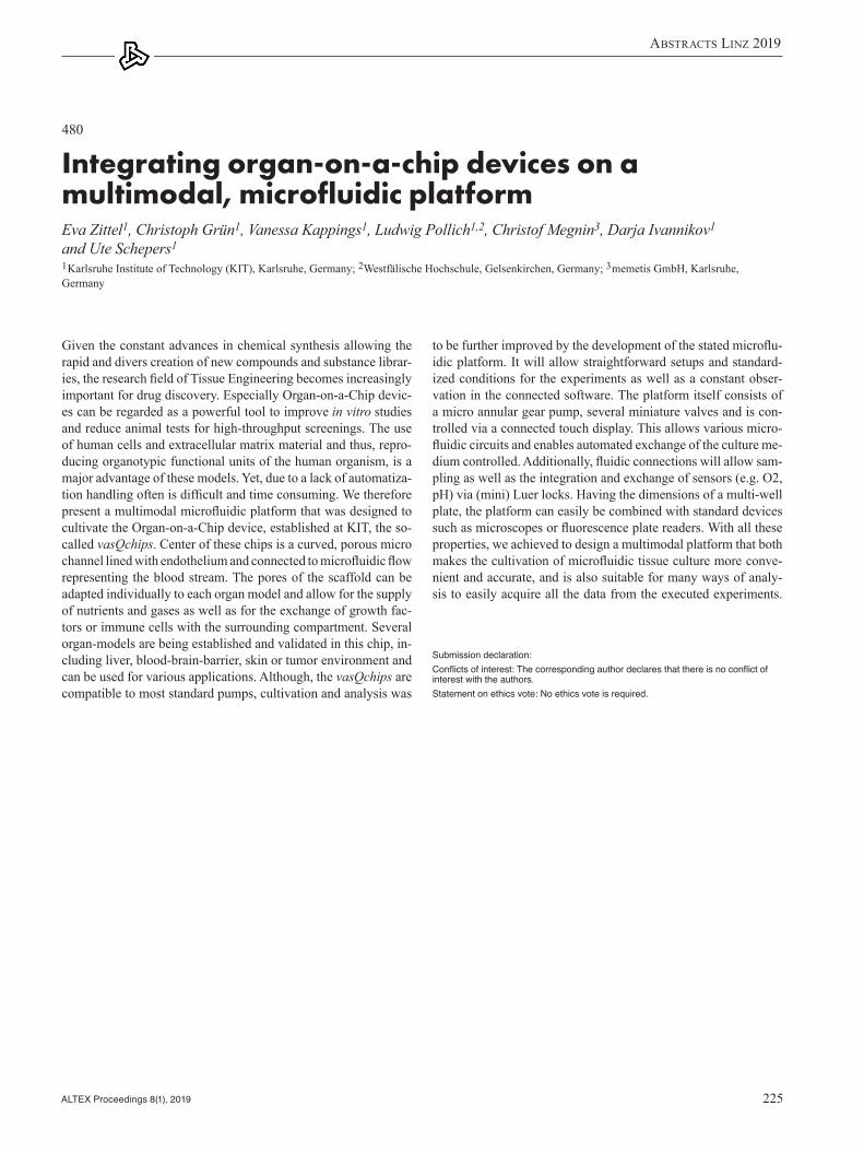
AbstrActs Linz 2019
ALTEX Proceedings 8(1), 2019 225
to be further improved by the development of the stated microflu-idic platform. It will allow straightforward setups and standard-ized conditions for the experiments as well as a constant obser-vation in the connected software. The platform itself consists of a micro annular gear pump, several miniature valves and is con-trolled via a connected touch display. This allows various micro-fluidic circuits and enables automated exchange of the culture me-dium controlled. Additionally, fluidic connections will allow sam-pling as well as the integration and exchange of sensors (e.g. O2, pH) via (mini) Luer locks. Having the dimensions of a multi-well plate, the platform can easily be combined with standard devices such as microscopes or fluorescence plate readers. With all these properties, we achieved to design a multimodal platform that both makes the cultivation of microfluidic tissue culture more conve-nient and accurate, and is also suitable for many ways of analy-sis to easily acquire all the data from the executed experiments.
Submission declaration: Conflicts of interest: The corresponding author declares that there is no conflict of interest with the authors.Statement on ethics vote: No ethics vote is required.
Given the constant advances in chemical synthesis allowing the rapid and divers creation of new compounds and substance librar-ies, the research field of Tissue Engineering becomes increasingly important for drug discovery. Especially Organ-on-a-Chip devic-es can be regarded as a powerful tool to improve in vitro studies and reduce animal tests for high-throughput screenings. The use of human cells and extracellular matrix material and thus, repro-ducing organotypic functional units of the human organism, is a major advantage of these models. Yet, due to a lack of automatiza-tion handling often is difficult and time consuming. We therefore present a multimodal microfluidic platform that was designed to cultivate the Organ-on-a-Chip device, established at KIT, the so-called vasQchips. Center of these chips is a curved, porous micro channel lined with endothelium and connected to microfluidic flow representing the blood stream. The pores of the scaffold can be adapted individually to each organ model and allow for the supply of nutrients and gases as well as for the exchange of growth fac-tors or immune cells with the surrounding compartment. Several organ-models are being established and validated in this chip, in-cluding liver, blood-brain-barrier, skin or tumor environment and can be used for various applications. Although, the vasQchips are compatible to most standard pumps, cultivation and analysis was
480
Integrating organ-on-a-chip devices on a multimodal, microfluidic platformEva Zittel1, Christoph Grün1, Vanessa Kappings1, Ludwig Pollich1,2, Christof Megnin3, Darja Ivannikov1 and Ute Schepers1
1Karlsruhe Institute of Technology (KIT), Karlsruhe, Germany; 2Westfälische Hochschule, Gelsenkirchen, Germany; 3memetis GmbH, Karlsruhe, Germany

AbstrActs Linz 2019
ALTEX Proceedings 8(1), 2019 226
these two tissues. These include different cells layers, embedded spheroids or hydrogel cell cultures as well as 3D bio-printed cel-lular scaffolds. During our investigations with different drug car-riers, we found that the 3D alignment especially of the endothelial cells in the chip has a major impact on their capability of transport and incorporation of drugs. While other setups do not recapitu-late such specific micro environments, the cells within our chip form their natural morphology and exhibit in vivo like behavior. Like this, the introduced vasQchip presents a versatile platform for the screening of many kinds of substances in different spe-cific tissues, as it allows the incorporation of various organotyp-ic endothelial and epithelial cells. In our projects, we therefore constantly work on the establishment and validation of different organ models including liver, blood-brain-barrier, skin or tumor environment, as well as various testing setups concerning perme-ability, transendothelial modification or influence of shear forces.
Submission declaration: Conflicts of interest: The corresponding author declares that there is no conflict of interest with the authors.Statement on ethics vote: No ethics vote is required.
Constant advances in chemical synthesis allow the rapid and div-ers creation of new compounds and substance libraries. Yet, in order to select potential drugs, available screening methods still need to be improved to make them faster, less expensive and safer. While common methods consist of either very simplified cell cul-tures or animal experiments, the field of Tissue Engineering offers new appropriate models to enhance the spectrum of test systems. Human cell material and ECM are used in more complex micro environments, creating a more in vivo like surrounding for cell culturing and thus, representing tissues, organs and eventually the human body in vitro as closely as possible. During the evaluation of drugs, their transportation within an organism and towards the target is a crucial process. As the interplay between blood vessel and adjacent organ tissue has an essential role here, our group at KIT developed a microfluidic chip device, the vasQchip, mimick-ing exactly this area of interest. Center of the chip is a curved, po-rous micro channel lined with endothelium and connected to mi-crofluidic flow representing the blood stream. The round shape of this channel enables an evenly spread shear stress within the sys-tem which can be adjusted via different flow rates for the individu-al conditions needed. In the surrounding compartment, an organo-typic 3D culture can be established, allowing interaction between
474
Testing compound transportation in vitro within a microfluidic blood vessel modelEva Zittel, Vanessa Kappings, Christoph Grün, Darja Ivannikov, Carmen Seidl and Ute SchepersKarlsruhe Institute of Technology (KIT), Karlsruhe, Germany

AbstrActs Linz 2019
ALTEX Proceedings 8(1), 2019 227
ibly sealed chip with a module-based architecture that provides an easy to use workflow, an efficient and precise cell seeding and a removable culture insert that can be transferred between modules. Finally, to characterize the barrier integrity and permeability of the produced skin, electrodes are integrated on the chip to directly quantify the transepithelial electrical resistance (TEER).
In the future, this innovative platform could reduce the depen-dence on animal models and provide a new in vitro tissue system compatible with long-term studies to study skin diseases and eval-uate the safety and efficacy of novel drugs and technologies.
References[1] Groeber, F., Holeiter, M., Hampel, M. et al. (2011). Adv Drug
Deliv Rev 63, 352-366. doi:10.1016/j.addr.2011.01.005 [2] Ackermann, K., Borgia, S. L., Korting, H. C. et al. (2010).
Skin Pharmacol Physiol 23, 105-112. doi:10.1159/000265681
Submission declaration: Conflicts of interest: The corresponding author declares that there is no conflict of interest with the authors.Statement on ethics vote: No ethics vote is required.
In vitro human skin models are gaining attention for their impor-tance as tools for basic research and for the pharmaceutical and cosmetic industries. However, current commercially available hu-man skin models are only suitable for short-term studies due to the time-dependent contraction of their collagen gel-based matrix [1]. Other limitations include the use of non-human extracellular matrix components and lack of dynamical flow systems and me-chanical forces [2].
In recent years, advances in biomaterials and microfluidics technology made it possible for the culture of artificial skin to move a step ahead giving rise to the development of microfluidic skin-on-chip platforms. These systems are able to reproduce key aspects of the in vivo cellular microenvironment by including flu-id flow and finely tuned forces.
This project aims at developing an innovative microfluidic sys-tem to grow and sustain a physiologically relevant human skin model. This approach begins with the production of a fully-hu-manized skin model by combining the production of a fibroblast derived matrix and the use of an inert porous scaffolds for long-term, stable cultivation, without using animal components. This technique is then combined with the use of a biomimetic “organ-on-a-chip” system which includes dynamic perfusion for continu-ous supply of nutrients and metabolites. Also, we present a revers-
428
Fully-humanized skin-on-a-chip with a modular architecture for biomedical applicationsPatricia Zoio1, Abel Oliva1 and João Conde2
1Instituto de Tecnologia Química e Biológica António Xavier (ITQB-NOVA), Oeiras, Portugal; 2INESC Microsistemas e Nanotecnologias, Lisboa, Portugal

AbstrActs Linz 2019
ALTEX Proceedings 8(1), 2019 228
Author IndexAchenbach, John C. 222Adjaye, James 200Adriaens, Els 115Adriani, Giulia 28Ahluwalia, Arti 13Aiello, Maurizio 181, 182Aigelsreiter, Ariane 191Ajvazi, Edip 35Akbarsha, Mohammad Abdulkader 1Alepee, Nathalie 216Altmann, Stephan 2Al-Zeer, Munir 15Amasheh, Salah 25Anderson, David 125Andresen, Niek 3Anzalone, Lucia 42Appelt-Menzel, Antje 60Archibald, Kathy 4Armento, Alex 133Athanassiou, Athanassia 17Ausserlechner, Michael 66, 146Auste, Arne 12Ayehunie, Seyoum 133Ayyash, Muneef 47Azad, Mandeep 5
Bachelor, Michael 116Bachmann, Chiara 135Bailey, Fiona 95Bailey, Jarrod 6, 7, 8Baines, Julia 9Bakchoul, Tamam 172Baker, Elizabeth 10, 11Balks, Elisabeth, 12, 192Balls, Michael 6Bals, Robert 73Baltazar, Maria 37, 142Banno, Yutaka 101, 204Bassi, Anna Maria 13, 207, 211Baumgart, Jan 114, 148Baumgart, Nadine 114, 148Baumgartl-Simons, Christiane 14Bechold, Julian 2Beisswenger, Christoph 73Belik, Vitaly 3Benetto, Simone 129Berberich-Siebelt, Friederike 59Berg, Johanna 15, 193Bernasconi, Camilla 138, 160Bert, Bettina 32
Author Index
Bertelsen, Thomas 16Bertoglio, Federico 152Bertolino, Marco 183Bertorelli, Rosalia 17Bettinelli, Alexandra 15Betzer, Simone 68Bicherel, Pascal 18, 19Biederlack, Julia 20Bienert, Michaela 154Birk, Barbara 21, 22, 216Bischoff, Sabine 208Blümlein, Katharina 198Bohlen, Marie-Léonie 23Bohndorf, Martina 200Bopp, Stephanie 161Bowden, Anthony 142Bowe, Gerry 138Braun, Armin 48Braun, Paula 45Braunbeck, Thomas 24, 121, 122Breves, Gerhard 135Breyfolgle, Bridget 116Briatico-Vangosa, Francesco 152Brown, Jeffrey 99Brück, Dietmar 189Brüggemann, Oliver 35Brunner, Nora 25Buck, Volker 154Buratti, Paolo 181, 182Burdzinski, Wiktor 155Busquet, Francois 26, 27Bussolino, Federico 129Buttgereit, Frank 39, 41, 112, 155, 156, 157
Cable, Sophie 37, 142 Cagiola, Monica 42Campia, Ivana 160Campisi, Marco 28Candinas, Daniel 50Capinha, Liliana 84Carmagnola, Irene 29Carta, Giada 84Carvalho-Wodarz, Cristiane 80Casey, Warren 139Castellano, Maila 183Cavaletti, Guido 13Cavanagh, Brenton 159Cavanaugh, Sarah E. 163Cervetto, Chiara 130Chen, Zhi Jie 30
Cheng, Shu Jun 30, 86, 87Chesne, Christophe 45, 194Chiang, Chiwan 31Chiono, Valeria 13, 28, 29, 82Chmielewska, Justyna 32Cho, Jae-Woo 67Choi, Heung-Koo 85Choi, Hyun-Sung 85Choi, Tae Hyun 33Chrz, Jan 45, 88Chushak, Yaroslav 34Ciardelli, Gianluca 82Cocuzza, Matteo 129Coecke, Sandra 138Cohen, Merav 47Cole, Tom 138Coleman, Robert 4Conde, João 227Conlon, Ross 159Constant, Samuel 35Corte, Giuliano Mario 36, 81Cotter, Mabel 37Creusot, Nicolas 38Csukás, Domokos 203Cull, Tom 37Culot, Maxime 219Curtin, Caroline 159Czekala, Lukasz 195
Damerau, Alexandra 39, 41, 112, 155, 156, 157 Damonte, Gianluca 183Daniel, Gabi 45Deag, Zoe 142Debatis, Michelle 83Degrassi, Cristina 181Dehouck, Lucie 219Delagrange, Mona 37Delannois, Frederique 40Dent, Matthew 139Dettling, Sarah 172Di Paolo, Antonella 42 Diamantara, Kostantina 20Diaspro, Alberto 17Dickmann, Jana 114Diesing, Karoline 39, 41, 155, 156, 157Dietrich, Ann-Christin 15, 193Dodkin, Christina 43Domazet, Sandra 118 Dong, Jingjing 68 Douziech, Mélanie 44

AbstrActs Linz 2019
ALTEX Proceedings 8(1), 2019 229
Drake, Tamara 4Dressler, Dirk 45Dubel, Stefan 99Dura, Adelaide 162Dvorakova, Marketa 46, 88
Ebert, Regina 2Edwards, Alex 95Efstratiou, Androulla 99Egger, Dominik 109Ehrig, Rainald 112Ehrlich, Avner 47Elenschneider, Leroy 48Ellis, Lee 222Emri, Ákos 214Engelhart-Jentzsch, Karin 45Enkelmann, Astrid 208Epars, Maxime 170Erichsen, Lars 200Erickson, Nancy 154Ertl, Peter 60Escher, Sylvia E. 198Eskes, Chantra 49
Fabian, Eric 68Fangmann, Josef 48Fechler-Bitteti, Mona 15Fechner, Henry 15, 193Fehér, Daniella 203Ferencz, Andrea 203, 214Ferrari, Dario 50Fertschai, Ismene 51Fiegl, Heidelinde 66Filipič, Bratko 52Filipova, Dilyana 53Filor, Viviane 54Fiore, Gianfranco Beniamino 13Fischer, Lisa 112Fischer, Melanie 55Fischer, Stephan 56Fischer, Ute 172Focke, Lukas 57Forti, Katia 42Fotiadis, Dimitrios 100Frahm-Barske, Silke 79Frey, Silvia 224Friedl, Heinz-Peter 118Froment, Laurène 169Fuhrmann, Herbert 190, 196Fytianos, Kleanthis 170
Gabbert, Silke 58Gaber, Timo 39, 41, 112, 155, 156, 157Gabusi, Elena 128Gallacher, David J. 205Gallagher, Ciara 159
Gallus, Lorenzo 183Gammel, Maurice R. 215Gearhart, Jeffery 34Gehring, Frank 45Geiger, Fabienne 59Geijtenbeek, Teunis B. H. 220Gerasimenko, Tatyana 105Gerhartl, Anna 60Gericke, Corina 53Gerlach, Claudia 77, 78Giese, Christoph 61Giovine, Marco 183Gleich, Alexander 190Gosselet, Fabien 219Gostner, Johanna 118Götschel, Sebastian 112Gradišnik, Lidija 52Graffmann, Nina 200Grange, Emma 62Grasberger, Jonathan 113Grauthoff, Sven 127Greulich, Andrea 215Grillari, Regina 136Grillari, Johannes 136Grimm, Fabian 63Groeber-Becker, Florian 59Grohmann, Lisa 20Groß, Henrik 64, 75, 189Grün, Christoph 225, 226Grune, Barbara 32Grzeschik, Nina 64Gstraunthaler, Gerhard 143, 217Guenat, Olivier 50, 169, 170
Hackethal, Johannes 65Hagedorn, Paula 54Hagenbuchner, Judith 66, 146Hammer, Helen 68Han, Ji-Seok 67Hana, Kolářová 202Hanegraaff, Ian 45Hans, Fabian 75Hansen, Tanja 48, 198Harrill, Joshua 150Haupt, Tina 216Hayden, Patrick 83, 131Hebesberger, Thomas 191Heckmanns, Annika 68Heinl, Céline 32Heitzer, Ellen 191Hellwich, Olaf 3Helmke, Palle 60Hendriks, A. Jan 44Hendriksen, Coenraad 69Hennerbichler, Simone 65Herms, Fenja 90
Herrmann, Kathrin 70, 71, 72Herrmann, Felix 114Hesler, Michelle 73Hill, Erin 216Hiller, Thomas 15, 193Hilscherova, Klára 74, 147Hippenstiel, Stefan 20Hittinger, Marius 64, 75, 189Hlinkina, Tatsiana 76Hobi, Nina 169, 170Hoff, Paula 156, 157Hoffmann, Pascal 135Hofmann, Lisa 90Hohensee, Christiane 14, 77, 78Hohlbaum, Katharina 3, 79Hollender, Juliane 38Holloway, Marcelle 162Honecker, Anja 73Honscha, Walther 190Hope, Thomas J. 220Horstmann, Justus 80Hosoki, Miku 101Huang, Song 35Huijbregts, Mark A. J. 44Humpenöder, Melanie 36, 81Hundsberger, Harald 201Hurst, Jose 90, 127, 215Hust, Michael 99Huwer, Hanno 169, 170
Idrees, Ayesha 82Im, Jueng Eun 102Ioannidis, Konstantinos 47Ivannikov, Darja 225, 226
Jackson Jr., George R. 83Jaikumar, Gayathri 38Jakob, Franz 2Jang, Sumi 67Jayne, Kimberley 43Jedlovszky-Hajdú, Angéla 214Jennings, Paul 84, 143, 219Jeon, Byoungjun 33Jeon, Hye Jin 85Jiancong, Huang 86, 87Jianhong Feng 86, 87Jírová, Dagmar 46, 88, 95Jo, SeongYea 89, 98, 103Joachim, Stephanie C. 90, 127, 215Joore, Jos 31Juan, Du 86Juhos, Krisztina 203Jukes, Nick 91Jung, Nathalie 92
Kahrass, Hannes 158

AbstrActs Linz 2019
ALTEX Proceedings 8(1), 2019 230
Kaiser, Nadine 114Kalman, Judit 93Kamm, Roger Dale 28Kanamori, Toshiyuki 94Kandarova, Helena 95, 96, 97, 115, 116, 182 Kang, Eun-Hye 89, 98, 103Kang, Kyung Soo 102Kapoor, Dipti 99Kappings, Vanessa 225, 226Karanasiou, Georgia 100Kasper, Cornelia 109Kassab, Rayan 131Kawakami, Hikaru 101Kejlova, Kristina 88, 95Keller, Johannes 22Keogh, Adrian 50Kern, Petra 216Khristichenko, Anna 105Kiebler, Tobias 127Kietzmann, Manfred 54Kim, Hyang Yeon 102Kim, Hyemin 89, 98, 103Kim, Ji-Woo 89, 98, 103Kim, Kyu-Bong 102Kim, Woo-Jin 67Kim, Yong-Bum 67Kim, Younhee 67Kindeeva, Olga 105King, Henry 44 Kirchsteiger, Maria 60Klausner, Mitchell 83, 131, 133Kliphuis, Saskia 210Knebel, Jan 48, 198Knoespel, Fanny 193Knoll, Thorsten 73Knudsen, Lisbeth 104Knyazev, Evgeny 105Kohl, Yvonne 73Kojima, Hajime 106, 107Kolle, Susanne N. 22, 58, 216Kostal, Jakub 108Kour, Kawardeep 5Kramer, Nynke 149Kreir, Mohamed 205Kreß, Sebastian 109Krimmling, Tanja 110, 140Kristína, Kejlová 202Kruip, Carina 12Kühn, Sandra 90Kukic, Predrag 37Kurreck, Jens 15, 193
Ladwig-Wiegard, Mechthild 36, 81Lambrigts, Denis 40 Lamplmair, Franz 111
Landry, Timothy 133Landsiedel, Robert 22, 58, 68, 216Lang, Annemarie 20, 39, 41, 112, 155, 156, 157Langezaal, Ingrid 138, 139Lanz, Henriette 31Lanzerstorfer, Peter 113Lass-Flörl, Cornelia 220Lau, Clara Bik San 117Ledermann, Christina 77, 78Lee, Byoung-Seok 67Lee, Jung Dae 102Lee, Kwon-Seob 85Lee, Mi-Young 37Lee, Sharon W. L. 28Lehmann, Judith 135Lehr, Claus-Michael 64, 75, 80, 169, 170, 189Leidinger, Charlotte Sophie 114Leitgeb, Tamara 118Letasiova, Silvia 83, 95, 115, 116, 131, 132 Leung, Ping Chung 117Lewejohann, Lars 3Li, Lucia 117Li, Ruoya 194Liang, Bo 117Liebsch, Manfred 117Lillicrap, Adam 55Lin, Grace 60, 118Linzey, Clair 119Liska, Roman 120, 139Liskova, Alzbeta 95Loginovskaya, Ludmila 76Loizou, George 137Long, Nathaniel F. 131Loretz, Brigitta 75Lörracher, Ann-Kathrin 121, 122Louhimies, Susanna 124, 125, 126, 127 Lu, Hua Rong 205Lubitz, Annika 61
Mach, Mojmír 96Ma-Hock, Lan 22Mainardi, Andrea 213Maione, Anna 83, 131Maliha, Ana Maria 90, 127Manferdini, Cristina 128Marasso, Simone Luigi 129 Marcoli, Manuela 130Marengo, Kaitlyn 131Marian, Rucki 202Marini, Irene 172Markéta, Dvořáková 202Markus, Jan 83, 132, 133, 182, 188 Marlinghaus, Lennart 82
Marqeño, Anna 165Marrella, Alessandra 181, 182Marshall, Lindsay 134Martin, Solenne 194Martinelli, Eleonora 35Martinoia, Sergio 130Martins, Soraia 200Marx, Uwe 199Marx-Stoelting, Philip 221Mathea, Miriam 58Mattu, Clara 28Maura, Guido 130May, Nina T. 135Mazidi, Zahra 136Mazija, Hrvoje 52McNally, Kevin 137Megnin, Christof 225Meißner, Jessica 54Mendoza, Emilio 161Mensch, Alexander 15Merle, Roswitha 36, 81Mertz, Marcel 158Metz, Julia Katharina 75Metzger, Marco 60Michel, Geert 154Middleton, Alistair 37Milasova, Tatiana 116Milcamps, Anne 138, 139Milec, Lucia 96Millo, Enrico 183Mirata, Serena 183Moer, Jana 140Mojzisek, Martin 181Mólnár, Kristóf 214Mombello, Domenico 129Moos, Bärbel 68Morash, Mike 222Morilla, Maria Jose 186Motschnig, Tomas 141Mühlebach, Michael 12Muller, Iris 142Mundhenk, Lars 154Murphy, Cormac 143Mut, Jürgen 2
Na, Ellen 154Nahmias, Yaakov 47Naqvi, Kubra 12Nardelli, Laurent 216Nastrucci, Candida 144, 145Natsch, Andreas 216Navas, José M. 93Ncube, Audrey 200Neuhaus, Winfried 60, 118Neumann, Gaby 53Nguyen, Lisa 200

AbstrActs Linz 2019
ALTEX Proceedings 8(1), 2019 231
Nicolas, Arnaud 31Nielsen, Rasmus Normann 212Nikulin, Sergey 105Nishikubo, Nanae 204Nolan, John C. 159Note, Reine 47Nothdurfter, Daniel 146Novak, Jiri 147Nowak, Norman 198Nuber, Maximilian 148Nunes, Carolina 149, 161Nyffeler, Johanna 150
O’Brien, Fergal J. 159Occhetta, Paola 213Odum, Jenny 139Oelgeschläger, Michael 221Oktivaningrum, Rafika 44Oldenkamp, Rik 44Oliva, Abel 227Oredsson, Stina 151Osaki, Tatsuya 28Ouedraogo, Gladys 47
Pacheco, Daniela 152Pamies, David 149Paparella, Martin 153Park, Han-Jin 89, 98, 103Park, Heejin 67Partosa, Natascha 22Pathan, Saif G. 131Paul Friedman, Katie 150Pelassa, Simone 130Pérez-Albaladejo, Elisabet 165Peter, Dominique 154Peters, Matt 133Petrini, Paola 152Petrov, Vladimir 105Petry, Monique 54Pfeiffenberger, Mori-tz 39, 41, 155, 156, 157Pfitzner, Inka 45Pfützner, Marcel 36, 81Phaneuf, Matthew D. 131Pichl, Dorothea 114Pietschmann, Ines 158Pípal, Marek 74Pippin, John J. 163Pirri, Candido Fabrizio 129Piskareva, Olga 159Pistollato, Francesca 138, 160, 161, 162, 163Plasenzotti, Roberto 141, 164, 175Plendl, Johanna 36, 81Pollich, Ludwig 225Porte, Cinta 165
Posch, Wilfried 220Possenti, Luca 28Pötz, Oliver 68Pound, Pandora 166Poyanena, Anastasiya 76Pozzolini, Marina 183Pracser, Nadja 60Prakash, Nilima 167Price, Anna 161Priebojová, Jana 74Primo, Luca 129Proença, Susana 149Proust, Yara-Maria 169Prykhodko, Olena 132Puliafito, Alberto 129Pullirsch, Dieter 168Püschel, Gerhard 185
Raabe, Hans 216Ragas, Ad 44Raggi, Giulia 169, 170Rahman, Md Shaifur 200Ram, Rebecca 166Rapet, Aude 169, 170Rasponi, Marco 213Redl, Heinz 65Redmond, Craig 171Rehberger, Domenik 172Reid, Kirsty 173, 174Reininger-Gutmann, Birgit 164, 175Ren, Peofeng 68Retter, Ida 20Reuter, Andreas 12, 192Rex, André 79Reynolds, Georgia 37Reynolds, Joe 37Rhodes, Nadine 118Richtig, Erika 191Riffle, Brandy 68Rinner, Beate 191Ritzmann, Felix 73Riu, Anne 47Roberts, David 176, 177Röblitz, Susanna 112Röhrs, Viola 15, 193Roldan, Nuria 169, 170Romero, Eder L. 186Rossi, Angela 118Rothbauer, Mario 60Rucki, Marian 45, 88, 216Rudd, Kathryn 195Ruf, Christopher 75, 189Runge, Dieter 110, 140Ruschig, Maximilian 178Russell, Paul 179, 180Ruwona, Tinashe 216
Ryan, Cindy 216
Saccà, Sergio C. 207, 211Sahigara, Faizan 19Sakai, Yasuyuki 184Sakamoto, Manato 204Sakazaki, Fumitoshi 204Sakharov, Dmitry 105Salber, Jochen 82Salis, Annalisa 183Sams, Craig 137Samuga, Amita 68Sandner, Georg 113Sándor, József 203Scaglione, Silvia 181, 182Scarfì, Sonia 183, 207, 211Schäfer-Korting, Monika 186Scharnowske, Lara 75Scheidecker, Benedikt 184Schenke, Maren 185Schentarra, Eva-Maria 172Schepers, Ute 225, 226Schiffner, René 208Schilrreff, Priscila 186Schindler, Stefanie 187Schirmer, Kristin 38, 55, 56Schjeide, Brit-Maren 185Schlenke, Peter 191Schlüter, Sebastian 15Schmidt, Christian 188Schmidt, Felix 68Schmidt, Konrad 15Schmieder, Natascha 68Schmitz, Inge 82Schneider, Marc 64, 189Schneider-Daum, Nicole 75, 80, 169, 170Schnichels, Sven 90, 127, 215Schnur, Sabrina 189Schoeniger, Axel 190, 196Schönfelder, Gilbert 32, 221Schröder, Klaus 35Schrom, Silke 191Schuenborg, Thomas 192Schug, Hannah 56Schwarz, Katharina 198Schwarzkopf, Konrad 64Seeger, Bettina 135, 185Sehnal, Luděk 74Seibel, Jürgen 2Seidl, Carmen 226Seidle, Troy 134Seiler-Wulczyn, Andrea 117Sellman, Zachary 131Sesardic, Thea 99Ševčík, Václav 45Severi, Giulio 42

AbstrActs Linz 2019
ALTEX Proceedings 8(1), 2019 232
Shaef, Ida 15, 193Shaid, Shahjahan 40Sharma, Ashwani 194Shasha, Ma 86, 87Sim, Bushra 95Simms, Liam 195Sittel, Anna-Maria 196Smith, Adrian 197Smutná, Marie 74Somogyvari, Ferenc 52Sonnenschein, Nico 198Spano, Raffaele 17Spicher, Carolin 14, 77, 78Spielmann, Horst 117, 199Spiric, Jelena 12, 192Spitzhorn, Lucas-Sebastian 200Stadnicka-Michalak, Julita 55Steinbrenner, Jutta 22Steubing, Jonathan 172Steubing, Rosemarie 172Stevens, Zachary 133Stevenson, Matthew 195Stickings, Paul 99Stierschneider, Anna 201Stolper, Gina 131Stroka, Deborah 50Stübl, Flora 113Stucki, Andreas 169Stucki, Janick 169, 170Suarato, Giulia 17Suarez Vargas, Natalia 152Sun, Qiuhua 117Sung, Baeckkyoung 23Surdo, Salvatore 17Svobodová, Lada 45, 88, 202Szabó, Györgyi 203, 214
Tanaka, Ryuichiro 101, 204Tang, Dawei 37Tang, Ling Yin 117Teasdale, Ian 35Tedesco, Mariateresa 130Teisman, Ard 205Teuschl, Andreas 65Thalmann, Beat 172Theiventhran, Mathura 142Thöne-Reineke, Christa 3, 36, 79, 81, 114, 154, 206Tingting, Luo 86, 87Tirendi, Sara 207, 211Tolksdorf, Beatrice 193Torrent, Fernando 93 Toth, Sandor 52Treasure, Carol 95Trelles-Sticken, Edgar 195
Trietsch, Sebastiaan 31Trietschel, David 208Trucco, Diego 128Tumbev, Yosif 59Turner, Jan 4
Ullmann, Kristina 79Ullrich, Anett 110, 140
van der Valk, Jan 209, 210, 217van Ravenzwaay, Bennard 22, 68Van Rompay, An 115van Uden, Sebastião 152van Zelm, Rosalie 44Vanfleteren, Jan 47Varani, Gabriele 181Verkaart, Sjoerd 216Vernazza, Stefania 207, 211Verstraelen, Sandra 115Vicini, Silvia 183Viebahn, Richard 82Vindahl Olsen, Birgitte 212Violet, Norman 221Visai, Livia 13, 152Visentin, Sonja 152Visone, Roberta 213Vladetic, Alexandra 60, 118Vogl, Silvia 221von Briesen, Hagen 73von Köckritz-Blickwede, Maren 54Voniatis, Constantinos 203, 214Vulto, Paul 31
Wagner, Kristina 217Wagner, Natalie 215Wagner, Sylvia 73Waidmann, Marc 172Wambaugh, John 150Wang, Chi C. 117Wang, Mao 68Wang, Rongyun 117Wareing, Britta 22, 216Weber, Marie-Christin 112Weber, Tilo 217Weber, Zia 15Wéber, György 203Weghuber, Julian 113Wehmeier, Oliver 57Weich, Kerstin 218Wellens, Sara 219Wenzel, Esther 99Westerink, Walter 216Whelan, Maurice 160Wieczorek, Roman 195Wiegrebe, Alexander 48
Wieser, Matthias 136Wiesner, Christoph 201Wiest, Joachim 188, 217Wilflingseder, Doris 220Willett, Catherine 134, 179Willis, Clinton 150Wilmes, Anja 143Wilschut, Karlijn 31Windbergs, Maike 92Wiszniewski, Ludovic 35Wittkowski, Paul 221Wittler, Lars 154Wittwehr, Clemens 160Wizenmann, Andrea 167Wohlleben, Wendel 22Wolf, Natalia 2Wöllhaf, Manuel 3Woodland, Cindy L. A. 222Worth, Andrew 161Wruck, Wasco 200
Yamaguchi, Shoji 101Yang, Mi-Jin 67
Zaderer, Viktoria 220, 223Zäh, Ralf-Kilian 189Zamprogno, Pauline 169Zeimet, Alain 66Zemanova, Miriam A. 224Ziccarelli, Anna 152Zietek, Tamara 53Zimmermann, Heiko 73Zittel, Eva 225, 226Zoio, Patricia 227Zoschke, Christian 186Zoso, Alice 29Zuang, Valérie 138Zurich, Marie-Gabrielle 149

ALTEX Proceedings 8, 1/2019
ImprInt
Issued byALTEX Edition, Kreuzlingen, Switzerland
Board:Daniel Favre Franz P. Gruber Gerhard Gstraunthaler Thomas Hartung Goran Krummenacher Beatrice RothKristina Wagner
Members:The members of the Society ALTEX Edition can be found at www.altex.org
ALTEX Edition Editorial Office EuropeSonja von Aulock (Editor in chief, CEO)Petra Mayr (Editor TIERethik)Carolin Rauter (Technical editor) Goran Krummenacher (Webmaster)
AddressALTEX EditionRomanshornerstrasse 908280 Kreuzlingen, Switzerlande-mail: [email protected]
ALTEX Edition Editorial Office USAMartin L. Stephens (North American Editor)Thomas HartungMichael Hughes
AddressJohns Hopkins University Baltimore615 N Wolfe Street, W7032 BaltimoreMD 21020, USAe-mail: [email protected]
LayoutH. P. Hoesli
ALTEX Proceedings is published online:www.altex.org
ALTEX Proceedingswww.altex.org
Vol. 8, No. 1 (2019)
ALTEX Proceedings publishes Abstract Books and Proceedings of scientific conferences and symposia on the development and pro-motion of alternatives to animal experiments according to the 3R concept of Russell and Burch: Replace, Reduce, and Refine in coop-eration with the organizers of the respective meeting.
© ALTEX Edition, Kreuzlingen, Switzerland
ACKNOWLEDGEMENTS – The Co-Chairs, the organisers and the Scientifi c Committee would like to express their gratitude to all those who are generously supporting the congress!
www.eusaat-congress.eu10 – 13 October 2019 – University of Linz, Austria
Supporting Sponsors
Main Sponsors
Supporters
ACADEMYFOR ANIMAL WELFARE
Logos and names of sponsors reproduced with their permission.
LINZ 2019 22nd European Congress on Alternatives to Animal Testing
EUSAAT 201919th Annual Congress of EUSAAT