volume 6 number 30 8 august, 2012 - repository.ugm.ac.id_nitrogen... · journal of medicinal plants...
TRANSCRIPT

Journal of
Medicinal Plants Research
Volume 6 Number 30 8 August, 2012
ISSN 1996-0875

ABOUT JMPR The Journal of Medicinal Plant Research is published weekly (one volume per year) by Academic Journals. The Journal of Medicinal Plants Research (JMPR) is an open access journal that provides rapid publication (weekly) of articles in all areas of Medicinal Plants research, Ethnopharmacology, Fitoterapia, Phytomedicine etc. The Journal welcomes the submission of manuscripts that meet the general criteria of significance and scientific excellence. Papers will be published shortly after acceptance. All articles published in JMPR are peer-reviewed. Electronic submission of manuscripts is strongly encouraged, provided that the text, tables, and figures are included in a single Microsoft Word file (preferably in Arial font).
Submission of Manuscript Submit manuscripts as e-mail attachment to the Editorial Office at: [email protected]. A manuscript number will be mailed to the corresponding author shortly after submission. The Journal of Medicinal Plant Research will only accept manuscripts submitted as e-mail attachments. Please read the Instructions for Authors before submitting your manuscript. The manuscript files should be given the last name of the first author.

Editors Prof. Akah Peter Achunike Editor-in-chief Department of Pharmacology & Toxicology University of Nigeria, Nsukka Nigeria Associate Editors Dr. Ugur Cakilcioglu Elazıg Directorate of National Education Turkey. Dr. Jianxin Chen Information Center, Beijing University of Chinese Medicine, Beijing, China 100029, China. Dr. Hassan Sher Department of Botany and Microbiology, College of Science, King Saud University, Riyadh Kingdom of Saudi Arabia. Dr. Jin Tao Professor and Dong-Wu Scholar, Department of Neurobiology, Medical College of Soochow University, 199 Ren-Ai Road, Dushu Lake Campus, Suzhou Industrial Park, Suzhou 215123, P.R.China. Dr. Pongsak Rattanachaikunsopon Department of Biological Science, Faculty of Science, Ubon Ratchathani University, Ubon Ratchathani 34190, Thailand.
Prof. Parveen Bansal Department of Biochemistry Postgraduate Institute of Medical Education and Research Chandigarh India. Dr. Ravichandran Veerasamy AIMST University Faculty of Pharmacy, AIMST University, Semeling – 08100, Kedah, Malaysia. Dr. Sayeed Ahmad Herbal Medicine Laboratory, Department of Pharmacognosy and Phytochemistry, Faculty of Pharmacy, Jamia Hamdard (Hamdard University), Hamdard Nagar, New Delhi, 110062, India. Dr. Cheng Tan Department of Dermatology, first Affiliated Hospital of Nanjing Univeristy of Traditional Chinese Medicine. 155 Hanzhong Road, Nanjing, Jiangsu Province, China. 210029 Dr. Naseem Ahmad Young Scientist (DST, FAST TRACK Scheme) Plant Biotechnology Laboratory Department of Botany Aligarh Muslim University Aligarh- 202 002,(UP) India. Dr. Isiaka A. Ogunwande Dept. Of Chemistry, Lagos State University, Ojo, Lagos, Nigeria.

Editorial Board
Prof Hatil Hashim EL-Kamali Omdurman Islamic University, Botany Department, Sudan. Prof. Dr. Muradiye Nacak Department of Pharmacology, Faculty of Medicine, Gaziantep University, Turkey. Dr. Sadiq Azam Department of Biotechnology, Abdul Wali Khan University Mardan, Pakistan. Kongyun Wu Department of Biology and Environment Engineering, Guiyang College, China. Prof Swati Sen Mandi Division of plant Biology, Bose Institute India. Dr. Ujjwal Kumar De Indian Vetreinary Research Institute, Izatnagar, Bareilly, UP-243122 Veterinary Medicine, India.
Dr. Arash Kheradmand Lorestan University, Iran. Prof Dr Cemşit Karakurt Pediatrics and Pediatric Cardiology Inonu University Faculty of Medicine, Turkey. Samuel Adelani Babarinde Department of Crop and Environmental Protection, Ladoke Akintola University of Technology, Ogbomoso Nigeria. Dr.Wafaa Ibrahim Rasheed Professor of Medical Biochemistry National Research Center Cairo Egypt.

Electronic submission of manuscripts is strongly encouraged, provided that the text, tables, and figures are included in a single Microsoft Word file (preferably in Arial font). The cover letter should include the corresponding author's full address and telephone/fax numbers and should be in an e-mail message sent to the Editor, with the file, whose name should begin with the first author's surname, as an attachment. Article Types Three types of manuscripts may be submitted: Regular articles: These should describe new and carefully confirmed findings, and experimental procedures should be given in sufficient detail for others to verify the work. The length of a full paper should be the minimum required to describe and interpret the work clearly. Short Communications: A Short Communication is suitable for recording the results of complete small investigations or giving details of new models or hypotheses, innovative methods, techniques or apparatus. The style of main sections need not conform to that of full-length papers. Short communications are 2 to 4 printed pages (about 6 to 12 manuscript pages) in length. Reviews: Submissions of reviews and perspectives covering topics of current interest are welcome and encouraged. Reviews should be concise and no longer than 4-6 printed pages (about 12 to 18 manuscript pages). Reviews are also peer-reviewed. Review Process All manuscripts are reviewed by an editor and members of the Editorial Board or qualified outside reviewers. Authors cannot nominate reviewers. Only reviewers randomly selected from our database with specialization in the subject area will be contacted to evaluate the manuscripts. The process will be blind review. Decisions will be made as rapidly as possible, and the journal strives to return reviewers’ comments to authors as fast as possible. The editorial board will re-review manuscripts that are accepted pending revision. It is the goal of the JMPR to publish manuscripts within weeks after submission.
Regular articles All portions of the manuscript must be typed double-spaced and all pages numbered starting from the title page. The Title should be a brief phrase describing the contents of the paper. The Title Page should include the authors' full names and affiliations, the name of the corresponding author along with phone, fax and E-mail information. Present addresses of authors should appear as a footnote. The Abstract should be informative and completely self-explanatory, briefly present the topic, state the scope of the experiments, indicate significant data, and point out major findings and conclusions. The Abstract should be 100 to 200 words in length.. Complete sentences, active verbs, and the third person should be used, and the abstract should be written in the past tense. Standard nomenclature should be used and abbreviations should be avoided. No literature should be cited. Following the abstract, about 3 to 10 key words that will provide indexing references should be listed. A list of non-standard Abbreviations should be added. In general, non-standard abbreviations should be used only when the full term is very long and used often. Each abbreviation should be spelled out and introduced in parentheses the first time it is used in the text. Only recommended SI units should be used. Authors should use the solidus presentation (mg/ml). Standard abbreviations (such as ATP and DNA) need not be defined. The Introduction should provide a clear statement of the problem, the relevant literature on the subject, and the proposed approach or solution. It should be understandable to colleagues from a broad range of scientific disciplines. Materials and methods should be complete enough
to allow experiments to be reproduced. However, only truly new procedures should be described in detail; previously published procedures should be cited, and important modifications of published procedures should be mentioned briefly. Capitalize trade names and include the manufacturer's name and address. Subheadings should be used. Methods in general use need not be described in detail.
Instructions for Author

Results should be presented with clarity and precision. The results should be written in the past tense when describing findings in the authors' experiments. Previously published findings should be written in the present tense. Results should be explained, but largely without referring to the literature. Discussion, speculation and detailed interpretation of data should not be included in the Results but should be put into the Discussion section. The Discussion should interpret the findings in view of the results obtained in this and in past studies on this topic. State the conclusions in a few sentences at the end of the paper. The Results and Discussion sections can include subheadings, and when appropriate, both sections can be combined. The Acknowledgments of people, grants, funds, etc should be brief. Tables should be kept to a minimum and be designed to be as simple as possible. Tables are to be typed double-spaced throughout, including headings and footnotes. Each table should be on a separate page, numbered consecutively in Arabic numerals and supplied with a heading and a legend. Tables should be self-explanatory without reference to the text. The details of the methods used in the experiments should preferably be described in the legend instead of in the text. The same data should not be presented in both table and graph form or repeated in the text. Figure legends should be typed in numerical order on a separate sheet. Graphics should be prepared using applications capable of generating high resolution GIF, TIFF, JPEG or Powerpoint before pasting in the Microsoft Word manuscript file. Tables should be prepared in Microsoft Word. Use Arabic numerals to designate figures and upper case letters for their parts (Figure 1). Begin each legend with a title and include sufficient description so that the figure is understandable without reading the text of the manuscript. Information given in legends should not be repeated in the text. References: In the text, a reference identified by means of an author‘s name should be followed by the date of the reference in parentheses. When there are more than two authors, only the first author‘s name should be mentioned, followed by ’et al‘. In the event that an author cited has had two or more works published during the same year, the reference, both in the text and in the reference list, should be identified by a lower case letter like ’a‘ and ’b‘ after the date to distinguish the works.
Examples:
Abayomi (2000), Agindotan et al. (2003), (Kelebeni, 1983), (Usman and Smith, 1992), (Chege, 1998;
1987a,b; Tijani, 1993,1995), (Kumasi et al., 2001) References should be listed at the end of the paper in alphabetical order. Articles in preparation or articles submitted for publication, unpublished observations, personal communications, etc. should not be included in the reference list but should only be mentioned in the article text (e.g., A. Kingori, University of Nairobi, Kenya, personal communication). Journal names are abbreviated according to Chemical Abstracts. Authors are fully responsible for the accuracy of the references. Examples: Chikere CB, Omoni VT and Chikere BO (2008). Distribution of potential nosocomial pathogens in a hospital environment. Afr. J. Biotechnol. 7: 3535-3539. Moran GJ, Amii RN, Abrahamian FM, Talan DA (2005). Methicillinresistant Staphylococcus aureus in community-acquired skin infections. Emerg. Infect. Dis. 11: 928-930. Pitout JDD, Church DL, Gregson DB, Chow BL, McCracken M, Mulvey M, Laupland KB (2007). Molecular epidemiology of CTXM-producing Escherichia coli in the Calgary Health Region: emergence of CTX-M-15-producing isolates. Antimicrob. Agents Chemother. 51: 1281-1286. Pelczar JR, Harley JP, Klein DA (1993). Microbiology: Concepts and Applications. McGraw-Hill Inc., New York, pp. 591-603.
Short Communications Short Communications are limited to a maximum of two figures and one table. They should present a complete study that is more limited in scope than is found in full-length papers. The items of manuscript preparation listed above apply to Short Communications with the following differences: (1) Abstracts are limited to 100 words; (2) instead of a separate Materials and Methods section, experimental procedures may be incorporated into Figure Legends and Table footnotes; (3) Results and Discussion should be combined into a single section. Proofs and Reprints: Electronic proofs will be sent (e-mail attachment) to the corresponding author as a PDF file. Page proofs are considered to be the final version of the manuscript. With the exception of typographical or minor clerical errors, no changes will be made in the manuscript at the proof stage.

Fees and Charges: Authors are required to pay a $600 handling fee. Publication of an article in the Journal of Medicinal Plant Research is not contingent upon the author's ability to pay the charges. Neither is acceptance to pay the handling fee a guarantee that the paper will be accepted for publication. Authors may still request (in advance) that the editorial office waive some of the handling fee under special circumstances. Copyright: © 2012, Academic Journals. All rights Reserved. In accessing this journal, you agree that you will access the contents for your own personal use but not for any commercial use. Any use and or copies of this Journal in whole or in part must include the customary bibliographic citation, including author attribution, date and article title. Submission of a manuscript implies: that the work described has not been published before (except in the form of an abstract or as part of a published lecture, or thesis) that it is not under consideration for publication elsewhere; that if and when the manuscript is accepted for publication, the authors agree to automatic transfer of the copyright to the publisher. Disclaimer of Warranties In no event shall Academic Journals be liable for any special, incidental, indirect, or consequential damages of any kind arising out of or in connection with the use of the articles or other material derived from the JMPR, whether or not advised of the possibility of damage, and on any theory of liability. This publication is provided "as is" without warranty of any kind, either expressed or implied, including, but not limited to, the implied warranties of merchantability, fitness for a particular purpose, or non-infringement. Descriptions of, or references to, products or publications does not imply endorsement of that product or publication. While every effort is made by Academic Journals to see that no inaccurate or misleading data, opinion or statements appear in this publication, they wish to make it clear that the data and opinions appearing in the articles and advertisements herein are the responsibility of the contributor or advertiser concerned. Academic Journals makes no warranty of any kind, either express or implied, regarding the quality, accuracy, availability, or validity of the data or information in this publication or of any other publication to which it may be linked.

International Journal of Medicine and Medical SciencesJournal of Medicinal Plants Research
Table of Contents: Volume 6 Number 30 8 August, 2012
ences ARTICLES
Research Articles Effects of irrigation treatment and Azospirillum inoculation on yield and yield component of black cumin (Nigella sativa L.) 4553 Mohammad Reza Haj Seyed Hadi, Mohammad Taghi Darzi and Zohreh Ghandehari Antioxidant activity and phenolic content of germinated lentil (Lens culinaris) 4562 Maryam Gharachorloo, Babak Ghiassi Tarzi, Marzieh Baharinia and Amir Homan Hemaci In vitro evaluation of hemostatic properties of the sap of Jatropha multifida L. (Euphorbiaceae) 4567 Jean Robert Klotoé, Victorien Tamègnon Dougnon, Jean-Marc Atègbo, Ferdinand Adounkpè, Julien Sègbo, Brice Fanou, Lauris Fah, Patrick Aléodjrodo Edorh, Carlos Dandjesso, Hornel Koudokpon, Donatien Klouyo, Frédéric Loko and Karim Dramane
Phytochemical screening of some indigenous medicinal plant species used in the management of diabetes mellitus in Ghana 4573 Y. Ameyaw, V. Y. A. Barku, J. Ayivor and A. Forson Preliminary phytochemical and biological screening of methanolic and acetone extracts from Leonotis nepetifolia (L.) R. Br. 4582 Danuta Sobolewska, Paweł Paśko, Agnieszka Galanty, Justyna Makowska-Wąs, Kinga Padło and Wojciech Wasilak

ARTICLES Effect of shading, nitrogen and magnesium fertilizer on phyllanthin and total flavonoid yield of Phyllanthus niruri in Indonesia soil 4586 Eko Hanudin, Hardhina Wismarini, Triana Hertiani and Bambang Hendro Sunarminto
Table of Contents: Volume 6 Number 30 8 August, 2012
ences

Journal of Medicinal Plants Research Vol. 6(30), pp. 4586-4592, 8 August, 2012 Available online at http://www.academicjournals.org/JMPR DOI: 10.5897/JMPR12.591 ISSN 1996-0875 ©2012 Academic Journals
Full Length Research Paper
Effect of shading, nitrogen and magnesium fertilizer on phyllanthin and total flavonoid yield of Phyllanthus
niruri in Indonesia soil
Eko Hanudin1*, Hardhina Wismarini1, Triana Hertiani2 and Bambang Hendro Sunarminto1
1Department of Soil Sciences, Faculty of Agriculture, Universitas Gadjah Mada (UGM), Yogyakarta, Indonesia.
2Laboratory of Pharmaceutical Biology, Faculty of Pharmacy, Universitas Gadjah Mada (UGM), Yogyakarta, Indonesia.
Accepted 19 June, 2012
Phyllanthus niruri is widely used as traditional medicine especially in Indonesia. The need to produce a standardized herbal medicine raw material has urged a research to study the effect of shading, nitrogen (N) and magnesium (Mg) fertilizer dosage on phyllanthin and total flavonoid yield of P. niruri. Pot experiment was conducted by using Inceptisols soil from Yogyakarta, Indonesia. The study was carried out in a split plot design with three replications. The main plot was shaded with 3 levels (0, 60 and 95%). The subplots was urea and magnesium suphate (MgSO4) fertilizer (proportion 5 : 1) with 5 levels (0, 1+0.2, 2+0.4, 3+0.6 and 4+0.8). The period of harvesting were 1, 2 and 3 months after planting. The results indicated that application of 3 g urea, 0.6 g MgSO4 and 60% paranets shading could improve the plant growth, N-Mg uptake, total flavonoid and phyllanthin yield of P.niruri. Based on the regression curve, the optimum dosage that produced the highest total flavonoid yield was 2.68 g of urea and 0.54 g of MgSO4, while for obtaining the highest phyllanthin yield was 2.39 g of urea and 0.48 g of MgSO4 per plant. N and Mg absorption were positively correlated with total flavonoid and phyllanthin yield. The best period of harvest to obtain the highest total flavonoid and phyllanthin yield was 3 months after planting (plant age ± 4 months). Key words: Phyllanthus niruri, shading, nitrogen (N) and magnesium (Mg) fertilizer, phyllanthin, total flavonoid.
INTRODUCTION Phyllanthus niruri (Euphorbiaceae) is a small plant widely distributed in tropical and subtropical regions in Central and South America (Brazil) and Asia (including India, Malaysia and Indonesia). The other species commonly found in the countries are: P. niruri L., P. tenellus Roxb., Phyllanthus urinaria L., P. caroliniensis Walt., P. amarus Schum. and Thonn., and P. stipulatus (Raf.) Webster (Martin et al., 2011). Many components of P. niruri have been identified, including groups of active substances such as alkaloids, tannin, lignans, phenols, steroids, flavonoids, triterpenes (Barros et al., 2003). The major lignans of this plant, namely, phyllanthin and hypophyllanthin, have shown to be anti hepatotoxic against carbon tetrachloride and galactosamine *Corresponding author. E-mail: [email protected]. Tel/Fax: +62274548814.
-induced hepatotoxicity in primary cultured rat hepatocytes (Syamsundar et al., 1985). Phyllanthin and hypophyllanthin can be extracted from P. niruri with solvent methanol, ethyl acetate and chloroform (Tripathi et al., 2006). The plants of genus Phyllanthus have long been used in folk medicine to treat kidney and urinary bladder stones, intestinal infections, diabetes and hepatitis B (Nayak et al., 2010). The whole plant is used as diuretic, febrifuge and in stomach trouble (Kotoky and Kanjilal, 2005). In recent years, the interest of these plants has increased considerably, due to its hepatoprotective action.
Plants need light intensity in different amount. Light intensity can affect micro environment of a plant, such as temperature and air humidity; that have influence to metabolism in plant which would involve the help of various enzymes. The enzymes would work actively in sufficient light intensity (Tang, 1997). Biosynthesis of flavonoids in plants is influenced by several factors,

namely nutrients, temperature, and light. Some metabolic process of flavonoids are controlled by plant growth regulators and light (Robinson, 1991). Flavonoids and anthocyanins biosynthesis are occurred likely in bright conditions (Morris et al., 1985). According to Padua et al. (1999), formations of flavonoid compounds in plants are usually related to the plant defensive mechanism to protect them self from high light intensity.
Nitrogen (N) as an essential macronutrient has an important role as a constituent of chlorophyll and protein synthesis. Amino acids as building blocks of protein are precursors of secondary metabolites formation (flavonoids and phyllanthin). Apart from being a constituent of chlorophyll, magnesium (Mg) also plays a role in N metabolism. Mg has some role in activating the enzyme associated with metabolic process, respiratory enzymes, and work as a catalyst. In addition, Mg serves as a cofactor in enzymes, especially to activate the phosphorylase (Mengel and Kirkby, 1987).
P. niruri can grow on various types of soil, especially sandy soil, and usually grows wild in damp areas. Inceptisols is one of soil orders which is most widely spread in Indonesia, about 70.25 million ha or 37.5% of Indonesia's land area. This soil has a diverse natural productivity because they do not have special physical and chemical characteristics. Therefore, inceptisol utilization for the future must be increased to the maximum.
The objectives of this research were to study the effect of shading, N and Mg fertilizer on phyllanthin and total flavonoid yield, to obtain the optimum dosage of N and Mg fertilizer, the best environment and period of harvest for the highest phyllanthin and total flavonoid content of P. niruri.
MATERIALS AND METHODS
A pot experiment was arranged in a split plot design in triplicates. Each polybag contained 10 kg of inceptisols soil. Main plots were shading with 3 levels: full light or without shading (S0); 60% shading with paranets (S1); and 95% shading with tree stands (S2). N and Mg fertilizer dosage (5:1) as the sub plots have 5 levels: 0 g urea and 0 g of magnesium sulphate (MgSO4) (F0); 1 g of urea and 0.2 g of MgSO4 (F1); 2 g of urea and 0.4 g of MgSO4 (F2); 3 g of urea and 0.6 g of MgSO4 (F3); 4 g of urea and 0.8 g of MgSO4 (F4). Basal fertilizers were also applied such as 300 g manure, 2 g SP-36 and 2 g KCl. The species P. niruri used in the experiment was collected in its natural habitat in Sleman, Yogyakarta and identified by comparison with reference specimens (herbarium no: 23) in the Laboratory of Pharmacognocy, Faculty of Pharmacy, Universitas Gadjah Mada, Yogyakarta, Indonesia. The plants were 1 month old after sowing, and the crops were harvested three times: 1, 2 and 3 months after planting.
The soil chemical characteristics were pH-H2O 7.00; very low in organic carbon 0.49% and total N 0.09%; very high in available P 45.33 ppm and available Na 1.05 cmol (+)/kg; high in available K+, Ca2+, Mg2+ (0.97, 15.83, 4.03 cmol (+)/kg, respectively). The yield of phyllanthin and total flavonoid were calculated by multiplying shoots dry weight and the respective phyllanthin and total flavonoid contents.
Hanudin et al. 4587 Estimation of total flavonoids contents The alumunium chloride colorimetric method was adapted from the procedure reported by Chang et al. (2002). Quercetin was used to make the calibration curve. Standard solution was made by dissolving 10 mg of quercetin dihydrate (minimum 98% high performance liquid chromatography (HPLC), Sigma, Germany) in 80% ethanol to get a solution with concentration 1 mg/ml. Then, the solution was diluted to 10, 25, 50, 80, 100 and 125 µg/ml. The diluted standard solutions (0.5 ml) were separately mixed with 1.5 ml of 95% ethanol, 0.1 ml of 10% alumunium chloride, 0.1 ml of 1 M potassium acetate and 2.8 ml of distilled water. The mixed solutions were incubated at room temperature for 30 min. The blank was made without the addition of quercetin.
Test solution was made in a test tube by mixing 0.5 ml of plant extracts with 1.5 ml of 95% ethanol, 0.1 ml of alumunium chloride, 0.1 ml of 1 M potassium acetate, and 2.8 ml distilled water. The mixed solutions were incubated at room temperature for 30 min. The blank was made without the addition of alumunium chloride. The absorbances of standard and test solutions were measured by using ultraviolet/visible (UV/Vis) spectrometer (Lambda EZ 150, Perkin Elmer, USA) at wavelength of 415 nm. Total flavonoids contents were calculated as quercetin and done in triplicate. Estimation of phyllantin content Phyllanthin content was determined by densitometry thin-layer chromatography (TLC) method (Khan et al., 2010). Samples were diluted with methanol (pro analyses grade, Merck, Darmstadt, Germany) by 10 mg/ml of dilution volume, then mixed with vortex. Samples spotted with a volume 30 to 40 µl on silica gel precoted alumunium plate F 254 (Merck, Darmstadt, Germany) using microsyringe. Standard phyllanthin (99.9% purity, ChromaDexTM, USA) was spotted with volumes 1, 2, 4 and 6 µl. Linear ascending development was carried out in a chamber, saturated with mobile phase. The mobile phase was consisted of toluene : ethyl acetate (2:1, v/v). The optimized chamber saturation time for mobile phase was 15 to 30 min at room temperature. The length of chromatogram run was 8 cm. Spot detection was done qualitatively by using UV light at 254 nm. Spots on the TLC plates were measured by using TLC Scanner (TLC Scanner 3, CAMAG, Switzerland) at wavelength of 282 nm.
RESULTS AND DISCUSSION The growth and N-Mg absorption by plant Shading and fertilizer dosage of N and Mg effected significantly on shoot fresh and dry weight of P. niruri. 60% shading with paranets resulted the highest shoot fresh and dry weight than the other treatments and the lowest one was treatment with 95% tree stands shading. Figure 1 shows that the best growth of the plant was obtained as added 3 g urea, 0.6 g MgSO4 and 60% paranets shading.
Applying 60% shading may give a good condition for P. niruri to hold the process of photosynthesis. In the condition, light intensity was adequate to support the plant growth. Application of urea and MgSO4as sources of N and Mg significantly contributed to the plant growth as such. It has been known well that the both macronutrient has an important role in supporting

4588 J. Med. Plants Res.
0.00
2.00
4.00
6.00
8.00
10.00
12.00
Sho
ot
fre
sh a
nd
dry
we
igh
t (g
)
Treatments
fresh weight
dry weight
Figure 1. The shoot fresh and dry weight of P. niruri (g).
0.00
5.00
10.00
15.00
20.00
25.00
30.00
35.00
40.00
N a
nd
Mg
up
take
(mg/
pla
nt)
Treatments
N uptake
Mg uptake
Figure 2. The N and Mg uptake by the plant (mg/plant).
Phyllanthin standard
Figure 3. UV spectra of phyllanthin compounds in standard and samples, measured by TLC Scanner.
metabolic process in the plant leaves. Besides, N and Mg are also the structural components of chlorophyll.
Figure 2 shows that data of plant fresh and dry weight was in line with the amount of N and Mg taken by the plant. Application of 3 g urea, 0.6 g MgSO4 and 60%
paranets shading resulted the highest value in absorption of N and Mg by the plant. Plants would grow well if nutrient uptake in the shoot was high. In the leaf, some metabolism process occurred which would produce energy for plant growth and development. Deficiency in N could induce stunted cell division and consequently inhibited plant growth. If plants absorb large amount of N, it would accelerate the formation of amino acids which are the precursor of several secondary metabolites. Mg has a role in N metabolism and as an activator of many enzymes. The increasing of Mg uptake in plants would increase protein content in roots or shoot of plants (Mengel and Kirkby, 1987). N absorption would reach a maximum when the plants were young and gradually decreased with increasing the plants age (Thompson and Troeh, 1978).
Total flavonoid and phyllanthin yield UV spectra of phyllanthin compounds in standard and samples are shown in Figure 3. Total flavonoid and phyllanthin yield increased from the first until the third period of harvest, and the highest peak was at the third harvest (Figure 4). In each harvest, treatment with 60% paranets shading resulted in the highest value of total flavonoid and phyllanthin yield. Sixty percent (60%) paranets shading gave the best environment for P. niruri growth, produced the highest plant biomass. This result was in accordance with a research reported by Nirwan (2007). It showed that 3 months of 50% shading and 1 month of full light resulted the highest total flavonoid content yield in Gynura pseudochina, 1.61 g/plant and quercetin yield 0.02 g/plant.
In shading conditions, plants would do the compensation, include decreased respiration rate, increased in leaf area in order to obtain a larger surface to absorb light, and increased the rate of photosynthesis in each unit of light energy (Gardner et al., 1985). The role of light in tissue differentiation is especially important on the synthesis and accumulation of secondary metabolites. The synthesis of polyphenols in tissue culture was stimulated by light, the formation of special oil channels in which essential oils secreted inside these channels, also stimulated by light (Siebert and Kadkade, 1980).
The biosynthesis of secondary metabolites varies among plants, even in different organs of plants and their biosynthesis depends on the environmental factors in which they grow. Differences in biosynthesis can result from both genetic and phenotypic variations. Phenotypic variation is especially pronounced in the physiological responses of a plant under growth conditions (Khan et al., 2010). The variation in biosynthesis of secondary metabolites in different parts of plant may be due to expression or activity of genes or enzymes (Jiang et al., 2006). In dynamic environments, plants can respond to the changing conditions through altered production of

Hanudin et al. 4589
0.000
5.000
10.000
15.000
20.000
25.000
30.000
1st harvest
2nd harvest
3rd harvest
Tota
l fla
vono
id y
ield
(mg/
plan
t)
without shading (full light)
60% paranets shading
95% tree stands shading
0.0000
0.2000
0.4000
0.6000
0.8000
1.0000
1.2000
1.4000
1.6000
1.8000
2.0000
1st harvest
2nd harvest
3rd harvest
Phyl
lant
hin
yiel
d (m
g/pl
ant)
without shading (full light)
60% paranets shading
95% tree stands shading
Figure 4. Total flavonoid and phyllanthin yield (mg/plant) at various shading.
y = -0.059x + 3.41R² = 0.056
y = -0.993x2 + 4.035x + 3.722R² = 0.590
y = -1.643x2 + 8.793x + 3.884R² = 0.968
0
2
4
6
8
10
12
14
16
18
0 1 2 3 4 5
Fla
von
oid
tota
l yile
d (
mg/
pla
nt)
N and Mg fertilizer dosage (g/plant)
1st harvest
2nd harvest
3rd harvest
Figure 5. Total flavonoid yield (mg/plant) at various N and Mg fertilizer dosage.
secondary metabolites (Khan et al., 2011). Different rate of photosynthesis at various shading may affect the varied contents of phyllanthin and total flavonoid. Another factor that could have contributed of phyllanthin yield could be the ideal conditions for the optimized growth of the plants and period of harvest suitable to produce high phyllanthin content compared to plants grown in wild conditions as reported by Sane et al. (1997) and Murali et al. (2001) cited in Annamalai and Lakshmi (2009).
Figure 5 shows relationship between N and Mg fertilizer dosage and total flavonoid yield that shape a parabola curve. Calculation of determination coefficient resulted positive values, and the highest one was obtained at the 3rd harvest. Period of harvest significantly influenced the total flavonoid yield. The highest was harvest of 3 months after planting. Based on the equation at 3rd harvest Y = -0.164x
2 + 0.879x + 0.388, we can calculate the optimum
y = -0.022x2 + 0.075x + 0.112R² = 0.242
y = -0.042x2 + 0.170x + 0.23R² = 0.231
y = -0.227x2 + 1.051x + 0.090R² = 0.956
0
0.2
0.4
0.6
0.8
1
1.2
1.4
1.6
0 1 2 3 4 5
Ph
ylla
nth
in y
ield
(mg/
pla
nt)
N and Mg fertilizer dosage (g/plant)
1st harvest
2nd harvest
3rd harvest
Figure 6. Phyllanthin yield (mg/plant) at various N and Mg fertilizer dosage.
fertilizer dosage that obtained the highest total flavonoid yield: 2.68 g of urea and 0.54 g of MgSO4 per plant. In the synthesis of secondary metabolites, the role of Mg as a catalyst for biosynthesis pathway from malonate acetate to Acetyl CoA. Combination of the both biosynthetic pathway of malonic acetate and shikimic acid resulted in flavonoid compounds (Torsell, 1997).
Application of N and Mg fertilizer resulted a similar trend in phyllanthin yield (Figure 6). Correlation between N and Mg fertilizer dosage with phyllanthin yield could be known by calculating the determination coefficient. Period of harvest significantly influenced the phyllanthin yield of the plant. The highest was harvest at 3 months after planting. From equation at 3rd harvest Y = -0.022x
2 +
0.105x + 0.009, could be calculated the optimum fertilizer dosage that resulted the highest phyllanthin yield: 2.39 g

4590 J. Med. Plants Res.
y = 0.173x2 - 0.059x + 0.101R² = 0.854
0.000
0.200
0.400
0.600
0.800
1.000
1.200
1.400
1.600
1.800
2.000
0.00 2.00 4.00
Tota
l fla
von
oid
yie
ld (g
/pla
nt)
N uptake (g/plant)
y = 2.654x2 + 0.393x + 0.023R² = 0.683
0.000
0.200
0.400
0.600
0.800
1.000
1.200
1.400
1.600
1.800
2.000
0.00 0.50 1.00
Tota
l fla
von
oid
yie
ld (g
/pla
nt)
Mg uptake (g/plant)
Figure 7. Relationship between N and Mg uptake with total flavonoid yield.
of urea and 0.48 g of MgSO4 per plant. Phyllanthin is one of the major lignans in P. niruri.
Lignan was a derivative of fenilpropanoid, which synthesized from amino acids phenylalanine and tyrosine (Robbers et al., 1996). N as essential macronutrient has an important role in synthesis of amino acids and protein in plants (Epstein and Bloom, 2005). In the synthesis of secondary metabolites, the role of N act as a building material, especially for synthesis of amino acids (phenylalanine and tyrosine) through biosynthetic pathway of shikimic acid in form of phenyl propane (C6 – C3) (Winkel, 2006). Rendig and Broadbent (1979) cited in Blumenthal et al. (2008) found that N fertilizer application decreased tryptophan, lysine, glysine, arginine, and threonine in protein, while concentrations of alanine, phenylalanine, tyrosine, glutamic acid, and leucine were increased. Effect of N-Mg uptake with total flavonoid and phyllanthin yield Effect between N and Mg uptake with total flavonoid yield is shown in Figure 7. Based on the figure, N and Mg uptake have positive correlation with total flavonoid yield. It means the higher N and Mg uptake the higher total flavonoid yield. The affect could be understood well, because N is an essential macronutrient, and in plants, N was changed to amino acids, amides, and amines which have role as building blocks and intermediary compounds (Marschner, 1995). Hence, N plays an important role in vegetative growth and basic metabolism in plant, which might be directly or indirectly involved in production of secondary metabolites (Baricevic and Zupancic, 2002; Fitter and Hay, 1991). Ozguven et al. (2008) showed that N fertilization had a significant effect on artemisinin
content of Artemisia annua and maximum of artemisinin was obtained at 80 kg/ha. Another research by Babalar et al. (2010), observed that N fertilization has a significant effect, not only on plants growth, but also on rosmarinic acid of Satureja hortensis. Application of 150 kg/ha N has shown as the best treatment for the production of rosmarinic acid.
Optimum N fertilization can support the formation of plant vegetative organs, for example, leave, stem and branch; so, can increase the plant biomass and also increase the total flavonoid and phyllanthin yield, because the value of total flavonoid and phyllanthin yield was computed by multiplying the dry weight and total flavonoid and phyllanthin content. According to some researchers, Annamalai and Lakshmi (2009), Khan et al. (2010) and Nitnaware et al. (2011) showed that the concentration of phyllanthin was higher in leave, fruit, stem and root. Another research by Fitrani (2010), interaction between shading and Mg fertilization significantly affected plant growth, leaves fresh weight, and anthocyanins yield of daun dewa (G. pseudochina), and the highest value were produced in 3 months of 50% shading and 2.80 g/plant of MgSO4.H2O. The increases of anthocyanins yield per plant were caused by plants growth rapidly and produced higher total biomass.
Effect of N and Mg absorption with phyllanthin yield is shown in Figure 8. The values indicated that N and Mg uptake have positive correlation with phyllanthin yield; it means that the increase of N and Mg uptake would promote the phyllanthin yield. As N, Mg is also a macronutrient that is essential for plants. Mg plays a critical role in nearly all parts of plant metabolism and protein synthesis, and is an essential constituent of chlorophyll (Havlin et al., 1999). Chlorophyll has an important role in photosynthesis, which produced the primary metabolites as a precursor of secondary
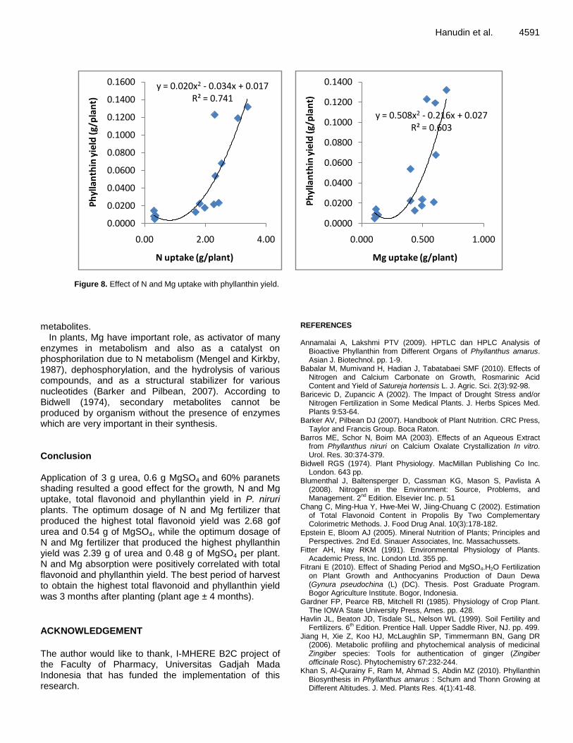
Hanudin et al. 4591
y = 0.020x2 - 0.034x + 0.017R² = 0.741
0.0000
0.0200
0.0400
0.0600
0.0800
0.1000
0.1200
0.1400
0.1600
0.00 2.00 4.00
Ph
ylla
nth
in y
ield
(g/
pla
nt)
N uptake (g/plant)
y = 0.508x2 - 0.216x + 0.027R² = 0.603
0.0000
0.0200
0.0400
0.0600
0.0800
0.1000
0.1200
0.1400
0.000 0.500 1.000
Ph
ylla
nth
in y
ield
(g/
pla
nt)
Mg uptake (g/plant)
Figure 8. Effect of N and Mg uptake with phyllanthin yield.
metabolites.
In plants, Mg have important role, as activator of many enzymes in metabolism and also as a catalyst on phosphorilation due to N metabolism (Mengel and Kirkby, 1987), dephosphorylation, and the hydrolysis of various compounds, and as a structural stabilizer for various nucleotides (Barker and Pilbean, 2007). According to Bidwell (1974), secondary metabolites cannot be produced by organism without the presence of enzymes which are very important in their synthesis. Conclusion Application of 3 g urea, 0.6 g MgSO4 and 60% paranets shading resulted a good effect for the growth, N and Mg uptake, total flavonoid and phyllanthin yield in P. niruri plants. The optimum dosage of N and Mg fertilizer that produced the highest total flavonoid yield was 2.68 gof urea and 0.54 g of MgSO4, while the optimum dosage of N and Mg fertilizer that produced the highest phyllanthin yield was 2.39 g of urea and 0.48 g of MgSO4 per plant. N and Mg absorption were positively correlated with total flavonoid and phyllanthin yield. The best period of harvest to obtain the highest total flavonoid and phyllanthin yield was 3 months after planting (plant age ± 4 months).
ACKNOWLEDGEMENT
The author would like to thank, I-MHERE B2C project of the Faculty of Pharmacy, Universitas Gadjah Mada Indonesia that has funded the implementation of this research.
REFERENCES Annamalai A, Lakshmi PTV (2009). HPTLC dan HPLC Analysis of
Bioactive Phyllanthin from Different Organs of Phyllanthus amarus. Asian J. Biotechnol. pp. 1-9.
Babalar M, Mumivand H, Hadian J, Tabatabaei SMF (2010). Effects of Nitrogen and Calcium Carbonate on Growth, Rosmarinic Acid Content and Yield of Satureja hortensis L. J. Agric. Sci. 2(3):92-98.
Baricevic D, Zupancic A (2002). The Impact of Drought Stress and/or Nitrogen Fertilization in Some Medical Plants. J. Herbs Spices Med. Plants 9:53-64.
Barker AV, Pilbean DJ (2007). Handbook of Plant Nutrition. CRC Press, Taylor and Francis Group. Boca Raton.
Barros ME, Schor N, Boim MA (2003). Effects of an Aqueous Extract from Phyllanthus niruri on Calcium Oxalate Crystallization In vitro. Urol. Res. 30:374-379.
Bidwell RGS (1974). Plant Physiology. MacMillan Publishing Co Inc. London. 643 pp.
Blumenthal J, Baltensperger D, Cassman KG, Mason S, Pavlista A (2008). Nitrogen in the Environment: Source, Problems, and Management. 2
nd Edition. Elsevier Inc. p. 51
Chang C, Ming-Hua Y, Hwe-Mei W, Jiing-Chuang C (2002). Estimation of Total Flavonoid Content in Propolis By Two Complementary Colorimetric Methods. J. Food Drug Anal. 10(3):178-182.
Epstein E, Bloom AJ (2005). Mineral Nutrition of Plants; Principles and Perspectives. 2nd Ed. Sinauer Associates, Inc. Massachussets.
Fitter AH, Hay RKM (1991). Environmental Physiology of Plants. Academic Press, Inc. London Ltd. 355 pp.
Fitrani E (2010). Effect of Shading Period and MgSO4.H2O Fertilization on Plant Growth and Anthocyanins Production of Daun Dewa (Gynura pseudochina (L) (DC). Thesis. Post Graduate Program. Bogor Agriculture Institute. Bogor, Indonesia.
Gardner FP, Pearce RB, Mitchell RI (1985). Physiology of Crop Plant. The IOWA State University Press, Ames. pp. 428.
Havlin JL, Beaton JD, Tisdale SL, Nelson WL (1999). Soil Fertility and Fertilizers. 6
th Edition. Prentice Hall. Upper Saddle River, NJ. pp. 499.
Jiang H, Xie Z, Koo HJ, McLaughlin SP, Timmermann BN, Gang DR (2006). Metabolic profiling and phytochemical analysis of medicinal Zingiber species: Tools for authentication of ginger (Zingiber officinale Rosc). Phytochemistry 67:232-244.
Khan S, Al-Qurainy F, Ram M, Ahmad S, Abdin MZ (2010). Phyllanthin Biosynthesis in Phyllanthus amarus : Schum and Thonn Growing at Different Altitudes. J. Med. Plants Res. 4(1):41-48.

4592 J. Med. Plants Res. Khan S, Singla RK, Abdin MZ (2011). Assessment of Phytochemical
Diversity in Phyllanthus amarus Using HPTLC Fingerprints. Indo-Global J. Pharm. Sci. 1(1):1-12.
Kotoky R, Kanjilal PB (2005). 2-Methylhexadec-2-ene- from Phyllanthus niruri Linn. Indian J. Chem. 44:434-435.
Marschner H (1995). Mineral Nutrition of Higher Plants. 2nd
Edition. Academic Press. San Diego. pp. 889.
Martin LRR, Pereira-Filho ER, Cass QB (2011). Chromatographic profiles of Phyllanthus aqueous extracts samples: a proposition of classification using chemometric models. Anal. Bioanal. Chem. 400:469-481.
Mengel K, Kirby EA (1987). Principles of Plant Nutrition. International Potash Inst. Bern. Switzerland. pp. 436-437.
Morris P, Scragg AH, Smart NJ, Stafford A (1985). Secondary Product Formation By Cell Suspension Cultures, in Dixon. R.A. (Ed.), Plant Cell Culture. IRL Press. Oxford, Washingthon DC. pp. 230.
Nayak PS, Upadhyay A, Dwivedi SK, Rao S (2010). Quantitative Determination of Phyllanthin in Phyllanthus amarus by High Performance Thin Layer Chromatography. Boletin Latinoamericano y del Caribe de Plantas Medicinales y Aromaticas. 9(5):353-358.
Nirwan (2007). Flavonoid Poduction of in vitro Daun Dewa (Gynura pseudochina (L.) DC) in Shading Conditions and Fertilizing. Dissertation. Abstract. Post-Graduate Program. Bogor Agriculture Institute. Bogor, Indonesia.
Nitnaware KM, Naik DG, Nikam TD (2011). Thidiazuron-induced Shoot Organogenesis and Production of Hepatoprotective Lignan Phyllanthin and Hypophyllanthin in Phyllanthus amarus. Plant Cell Tiss. Organ Cult. 104:101-110.
Ozguven M, Sener B, Orhan I, Sekeroglu N, Kirpik M, Kartal M, Pesin I, Kaya Z (2008). Effects of Varying Nitrogen Doses in Yield Components and Artemisinin Content of Artemisia annua L. Ind. Crop. Prod. 27:60-64.
Padua de L.S, Bunyaprapharsara N, Lemmens RHMJ (1999). Medicinal and Poisonous Plant I. Plant Resources of South East Asia. Bogor.
Robbers JE, Speedie KM, Tyler VE (1996). Pharmacognosy and
Pharmacobiotechnology. Williams and Wilkins. Baltimore 337 pp. Robinson T (1991). The Organic Constituent of Higher Plants. Bandung
Technology University, Bandung (in Indonesia). Siebert M, Kadkade PG (1980). Enviromental Factors, in Staba, E. J.
(Ed.), Plant Tissue Culture as a Source of Biochemical. CRC Press Inc. Boca Raton, Florida. pp. 123-141.
Syamsundar KV, Singh B, Thakur RS, Husain A, Kiso Y, Hikino H (1985). Antihepatotoxicity Principles of Phyllanthus niruri Herbs. J. Ethnopharmacol. 14:41-44.
Tang Y (1997). Light in Plant Ecophysiology. M.N.V. Prasad (Edt.). John Willey and Sons, Inc. USA. pp. 3-40.
Thompson LM, Troeh FR (1978). Soils and Soil Fertility. Fourth Edition. McGraw- Hill Inc, United States of America. pp. 489.
Torsell K (1997). Natural Product Chemistry : A Mechanic, Biosynthetic and Ecological Approach. Stockholm, Apotekarsocieteter. Swedish. pp. 117-173.
Tripathi AK, Ram KV, Anil KG, Madan MG, Suman PSK (2006). Quantitative Determination of Phyllanthin and Hypophyllanthin in Phyllanthus Species By High-Perfomance Thin Layer Chromatography. Phytochem. Anal. 17:394-397.
Winkel BSJ (2006). The Biosynthesis of Flavonoids. In the Science of Flavonoids. Erich Grotewold (Eds.). Spinger-Science-Business Media, Inc. New York. pp. 71-122.