volume 4, issue 5, 947-967. research article issn 2277
TRANSCRIPT

www.wjpr.net Vol 4, Issue 5, 2015
947
Srividya et al. World Journal of Pharmaceutical Research
IN VITRO AND IN VIVO ANTI HEPATOTOXIC EVALUATION OF
ALPINIA GALANGA ON D- GALACTOSAMINE INDUCED TOXICITY
Srividya Ammayappan Rajam*1, Sameer Kumar Varma
1, Vishnuvarthan
Vaithiyalingam Jagannathan2
1Department of Pharmaceutical Biotechnology, JSS College of Pharmacy, Ooty- 643001.
Tamilnadu, India.
2Department of Pharmacy, SRM University, Katangulathur Chennai.
ABSTRACT
Alpinia galangal is a rhizome belongs to the family Zingeberaceae.
Qualitative phytochemical analysis of plant extracts showed the
presence of majority of the compound including terpenoids,
flavonoids, alkaloids, tannins, saponins and glycosides. The hydro
alcoholic hot extract has shown high total phenol content 14.50 ±
0.210 mg/g and hydro alcoholic cold shown 12.59 ± 0.295 mg/g.
Alpinia galangal hydro alcoholic extract prepared by hot maceration
showed potent antioxidant activity with IC value 7.7 ± 0.121 μg/ml.
The antihepatotoxicity produced by the extract at concentration of 200
and 400 μg/ml was effective against the D- Galactosamine induced
hepatotoxicity, whereas at the concentration of 600 and 800 μg/ml it
was found to be cytotoxic for both the hydro alcoholic extracts
prepared by cold and hot maceration process. A significant increase in
the levels of ASAT, ALAT, ALP, total Bilirubin, direct Bilirubin (P<0.001) and a
significant reduction in the levels of TGL, total proteins and albumin (P< 0.001) was
observed in hepatocytes exposed to D- Galactosamine when compared to normal hepatocytes.
These cells when treated alone with different extract of Alpinia Galanga showed a significant
restoration of the altered biochemical parameters towards the normal (P< 0.001) when
compared to D- Galactosamine treated group) and were dose dependent. A similar result was
obtained with the silymarin. The antihepatotoxic effect of extracts of Alpinia galangal was
found to be 200 – 400 μg/ml concentration.
KEYWORDS: Alpinia galangal, anti hepatoxicity, antioxidant, cytotoxic.
World Journal of Pharmaceutical Research SJIF Impact Factor 5.990
Volume 4, Issue 5, 947-967. Research Article ISSN 2277– 7105
Article Received on
18 Feb 2015,
Revised on 15 March 2015, Accepted on 07 April 2015
*Correspondence for
Author
Dr. Srividya
Ammayappan Rajam
Department of
Pharmaceutical
Biotechnology, JSS
College of Pharmacy,
Ooty- 643001. Tamilnadu,
India.

www.wjpr.net Vol 4, Issue 5, 2015
948
Srividya et al. World Journal of Pharmaceutical Research
INTRODUCTION
The liver is a vital organ present in vertebrates and some other animals, and is typically the
largest visceral organ. The liver plays a major role in transforming and clearing chemicals
and is susceptible to the toxicity from these agents. Some medicinal agents, when taken in
overdoses and sometimes even when introduced within therapeutic ranges, may injure the
organ. Chemicals that cause liver injury are called hepatotoxins.[1]
Chemicals often cause
subclinical injury to liver which manifests only as abnormal liver enzyme tests. Drug-induced
hepatotoxicity represents a major clinical problem accounting for 50% of all cases of acute
liver failure. Although the majority of cases of acute liver failure are due to intentional or
unintentional misuse, 16% are idiosyncratic.[2]
Some of the inorganic compounds producing
hepatotoxicity are arsenic, phosphorus, copper and iron. The organic agents include certain
naturally occurring plant toxins such as pyrrolizidine alkaloids, In addition, exposure
mycotoxins and bacterial toxins also produce hepatotoxicity. In addition, exposure to
hepatotoxic compounds the other factors which produce hepatotoxicity may be occupational
environmental or domestic that could be accidental, homicidal or suicidal ingestion.[3]
Drugs continue to be pulled from the market with disturbing regularity because of late
discovery of hepatotoxicity.[4]
The mechanism of hepatic injury has been proposed to involve
2 pathways-direct hepatotoxicity and adverse immune reactions. In most instances, hepatic
injury is initiated by the bioactivation of drugs to chemically reactive metabolites, which have
the ability to interact with cellular macromolecules such as proteins, lipids, and nucleic acids,
leading to protein dysfunction, lipid peroxidation, DNA damage and oxidative stress.
Additionally, these reactive metabolites may induce disruption of ionic gradients and
intracellular calcium stores, resulting in mitochondrial dysfunction and loss of energy
production. Its dysfunction releases excessive amount of oxidants which in turn injures
hepatic cells.[5]
Hepatic cellular dysfunction and death also have the ability to initiate
immunological reactions, including both innate and adaptive immune responses.
Stress and damage to hepatocytes result in the release of signals that stimulate activation of
other cells, particularly those of the innate immune system, including Kupffer cells (KC),
natural killer(NK) cells, and NKT cells. These cells contribute to the progression of liver
injury by producing proinflammatory mediators and secreting chemokines to further recruit
inflammatory cells to the liver. It has been demonstrated that various inflammatory cytokines,
such as tumor necrosis factor (TNF) α, interferon (IFN), and interleukin (IL)-1, produced

www.wjpr.net Vol 4, Issue 5, 2015
949
Srividya et al. World Journal of Pharmaceutical Research
during hepatic injury are involved in promoting tissue damage.[6]
However, innate immune
cells are also the main source of IL-10, IL-6, and certain prostaglandins, all of which have
been shown to play a hepatoprotective role.[7]
In this paper the hepatoprotective activity of
Alpinia galangal has been carried out in on D- galactosamine induced toxicity in albino rats
of wister strain in both in -vitro and in-vivo.
MATERIALS AND METHODS
Alpinia galangal belongs to the family Zingiberaceae The creamy white rhizomes contain a
volatile essential oil quite similar to that of the ginger.
The chemical constituents of this oil are methyl cinnamate, cineole, eugenol, camphor and d-
pinene. Present are also: phoblaphenes, starch, sulphates and chlorides. The essential oil is
active again gram positive and gram negative microorganisms. Broncho-spasm induced by
pilocarpine is counteracted by small doses of a tincture of galanga. The seeds have anti-ulcer
activity. It is an excellent candidate for the development of a remedy for opportunistic fungal
infections in AIDS patients. Anti-tumor and anti-dementia effects have been observed in
rodents.
The rhizome is used against rheumatism, bronchial catarrh, bad breath and ulcers whooping
colds in children, throat infections, to control incontinence and fever. It is a potent
antioxidant. Alpinia species show promise as anti-fungal, hypertensive, enhancers of sperm
count and motility. Homoeopaths use it as a stimulant. It has some reputation as a remedy for
perineal relaxation with hemorrhoids and for a lax and pendulous abdomen. It is used as a
snuff to treat cold and flu symptoms. Galangal Root has also been used as a digestive aid,
especially in combating dyspepsia and flatulence.[8]
Collection and authentification of plant material
Both the plants were collected in the Kannur Kerala, during the month of May 2013. The
plant was authenticated by Dr. S. Rajan, Botanical Survey of India, Medicinal Plant Survey
and Collection Unit, Government Arts College, Ootacamund, Tamil Nadu, and India.
Preparation of plant extracts[9,10]
The collected rhizomes and leaves of the plant were dried under shade then they were
chopped and coarsely powdered. Extraction was performed by soxhlet extraction, cold
maceration and hot maceration process.
www.wjpr.net Vol 4, Issue 5, 2015
950
Srividya et al. World Journal of Pharmaceutical Research
1) Soxhlet extraction
Coarsely powdered plant material was extracted by soxhlet apparatus with a range of solvents
from non polar to polar.
2) Cold maceration
Shade dried coarsely powdered material (rhizome & leaves) were macerated for 7 days with
50% ethanol.
3) Hot maceration
The powdered (rhizome& leaves) were macerated with 50% ethanol for 6 hr at 60oC.
Phytochemicals Studies of Extracts[11,12]
A) Qualitative Phytochemical analysis
A systematic and complete study of crude drugs should include a complete investigation of
both primary and secondary metabolites derived from plant metabolism. The different
qualitative chemical tests and to be performed for establishing profiles of given extracts for
their nature of chemical composition. The ethanol extracts obtained as above were tested for
the following qualitative chemical tests for the identification of various phytoconstituents.
B) Quantitative phytochemicals analysis[13]
Estimation of Total Phenolic Content
400 µl of the each dilution of the extracts were separately mixed with 2 ml of Folin-
Ciocalteau reagent and 1.6 ml of sodium carbonate. After shaking, it was incubated for 2 h at
370C. The absorbance was measured at 750 nm (Shimadzu UV-160 A Spectrophotometer,
Shimadzu Corporation, Japan). Using Gallic acid monohydrate, standard curve was prepared
and linearity was obtained. The total phenol content was expressed as Gallic acid equivalent
in mg/g or % w/w of the extracts.
Estimation of Total Flavanol Content
0.5 ml of the extracts was separately mixed with 1.5 ml methanol, 0.1 ml of 10% aluminum
chloride, 0.1 ml of 1M potassium acetate and 2.8 ml of distilled water. After incubation at
room temperature for 30 min, the absorbance of the reaction mixture was measured at 415 nm
with a Shimadzu UV-160A Spectrophotometer (Shimadzu Corporation, Japan). Using rutin,
standard curve was prepared and linearity was obtained in the range of 1000-10 µg/ml. The
total flavanol content was expressed as rutin equivalent in mg/g or % w/w of the extracts.

www.wjpr.net Vol 4, Issue 5, 2015
951
Srividya et al. World Journal of Pharmaceutical Research
In Vitro Antioxidant Screening of Extracts
DPPH radical scavenging activity[14,15]
The assay was carried out in a 96 well microtitre plate. To 200 l of DPPH solution, 10 l of
each of the extract or standard solution was added separately in wells of the microtitre plate.
The plates were incubated at 37 oC for 30 min and the absorbance was measured at 490 nm
using ELISA reader.
Calculation
The absorbance was measured spectrophotometrically against the corresponding blank
solution. The percentage inhibition was calculated by using the following formula.
OD control - OD sample
Radical scavenging activity (%) = x 100
OD control
IC50, which is the concentration of the sample required to scavenge 50% of free radicals was
calculated.
Measurement of Reducing power ability
Measurement of reducing power ability was investigated in Fe3+
- Fe2+
transformation in the
presence of the extracts.
1 ml of extract, 2.5 ml of phosphate buffer, 2.5 ml of K3Fe(CN)6 were incubated at 500C for
20 min, 2.5 ml of TCA was added to the mixture and centrifuged for 10 min at 3000 rpm.
From the upper layer 2.5 ml was taken and diluted with 2.5 ml of distilled water and shaken
with 0.5 ml fresh FeCl3. The absorbance was measured at 700 nm after 20 min the blank
solution contained distilled water instead of the samples.
Evaluation of Total Antioxidant capacity[16]
The total antioxidant capacity was determined by phosphomolybdenum method and is based
on the reduction of Mo (VI) to Mo (V) by the antioxidant compounds and the formation of a
green Mo (V) complex which has the maximal absorption at 695nm.
An aliquot of 0.1 ml of the sample solution containing a reducing species in DMSO
was combined in an assay tube with 1 ml of reagent solution (0.6 M sulphuric acid, 28
mM sodium phosphate, and 4 mM ammonium molybdate). The tubes were capped and
incubated in water bath at 95 °C for 90 min. The samples were cooled to room

www.wjpr.net Vol 4, Issue 5, 2015
952
Srividya et al. World Journal of Pharmaceutical Research
temperature, and the absorbance of each solution was measured at 695 nm. The total
antioxidant capacity was expressed as mM equivalent of ascorbic acid.
In vitro antihepatotoxic activity on isolated rat hepatocytes[17]
The HEPES buffer and collagenase solution were warmed in a water bath (38oC-39
oC to
achieve 370
C in the liver) The pump flow rate was adjusted to 30 ml/min. The rat (180-200
g) was anaesthetized by intra peritoneal administration of Phenobarbital sodium 35 mg/kg
b.w. The abdomen was opened and a loosely tied ligature was placed around the portal vein
approximately 5 mm from the liver, and the cannula was inserted up to the liver and then the
ligature was tightened, and heparin (1000 IU) was injected into the femoral vein. Subhepatic
vessels were rapidly incised to avoid excess pressure and 600 ml of calcium free HEPES
buffer was perfused at a low rate of 30 ml/min for 20 minutes. The liver swells during this
time slowly changing color from dark red to greyish white. 300 ml of collagenase solution
were perfused at a flow rate of 15 ml/min for 20 minutes during which the lobes swell. The
lobes were removed and washed HEPES buffer, after disrupting the Glison capsule. The cell
suspension was centrifuged at 1000 RPM to remove the collagenase, damaged cells and non-
parenchymal cells. The hepatocytes were collected in Ham’s F12 medium enriched with
0.2% bovine albumin, 10 μg/ml bovine insulin and 0.2% of dexamethasone.
Cell counting[18]
Tryphan Blue dye exclusion technique
Trypan blue is a dye, which is capable of penetrating the dead cells; dead cells take up the
blue stain, where as viable cells do not. This method gives an exact number of dead cells and
viable cells. In this procedure One drop of cell suspension was mixed with one drop of
tryphan blue (0.4%) dye on a slide and mixed well for one minute. A drop of the mixture was
loaded on a haemocytometer and the viable and non-viable count was recorded (live cells
don’t take stain where as dead cells get stained). The observations were recorded in a couple
of minutes; else the live cells would degenerate and take up the stain.
The percentage viability was calculated as
Total cells – Dead cells
% Viability = X 100
Total cells

www.wjpr.net Vol 4, Issue 5, 2015
953
Srividya et al. World Journal of Pharmaceutical Research
Determination of Hepatoprotective Activity on Freshly Isolated Rat Hepatocytes by
Estimating the Bio-chemical Parameters[18]
The hepatocytes isolated were incubated for 30 minutes at 37oC for stabilization. The cells
were then diluted in F12 Coons modified medium to obtain a cell count 5x10⁵ cells/ml. 100
μl of this cell suspension was seeded in each well of 96 well plates in each well. After 2
hours of pre-incubation, the medium was replaced with fresh medium. Then the hepatocytes
were pretreated with extracts 15 min before galactosamine - induced treatment (50 μl of D-
galactosamine and 50 μl of different extract concentration into each well). Hepatocytes
injury was induced by incubation of hepatocytes with 30 mM D-galactosamine for 24 hours
by incubating at 37oC. After incubation, the toxicant and drug treated cell suspensions were
pooled into eppendroff tubes and centrifuged. The Asparate Aminotransferase Alanine
Aminotransferase, Alanine Aminotransferase Alkaline Phosphatase enzyme levels as well as
total protein and total bilirubin and total protein levels were determined in supernatant using
Ecoline diagnostic kits.
In vivo antioxidant and hepatoprotective studies[19,20,21,22,23]
Selection and maintenance of animals
Healthy adult albino rats of Wistar strain weighing 150-200 g were obtained from J.S.S.
College of Pharmacy, Animal house, Ooty, India. The animal house was well ventilated and
animals had 12 ± 1 hour day and night schedule with temperature between 20 ± 20C. The
animals were housed in large spacious hygienic cages during the course of the experimental
period. The animals were fed with rat feed supplied by M/S. Hindustan Lever Ltd.,
Bangalore. Experiments were carried out after getting permission from the institutional
Ethical Committee of JSS college of Pharmacy, Ooty.
Preparation of extracts dose
S. No. Plant name Extract name Concentration
(mg/kg/b.w.)
1 Alpinia galanga 50% hydro alcoholic, hot maceration 200 and 400 mg/ml
2 Alpinia galanga 50% hydroalcoholic cold maceration 200 and 400 mg/ml
Preparation of standard
Single dose of 25mg/kg b.w. of silymarin product for 13 days.

www.wjpr.net Vol 4, Issue 5, 2015
954
Srividya et al. World Journal of Pharmaceutical Research
Preparation and Induction of Hepatotoxicity
D-galactosamine HCl (400mg/kg b.w.) was administered intraperitonially on the 14th
day to
induce liver damage.
Randomization, Numbering and Grouping of Animals
The animals were divided into nine groups with four animals in each (equal number of both
male and female) and they were treated as described below.
GROUP I Served as solvent control which received double distilled water (1ml/kg.b.w)
and 0.3% sodium carboxy methyl cellulose (CMC).
GROUPII Served as negative control which received (1ml/kg.b.w) of double distilled
water and 0.3% CMC orally once a day for 13 days. On the 14th
day, D-galactosamine
HCl (400mg/kg b.w.) was given.
GROUP III Received a single dose of 25mg/kg b.w. of silymarin product for 13 days
followed by treatment with the toxicant on the 14th day.
GROUP IV Received a single dose of (200mg/kg b.w.) of hydro alcoholic hot macerated
extract of A. galanga for 13 days followed by treatment with the toxicant on the 14th day.
GROUP V Received a single dose of (400mg/kg b.w.) of hydro alcoholic hot macerated
extract of A. galanga for 13 days followed by treatment with the toxicant on the 14th day.
GROUP VI Received a single dose of (200mg/kg b.w.) hydro alcoholic cold macerated
extract of A. galanga for 13 days followed by treatment with the toxicant on the 14th day.
GROUP VII Received a single dose of (400mg/kg b.w.) hydro alcoholic cold macerated
extract of A. galanga for 13 days followed by treatment with the toxicant on the 14th day.
Isolation of blood
The rats were anesthetized using thiopental and the blood was collected from abdominal
artery and after collection, the blood was kept at 370C in the incubator for 30 min later, it was
cold centrifuged at 2000 rpm for 15 min to get clear supernatant serum, which was used for
the biochemical estimations.

www.wjpr.net Vol 4, Issue 5, 2015
955
Srividya et al. World Journal of Pharmaceutical Research
Preparation of Liver and Kidney homogenates
The liver and kidneys were removed, weighed and homogenized immediately with Elvenjan
homogenizer fitted with Teflon plunger, in ice chilled 10% KCl solution (10 ml/g of tissue).
After centrifugation at 2000 rpm for 10 min, clear supernatant was used for the determination
of SOD, CAT, TBARS and protein estimations. SOD and CAT levels were determined
immediately after centrifugation and malondialdehyde (MDA) as TBA-RS and protein
content were estimated in frozen samples.
In vivo antihepatotoxic and antioxidant evaluation[24]
Estimation of in vivo antioxidant enzymes levels in homogenates
Catalase estimation
2.25 ml of potassium buffer (65mM, pH 7.8) and 100 µl of the tissue homogenate or sucrose
(0.32 M) were incubated at 250C for 30 min. 0.65 ml of H2O2 (75mM) was added to initiate
the reaction. The change in absorption at 240 nm was measured for 2-3 min, and dy / dx for 1
min for each assay was calculated and the results are expressed as CAT units / mg of tissue.
SOD estimation
Assay mixture contained 0.1mL of supernatant, 1.2mL of sodium phosphate buffer (pH 8.3;
0.052 M), 0.1mL of Phenazine methosulphate (186 µm), 0.3mL of nitro blue tetrazolium, 300
µM, 0.2mL of NADH (750 µM). Reaction was started by addition of NADH. After
incubation at 30 0
C for 90 s, the reaction was stopped by addition of 0.1mL of glacial acetic
acid. Reaction mixture was stirred vigorously with 4.0mL of n-butanol. Colour intensity of
the chromogen in the butanol was measured spectrophotometrically at 560 nm and
concentration of SOD was expressed as U/mg of protein.
Thiobarbituric acid reactive substances (TBA-RS) estimation
Acetic acid 1.5mL (20%; pH 3.5), 1.5mL of thiobarbituric acid (0.8%) and 0.2mL of sodium
lauryl sulphate (8.1%) were added to 0.1mLof supernatant and heated at 1000C for 60 min.
Mixture was cooled and 5mL of n-butanol-pyridine (15:1) mixture, 1mL of distilled water
was added and vortexed vigorously. After centrifugation at 1200×g for 10 min, the organic
layer was separated and absorbance was measured at 532 nm using a spectrophotometer.
MDA is an end product of lipid peroxidation, which reacts with thiobarbituric acid to form
pink chromogen–thiobarbituric acid reactive substance. It was calculated using a molar
extinction coefficient of 1.56×10⁵M−1
cm−1
and expressed as nanomoles of TBARS/mg of
protein.
www.wjpr.net Vol 4, Issue 5, 2015
956
Srividya et al. World Journal of Pharmaceutical Research
Protein estimation
100 µl of homogenates, 900 µl of distilled water, and 5ml of Bradford reagent. Mix well and
allow the color to develop for at least 5 min but no longer than 30 min. read the absorbance at
595 nm. Plot a standard curve using the standard protein absorbance Vs concentration.
Calculate the protein in the experimental sample using the standard curve.
Estimation of biochemical parameters from serum
ASAT, ALAT, ALP, total protein, albumin, LDH, total bilirubin, direct bilirubin and
triglycerides were assayed using Ecoline diagnostic kit.
Statistical analysis
The results of in vitro antihepatotoxic & in vivo antihepatotoxic and antioxidant activity were
analyzed statistically using one way analysis of variance (ANOVA) followed by Dunnett’s t-
test.
RESULT AND DISCUSSION
Collection and authentification of plant
The Rhizomes of Alpinia galangal was collected from the Kerala, in the month of May 2013.
The plant was authenticated by Dr. S. Rajan, Botanical Survey of India, Medicinal Plant
Survey and Collection Unit, Government Arts College, Ootacamund, Tamil Nadu, and India.
Preparation of plant extracts
The dried parts of rhizome of Alpinia galanga were chopped and coarsely powdered, the
powdered materials were extracted separately using different solvent according to polarity by
Soxhletion and hot and cold maceration. All the extracts were stored in refrigerator till further
use. The percentage yields were given in table 1.
Table 1. Percentage yield of rhizome of Alpinia galanga extracts
S. No. Extraction
process
Powder
weight Solvent used
Weight of
extract % Yield
1
Soxhlet
extraction
100 g Petroleum ether 3.85 g 3.8%
2 100 g Chloroform 7.86 g 7.8%
3 100 g Ethanol 9.50 g 9.5%
4 100 g Methanol 9.45 g 9.4%
5 Cold
maceration 100 g
Hydro alcoholic
(50%) 8.54 g 8.5%
6 Hot
maceration 200 g
Hydro alcoholic
(50%) 19.25 g 9.7%

www.wjpr.net Vol 4, Issue 5, 2015
957
Srividya et al. World Journal of Pharmaceutical Research
Phytochemicals studies of extracts
A) Qualitative Phytochemicals analysis
Qualitative Phytochemical analysis of plant extracts showed the presence of majority of the
compounds including terpenoids, flavanoids, alkaloids, tannins, saponins and glycosides. The
results from chemical tests are shown in table no 2.
Table 2. Analysis of Alpinia galanga extracts
Tests
Alpinia galanga
Pet. ether Chloroform Ethanol Methanol Hot
maceration
Cold
maceration
Alkaloids _ _ + _ + +
Carbohydrates _ _ _ + + +
Flavanoids + + + + + +
Terpenoids + + + _ + +
Glycosides _ _ + _ + +
Phytosterols + + _ _ _ _
Proteins and
aminoacids + _ _ _ + +
Saponins _ _ _ _ + +
Phenolic
compounds
and Tannins
+ + + + + +
Estimation of Total Phenolic Content
Among the six extracts, the hydro alcoholic (hot) extract has shown high total phenol content
14.50 ± 0.210 mg/g, and hydro alcoholic (cold) shown 12.59 ± 0.295 mg/g. The other four
extracts showed total phenol content in the range of 6.42 ± 0.058 mg/g to 10.59 ± 0.230
mg/g. The results from total phenol content are shown in table no.3.
Table 3. Total phenol estimation of different extracts of Alpinia galanga
S. No. Extract Total phenol (mg/g)
1 Petroleum Ether 6.42 ± 0.058
2 Chloroform 8.21 ± 0.078
3 Ethanol 11.43 ± 0.363
4 Methanol 10.59 ± 0.230
5 Hydro alcoholic (cold) 12.59 ± 0.295
6 Hydro alcoholic (hot) 14.50±0.210
Estimation of total Flavanol content
Among the six extracts, the hydro alcoholic (hot) extract has shown high total flavanol
content 285.57 ± 0.188 mg/g, and hydro alcoholic (cold) shown 174.41 ± 0.383 mg/g. The

www.wjpr.net Vol 4, Issue 5, 2015
958
Srividya et al. World Journal of Pharmaceutical Research
other four extracts showed total flavanol content in the range of 49.62 ± 0.342 mg/g to 169.84
± 0.145 mg/g. The results from total flavanol content are shown in table 4.
Table 4. Total flavanol content of different extracts of Alpinia galanga:
S. No. Extract Total flavanol (mg/g)
1 Petroleum Ether 119.46 ± 0.453
2 Chloroform 169.84 ± 0.145
3 Ethanol 59.86 ± 0.115
4 Methanol 49.62 ± 0.342
5 Hydro alcoholic (cold) 174.41 ± 0.383
6 Hydro alcoholic (hot) 285.57 ± 0.188
In Vitro Antioxidant Screening of Extracts
DPPH radical scavenging activity
Among the six extracts of Alpinia galanga hot extract showed potent antioxidant activity
with IC50 value of 7.7 ± 0.121µg/ml The other five extracts showed antioxidant activity in the
range of 12.35 ± 0.176 µg/ml to 64.03 ± 5.179 µg/ml. the results from DPPH activity are
shown in table 5.
Table 5. DPPH radical scavenging activity of different extracts of Alpinia galanga
S. No. Extract IC50 (µg/ml)
1 Petroleum Ether 64.03 ± 5.179
2 Chloroform 33.44 ± 0.066
3 Ethanol 13.65 ± 0.136
4 Methanol 15.76 ± 0.237
5 Hydro alcoholic (cold) 12.35 ± 0.176
6 Hydro alcoholic (hot) 7.7 ± 0.121
7 Ascorbic acid 2.69 ± 0.05
Total antioxidant capacity
Among the six extracts of A. galanga hydro alcoholic (hot) extract showed potent antioxidant
capacity 0.24 ± 0.016 mM equivalent of Ascorbic acid in comparison to other extracts.
Table 6 Total antioxidant capacity of different extracts of A. galanga
S. No. Extract TAC (mM equivalent of Ascorbic acid)
1 Petroleum Ether 0.72 ± 0.019
2 Chloroform 0.69 ± 0.018
3 Ethanol 0.34 ± 0.017
4 Methanol 0.38 ± 0.011
5 Hydro alcoholic (cold) 0.30 ± 0.012
6 Hydro alcoholic (hot) 0.24 ± 0.016
www.wjpr.net Vol 4, Issue 5, 2015
959
Srividya et al. World Journal of Pharmaceutical Research
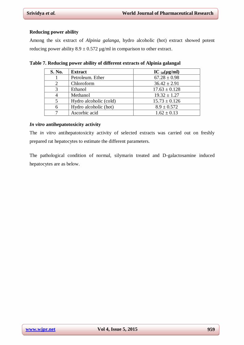
Reducing power ability
Among the six extract of Alpinia galanga, hydro alcoholic (hot) extract showed potent
reducing power ability 8.9 ± 0.572 µg/ml in comparison to other extract.
Table 7. Reducing power ability of different extracts of Alpinia galangal
S. No. Extract IC 50(µg/ml)
1 Petroleum. Ether 67.28 ± 0.98
2 Chloroform 36.42 ± 2.91
3 Ethanol 17.63 ± 0.128
4 Methanol 19.32 ± 1.27
5 Hydro alcoholic (cold) 15.73 ± 0.126
6 Hydro alcoholic (hot) 8.9 ± 0.572
7 Ascorbic acid 1.62 ± 0.13
In vitro antihepatotoxicity activity
The in vitro antihepatotoxicity activity of selected extracts was carried out on freshly
prepared rat hepatocytes to estimate the different parameters.
The pathological condition of normal, silymarin treated and D-galactosamine induced
hepatocytes are as below.
www.wjpr.net Vol 4, Issue 5, 2015
960
Srividya et al. World Journal of Pharmaceutical Research
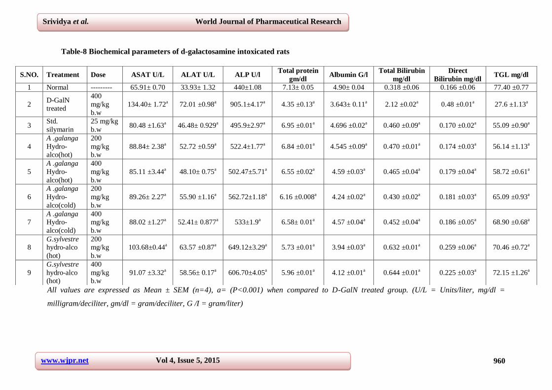
Table-8 Biochemical parameters of d-galactosamine intoxicated rats
All values are expressed as Mean ± SEM (n=4), a= (P<0.001) when compared to D-GalN treated group. (U/L = Units/liter, mg/dl =
milligram/deciliter, gm/dl = gram/deciliter, G /I = gram/liter)
S.NO. Treatment Dose ASAT U/L ALAT U/L ALP U/l Total protein
gm/dl Albumin G/l
Total Bilirubin
mg/dl
Direct
Bilirubin mg/dl TGL mg/dl
1 Normal --------- 65.91± 0.70 33.93± 1.32
440±1.08
7.13± 0.05
4.90± 0.04
0.318 ±0.06
0.166 ±0.06 77.40 ±0.77
2 D-GalN treated
400
mg/kg
b.w
134.40± 1.72a
72.01 ±0.98a
905.1±4.17a
4.35 ±0.13a
3.643± 0.11a
2.12 ±0.02a
0.48 ±0.01a
27.6 ±1.13a
3 Std.
silymarin
25 mg/kg
b.w 80.48 ±1.63
a 46.48± 0.929
a 495.9±2.97
a 6.95 ±0.01
a 4.696 ±0.02
a 0.460 ±0.09
a 0.170 ±0.02
a 55.09 ±0.90
a
4
A .galanga
Hydro-alco(hot)
200
mg/kg b.w
88.84± 2.38a
52.72 ±0.59a
522.4±1.77a
6.84 ±0.01a
4.545 ±0.09a
0.470 ±0.01a
0.174 ±0.03a
56.14 ±1.13a
5
A .galanga
Hydro-
alco(hot)
400
mg/kg
b.w
85.11 ±3.44a
48.10± 0.75a
502.47±5.71a
6.55 ±0.02a
4.59 ±0.03a
0.465 ±0.04a
0.179 ±0.04a
58.72 ±0.61a
6
A .galanga
Hydro-
alco(cold)
200
mg/kg
b.w
89.26± 2.27a
55.90 ±1.16a
562.72±1.18a
6.16 ±0.008a
4.24 ±0.02a
0.430 ±0.02a
0.181 ±0.03a
65.09 ±0.93a
7 A .galanga Hydro-
alco(cold)
400 mg/kg
b.w
88.02 ±1.27a
52.41± 0.877a
533±1.9a
6.58± 0.01a
4.57 ±0.04a
0.452 ±0.04a
0.186 ±0.05a
68.90 ±0.68a
8 G.sylvestre hydro-alco
(hot)
200 mg/kg
b.w
103.68±0.44a
63.57 ±0.87a
649.12±3.29a
5.73 ±0.01a
3.94 ±0.03a
0.632 ±0.01a
0.259 ±0.06a
70.46 ±0.72a
9
G.sylvestre
hydro-alco (hot)
400
mg/kg b.w
91.07 ±3.32a
58.56± 0.17a
606.70±4.05a
5.96 ±0.01a
4.12 ±0.01a
0.644 ±0.01a
0.225 ±0.03a
72.15 ±1.26a
www.wjpr.net Vol 4, Issue 5, 2015
961
Srividya et al. World Journal of Pharmaceutical Research
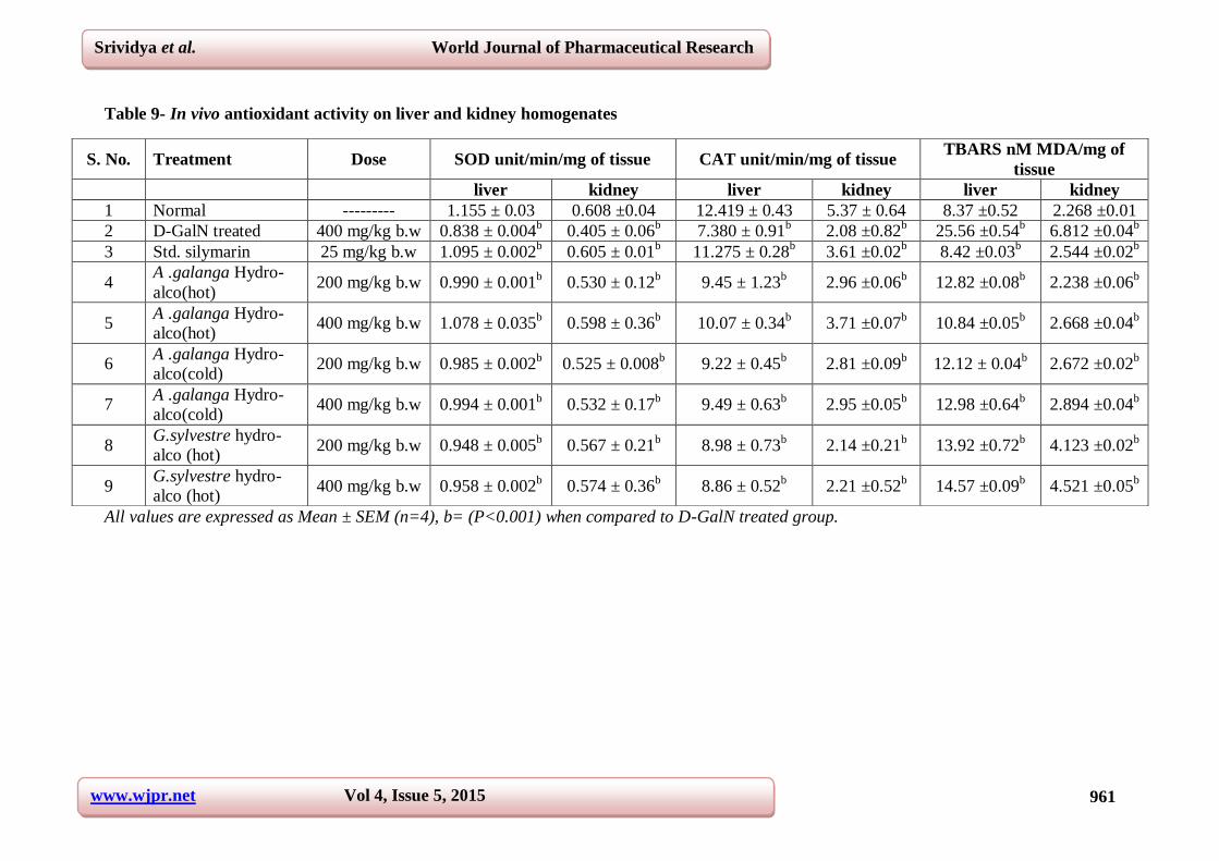
Table 9- In vivo antioxidant activity on liver and kidney homogenates
All values are expressed as Mean ± SEM (n=4), b= (P<0.001) when compared to D-GalN treated group.
S. No. Treatment Dose SOD unit/min/mg of tissue CAT unit/min/mg of tissue TBARS nM MDA/mg of
tissue
liver kidney liver kidney liver kidney
1 Normal --------- 1.155 ± 0.03
0.608 ±0.04
12.419 ± 0.43
5.37 ± 0.64
8.37 ±0.52
2.268 ±0.01
2 D-GalN treated 400 mg/kg b.w 0.838 ± 0.004b
0.405 ± 0.06b
7.380 ± 0.91b
2.08 ±0.82b
25.56 ±0.54b
6.812 ±0.04b
3 Std. silymarin 25 mg/kg b.w 1.095 ± 0.002b
0.605 ± 0.01b
11.275 ± 0.28b
3.61 ±0.02b
8.42 ±0.03b
2.544 ±0.02b
4 A .galanga Hydro-
alco(hot) 200 mg/kg b.w 0.990 ± 0.001
b 0.530 ± 0.12
b 9.45 ± 1.23
b 2.96 ±0.06
b 12.82 ±0.08
b 2.238 ±0.06
b
5 A .galanga Hydro-
alco(hot) 400 mg/kg b.w 1.078 ± 0.035
b 0.598 ± 0.36
b 10.07 ± 0.34
b 3.71 ±0.07
b 10.84 ±0.05
b 2.668 ±0.04
b
6 A .galanga Hydro-
alco(cold) 200 mg/kg b.w 0.985 ± 0.002
b 0.525 ± 0.008
b 9.22 ± 0.45
b 2.81 ±0.09
b 12.12 ± 0.04
b 2.672 ±0.02
b
7 A .galanga Hydro-
alco(cold) 400 mg/kg b.w 0.994 ± 0.001
b 0.532 ± 0.17
b 9.49 ± 0.63
b 2.95 ±0.05
b 12.98 ±0.64
b 2.894 ±0.04
b
8 G.sylvestre hydro-
alco (hot) 200 mg/kg b.w 0.948 ± 0.005
b 0.567 ± 0.21
b 8.98 ± 0.73
b 2.14 ±0.21
b 13.92 ±0.72
b 4.123 ±0.02
b
9 G.sylvestre hydro-
alco (hot) 400 mg/kg b.w 0.958 ± 0.002
b 0.574 ± 0.36
b 8.86 ± 0.52
b 2.21 ±0.52
b 14.57 ±0.09
b 4.521 ±0.05
b

www.wjpr.net Vol 4, Issue 5, 2015
962
Srividya et al. World Journal of Pharmaceutical Research
Normal hepatocytes Silymarin treated 250µg/ml
30 mM D-galactosamine induced
Effect of hot macerated hydro alcoholic (50%) extract of Alpinia galanga on rat
hepatocytes
The hepatocytes were treated with different concentration of hydro alcoholic hot macerated
extract are as follows,
200 µg/ml concentration 400 µg/ml concentration

www.wjpr.net Vol 4, Issue 5, 2015
963
Srividya et al. World Journal of Pharmaceutical Research
600 µg/ml concentration 800 µg/ml concentration
From the above study the antihepatotoxicity produced by extract at concentration of 200 and
400 µg/ml was effective against the D-galactosamine induced hepatotoxicity, where as at
concentration of 600 and 800 µg/ml it is found cytotoxic.
Effect of cold macerated hydro alcoholic (50%) extract of Alpinia galanga on rat
hepatocytes
The hepatocytes were treated with different concentration of hydro alcoholic hot macerated
extract are as follows,
200µg/ml concentration 400 µg/ml concentration
600 µg/ml concentration 800 µg/ml concentration

www.wjpr.net Vol 4, Issue 5, 2015
964
Srividya et al. World Journal of Pharmaceutical Research
From the above study the antihepatotoxicity produced by extract at concentration of 200 and
400 µg/ml was effective against the D-galactosamine induced hepatotoxicity, whereas at
concentration of 600 and 800 µg/ml it is found to be cytotoxic.
The effects of the different extract of Alpinia galanga on freshly prepared isolated rat
hepatocytes intoxicated with D-galactosamine are recorded in table 8. A significant increase
in the levels of ASAT, ALAT, ALP, total bilirubin, direct bilirubin (P < 0.001) and a
significant reduction in the levels of TGL, total proteins and albumin ( P < 0.001) was
observed in hepatocytes exposed to D-galactosamine when compared to normal hepatocytes.
These cells when treated alone with the different extract of Alpinia galanga showed a
significant restoration of the altered biochemical parameters towards the normal (P < 0.001,
when compared to D-galactosamine treated group) and were dose dependent. A similar result
was obtained when D-galactosamine intoxicated hepatocytes were treated with the Silymarin.
However, the antihepatotoxic effect of extracts of the Alpinia galanga was observed at 200-
400 µg/ml concentration.
In vivo antihepatotoxicity & antioxidant activity
The effects of different extract of Alpinia galanga (rhizomes) on D-galactosamine intoxicated
rats are recorded in table 9. Intoxication of rats treated with D-galactosamine significantly ( P
< 0.001) altered the biochemical parameters when compared with D-galactosamine induced
rats. However, the Silymarin at 25 mg/kg body weight exhibited better results with no
mortality.
Lipid peroxidation level of liver homogenates significantly increased (p < 0.001) in D-GalN
treated rats when compared to control rats. Treatment with plant extract of 400 mg/kg dose of
Alpinia galanga hot macerated, cold macerated extract and silymarin (25 mg/kg) showed
significant (p < 0.001) decrease in LPO when compared with D-GalN treated rats are
recorded in table 9.
The plant extract at 200 mg/kg dose also showed good significant (p < 0.001) decrease in
LPO in liver homogenate when compared D-GalN treated rats as mentioned.
Administration of D-GalN caused a significant (p < 0.001) decrease SOD and CAT levels in
rats when compared with normal animal. The plant extract at 400 mg/kg showed significant
(p < 0.001) increase in SOD and CAT when compared to D-galactosamine treated rats. The

www.wjpr.net Vol 4, Issue 5, 2015
965
Srividya et al. World Journal of Pharmaceutical Research
standard drug, silymarin treated rats also showed significant (p < 0.001) increase in SOD and
CAT when compared to D-GalN treated rats.
CONCLUSION
Drug discovery and development consists of a series of processes starting with the
demonstration of pharmacological effects in experimental cell and animal models and ending
with drug safety and efficacy studies in patients. A main limitation is often the unacceptable
level of toxicity with the liver as the primary target organ. The study of anti hepatotoxic
effect of Alpinia galangal showed its activity that is equivalent to that of standard drug
Silymarin. Further studies could be continued to isolate the active constituents which are
responsible for the anti hepatotoxic activity.
ACKNOWLEDGEMENT
Authors would like to thank JSS University, Mysore and JSS college of Pharmacy, Ooty for
providing the facilities to carry out this research.
REFERENCE
1. Ammayappan Rajam Srividya , Navangul Mansi , Vaithiyalingam jagannathan
vishnuvarthan In-Vitro and In-Vivo Anti-Hepatotoxic Evaluation of Curcuma
Aromatica on D-Galactosamine Induced Toxicity International Journal for
Pharmaceutical Research Scholars V-3, I-4, 2014 153- 164.
2. Maity T, Ahmad A. Protective effect of Mikania scandens (L.) Willd. against isoniazid
induced hepatotoxicity in rats. Int J Pharm Pharm Sci 2012; 4: 466-469.
3. Bell LN, Chalasani N. Epidemiology of idiosyncratic drug induced liver injury. Semin
Liver Dis 2009; 29: 337-347.
4. Sumanth M. Screening models for hepatoprotective agents. Pharm Rev 2007; 2: 2-5.
5. Tripathi M, Singh BK, Mishra C, Raisuddin S, Kakkar P. Involvement of mitochondria
mediated pathways in hepatoprotection conferred by Fumaria parviflora Lam. Extract
against nimesulide induced apoptosis in vitro. Toxicol In Vitro 2010; 24: 495-508.
6. Tarantino G, Di Minno MN, Capone D. Drug-induced liver injury: is it somehow
foreseeable? World J Gastroenterol 2009; 15: 2817- 2833.
7. Gao B, Radaeva S, Park O. Liver natural killer and natural killer T cells: immunobiology
and emerging roles in liver diseases. J Leukoc Biol 2009; 86: 513-528.

www.wjpr.net Vol 4, Issue 5, 2015
966
Srividya et al. World Journal of Pharmaceutical Research
8. Srividya AR, Dhanabal SP, Satish kumar MN, Parth kumar H. Bavadia, Antioxidant and
Antidiabetic Activity of Alpinia Galanga. International Journal of Pharmacognosy and
Phytochemical Research 2010; 3(1): 6-12.
9. Mukherjee pulok K. Quality control of herbal drugs. Edn 1, Nirali publications, Mumbai,
2002.
10. Srividya AR.Sumithra Ganesh, S. Antioxidant, antimicrobial and cytotoxic property of
Melianthus major leaves. J Global Pharma Tech, 2010; 2(10): 94-97.
11. Srividya AR, Sameer Kumar Varma, Dhanapal SP, Vadivelan R, Vijayan P. In Vitro
and In Vivo Evaluation of Hepatoprotective Activity of Gymnema Sylvestre Int J Pharm
Sci and Nanotechnology. 2010; 2(4): 768- 73.
12. Ammayappan rajam Srividya., Palanisamy Dhanabal., Parth Kumar bhavadia.,
Vaithiyalingam Jagannathan vishnuvarthan, Muthureddy Natarajan Sathish Kumar.(2012)
Antioxidant and antidiabetic activity of Curcuma aromatica. Int. J of Research in
Ayurveda and Pharmacy, 2012; 3(3), 401- 05.
13. Srividya AR Balai PH., Raghu H, Vijayan, P. Antioxidant and antiadipogenic activity
of Taraxacum officinale in vitro screening methods. The Pharmacist 2008; 3 (1): 41-44.
14. Topcu Gulacu, Yesilyurt Volkan, Halfon Belkis and Ozturk Mehmet. (2008). Antioxidant
potential and phenolic constituent of Salvia cedronella. Food Chemistry, 2008; 108: 31-9.
15. Srividya AR, Dhanabal SP, Satish kumar MN, Vishnuvarthan VJ. Relationship between
the Curcumin and antioxidant activity in Curcuma aromatica and Curcuma zedoaria
rhizomes. The Journal of Free Radicals and Antioxidants, 2013, 139, 186-98.
16. Srividya AR, Dhanabal SP, Jeevitha S, Vishnuvarthan VJ and Rajesh Kumar R.
Relationship between antioxidant properties and chemical composition of Abutilon
indicum. Ind J Pharma sci. 2012; 74(2), 163-67.
17. Harish H, Shivanandappa T. Antioxidant activity and hepatoprotective potential of
Phyllanthus niruri. Food Chemistry, 2006; 95: 180–85.
18. Freshney R. Ian. (2002). Culture of animal cells: A manual of basic technique (p.p. 246,
386).
19. Toshio Morikawa, Yingni Pan, Kiyofumi Ninomiya, Katsuya Imura, Hisashi Matsuda,
Masayuki Yoshikawa, Dan Yuan b, Osamu Muraoka a Acylated phenylethanoid
oligoglycosides with hepatoprotective activity from the desert plant Cistanche tubulosa.
Bioorg. Med Chem. 2010; 18: 1882–90.
20. Tolulope Olaleye M, Afolabi C. Akinmoladun , Adebayo A. Ogunboye, Afolabi A.
Akindahunsi Antioxidant activity and hepatoprotective property of leaf extracts of
www.wjpr.net Vol 4, Issue 5, 2015
967
Srividya et al. World Journal of Pharmaceutical Research
Boerhaavia diffusa Linn against acetaminophen-induced liver damage in rats. Food and
Chemical Toxicology, 2010; 48: 2200–05.
21. Pannarat Akanitapichat, Kallayanee Phraibung, Kwunchai Nuchklang, Suparichart
Prompitakkul. Antioxidant and hepatoprotective activities of five eggplant varieties. Food
and Chemical Toxicology. 2010; 48: 3017–21.
22. Sabira SM, Rochab JBT Antioxidant and hepatoprotective activity of aqueous extract of
Solanum fastigiatum (false “Jurubeba”) against paracetamol-induced liver damage in
mice. J Ethnopharmacol. 2008; 120: 226–32.
23. Daonian Zhoua, Jinlan Ruana, Yaling Caia, Zhaomei Xionga, Wei Fua, Anhua W
Antioxidant and hepatoprotective activity of ethanol extract of Arachniodes exilis
(Hance) Ching. J Ethnopharmacol, 2010; 129: 232–37.
24. Amit Khatria, Arun Gargb, Shyam S. Agrawal Evaluation of hepatoprotective activity of
aerial parts of Tephrosia purpurea L. and stem bark of Tecomella undulate J
Ethnopharmacol, 2009; 122: 1–5.