voltammetric study of cefotaxime at the macroscopic and
TRANSCRIPT

Vol.:(0123456789)1 3
https://doi.org/10.1007/s00604-021-05072-w
ORIGINAL PAPER
Voltammetric study of cefotaxime at the macroscopic and miniaturized interface between two immiscible electrolyte solutions
Konrad Rudnicki1 · Karolina Sobczak1 · Magdalena Kaliszczak2 · Karolina Sipa1 · Emilia Powałka1 · Sławomira Skrzypek1 · Lukasz Poltorak1 · Gregoire Herzog2
Received: 16 September 2021 / Accepted: 17 October 2021 © The Author(s) 2021
AbstractThe electrochemical behavior of cefotaxime (CTX+) was investigated at the polarized macro- and micro-interface between two immiscible electrolyte solutions (ITIES) by cyclic voltammetry and alternating current voltammetry. Miniaturization was achieved with fused silica microcapillary tubing entrapped in a polymeric casing. Scanning electron microscopy (SEM) was employed for the fabricated LLI support characterization. Voltammetric investigation of CTX+ at macro- and μ-ITIES allowed the determination of many physicochemical parameters, such as formal Galvani potential of the ion transfer reaction ( Δaq
org�� ), diffusion coefficients (D), formal free Gibbs energy of the ion transfer reaction (∆G′aq → org), and water-1,2-dichlo-
roethane partition coefficient ( logPCTX+water∕DCE
). Additionally, based on the results obtained the analytical parameters including voltammetric sensitivity, limits of detection and the limits of quantification (in micromolar range) were calculated. The applicability of the developed procedures was verified in spiked still mineral and tap water samples.
Keywords ITIES · Electrified liquid-liquid interface · Fused silica capillaries · Cefotaxime · AC voltammetry
Introduction
Cephalosporins (CFS) are semi-synthetic antibiotics of the β-lactam family. Their mechanism of action is based on the reduction of bacterial peptidoglycans consequently blocking their growth [1]. These antibiotics have a broad spectrum of activity against Gram-positive and Gram-negative aero-bic and anaerobic bacteria. Cefotaxime (CTX, Fig. 1C) is the most active among third-generation CFS, particularly against Gram-negative bacteria as compared with the 1st
and 2nd CFS generations [2]. In humans, these antibiotics are often used to treat urinary and respiratory tract bacte-rial infections. Preventive treatment of bacterial infections in cattle with CFS is a direct threat of their residues in food, especially meat and dairy products [3]. Antimicrobial resist-ance of bacteria together with the potential allergic reac-tions caused by CTX+ along with other members of β-lactam family are certainly a societal problem. In recent years, the consumption of antibiotics has increased dramatically lead-ing their existence in natural environment. Especially due to this reasons, these chemical species must be monitored in the surrounding environments, human and animal biological fluids, or foodstuffs [2, 4].
A number of methods have been reported to date for the determination of CTX+. Examples include high-perfor-mance liquid chromatography (HPLC) [5, 6], liquid chroma-tography (LC) [7], chemiluminescence sensing [8], and also electroanalytical methods [4, 9, 10]. Low cost, simple instru-mentation, existence of relatively easy miniaturization proto-cols, and integrability into electronic circuits speak for elec-trochemical techniques which are especially attractive from the presumptive sensing point of view. Electroanalytical
* Konrad Rudnicki [email protected]
* Lukasz Poltorak [email protected]
* Gregoire Herzog [email protected]
1 Department of Inorganic and Analytical Chemistry, Faculty of Chemistry, University of Lodz, Tamka 12, 91-403 Lodz, Poland
2 Université de Lorraine, CNRS, LCPME, Nancy, France
/ Published online: 9 November 2021
Microchimica Acta (2021) 188: 413
1 3
sensing at solid electrodes can reach high or even very high selectivity usually for the cost of the elaborated surface engi-neering [11]. Detection of different chemical species, such as CTX+, at the solid electrodes, can be obstructed by the redox active interferences co-existing in real samples. (e.g., phenols passivating carbon-based electrodes are inactive at the ITIES when deprived from other ionizable functionali-ties) [12].
Electrochemistry at the interface between two immisci-ble electrolyte solutions (ITIES) provides an alternative to a conventional sensing at the solid electrified junctions [13]. Detection at ITIES is not limited to oxidation/reduction reac-tions; its mechanism is frequently related to ionic species transferring across the polarizable liquid-liquid interface (LLI). It is often used to detect charged molecules that are electrochemically inactive at conventional solid electrodes [14]. Moreover, since the ionic partitioning governed by the molecular lipophilicity is directly related to the Galvani potential difference of the ion transfer reaction, the signals originating from hydrophilic-hydrophobic analytes can be separated which in turn may improve detection selectivity. For all these reasons, ITIES was used to study many classes of chemical species, e.g., proteins [15], antibiotics [16], drugs [17], or polyelectrolytes [18].
Within last two decades, significant attention was given to the development of a variety of ITIES minia-turization protocols [19]. LLI downscaling brings a few benefits to electroanalytical studies. First of all, under proper geometrical conditions, the mass transfer of the analyte towards the microscopic junction is improved as compared with the macroscopic systems. This in turn improves the detection sensitivity. Capacitive currents drop with the ITIES surface area further lowering the voltammetric limits of detection. Small internal vol-umes of the miniaturized ITIES platforms drastically reduce amount of consumed reagents, especially toxic organic phase [16]. Finally, the properties of the min-iaturized ITIES can be tuned by the surface chemistry
of the supports. Lisiqi Xie et al. constructed nanoITIES formed within an ultrathin (80 nm thickness) isoporous silica membrane patterned with channels having 2–3 nm as the inner diameter and showed that the interfacial transfer of model ions was affected by the membrane surface charge controlled by the aqueous phase pH [20]. Size and charge sieving at the ITIES can be also achieved with silica deposits [21] or charged polymeric cushions [22]. The examples of other platforms used as an ITIES support include laser ablated pore(s) formed within poly-meric [23] films or glass sheets [15], ion beam patterning of silicon nitride wafers [24], etched metal microwire electrodes formed in a glass casing [25], or pulled glass capillaries [26]. All these examples require the access to fabrication facilities and are burthen with rather timely preparation procedures. We have recently proposed a very fast μITIES fabrication protocol which uses fused silica micro-capillaries having 25 μm as an inner diameter. We have found that these platforms can be successfully used for electroanalytical applications, e.g., fluoroquinolone antibiotics screening [16, 27], food quality control [14], or as a support to electrochemically facilitated formation of nylon-6,6 at the ITIES [28].
In this work, we have miniaturized the ITIES formed at the water || 1,2-dichloroethane solutions to study interfacial behavior of one of the CFSs antibiotics — cefotaxime. Cyclic voltammetry (CV) allowed the determination of a number of electroanalytical and physicochemical parame-ters, e.g., formal Galvani potential of the ion transfer reac-tion ( Δaq
org�� ), diffusion coefficients (D), formal free Gibbs
energy of the ion transfer reaction (∆G′aq → org), and water-1,2-dichloroethane partition coefficient ( logPCTX+
water∕DCE ). We
also applied alternating current voltammetry (ACV) to study CTX+ in model and spiked real mineral water and tap water. The obtained results were validated and analytical parameters, this is limits of detection (LOD), limits of quantification (LOQ), linear dynamic range (LDR), and detection sensitivities were tabulated.
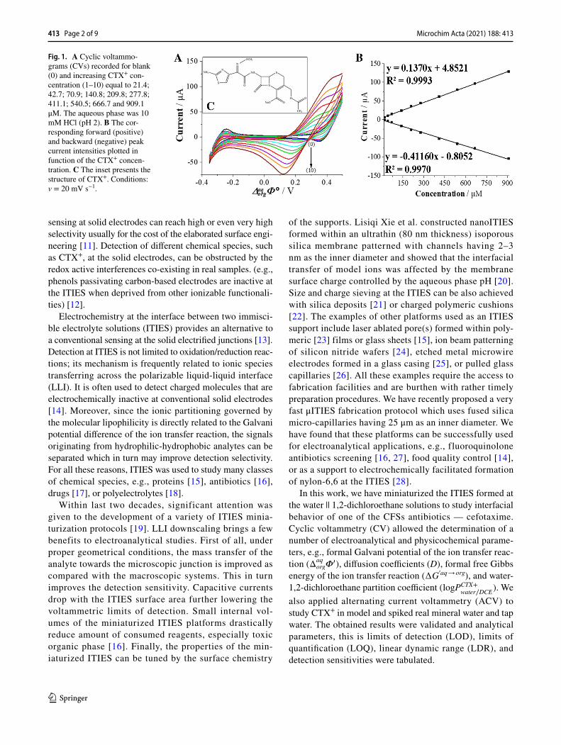
Fig. 1. A Cyclic voltammo-grams (CVs) recorded for blank (0) and increasing CTX+ con-centration (1–10) equal to 21.4; 42.7; 70.9; 140.8; 209.8; 277.8; 411.1; 540.5; 666.7 and 909.1 μM. The aqueous phase was 10 mM HCl (pH 2). B The cor-responding forward (positive) and backward (negative) peak current intensities plotted in function of the CTX+ concen-tration. C The inset presents the structure of CTX+. Conditions: v = 20 mV s−1.
413 Page 2 of 9 Microchim Acta (2021) 188: 413
1 3
Methods and materials
Apparatus
All electroanalytical measurements were carried out with the AUTOLAB–PGSTAT302N) equipped with the ECD mod-ule (Metrohm Autolab B.V., the Netherlands) controlled via NOVA 1.11.1 software. MacroITIES studies were performed in a four-electrode electrochemical system equipped with two platinum (Pt) wires acting as the counter electrodes and two silver/silver chloride wires (Ag/AgCl) applied as the reference electrodes (Scheme 1). The formal Galvani poten-tial of the ion transfer was calculated using data from cyclic voltammograms with the potential axis calibrated using standard Galvani potential of tetrapropylammonium cation ( Δaq
org�0TPrA+ = −0.091 V ) [29].
The set-up used for μITIES polarization contained three electrodes, two in the aqueous phase and one in the organic phase. In the aqueous phase, the platinum wire (Pt) and sil-ver/silver chloride wire (Ag/AgCl) were employed as the counter and reference electrode, respectively. In the organic phase, the platinum wire (Pt) was used as the auxiliary and the pseudo-reference electrode (Scheme 2). The μITIES system was constructed using a small piece of methyl deac-tivated (internal walls of the tubing are commercially func-tionalized with methyl groups) fused silica microcapillary tubing (FSMT) with inner diameter = 25 μm. The fabrica-tion protocol of μ-platforms preparation is described else-where [27].
AC voltammetry measurements were performed under the following experimental conditions: frequency (f) 1 Hz, the amplitude (E) = 10 mV and the step potential (ΔE) = 10 mV. It was assumed that the μITIES system behaves as a Randles circuit. The analytical signal (ϒ) applied to
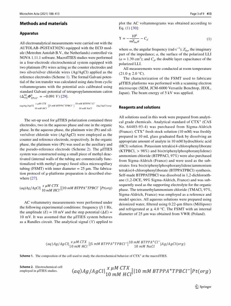
(aq)Ag∕AgCl|x �M CTX
10 mM HCl
||||5 mM BTPPA+TPBCl− |
|
10 mM BTPPA+Cl−
10 mM NaCl
|Ag∕AgCl (org)
(aq)Ag∕AgCl|x �M CTX
10 mM HCl||||10 mM BTPPA+TPBCl− ||Pt(org)
plot the AC voltammograms was obtained according to Eq. (1) [30]:
where ω, the angular frequency (rad·s−1); ZIm, the imaginary part of the impedance; a, the surface of the polarized LLI (a = 1.39 cm2); and Cd, the double layer capacitance of the polarized LLI.
All measurements were conducted at room temperature (21.0 ± 2.0 °C).
The characterization of the FSMT used to fabricate μITIES platforms was performed with a scanning electron microscope (SEM, JCM-6000 Versatile Benchtop, JEOL, Japan). The beam energy of 5 kV was applied.
Reagents and solutions
All solutions used in this work were prepared from analyti-cal grade chemicals. Analytical standard of CTX+ (CAS No. 64485-93-4) was purchased from Sigma-Aldrich (France). CTX+ fresh stock solution (10 mM) was freshly prepared in 10 mL glass graduated flask by dissolving an appropriate amount of analyte in 10 mM hydrochloric acid (HCl) solution. Potassium tetrakis(4-chlorophenyl)borate (KTPBCl, > 98%) and bis(triphenylphosphoranylidene) ammonium chloride (BTPPACl, 97%) were also purchased from Sigma-Aldrich (France) and were used as the sub-strates fora bis(triphenylphosphoranylidene)ammonium tetrakis(4-chlorophenyl)borate (BTPPATPBCl) synthesis. Self-made BTPPATPBCl was dissolved in 1,2-dichloroeth-ane (1,2-DCE, 99% Sigma-Aldrich, France) and was sub-sequently used as the supporting electrolyte for the organic phase. The tetramethylammonium chloride (TMACl, 97%, Sigma-Aldrich, France) was employed as a reference and model species. All aqueous solutions were prepared using deionized water, filtered using 0.22-μm filters (Millipore) and refrigerated at ± 4.0 °C. The FSMT with an internal diameter of 25 μm was obtained from VWR (Poland).
(1)Υ = −106
ωZIma− Cd
Scheme 1. The composition of the cell used to study the electrochemical behavior of CTX+ at the macroITIES.
Scheme 2. Electrochemical cell employed in μITIES studies.
Page 3 of 9 413Microchim Acta (2021) 188: 413
1 3
Results and discussion
macroITIES studies
At first, electrochemical behavior of CTX (the inset — Fig 1A) was studied at the macroITIES with CV and ACV. Cho-sen CVs recorded at macroITIES for increasing concentra-tions of CTX+ (always initially present in the aqueous phase) are shown in Fig. 1A. When dissolved in 10 mM HCl solu-tion (pH set to 2) CTX is almost fully protonated (pKa1 = 3.42) [31]. Based on its structure inspection (inset of Fig. 1C), we have concluded that at given pH, CTX should exist mostly in a cationic form (concentration fraction of cationic form ~ 96%, see Fig. S1) with a delocalized positive charge located in between primary amine and nitrogen atom from the aminothiazole heterocycle. The carboxylate anions located on the opposite side of the CTX structure will be neutral. As such, positively charged CTX (CTX+) molecules may undergo the interfacial transport between the aqueous and the organic phases. During voltammetric studies the LLI was polarized from less to more positive potentials on the forward scan giving positive currents being related to the CTX+ transfer from the aqueous to the organic phase. As can be seen in Fig. 1A, the observed process was reversible with the forward and reversed current being close to unity and the peak-to-peak separation (ΔEp) extrapolated to 56 mV (see the intercept of Fig. S2 showing the increasing peak-to-peak separation plotted in function of the CTX+ concentration). The latter value is close to the theoretical 59 mV·z−1 (z = 1), and it points out that the CTX molecule is mono-charged. From the slope of the linear dependency of the signal current (Is) plotted against voltammetric scan rate (v) (Fig. S3) and the Randles-Ševčík equation, we have calculated the aque-ous and the organic diffusion coefficients (D). Obtained val-ues were equal to Daq→org = 2.55 × 10⁻⁶ cm2·s−1 and Dorg→aq = 4.62 × 10⁻⁷ cm2·s−1. Another parameter that can be deter-mined from CVs is the formal Galvani potential of the ion transfer ( Δaq
org�� ). This parameter is closely related to the
hydrophobicity/hydrophilicity of studied molecule. For cati-onic species, the higher value of the Δaq
org�� indicate their
greater hydrophilicity [16]. The Δaqorg�
� can be further used (Eq. 2) to calculate the formal water – 1,2-DCE partition coefficient ( logPCTX+
water∕DCE ) [32]:
where Δaqorg�
� , the formal Galvani potential of the ion trans-fer reaction (in V); zi, charge of the studied molecule; F, the Faraday constant (96485 C·mol−1); R, the gas constant (8.314 J mol−1·K−1); and T, the temperature (298 K). The calculated logPCTX+
water∕DCE for CTX+ is equal to − 4.48 which
points out high hydrophilicity of the analyte, while based on the logPCTX
water∕octanol = 0.64 available in the veterinary sub-
stances database [33], it can be stated that CTX is a weakly hydrophobic compound. Finally, the Δaq
org�� was used to
determine the formal Gibbs free energy of the ion transfer interfacial reaction (∆G′, aq → org), according to Eq. 3:
All physicochemical and electroanalytical parameters determined for CTX+ are collected and summarized in Table 1.
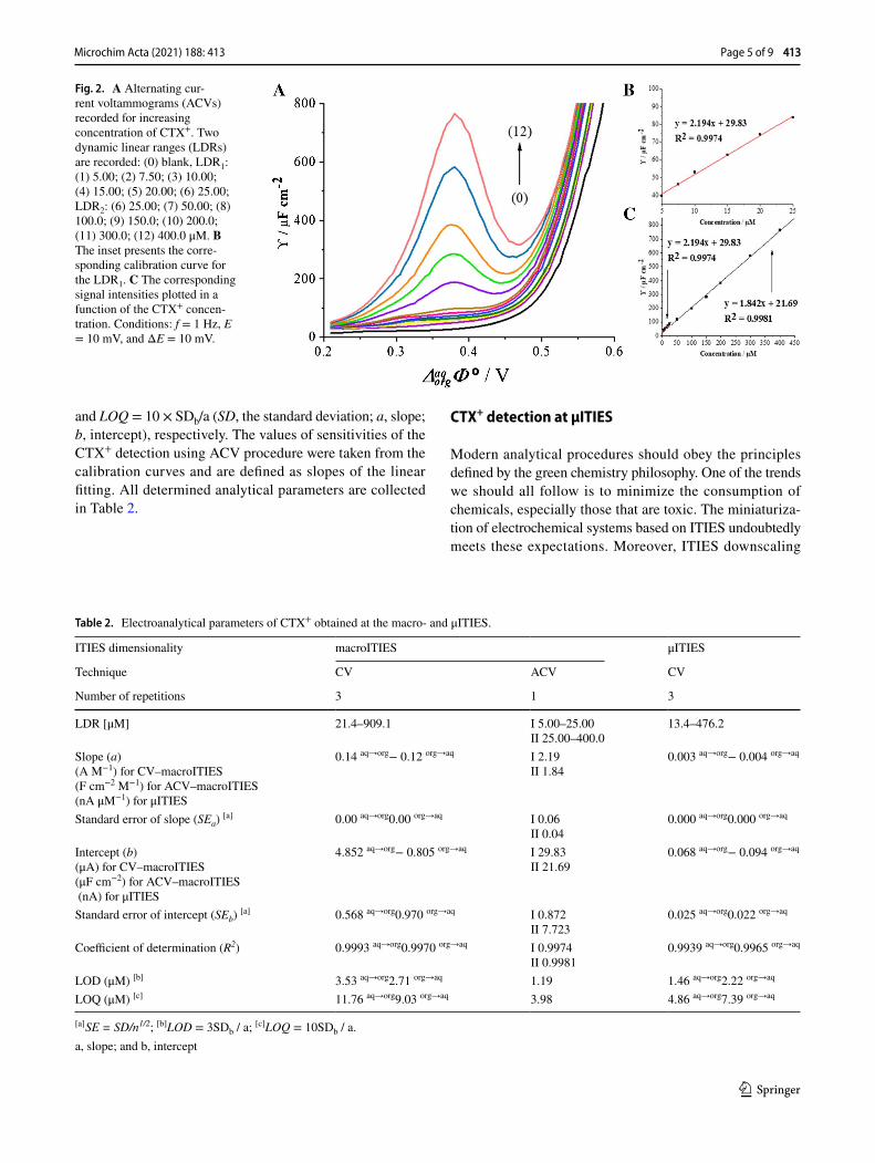
Recently, Suárez-Herrera and Scanlon [34] applied ACV technique to quantitatively analyze simple ion transfer reac-tions across the electrified LLI. Measurements in AC vol-tammetry technique are based on the registration of imped-ance spectra and then converting them to analytical signals. The employment of electrochemical impedance spec-troscopy (EIS) allows for the complete elimination of the capacitive current and thus a significant improvement of the sensitivity of the applied technique. In this work, we have applied the optimized ACV procedure for the determina-tion of CTX+ at the macroITIES. Figure 2A shows the ACV curves recorded at the polarized 10 mM HCl || 1,2-DCE interface in the conventional macroITIES cell (Scheme 1) under conditions described in the “Apparatus” section. Fig-ure 2B and C represent the calibration curve exhibiting two linear dynamic ranges, first from 5.00 to 25.00 μM and sec-ond from 25.00 to 400.0 μM. The existence of two slopes defined by a signal originating only from a simple ion trans-fer reaction is unlikely, and hence, we deduced that CTX+ may undergo weak adsorption process to the ITIES leading to its interfacial preconcentration. The LOD (1.19 μM) and LOQ (3.98 μM) values for CTX+ were calculated from the calibration curve liner fit parameters using LOD = 3 × SDb/a
(2)logPCTX+water∕DCE
= −Δ
aqorg�
�ziF
2.303RT
(3)ΔG�,aq→org = ziFΔaqorg
��
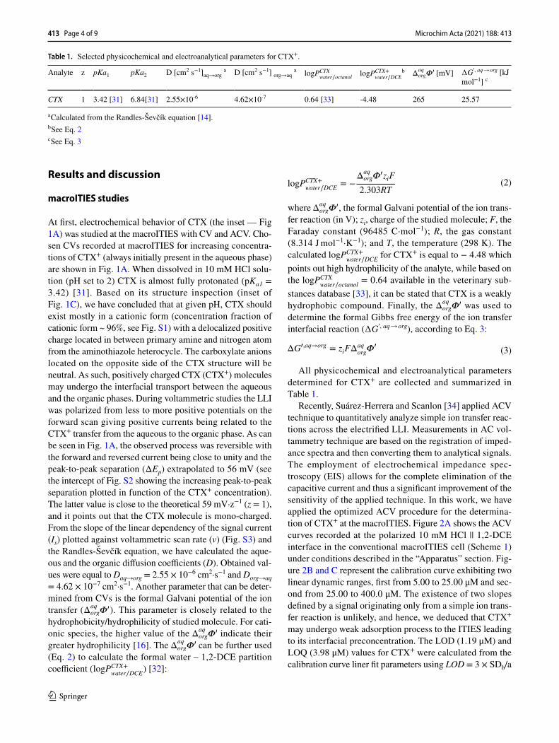
Table 1. Selected physicochemical and electroanalytical parameters for CTX+.
a Calculated from the Randles-Ševčík equation [14].b See Eq. 2c See Eq. 3
Analyte z pKa1 pKa2 D [cm2 s−1]aq→org a D [cm2 s−1] org→aq a logPCTXwater∕octanol
logPCTX+water∕DCE
b Δaqorg�
� [mV] ∆G′, aq → org [kJ mol−1] c
CTX 1 3.42 [31] 6.84[31] 2.55×10⁻⁶ 4.62×10⁻⁷ 0.64 [33] -4.48 265 25.57
413 Page 4 of 9 Microchim Acta (2021) 188: 413
1 3
and LOQ = 10 × SDb/a (SD, the standard deviation; a, slope; b, intercept), respectively. The values of sensitivities of the CTX+ detection using ACV procedure were taken from the calibration curves and are defined as slopes of the linear fitting. All determined analytical parameters are collected in Table 2.
CTX+ detection at μITIES
Modern analytical procedures should obey the principles defined by the green chemistry philosophy. One of the trends we should all follow is to minimize the consumption of chemicals, especially those that are toxic. The miniaturiza-tion of electrochemical systems based on ITIES undoubtedly meets these expectations. Moreover, ITIES downscaling
Fig. 2. A Alternating cur-rent voltammograms (ACVs) recorded for increasing concentration of CTX+. Two dynamic linear ranges (LDRs) are recorded: (0) blank, LDR1: (1) 5.00; (2) 7.50; (3) 10.00; (4) 15.00; (5) 20.00; (6) 25.00; LDR2: (6) 25.00; (7) 50.00; (8) 100.0; (9) 150.0; (10) 200.0; (11) 300.0; (12) 400.0 μM. B The inset presents the corre-sponding calibration curve for the LDR1. C The corresponding signal intensities plotted in a function of the CTX+ concen-tration. Conditions: f = 1 Hz, E = 10 mV, and ΔE = 10 mV.
Table 2. Electroanalytical parameters of CTX+ obtained at the macro- and μITIES.
[a] SE = SD/n1/2; [b]LOD = 3SDb / a; [c]LOQ = 10SDb / a.a, slope; and b, intercept
ITIES dimensionality macroITIES μITIES
Technique CV ACV CV
Number of repetitions 3 1 3
LDR [μM] 21.4–909.1 I 5.00–25.00II 25.00–400.0
13.4–476.2
Slope (a)(A M−1) for CV–macroITIES(F cm−2 M−1) for ACV–macroITIES(nA μM−1) for μITIES
0.14 aq→org− 0.12 org→aq I 2.19II 1.84
0.003 aq→org− 0.004 org→aq
Standard error of slope (SEa) [a] 0.00 aq→org0.00 org→aq I 0.06II 0.04
0.000 aq→org0.000 org→aq
Intercept (b)(μA) for CV–macroITIES(μF cm−2) for ACV–macroITIES (nA) for μITIES
4.852 aq→org− 0.805 org→aq I 29.83II 21.69
0.068 aq→org− 0.094 org→aq
Standard error of intercept (SEb) [a] 0.568 aq→org0.970 org→aq I 0.872II 7.723
0.025 aq→org0.022 org→aq
Coefficient of determination (R2) 0.9993 aq→org0.9970 org→aq I 0.9974II 0.9981
0.9939 aq→org0.9965 org→aq
LOD (μM) [b] 3.53 aq→org2.71 org→aq 1.19 1.46 aq→org2.22 org→aq
LOQ (μM) [c] 11.76 aq→org9.03 org→aq 3.98 4.86 aq→org7.39 org→aq
Page 5 of 9 413Microchim Acta (2021) 188: 413

1 3
meeting specific geometrical recruitments brings a few addi-tional benefits to electroanalytical sensing: (I) stabilization of the electrified LLI by the employed support (capillary forces and surface wettability of the used supports define the position of the soft junction); (II) improved detection sensitivity of the voltammetric methods (shape of the dif-fusion zones established above miniaturized LLI enhances the mass transport from the bulk solution to the polarizable junction); and (III) better analytical performance in terms of LOD and LOQ (reduction of the electroactive surface area = smaller capacitive current) [27, 35]. Therefore, in this work, we have used the FSTM as a polarized LLI support and further applied resulting platform in CTX+ detection.
The μ-devices after preparation and characterization were employed to investigate the interfacial behavior of CTX+ in 10 mM HCl solution (pH = 2). To evaluate the utility of CV procedure for CTX+ determination at the polarized μITIES, CTX+ concentration (cCTX+) dependency was recorded (Fig. 3A), and the corresponding calibration curves of Is vs. cCTX+ were plotted (the inset of Fig. 3A). The asymmetric shape of recorded voltammograms is caused by the coexist-ence of a hemispherical (CTX+ transfer from the aqueous to the organic phase, backward scan, Fig. 3B) and linear diffusion (CTX+ transfer from the organic to the aqueous phase, forward scan, Fig. 3C) and on each side of the LLI. Such behavior is described in other published works [14, 27]. The signal limiting the potential window on the less positive potential window side (~− 0.25 V) originate from the interfacial transfer of Cl- anions or BTPPA+ cations (Cl- transfer from the aqueous to the organic phase or BTTPA+ from the organic to the aqueous phase is expected to be recorded as the negative current). Calibration curve was linear within the entire studied concentration range, this is from 13.32 to 476.2 μM, for both the positive and negative signal intensities. The calculated LOD values for the forward
and backward signal currents are equal to 1.5 and 2.2 μM, respectively. Remaining determined analytical parameters, LOQs, and sensitivities are summarized in Table 2.
Real sample analysis
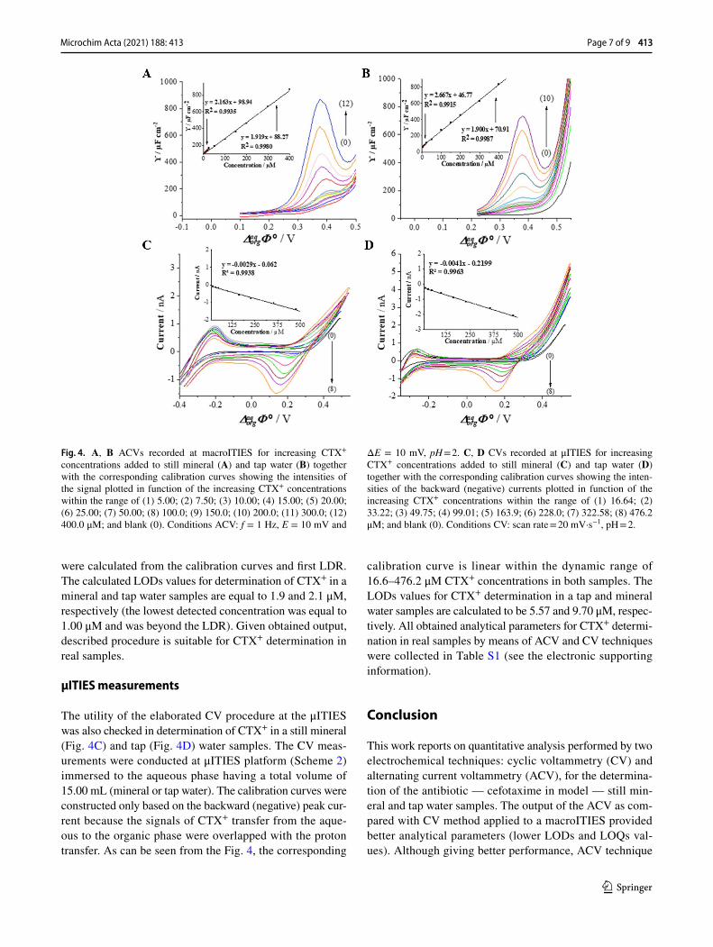
To verify the applicability of the developed method, CTX+ was determined in a spiked tap and mineral water samples using CV and ACV techniques (Fig. 4). The pH of collected water samples was first adjusted with a 1 M HCl solution until pH = 2 was reached. Next, appropriate aliquots of CTX+ stock solutions were added. The samples did not require any further purification to remove salts or any other contaminants. The chemical composition of real tap and mineral water samples includes inorganic cations that may undergo interfacial ion transfer further affecting the positive current values limiting the potential window on more posi-tive potential site. We assume that the existence of additional cations in the aqueous phase is a consequence of overlaid positive current signals recorded for real samples shown in Fig. 4C/D. As such, the backward signals were analyzed for CTX+ quantification.
AC voltammetry measurements
The ACV studies were performed in a traditional macroI-TIES cell (Scheme 1), wherein the aqueous phase com-partment of the cell was filled with 3.50 mL of a sample (mineralor tap water). Figure 4 presents the ACVs recorded upon addition of the appropriate aliquot of the stock solu-tion of CTX+ to the still mineral water (Fig. 4A) and tap water (Fig. 4B) samples. The analytical signals (positive or negative currents) increased linearly with CTX+ concentra-tion in two linear dynamic ranges of 5.00 to 25.00 μM and 25.00 to 400.0 μM in both samples. The LODs and LOQs
Fig. 3. A Representative CVs recorded for increasing CTX+ concentrations of (1) 13.3; (2) 26.6; (3) 49.8; (4) 82.6; (5) 147.8; (6) 228.0; (7) 353.7; (8) 476.2 μM; and blank (0) in 10 mM HCl (pH = 2) used as the aqueous phase. The inset presents forward and back-ward Is intensities plotted in a function of increasing cCTX+. B The scheme of interfacial mass transfer of CTX+ controlled by the hemispherical diffusion. C The backward CTX+ transfer occurring inside the FSTM governed by a linear diffusion. Conditions: v = 20 mV s−1.
413 Page 6 of 9 Microchim Acta (2021) 188: 413
1 3
were calculated from the calibration curves and first LDR. The calculated LODs values for determination of CTX+ in a mineral and tap water samples are equal to 1.9 and 2.1 μM, respectively (the lowest detected concentration was equal to 1.00 μM and was beyond the LDR). Given obtained output, described procedure is suitable for CTX+ determination in real samples.
μITIES measurements
The utility of the elaborated CV procedure at the μITIES was also checked in determination of CTX+ in a still mineral (Fig. 4C) and tap (Fig. 4D) water samples. The CV meas-urements were conducted at μITIES platform (Scheme 2) immersed to the aqueous phase having a total volume of 15.00 mL (mineral or tap water). The calibration curves were constructed only based on the backward (negative) peak cur-rent because the signals of CTX+ transfer from the aque-ous to the organic phase were overlapped with the proton transfer. As can be seen from the Fig. 4, the corresponding
calibration curve is linear within the dynamic range of 16.6–476.2 μM CTX+ concentrations in both samples. The LODs values for CTX+ determination in a tap and mineral water samples are calculated to be 5.57 and 9.70 μM, respec-tively. All obtained analytical parameters for CTX+ determi-nation in real samples by means of ACV and CV techniques were collected in Table S1 (see the electronic supporting information).
Conclusion
This work reports on quantitative analysis performed by two electrochemical techniques: cyclic voltammetry (CV) and alternating current voltammetry (ACV), for the determina-tion of the antibiotic — cefotaxime in model — still min-eral and tap water samples. The output of the ACV as com-pared with CV method applied to a macroITIES provided better analytical parameters (lower LODs and LOQs val-ues). Although giving better performance, ACV technique
Fig. 4. A, B ACVs recorded at macroITIES for increasing CTX+ concentrations added to still mineral (A) and tap water (B) together with the corresponding calibration curves showing the intensities of the signal plotted in function of the increasing CTX+ concentrations within the range of (1) 5.00; (2) 7.50; (3) 10.00; (4) 15.00; (5) 20.00; (6) 25.00; (7) 50.00; (8) 100.0; (9) 150.0; (10) 200.0; (11) 300.0; (12) 400.0 μM; and blank (0). Conditions ACV: f = 1 Hz, E = 10 mV and
ΔE = 10 mV, pH = 2. C, D CVs recorded at μITIES for increasing CTX+ concentrations added to still mineral (C) and tap water (D) together with the corresponding calibration curves showing the inten-sities of the backward (negative) currents plotted in function of the increasing CTX+ concentrations within the range of (1) 16.64; (2) 33.22; (3) 49.75; (4) 99.01; (5) 163.9; (6) 228.0; (7) 322.58; (8) 476.2 μM; and blank (0). Conditions CV: scan rate = 20 mV·s−1, pH = 2.
Page 7 of 9 413Microchim Acta (2021) 188: 413

1 3
requires further data processing and is more time-consum-ing. The application of the μITIES-based setup for the real sample analysis additionally provided improved stability of the soft junction supported with a solid support, reduced volume of the organic phase, and assured compact size of the sensing set-up. Cefotaxime detection at μITIES with CV provided improved electroanalytical parameters comparable to these obtained with ACV at macroITIES. Hence, minia-turized systems were further applied for cefotaxime detec-tion in real samples. Since we did not observe any interfer-ence of the real sample matrix during cefotaxime detection, these did not require any further preparation.
Supplementary Information The online version contains supplemen-tary material available at https:// doi. org/ 10. 1007/ s00604- 021- 05072-w.
Funding This study is financially supported by the National Sci-ence Center (NCN) in Cracow, Poland (Grant no. UMO–2018/29/N/ST4/01054), and Embassy of France in Poland.
Declarations
Conflict of interest The authors declare no competing interests.
Open Access This article is licensed under a Creative Commons Attri-bution 4.0 International License, which permits use, sharing, adapta-tion, distribution and reproduction in any medium or format, as long as you give appropriate credit to the original author(s) and the source, provide a link to the Creative Commons licence, and indicate if changes were made. The images or other third party material in this article are included in the article's Creative Commons licence, unless indicated otherwise in a credit line to the material. If material is not included in the article's Creative Commons licence and your intended use is not permitted by statutory regulation or exceeds the permitted use, you will need to obtain permission directly from the copyright holder. To view a copy of this licence, visit http:// creat iveco mmons. org/ licen ses/ by/4. 0/.
References
1. Samanidou VF, Tsochatzis ED, Papadoyannis IN (2008) HPLC determination of cefotaxime and cephalexine residues in milk and cephalexine in veterinary formulation. Microchim Acta 160:471–475. https:// doi. org/ 10. 1007/ s00604- 007- 0820-1
2. Salem H, Samir E (2018) Determination of cefotaxime, cefopera-zone, ceftazidime and cefadroxil using surface plasmon resonance band of silver nanoparticles. Br J Pharm Sci 54:1–9. https:// doi. org/ 10. 1590/ s2175- 97902 01800 03175 65
3. Yang G, Zhao F, Zeng B (2014) Electrochemical determination of cefotaxime based on a three-dimensional molecularly imprinted film sensor. Biosens Bioelectron 53:447–452. https:// doi. org/ 10. 1016/j. bios. 2013. 10. 029
4. Zhang F, Gu S, Ding Y et al (2013) Electrooxidation and deter-mination of cefotaxime on Au nanoparticles/poly (L-arginine) modified carbon paste electrode. J Electroanal Chem 698:25–30. https:// doi. org/ 10. 1016/j. jelec hem. 2013. 03. 010
5. Arabsorkhi B, Sereshti H (2018) Determination of tetracycline and cefotaxime residues in honey by micro-solid phase extraction
based on electrospun nanofibers coupled with HPLC. Microchem J 140:241–247. https:// doi. org/ 10. 1016/j. microc. 2018. 04. 030
6. Aleksić MM, Kapetanović V, Atanacković J et al (2008) Simul-taneous determination of cefotaxime and desacetylcefotaxime in real urine sample using voltammetric and high-performance liquid chromatographic methods. Talanta 77:131–137. https:// doi. org/ 10. 1016/j. talan ta. 2008. 05. 047
7. Burrer A, Findeisen P, Jäger E et al (2015) Rapid detection of cefotaxime-resistant Escherichia coli by LC-MS. Int J Med Micro-biol 305:860–864. https:// doi. org/ 10. 1016/j. ijmm. 2015. 08. 004
8. Chen D, Wang H, Zhang Z et al (2011) Chemiluminescence determination of cefotaxime sodium with flow-injection analy-sis of cerium (IV)-rhodamine 6G system and its application to the binding study of cefotaxime sodium to protein with on-line microdialysis sampling. Spectrochim Acta - Part A Mol Biomol Spectrosc 78:553–557. https:// doi. org/ 10. 1016/j. saa. 2010. 10. 014
9. Aleksić MM, Kapetanović V (2006) Voltammetric behavior and square-wave voltammetric determination of cefotaxime in urine. J Electroanal Chem 593:258–266. https:// doi. org/ 10. 1016/j. jelec hem. 2006. 06. 011
10. Bottari F, Blust R, De Wael K (2018) Bio(inspired) strategies for the electro-sensing of β-lactam antibiotics. Curr Opin Electro-chem 10:136–142. https:// doi. org/ 10. 1016/j. coelec. 2018. 05. 015
11. Karabozhikova V, Tsakova V (2019) Electroanalytical determi-nation of caffeic acid – factors controlling the oxidation reaction in the case of PEDOT-modified electrodes. Electrochim Acta 293:439–446. https:// doi. org/ 10. 1016/j. elect acta. 2018. 10. 067
12. Gao G, Vecitis CD (2013) Electrocatalysis aqueous phenol with carbon nanotubes networks as anodes: electrodes passivation and regeneration and prevention. Electrochim Acta 98:131–138. https:// doi. org/ 10. 1016/j. elect acta. 2013. 02. 127
13. Chen R, Xu K, Shen M (2020) Electrochimica Acta Avocado oil, coconut oil, walnut oil as true oil phase for ion transfer at nanoscale liquid /liquid interfaces. Electrochim Acta 357:136788. https:// doi. org/ 10. 1016/j. elect acta. 2020. 136788
14. Rudnicki K, Sobczak K, Borgul P et al (2021) Determination of quinine in tonic water at the miniaturized and polarized liquid – liquid interface. Food Chem 364:130417. https:// doi. org/ 10. 1016/j. foodc hem. 2021. 130417
15. Alvarez De Eulate E, Strutwolf J, Liu Y et al (2016) An elec-trochemical sensing platform based on liquid-liquid microinter-face arrays formed in laser-ablated glass membranes. Anal Chem 88:2596–2604. https:// doi. org/ 10. 1021/ acs. analc hem. 5b030 91
16. Rudnicki K, Poltorak L, Skrzypek S, Sudhölter EJR (2019) Ion transfer voltammetry for analytical screening of fluoroquinolone antibiotics at the water – 1,2-dichloroethane interface. Anal Chim Acta. https:// doi. org/ 10. 1016/j. aca. 2019. 07. 065
17. Poltorak L, Rudnicki K, Kolivoška V et al (2021) Electrochemical study of ephedrine at the polarized liquid-liquid interface sup-ported with a 3D printed cell. J Hazard Mater 402:123411. https:// doi. org/ 10. 1016/j. jhazm at. 2020. 123411
18. Riva JS, Cámara CI, Juarez AV, Yudi LM (2014) Electrochemical behaviour of cationic polyelectrolytes at a polarized liquid/liquid interface. J Appl Electrochem 44:1381–1392. https:// doi. org/ 10. 1007/ s10800- 014- 0747-2
19. Liu S, Li Q, Shao Y (2011) Electrochemistry at micro- and nano-scopic liquid/liquid interfaces. Chem Soc Rev 40:2236–2253. https:// doi. org/ 10. 1039/ c0cs0 0168f
20. Xie L, Huang X, Lin X, Su B (2017) Nanoscopic liquid/liquid interface arrays supported by silica isoporous membranes: trans-membrane resistance and ion transfer reactions. J Electroanal Chem 784:62–68. https:// doi. org/ 10. 1016/j. jelec hem. 2016. 12. 007
21. Poltorak L, Morakchi K, Herzog G, Walcarius A (2015) Elec-trochemical characterization of liquid-liquid micro-interfaces
413 Page 8 of 9 Microchim Acta (2021) 188: 413

1 3
modified with mesoporous silica. Electrochim Acta 179:9–15. https:// doi. org/ 10. 1016/j. elect acta. 2015. 01. 129
22. Borgul P, Rudnicki K, Chu L et al (2020) Layer-by-layer (LbL) assembly of polyelectrolytes at the surface of a fiberglass mem-brane used as a support of the polarized liquid–liquid interface. Electrochim Acta 363:137215. https:// doi. org/ 10. 1016/j. elect acta. 2020. 137215
23. Stockmann TJ, Olaya AJ, Méndez MA et al (2011) Evalua-tion of Gibbs energy of dioxouranium transfer at an electrified liquid|liquid interface supported on a microhole. Electroanalysis 23:2677–2686. https:// doi. org/ 10. 1002/ elan. 20110 0401
24. Scanlon MD, Strutwolf J, Blake A et al (2010) Ion-transfer elec-trochemistry at arrays of nanointerfaces between immiscible elec-trolyte solutions confined within silicon nitride nanopore mem-branes. Anal Chem 82:6115–6123. https:// doi. org/ 10. 1021/ ac100 8282
25. Poltorak L, Sudhölter EJR, de Puit M (2019) Electrochemical cocaine (bio)sensing From solid electrodes to soft junctions. TrAC - Trends Anal Chem 114:48–55. https:// doi. org/ 10. 1016/j. trac. 2019. 02. 025
26. Iwai NT, Kramaric M, Crabbe D, et al (2018) GABA Detection with nano-ITIES pipet electrode: a new mechanism, water/DCE–octanoic acid interface. Anal Chem 90(5):3067–3072. https:// doi. org/ 10. 1021/ acs. analc hem. 7b030 99
27. Rudnicki K, Poltorak L, Skrzypek S, Sudhölter EJR (2018) Fused silica microcapillaries used for a simple miniaturization of the electrified liquid-liquid interface. Anal Chem 90:7112–7116. https:// doi. org/ 10. 1021/ acs. analc hem. 8b013 51
28. Kowalewska K, Sipa K, Leniart A et al (2020) Electrochemistry at the liquid–liquid interface rediscovers interfacial polycondensation
of nylon-6,6. Electrochem Commun 115:106732. https:// doi. org/ 10. 1016/j. elecom. 2020. 106732
29. Samec Z (2004) Electrochemistry at the interface between two immiscible electrolyte solutions (IUPAC technical report). Pure Appl Chem 76:2147–2180
30. Lu G, Despas C, Liu L, Herzog G (2020) Ametryn detection by proton assisted transfer at a single micro-interface between two immiscible electrolyte solutions. J Electroanal Chem 877:114745. https:// doi. org/ 10. 1016/j. jelec hem. 2020. 114745
31. https:// go. drugb ank. com/. Accessed 18 Aug 2021 32. Poltorak L, Sudhölter EJR, de Smet LCPM (2017) Effect of charge
of quaternary ammonium cations on lipophilicity and electroana-lytical parameters: task for ion transfer voltammetry. J Electroanal Chem 796:66–74. https:// doi. org/ 10. 1016/j. jelec hem. 2017. 04. 051
33. http:// sitem. herts. ac. uk/ aeru/ vsdb/ Repor ts/ 1940. htm. Accessed 10 Aug 2021
34. Suárez-Herrera MF, Scanlon MD (2020) Quantitative analysis of redox-inactive ions by AC voltammetry at a polarized inter-face between two immiscible electrolyte solutions. Anal Chem 92:10521–10530. https:// doi. org/ 10. 1021/ acs. analc hem. 0c013 40
35. van den Brink FTG, Olthuis W, van den Berg A, Odijk M (2015) Miniaturization of electrochemical cells for mass spectrometry. TrAC Trends Anal Chem 70:40–49. https:// doi. org/ 10. 1016/J. TRAC. 2015. 01. 014
Publisher’s note Springer Nature remains neutral with regard to jurisdictional claims in published maps and institutional affiliations.
Page 9 of 9 413Microchim Acta (2021) 188: 413