voltage dependent ionic currents in frog cultured …. physiol. biophys. (1993), 12, 231—247 231...
TRANSCRIPT
Gen. Physiol. Biophys. (1993), 12, 231—247 231
Voltage Dependent Ionic Currents in Frog Cultured Skeletal Myocytes
V. I. LUKYANENKO1, I. E. KATINA2, G. A. NASLEDOV1* and A. V. LONSKY2
1 Sechenov Institute of Evolutionary Physiology and Biochemistry, Academy of Sciences of Russia, Thorez Av. 44, 194223 St.-Petersburg, Russia
2 Physiological Institute, St.-Petersburg University, University emb. 7/9, 199034 St.-Petersburg, Russia
A b s t r a c t . The voltage dependent ionic currents in cultured embryonic skeletal myocytes at stages of development ranging from 1 to 6 day were studied using the whole-cell patch clamp technique. Sodium (Jfja) and calcium ( lea) inward and potassium (IK) outward currents were observed at all stages. 7 N 3 did not differ from tha t described in adult frog striated muscle fibres. Slow 7c a was mediated by current through dihydropyridine sensitive Ca channels and it did not differ in its kinetics from corresponing slow IQH in frog adult twich muscle fibres. In about 10% of cells examined for IQH, this current was significantly slower and similar to 7c a
described in frog tonic muscle fibres. In some cases two slow calcium currents with distinguishable kinetics were recorded in the same myocytes. Fast dihydropyridin-insensitive noninactivating 7c a could also be observed. At least 6 types of IK were registered, with approximate time-to-peak (at test pulse of —10 mV) 5, 12, 20, 30, 50 ms (fast IK) and more than 7 s (slow IK). Three of them (5, 20 and 30 ms) predominated in 3-day cultures and disappeared in 6-day-old cultures. IK
in myocytes did not correspond fully in the kinetics to 7K reported in adult frog skeletal muscles. Channels associated with transient fast and noninactivating slow 7j< were shown to be highly sensitive to low temperature (+5°C) .
K e y words: Frog embryonic myocytes — Skeletal myocytes culturing — Ionic
currents in myocytes
I n t r o d u c t i o n
Detialed understanding of ionic currents in embryonic myoblasts developing in cul-
* Correspondence to: G. A. Nasledov, Sechenov Institute of Evolutionary Physiology and Biochemistry, Academy of Sciences of Russia, Thorez pr. 44, 194223 St.-Petersburg, Russia
232 Lukyanenko et al.
ture may help elucidate the development of ionic channel functions during the early
stages of myogenesis. Voltage dependent sodium, potassium and calcium channels
have been described in cultured embryonic myocytes. In the course of development
of myocytes in culture, sodium channels do not change their major kinetic proper
ties; their characteristics are similar to those in adult frog muscle fibres (DeCino
and Kidokoro 1985). Only one of the C a 2 + currents described in frog skeletal mus
cles (Sanchez and Stefani 1978, 1983; Cota and Stefani 1986) has been observed
in pr imary culture of frog myocytes: the current through dihydropyridine sensitive
channels (Moody-Corbett et al. 1989). Later, Moody-Corbett and Virgo (1991)
have reported that Xenopus skeletal muscle cells in culture develop a transient
current, which differs from fast noninactivating calcium current of adult frog mus
cle fibres (Cota and Stefani 1986; Garcia and Stefani 1987). This is similar to the
fast transient current described in rat myocytes (Beam et al. 1986; Cognard et al.
1986).
In frog myocytes developing in primary culture 4 types of outward 7K have
been described (Moody-Corbett and Gilbert 1988; Gilbert and Moody-Corbett
1989); however, their characteristics are not fully known as yet, and the kinetic
properties of these currents do not directly correspond to delayed rectifier currents
in adult frog muscle fibres (Adrian et al. 1970; Lynch 1985). In chicken muscle fibre
culture, 7 types of voltage-sensitive K + channels have been reported, the relative
densities of which changed during the culture development (Zemkováet . al. 1989).
The above da ta indicate the need of more detailed mapping of t ransmembrane
ionic currents appearing during the development of frog muscle cell. The present
investigations performed in frogs with the ionic currents in adult tissue being most
completely described provide more information on the development of ionic channel
functions during myogenesis.
Some of the da ta have already been published in short form in Russian (Lukya
nenko et al. 1992a,b).
M a t e r i a l s and M e t h o d s
Cell culture
Standard frog embryo muscle cell cultures were prepared from early neurula embryos of Rana temporaria. The dorsal portions of the embryos were dissected in 60% Medium 199M (Institute of Polyomyelitis and Encephalitis, Academy of Medical Sci. of the CIS) with 2% fetal calf serum (Ecophond, CIS), and 50 U/ml penicillin and 50 jig/ml streptomycin (CIS), and washed during 10 min in calcium-magnesium free salt solution containing (in mmol/1): NaCl 50.4; KC1 0.67; K H 2 P 0 4 0.86; NaHP0 4 16; NaHCOs 2.4; EDTA 1.9 (Freed and Mezger-Freed 1970). During the dissociation into single cells the ectoderm was striped and removed, and mesodermal and neural cells were transferred for cultur-ing on glass, in separate 40 mm Petri dishes for every embryo. The growth medium contained: Medium 199M 55%; fetal calf serum 10%; penicillin 50 U/ml and strepto-
Ionic Currents in Frog Myocytes 233
mycin 50 fig/ml. These constituents almost completely prevent both myocyte division (Teylor-Papadimitriou and Rosengurt 1979) and fusion. The culture was kept at 20 °C under sterile conditions. The myoblasts plated on the glass bottom of the chamber turned spindle-shaped, 1-2 //m in diameter and 15-40 fj,m in length; their size almost doubled by the last days (5-6) of culturing.
The experiments were performed with cells from 1 to 6-day-old cultures. The myocytes selected for experiments did not show any connections with neuroblasts or with each other.
Patch-clamp recording and data analysis
The conventional whole-cell voltage-clamp recording procedure was used. The voltage-clamp circuit was similar to that described by Hamill et al. (1981),
with a 5 Gfi head stage feed-back resistor. The linear component of the leakage current was substracted electronically. The fast component of capacity currents associated with electrode and electrode holder was fully compensated, the slow component associated with cell capacity could be compensated only partly due to the large size and complex shape of the cells. The resistance of patch electrodes filled with standard solution ranged between 3 and 7 Mfi. The seal resistance was 5-30 Gfi and the input resistance of cells ranged between 1-5 Gfi. The experiments were started 10-15 min after the whole-cell recording configuration was established. The membrane potential was held at —80 mV.
The experiments were on-line computer-controlled (voltage pulse delivery and recording of current responses). The current signal was sampled using a ±10 bit A/D converter, at sampling intervals 0.1-10 ms.
The potassium conductance was calculated assuming linear I/V relationship:
GK = IK/(E-ER), (1)
where ER is the current reversal potential. The voltage dependency of peak potassium conductance (Gp) and steady-state inactivation were fitted by Boltzmann function (2) using least squares criterion:
GK(E) = GK/{l + exp[(E-E05)/k}}, (2)
where G K is the conductance at a given membrane potential (E), G K is the maximal peak conductance, Eo 5 is the mid-point on the curve (the value of E at which GK/GK = 0.5), and k is the slope factor of the curve.
Experiments were performed at room temperature (18-20 °C). Mean ± standard error of the mean are given.
Solutions and chemicals
The basic external solution contained (in mmol/1): NaCl 120; KC1 1.5; CaCl2 2; HEPES 8; pH 7.4 adjusted with NaOH. The pipettes were filled with a solution containing (in mmol/1): KC1 110; CaCl2 2; MgCl2 1; K2EGTA 10; HEPES 8; pH 7.2 adjusted with KOH. In some cases KC1 was replaced by CsCl in equimolar amounts. Dihydropyridine (DHP) antagonist nifedipine (Sigma) and agonists CGP-IOS (Institute for Organic Synthesis, Riga, Latvia) was used to identify DHP-sensitive type of calcium channels (the agonistic effects of this DHP derivative has been described by Shvinka et al. 1990). CGP-IOS and nifedipine were preliminarily dissolved in dimethylsulphoxide at 10 mmol/1 and subsequently in external solution yield final concentrations between 20-100 /xmol/l.
234 Lukyanenko et al.
Results
Sodium current
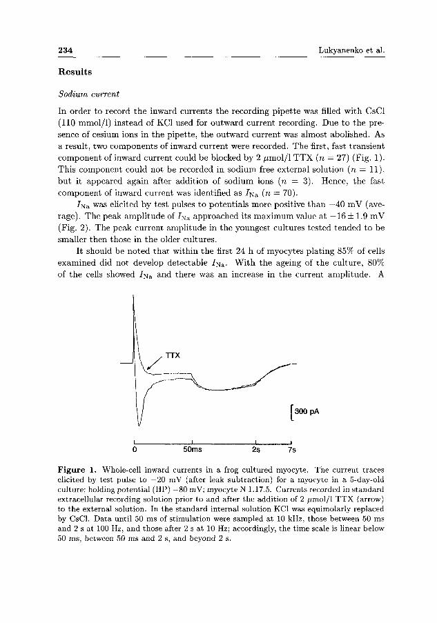
In order to record the inward currents the recording pipette was filled with CsCl (110 mmol/1) instead of KC1 used for outward current recording. Due to the presence of cesium ions in the pipette, the outward current was almost abolished. As a result, two components of inward current were recorded. The first, fast transient component of inward current could be blocked by 2 /xmol/1 T T X (n — 27) (Fig. 1). This component could not be recorded in sodium free external solution (n — 11), but it appeared again after addition of sodium ions (n = 3). Hence, the fast component of inward current was identified as 7 N S (n = 70).
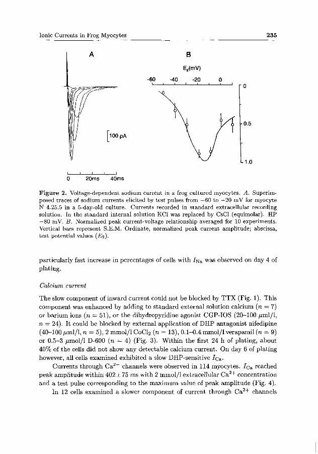
7[va was elicited by test pulses to potentials more positive than —40 mV (average). The peak ampli tude of I^a approached its maximum value at —16 ± 1.9 mV (Fig. 2). The peak current amplitude in the youngest cultures tested tended to be smaller then those in the older cultures.
It should be noted that within the first 24 h of myocytes plating 85% of cells examined did not develop detectable 7Na. Wi th the ageing of the culture, 80% of the cells showed 7Na and there was an increase in the current ampli tude. A
i 1 1 i 0 50ms 2s 7s
Figure 1. Whole-cell inward currents in a frog cultured myocyte. The current traces elicited by test pulse to —20 mV (after leak subtraction) for a myocyte in a 5-day-old culture; holding potential (HP) —80 mV; myocyte N 1.17.5. Currents recorded in standard extracellular recording solution prior to and after the addition of 2 ^imol/1 TTX (arrow) to the external solution. In the standard internal solution KC1 was equimolarly replaced by CsCl. Data until 50 ms of stimulation were sampled at 10 kHz, those between 50 ms and 2 s at 100 Hz, and those after 2 s at 10 Hz; accordingly, the time scale is linear below 50 ms, between 50 ms and 2 s, and beyond 2 s.
Ionic Currents in Frog Myocytes 235
A B
E2(mV)
-60 -40 -20 0 I 1 1 I I I L.
I 1 I 1 I
0 20ms 40ms
Figure 2. Voltage-dependent sodium current in a frog cultured myocytes. A. Superimposed traces of sodium currents elicited by test pulses from —60 to +20 mV for myocyte N 4.25.5 in a 5-day-old culture. Currents recorded in standard extracellular recording solution. In the standard internal solution KC1 was replaced by CsCl (equimolar). HP —80 mV. B. Normalized peak current-voltage relationship averaged for 10 experiments. Vertical bars represent S.E.M. Ordinate, normalized peak current amplitude; abscissa, test potential values (£ 2 ) .
particularly fast increase in percentages of cells with 7Na was observed on day 4 of
plating.
Calcium current
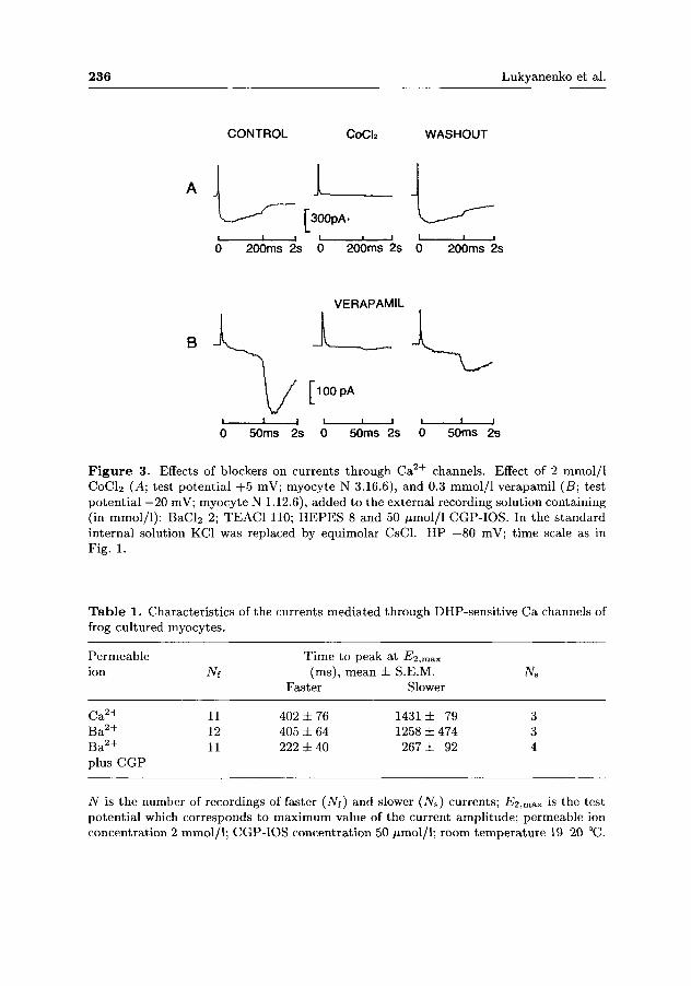
The slow component of inward current could not be blocked by T T X (Fig. 1). This component was enhanced by adding to s tandard external solution calcium (n = 7) or barium ions (n = 51), or the dihydropyridine agonist CGP-IOS (20-100 fiml/l,
n — 24). It could be blocked by external application of D H P antagonist nifedipine (40-100 /iml/1, n = 5), 2 mmol/1 CoCl2 (n = 13), 0.1-0.4 mmol/1 verapamil (n = 9) or 0.5-3 /xmol/1 D-600 (n = 4) (Fig. 3). Within the first 24 h of plating, about 40% of the cells did not show any detectable calcium current. On day 6 of plating however, all cells examined exhibited a slow DHP-sensitive 7c a-
Currents through C a 2 + channels were observed in 114 myocytes. 7c a reached peak amplitude within 402 ± 75 ms with 2 mmol/1 extracellular C a 2 + concentration and a test pulse corresponding to the maximum value of peak amplitude (Fig. 4).
In 12 cells examined a slower component of current through C a 2 + channels
236 Lukyanenko et al.
CONTROL CoCIa WASHOUT
I I 300pA.
200ms 2s 0 200ms 2s 0 200ms 2s
VERAPAMIL
B
50ms 2s 50ms 2s
Figure 3. Effects of blockers on currents through Ca2 + channels. Effect of 2 mmol/1 CoCl2 (̂ 4; test potential +5 mV; myocyte N 3.16.6), and 0.3 mmol/1 verapamil (B; test potential —20 mV; myocyte N 1.12.6), added to the external recording solution containing (in mmol/1): BaCl2 2; TEAC1 110; HEPES 8 and 50 fimol/l CGP-IOS. In the standard internal solution KC1 was replaced by equimolar CsCl. HP —80 mV; time scale as in Fig. 1.
Table 1. Characteristics of the currents mediated through DHP-sensitive Ca channels of frog cultured myocytes.
Permeable ion
Ca2+ Ba 2 +
Ba 2 +
plus CGP
iVf
11 12 11
Time to peak (ms),
Faster
402 ± 76 405 ± 64 222 ± 40
mean at -E2,max
± S.E.M. Slower
1431 ± 79 1258 ± 474 267 ± 92
Ns
3 3 4
AT is the number of recordings of faster (Ns) and slower (Ns) currents; £ľ2,max is the test potential which corresponds to maximum value of the current amplitude; permeable ion concentration 2 mmol/1; CGP-IOS concentration 50 /imol/1; room temperature 19-20 °C.

Ionic Currents in Frog Myocytes 237
B E2 (mV)
150pA
50ms 2s 7s
-40 -20 0 i 1 1 1 i ' i g
0.5
L1.0
Figure 4. Slow DHP-sensitive calcium current in a frog cultured myocyte. A. Whole-cell currents (without leak subtraction) elicited by test potentials from —40 to —5 mV (indicated at each recording) for myocyte N 3.23.5 in a 3-day-old culture recorded with 2 mmol/1 CaCl2 in the external recording solution. In the internal standard solution KC1 was replaced by equimolar CsCl. HP —80 mV; time scale as in Fig. 1. B. The normalized peak current-voltage relationship averaged for 15 experiments with 2 mmol/1 CaCl2 in the external recording solution. Vertical bars represent S.E.M. Ordinate, normalized peak current'amplitude; abscissa, test potential values (i?2).
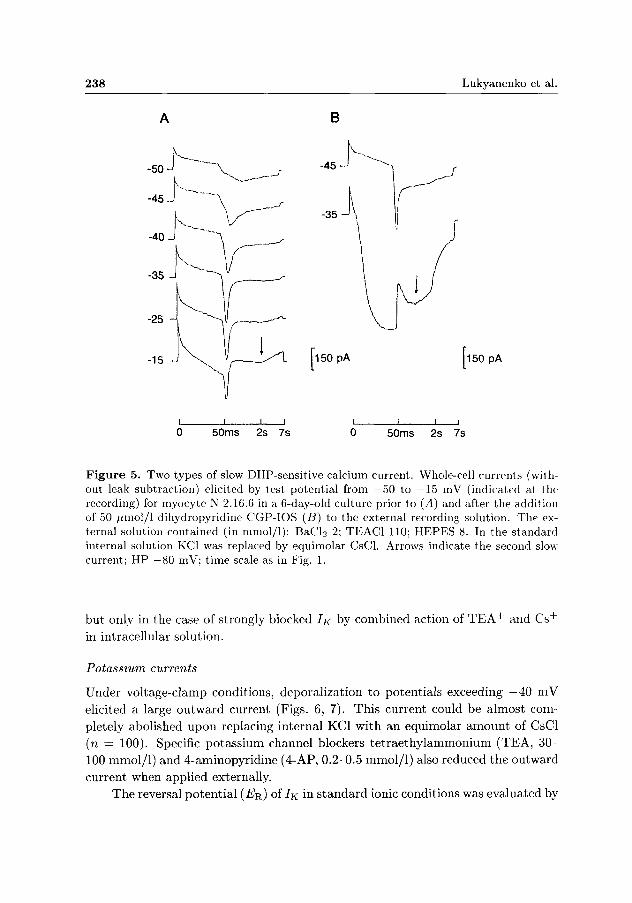
was recorded ( P < 0.001). Under the same experimental conditions the current amplitude of this component reached a maximum value within 1431 ± 79 ms (Table 1). This component could be blocked by external application of 2 mmol/1 C0CI2 (n — 3) or 3 yumol/1 D-600 (n — 3). It was enhanced by external addition of barium ions (n = 10) or 50 /xmol/1 CGP-IOS (n = 2). Both types of slow DHP-sensitive currents in the same cell were manifested in 5 myocytes (Fig. 5), and in 2 of them both currents were recorded at normal external calcium ion concentration (2 mmol/1 C a 2 + ) .
DHP agonist CGP-IOS (20-100 ^rnol/1) increased both types of inward current
in all cases (n = 25), accelerated the current activation, and shifted the membrane
potential of activation 10-15 mV to more negative values (Fig. 5). Inactivation t ime
constants (r,n) for bo th types of 7c a could not be estimated due to the currents
superposition and to the continuation of outward current. Insensitive to nifedipine
and displied high t ime constant of decay (500-800 ms) fast 7c a also be registered,
238 Lukyanenko et al.
A B
i i i i i i i i
0 50ms 2s 7s 0 50ms 2s 7s
Figure 5. Two types of slow DHP-sensitive calcium current. Whole-cell currents (without leak subtraction) elicited by test potential from —50 to —15 mV (indicated at the recording) for myocyte N 2.16.6 in a 6-day-old culture prior to (A) and after the addition of 50 /nnol/1 dihydropyridine CGP-IOS (B) to the external recording solution. The external solution contained (in mmol/1): BaCl2 2; TEAC1 110; HEPES 8. In the standard internal solution KC1 was replaced by equimolar CsCl. Arrows indicate the second slow current; HP —80 mV; time scale as in Fig. 1.
but only in the case of strongly blocked IK by combined action of T E A + and C s +
in intracellular solution.
Potassium currents
Under voltage-clamp conditions, deporalization to potentials exceeding —40 mV
elicited a large outward current (Figs. 6, 7). This current could be almost com
pletely abolished upon replacing internal KC1 with an equimolar amount of CsCl
(n = 100). Specific potassium channel blockers te t raethylammonium (TEA, 30 -
100 mmol/1) and 4-aminopyridine (4-AP, 0.2-0.5 mmol/1) also reduced the outward
current when applied externally.
The reversal potential (7?R,) of 7K in s tandard ionic conditions was evaluated by

Ionic Currents in Frog Myocytes 239
A CONTROL B C
\ TEACI 4-AP
I 200 pA
i _ j i i i l l i i 1 1——i
0 50ms 2s 7s 0 50ms 2s 7s 0 50ms 2s 7S
Figure 6. Effects of K + channel blockers on outward currents. Whole-cell current traces recorded from myocyte N 4.19.6 in a 1-day-old culture with standard external and internal recording solutions prior to the addition of drugs (control; A), after the addition of 110 mmol/1 TEACI (B) and 0.5 mmol/1 4-aminopyridine (C) to the external solution. Test pulse —20 mV; HP —80 mV; time scale as in Fig. 1.
constructing instantaneous I —V relationship (conditioning potential E\ = 10 mV;
test potentials, E2 = —10—1-20 mV). The value of ER was practically independent
of the duration of E\ (30 and 150 ms) and averaged —77.5 ± 0.54 mV (n = 8).
A permeability ratio P-^a,/ PK = 0.03 was calculated from a modified Goldman-
Hodgkin-Katz equation.
The t ime course of outward 7K was complex with usually more than one peak
(Fig. 6). It depended both on the test and on the holding potential , and was
different for cells at different stages and for different conditions of cultivation.
An analysis of the kinetics and pharmacological properties showed 9 components of outward current which differred one from another by the t ime courses of activation (Table 2). At test potentials near —10 mV all the components can be divided into two groups: fast components ( / ) with time-to-peak less than 70 ms ( / 5 , / 1 2 , / 2 0 , / 3 0 , / 5 0 ) , and slow (s) components with longer time-to-peak (s l90 , s700, s2000, S). The figures represent the approximate time-to-peak; no steady-state level was observed for component 5 , even at a duration of E2 as long as 7 s. The components of the fast and the slow group differ also in their pharmacological properties: the fast components are more sensitive to TEA (30-110 mmol/1) whereas the slow ones are more sensitive to 4-AP (0.2-0.5 mmol/1) (Fig. 6). Three of the four slow components ( s l90 , s 700 and s2000) were recorded rather rarely and only at high concentrations of inhibitors (110 mmol/1 TEACI and /o r 8-30 mmol/1 BaClž), when all other outward current components were completely abolished.
As a rule, 2-3 or more components were present simultaneously in the same

240 Lukyanenko et al.
B
o -10
-20
-30
-40 ^
_ i _
=^A 300 pA
50ms 2s 7s
-100
50ms 2s
G„/GD
Figure 7. Voltage dependence of peak conductance (Gp) and steady-state inactivation of potassium current (component type /12). A. Currents (after leak subtraction) elicited by test potentials from —40 to 0 mV (indicated at the recording). HP —80 mV; 6-day-old cultured myocyte N 1.31.5; standard external and internal recording solutions. B. Current traces (after leak sutraction) elicited by test potentials of —10 mV at condition potentials (duration 10 s) from —100 to —50 mV (indicated at the recording). HP —80 mV; 2-day-old cultured myocyte N 1.7.6; standard external recording soluiion with 0.5 mmol/1 4-AP; time scale as Fig. 1. C. The right curve corresponds to the voltage dependence of Gp normalized to maximal conductance (Gp) at large positive potentials (Gp/Gp) as a function of membrane voltage Em (n = 6) . The left curve reflects steady state inactivation (/icx.); values of peak current at given Em normalized to maximal currents at Em = —100 mV (n = 4). Mean values ± S.E.M (bars); the lines are the best fit to Boltzmann function (2), where for Gv/Gp: E0 5 = — 2 and k = 13; and for h^: E0 5 = —61 and k = 7.
cell, and it was difficult to separate properties of the individual components. In
some cases, only one of the fast and/or one of the slow currents was predominant.
Fig. 7 illustrates the properties of outward current in cells with predominant / 1 2
type component. Currents for various depolarizing test pulses (752 = —40 — 0 mV)
Ionic Currents in Frog Myocytes 241
from holding potential of —80 mV are shown in Fig. 7 A, and currents at test potential E<i — - 1 0 mV from various conditioning levels (E\ — - 1 0 0 50 mV) in Fig. 7 B. These d a t a allowed t o plot peak conductance and steady-state inactivation voltage relations. Averaged d a t a from an /12 experiment are shown in Fig. 7 C; t h e right curve corresponds to the voltage dependence of peak conductance Gp
normalized t o maximum conductance (Gp) at large positive potentials (n = 6). T h e left curve reflects the steady-state inactivation (values of peak current at a given Ei are normalized to maximum currents at Ei = —100 mV (n = 4). T h e results of an analogous analysis for some components are summarized in Table 2 ( n = 3 - 7 , for each component type).
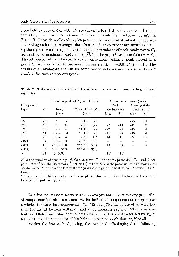
Table 2. Stationary characteristics of the outward current components in frog cultured myocytes.
Time to peak at ŕľ2 = —10 mV Curve parameters (mV) Component Peak Steady-state type N Range Mean ± S.E.M. conductance inactivation
/5 /12 /20 /30 /50 sl90 s700 s2000 S
25 58 66 44 20 9 11 7 33
(ms)
4-10-19-29-40-110-450-1500-
8 15 25 34 70 250 1110 2550
> 7000
(ms)
6.4 ± 12.8 ± 21.4 ± 30.4 ± 49.0 ± 190.0 ± 734.0 ±
0.4 0.2 0.2 0.2 1.4 18.4 56.7
1865.0 ±165.0 -
EQ 5
-
-2 -22 -14 -16 --18 --44*
*G
-
-13 -9 -8 -11 --8 --11*
Eo 5
-55 -61 -43 -39 -74 ----
A:h
6 7 9 9 8 ----
N is the number of recordings; /, fast; s, slow; £ľ2 is the test potential; Eo 5 and k are parameters from the Boltzmann function (2), where Eo 5 is the potential at half-maximum conductance, k is the slope factor (these parameters give the best fit to Boltzmann function). * The curves for this type of current were plotted for values of conductance at the end of long (7 s) depolarizing pulses.
In a few experiments we were able t o analyze not only stat ionary properties of components but also to est imate T;„ for individual components or the group as a whole. For three fast components, / 5 , /12 and /30 , the values of r;n were less t h a n 100 ms (at TJ2 near —10 mV), and for components /20 and /50 they were as high as 300-600 ms. Slow components s l 9 0 and s 700 are characterized by Tm of 500-2000 ms, the component s2000 being inactivated much slowlier, if at all.
Within the first 24 h of plating, the examined cells displayed the following

242 Lukyanenko et al.
current types: /12 (48% of myocytes), /20 (50%), /30 (70%), /50 (53%) and S (78%), but not type /5 . As the age of the culture increased to 3 days, type /5 appeared in most cells (80%) and types /20 and /30 became more prominent (82% each) than other types of outward components. By the 4th day the proportion of cells with detectable currents /20 and /30 rapidly decreased. By the 5th day the proportion of myocytes with type /5 currents also decreased. The percentage of myocytes with 7K types /12 and S increased from the second day through sixth day of plating (100%).
It is interesting to note that, in contrast to myocytes kept under conventional conditions at +20°C, myocytes, kept for 5-7 days at +5°C before plating, did not exhibit the outward current components /5 and S (n = 24).
Discussion
Sodium current. Sodium current registered in our experiments exhibits kinetics and pharmacological properties very similar to those described for frog embryonic myocytes (DeCino and Kidokoro 1985) and skeletal muscle fibres of adult frogs (Adrian et al. 1970; Campbell and Hille 1976; Stefani and Chiarandini 1982).
The percentages of cells with sodium current were low on the first day of culturing (15%), increasing rapidly in the course of culture development with a simultaneous increase of the amplitude. This is assumed to be a result of an increase in N a + channel density in the myocyte membrane with the cell culture age (DeCino and Kidokoro 1985). Particularly rapid increases of the percentages of cells with 7Na occurrence were observed on days 4-5 of culturing. These data point to the existence of a distinct developmental stage at which functioning sodium channels appear in membrane of skeletal myocytes. Similar results were obtained with chicken embryonic muscle cells. Using labelled channel blockers sodium channels were shown to appear after 2-3 days of culturing, reaching maximum amounts by day 5-7 (Frelin et al. 1981; Strichartz et al. 1983).
Calcium currents. We found that myocytes in culture show a slow inward ionic current through C a 2 + channels that is voltage operated and sensitive to dihydropy-ridine (DHP). This current was similar to that mediated by ionic flux through DHP-sensitive C a 2 + channels in twitch skeletal muscle of the adult frog (Stanfield 1977; Sanchez and Stefani 1978, 1983; Aimers and Palade 1981; Aimers et al. 1981; Stefani and Chiarandini 1982; Beaty et al. 1987; Henček et al. 1988; Feldmeyer et al. 1990).
Moody-Corbett et al. (1989) and Moody-Corbett and Virgo (1991) reported that Xenopus embryonic muscle cells grown in culture develop DHP-sensitive slow and DHP-insensitive fast transient currents through calcium channels. The former is similar to the slow current described in the present study and in adult frog

Ionic Currents in Frog Myocytes 243
muscle. The latter in turn is similar to the fast transient current described in rat and mice cultured myocytes (Beam et al. 1986; Cognard et al. 1986; Shimahara and Bournaud 1991), and it differs from the fast noninactivating current described in adult frog muscle (Cota and Stefani 1986; Beaty et al. 1987; Garcia and Stefani 1987; Henček et al. 1988).
No fast 7ca was observed in our experiments under described experimental conditions. The amplitude of fast 7c a is known to be n-times smaller than the amplitude of slow 7c a. In our study even slow 7c a had a low amplitude and we had to replace standard extracellular solution by a solution containing B a 2 + in order to increase current through DHP-sensitive channels (Aimers and McCleskey 1984); also, remains of currents through K + channels had be blocked by TEA. However, B a 2 + do not increase current through T-type C a 2 + channel (Fox et al. 1987), and TEA inhibits it (Beaty et al. 1987; Garcia and Stefani 1987). It is likely that the high concentrations of CsCl and EGTA used in the internal recording solution affected the appearence of the fast current (Beaty et a. 1987). We have observed fast noninactivated, DHP insensitive 7c a in addition experiments with strongly blocked 7K by TEA + and Cs + in intracellular solution. These results will be described separately.
A second slow current, characterized by slower activation and inactivation and a lower amplitude, was seen in 12 myocytes, on days 4-6 of culturing (Fig. 5^4). This current also appeared to be mediated via DHP-sensitive C a 2 + channels (Fig. 55), however, its characteristics were less clear since it was quite infrequent. By its activation time, the second slow current is similar to one described in tonic skeletal muscle fibres of adult frog. Huerta and Stefani (1986) have shown that the amplitude of slow 7c a in frog tonic muscle fibres reached a maximum value within 1000 ms. Also, Henček et al. (1988) have reported that it took the amplitude of the slow current in tonic fibres more than 1 s to reach a maximum value, whereas in twitch fibres time-to-peak was only 200-700 ms (Stanfield 1977; Aimers and Palade 1981; Stefani and Chiarandini 1982; Henček et al. 1988).
Our results can be interpreted in terms of a hypothesis that in some myocytes DHP-sensitive C a 2 + channels specifically associated with twitch and tonic types of adult muscle fibres coexist. Fig. 5 shows two components of DHP-sensitive inward slow current in the same myocyte. Aimers and McCleskey (1984) have reported current traces with two slow components in external solution containing mixtures of 10 mmol/1 C a 2 + and 10 mmol/1 Ba 2 + , as a result of anomalous mole-fraction behavior (AMFB). We recorded two slow components not only upon substituting standard external solution by a solution with 2-8 mmol/1 Ba 2 + , but also in standard solution (2 mmol/1 Ca 2 + ; n=2). The mode of substitution of extracellular solution used in our experiments allowed us to suggest that in our case the coexistence of slow currents was not a result of AMFB. Moody-Corbett et al. (1989) and Moody-Corbett and Virgo (1991) did not record a second slow current in Xenopus
244 Lukyanenko et al.
myocytes. Presumably, the reason may have been relatively short depolarizing
pulses (only 500 ms), whereas 7 s were used in our experiments.
Potassium currents. In our experiments frog embryonic muscle cells in culture
exhibited 9 components of voltage operated potassium current. Three of them,
slow components s l90 , s 700 and s2000, had low frequencies of occurrence and were
evident only in the presence of high concentrations of potassium channel blockers
( T E A + and/or B a 2 + ) , when other outward tu r ren t components were suppressed.
The components / 5 , / 1 2 , / 2 0 , / 3 0 , / 5 0 , and S can be considered as separate
types of currents through corresponding potassium ionic channels, all of them being
sensitive to specific K-channels blockers. This conclusion is based on the differences
in t ime of activation of the currents, as well as on some other differences. Namely,
5- type of 7K exhibits the most negative range of activation (7?o.5 = —44). Types
/ 5 , / 2 0 and / 3 0 of 7K disappeared on day 6 of culturing, but / 1 2 and S were
registered at that t ime in 100% of myocytes. Types / 2 0 and / 3 0 , in spite of
their similarity in t ime to peak value, are characterized by different parameters
of conductance, different stat ionary inactivation curves (Table 2), and by different
speeds of inactivation. Current / 5 0 differs from other fast 7K by its range of steady-
s ta te inactivation curve being the most negative one (75o.5 = —74 mV), which is
close to the reversal potential ( — 77.5 mV).
The absence of / 5 and S currents after preincubation of myocytes at +5°C
during 5-7 days also points to the existence of separate types of potassium channels.
It might be suggested tha t K + channels / 5 , which did not display any activity at
the beginning of normal cell incubation, could not develop under low temperature.
Activity of S type of channels was observed in 80% of myocytes on the first day
of normal culturing, but in none of them (n = 24) after cold preincubation. It
means that the already developed 5-type activity of 7K can be supressed by low
temperature .
In recent years several types of 7K were described in embryonic muscle cells.
Moody-Corbett and Gilbert (1988) and Gilbert and Moody-Corbett (1989) regis
tered 4 voltage dependent outward 7K in embryonic myocytes of Xenopus using
whole-cell configuration. By their kinetic and pharmacological properties, these
currents appear to be similar to our currents / 5 , / 1 2 , / 5 0 and S. Zemková et al.
(1989) examined single K + channel currents during differentiation of chicken em
bryonic muscle cells in vitro and identified 7 types of K + channels. These authors
described also differences between myoblasts and myotubes concerning the per
centages of the individual channel types; however muscle cells with blocked fusion,
showed the same change in channel population as shown in the present paper.
None of the IK components found in the present s tudy could be directly com
pared to currents in adult frog muscle fibres, where the existence of two types of
delayed rectifier currents (fast and slow) were described (Adrian et al. 1970; Stan-
Ionic Currents in Frog Myocytes 245
field 1970; Aimers and Palade 1981; Lynch 1985). Nevertheless, the properties of
the fast and the slow group as a whole are nearly compatible with the properties
of the fast and slow 7K in adult frog muscle. It is likely tha t the fast and the slow
delayed rectifier currents in adult skeletal muscle fibres are a result of a coexistence
of several 7K types.
In terms of conventional classification (Rudy 1988), three fast components
( / 5 , / 1 2 and /30 ) could be considered as fast transient type, which differs from
typical "^4" currents by its low sensitivity to 4-AP. Components / 2 0 and / 5 0 are
closely related to fast delayed rectifier, and the 5-type may be refferred to as a
slow delayed rectifier.
R e f e r e n c e s
Adrian, R. H., Chandler W. K., Hodgkin A.L. (1970): Voltage clamp experiments in striated muscle fibres. J. Physiol. (London) 208, 607—644
Aimers W., Palade P. T. (1981): Slow calcium and potassium currents across frog muscle membrane: measurements with a vaseline-gap technique. J. Physiol. (London) 312, 159—176
Aimers W., McCleskey E. W. (1984): Non-selective conductance in calcium channels of frog muscle: calcium selectivity in a single-file pore. 353, 585-608 J. Physiol. (London) 353, 585—608
Aimers W., Fink R., Palade P. T. (1981): Calcium depletion in frog muscle tubules: the decline of calcium current under maintained depolarization. J. Physiol. (London) 312, 177—207
Beam K. G., Knudson C. M., Powell J. A. (1986): A lethal mutation in mice eliminates the slow calcium current in skeletal muscle cells. Nature 320, 168—170
Beaty G. N., Cota G., Siri L. N., Sanchez J. A., Stefani E. (1987): Skeletal muscle C a + +
channels. In: Structure and Physiology of the Slow Inward Calcium Channel, pp. 123—140, Allan R. Liss., New York
Campbell D. T., Hille B. (1976): Kinetic and pharmacological properties of the sodium channel of frog skeletal muscle. J. Gen. Physiol. 67, 309—323
Cognard C , Lazdunski M., Romey G. (1986): Different types of Ca2 + channels in mammalian skeletal muscle cells in culture. Proc. Nat. Acad. Sci. USA 83, 517—521
Cota G., Stefani E. (1986): A fast-activated inward calcium current in twitch muscle fibres of the frog (Rana montezume). J. Physiol. (London) 370, 151—163
DeCino P., Kidokoro Y. (1985): Development and subsequent neural tube effects on the excitability of cultured Xenopus myocytes. J. Neurosci. 5, 1471—1482
Feldmeyer D., Melzer W., Pohl B., Zolner P. (1990): Fast gating kinetics of the slow Ca2 +
current in cut skeletal muscle fibres of the frog. J. Physiol. (London) 425, 347—367 Fox A. P., Nowycky M. C , Tsien R. W. (1987): Kinetic and pharmacological properties
distinguishing three types of calcium currents in chick sensory neurones. J. Physiol. (London) 394, 149—172
Freed J. J., Mezger-Freed L. (1970): Culture methods for anuran cells. In: Methods in Cell Physiology. Vol. 4. (Ed. D. M. Prescot), pp. 19—48, Acad. Press., New York

246 Lukyanenko et al
Frelin C , Lombet A , Vigne P , Romey G , Lazdunsky M (1981) The appearance of voltage-sensitive N a + channels during the in vitro differentiation of embryonic chick skeletal muscle cells J Biol Chem 256, 12355—12361
Garcia J , Stefani E (1987) Appropriate conditions to record activation of fast C a 2 +
channels in frog skeletal muscle (Rana pipiens) Pflugers Arch 408, 646—648 Gilbert R , Moody-Corbett F (1989) Four outward potassium currents in Xenopus skele
tal muscle cells in culture Can J Physiol Pharmacol 67, N 5, Axin—Axiv Harnill O P , Marty A , Neher E , Sakmann B , Sigworth F J (1981) Improved patch-
clamp techniques for high-resolution current recording from cells and cell-free membrane patches Pflugers Arch 391, 85—100
Henček M , Zacharova D , Zachar J (1988) Fast calcium currents in cut skeletal fibres of the frogs Rana tempona and Xenopus laevis Gen Physiol Biophys 7, 651—658
Huerta M , Stefani E (1986) Calcium action potentials and calcium currents in tonic muscle fibres of the frog (Rana pipiens) J Physiol (London) 372, 293—301
Lukyanenko V , Nasledov G A , Katina I E , Lonsky A V (1992a) Voltage dependent inward ionic currents in cultured frog myoblasts Zh Evol Biochim Fiziol 28, 120—122 (in Russian)
Lukyanenko V , Katma I E , Nasledov G A , Lonsky A V (1992b) Voltage dependent outward ionic currents in cultured frog myoblasts Neurofiziologiya 24, 109—113 (in Russian)
Lynch C (1985) Ionic conductances in frog short skeletal muscle fibres with slow delayed rectifier currents J Physiol (London) 368, 359—378
Moody-Corbett F , Gilbert R (1988) Potassium current of embryonic muscle cells in culture Biophys J 53, N2, 546a
Moody Corbett F , Virgo N S (1991) Exposure to nerve does not affect the appearence of calcium currents in embryonic muscle NeuroReport 2, 437—440
Moody-Corbett F , Gilbert R , Akbarah H Hall J (1989) Calcium current in embryonic Xenopus muscle cells in culture Can J Physiol Pharmacol 67, 1259—1264
Rudy B (1988) Diversity and ubiquity of K channels Neuroscience 25, 729—749 Sanchez J A , Stefani E (1978) Inward calcium current in twitch muscle fibres of the
frog J Physiol (London) 283, 197—209 Sanchez J A Stefani E ((1983) Kinetic properties of calcium channels of twitch muscle
fibres of the frog J Physiol (London) 337, 1—17 Shimahara T Bournaud R (1991) Barium currents m developmg skeletal muscle cells
of normal and mutant mice foetuses with "muscular dysgenesis" Cell Calcium 12, 727—733
ShvinkaN E Gyorke S , Nasledov G A (1990) Effects of dihydropyndme C GP on cm-rents through the calcium channels in frog skeletal muscle Gen Physiol Biophys 9, 83—86
Stanfield P R (1970) The effect of the tetraethylammomum ion on the delayed currents of frog skeletal muscle J Physiol (London) 209, 209—229
Stanfield P R (1977) A calcium dependent mward current in frog skeletal muscle fibres Pflugers Arch 368, 267—270
Stefani E , Chiarandini D (1982) Ionic channels m skeletal muscle Annu Rev Physiol 44, 357—372
Strichartz G , Bar-Sagi D , Prives J (1983) Differential expression of sodium channel activities during the development of chick skeletal muscle cells in culture J Gen Physiol 82, 365—385
Ionic Currents in Frog Myocytes 247
Teylor-Papadimitriou J., Rosengurt E., (1979): The role of thymidine uptake in the control of cell proliferation. Exp. Cell Res. 119, 393—396
Zemková H., Vyskočil F., Tolar M., Vlachová V., Ujec E. (1989): Single K+ currents during differentiation of embryonic muscle cells in vitro. Biochim. Biophys. Acta 986, 146—150
Final version accepted March 5, 1993