vol.44 no.3 march 2001 - med.or.jp no.3 march 2001 a journal of medical sciences of japan and other...
TRANSCRIPT

Vol.44 No.3March
2001
A Journal of Medical Sciences ofJapan and Other Asian Countries
CONTENTS
Feature:Health Investment Project: Part Two
(6) Obesity and lifestyle Sanae FUKUDA et al. . . . . . . . . . 97
(7) Peptic ulcers and lifestyle Madoka NAKAJIMA et al. . . . . . 103
(8) Allergic diseases and lifestyle Satoshi TSUJITA et al. . . . . . . . . 108
(9) Non-insulin-dependent diabetes(10) mellitus and lifestyle Ichiro NAKAMOTO et al. . . . . . . 119
(10) Behavioral science for health(10) education Yoshiharu AIZAWA
Hitomi KARUBE . . . . . . . . . . . . . 127
Progress in Clinical Medicine
Interaction between grapefruit(10) juice and drugs Junichi AZUMA . . . . . . . . . . . . . . 136
Topical use of steroids in the aged Haruko HINO . . . . . . . . . . . . . . . . 142

ASIAN MEDICAL JOURNAL
Published by the Japan Medical Association2-28-16, Honkomagome, Bunkyo-ku, Tokyo 113-8621, JapanPresident: TSUBOI, Eitaka, M.D.Secretary General: KUMAGAI, Fujio, M.D.
Editorial correspondence and contribution to:AMJ Editorial OfficeInternational Affairs DivisionJapan Medical Association2-28-16, Honkomagome, Bunkyo-ku, Tokyo 113-8621, JapanTel: +81-(0)3-3946-2121Fax: +81-(0)3-3946-6295E-mail: [email protected]: http://www.med.or.jp/english/
Printed by Japan Printing Co., Ltd.Subscription Rate: Single Issue: ¥600
One Year: ¥7,200

OBESITY AND LIFESTYLE
Sanae FUKUDA, Tatsuya TAKESHITA and Kanehisa MORIMOTO
Department of Social and Environmental Medicine, Course of Social Medicine,Osaka University Graduate School of Medicine
1. Introduction
The Japanese lifestyle has undergone remarkable changes as Japan achievedenormous economic growth. Westernization of the diet is an example of suchchanges. Another recent concern has been the decrease of physical activity.l) Onemore important change that cannot be overlooked is that people have becomeexposed to various stresses at school, work, and/or home. These changes havegiven rise to various diseases, particularly those that are considered to be closelyrelated to lifestyle. The increasing prevalence and mortality due to these diseaseshave become a matter of concern.
“Obesity,” as discussed in this article, is a lifestyle-related disease and is alsoa risk factor for the development of other lifestyle-related diseases, including dia-betes, cardiovascular disease, and cancer. In Japan, the prevalence of obesity andthe BMI* have been increasing steadily in all populations, except for women intheir 20s to 30s (Fig. 1).2) Prevention of obesity through modification of lifestyle isan important goal because it would also assist in the prevention of other lifestyle-related diseases. In this article, risk factors for the development of obesity, particu-larly those related to lifestyle, are discussed.
2. Obesity and Lifestyle
2-1. DietThe diet has considered as an important risk factor for obesity. Large-scale
F E A T U R EHEALTH INVESTMENT PROJECT: Part Two
(6)
* The body mass index (BMI) is widely used to assess the degree of obesity. In the “NationalNutrition Survey” performed by the Ministry of Health and Welfare, the degree of obesity wasinitially rated from the thickness of subcutaneous fat at two sites (the 1994 National NutritionSurvey Report, 1996). This method was replaced by the BMI (BMI�weight(kg)/height(m)2)since 1995. The WHO has proposed a classification that divides obesity into three gradesdepending on the BMI: grade 1 (BMI 25.0–29.9), grade 2 (30.0–39.9), and grade 3 (40.0–).According to the classification currently used by the Japan Society for the Study of Obesity, thestandard BMI is 22. A BMI exceeding 26.4 is defined as “obesity,” a BMI between 24.2 and 26.4is “overweight,” a BMI between 19.8 and 24.2 is “normal,” and a BMI of less than 19.8 is“lean”. This classification is used in the “National Nutrition Survey” performed by the Ministryof Health and Welfare.
This article and following three articles are revised English version of papers originally publishedin the Journal of the Japan Medical Association Vol. 121, 1999.

S. FUKUDA et al. Asian Med. J. 44(3), 200198
epidemiological studies on diet and obesity have been performed in many countries.With respect to the frequency of meals, it has been reported that the preva-
lence of obesity rises as the frequency of meals decreases.3) However, this issuedoes not yet seem to have been settled because some other studies have come tothe opposite conclusion.
In large-scale surveys performed in various parts of Europe and the UnitedStates, it has been demonstrated that the body weight increases as the total dietarycalorie intake increases.4)
In large-scale surveys such as the National Health and Nutrition ExaminationSurvey (NHANES) performed in the United States and other surveys performedin Germany, Scotland, and Denmark, the BMI or the amount of subcutaneous fatwas higher in the high-fat diet group than in the low-fat diet group.5) In regionalsurveys performed in Tennessee and North California in the United States and inFinland (Odds ratio (OR)�1.7), the weight gain of the high-fat diet group wassignificantly greater than that of the low fat diet group.4)
According to several large-scale surveys, including the European prospectiveinvestigation and the American Cancer Prevention Study (ACPS) II, the consump-tion of a large amount of meat results in weight gain.6,7) An investigation by Kahnet al. showed that the risk of obesity in relation to increased consumption of meatwas estimated by an OR of 1.46.7) In contrast, the mean BMI of vegetarians is low.A survey performed on 10,000 subjects in Norway and the ACPS II (OR�0.81)have suggested that it is possible to reduce the BMI by eating a large amount offruit and vegetables.6,7)
Fig. 1 Annual changes of BMISource: National Institute of Health and Nutrition ResearchSource: (The 1997 edition of the Health and Welfare White Book)
(Men)
(Women)
A preference for being slim is specifically prevalentamong young women.
40s50s30s60s20s�70
60s50s40s�70
30s
20s
year
year

Table 1 Smoking and Obesity (Including Weight Loss and Gain)Odds ratios (ORs) calculated by taking the OR for non-smokers as 1.
Survey OR Parameter Reference
Ontario Smokers 0.8 BMI 16)Ex-smokers 1.10
NHANES III Men Smokers 0.52 BMI 17)Ex-smokers 2.42
Women Smokers 0.50Ex-smokers 2.02
CARDIA Smokers 1.56 Weight loss 18)
Survey in Smokers 0.67 BMI 21)monozygotic Ex-smokers 1.20twins
2-2. ExerciseA beneficial effect of physical activity on obesity has been demonstrated in
many studies.A study performed on 3,132 individuals at seven health centers delineated the
association between exercise and obesity in the Japanese. This study showed thatthe prevalence of obesity was lower among individuals who were in the habit ofperforming exercise, and the risk of obesity in this group was low (OR�0.48).8)
Many studies have shown that the prevalence of obesity, the mean BMI, or thebody weight decrease as the amount of exercise increases.4,9,10) Among persons intheir 20s from the Coronary Artery Risk Development in Young Adults (CAR-DIA) study, there was a significant association between an increase of exerciseover 2 years and weight loss. The risk of weight gain was decreased by jogging(OR�0.57 in men) and aerobics (OR�0.59 in men), but was not significantlyreduced by playing a team sport or tennis.7,10,11)
2-3. StressThe direct association between stress and obesity is not so strong,12) but some
reports have supported a direct influence of mental stress on the development ofobesity.13–15) The CARDIA study, a higher Cook-Medley hostility score was signifi-cantly correlated with a higher waist-to-hip ratio.13) In the NHANES I study,people who gained weight were less happy than those who neither gained nor lostweight (OR for unhappiness: 1.54 for obese vs. 2.03 for non-obese).14) In manyreports, however, stress was concluded to have no influence on the degree ofobesity.12) Because the methods used to assess stress have varied among studies,direct comparison is difficult. In addition, the influence of other relevant factors,such as dietary habits, cannot be ignored.
2-4. SmokingMany epidemiological studies performed in Europe and the United States
appear to indicate that smoking reduces obesity (Table 1). In all of these surveys,
HEALTH INVESTMENT PROJECT 99

including a health survey performed in Ontario on 20,306 subjects, NHANES I andIII, the CARDIA study, and a study on 1,911 pairs of monozygotic twins, currentsmokers were the leanest whereas ex-smokers the most obese.16–21) However, a 10-year follow-up study performed in the United States showed that the OR of smok-ers for an increase of BMI was 0.8, indicating no significant difference betweensmokers and ex-smokers.7) In Australia, the recent increase of BMI has beenreported as not being attributable to the decreasing prevalence of smoking.22)
Body weight appears to increase for several years after ceasing to smoke.Despite this, anti-smoking campaigns are still useful if the risk of smoking withrespect to the development of cardiovascular disease and cancer is considered. Itappears necessary to provide appropriate measures for the prevention of weightgain when smokers are trying to quit the habit.
2-5. AlcoholWith respect to the association between alcohol intake and obesity, many
large-scale studies have been performed, including the health study in Ontario,NHANES I, the study on monozygotic twins, and the ACPS. In NHANES I,23) theOR for weight gain by heavy drinkers (defined as intake of alcohol twice daily ormore) was 0.9 when the risk for those who did not drink was set at 1 and there wasno significant difference. In the study on monozygotic twins cited above (see“Smoking”), when the risk of obesity in subjects who did not drink was set at 1,the OR for heavy drinkers (alcohol intake of 0.99 ounces or more per day) was1.43 and the OR for non-drinkers was 2.14, showing no significant difference.21)
2-6. Childhood obesityThere have been many reports that obese children are at high risk of becom-
ing obese adults.24) The risk has been estimated as very high, with OR values of 2.0to 6.7. Thus, prevention and management of childhood obesity is considered to beone of the mainstays for primary prevention of adult obesity. To prevent adultobesity, children should be encouraged to lead a healthy lifestyle. Obesity andhyperlipidemia are likely to be the two major risk factors for lifestyle-relateddiseases at school age.
3. Summary
The relationship between lifestyle and obesity is summarized in Table 2.Genetic factors and social factors, such as inadequate availability of facilities
for exercise and recreation, development of transport that reduces the distancethat people have to walk, and instant availability of food over 24 hours whateverpeople want to eat, are other causes of obesity that are not discussed in this article.
The value of preventing obesity can be considered in relation to the followingthree factors. First, it can prevent the development of so-called lifestyle-relateddiseases. Second, a reduction of medical costs can be achieved by such prevention.Third, improvement in the quality of life can be achieved, both for individuals andat the population level.
To prevent obesity, individual and population-based health education needs to
S. FUKUDA et al. Asian Med. J. 44(3), 2001100

be provided so that people can obtain the basic knowledge necessary to establisha healthy lifestyle that does not lead to obesity.
REFERENCES
1) Japanese Ministry of Health and Welfare (Ed.): The 1997 edition of the Health andWelfare White Book: Aiming at Improvement of “Health” and “Quality of Life”.Indexes of Health and Welfare (Supplementary issue).
2) Health and Welfare Statistics Association: Trends of National Health. 1998, 45(9).3) Bellisle, F., McDebitt, R. and Prentice, A.M.: Meal frequency and energy balance.
Br J Nutr 77: S57–S70, 1997.4) Williamson, D.F.: Dietary intake and physical activity as “predictors” of weight gain
in observational, prospective studies of adults. Nutr Rev 54: S101–S109, 1996.5) Lissner, L. and Heitmann, B.L.: Dietary fat and obesity: Evidence from epidemiology.
Eur J Clin Nutr 49: 79–90, 1995.6) Key, T. and Davey, G.: Prevalence of obesity is low in people who do not eat meat.
BMJ 28: 816, 1996.7) Kahn, H.S., Tatham, L.M., Rodriguez, C., Calle, E.E., Thun, M.J. and Heath, C.W. Jr.:
Stable behaviors associated with adults’ 10-year change in body mass index and like-lihood of gain at the waist. Am J Public Health 87: 747–754, 1997.
8) Hiraoka, J., Ojima, T., Nakamura, Y. and Yanagawa, H.: A comparative epidemiologi-cal study of the effects of regular exercise on health level. J Epidemiol 8: 15–23, 1998.
9) French, S.A., Jeffery, R.W., Forster, J.L., McGovern, P.G., Kelder, S.H. and Baxter,J.E.: Predictors of weight change over two years among a population of workingadults: The healthy worker project. Int J Obes 18: 145–154, 1994.
10) Williamson, D.F., Madans, J., Anda, R.F., Kleinman, J.C., Kahn, H.S. and Byers, T.:Recreational physical activity and ten-year weight change in a US national cohort.Int J Obes 17: 279–286, 1993.
11) Wilmore, J.H.: Increasing physical activity: Alterations in body mass and composition.Am J Clin Nutr 63: 456s–460s, 1996.
HEALTH INVESTMENT PROJECT 101
Table 2 Summary of the Relationship between Lifestyle and Obesity
Lifestyle Effects on obesity:� Tendency to decrease� Tendency to increase�� Significant decrease�� Significant increase
Regular exercise, ��Adequate physical activity
Stress � Indirect influence
High-fat diet, consumption of meat ��
Vegetable-rich diet ��
Smoking ��
Cessation of smoking ��
Drinking �
Childhood obesity ��

S. FUKUDA et al. Asian Med. J. 44(3), 2001102
12) Foster, G.D., Sarwer, D.B. and Wadden, T.A.: Psychological effects of weight cyclingin obese persons: A review and research agenda. Obes Res 5: 474–488, 1997.
13) Kaye, S.A., Folsom, A.R., Jacobs, D.R. Jr., Hughes, G.H. and Flack, J.M.: Psychosocialcorrelates of body fat distribution in black and white young adults. Int J Obes 17: 271–277, 1993.
14) Rumpel, C., Ingram, D.D., Harris, T.B. and Madans, J.: The association betweenweight change and psychological well-being in women. Int J Obes 18: 179–183, 1994.
15) Rosmond, R., Lapidus, L., Marin, P. and Bjorntorp, P.: Mental distress, obesity andbody fat distribution in middle-aged men. Obes Res 4: 245–252, 1996.
16) �Stbye, T., Pomerleau, J., Speechley, M., Pederson, L.L. and Speechley, K.N.: Corre-lates of body mass index in the 1990 Ontario health survey. Can Med Assoc J 152:1811–1817, 1831, 1995.
17) Flegal, K.M., Ttoiano, R.P., Pamuk, E.R., Kuczmarski, R.J. and Campbell, S.M.: Theinfluence of smoking cessation on the prevalence of overweight in the United States.N Engl J Med 333: 1165–1170, 1995.
18) Bild, D.E., Sholinsky, P., Smith, D.E., Lewis, C.E., Hardin, J.M. and Burke, G.L.:Correlates and predictors of weight loss in young adults: The CARDIA study. Int JObes 20: 47–55, 1996.
19) Niedhammer, I., Lert, F. and Marne, M.J.: Prevalence of overweight and weight gainin relation to night work in a nurses’ cohort. Int J Obes 20: 625–633, 1996.
20) Williamson, D.F., Madans, J., Anda, R.F., Kleinman, J.C., Giovino, G.A. and Byers, T.:Smoking cessation and severity of weight gain in a national cohort. N Engl J Med 324:739–745, 1991.
21) Eisen, S.A., Lynos, M.J., Goldberg, J. and True, W.R.: The impact of cigarette andalcohol consumption on weight and obesity. Arch Intern Med 153: 2457–2463, 1993.
22) Boyle, C.A., Dobson, A.J., Egger, G. and Magnus, P.: Can the increasing weight ofAustralians be explained by the decreasing prevalence of cigarette smoking? Int JObes 18: 55–60, 1994.
23) Liu, S., Serdula, M.K., Williamson, D.F., Mokdad, A.H. and Byers, T.: A prospectivestudy of alcohol intake and change in body weight among US adults. Am J Epidemiol140: 12–20, 1994.
24) Serldula, M.K., Ivery, D., Coates, R.J., Freedman, D.S., Willamson, D.F. and Byers, T.:Do obese children become obese adults? A review of the literature. Prev Med 22: 167–177, 1993.

PEPTIC ULCERS AND LIFESTYLE
Madoka NAKAJIMA, M.D., Tatsuya TAKESHITA, M.D.and Kanehisa MORIMOTO, M.D.
Department of Social and Environmental Medicine, Course of Social Medicine,Osaka University Graduate School of Medicine
1. Introduction—Which causes peptic ulcers,1. Introduction—Helicobacter pylori (HP) or lifestyle?
Table 1 shows changes in the number of articles on relations of lifestyle factorsto the occurrence of peptic ulcers. It should be noted that the number of articleson stress rapidly decreased during the period from 1988 to 1997. In peptic ulcerresearch, the articles including HP appeared from about 1990 onward, showingthat the development of the diagnostic and therapeutic methods in relation tomainly HP is progressing.
On the other hand, the results of the survey of the number of articles sug-gested that the number of articles on lifestyle tended to decrease in concert withthe time when HP was debated intensely. This point is a serious situation from theaspect of preventive medicine; i.e., HP infection is certainly an important factor forthe occurrence of ulcers, and a critical factor for suppression of the occurrencefrom the viewpoint of the secondarily preventive approach. However, HP infectioncan not be the only factor to the occurrence of peptic ulcer. Some attacking factors
F E A T U R EHEALTH INVESTMENT PROJECT: Part Two
(7)
Table 1 Changes in the Number of Articles on the Relations of the Occurrence of Peptic UlcersTable 1 to Lifestyle (From the Medical Literature Database “MEDLINE”)
1968–1977 1978–1987 1988–1997(In relation to HP)
Total number of peptic ulcer cases 9,969 9,481 9,840(1,937)
Smoking 153 228 382(107)
Stress 226 219 5915(10)
Nutrition, Diet 545 503 5055(80)
Alcohol drinking 547 540 5635(16)
Exercise 517 512 51155(0)
Socioeconomic factors 548 531 5605(30)

M. NAKAJIMA et al. Asian Med. J. 44(3), 2001104
and enhancing factors must be involved with the occurrence. It has also becomeapparent that HP infection itself is influenced by lifestyle and environmentalfactors.
In this study, the influence of lifestyle-related factors on the onset of pepticulcer or HP infection is surveyed on the basis of the data of literature search froma viewpoint of primary prevention.
2. Relations of the Occurrence of Peptic Ulcer to Lifestyle
Gastric ulcer tends to occur more markedly in the Japanese than in Westernpeople.1) The reason for the difference in the incidence between the Japaneseand Western people is estimated to lie on lifestyle from the comparative studiesbetween the Japanese and emigrants of Japanese descent. The high rate of smokingand stress of working environment in the Japanese are enumerated as risk factorsfor gastric ulcer. The difference between the Japanese and Western people is char-acteristic of gastric ulcer, but is not of duodenal ulcer. From these features, theeating habit specific to the Japanese, i.e., overintake of salty foods, is also believedto be one of the risk factors for gastric ulcer.
2-1. SmokingOf lifestyle, smoking is the highest risk factor for peptic ulcer.2,3) As shown in
Table 2, the risk of the onset in smokers is twice or higher, showing that there isa dose-response relationship between the number of cigarettes smoked and theonset of peptic ulcer.2) Some reports have shown that the risk of HP infection ishigh in smokers [relative risk (RR)�1.20–1.57], while smoking plays a role inenhancing the risk of ulcer in the situation where ulcer may occur.4,9) Smoking actson the mucosa to induce the condition in which the mucosa is easily infected withHP, thereby weakening the defence mechanism of the mucosa.
2-2. StressAs described above, the involvement of factors like stress, which are hardly
determined, with the onset mechanism of peptic ulcer has tended to be neglected,since attention was paid to HP as the cause of peptic ulcer.5) Indeed, any directcausal relationship between stress and HP infection has not been known. However,
Table 2 Relations of Lifestyle and Occurrence of Peptic Ulcers (Outline)
Risk of peptic ulcers Risk of HP infection(Risk level) (Risk level)
Smoking ��� 2.2–3.4 � 1.20–1.57
Stress �� 1.4–2.9 —
Salty food intake � 1.2–1.5 � 1.19–1.92
Alcohol drinking � 0.6–1.2 �� 0.33–0.85
Exercise � 1.1 —

effects of stress as an enhancing factor in ulcer has been clarified by many experi-mental studies and from epidemiological findings.
According to Anda et al.,6) the risk of peptic ulcer due to subjective stress isincreased with the extent of stress perceived (Table 3). Levenstein et al.7) havepresented interesting data showing that even concrete stress items in women inU.S.A., which included “fear of losing employment [odds ratio (OR)�2.4]”, and“worries about problems with their children (OR�2.7)”, showed higher risks thanother items.
2-3. Diet and nutrition2-3-1. Salty foods intake
The risk of peptic ulcer due to salty foods is not so high as that of the onsetdue to smoking or stress (RR�1.2–1.5),2) but Tsugane et al.8) have shown that therisk of HP infection is increased when the frequency of consumption of saltedvegetables and miso soup is high (Table 4). As a factor for the decrease in theability to defend, salty foods intake becomes a factor for the facilitated ulcer for-mation in the coexistence with enhancing factors such as smoking and stress. In thisregard, specific attention should be paid to the dietary habit including salty foodsintake as a potent risk factor, because the habit is essential to daily living.
HEALTH INVESTMENT PROJECT 105
Table 3 Risk Level* of Peptic Ulcers Due to Subjective Stress
The amount of subjective stress Relative risk level** (95% confidence interval)
Absent 1.0 (1.0–2.1)Slightly present 1.4 (1.0–2.1)Present (routinely) 1.9 (1.3–2.8)Present (frequently) 2.3 (1.4–3.7)Considerably present 2.4 (1.5–3.9)Almost intolerably present 2.9 (1.2–7.5)
*Modified from Ref. 6)**The values obtained by correction of other factors, Trend test ( p�0. 0001)
Table 4 Relations of the Frequency of Consumption of Salted Vegetables andMiso Soup to the Risk of HP Infection
Frequency of consumption Risk level (OR) (95% confidence interval)
Salted vegetables�one day/week 1.001–2 days/week 1.19 (0.65–2.19)3–4 days/week 1.92 (0.98–3.79)5–7 days/week 1.90 (1.10–3.30)
Trend test p�0.015
Miso soup�5 days/week 1.005–7 days/week 1.60 (1.03–2.49)
*Modified from Ref. 8)

2-3-2. Other food productsKato et al.2) have investigated relations of gastric ulcer and duodenal ulcer to
intake of various food products. There are no food products other than salty foods,which show significantly high risk.
According to Kato et al. coffee has no influence on the occurrence of pepticulcer, but Brenner et al.,4) have shown that the risk of HP infection is significantlyincreased (OR�2.49) by the daily intake of three or more cups of coffee.
2-4. Alcohol drinkingSome reports have shown that alcohol drinking is not a risk factor for the
occurrence of peptic ulcer, rather a factor for the decrease in the risk of HPinfection. According to Brenner et al.,4) the risk of HP infection is decreased(OR�0.33–0.85) with the amount of alcohol consumption, and this tendency isobserved regardless of the type of alcohols (beer, wine, etc.).
2-5. Influence of other environmental factors on the onset of peptic ulcerWith regard to HP infection, its relation to socioeconomic factors has been
widely investigated from the aspect of sanitary problems.Many reports have also shown that the frequency of HP infection is high in
the elderly people. In their 50s to 60s, the frequency is particularly high. Thistendency is believed to be attributable to the environmental factors specific tothese age groups, rather than aging. Details of this regard have been assessed bythe EUROGAST Study Group which has dealt with the residents in 17 areas inWestern countries and Japan.9)
The results have shown that among the factors investigated educational levelis a factor, which significantly increases the risk of HP infection, even after the ageis adjusted. The Study Group has indicated that the influence of smoking andobesity is also derived from the educational level. This is proved by the results ofthe survey, which showed that the educational level does not become a factor forthe increase in the risk of HP infection in the Japanese areas where educationalenvironments have been completed.
Educational environment has been investigated by studies in twins.10,11) Accord-ing to these studies, the influence of environments, which have been held in com-mon in childhood, was observed in only about 20%, showing that the influence ofthe subsequent living environment of an individual was more marked than that ofthe common environment.
These findings may be observed for diseases other than peptic ulcer, indicatinga new problem about relations of the lifestyle after grown-up to the domestic andeducational environment in childhood which determines it.
3. Conclusion
The influence of lifestyle-related factors on the onset of peptic ulcers is sur-veyed. Only little has been known on the health-promoting activities, as comparedto risk enhancing factors, probably because the diagnostic and therapeutic methodsfor the disease have been clinically established.
M. NAKAJIMA et al. Asian Med. J. 44(3), 2001106

HEALTH INVESTMENT PROJECT 107
In recent years, the preventive effect of green tea on diseases is drawingattention, and its suppressive effect on HP infection has become apparent withregard to peptic ulcers. It is hoped that details of the relations of lifestyle to HPinfection will be elucidated by longitudinal studies, which will comprehend allthree points including the onset of peptic ulcers, HP infection, and lifestyle, andthat the findings will contribute to the primary prevention of the peptic ulcer.
REFERENCES
1) Kurata, J. et al.: Peptic ulcer disease mortality. J Clin Gastroenterol 18: 145–154, 1994.2) Kato, I. et al.: A prospective study of gastric and duodenal ulcer and its relation to
smoking, alcohol, and diet. Am J Epidemiol 135: 521–530, 1992.3) Menzel, M. et al.: Relative risks of age, gender, nationality, smoking, and Helicobacter-
pylori-infection in duodenal and gastric ulcer and interactions. Z Gastroenterol 33:193–197, 1995.
4) Brenner, H. et al.: Relation of smoking and alcohol and coffee consumption to activeHelicobacter pylori infection: Cross sectional study. BMJ 315: 1489–1492, 1997.
5) Levenstein, S.: Stress and peptic ulcer: Life beyond Helicobacter. BMJ 316: 538–541,1998.
6) Anda, R.F. et al.: Self-perceived stress and the risk of peptic ulcer disease. Arch InternMed 152: 829–833, 1992.
7) Levenstein, S. et al.: Sociodemographic characteristics, life stressors and peptic ulcer.J Clin Gastroenterol 21: 185–192, 1995.
8) Tsugane, S. et al.: Salty food intake and risk of Helicobacter pylori infection. Jpn JCancer Res 85: 474–478, 1994.
9) The EUROGAST Study Group: Epidemiology of, and risk factors for, Helicobacterpylori infection among 3,194 asymptomatic subjects in 17 populations. Gut 34: 1672–1676, 1993.
10) Raiha, I. et al.: Lifestyle, stress, and genes in peptic ulcer disease. Arch Intern Med 158:698–704, 1998.
11) Malaty, H.M. et al.: Helicobacter pylori infection: Genetic and environmental influ-ences. Ann Intern Med 120: 982–986, 1994.

ALLERGIC DISEASES AND LIFESTYLE
Satoshi TSUJITA, Tatsuya TAKESHITA and Kanehisa MORIMOTO
Department of Social and Environmental Medicine, Course of Social Medicine,Osaka University Graduate School of Medicine
1. Introduction
Increases in the morbidity rates of allergic diseases in Japan have beenrevealed by a variety of epidemiological surveys in recent years.1–5) The morbidityrate of adult bronchial asthma has increased from approximately 1% to 3% overthe past 30 years, and the childhood asthma rate stands at approximately 5%.Although allergic rhinitis, which is mostly caused by Japanese cedar (Cryptomeriajaponica) pollen, was hardly ever encountered before World War II, it is now saidto affect about 10% of the population, and atopic dermatitis is said to have beenincreasing not only among children but adults as well.1)
According to a survey conducted by the Epidemiology Group of the Compre-hensive Research Project of the Japanese Ministry of Health and Welfare, thecurrent prevalence rate of allergic diseases in Japan is approximately 30%, andapproximately 1 out of every 3 persons in the country has contracted some formof allergic disease.6) Increases in allergic diseases have not just been observedin Japan, but similar increases have also been noted in advanced Westerncountries,7–11) and they are even being observed in the urban areas of developingnations.11)
2. Genetic Factors and Environmental Factors
Bronchial asthma, atopic dermatitis, and allergic rhinitis, the allergic diseaseswhose increases have become a problem, correspond to class I of classes I–IVin Gell and Cooms’ classification of allergic diseases,12) and IgE antibodies areinvolved in their development. Many of the patients have a predisposition to tendto produce IgE antibodies to allergens that are normally present in the environ-ment, such as dust, mites, molds, and pollen. This predisposition is called “atopy”,13)
and these diseases are referred to as “atopic diseases”.Atopic diseases exhibit family clustering, and the risk of morbidity increases
when parents or siblings have atopic diseases (odds ratio: 2.1–7.0).14–28) Based ongene linkage analyses, etc., a number of candidate genes that support this geneticpredisposition have been reported,29–33) and they consist of genes associated withIgE production or the allergic response (Table 1).
In summarizing the process that occurs in an allergic reaction,30) first, an aller-gen is incorporated and broken down by an antigen-presenting cell (APC), such as
F E A T U R EHEALTH INVESTMENT PROJECT: Part Two
(8)

the Langerhans cells that are present in mucous membranes. The antigenic pep-tides generated are then presented to the T-cell antigen receptors (TCRs) of CD4�
T cells by the APC class II major histocompatibility complex (MHC), and as aresult, naive T cells differentiate into helper T cells (Th2) and mature. The Th2 cellssecrete various cytokines that are intimately associated with allergic reactions,namely, interleukin 3 (IL-3), IL-4, IL-5, IL9, and IL-13, and GM-CSF (granulocyte-macrophage colony stimulating factor). IL-4 and IL-13 induce a B-cell isotypeswitch, and IgE is produced. IgE binds to cells that express high-affinity IgE recep-tors (Fc�RI), such as mast cells. When the IgE on the receptors is bridged by amultivalent antigen, receptor aggregation occurs. The mast cells release activeamines, such as histamine, leukotrienes, prostaglandins, proteases, heparin, etc.,and then secrete IL-4, IL-5, IL-9, IL-13, etc. Eosinophils are mobilized, and allergicinflammation is induced.
Candidates for genes linked to atopic diseases are the genes for the cytokines,such as IL-4, IL-5, IL-9, and IL-13, involved in the process of this allergic reactionand their receptors, MHC, TCR, and Fc�RI, and mutations in them are thought toincrease susceptibility to atopic diseases.
However, the development of disease, including atopic diseases, is generallygoverned by genetic predisposition and environmental factors. Since it seems hardto believe that the gene pools of Japanese and Western populations have changedmuch over the past 30 years, the increase in atopic disease in recent years appearsto be attributable to changes in environmental factors.8–11)
3. Westernization of Lifestyle
Urban living and Westernization of lifestyle have been pointed as causes ofthe increase in atopic diseases.1–5,9–11) The basis for these claims is said to be the highasthma morbidity rate among socio-economically affluent people with high medi-
HEALTH INVESTMENT PROJECT 109
Table 1 Candidates for Genes Associated with Atopic Diseases (References 29–33)
Chromosome region Candidate gene Functional association
5q31 IL-3, IL-4, IL-5, IL-9, IL-13, Th2 cytokine functions (induction of class switch toGM-CSF IgE, activation of mast cells, eosinophils, and basophils)1
5q32 �2-adrenoceptor Bronchodilation
6p21 HLA Antigen presentationTNF� Inflammatory cytokines
7q35 TCR� T-cell activationThromboxane A2 synthase Thromboxane A2 synthesis
11q13 Fc�RI Signal transmission to mast cells, etc.
12q14.3–q24.1 IFN� Th2 cell suppressionMast cell growth factor Mast cell activation
14q11.2–13 TCR�/� T-cell activation
16p12.1–11.2 IL-4R IL-4 signal transmission
S. TSUJITA et al. Asian Med. J. 44(3), 2001110
cal care and sanitation standards,34) the higher atopic disease morbidity rate amongurban children than rural children,35,36) and the children of families that haveimmigrated to advanced, industrialized countries from developing countries withlow atopic disease morbidity rates having atopic disease morbidity rates as high asthose of children in advanced countries.37,38)
Another plausible basis for this comes from surveys of atopic diseases inGermany, which was reunited in 1990. It was shown that the residents of the citiesof the former East Germany, both adults39–41) and children,42–44) have lower atopicdisease morbidity rates, prick-test positivity rates, and blood IgE concentrationsthan the urban residents of the former West Germany. Their lower levels of atopicdisease appear to be attributable to the fact that Germany was divided into Eastand West in 1949 and that for approximately 40 years, until they were reunited in1990, the residents of the former East Germany had been in a living environmentthat differed from the Western lifestyle of the advanced Western countries.
The following can be cited as results of surveys that support this line of rea-soning. The atopic disease morbidity rates in East and West Germany were similaramong persons who were born between 1946 and 1961, when there is not thoughtto have been much difference in lifestyle between East and West Germany, butdifferences were observed between people who were born between 1962 and 1971,a period when the lifestyle in West Germany underwent considerable changes asa result of economic development,4) and an increase in atopic diseases wasobserved in children in former East German cities who were born around the timeof reunification and reared in a Westernized lifestyle.44)
How changes in which environmental factors as a result of this urbanizationand Westernization of the lifestyle might interact with hereditary factors toincrease the atopic disease morbidity rate have been investigated from a variety ofdifferent angles, but the factors that are associated, far from being simple, are quitecomplex.9–11,15,45)
4. Increases in Allergen Load
Because of the trend toward reinforced concrete/steel-frame construction andconversion to high-rise buildings in cities, as well as aluminum sashes being usedto increase the efficiency of indoor heating and cooling, residential structures havebecome more airtight. The result, however, has been to turn the indoors into afavorable environment for mites and molds, and that has increased the indoorallergen load.46–51) Moreover, the boom in pet popularity in recent years has con-tinued, and because of housing conditions, dogs and cats and other pets are nowbeing cared for indoors. Not only is that said to have increased the indoor dog andcat allergen load, but to have created conditions that favor the proliferation ofmites and molds as well.48) The increase in the indoor load of these allergens mayincrease the opportunities for sensitization of persons who have an allergic predis-position. Another problem is unique to Japan: the increase in Japanese cedarpollen allergens associated with cedar afforestation.51)

5. Air Pollution
In addition to the air pollution associated with the growth of industrial activityin cities, the concentrations of DEPs (diesel exhaust particles), SO2, NOx, andozone in the air have risen as a result of increases in motor vehicle traffic. Theseair pollutants are said to not only irritate the airway mucosa directly and triggerbronchial asthma, but because of their adjuvant action, to promote sensitization toallergens, such as pollen and mites, and the production of IgE.9,45,50,52)
Smoking also increased the atopic disease morbidity rate (odds ratio: 1.1–4.9),17–27) and there are even reports that parental smoking increases the bronchialasthma morbidity rate of their children.26,53) This pollution of indoor and outdoorair appears to increase atopic disease hand-in-hand with the increase in environ-mental allergen load.
6. Westernization of the Diet
The diet is not only the cause of food allergies because it contains allergens,but an unbalanced diet alters the pathogenesis of allergic diseases. Allergic reac-tions generally tend to be frequent in regions with high nutrition levels. Economicgrowth in Japan has been associated with an increase in fat, animal protein, andsugar as a proportion of total energy intake, while the proportion of starches andplant proteins, which contain large amounts of vitamins and dietary fiber, hasdecreased instead.54)
Fat intake, in particular, has increased sharply, and the intake of n-6 fattyacids, as typified by linoleic acid, has risen to 6 times the required amount. Then-6 fatty acids are said to increase the production of arachidonic acid, and to inturn increase the production of leukotrienes and prostaglandins, which are associ-ated with inflammation, and to be associated not only with increases in allergicdiseases but with increases in cancer.55,56)
By contrast, the n-3 fatty acids, such as �-linoleic acid, EPA (eicosapentaenoicacid), and DHA (docosahexaenoic acid), decrease the production of leukotrienesand inhibit the development of allergies, however, there is said to have been arelative decrease in their intake.54) Another study examined the blood lipid com-position of atopic disease patients and showed a decrease in the n-3/n-6 ratio.57)
Sodium intake is also said to be associated with bronchial asthma.58,59) However,the data supporting the above findings are still insufficient.9)
7. Stress
Stress is an unavoidable problem in modern society. The ways in which psy-chological stress contributes to the development and course of allergic diseases canbe divided into three sets of factors: inducing factors, which contribute directly tothe development of the disease, preliminary factors, which facilitate the occurrenceof allergy, and persistence-aggravating factors, to which the patient’s psychologicalstate after developing the disease is related.60) Asthma attacks sometimes occur inresponse to suggestion or conditioning, and cholinergic neurons are claimed to be
HEALTH INVESTMENT PROJECT 111

involved because these phenomena are suppressed when atropine is administeredadvance.60)
Bronchial asthma is also associated with different stresses according to lifestage. In early childhood, problems such as inappropriate parent-child relations,e.g., overprotection, excessive interference, etc., doing poorly in school, being bul-lied, etc., in adolescence, problems such as trouble with the family (parents), andmaladjustment at school, difficulty getting a job, and in adulthood, problems suchas overadaptation to the workplace, depression etc., are said to aggravate thesymptoms of bronchial asthma.61–66) Psychological stress acts as an aggravating fac-tor in allergic rhinitis and atopic dermatitis as well,60) and there is even a reportthat when strong aggressiveness was managed, intractable urticaria was relievedand serum IgE decreased.66)
8. Exercise
Exercise-induced asthma, in which dyspnea associated with wheezing occurstransiently after exercise, is the best known problem in the relationship betweenexercise and asthma. However, it is claimed that when exercise is avoided becauseof this, more time is spent indoors, and that as a result the duration of contact withallergens, such as mites and molds, increases. This causes allergic inflammation,asthma attacks are induced, and airway hypersensitivity also increases.67) In addi-tion to a decrease in exercise capacity, other problems, such as reduced opportu-nities to socialize with friends, also arise. Actively engaging in an appropriate levelof exercise, on the other hand, has the effect of alleviating bronchial asthma, andthis is known as physical training therapy.67,68)
9. Infection Reduction
Thanks to improvements in sanitation and improved medical standards inadvanced industrialized countries, infectious diseases caused by microorganismsand parasites have decreased. On the contrary, this has shifted the T-lymphocyteTh1-Th2 balance in the direction of increased Th2 cells and IgE production hasbeen facilitated, probably leading to the increase in atopic diseases.9–11) This hasbeen explained by the fact that the prevalence rate of atopic diseases is low amongpeople who have contracted measles, hepatitis A, or tuberculosis during the child-hood.9–10) The fact that there were few siblings among atopic disease patients is alsosaid to suggest that there were few opportunities for infection by microorganismsor parasites.9)
10. Maternal Diet During Pregnancy
There is no direct communication between the blood of the fetus, which cir-culates within the placental villi, and the blood of the mother in the spacesbetween the villi. However, in the final stage of pregnancy, the placenta undergoesaging, and allergens in the blood of the mother pass through the placenta relativelyeasily and sensitize the fetus.69-71) It has been pointed out that antigenic peptides
S. TSUJITA et al. Asian Med. J. 44(3), 2001112

HEALTH INVESTMENT PROJECT 113
bound to maternal IgG antibodies may pass through the placenta and sensitize thefetus.70)
Actually, T cells that react with food allergens and environmental allergenshave been detected in umbilical cord blood, regardless of whether there is anyatopic predisposition or not.70,71) However, while there are studies that considerdietary restrictions in the final stage of pregnancy, e.g., not eating eggs, to beeffective, the incidence of allergy in children due to transplacental sensitization isless than 1% of newborn infants, and there is research claiming that it is impossibleto hope to prevent childhood allergies by dietary restriction in the final stage ofpregnancy.69,70)
11. Diet of Mother and Child during Breast-feeding
Because the digestive organs of infants are anatomically and functionallyimmature, they readily absorb food allergens, causing food allergies. By contrast,the proteins in breast milk, even if they are absorbed, never sensitize infants. It istherefore considered preferable to feed infants nothing but breast milk during thefirst six months after birth as a means of preventing sensitization during infancy,and there is a report that strictly feeding infants breast milk for the first six monthsactually cut the atopic disease morbidity rate of children with an atopic predispo-sition in half.73)
Nevertheless, there is also a report that the results of an epidemiologic surveyshowed that while children fed breast milk had a significantly lower prevalence ofbronchial asthma than children fed an artificial diet, they tended to have a higherprevalence of atopic dermatitis.74) Another report states that the facial eczema ofchildren with an atopic predisposition who were fed breast milk was sometimesaggravated when their mothers ate eggs, and that when the mothers eliminatedeggs from their diet, their children’s symptoms resolved, as well as that when themother-child egg-free diet was started before 6 months after birth a significantlysmaller proportion of children subsequently developed mite allergy or wheezingthan when it was started between 6 and 12 months after birth.75)
On the other hand, although the breast milk of mothers who drank cow milkcontained a minute amount of cow milk �-lactoglobulin, it is said to induce toler-ance more than sensitization.76) There is also a report that consumption of a largevolume of cow milk during infancy inhibits the production of IgE.77)
12. Conclusion
The major effects of lifestyle on the relative risk of atopic diseases are shownin Table 2.
Measures that reduce the amounts of allergens in the environment and in foodwould seem to be the most easy to comprehend and practical approaches to theprevention of atopic diseases.75,78,79) The fact that sensitization of small children islinked to later atopic diseases70) makes measures to prevent allergen exposure inearly childhood particularly important. Atopic diseases can be significantly pre-vented in small children who are predisposed to atopy if dietary restrictions are

implemented by feeding them appropriate allergen-free foods,75,79) the use of rugsand stuffed animals, which become a habitat for mites, is abandoned, no pets arekept, and indoor mite allergens are reduced by thorough cleaning.78,79) Carrying outthese measures should be even more effective if air pollution by smoking wereprevented. While controversy still exists in regard to maternal dietary restrictionsduring pregnancy and breast-feeding, it is preferable to refrain from consuminglarge quantities of eggs or milk, which often become allergens, and to strive to eata balanced diet.69,72)
Nevertheless, since the maximal effect desired can never be achieved, even bymaking great efforts to eliminate allergens, some are of the opinion that otherstrategies are needed.80) For example, it has been advocated that, more than focus-ing on the factors that promote sensitization, a search should be made for factorsthat induce tolerance, and that by adjusting the intestinal microflora, the immunesystem should be shifted to a predominantly Th1 reaction.80)
In regard to adults, Shirakawa and Morimoto investigated associationsbetween various lifestyle parameters and serum IgE concentrations82–84) and discov-ered both factors that increased IgE (subjective stress level, amount of exercise,working hours, having hobbies or not) and factors that decreased IgE (drinkingalcoholic beverages, smoking, a feeling of being too busy). They advocated anHPIA (health practice index for allergic reactions) that combines their effects. Thisindex makes it possible to conduct a comprehensive comparative assessment of theadvantages and disadvantages of the lifestyles of various individuals on an indi-vidual basis, and it seems possible to utilize it to design individual lifestyleimprovement measures aimed at preventing allergic diseases.
REFERENCES
1) Akiyama, K.: Actual status of increasing allergic diseases: From the epidemiologicalviewpoint—Introduction. Japanese Journal of Allergology 44: 307, 1995. (in Japanese)
2) Heike, T.: Actual status of increasing allergic diseases: From the epidemiological view-point—Actual status of childhood bronchial asthma. Japanese Journal of Allergology44: 307, 1995. (in Japanese)
3) Nakagawa, T., Takahashi, H. and Akiyama, K.: Actual status of increasing allergicdiseases: From the epidemiological viewpoint—Actual status of adult bronchialasthma. Japanese Journal of Allergology 44: 308, 1995. (in Japanese)
S. TSUJITA et al. Asian Med. J. 44(3), 2001114
Table 2 Lifestyle and Changes in Relative Risk of Developing Atopic Diseases (References 14–28)
Lifestyle Bronchial asthma Atopic dermatitis Allergic rhinitis
Smoking �� (OR2.0–3.1) �? (OR1.1) �� (OR4.9)
Exercise insufficient�, appropriate� �? �?
Food n-6 fatty acids�, salt� n-6 fatty acids� n-6 fatty acids�n-3 fatty acids�, salt� n-3 fatty acids� n-3 fatty acids�
Stress � � �
4) Ueda, H.: Actual status of increasing allergic diseases: From the epidemiological view-point—Actual status of atopic dermatitis. Japanese Journal of Allergology 44: 308,1995. (in Japanese)
5) Ukai, K.: Actual status of increasing allergic diseases: From the epidemiological view-point—Actual status of allergic rhinitis. Japanese Journal of Allergology 44: 309, 1995.(in Japanese)
6) Fujisawa, T. and Akiyama, K.: Epidemiology of allergic diseases: Comments from thechairman. Japanese Journal of Allergology 46: 224, 1995. (in Japanese)
7) Sears, M.R.: Descriptive epidemiology of asthma. Lancet 350(suppl II): 1–4, 1997.8) Pershagen, G.: Challenges in epidemiologic allergy research. Allergy 52: 1045–1049,
1997.9) Hopkin, J.M.: Mechanisms of enhanced prevalence of asthma and atopy in developed
countries. Curr Opin Immunol 9: 788–792, 1997.10) Hopkin, J.M.: The rise of asthma and atopy. Q J Med 91: 169–170, 1998.11) Holgate, S.T.: Asthma and allergy—Disorders of civilization? Q J Med 91: 171–184,
1998.12) Gell, P.G.H. and Cooms, R.R.A. (eds.): Clinical Aspects of Immunology. Oxford,
Blackwell, 1964.13) Peys, J.: Atopy. In: Gell, P.G.H., Cooms, R.R.A., Lachmann, P.J., eds. Clinical Aspects
of Immunology. 3rd ed. Oxford, Blackwell Scientific, 1975, pp.877–902.14) Ronmark, E., Lundback, B., Jonsson, E. and Platts Mills, T.: Asthma, type I allergy
and related conditions in 7 and 8 year old children in northern Sweden: Prevalencerates and risk factor pattern. Respir Med 92: 316–324, 1998.
15) Saraclar, Y., Sekerel, B.E., Kalayci, O. et al..: Prevalence of asthma symptoms inschool children in Ankara, Turkey. Respir Med 92: 203–207, 1998.
16) Tariq, S.M., Matthews, S.M., Hakim, E.A. et al.: The prevalence of and risk factors foratopy in early childhood: A whole population birth cohort study. J Allergy ClinImmunol 101 :587–593, 1998.
17) Ronmark, E., Lundback, B., Jonsson, E. et al.: Incidence of asthma in adults—Reportfrom the Obstructive Lung Disease in Northern Sweden Study. Allergy 52: 1071–1078,1997.
18) Norrman, E., Nystrom, L., Jonsson, E. and Stjernberg, N.: Prevalence and incidenceof asthma and rhinoconjunctivitis in Swedish teenagers. Allergy 53: 28–35, 1998.
19) Bodner, C.H., Ross, S., Little, J. et al.: Risk factors for adult onset wheeze: A casecontrol study. Am J Respir Crit Care Med 157: 35–42, 1998.
20) Abramson, M., Kutin, J.J., Raven, J. et al.: Risk factors for asthma among young adultsin Melbourne, Australia. Respirology 1: 291–294, 1996.
21) Genes for asthma? An analysis of the European Community Respiratory HealthSurvey. Am J Respir Crit Care Med 156: 1773–1780, 1997.
22) Bergmann, R.L., Edenharter, G., Bergmann, K.E. et al.: Predictability of early atopycord blood IgE and parental history. Clin Exp Allergy 27: 752–760, 1997.
23) Olesen, A.B., Ellingsen, A.R., Olesen, H. et al.: Atopic dermatitis and birth factors:Historical follow up by record linkage. BMJ 314: 1003–1008, 1997.
24) Aberg, N., Sundell, J., Eriksson, B. et al.: Prevalence of allergic diseases in schoolchil-dren in relation to family history, upper respiratory infections, and residential charac-teristics. Allergy 51: 232–237, 1996.
25) Forastiere, F., Agabiti, N., Corbo, G.M. et al.: Socioeconomic status, number of sib-lings, and respiratory infections in early life as determinants of atopy in children.Epidemiology 8: 566–570, 1997.
26) Ehrilich, R.I., Du Toit, D., Jordaan, E. et al.: Risk factors for childhood asthma andwheezing. Importance of maternal and household smoking. Am J Respir Crit CareMed 154: 681–688, 1996.
27) Mills, C.M., Srivastava, E.D., Harvey, I.M. et al.: Cigarette smoking is not a risk factorin atopic dermatitis. Int J Dermatol 33: 33–34, 1994.
28) Dold, S., Wjst, M., von Mutius, E. et al.: Genetic risk for asthma, allergic rhinitis, and
HEALTH INVESTMENT PROJECT 115

atopic dermatitis. Arch Dis Child 67: 1018–1022, 1992.29) Shirakawa, T., Hopkin, J., Mao, X-Q and Morimoto, K.: Atopy genes. Mebio 5: 358–
367, 1996. (in Japanese)30) Hamaguchi, H.: Genes involved in allergy. Idenshi Igaku 3: 27–31, 1999. (in Japanese)31) Holgate, S.T.: The cellular and mediator basis of asthma in relation to natural history.
Lancet 350(suppl II): 5–9, 1997.32) Holgate, S.T.: Asthma genetics: Waiting to exhale. Nat Genet 15: 227–229, 1997.33) Tsuchiya, N.: Genetic analysis of allergic diseases: Shindan To Chiryou 87: 221–229,
1999. (in Japanese)34) Gregg, I.: Epidemiologic aspects. eds. Clark, T.J.H. and Godfrey, S. In: Asthma, 2nd
edn. Chapman and Hall, London, 1983, pp.242–284.35) Van Niekerk, C.H., Weinberg, E.G., Shore, S.C. et al.: Prevalence of asthma: A com-
parative study of urban and rural Xhosa children. Clin Allergy 9: 319–324, 1979.36) Yemaneberhan, H., Bekele, Z., Venn, A. et al.: Prevalence of wheeze and asthma and
relation to atopy in urban and rural Ethiopia. Lancet 350: 85–90, 1997.37) Morrison Smith, J., Harding, L.K. and Cumming, G.: The changing prevalence of
asthma in school children. Clin Allergy 1: 57–61, 1971.38) Waite, D.A., Egles, E.F., Tonkin, S.L. and O’Donnell, T.U.: Asthma prevalence in
Tokelauan children in two environments. Clin Allergy 10: 71–75, 1980.39) Heinrich, J., Nowak, D., Beck, E. et al.: Comparison of bronchial responsiveness and
atopy in two urban adult populations in Eastern and Western Germany. Am J RespirCrit Care Med 149: A915, 1994.
40) Nowak, D., Heinrich, J., Jorres, R. et al.: Prevalence of respiratory symptoms, lungfunction and atopy among adults: West and East Germany. Eur Respir J 9: 2541–2552,1996.
41) Heinrich, J., Nowak, D., Wassmer, G. et al.: Age-dependent differences in the preva-lence of allergic rhinitis and atopic sensitization between an eastern and westernGerman city. Allergy 53: 89–93, 1998.
42) von Mutius, E., Fritzsch, C., Weiland, S.K. et al.: Prevalence of asthma and allergicdisorders among children in united Germany: A descriptive comparison. BMJ 305:1395–1399, 1992.
43) von Mutius, E., Martinez, F.D., Fritzsch, C. et al.: Prevalence of asthma and atopy intwo areas of West and East Germany. Am J Respir Crit Care Med 149: 358–364, 1994.
44) von Mutius, E., Weiland, S., Fritzsch, C. et al.: Increasing prevalence of hay fever andatopy among children in Leipzig, East Germany. Lancet 351: 862–866, 1998.
45) Becklake, M.R. and Ernst, P.: Environmental factors. Lancet 350(suppl II): 10–13,1997.
46) Osaka, F.: Relationship between allergic diseases and life environment and lifestyle inschool children. Japanese Journal of Allergology 44: 1–6, 1995. (in Japanese)
47) Nitta, H.: Characteristics of urban environment and allergic diseases. The Allergy inPractice 14; 250–254, 1994. (in Japanese)
48) Torii, S. and Ishiguro, A.: Indoor environment in urban districts and bronchial asthma.The Allergy in Practice 14; 255–257, 1994. (in Japanese)
49) Peat, J.K., Dickerson, J. and Li, J.: Effects of damp and mould in the home on respi-ratory health: A review of the literature. Allergy 53: 120–128, 1998.
50) Ori, T., Yamamoto, K., Kawazaki, S. et al.: Air pollution and allergy (environment andallergy). Japanese Journal of Allergology 47: 921, 1998. (in Japanese)
51) Yamaguchi, H.: Evaluation of immediate hypersensitivity and environmental factorsby intracutaneous skin tests and specific IgE antibodies in allergic children: Part 1—The annual change in immediate hypersensitivity measured by intracutaneous skintests and radioallergosorbent test. Japanese Journal of Allergology 42: 571–581, 1993.(in Japanese)
52) Rusznak, C., Devalia, J.L., Wang, J. and Davies, R.J.: Pollution-induced airway diseaseand the putative underlying mechanisms. Clin Rev Aller Immunol 15: 205–215, 1997.
53) Nafstad, P., Kongerud, J., Botten, G. et al.: The role of passive smoking in the devel-
S. TSUJITA et al. Asian Med. J. 44(3), 2001116

opment of bronchial obstruction during the first 2 years of life. Epidemiology 8: 293–297, 1997.
54) Ikezawa, Y.: Eating habit increasing allergic diseases. Japanese Journal of Allergology47: 197, 1998. (in Japanese)
55) Okuyama, H.: Fatty acid and allergy: Allergic hypersensitivity as reflected by over-eating syndrome of linoleic acid. Japanese Journal of Allergology 47: 274, 1998. (inJapanese)
56) Okuyama, H.: Linoleic acid and overeating syndrome: Allergy, cancer or adult diseaseto brain activity. Kyoiku To Igaku 44: 1012–1017, 1996. (in Japanese)
57) Sakai, K. and Okuyama, H.: Fatty acid compositions of plasma lipids in atopicdermatitis/asthma patients. Jpn J Allergol 43: 37–43, 1994.
58) Britton, J., Pavord, I. and Richards, K.: Dietary sodium intake and the risk of airwayhyperreactivity in a random adult-population. Thorax 49: 875–880, 1994.
59) Schwarz, J. and Weiss, S.T.: Dietary factors and their relationship to respiratorysymptoms: The Second National Health and Nutrition Examination Survey. Am JEpidemiol 132: 67–76, 1990.
60) Kubo, C.: Psychological factors and allergy. Japanese Journal of Allergology 45: 1–4,1996. (in Japanese)
61) Akasaka, T.: Infantile stress and bronchial asthma. The Allergy in Practice 18; 340,1998. (in Japanese)
62) Nishio, T. and Nishima, S.: Pediatric stress and bronchial asthma. The Allergy in Prac-tice 18: 341–344, 1998. (in Japanese)
63) Togawa, H.: Adolescent stress and bronchial asthma. The Allergy in Practice 18: 345–349, 1998. (in Japanese)
64) Ehana, S.: Middle-aged stress and bronchial asthma. The Allergy in Practice 18: 350–353, 1998. (in Japanese)
65) Hara, S.: Aged stress and bronchial asthma. The Allergy in Practice 18: 354–358, 1998.(in Japanese)
66) Sugao, Y., Kawamura, H., Irie, M. et al.: A case of intractable urticaria which im-proved by the management of severe aggressive feeling and exhibited a decreasedserum IgE level. Shinshin-i 34: 335–338, 1994. (in Japanese)
67) Sugimoto, H.: Importance of physical training and exercise therapy. The Allergy inPractice 17: 34–37, 1997. (in Japanese)
68) Sugimoto, H.: A method of physical training for asthmatic children. Japanese Journalof Allergology 46: 1–6, 1997. (in Japanese)
69) Yamaguchi, K. and Baba, M.: Pregnancy and allergy, 2: From the pediatric field. TheAllergy in Practice 11: 816–818, 1991. (in Japanese)
70) Holt, P.G.: Primary allergic sensitization to environmental antigens: Perinatal T cellpriming as a determinant of responder phenotype in adulthood. J Exp Med 183: 1297–1301, 1996.
71) Prescott, S., Mcaubas, C., Smallacombe, T. et al.: Development of allergen-specific T-cell memory in atopic and normal children. Lancet 353: 196–200, 1999.
72) Iwasaki, E.: Prevention of the occurrence of pediatric asthma: Maternal nutrition andallergy in the neonate. The Allergy in Practice 17: 291–294, 1997. (in Japanese)
73) Businco, L., Cantani, A., Meglio, P. and Bruno, G.: Prevention of atopy: Results of along-term (7 month to 8 years) follow-up. Ann Allergy 59: 183–186, 1987.
74) Arita, M., Mikawa, H., Shirataka, M. et al.: Relationship between infantile occurrenceof atopic disease and nutritional method from the epidemiological study. JapaneseJournal of Allergology 46: 354–369, 1997. (in Japanese)
75) Ito, S.: Attempt to inhibit the development of allergy march, 4: From the standpointof food allergens. The Allergy in Practice 14: 335–338, 1994. (in Japanese)
76) Vandenplas, Y.: Myths and facts about breastfeeding: Does it prevent later atopicdisease? Acta Paediatr 86: 1283–1287, 1997.
77) Kataoka, Y., Fubukaku, T., Endo, K. et al.: Excessive intake of milk during theinfantile inhibits the production of anti-milk antibody (IgE). Japanese Journal of
HEALTH INVESTMENT PROJECT 117

Allergology 46: 233, 1997. (in Japanese)78) Toshima, K., Doi, S., Yamamoto, K. et al.: Attempt to inhibit the development of
allergy march, 5: From the standpoint of inhalation allergens. The Allergy in Practice14; 339–343, 1994. (in Japanese)
79) Mukoyama, N.: Prediction, Prevention, and treatment of childhood bronchial asthma,1: Occurrence prevention and prevention. The Allergy in Practice 17: 16–20, 1997. (inJapanese)
80) Bjorksten, B.: Allergy priming early in life. Lancet 353: 167, 1999.81) von Mutius, E.: Towards prevention. Lancet 350: 14–17, 1997.82) Shirakawa, T., Hayakawa, K., Shimizu, T. and Morimoto, K.: Association of life style
with high risk of hyperimmunity and of immunosuppression mediated by IgE. J ClinEpidemiol 46: 1059–1065, 1996.
83) Shirakawa, T. and Morimoto, K.: Effect of lifestyle on levels of specific IgE antibodies.Allergy 48: 177–182, 1993.
84) Shirakawa, T. and Morimoto, K.: Lifestyle effect on total IgE. Allergy 46: 561–569,1991.
S. TSUJITA et al. Asian Med. J. 44(3), 2001118

NON-INSULIN-DEPENDENTDIABETES MELLITUS AND LIFESTYLE
Ichiro NAKAMOTO, Tatsuya TAKESHITA, M.D.and Kanehisa MORIMOTO, D.M.Sc
Department of Social and Environmental Medicine, Course of Social Medicine,Osaka University Graduate School of Medicine
1. The Present Status of Non-insulin-dependent Diabetes Mellitus1. in Japan
The incidence of diabetes mellitus (DM) varies considerably with nations; theincidence of DM in the total population of Japan is approximately 2%. The pro-portion of the patients with non-insulin-dependent DM (NIDDM) in DM patientsin Japan accounts for approximately 95%.1) According to the investigation of theactual condition of DM sponsored by the Japanese Ministry of Health and Wel-fare,2) 6,900,000 people were strongly suspected of having DM, and the number ofthe people, in whom DM was strongly suspected or the possibility of DM devel-oping was not ruled out, was estimated to be 13,700,000 people. The morbidity rateis approximately 10% in the population 40 years of age or older. The number ofthe population, who have no DM at present and whose condition may progress toDM several years later unless they receive appropriate preventive therapy, is abouttwice as large as the current number of DM patients. Taking rapid aging intoconsideration, the number of DM patients is predicted to increase in the future.
The current medical examination of DM mainly includes the tertiary prophy-laxis aiming at prevention of progression of DM. Considering the present state ofDM, however, much attention should be paid to the primary prophylaxis (theonset-preventing activity) of DM and the secondary prophylaxis (remission andthe inhibition of progression by early detection and treatment of DM).
2. The Onset Mechanisms and Prevention of NIDDM
The onset mechanisms of NIDDM include hereditary factors and the acquire-ment of resistance to insulin via obesity due to various lifestyles. The acquirementis followed by hyperinsulinemia and impaired glucose tolerance, and insulin secre-tion failure occurs, eventually progressing to NIDDM.
It has been clarified by a study on twins that hereditary factors play an impor-tant role in the onset of NIDDM. It is impossible at present to prevent NIDDMby correcting gene abnormality. From a viewpoint of prevention, it is important tochange lifestyle including diet, exercise, smoking, and drinking.
F E A T U R EHEALTH INVESTMENT PROJECT: Part Two
(9)
I. NAKAMOTO et al. Asian Med. J. 44(3), 2001120
3. High-risk Group—With reference to impaired glucose tolerance3. (borderline type)
Impaired glucose tolerance (IGT), whose blood sugar level is borderline typebetween the normal and DM types, is the precursor stage of DM, and with hyper-insulinemia indicating the slightly high blood sugar level and insulin resistance. Thepossibility of IGT progressing to NIDDM is high. Akazawa et al.3) estimated themorbidity of IGT to be 22.8% in the people 40 years of age or older from thenationwide survey. It is very useful for prevention of NIDDM to take countermea-sures against the onset in the stage of IGT.
4. Risk of NIDDM and Lifestyle
4-1. AgeThe risk of NIDDM increases as age advances. A physiological factor, i.e.,
decrease in glucose tolerance with aging, underlies the phenomenon. Ito4) hasreported the epidemiological surveys of DM by 75 g glucose tolerance test on theresidents of each district in Japan. The results of all surveys showed the increasein the morbidity of DM with aging. Since the risk of DM increases at age 40 yearsor over, the primary prevention under 40 years old is important.
4-2. ObesityA variety of epidemiological surveys have pointed at obesity as a potent risk
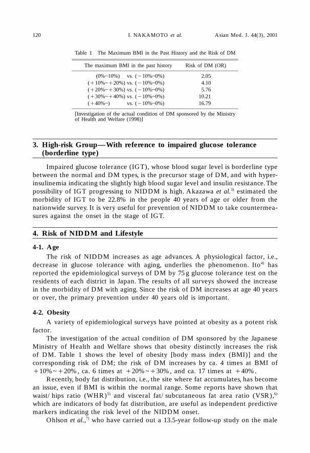
factor.The investigation of the actual condition of DM sponsored by the Japanese
Ministry of Health and Welfare shows that obesity distinctly increases the riskof DM. Table 1 shows the level of obesity [body mass index (BMI)] and thecorresponding risk of DM; the risk of DM increases by ca. 4 times at BMI of�10%~�20%, ca. 6 times at �20%~�30%, and ca. 17 times at �40%.
Recently, body fat distribution, i.e., the site where fat accumulates, has becomean issue, even if BMI is within the normal range. Some reports have shown thatwaist/hips ratio (WHR)5) and visceral fat/subcutaneous fat area ratio (VSR),6)
which are indicators of body fat distribution, are useful as independent predictivemarkers indicating the risk level of the NIDDM onset.
Ohlson et al.,7) who have carried out a 13.5-year follow-up study on the male
Table 1 The Maximum BMI in the Past History and the Risk of DM
The maximum BMI in the past history Risk of DM (OR)
(0%~10%) vs. (�10%~0%) 2.05(�10%~�20%) vs. (�10%~0%) 4.10(�20%~�30%) vs. (�10%~0%) 5.76(�30%~�40%) vs. (�10%~0%) 10.21(�40%~) vs. (�10%~0%) 16.79
[Investigation of the actual condition of DM sponsored by the Ministryof Health and Welfare (1998)]

HEALTH INVESTMENT PROJECT 121
Swedes by dividing the subjects according to BMI (3 groups) and WHR (3 groups)and by combining each group with the risk of the DM onset, have reported thatthe risk of DM increases as WHR increases in each BMI group and that theincidence of NIDDM is 15.2% at high BMI and WHR, while the incidence is only0.5% at low BMI and WHR. In the coexistence of abdominal obesity and systemicobesity, DM is more likely to develop. With regard to BMI and WHR, Warne etal.,8) who have conducted a follow-up study in the residents of Pima, have reportedthat WHR is the indicator of obesity in men and BMI in women.
4-3. DietThe increase in the incidence and morbidity of NIDDM in recent years is
related to the westernization of the Japanese conventional lifestyle. The increase infat intake and the decrease in routine physical activities are related to thesechanges of lifestyle. Kawade9) has shown that the morbidity of DM in the immi-grants of Japanese descent in Hawaii was significantly higher than that in theJapanese residents of Hiroshima as control group, and pointed out the following asthe causes: Physical activity was low and intake of animal fat, sucrose, and fructosewas high in the immigrants of Japanese descent, though there was no difference intotal energy intake between the immigrant group and the control group.
With regard to lipids in dietary components, high fat diet is known to decreasethe insulin sensitivity and to increase the risk of NIDDM. Recent follow-upstudies10,11) have also reported that high fat diet is a risk factor for NIDDM andingestion of fishes, vegetables, and dietary fibers suppresses NIDDM.
Proteins and amino acids have been reported to promote insulin secretion, butthere have been no reports mentioning the possibility of proteins and amino acidsinducing insulin secretory disturbance.
With regard to diet, not only what to eat, but also when and how to eatbecomes an issue. Of epidemiological surveys in Japan, Miyagawa12) has reportedthat the risk of DM is high in the women who have no breakfast, and Watanabeet al.13) have reported that working men in big cities frequently take food and drinkat 9:00pm or later, and that the risk of DM is high in the people who drink threecups of coffee or more a day with sugar, who like sweets, and who eat hurriedmeals. Kato et al.14) have reported that, in male, there is a significantly highcorrelationship between the risk of DM and the meals with much seasoning athome.
4-4. ExerciseThe concept that the reduced energy consumption due to underexercise causes
obesity, which is followed by insulin resistance and DM, is widely accepted.According to the investigation of the actual condition of DM sponsored by the
Ministry of Health and Welfare (Table 2), the risk of obesity in the people whowalk less than 4,000 steps a day is about 3 times as high as that in the people whowalk 12,000 or more steps. These data indicate the possibility of underexercisecausing DM via obesity. A study by Helmrich et al.15) has also shown that theincrease in physical activity prevents the onset and progression of NIDDM.
With regard to the contents of exercises, Sato et al.16) have reported that not

all the exercises are effective for prevention of DM or NIDDM; aerobic exerciseslike jogging and swimming are rather effective for improvement in the individual’sinsulin sensitivity than anaerobic exercises such as weight lifting. Sato et al.16) havefurther concluded that the criteria for therapeutic exercise aiming at improvementin the insulin sensitivity in the people with IGT are that aerobic systemic exercisesat 50% Vo2 max (in general, 130 pulses/min, 120/min, and 110/min for the peoplein their 20s–30s, 40s–50s, and 60s–70s, respectively) are performed for at least 10–30 minutes at one setting at least 3 times per week.
Yamanouchi et al.17) have reported that continuous exercise increases theindividual’s insulin sensitivity. The improvable effect of the training on the insulinsensitivity decreases within 3 days after discontinuation of the training, and italmost disappears in a week.16) Therefore, continuation and establishment of thehabit of doing exercise is important.
Intervention trials18) regarding prevention of DM by walking and exerciseusing dumbbells were performed at Ogunicho in Yamagata Prefecture as an inves-tigation of the feasibility of exercise. As long as the people with IGT were includedas the subjects in the trials, those who frequently participated in the exercise andwho did the exercise using dumbbells hard, showed improvements in the physicalpower and indicators.
Berger et al.19) have reported that the combined therapy involving diet therapyof DM and therapeutic exercise prevents the onset of NIDDM in the groups withhereditary predispositions of hyperinsulinemia and central obesity. The investiga-tors have also reported that training is particularly effective for prevention ofarteriosclerosis as well, via the increased activity of the fibrinolytic system.
4-5. SmokingIt has conventionally been believed that there is no difference in the rate of
smoking people among DM patients and that among healthy people.20) It has notbeen proved whether smoking has influence on the onset of DM. A recent studyby Rimm et al.21) has concluded that smoking is an overt risk factor for DM, andshown that the relative risk value (RR) to non-smokers increased to 1.94 (the 95%confidence interval, 1.25–3.03) by smoking of at least 25 cigarettes a day in maleCaucasians.
4-6. Drinking alcoholIn a study at Hisayamacho, Kiyohara et al.22) have reported that alcohol con-
I. NAKAMOTO et al. Asian Med. J. 44(3), 2001122
Table 2 Relationship between Exercise and the Risk of Obesity
Quantity of motion (steps/day) Risk of obesity (OR)
(8,000–12,000 steps) vs. (12,000 steps or more) 1.68(4,000–8,000 steps) vs. (12,000 steps or more) 2.28
(Less than 4,000 steps) vs. (12,000 steps or more) 2.76
[Investigation of the actual condition of DM sponsored by the Ministry ofHealth and Welfare (1998)]

sumption of at least 20 g/day (ca. 180 ml for sake) is a significant risk factor for theonset of IGT, and that drinking of a small amount (less than 20 g/day) of alcoholsuppresses the onset of IGT. According to this study, the RR to the risk level inthe male drinkers of a small amount of alcohol, whose morbidity of DM waslowest, was 3.7 in the male drinkers of the moderate amount of alcohol or moreand was 1.8 in the male non-drinkers. Thus, the risk of DM was significantly highin the drinkers of the moderate amount or more of alcohol. In the female non-drinkers, the RR to that in the female drinkers was 1.8, showing the same tendencyas that of the male non-drinkers.
Kato et al.14) have described that the risk of DM in women is significantly highby drinking 180 ml (sake) of alcohol or more a day.
Perry et al.23) have reported that consumption of a moderate amount ofalcohol improves insulin resistance and suppresses the onset of DM.
As described above, drinking a small amount of alcohol shows a tendency toprevent DM and heavy drinking shows a tendency to be a risk factor for the onsetof DM.
4-7. HypertensionIn the survey at Aitoucho,24) the RR to NIDDM was 1.32 in the male and
female hypertensive subjects. Kuzuya et al.,25) who analyzed the data from thepeople who had undergone medical examination, have reported that hypertensionis an independent risk factor for NIDDM and the OR is 1.62. Nanjo–26) has reportedthat the morbidity of IGT was 34.6% high and the morbidity of DM was alsosignificantly high in hypertension patients and the people with a family history ofhypertension. These findings and observations support the presence of multiplerisk factor syndrome via insulin resistance.
4-8. Stress and occupationsAt present, the involvement of stress on the onset of DM is widely recognized
from clinical experience. However, there have been only a few research on thedirect involvement of stress on the onset of DM by epidemiological survey, prob-ably because it is difficult to measure stress.
Watanabe et al.13) have reported that the OR to that in the technological andclerical workers was 1.43 in the people engaged in management, sales, and inde-pendent business, which showed the elevated risk of DM.
As a result of a case-control study, Uehata et al.27) have enumerated the fol-lowing as risk factors for DM, which showed significantly high OR: Drinking ofcoffee in large quantities, stress of duties (compression by jobs, troubles with jobs,etc.), subjective stress (fatigue, anxiety, mental and physical exhaustion, etc.),experience of divorce, etc.
5. Conclusion
The number of DM patients is increasing steadily in Japan. Pressure on thefamily members due to progression of complications and the resultant medicalexpenses are increasingly enormous, as well as deterioration in the patient’s quality
HEALTH INVESTMENT PROJECT 123

Table 3 General Summarization List of NIDDM
� Indicators of health
Risk factor Risk level Risk value 95% confidence interval
Sex Male � 1.68 1.02–1.65
Age 40 years of age or over � 1.58 1.16–2.14
Family history Family history of DM � 1.65 1.16–2.36
BMI ��24 � 1.76 1.32–2.26
WHR Males�0.9, Females�0.8 � 1.47 1.14–1.91
Hypertension ��140/90 mmHg � 1.92 1.39–2.65
Serum cholesterol �T-Cho�220 mg/dl � 1.49 1.12–1.98
Triglycerides �TG�150mg/dl � 1.84 1.40–2.41
Insulin �F-IRI�12U/ml ��� 3.91 2.82–5.42
Blood sugar level 90–109mg/dl ��� 4.15 2.86–6.07
Occupations Management, Sales, � 1.43* 1.03–1.98Independent business
�High-risk behavior
Risk factor Risk level Risk value 95% confidence interval
Smoking habit Presence � 1.42 1.03–1.82
Alcohol drinking habit Moderate amount or more � 1.80 1.34–2.42
Breakfast No breakfast �� 2.75** 1.05–7.24
Food intake at 2–3 times/week �Every day � 1.64* 1.19–2.259:00 pm or later
Quickness about meals Quick � Ordinary � 1.55* 1.09–2.17
Coffee with sugar At least 3 cups per day �� 2.10* 1.48–3.00
Tastes for sweets Liking for sweets �� 2.07* 1.40–3.07
�Behavior aiming at health
Risk factor Risk level Risk value 95% confidence interval
Physical activities Moderate �� 0.40 0.2–0.8
*: Only males **: Only females
I. NAKAMOTO et al. Asian Med. J. 44(3), 2001124
of life. Since lifestyle relates to the progression of NIDDM, it can also be pre-vented by improvement in lifestyle. In order to demonstrate this, investigation ofthe feasibility by intervention trials including changes in behavior is needed.Malmö study28) and DaQing study29) are representatives for large-scaled interven-tion trials via diet therapy and therapeutic exercise, which aim at preventing theonset of NIDDM.
The Malmö study included a 5-year intervention trial via diet therapy andtherapeutic exercise on IGT patients. The incidence of NIDDM was 10. 6% in the

group with the intervention, which was significantly lower than 28.6% in the groupwithout the intervention.
The DaQing study in China included a 6-year intervention trial by dividingIGT subjects into 4 groups, i.e., the diet, exercise, diet�exercise, and controlgroups. As for the contents of the exercise used for the intervention, exercise in thespare time was increased by 80–160 kcal a day. The incidence of NIDDM in everygroup with the intervention was lower than that in the control group.
The data of these surveys suggest that correction of lifestyles mainly by diettherapy and therapeutic exercise is adequately effective for prevention of the onsetof NIDDM. Such primary prevention may lead to reduction in medical expenses.It is therefore important to detect the people with IGT early, who are at the highrisk group of NIDDM, and to guide them in the improvement of lifestyle mainlyby diet therapy and therapeutic exercise. With regard to the diet and exercisetherapy, social supportive measures should also be planned, because it is importantto continue them and it is difficult to keep practicing them with personal effortsalone.
Since the risk of DM rapidly increases at age 40 years or over, guidance ofhealth at age 39 years or under is also considered to be important.
In conclusion, general summarization list of influences of lifestyle on the onsetof NIDDM is shown in Table 3.
REFERENCES
1) The tendency of national health. Welfare Statistics Society, Tokyo, 1998. (in Japanese)2) The outline of investigation of the actual condition of diabetes mellitus (for a prompt
report). Lifestyle-related Diseases Countermeasures Room, Health Service Bureau,Ministry of Health and Welfare, 1998. (in Japanese)
3) Akazawa, Y.: Epidemiology of diabetes mellitus. (Ed.) The Japan Diabetes Society,Progress in Study of Diabetes Mellitus (The 30th Series), Shindan to Chiryosha, Tokyo,1992, pp.25–35. (in Japanese)
4) Ito, H.: The incidence and prognosis of diabetes mellitus in the elderly. Medicina 30:1402–1404, 1993. (in Japanese)
5) Peiris, A.N. et al.: Glucose metabolism in obesity: Influence of body fat distribution.J Clin Endocrinol Metab 67: 760–767, 1988.
6) Tokunaga, K. et al.: A novel technique for the determination of body fat by computedtomography. Int J Obes 7: 437–445, 1983.
7) Ohlson, L.O. et al.: The influence of body fat distribution on the incidence of diabetesmellitus. 13.5 years of follow-up of the participants in the study of men born in 1913.Diabetes 34: 1055–1058, 1985.
8) Warne, D.K. et al.: Comparison of body size measurements as predictors of NIDDM.Diabetes Care 18: 435–439, 1995.
9) Kawade, R.: Diabetes mellitus in Japan and in immigrants of Japanese descent inHawaii. Tonyobyogaku 1978, Shindan to Chiryosha, Tokyo, 1978, pp.88–108. (inJapanese)
10) Marshall, J.A. et al.: Dietary fat predicts conversion from impaired glucose toleranceto NIDDM: The San Luis Valley Diabetes Study. Diabetes Care 17: 50–56, 1994.
11) Feskens, E.J.M. et al.: Dietary factors determining diabetes and impaired glucosetolerance. A 20-year follow-up of Finnish and Dutch cohorts of the Seven CountriesStudy. Diabetes Care 18: 1104–1112, 1995.
HEALTH INVESTMENT PROJECT 125

12) Miyagawa, M.: Risk factors for diabetes mellitus evaluated by long-term observations.Nippon Eiseigaku Zasshi (Japanese Journal of Hygiene) 50: 986–997, 1995. (inJapanese)
13) Watanabe, M. et al.: The borderline type of impaired glucose tolerance and the situ-ation of food consumption in male workers in large cities—Relations to lifestyle.Nippon Ko–shu– Eisei Zasshi (Japanese Journal of Public Health) 40: 969–979, 1993. (inJapanese)
14) Kato, I. et al.: A follow-up study of the correlations between lifestyle and majordiseases that frequently occur in adults. Nippon Ko–shu– Eisei Zasshi (Japanese Journalof Public Health) 36: 662–667, 1989. (in Japanese)
15) Helmrich, S.P. et al.: Physical activity and reduced occurrence of non-insulin-depen-dent diabetes mellitus. N Engl J Med 325: 147–152, 1991.
16) Sato, Y. et al.: Physical training therapy. Nippon Rinsho (Japanese Journal of ClinicalMedicine) 54: 2745–2749, 1996. (in Japanese)
17) Yamanouchi, K. et al.: Physical training therapy for DM (12th Report). Tonyobyo(Journal of the Japan Diabetes Society) 30: 561–565, 1987. (in Japanese)
18) Tominaga, M. et al.: Prevention of diabetes mellitus by establishment of the habit ofdoing exercise at Ogunicho, Yamagata Prefecture. A Report of a Research on theDiabetes Mellitus Survey Sponsored by the Ministry of Health and Welfare in 1996. (inJapanese)
19) Berger, M. et al.: Diabetes & Exercise ’90. Diabetes Care 15: 1675–1823. 1992.20) Kesson, C.M. et al.: Smoking in diabetics. Lancet 1: 504–505, 1979.21) Rimm, E.B. et al.: Prospective study of cigarette smoking, alcohol use, and the risk of
diabetes men. BMJ 310: 555–559, 1995.22) Kiyohara, H. et al.: The influence of the alcohol intake volume on the morbidity of
impaired glucose tolerance: A Hisayamacho study. A Report of a Research on theDiabetes Mellitus Survey Sponsored by the Ministry of Health and Welfare in 1996. (inJapanese)
23) Perry, I.J. et al.: Prospective study of risk factors for development of non-insulindependent diabetes in middle aged British men. BMJ 310: 560–564, 1995.
24) Maekawa, S. et al.: An Aitou study—Analysis of the morbidity of diabetes mellitusand the risk factors by a prospective cohort study. Tonyobyo (Journal of the JapanDiabetes Society) 35: 241–248, 1982. (in Japanese)
25) Kuzuya, H. et al.: Analysis and countermeasures against multi-risk factor syndrome.A Report of a Research on the Diabetes Mellitus Survey Sponsored by the Ministry ofHealth and Welfare in 1996. (in Japanese)
26) Nanjo–, K.: Epidemiology of diabetes mellitus (The influence of hypertension anda family history of hypertension on the onset of diabetes mellitus). A Report of aResearch on the Diabetes Mellitus Survey Sponsored by the Ministry of Health andWelfare in 1996. (in Japanese)
27) Uehata, T. et al.: Assessment of stress factors for the onset of NIDDM. A Report ofa Research on the Diabetes Mellitus Survey Sponsored by the Ministry of Health andWelfare in 1993. (in Japanese)
28) Eriksson, K.F. et al.: Prevention of type 2 (non-insulin-dependent) diabetes mellitusby diet and physical exercise. The 6-year Malmö feasibility study. Diabetologia 34: 891–898, 1991.
29) Pan, X-R. et al.: Effects of diet and exercise in preventing NIDDM in people withimpaired glucose tolerance. The DaQing IGT and diabetes study. Diabetes Care 20:537–544, 1997.
I. NAKAMOTO et al. Asian Med. J. 44(3), 2001126

F E A T U R EHEALTH INVESTMENT PROJECT: Part Two
(10)
BEHAVIORAL SCIENCEFOR HEALTH EDUCATION
Yoshiharu AIZAWA* and Hitomi KARUBE**
*Professor, Department of Preventive Medicine and Public Health,Kitasato University School of Medicine
**Professor, University of Human Arts and Science
1. Introduction
Japan’s aging of population, changes in living environment, and diversificationof sense of values in recent years are accompanied by gradual increases of peopledeveloping lifestyle-related diseases and those complaining of fatigue andstresses.1) Regular health checkups at work places reveal that the ratio of thosewith abnormal findings is increasing yearly and the result of health examinationsin 1999 found 41.7% of workers with some abnormal findings.2)
On the other hand, the society has achieved materialistic affluence, higherincome, and increased leisure hours. Against such background, people’s sense ofvalues continues to diversify and individual needs are becoming more sophisti-cated.1,2) Patients are changing their attitude toward doctors; they no longer wishto seek or appreciate paternalistic guidance or orders from their doctors, but wantrelationship under which doctors respect and accept their individual lifestyle andpersonality and treat them as equals.3,4)
What is behavioral science? It may be defined as an academic discipline thatattempts to elucidate systematically and demonstratively the human attitudes andhabits as represented how people as individuals or as members of a group reactphysiologically and psychologically in addressing the environment.3,5)
According to a conventional way of thinking, health management and behav-ioral science are understood as two different entities without any point of contactor fusion. As discussed above, however, changes in people’s daily life and thedisease structure now require techniques to assist and support people to make freedecisions in better mental conditions.
The authors have therefore reviewed and studied many Japanese and foreignreferences in order to establish effective techniques for health investment anddevised a method of “applying behavioral intervention in health education”. Wepresent here the review of studies in this field.
2. From Adult Diseases to Lifestyle-related Diseases
Conventional measures for adult type diseases addressed cancers, cerebrovas-

Y. AIZAWA and H. KARUBE Asian Med. J. 44(3), 2001128
cular diseases, and heart diseases that increased with age, and attached an impor-tance to secondary preventive measures of early detection and early treatment.Views were expressed, however, that the term “adult diseases” gives the impres-sion that “they are diseases that develop after one gets old”, and it was decided touse the term “lifestyle-related diseases” based on the report of Public HealthCouncil of December 1996 on “Basic direction for disease prevention in view oflifestyles” and to promote disease measures focusing on primary prevention.1,6)
Factors related to the onset and prognosis of diseases may roughly be clas-sified into three; genetic factors, environmental factors and lifestyle factors.Lifestyles that are known to be closely related to occurrence of diseases are smok-ing and lung cancer and pulmonary emphysema, excessive animal fat intake andcolon cancer, excessive salt intake and strokes, obesity and diabetes mellitus, andalcohol intake and cirrhosis. In other words, lifestyle-related diseases is one way ofregarding diseases that shows the onset or progress of a disease may be preventedby improving lifestyle.
In order to ensure that our clients will implement the behavior of improvingtheir lifestyle, we, health professionals, should not only diffuse accurate knowledgebut also enable behavioral changes. The term “behavioral change” means changingof a behavior or habit and may be divided into “pre-contemplation stage”, “con-templation stage”, “preparation stage”, “action stage” and “maintenance stage” asproposed by Prochaska (Table 1). Stopping to smoke may be carried out as inFig. 1.
As discussed above, primary prevention means an attempt to prevent diseaseby acquiring good habits in life. The term “habit” is said to exceed the concept ofdisease prevention and to mean building the body and mind for becoming a betterhuman being.6)
3. Choosing Lifestyle and Behavior
Lifestyle means accumulation of daily choices. There are lifestyles consideredgood for health and those that are considered bad for health.1) One example of thelatter is cigarette smoking. To switch this lifestyle to “stopping smoking” requires
Table 1 Stages of Behavioral Changes (J.O. Prochaska)6)
Pre-contemplation stage:(Those who have no desire to implement behavior in the predictable stage)
Contemplation stage:(Those who intend to implement behavior during the six months to come,those who are troubled about sacrifices and merits of changes)
Preparation stage:(Those who have realistic understanding about risks)
Action stage:(Those who need the utmost efforts during the six months when outsiderscan observe the changes)
Maintenance stage:(Efforts should be continued for six to twelve months)

a certain motivation.8–10) To start and continue a behavior for maintaining andpromoting so-called healthy state or health activities such as stopping ordecreasing smoking, decreasing salt intake, taking physical exercises, stopping ofdrinking, decreasing drinking, decreasing fat intake, and brushing teeth requireconsiderable motives and background. From the viewpoint of behavioral science,health guidance means how to support people’s subjective efforts for promotingand recovering their health.8–11) The important thing for dealing with lifestyle-related diseases is to develop behavioral science techniques by finding out whatkind of interventions are effective for health guidance and how they lead tochanges in lifestyles.
In determining behaviors, rationale for implementing a behavior and its effec-tiveness should be recognized well by people who are implementing. Only after theclear-cut reason and effectiveness are recognized, they are motivated and becomepositive toward such behavior. Such reason and efficacy are not to be merelypreached or educated by health professionals, but they should be realized, con-firmed and implemented by the subjects themselves. We health professionals arealways in a position to support them.12)
Then, how do we actually proceed with the above-mentioned series of worksof confirmation? For instance, the subject may be asked to write a list of reasonsand effects of a certain behavior and helped to choose the behavior. This methodenables the subject to continue a certain health activity and acquire the habit ofsuch activity until the time when he/she is convinced with the reason and effects,and even to enhance positive willingness.11)
4. How to Incite Desire for Healthy Behavior
As discussed above, health guidance based on behavioral science means tosupport and assist the subject in many areas. It means to help him/her not tobecome frustrated or lose confidence as he/she is motivated to start the behavior,experiences the psychological burden in continuing the behavior, to firmly estab-lish the rationale for the behavior and to confirm its effectiveness.5,13)
One should take note of individual differences both physically and psychologi-
HEALTH INVESTMENT PROJECT 129
Fig. 1 Behavioral change toward smoking cessation (Hinohara, 1998)6)
New habit
Cannot stopsmoking
50%
Old habit Pre-contemplationstage
Contemplationstage
Implementingstage
Continuingstage
Preparationstage
Reformingstage

cally in those starting or about to start a health activity. Their environment in life,work, and home differs and their lifestyle and sense of values regarding life alsodiffer. Therefore, advice or assistance deemed pertinent by the supporter may beinterpreted as criticism, slander or even contempt depending on sensitivity orphysical and psychological conditions of those receiving such advice, etc., and mayinvite the result contrary to original purposes. The quality of mutual relationshipbetween the party behaving and those assisting can be an extremely importantkey.14,15)
4-1. Level of changesBehavioral science captures changes occurring to humans at four levels. They
are changes in knowledge, changes in attitude, changes in behavior, and changesin group behavior or organizational behavior.16)
Figure 2 shows the time required for these changes to occur without pressureor submission and the relative difficulties. As shown, changes in knowledge occurmost easily, followed by changes in attitude. Factors constituting attitude are dif-ferent from those of knowledge in that emotional factors such as positiveness ornegativeness are included. On the other hand, changes in behavior entail moredifficulty and time compared to these two changes. Changes in group or organiza-tional behavior are most difficult and time-consuming among these four changes.
4-2. BehaviorBehavior is basically preceded by an objective. In other words, our behavior
is motivated by a desire to achieve an objective. However, rationale for behavioris not necessarily clear in conscience. Freud is one of those who first recognized theimportance of subconscious motive. According to him, motive is defined as a desireinherent in an individual, a sense of psychological dissatisfaction, drive or impulse.
On one hand, an objective exists outside an individual. Objectives aredescribed as “desirable” compensations that direct the motive, and psychologists
Fig. 2 Time required for change and degree of difficulty16)
(High) Group behavior
Degree of Individual behavior
difficulty Attitude
(Low) Knowledge
(Short) Time required (Long)
�
�
�
� � {Motive Activity to achieve objective
Behavior
Objective Activity aimed
�
Fig. 3 How motivation works16)
Y. AIZAWA and H. KARUBE Asian Med. J. 44(3), 2001130

define them as incentives.Figure 3 shows the relation among motive, objective, and behavior. The most
intense motive induces behavior to realize or achieve an objective, but since not allthe objectives can be realized, the dotted line indicates them.16)
On the other hand, factors that determine human behavior are the precedingfactor, the resulting factor and the cognitive factor according to Bandura.17) Heattached the greatest importance to the expectant function as a preceding factorand divided it into the expected result and the expected efficacy, and clearlyshowed that they are the factors that affect behavioral changes. An individual’scognition of expected efficacy before implementing behavior is described as self-efficacy and studies made clear that this is the determinative factor for starting andcontinuing a behavior.18,19) Cognition of how much self-efficacy is possessed by anindividual is known to be useful for predicting behavioral changes, to restrainemotional reaction, and to ameliorate stress reactions.18)
4-3. Staging and motivation of desireBehavior in a specific scene is generally determined by the desire that is
strongest at that time. Therefore, it is necessary to understand important desiresthat are common to all humans. As for the intensity of specific desires, Maslowproposed that physiological desires, desires for safety or security, desires for societyor solidarity, self respect and self realization (to maximize capabilities of the self)generate in a stepwise fashion. In other words, it is necessary to see in which stagethe desire of the subject or the group is. Figure 4 shows this theory.
4-4. Support for health activitiesWell known methods of support for health activities are described below.
(1) Method of association of motivesThis method associates the motive for an activity, which the subjects find most
important in life with the motive for health activities in order to enhance motiva-tion for health activities.3)
(2) Self reward and punishmentThis method rewards or punishes the self for continuing or failing an activity
in order to intensify motivation for health activities.3)
(3) Support networkingA short or long-term network system is established among a group engaged
Fig. 4 Motivation: Relation between theory of environment and Maslow’s stages of desire16)
Factor of desire
Self-realizationSelf-respect
SocialSafety (solidarity)
Physiological (security) Environmentalfactor factor
�
�
HEALTH INVESTMENT PROJECT 131

in a specific health activity for supporting each other by personal contacts. Thesupporters may support or assist this system.3,20,21)
(4) AutosuggestionMotivation is intensified by imagining the ideal self, who is achieved by imple-
mentation of a health activity.3)
5. Behavioral Science Approach to Management
Physicians are the leaders of health professionals and are naturally expectedto manage the health team with expertise.
Capabilities in at least three areas are considered necessary for good manage-ment; those of specialties, those of personal management, and those ofconceptualization.16) Capabilities in specialties mean those for using the knowl-edge, methods, techniques, and tools required for certain business, and they can beacquired by experience, education, and training. Capabilities in personal manage-ment mean those for working with or through others and to make judgments andinclude understanding of psychology of motivation and effective application of theleadership. Capabilities in conceptualization mean understanding the complexstructure of organization as a whole and where and how the self is incorporated inthe organization. These capabilities differ according to the levels in the organiza-tion (Fig. 5).
6. What is Behavioral Medicine?
Behavioral medicine came into the limelight as medicine was re-examinedduring the past 30-odd years in the United States and is an academic discipline thatentails practical activities. The review is still continuing,22) and has rapidly devel-oped after the late 1970s. International Society of Behavioral Medicine was estab-lished in 1990 and Japanese Society of Behavioral Medicine in 1992. The Charterof the International Society of Behavioral Medicine (revised in 1993) defines that“behavioral medicine is a discipline which aims to advance and integrate studies ofpsychology, sociology, behavioral science, and biomedicine regarding health and
Fig. 5 Management skills required at different levels of organization16)
Conceptualization
Topmanagement
Middlemanagement
Front linesupervisor
Personal managem
ent skills
Professional skills
Y. AIZAWA and H. KARUBE Asian Med. J. 44(3), 2001132

disease, and to apply them to disease prevention, elucidation of etiology, diagnosis,treatment, and rehabilitation”. This is a new inter-disciplinary area across clinicalsciences, socio-psychological behavioral science, basic medicine, public health, andpreventive medicine.23)
The reason why behavioral medicine is attracting attention today is discussedbelow.
6-1. Reflection on conventional psychosomatic medicinePsychosomatic medicine is interested mainly in elucidation of psychological
factors related to the cause and onset of diseases. In other words, the treatmentaims to enable patients to understand their psychological complications. Such at-tempts to clarify the psychodynamic structure have not necessarily been successful.
6-2. Importance of methods for behavioral interventionOne factor which promoted development of behavioral medicine is the change
in disease structure. As acute diseases have been overcome to a certain degree andchronic diseases now play the principal role, approaches toward multiple factorsbecame necessary. Epidemiological studies revealed that certain behaviors andlifestyles can become risk factors for cancer, coronary diseases and diabetesmellitus, and behavioral interventions have come to be recognized as effectivemeans for preventing these diseases.19,20)
6-3. Bio-feedback and autonomous system trainingThese methods were developed for behavioral interventions discussed above
and are said to be effective for headaches and chronic pains that have shownstrong resistance toward conventional treatment.
7. Introduction of Consultation and Liaison Psychiatry
In the field of mental health and medicine, which is gaining importance inrecent years, a theory of consultation and liaison psychiatry is considered veryimportant.5,13,25–27) Generally, it is called liaison psychiatry, but the two entities differin definition in a strict sense. Glickman and Hackett discuss differences as follows:“A consultation psychiatrist is a fire fighter who hurries to the site and distin-guishes fire while a liaison psychiatrist is an equivalent of a fire department inspec-tor who prevents or detects fires early by inspections and regular fire drills.”
8. Conclusion
As discussed above, our daily life is full of changes and the time flies.Lifestyles of individuals are diverse and their sense of values toward life and laborhas diversified. In Japan, people have become affluent and medicine and technol-ogy are leading the rest of the world.
When shifting our viewpoint to the actual scene of practical medicine, acutediseases have been controlled to some degree and chronic diseases are now themain streams. Conventional ways of doctors to unilaterally address therapy to
HEALTH INVESTMENT PROJECT 133

patients of terminal illnesses and intractable diseases have changed and now reflectpatients’ sense of values and views of life.
From the viewpoint of disease prevention, the most important challenge ishow to address lifestyle-related diseases. Complex and diverse social life is stillproceeding to reflect the disturbed rhythms of life, insufficient physical exercises,and accumulation of stresses. It is high time to re-examine the relation betweenlifestyle that are good for health and disease prevention.
In order to carry out specific health activities, there should be good humanrelationship between health professionals and patients in motivating patients andpresenting the reason for implementation, by having them understand, appreciate,recognize, and continue the habits for an extended period of time. We believe thatit is essential to introduce and apply behavioral science discussed above andbehavioral medicine, which is application of behavioral science to medicine.
REFERENCES
1) Kosei Tokei Kyokai: Trend in National Health. 1998. (in Japanese)2) Japan Industrial Safety and Health Association: Handbook for Occupational Health.
1998. (in Japanese)3) Munakata, K.: New health guidance based on behavioral science. Kango Tenbo 16:
385–392, 1991. (in Japanese)4) Munakata, K.: Medical behavioral science: A new medical paradigm. Ed. Hasegawa,
H. and Munakata, K.: Behavioral Science and Medical Care. Kobundo, Tokyo, 1996;pp.7–33. (in Japanese)
5) Hoyle, L. and Morton, F.R.: Major trends psychosomatic medicine: The psychiatristsinvolving role in medicine. Ann Intern Med 87: 233–239, 1977.
6) Hinohara, S. and Nishikawa, Y.: Talk on the term “lifestyle-related diseases”. Gendaino Esprit 373: 5–37, 1998. (in Japanese)
7) Fukushima, O. et al.: Motivation in real scene. Ed. Kagawa, M.: Basis and Practice ofMotivation. Kawashima Shoten, Tokyo, 1997; pp.273–338. (in Japanese)
8) Killen, J.D., Portmann, S.P., Davis, L. and Varady, A.: Nicotine patch and self-helpvideo for cigarette smoking cessation. Consult Clin Psychol 65: 663–672, 1997.
9) Ockene, J.K. and Zapka, J.G.: Physician-based smoking intervention: A rededicationto a five-step strategy to smoking research. Addict Behav 22: 835–848, 1997.
10) Basen, E.X., Hudmon, K.S., Trip, M. and Chamberlain, R.: Work-site health andsafety climate: Scale development and effects of a health promotion intervention. PrevMed 27: 111–119, 1998.
11) Kagawa, M. et al.: What is motivation? Ed. Kagawa, M.: Basis and Practice of Moti-vation. Kawashima Shoten, Tokyo, 1997; pp.3–26. (in Japanese)
12) Ishikawa, T. et al.: Basic problems of motivation in humans. Ed. Kagawa, M. et al.:Basis and Practice of Motivation. Kawashima Shoten, Tokyo, 1997; pp.145–215.(in Japanese)
13) Szasz, T.S. and Hollender, M.H.: A contribution to the philosophy of medicine: Thebasic models of the doctor-patient relationship. Arch Intern Med 65: 598–592, 1955.
14) Klein-man, A., Eisenberg, L. and Good, B.: Culture, illness and care-clinical lessonsfrom anthropologic and cross-cultural research. Ann Intern Med 88: 251–258, 1978.
15) Boyd, J.R., Covington, T.R., Stanszek, W.F. and Coussons, R.T.: Drug defaulting: Part2. Analysis of noncompliance patterns. Am J Hosp Pharm 131: 485–491, 1974.
16) Hersey, P. and Blanchard, K.H. (Trans. Yamamoto, S., Mizuno, M. and Narita, I.):Management of Organizational Behavior. Seisansei Shuppan, Tokyo, 1978. (inJapanese)
Y. AIZAWA and H. KARUBE Asian Med. J. 44(3), 2001134

17) Bandura, A.: Self-efficacy: Toward a unifying theory of behavioral change. PsycholRev 84: 191–215, 1977.
18) Chen, J. et al.: Study on apathy at work place: Apathy at work place and its relationwith self efficacy. Ind Psychiatric Health 7: 45–60, 1999. (in Japanese)
19) Burke, L.E., Dunbar-Jacob, J.M. and Hill, M.N.: Compliance with cardiovasculardisease prevention strategies: A review of the research. Ann Behav Med 19: 239–263,1997.
20) Foreyt, J.P. and Poston, W.S.: The role of the behavioral counselor in obesity treat-ment. J Am Diet Assoc 98(Suppl): S27–30, 1998.
21) Aral, S.O. and Peterman, T.A.: Do we know the effectiveness of behavioral interven-tions? Lancet 351(Suppl): 33–36, 1998.
22) Nishikawa, Y.: What is behavioral medicine? Gendai no Esprit 373: 38–48, 1998.(in Japanese)
23) Kobayashi, A.: International trend in behavioral science studies. Gendai no Esprit 373:49–59, 1998. (in Japanese)
24) Rasmussen, S.A. and Eisen, J.L.: Treatment strategies for chronic and refractoryobsessive-compulsive disorder. J Clin Psychiatry 58(Suppl): 9–13, 1997.
25) Yamawaki, N.: Introduction. Ed. Kazamatsuri, G.: Consultation �Liaison PsychiatryTreatment. Nakayama Shoten, Tokyo, 1998; pp.3–7. (in Japanese)
26) Kasahara, T. et al.: Psychiatry symptoms encountered in general ward. Ed.Kazamatsuri G.: Consultation �Liaison Psychiatry Treatment, Nakayama Shoten,Tokyo, 1998; pp.11–154. (in Japanese)
27) Uchitomi, Y. et al.: Liaison psychiatry medicine for patients with cancer and AIDS.Ed. Kazamatsuri, G.: Consultation �Liaison Psychiatry Treatment, Nakayama Shoten,Tokyo, 1998; pp.297–344 (in Japanese)
HEALTH INVESTMENT PROJECT 135

136
* This article is a revised English version of a paper originally published in the Journal of theJapan Medical Association (Vol. 123 No. 7, 2000, pages 1013–1016). The Japanese text is atranscript of a lecture originally aired on November 26, 1999, by the Nihon Shortwave Broad-casting Co., Ltd., in its regular program “Special Course in Medicine”.
** Professor, Postgraduate School of Pharmaceutical Science, Osaka University
INTERACTION BETWEENGRAPEFRUIT JUICE AND DRUGS*
Junichi AZUMA**
Asian Med. J. 44(3): 136–141, 2001Abstract: There is a possibility that even a glass of grapefruit juice (GFJ) takenseveral hours before oral medication may enhance the bioavailability of many drugs,exaggerate the drug action, and increase the toxic effect by exaggerating the potencyof the drug. This can be explained by inhibition of CYP3A4 in the small intestineby GFJ, thus suppressing drug metabolism. This interaction with GFJ is likely tooccur mainly with those drugs that are metabolized by CYP3A4 and are associatedwith low bioavailability when given via an oral route. Thorough instruction onmedication is necessary when these drugs are administered to patients. Examples ofthese drugs include: dihydropyridine calcium antagonists, terfenadine, cyclosporine,and carbamazepine.
Key words: Grapefruit juice; Cytochrome P450 (CYP); CYP3A4;Drug interactions; Enzyme inhibition
Introduction
Even a glass of grapefruit juice taken several hours before medication mayincrease the bioavailability of many drugs, raise the blood concentration of thedrug, enhance drug potency, or exaggerate the toxic action of a certain drug. Thisoccurs because of the inhibition of metabolic activity of CYP3A4, a molecularspecies of drug metabolizing enzyme, P450, that exists in the small intestine.
A drug that has been administered via an oral route is absorbed through thedigestive tract, reaches the liver via the portal system, appears in the peripheralcirculation, and is then distributed throughout the body. Part or most of a drug issubjected to a first-pass metabolism in the liver and small intestine. Many drugs arehighly oil-soluble and they have to be metabolized by a metabolic enzyme calledcytochrome P450 into water-soluble compounds that can be excreted in the urineor bile. The most important drug-metabolizing enzymes in the human are thefollowing 5: CYP1A2, CYP2C9, CYP2C91, CYP2D6, and CYP3A4. Among theP450 enzymes that exist in the human liver, the amount of CYP3A4 is the greatest(about 30%). CYP3A4 also exists in the small intestine and is the most importantenzyme, participating in more than one-half of the pharmaceutical agents.
Progress in Clinical Medicine

Drug Interactions: Grapefruit Juice
The development of adverse effects of drugs caused by “drug interactions”,which has attracted attention recently, occurs when 2 or more drugs are takensimultaneously or almost at the same time. A typical example is an adverse effectof sorivudine, which is caused when the process in which 5 FU, an antineoplasticagent, is metabolized and inactivated is inhibited by the metabolites of sorivudine.If a drug-metabolizing enzyme is inhibited, the blood concentration of the activeunchanged drug increases, thus amplifying the pharmacological action or causingadverse effects to develop.
An abnormal rise in the blood concentration of a drug is called an interactionby “inhibition” of a drug-metabolizing enzyme. The blood concentration of a drugthat has been affected by inhibition rises but a clinical problem arises only withdrugs (such as fluorouracil) that have a narrow safety range for their blood con-centration and may readily develop serious adverse effects.
Recently, the drug interaction of grapefruit juice has been frequently reported.An interesting action of this juice in increasing the bioavailability of an orallytaken drug was accidentally discovered about 10 years ago when it was given tocover the taste of ethanol during a study of the interaction between felodipine (adihydropyridine type Ca blocker) and ethanol.1)
Later, it was shown that grapefruit juice raises the mean area under thefelodipine plasma concentration-time curve (AUC) approximately 3-fold when thefelodipine was administered orally to patients with borderline hypertension. It wasreported that grapefruit juice exaggerated the antihypertensive effects of felodi-pine, raised the heart rate in clinical patients, and was frequently responsible foradverse effects due to vasodilation.2) Grapefruit juice significantly raised the maxi-mum plasma concentration (Cmax) of felodipine but did not change its half-life(t1/2). Because this juice had no effect on the pharmacokinetics of felodipinewhen administered via an intravenous route, it was concluded that the action ofgrapefruit juice stems from its metabolic inhibition in the stage before the drugenters the circulating blood.
Mechanism Responsible for Interactions Caused by Grapefruit Juice
To discover the action of grapefruit juice on the drug-metabolizing enzymesof the small intestine and liver in man, a clinical study that is not possible in Japanwas conducted.3) Consequently, it became evident that this juice selectively partici-pated in post-translational regulation of CYP3A4 in the small intestine, reducedthe CYP3A4 content by about 50%, and compromised the metabolic activity ofthe enzyme. Specifically, drinking grapefruit juice for 5 consecutive days caused a3-fold increase in the AUC of felodipine and a 5-fold increase in Cmax. In thisinstance, there were no changes in the CYP3A4 mRNA level of the small intestine;but the protein contents of CYP3A4 and CYP3A5 were reduced by a mean of62%. No changes were noted in the activity of the hepatic CYP3A4—detected bya method called an erythromycin breath-test—or the protein contents forCYP2D6 or CYP1A1 in the small intestine. Thus it was concluded that grapefruit
DRUG-GRAPEFRUIT JUICE INTERACTION 137

juice selectively inhibits a molecular species of CYP3A in the small intestine andraises the bioavailability of felodipine.
Orange juice is not associated with the inhibitory actions described above.Because the expression of a molecular species of CYP3A is reduced by grapefruitjuice, it is evident that this inhibition is not a simple competitive action on themetabolism of the substrate. Grapefruit juice reduced the post-translationalCYP3A4 protein content without affecting CYP3A4 mRNA in the small intestine,suggesting that this is an irreversible enzyme inhibition with probable enhance-ment of CYP3A4 decomposition. To restore the CYP3A4 activity, de novo synthe-sis of the enzyme becomes necessary.
The potency of the inhibitory action of grapefruit juice varies in individuals:Some are not much affected while in others the AUC and Cmax of the plasmafelodipine rose to 6 times that when water was given instead of the juice. Becausethe expression of CYP3A4 protein in the small intestine is involved in the reactionand the inhibitory rate is exaggerated in individuals with greater CYP3A4 activity,it is suspected that this individual difference in the potency of the interactioncaused by the grapefruit juice is at least partly due to some genetic difference. Thedetails have yet to be elucidated.
It is presumed that in this phenomenon, a “certain component” in the grape-fruit juice participates in the inhibition of CYP3A4 activity. It is understood thatthis “certain component”, when taken orally, is trapped in the small intestinewithout reaching the liver and inhibits the CYP3A4 activity that is expressed bythe villous cells of the small intestine. Initially, it was believed that flavonoids, suchas naringen and naringenin, and 6�,7�-dihydroxybergamottin of the furanocou-marin type that are contained in grapefruit juice, were strongly suspected to becausative agents for inhibition. However, recent reports negated the possibilitythat these act as major causative agents in the interactions between grapefruit juiceand a drug.
Recently, a component that potently inhibits CYP3A4 was discovered in anethylacetate extract of grapefruit juice. Structural analysis revealed that this was anew furanocoumarin derivative.4) However, clinical studies will be needed to proveits clinical significance in drug interactions.
Drugs That Are Affected by Grapefruit Juice
The drugs that may interact with grapefruit juice are those that are metabo-lized by CYP3A4. The most frequently investigated are dihydropyridine-type Cablockers; their metabolic pathways are similar but the bioavailability of each drugvaries widely. Nisoldipine has the lowest bioavailability and is most likely to beaffected by grapefruit juice, followed by felodipine, nicardipine, and nitrendipine,while the effect on nifedipine and amlodipine are negligible.
It is known that the magnitude of the effect of grapefruit juice is proportionalto the first-pass effect (the greater the first-pass effect, the lower the bioavail-ability). It is difficult to determine an individual’s drug interaction for thedihydropyridine-type drugs with low bioavailabilities. Individual difference is atthe greatest for nisoldipine. Among nifedipine-type drugs, the effect of grapefruit
J. AZUMA Asian Med. J. 44(3), 2001138

juice is slightly more pronounced for enteric-coated preparations.In addition to these dihydropyridine-type drugs, it has been confirmed that
more than 20 drugs interact with grapefruit juice, resulting in clinical problems.5)
Even at this time, the results of a number of clinical studies attest to these effects.Among the drugs that are marketed in Japan and metabolized by CYP3A4, thosethat have known clinical problems due to their interactions with grapefruit juiceare: Terfenadine, saquinavir, cyclosporine, midazolam, triazolam, and verapamil(Table). The plasma concentrations of those drugs that are raised significantlywhen combined with itraconazole or erythromycin, known inhibitors of hepaticCYP3A4 activity, are also affected by grapefruit juice. Cisapride and astemizoleare also affected by grapefruit juice. It is believed that those drugs that are not verylikely to be affected with the combination of CYP3A4 inhibiting drugs are alsorelatively untouched by the actions of grapefruit juice.
There are several recent reports on the different effect of grapefruit juicebetween water-soluble HMG-CoA reductases and oil-soluble preparations thatconstitute metabolic substrates for CYP3A4.6) It is reported that grapefruit juicedoes not affect the pharmacokinetics of water-soluble preparations, while drinkingof the juice together with oil-soluble preparations that are metabolized mainly byCYP3A4 results in significant increases in the plasma concentrations of thesepreparations (AUC, 1.4 to 15 times that when taken with water, though varying indegree for each drug).
Notes on Therapy
When a drug that may undergo an interaction with grapefruit juice is to beprescribed, it is necessary to give appropriate instructions to patients at medica-tion. The following clinical notes are suggested.
The duration of the inhibitory action by grapefruit juice is an important clini-cal topic. The correlation between the consumption of grapefruit juice and
Table Drugs That Are Affected by Grapefruit Juice
• Dihydropyridine-type Ca blockers:nisoldipine, felodipine, nicardipine, nitrendipine, nifedipine, etc.
• Drugs acting on the central nervous system:benzodiazepam-type hypnotic sedatives (triazolam and midazolam),carbamazepine, and diazepam
• HMG CoA reductase inhibitors:lovastatin, simvastatin, etc.
• Immunosuppressive agents:cyclosporine and tacrolimus.
• Anti-allergy agents:terfenadine and astemizole
• HIV protease inhibitors:saquinavir
• Others:cisapride and verapamil
DRUG-GRAPEFRUIT JUICE INTERACTION 139

felodipine administration has been studied.7) When a glass (220 ml) of grapefruitjuice is consumed 4 hours before or at the time of drug administration, the ratesof increases of AUC and Cmax became the greatest. The potency of the interac-tion attenuated as the interval between grapefruit juice ingestion and felodipineadministration increased; but the Cmax of felodipine was still obviously high evenwhen the drug was administered 24 hours after grapefruit juice ingestion.
We conducted a study on the effect of grapefruit juice on nisoldipine pharma-cokinetics in 8 healthy Japanese subjects.8) When nisoldipine was administeredtogether with 250ml of grapefruit juice, the Cmax and AUC of the plasma concen-tration of the drug increased 3 and 4.5 times, respectively, in comparison with thecorresponding amounts when water was used instead of grapefruit juice. If thejuice was given, followed by the drug together with water one hour later, Cmaxand AUC increased about 4- and 5-fold, respectively. However, no notablechanges in pharmacodynamics were noted when grapefruit juice was ingested onehour after medication.
The half-life of the inhibitory effect of grapefruit juice has been generallyestimated to be 12 hours. Imbibing this juice on a single occasion may result in asustained effect for more than 24 hours, which calls for special clinical attention.
What is important next is the extent of changes in the dynamics of the bloodcontents of the affected drugs. The following types of drugs are considered to posea clinical risk when combined with grapefruit juice: (1) Those for which the phar-macological actions may be potentiated, causing excessive reactions or toxic re-sponses when their plasma concentrations exceed twice the desired level: examplesinclude some dihydroxypyridine-type Ca blockers, terfenadine, and saquinavir; (2)those drugs that are associated with sharply curved concentration (dosage)-response relationship; and (3) those with narrow therapeutic ranges and possiblepotentiation of the drug effects, resulting in excessive drug actions or toxic reac-tions; these should be watched carefully in clinically practice even when theirplasma concentrations may not be excessively high; examples include cyclosporine,midazolam, triazolam, and verapamil.
Finally, the following patient factors are cited in relation to drug interactionswith grapefruit juice: (1) Accentuated CYP3A4 activity of the small intestine; and(2) hepatic insufficiency.
The type and quantity of grapefruit juice consumed are also important. Inter-actions between commercially available grapefruit juice (frozen and concentrated,condensed or diluted, and fresh frozen) and felodipine have been proven; but theirmagnitude depends on the amount of the active components and may vary, evenwithin each brand or lot. Many of the studies have used grapefruit juice that wascondensed twice but it would be safe to assume that all grapefruit juice has thepotential to act in the way that has been described here.
Even a single glass of grapefruit juice imbibed several hours earlier may aug-ment the bioavailability, cause excessive drug effects, or exaggerate the toxic ac-tions of the drugs that have been cited above. Particular attention should be paidto these matters in clinical practice.
J. AZUMA Asian Med. J. 44(3), 2001140

REFERENCES
1) Bailey, D.G. et al.: Ethanol enhances the hemodynamic effects of felodipine. ClinInvest Med 12: 357–362, 1989.
2) Bailey, D.G. et al.: Interaction of citrus juices with felodipine and nifedipine. Lancet337: 268–269, 1991.
3) Lown, K.S. et al.: Grapefruit juice increases felodipine oral availability in humans bydecreasing intestinal CYP3A protein expression. J Clin Invest 99: 2545–2553, 1997.
4) Fukuda, K. et al.: Specific inhibitors in grapefruit juice: Furanocoumarin dimers ascomponents of drug interaction. Pharmacogenetics 5: 391–396, 1997.
5) Josefsson, M. et al.: Effect of grapefruit juice on the pharmacokinetics of amlodipinein healthy volunteers. Eur J Clin Pharmacol 51: 189–193, 1996.
6) Lilja, J.J. et al.: Grapefruit juice-simvastatin interaction: Effect on serum concen-trations of simvastatin, simvastatin acid, and HMG-CoA reductase inhibitors. ClinPharmacol Ther 64: 477–483, 1998.
7) Lundahl, J. et al.: Relationship between time of intake of grapefuruit juice and itseffect on pharmacokinetics and pharmacodynamics of felodipine in healthy subjects.Eur J Clin Pharmacol 49: 61–67, 1995.
8) Azuma, J. et al.: Effect of grapefruit juice on the pharmacokinetics of the calciumchannel blockers nifedipine and nisoldipine. Curr Ther Res 59: 619–634, 1998.
DRUG-GRAPEFRUIT JUICE INTERACTION 141

142
* This article is a revised English version of a paper originally published in the Journal of theJapan Medical Association (Vol. 122 No. 1, 1999, pages 57–60).
** Director, Department of Dermatology, Kanto Central Hospital
TOPICAL USE OF STEROIDS IN THE AGED*
Haruko HINO**
Asian Med. J. 44(3): 142–147, 2001Abstract: Steroid preparations for topical use include a variety of forms, amongthem hydrophobic ointments, emulsions, liquids, and gels. These drugs are chosenaccording to the status and site of the lesion and the patient’s age. There are fivegrades of steroid preparations for topical use in terms of the potency of their clinicaleffect. The skin of elderly persons tends to be dry and therefore does not absorbsteroids well. However, if steroids of high potency are used for a long period, elderlypatients are apt to suffer side effects of steroid application, including enhancedatrophy, purpura, xeroderma, capillary telangiectasia, delayed wound healing, andsusceptibility to infection, because their skin is atrophied. In the elderly, the skinbecomes dry as it looses its ability to retain water. To treat pruritus and asteatoticeczema under these conditions, steroid preparations of mild to moderate potencyand having a hydrophobic base should be used. If nummular eczema or autosensiti-zation dermatitis has occurred as a result of scratching, steroids of higher potencyshould be selected. Emulsions are not suitable for moist lesions like erosions andulcers, because of the issues of reabsorption and stimulation. Although seborrheiceczema and psoriasis are common in the elderly, the former requires short-termsteroids of moderate or lower potency, whereas the latter requires the cautious useof stronger steroids.
Key words: Steroid preparations for topical use; Local side effects of steroids;Indications of topical steroid therapy in elderly patients
Introduction
Among various drugs available for the treatment of skin diseases, adrenocor-tical hormones are the most common and most frequently used because of theirpotent anti-inflammatory action. Although systemic steroid therapy by oral admin-istration or injection is employed in some cases, topical steroids are generally usedin the treatment of most dermatologic diseases. Some cautions, however, areimportant with regard to steroid use. The condition of the skin as well as thepharmacology of adrenocortical hormones (steroids) should be borne in mindwhen applying steroids to the skin.

Steroid Preparations for Topical Use
1. Topical steroidsIn 1952, Sulzberger and Witten first reported the topical application of a steroid
in the treatment of dermatosis.1) The steroid used was 17-hydroxycorticosterone-21-acetate. Following their report, halogenation of the steroid skeleton at hydroxylgroups located at the C-6, -9, and -21 positions, esterification at C-17 and -21, andacetylation at C-16 and -17 were found to enhance the anti-inflammatory action ofsteroids, and various potent steroids for topical use have been developed. On theother hand, as more potent steroids have become available, accompanying sideeffects have arisen as a problem.2)
2. Types of topical steroid preparations and their effects on steroid efficacyVarious forms of topical steroids are available, and they are selected accord-
ing to the lesion to be treated.3)
(1) Hydrophobic ointmentsHydrophobic ointments do not contain water, but have fat or oil bases like
water-insoluble petrolatum. Although hydrophobic ointments are sticky, they areminimally irritating to the skin and are applicable to erosions and ulcers. Steroidointments with a petrolatum base are widely used, and steroid ointments of thistype are, in general, simply referred to as “steroid ointments”.(2) Emulsions
Emulsions, which are a mixture of normally non-mixable fat and water emul-sified with a surfactant, are divided into the oil-in-water (o/w) type and the water-in-oil (w/o) type. Steroid creams are mostly of the oil-in-water type and can bewashed away with water. Because of this, they are reabsorbed from the erosive orulcerative surface and therefore cannot be applied to a macerated surface. Creamsof the w/o type are more difficult to wash away with water than those of the o/wtype. Universal creams are used as bases of steroid creams.(3) Lotions
Lotions are o/w- or w/o-type emulsified liquids. Most steroid lotions are ofthe o/w type. In recent years, shakable steroid-suspension lotions have becomeavailable.(4) Gels
Steroid gels have solid or semisolid suspension bases that can be divided intohydrogel and lyogel bases. The former are transparent bases that form a capsulewhen applied to the skin, while the latter are also known as FAPG bases, and havea texture intermediate between ointments and creams.(5) Sprays
Steroid sprays are steroid solutions atomized by gas pressure. Although theyare advantageous in terms of extensive application, an important drawback is thatit is difficult to estimate the exact amount being applied.
3. Indications of topical steroid therapySteroid preparations for topical use are generally classified into five grades:
strongest, very strong, strong, medium or mild, and weak, in order of potency
TOPICAL USE OF STEROIDS IN THE AGED 143

H. HINO Asian Med. J. 44(3), 2001144
(Table 1).2,4,5) Even when the same steroid is used, ointments and creams may rankdifferently because of the base that was used.2,4)
Although steroid preparations may be selected on the basis of the diagnosis,most frequently the form and site of the lesion and the patient’s age are decisivefactors. While hydrophobic ointments are sticky and glossy, they can be used foralmost all lesions, including erythema, papules, chronically lichenified surfaces,erosions, and ulcers. Since cream bases are water-soluble, being reabsorbed afterhaving been dissolved in effusions, and are also irritants, they are not suitable forerosions and ulcers.3)
4. Side effects of topical steroids(1) Local side effects of topical application1) Side effects of steroids themselves
Table 1 Major Steroids for Topical Use Ranked According to Clinical Efficacy
(Extension of citation from Shimao2,5) and Aso4))
Drug effect Generic name Proprietary name
Group I a. clobetasol propionate Dermovate, Myalone(strongest) a. diflorasone diacetate Diflal, Diacort
a. mometasone furancarboxylate Fulmetaa. fluocinonide Topsym, Simaron, Biscosala. betamethasone dipropionate Rinderon DP, Dermosol DP
Group IIa. difluprednate Mysera. budesonide Budeson
(very strong)b. amcinonide Visdermb. diflucortolone valerate Texmetenb. hydrocortisone butyrate propionate Pandelb. betamethasone butyrate propionate Antebate
c. dexamethasone propionate Methadermc. dexamethasone valerate Voalla, Zalucsa. halcinonide Adcortin
Group III a. deprodone propionate Eclar(strong) d. betamethasone valerate Rinderon V, Betnevate, Hormezon, Muhibeta V
d. beclomethasone propionate Propaderm, Beclacind. fluocinolone acetonide Flucort, Fluzon, Coriphated. prednisolone valerate acetate Lidomex
d. triamcinolone acetonide Ledercort, Kenacort A, Tricinolon, Nogirond. flumethasone pivalate Locorten, Testohgen
Group IV d. clobetasone butyrate Kindavate(medium or mild) d. alclometasone propionate Almeta
d. hydrocortisone butyrate Locoid, Plancold. dexamethasone Decaderm
b. methylprednisolone acetate Veriderm Medrol AcetateGroup V d. prednisolone Prednisolon(weak) d. hydrocortisone acetate Cortes
a. Ointments and Creams are in the same group. b. Ointments rank one group above creams.c. Ointments rank one group below creams. d. Difference between ointments and creams is unknown.

Table 2 shows possible adverse reactions to topical steroids.2,4,5) The higher thegrade of steroid and the longer its period of application, the more often adversereactions occur. Adverse reactions are also more likely to occur when the steroidis applied by occlusive dressing therapy (ODT) than by simple topical application.
Contact dermatitis caused by steroids themselves is well known. If the diseasestate remains unchanged or is aggravated with the topical use of a steroid prepa-ration, it is necessary to suspect contact dermatitis due to the steroid.2) Side effects of the base or formulating ingredients
Surfactants used in the base, lanolin, propylene glycol, and supplementedaminoglycoside antibiotics such as neomycin may cause contact dermatitis.(2) Systemic side effects after topical application
Percutaneously absorbed steroids can exert systemic effects. When a strongsteroid at a dose of 15–30 g/day is applied by ODT, hypoadrenalism, eosinopeniaof the peripheral blood, accumulation of Na and water retention, and cushingoidsigns may occur. However, these changes resolve soon after the steroid is discon-tinued. It has been reported that hypoadrenalism seldom occurs after simple topi-cal application at a daily dose of less than 10 g.2)
To prevent systemic effects of steroids absorbed from the skin, various ante-drugs have been developed. Steroids that are unlikely to be absorbed from the skinor that lose glucocorticoid activity and are metabolized within a short period oftime, even if absorbed, are especially desirable. The recent development of topicalsteroids has been aimed at designing clinically effective preparations that are free
Table 2 Local Adverse Effects of Topically Applied Steroids
1. Adverse effects due to inhibitory action on cell growth and fibrosisSkin atrophyStriae atrophicansSteroid skin injuryXeroderma/ichthyosiform cutaneous symptomsDelayed wound repairAsteroid uloidCapillary telangiectasiaRosacea-like dermatitis/peristome dermatitisSteroid purpura/flushSteroid elastosisSteroid acne milium/colloid miliumPigmentation disorder
2. Adverse effects due to hormone actionAcneExcessive hair growth
3. Adverse effects due to induced decrease in immunocompetenceSusceptibility to bacterial, fungal and viral infections
4. OthersContact dermatitisPhotodermatosisGlaucoma/cataract (due to eye drops)
TOPICAL USE OF STEROIDS IN THE AGED 145

from undesirable systemic and local effects.4)
Steroid Preparations for Topical Use in Elderly Patients
1. Effects of topical steroids on the skin of elderly patientsIn the elderly, the skin is dry and steroids are poorly absorbed. However,
aged, atrophied skin is susceptible to steroid-induced skin atrophy, peliosis, andsteroid skin injury, changes that are difficult to resolve once they occur. Seniledermatoses follow a chronic course, are intractable, and require prolonged topicalapplication of steroids, which then can lead to adverse reactions to the drug.Steroids of an appropriate grade should be chosen according to the lesion andshould be applied for as short a period as possible.
The skin of the face is thin, particularly in the perioral and periocular areas,as is skin in the genitocrural and perianal regions, and axilla. Since these areas aresusceptible to steroid-induced atrophy, due caution is necessary in selecting thegrade of steroid to be applied.
2. Common major dermatoses requiring topical steroids in the aged and2. the usage of such steroids(1) Pruritus
This condition, in which the patient complains of severe itching although noparticular eruptions are present, is especially common in the winter. Pruritus mayaccompany skin dryness in diabetes mellitus, chronic liver disease, and dialysis orvisceral lesions including malignant tumors. Patients often suffer itching of the skinwhen they become warmer as a result of improved blood circulation, particularlyafter bathing, in bed, or while using an electric blanket. Patients may indeedscratch their skin until it becomes sore, a situation that is more apparent as peoplegrow older, representing a common problem among the elderly.
Application of an oily ointment or body oil after bathing, when the skin ismoist, is effective. When symptoms are severe, the use of a steroid ointment of themedium grade is recommended.(2) Asteatotic eczema
An eczematous lesion that develops from pruritus through scratching is calledasteatotic eczema. Application of a medium or strong steroid ointment is indi-cated. Simple embrocation once or twice a day is adequate. This lesion commonlyoccurs in winter, and is frequently located on the lateral sides of the lower legs andin the dorsolumbar region. In addition to people of middle or advanced age, thislesion has recently been seen in young people who work in dry, ventilated rooms.(3) Nummular eczema
Pruritus complicated by scratching and infection with staphylococci or otherbacteria forms a surface the size of a coin or egg yolk, accompanied with erosions,effusions, and sludgy crust formation.
Simple topical application of a steroid ointment is not adequate for this typeof lesion, and superposing zinc ointment on the steroid ointment is required. Afterthe crust has been eliminated, simple embrocation of the steroid ointment isemployed. Strong or very strong steroid ointments are appropriate. Strongest
H. HINO Asian Med. J. 44(3), 2001146

steroid ointment may also be used, but only for a short period of time.(4) Seborrheic eczema
Although this type of eczema is particularly common in the neonatal period,puberty, and adolescence, it frequently occurs in people of middle or advanced ageas well. The lesion involves the face or head, with the seborrheic area having anerythematous surface with pityroid desquamation. Antimycotic agents such asketoconazole may be used for facial lesions. In intractable cases, a weak ormedium steroid ointment may be used for a short period.(5) Psoriasis vulgaris
This lesion occurs relatively frequently in people of middle or advanced age.Although treatment may vary according to the disease state, a strong steroid oint-ment is usually applied to rashes on the trunk.
An occlusive dressing may be employed on some occasions.
Conclusion
The skin of the elderly does not permit good percutaneous absorption.Further, it is thin and apt to be adversely affected by the application of topicalsteroids. Thus, local adverse reactions are likely to occur, leading to skin atrophy,purpura, and capillary telangiectasia.
Steroid preparations for topical use are available in various forms, whichshould be chosen according to the characteristics of the cutaneous lesion in ques-tion. Ointment bases such as petrolatum are applicable to a variety of lesions. Incontrast, cream bases are not appropriate for moist lesions including erosions andulcers because they are dissolved in effusions and reabsorbed. Steroid preparationsfor topical use are classified into five grades in terms of the potency of clinicaleffect. It is necessary to choose a steroid agent of the grade suitable for the cuta-neous lesion and lesion site. Since cutaneous lesions in the aged are often intrac-table, due consideration of the potency and form of dosage is required when usingtopical steroid therapy.
REFERENCES
1) Sulzberger, M.B. and Witten, V.H.: The effect of topically applied compound F inselected dermatoses. J Invest Dermatol 19: 101–102, 1952.
2) Shimao, S. and Aso, M.: Application of steroids for topical use. In Dermatosis: Up-to-date Treatment. 87–88, Nankodo, Co., Ltd. 1987, pp.1–8. (in Japanese)
3) Hino, H.: Theory and practice of topical therapy. Nippon Hifuka Gakkai Zasshi 109:121–128, 1999. (in Japanese)
4) Aso, M. and Shimao, S.: Problems in steroids for topical use. Hifubyo Shinryo 10: 984–991, 1988. (in Japanese)
5) Shimao, S.: Problems in topical steroid therapy: Q & A. Hifubyo Shinryo 5: 77–83,1983. (in Japanese)
TOPICAL USE OF STEROIDS IN THE AGED 147