vol. 24 no. 1 winter 2003 pages 29–56 - law project for...
TRANSCRIPT

Vol. 24 No. 1 Winter 2003pages 29–56

Library of Congress Cataloging in Publication Data
The Journal of mind and behavior. – Vol. 1, no. 1 (spring 1980)–– [New York, N.Y.: Journal of Mind and Behavior, Inc.]
c1980–
1. Psychology–Periodicals. 2. Social psychology–Periodicals. 3. Philosophy–Periodi-cals. I. Institute of Mind and BehaviorBF1.J6575 150'.5 82-642121ISSN 0271-0137 AACR 2 MARC–S
Copyright and Permissions: © 2003 The Institute of Mind and Behavior, Inc., P.O. Box 522,Village Station, New York City, New York 10014. All rights reserved. Written permissionmust be obtained from The Institute of Mind and Behavior for copying or reprinting text ofmore than 1,000 words. Permissions are normally granted contingent upon similar permissionfrom the author. Printed in the United States of America.

29
©2003 The Institute of Mind and Behavior, Inc.The Journal of Mind and BehaviorWinter 2003, Volume 24, Number 1Pages 29–56ISSN 0271–0137
Broken Brains or Flawed Studies?
A Critical Review of ADHD Neuroimaging Research
Jonathan Leo
Western University of Health Sciences
David Cohen
Florida International University
A review of over thirty neuroimaging studies on children diagnosed with AttentionDeficit/Hyperactivity Disorder (ADD, ADHD) by Giedd, Blumenthal, Molloy, andCastellanos (2001) is organized around tables listing the main findings of studies usingdifferent types of neuroimaging. Like most researchers in this field, Giedd et al. con-clude that the evidence supports the involvement of right frontal–striatal circuitrywith cerebellar modulation in ADHD. However, Giedd et al. do not report on a con-founding variable of crucial interest in this field of research — whether subjects hadbeen previously treated with stimulants or other psychotropic drugs. In the presentpaper, we have redone five of the tables from the Giedd et al. review, adding informa-tion on the subjects’ prior medication exposure, as reported in the individual studiesincluded in the review. We found that most subjects diagnosed with ADD or ADHDhad prior medication use, often for several months or years. This substantial confoundinvalidates any suggestion of ADHD-specific neuropathology. Moreover, the fewrecent studies using unmedicated ADHD subjects have inexplicably avoided makingstraightforward comparisons of these subjects with controls.
Some of the most often cited literature in support of the medication ofchildren with stimulants such as amphetamine or methylphenidate comesfrom research utilizing modern neuroimaging techniques. Researchers in thisfield use several different imaging modalities to look for anatomical andphysiological differences in the brains of children diagnosed with AttentionDeficit Hyperactivity Disorder (ADHD). Images published in scientific jour-
Request for reprints should be sent to Jonathan Leo, Ph.D., Department of Anatomy, WesternUniversity of Health Sciences, 309 East Second Street, Pomona, California 91767. Jonathan Leomay be reached at [email protected]; David Cohen may be reached at [email protected]

30 LEO AND COHEN
nals and in the media supposedly show abnormalities (or differences) in thebrains of children diagnosed with ADHD. For clinicians, families, and thepublic who are wondering whether or not the ADHD diagnosis points to anunderlying disease, and whether its treatment requires drugs, the neuroimag-ing research and its accompanying images can be deciding factors.
Researchers have long tried to discover biological lesions in children diag-nosed with ADHD. Principally using neuropsychological studies, pharmaco-logical manipulations of brain chemistry, and attempts to find biochemicalcorrelates of the ADHD behavior cluster, investigators have produced “ahuge, diverse, and often conflicting literature,” but “no biological abnormalityhas ever been specifically and unambiguously linked to the disorder by con-ventional techniques” (Baumeister and Hawkins, 2001, pp. 3–4). However, incontrast to generally negative assessments of conventional research, assess-ments of the neuroimaging studies have been more positive. For example, areview by Faraone and Biederman concluded that “taken together, the brainimaging studies fit well with the idea that dysfunction in the frontosubcorti-cal pathways occurs in ADHD” (cited in Baumeister and Hawkins, p. 4).
Similarly, Giedd, Blumenthal, Molloy, and Castellanos (2001) summarizedover thirty ADHD neuroimaging studies. Although Giedd et al. note thatfew findings have been replicated and most studies have inadequate statisti-cal power, they conclude that, “Taken together, the results of the imagingand neuropsychological studies suggest right frontal–striatal circuitryinvolvement in ADHD with a modulating inf luence from the cerebellum”(p. 44). Most of Giedd et al.’s review consists of six tables summarizingresults from studies using different modalities to compare the brains of“ADHD” children to the brains of “normal” children.1 Computerized tomog-raphy (CT) and magnetic resonance imaging (MRI) are used to image vari-ous neuroanatomical structures. Images of glucose brain metabolism andcerebral blood f low are obtained using single photon emission computerizedtomography (SPECT) and positron emission tomography (PET). In eachtable, Giedd et al. identify the studies that have employed that particulartechnique, report on variables such as numbers of patients and controls, andsummarize the key findings.
Although positive findings on neuroimaging studies of psychiatric disor-ders, including ADHD, are usually given wide coverage in scientific publica-tions and the mass media, the fact remains that this body of research has notprovided support for a specific “biological basis” for ADHD. This is wellshown by Baumeister and Hawkins (2001) who report, “inconsistencies
1Giedd et al. (2001) presented six tables. Since one of their tables summarized studies examin-ing stimulant response we have not included it in our review because we are primarily inter-ested in studies investigating differences between ADHD and control subjects. Thus while theGiedd et al. review has six tables we only present five tables in our study.

BRAIN IMAGING AND ADHD 31
among studies raise questions about the reliability of the findings” (p. 2).These researchers noted that “the complexity of many of these studies and[the] methodologic variation among them” make it “difficult to discernwhether these inconsistencies are apparent or real” (p. 4). Baumeister andHawkins therefore isolated specific reported structural and functional abnor-malities and examined the congruence among studies with respect to each.“The principal conclusion is that the neuroimaging literature provides littlesupport for a neurobiological etiology of ADHD” (p. 4). Writing, forinstance, about the tendency for studies to find decreases in the size andactivity of the frontal lobes, Baumeister and Hawkins summarize that
Even in this instance, however, the data are not compelling. The number of indepen-dent replications is small, and the validity of reported effects is compromised by a lackof statistical rigor. For example, several of the major functional imaging studies failedto employ standard statistical controls for multiple comparisons. This means that manyof the reported findings are almost certainly spurrious. Moreover, considering the likelyexistence of bias toward reporting and publishing positive results, the literature proba-bly overestimates the occurrence of significant differences between subjects withADHD and control subjects. (p. 8, references omitted)
In addition, virtually all researchers in this field acknowledge that no brainscan can currently detect anomalies in any given individual diagnosed with aprimary mental disorder, nor can it help clinicians to confirm such a diag-nose. In the case of ADHD, for example, Giedd et al. (2001) concludeunequivocally that:
MRI is not currently diagnostically useful in the routine assessment or management ofADHD . . . . The brain imaging studies . . . are not currently specific enough to be useddiagnostically . . . . If a child has no symptoms of ADHD but a brain scan consistentwith what is found in groups of ADHD, treatment for ADHD is not indicated.Therefore, at the time of this writing, clinical history remains the gold standard ofADHD diagnosis. (p. 45)
Given this crucial limitation of the neuroimaging data, what is its utility inADHD? Giedd et al. (2001) believe that it “. . . may help to uncover thecore neuropathology of the disease . . .” (p. 45). However, Giedd et al. do notprovide in their tables information on a variable with undoubtedly weightyconsequences on the interpretation of such research: whether or not subjectsdiagnosed with ADD or ADHD had a prior use of stimulant or other psy-chotropic medications. For investigators directly or not directly involved inneuroimaging research, information on the prior use of medication in theexperimental subjects is simply too important to ignore. This is because anastronomical number of experimental and clinical studies on animals andhumans find that almost every studied psychotropic drug has been consis-tently shown to produce subtle or gross, transient or persistent effects on the

32 LEO AND COHEN
functioning and structure of the central nervous system. The very definitionof “psychotropic” (acting on the central nervous system to produce changesin thinking, feeling, and behaving) presumes such effects. The effects varydepending on several factors, typically including dose and duration of use, aswell as others such as the general state of the organism.
Sufficient evidence exists to view prior use of stimulants as a confoundingfactor in ADHD neuroimaging research. Research with rodents has docu-mented that dopamine depletion is one of the intermediate and long-termeffects of methylamphetamine and d-amphetamine treatment, but resultsabout methylphenidate’s effects have been mixed (Breggin, 1999; Wagner,Schuster, and Seiden, 1981; Wan, Lin, Huang, Tseng, and Wong, 2000;Yuan, McCann, and Ricaurte, 1997). However, several studies have con-firmed long-term pathology. In a study of young adult rats treated withmethylphenidate twice daily for four days, there was evidence of “attenuatedpresynaptic striatal dopamine function” 14 days after the end of treatment(Sproson, Chantrey, Hollis, Marsden, and Fonel, 2001). In another experi-ment, methylphenidate was administered for two weeks to very young andolder rats and the researchers found the density of dopamine transporters inthe striatum (but not in the midbrain) to be “significantly reduced after earlymethylphenidate administration (by 25% at day 45), and this declinereached almost 50% at adulthood (day 70), that is, long after termination ofthe treatment” (Moll, Hause, Ruther, Rothenberger, and Huether, 2001, RC121). Based on these findings Moll et al. (2001) concluded that “long-lastingchanges in the development of the central dopaminergic system [are] caused bythe administration of methylphenidate during early juvenile life” (p. 15).
There have been several important studies on the effects of stimulants inhumans. Volkow et al. (2001) utilized PET scans to look for functionalchanges following exposure to methylphenidate in healthy male subjectswith no known psychiatric history and no past history of drug or alcoholabuse. Each subject underwent one scan 60 minutes after orally ingestingplacebo and a second scan 60 minutes after orally ingesting 60 mg ofmethylphenidate. Methylphenidate induced large changes in the dopaminevolume in the striatum (but not in the cerebellum). Volkow et al. (2001)conclude: “These results provide direct evidence that oral methylphenidatesignificantly increases extracellular dopamine concentration in the humanbrain” (p. 3). If changes in the concentration of dopamine persist, however,they are likely to desensitize target areas to dopamine’s effects, leading to aloss of dopamine receptors (downregulation). In this regard, in another studyusing SPECT to estimate striatal dopamine (D2) receptor availability, non-drug treated children diagnosed with ADHD were scanned before and threemonths after methylphenidate treatment (Ilgin, Senol, Gucuyener, Gokcora,

BRAIN IMAGING AND ADHD 33
and Sener, 2001). The investigators found “D2 availability reduced signifi-cantly as a function of methylphenidate therapy in patients with ADHD inall four regions of the striatum.” More to the point, the authors conclude:“The effect of methylphenidate on D2 receptor levels in patients withADHD is similar to that observed in healthy adults: a downregulation phe-nomenon within 0 to 30%” (p. 755).
The stimulant-specific findings brief ly reviewed above illustrate the generalpoint that, when striving to establish whether cerebral pathology or dysfunc-tion is associated with a given psychiatric diagnosis, or to some symptoms orsigns making up the criteria for a given diagnosis, it is critical to be able torule out the probable impact on the brain of prior psychotropic drug use.This is especially the case in studies involving children, as significantchanges occur in the number and patterning of brain cells well into adoles-cence (Vitiello, 1998). The clearest way to rule out such an impact is toselect patients who have had no exposure to psychotropic medications, andto compare these patients to normal controls without the diagnosis.
In the search for biological causes of behavior disorders that characterizespsychiatric research — and perhaps as reassurance for the safety of thewidespread and long-term use of prescribed psychotropics — investigatorshave been prone to treat the variable of prior psychotropic drug use with lessobjectivity than its importance requires. They do so by “mentioning” thisimportant variable but downplaying its impact, or by mentioning the vari-able but not discussing its impact, or by not mentioning it at all. Thesestrategies are not used soley by authors who evaluate neuroimaging findingspostively, as even Baumeister and Hawkins’ (2001) highly critical reviewdoes not raise the issue of medication status of ADHD subjects in any of thethirty-one studies reviewed.
The question thus arises: How do researchers treat the confounding vari-able of prior drug exposure? Further, to what extent does this variable need tobe considered in studies used to support the belief that cerebral pathologyunderlies the ADHD diagnosis in children? To answer these questions, weretrieved all the reports cited in Giedd et al.’s (2001) review and extractedfrom each the relevant information. Below we present the same tables thatGiedd et al. present, with additional columns indicating how many ADHDpatients in each study were identified as having prior history of stimulant orother psychotropic drug use. Occasionally, we summarize or comment onsome of the studies, highlighting what we judged to be noteworthy aspects orfailings not raised by Giedd et al. Finally, we discuss two individual neu-roimaging studies published since the Giedd et al. review, one of which madenewspaper headlines to the effect that a biological basis of ADHD had beenestablished.

34 LEO AND COHEN
Findings
Computerized Tomography
The six studies listed in Table 1 used computerized tomography (CT) tomeasure various regions of the cortex. Computerized tomography scanningwas one of the first non-invasive brain imaging technologies developed, andthe studies in Table 1 were all conducted before 1986. Only three studiesincluded a control group. Four did not report the medication history of thepatients, and in one study unclear reporting prevented making this determi-nation.
Bergstrom and Bille (1978). The authors examined 50 children diagnosedwith Minimal Brain Dysfunction (one of the immediate precursor terms ofADD/ADHD). Bergstrom and Bille reported various abnormalities in 15
Table 1Computerized Tomography Studies of Attention Deficit/Hyperactivity Disorder,
Modified from Giedd et al. (2001)
Study Patients Controls Findings Medication History and Status of Patients
Bergstrom and 46 (minimal brain None 33% had Not reportedBille (1978) dysfunction) “abnormal”
ventricles
Thompson et al. 44 (minimal brain None 4.5% Not reported(1980) dysfunction) abnormal
Caparulo et al. 14 (DSM-III ADD) None 28% Not reported(1981) abnormal
Reiss et al. 7 (DSM-III ADD) 19 VBR larger Unclear reporting(1983) (neurological
patients)
Shaywitz et al. 35 (DSM-III ADD) 27 None Not reported(1983)
Nasrallah et al. 24 (hyperkinetic/ 27 None 100% previously (1986) minimal brain treated
dysfunction)
Note: Tables 1–5 are from J.N. Geidd, J. Blumenthal, E. Molloy, and F.X. Castellanos(2001). Brain Imaging of Attention Deficit/Hyperactivity Disorder. In J. Wassertein, andL.E. Wolf, and F.F. Lefever (Eds.), Adult Attention Deficit Disorder: Brain Mechanisms andLife Outcomes (Vol. 931, pp. 33–49) New York: New York Academy of Sciences.Copyright 2001 by New York Academy of Sciences., U.S.A. Adapted with permis-sion. To the original tables, we have added an additional column (in bold) containinginformation on the medication history of the subjects.

BRAIN IMAGING AND ADHD 35
cases, and provided detailed descriptions of three children, each manifesting“hypotonia, traces of persisting neonatal ref lexes, abnormal associated move-ments and motor and visuomotor incoordination after sensorimotor stress”(pp. 380–382). Case #1 was an 8-year old boy who at birth weighed merely1680 grams, had a one-minute APGAR score of 4, and suffered from perina-tal asphyxia. On CT, he showed dilation of the left lateral ventricle and fis-sure of sylvius (p. 380). Case #2 was an 11-year old boy whose CT showeddilation of the third ventricle. Case #3 was a 12-year old boy who, at ageseven, “. . . dribbled a lot” (p. 382). He also had “a huge arachnoid cyst inthe left temporal region” (p. 382). The children in this study have obviousneurological problems — some of which are suggestive of cerebral palsy,according to Shaywitz and colleagues (1983) — that go far beyond hyperac-tivity and inattention in typical classroom situations.
Shaywitz, Shaywitz, Byrne, Cohen, and Rothman (1983). As Table 1 shows,under the column “Findings” Giedd et al. (2001) reported “None.” This issomewhat misleading, as Shaywitz et al. (1983) actually did report finding nodifference between the ADHD group versus controls. These authors statedunambiguously: “Our findings suggest that when quantitative techniques,contrast populations, and blind analysis of CTs are employed, CTs of childrenwith ADD are indistinguishable from contrasts. It further suggests that ifanatomic abnormalities are present in ADD, they are not discernible usingpresent-day CT technology” (p. 1502).
Thompson, Ross, and Horwitz (1980). The brains of 44 children with mini-mal brain dysfunction and learning disabilities were scanned. Forty-two ofthese were normal and two (4.5%) exhibitied obvious organic pathology. Inthe first, “Delta CT examination of the brain revealed a focal area ofdecreased density deep in the right occipital region suggesting localized glio-sis and atrophy that may have been ischemic or post-inf lammatory innature” (p. 49). In the second, agenesis of the corpus callosum was observed.Thompson et al. concluded: “Most children evaluated with computedtomography can be expected to have normal scans. Little additional infor-mation will be provided from CAT scanning after neurologic and psychomo-tor testing” (p. 51).
Reiss et al. (1983). This study compared 20 psychiatric patients, anunknown number of which had an ADD diagnosis, to controls. Thirteen ofthe patients had diagnoses ranging from borderline personality disorder toschizophrenia, including separation anxiety and Tourette’s syndrome. Elevenpatients had had one or more psychiatric hospitalizations and ten had beentreated with psychoactive medications (including one with diphenylhydan-toin for three months, and another with thioridazine for five years). Theauthors do not report how many patients were diagnosed with ADD, norhow many of these were treated with medication.

36 LEO AND COHEN
Nasrallah et al. (1986). Again, under “Findings,” Giedd et al. (2001) report“None,” but this is incorrect. Nasrallah and colleagues measured four differ-ent physical characteristics: lateral ventricular size, third ventricle size, sulcalwidening, and cerebellar atrophy. They found statistically significant differ-ences for sulcal widening between patients and controls. In addition, theyreported that 25% of the patients had cerebellar atrophy, versus only 3.8% ofthe controls. However, Nasrallah et al. do not interpret their results to sup-port the hypothesis of ADHD-related neuropathology: “. . . since all of thehyperkinetic/minimal brain dysfunction patients had been treated with psy-chostimulants, cortical atrophy may be a long-term adverse effect of thistreatment” (p. 245). Nasrallah et al. (1986) also took the bold step of sug-gesting that future studies should investigate whether stimulants result instructural brain changes. It is also important to mention that seven of the 24patients had a history of alcohol abuse.
Magnetic Resonance Imaging
The fourteen studies listed in Table 2 all used magnetic resonance imaging(MRI). Two of the studies did not report on the issue of prior medication useby patients, and one did not report clearly. The eleven remaining studiesinvolved a total of 259 patients and 271 controls, with 247 of the patients(95%) having had prior medication use. Only two studies actually discussedprior drug use (Castellanos et al., 1994, 1996) but neither devoted more thantwo sentences to the topic.
It should be mentioned that four of the articles in Table 2 (Berquin et al.,1998; Casey et al., 1997; Castellanos et al., 1994, 1996) used the same poolof experimental subjects (or a portion of). Furthermore, these subjects wereoriginally part of yet another study, which compared methylphenidate todextroamphetamine in children diagnosed with ADHD (Elia, Borcherding,Rapoport, and Keysor, 1991).
Mostofsky, Reiss, Lockhart, and Denckla (1998). In this study, seven of the 12boys diagnosed with ADHD had a prior history of psychotropic drug use. Nodiscussion appears in this article about the potential problems with such use.
Castellanos et al. (1994). According to the authors, “Fifty-three of [57ADHD children] had been previously treated with psychostimulants, and 56participated in a 12-week double-blind trial of methylphenidate, dextroam-phetamine and placebo, as described elsewhere” (p. 608). In the discussionsection of the paper the authors caution: “Because almost all (93%) of thesubjects with ADHD had been exposed to stimulants, we cannot be certainthat our results are not drug related. A replication study with stimulant-naïveboys with ADHD is under way” (p. 614). This probably refers to the study byCastellanos et al. (2002) discussed below in detail.

Table 2Magnetic Resonance Imaging Studies of Attention Deficit/Hyperactivity Disorder,
Modified from Giedd et al. (2001)
Study Patients/ Findings Medication History and Status Controls of Patients
Hynd et al. 10/10 Normal R>L anterior frontal 100% previously treated (1990) width reversed in ADHD
Hynd et al. 7/10 Anterior and posterior corpus 100% previously treated (1991) callosum areas smaller in ADHD
Hynd et al. 11/11 L caudate wider than R in normal 100% previously treated(1993) subjects; reversed in ADHD
Giedd et al. 18/18 Rostrum, rostral body of corpus Not reported, but patients recruited(1994) callosum smaller in ADHD from day treatment program at NIMH
Castellanos 50/48 R caudate smaller and loss of 100% treated for 12 weeks prior et al. (1994) normal R> L caudate asymmetry to scans (78% longer treatment)
in ADHD
Semrud– 15/15 Splenium significantly smaller 100% previously treated Clikeman in ADHD group (posterior corpuset al. (1994) callosum)
Baumgardner 13/27 Rostral body of corpus callosum Not reportedet al. (1996) smaller in ADHD
Aylward et al. 10/11 Globus pallidus smaller in ADHD 100% previously treated, all on(1996) (significant on left) medication at time of scanning
Castellanos 57/55 Total cerebral volume, caudate, 93% previously treatedet al. (1996) globus pallidus smaller in ADHD
Filipek et al. 15/15 Caudates and R anterior superior 100% treated for at least six months(1997) white matter smaller in ADHD; prior to scans
posterior white matter volumes decreased only in stimulant non-responders
Casey et al. 26/26 Performance on response 88.5% previously treated (1997) inhibition tasks correlate with
anatomical measures of frontstriatal circuitry, particularly on right
Mataro et al. 11/19 Larger R caudate at time of No subject was receiving (1997) experiment medication
Berquin 46/47 Smaller posterior inferior 100% previously treated, et al. (1998) cerebellar vermal volume 100% on medication during
scanning
Mostofsky 12/23 Smaller posterior inferior 58% previously treated with MPH,et al. 1998) cerebellar vermal volume on medication at time of scanning
BRAIN IMAGING AND ADHD 37

38 LEO AND COHEN
Baumgardner et al. (1996). All of the ADHD children in this study werealso diagnosed with Tourette’s syndrome, making them atypical of the chil-dren being diagnosed with ADHD in North America. No medication historyis reported.
Single Photon Emission Tomography
The three articles cited in Table 3 (Lou, Henriksen, and Bruhn, 1984,1990; Lou, Henriksen, Bruhn, Borner, and Neilson, 1989), utilized singlephoton emission tomography (SPECT) scans. In the earliest study (Lou et al.1984), six of the 11 ADHD patients had a history of prior medication. Intheir follow-up, Lou et al. (1989) increased the patient sample and divided itinto two groups. The first group of six children was classified as “ADHD”while the second group of 13 children was classified as “ADHD plus” becauseits children presented additional conditions. For instance, three of the chil-dren had IQs between 50 and 70, five had motor problems such as apraxia,and nine had various forms of dysphasia. Out of the 19 children in the twoADHD groups, 13 (68%) had a prior history of medication withmethylphenidate.
Table 3Single Photon Emission Tomography Studies Using Inhaled 133Xenon,
Modified from Giedd et al. (2001)
Study Patients Control Findings Medication History Subjects and Status
Lou N = 13 N = 9, mostly Frontal hypoperfusion 54% treated with et al. 11 w/mixed ADD; siblings (3F) in all ADD; caudate 10 to 30 mg daily (1984) 8 “dsyphasic” hypoperfusion in 7/11 MPH, discontinued
w/ADD; central perfusion one week before increased in 6/6 after scanningmethylphenidate (MPH)
Lou N = 6 “pure N = 9 “Pure ADHD”: decreased 68% treated in “pureet al. ADHD”; N = 13 R striatal perfusion, ADHD” group, 69%(1989) ADHD plus other increased occipital and L treated in other
CNS dysfunction; sensorimotor and groups13 (includes 4 “pure auditory regions; MPHADHD”) scanned significantly increased L pre- and post- MPH striatal perfusion[total subjects: 19]
Lou N = 9 “pure ADHD” 15 contrast Normalized striatal and Not reported et al. (2F); N = 8 ADHD subjects posterior periventricular (1990) plus dysphasia (0 F) (6 new, 7F) perfusion decreased in
ADHD and ADHD plus; occipital perfusion increa-sed in “pure ADHD”

BRAIN IMAGING AND ADHD 39
These studies sought to answer two questions: (1) Is there is a difference inthe brains of ADHD children compared to controls? and (2) What is theeffect of methylphenidate on the brains of ADHD children? The ADHDchildren in these studies who had been receiving methylphenidate discontin-ued their medication for one week prior to the study, an event which compli-cates answering both questions. Regarding the first question, any differencein the brains of the ADHD versus control subjects could be attributed to themedication. Regarding the second question, Lou et al. (1989) claimed theywere examining the effect of methylphenidate on the brains of ADHD chil-dren. It would be more correct to say that the study examined the effect ofmethylphenidate on a group of children who had first been treated withmethylphenidate (for an indeterminate time), then taken off the drug for aweek of withdrawal, and then re-medicated. Thus, changes seen in the stria-tum of these patients could be attributed to either long-term medication use,withdrawal effects, the effect of retreatment, or the entire sequence of treat-ment, withdrawal and re-treatment.
In the third, and most recent study, Lou et al. (1990) included 24 childrenbut only nine were placed in the “pure” ADHD group. According to theauthors: “In retrospect, 11 of the children had a history of adverse, butmostly poorly described, antenatal and perinatal events such as vaginalhaemorrahage, pre-clampsia, weak prenatal cardiac sounds, prolonged labor,and perinatal asphyxia. In 2 cases the probable cause of brain dysfunctionwas head trauma and measles encephalitis . . .” (p. 8). Despite these obviousconfounds, data from these 11 children’s scans were not partitioned into adifferent group for more specific comparisons with controls or with the otherADHD subjects. Lou et al. (1990) do not mention prior medication use inthe patients although it appears that some of them also participated in theprevious studies (Lou et al., 1984, 1989).
Positron Emission Tomography of Glucose Cerebral Metabolism
The three articles in Table 4 were all published by Alan Zametkin’sresearch group. The earliest study (Zametkin et al., 1990) compared cerebralglucose metabolism between normal adults and adults with a history ofhyperactivity in childhood. However, several authors (DeGrandpre, 1999;Reid, Maag, and Vasa, 1994) have noted problems with this study. In particu-lar, control females have a higher rate of cerebral glucose metabolism thancontrol males, a finding that accounts entirely for the difference thatZametkin found between the ADHD patients and controls.
Zametkin et al. (1993) used ten adolescents diagnosed with ADHD, sevenof whom had a history of prior medication use. Ernst et al. (1994) extendedthe 1993 study by adding ten more ADHD subjects, eight with a history of
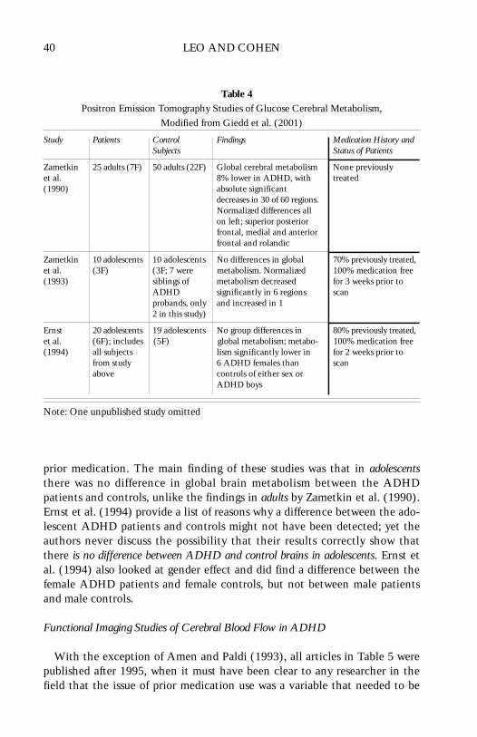
40 LEO AND COHEN
prior medication. The main finding of these studies was that in adolescentsthere was no difference in global brain metabolism between the ADHDpatients and controls, unlike the findings in adults by Zametkin et al. (1990).Ernst et al. (1994) provide a list of reasons why a difference between the ado-lescent ADHD patients and controls might not have been detected; yet theauthors never discuss the possibility that their results correctly show thatthere is no difference between ADHD and control brains in adolescents. Ernst etal. (1994) also looked at gender effect and did find a difference between thefemale ADHD patients and female controls, but not between male patientsand male controls.
Functional Imaging Studies of Cerebral Blood Flow in ADHD
With the exception of Amen and Paldi (1993), all articles in Table 5 werepublished after 1995, when it must have been clear to any researcher in thefield that the issue of prior medication use was a variable that needed to be
Table 4Positron Emission Tomography Studies of Glucose Cerebral Metabolism,
Modified from Giedd et al. (2001)
Study Patients Control Findings Medication History andSubjects Status of Patients
Zametkin 25 adults (7F) 50 adults (22F) Global cerebral metabolism None previouslyet al. 8% lower in ADHD, with treated(1990) absolute significant
decreases in 30 of 60 regions. Normalized differences all on left; superior posterior frontal, medial and anterior frontal and rolandic
Zametkin 10 adolescents 10 adolescents No differences in global 70% previously treated,et al. (3F) (3F; 7 were metabolism. Normalized 100% medication free (1993) siblings of metabolism decreased for 3 weeks prior to
ADHD significantly in 6 regions scanprobands, only and increased in 12 in this study)
Ernst 20 adolescents 19 adolescents No group differences in 80% previously treated,et al. (6F); includes (5F) global metabolism; metabo- 100% medication free (1994) all subjects lism significantly lower in for 2 weeks prior to
from study 6 ADHD females than scanabove controls of either sex or
ADHD boys
Note: One unpublished study omitted

BRAIN IMAGING AND ADHD 41
Table 5Functional Imaging Studies of Cerebral Blood Flow in ADHD,
Modified from Giedd et al. (2001)
Study Method Patients Controls Findings Medication History and Status of Patients
Amen and [99m-TC] 54 (8F) 18 (8F) Prefrontal deactivation Unclear on prior Paldi SPECT at rest significantly greater in history; medication (1993) and during ADHD (65% vs 5%) free during scan
math stress test
Sieg [123-I] SPECT 10 (3F) 6 (1F) L (vs R) blood flow Not reportedet al. at rest reduced in frontal and parietal(1995) regions in ADHD
Teicher T2 relaxation 11 6 Optimal dose methylphenidate See discussion and et al. times with children significantly increased R footnotes(2000) MRI caudate blood flow, decreased
R frontal cortical f low
Schweitzer [15O]water 6 male 6 male Task-related changes in rCBF 33% previously et al. PET, neural adults adults in men without ADHD were treated with MPH (2000) activation more prominent in the frontal but had been medi-
related to and temporal regions, but cation free for 2 and working rCBF changes in men with 10 years respectivelymemory ADHD were more widespread
and primarily located in the occipital regions
Bush f MRI, 8 adults 8 adults ADHD subjects failed to 100% previously et al. counting activate the anterior cingulate treated with various (1999) stroop cognitive division (ACcd) ADHD medications.
during the Counting Stroop. 48 hour washout ACcd activity higher in prior to scanscontrol group. ADHD subject did activate a frontostriatal–insular network, indicating ACcd hypoactivity was not caused by globally poor neuronal responsiveness.
Rubia f MRI 7 males 9 males ADHD adolescents showed Not reportedet al. lower power of response in (1999) the right medial prefrontal
cortex during a stop task and a motor timing task, and in the R inferior pre-frontal cortex and L caudate during the stop task.
Vaidya f MRI, two go/ 10 male 6 male ADHD impaired inhibitory Patients had a et al. no-go tasks children children control on two tasks. Off-drug 1 to 3 year history(1998) with and frontal–striatal activation of medication
without drug during response inhibition differed between ADHD andhealthy children.

42 LEO AND COHEN
examined. Nonetheless, four of the seven articles do not mention whetherthe patients had prior medication use.
Amen and Paldi (1993). Giedd et al. (2001) refer to a one page abstract byAmen and Paldi (1993). This study was later expanded upon in a moredetailed report (Amen and Carmichael, 1997), which is what we discusshere. The primary purpose of this study was to determine if there were simi-larities between reported PET and QEEG (quantified computerized EEG)findings in children diagnosed with ADHD. Because of dangers involved inexposing individuals to radioactive substances, the control subjects werepatients from a psychiatric outpatient clinic who were diagnosed with a psy-chiatric condition but not with ADHD. Amen and Carmichael (1997) statethat both ADHD and control patients were “medication free,” but no otherdetails are provided.
Amen, who is prominent in the ADHD marketing enterprise, has receivedsignificant media attention based on his theory that there are six types ofADHD, each of which has distinctive behavioral symptoms with distinctneuroanatomical pathologies that can be visualized with SPECT scans(Amen, 2001). To our knowledge, Amen has not published a study showingthat by using a brain scan, he can tell the difference between the brain of anADHD child and that of a normal child, or that he can use neuroimaging todiagnose any of his proposed six subdivisions of ADHD. Also problematic isthat Amen uses SPECT scans as a regular tool to diagnose ADHD. Withinpsychiatry, neuroimaging is typically used for research, not diagnosis, becausethis technology involves low doses of radiation. Thus its use in children with-out life-threatening conditions is controversial, as Amen and Carmichaelacknowledge (p. 84). Amen is one of only a handful of practitioners who useneuroimaging as an aid in the diagnosis of ADHD.
Teicher et al. (2000).2 This study used functional MRI relaxometry (fMRI)to assess blood volume in the striatum. It reportedly found differences in theputamen of 11 ADHD children compared to six healthy control children.Teicher et al. (2000) concluded: “On average, T2–RT was 3.1% higher inADHD children than control subjects in the left putamen . . . and 1.6% in theright putamen” (p. 471). However like many of the neuroimaging studies thisconclusion is tempered by the issue of prior medication exposure. Someexplanation of the experimental design of the Teicher et al. study is neces-sary.
The 11 children with ADHD were randomized to one of four groups:placebo, or methylphenidate at 0.5, 0.8, or 1.5 mg/kg in divided doses. The
2Giedd et al. cite a 1996 paper by Teicher that is not concerned with brain imaging. We arenot exactly sure which paper they meant to refer to but Teicher’s most significant paper is theone that we discuss here.

BRAIN IMAGING AND ADHD 43
children received the appropriate dose for a week after which they weretested for drug efficacy using objective measures of attention/activity andfMRI within 1–3 hours of their afternoon dose. The children then movedinto the next group for a week, received the appropriate dose and were againscanned and tested. All children were cycled through all four groups but theystarted out in different groups. The researchers then compared the fMRIresults of the unmedicated healthy control subjects and the ADHD subjectsfollowing their week of placebo treatment and it is this difference that isreported as significant. For instance in a scatterplot (Figure 1, p. 472) the T2relaxation times of the 11 ADHD children on placebo are compared to con-trols and Teicher et al. (2000) report that “The increased T2 relaxationtimes in the ADHD sample indicate diminished regional blood volume” (p. 472). However, one problem with this scatterplot (and the experiment) isthat the scans of the ADHD children performed during the week of theplacebo treatment are not comparable. The prescan interval for each childmight have been preceded by one, two, or three weeks of treatment withmethylphenidate. A child randomized to placebo and then scanned is notcomparable to a child administered three weeks of the drug followed byplacebo and then scanned. Teicher and colleagues grouped children whonever received drug treatment with children experiencing withdrawal fromdrug treatment.3 Since exact treatment protocol is not supplied for eachchild, precise interpretation of the results is impossible.
Studies Published Since the Giedd et al. Review
Two studies published since the Giedd et al. 2001 review are noteworthyfor what they could have accomplished — but did not. Both studies had thechance to compare non-medicated patients to appropriate controls yetmerely addressed “secondary” issues. Our analysis of these studies focuses onsifting through the “secondary” issues and asking: What about the essentialcomparison between non-medicated patients and controls?
Kim, Lee, Cho, and Lee (2001). These investigators used SPECT to exam-ine rCBF in 32 previously unmedicated ADHD children before and aftereight weeks of treatment with methylphenidate. Changes were detected inthe prefrontal cortex and the caudate nucleus. We have two concerns withthis study. First, the drug-induced changes in the striatum and frontal lobes
3It is difficult, if not impossible, to determine the exact design of this study — it is only bycommunicating directly with Teicher that we obtained information on the study design. Hesupplied only limited information so while we have done our best to fairly present his studywe are still unsure of the exact protocol. We also wonder how the reviewers of this paper wereable to judge its merit given the limited explanation of the methodology.

44 LEO AND COHEN
are reported to occur in the same cerebral circuit that Giedd et al. (2001)pointed to as the neuropathological locus of ADHD. But whereas Giedd et al.attributed such findings to an endogenous organic pathology, Kim et al.attributed their findings to methylphenidate exposure. Second, theresearchers did not include a control group of “normal” children to compareto the medication-free ADHD children. Kim et al. appear to be one of thefirst research groups to report on ADHD children not previously medicated,yet they have not run the essential comparison between non-medicated chil-dren and controls
It would have been more fruitful to have divided the procedure into twoparts: the first part simply comparing the scans of ADHD children to con-trols, and the second part examining the effect of medication on the scans ofADHD children, as in the study by Lou et al. (1990). Researchers may beperplexed that the scans of the children diagnosed with ADHD were notcompared to controls. Kim et al. state that it would have been unethical toexamine the effect of methylphenidate on a control group — and they arecorrect. However, their study would have been more significant if, prior to thedrug administration, they had compared the scans of the ADHD children to acontrol group.
Castellanos et al. (2002). This study, carried out between 1991 and 2001,reported that the brains of ADHD children are smaller compared to those ofcontrols. The study is significant because of the size of the sample (291 parti-
Entire Patient GroupFemale MaleN=63 N=89
Age, mean (SD), y 9.4 (2.6) 10.5 (3.1)Height,mean (SD),cm 134.9(15.0) 141.7(18.0)Weight, mean(SD),kg 33.0(12.2) 36.9(14.4)
Controls p value for Female Male patients vs. N=56 N=83 controls
10.0 (2.6) 10.9 (3.5) .13140.2(16.0) 147.3(20.3) .0135.8(12.5) 42.0(16.5) .02
Medicated Patients Non-medicated Patients
Age* 10.9 (2.7) 8.3 (2.6)Height Not provided Not providedWeight Not provided Not provided
� �
Figure 1: Participants’ physical characteristics in the Castellanos et al. (2002) study.
*p value for medicated versus non-medicated .001

BRAIN IMAGING AND ADHD 45
cipants) and because one third of the patients never received medication.Three groups were constituted: 49 unmedicated patients, 103 medicatedpatients, and 139 controls. Thus the authors had the opportunity to makenumerous comparisons: unmedicated versus medicated, unmedicated versuscontrols, medicated versus controls, and ADHD versus controls. The mostimportant — and we would say legitimate — comparison was betweenunmedicated patients and controls. However, compared to the controls, theunmedicated patients were two years younger, shorter and lighter.4
Castellanos et al. state that height and weight did not correlate with brainsize in their study. Yet in that study these variables were significantly corre-lated with the diagnosis of ADHD. Thus, although finding three biologicaldifferences between the ADHD children and controls, the researchers onlyfocused on brain size. Height and weight have never been shown to be partand parcel of ADHD, but the results from this study suggest otherwise.Conversely, if height and weight are only spuriously correlated with ADHD,then the appropriateness of the control group is called into question.5
Consider the following data as shown in Figure 1: the entire ADHDpatient group (medicated plus unmedicated) is significantly shorter andlighter than the control group; for the most important comparison in thepaper the subgroup of unmedicated patients is drawn from this already smallerand lighter group of patients; we are not told the height and weight of thissubgroup of unmedicated patients but we are told that they are almost twoyears younger than the entire patient group; and for this reason the unmedi-cated patients are probably also significantly shorter and lighter than thecontrol group. We say “probably” because for the most important comparisonin the article the subjects’ specific physical characteristics are not provided.The issue of height and weight is especially relevant here because mostresearch on brain size has found brain size to be correlated with body weight.Gould has pointed out that in studies that have incorrectly associated brainsize to other factors, the most common mistake has been in sample selectionand that “modern students of brain size have still not agreed on a proper mea-sure to eliminate the powerful effect of body size” (1996, p. 138). We suggest
4Regarding the age discrepancy, the authors reported that they conducted a secondary analysisrestricted to an age-matched subset of 24 unmedicated ADHD children and 54 controls and,“All measures essentially remained unchanged” (p. 1745). However, given a more appropriatecontrol group these types of secondary analyses would not have been required.
5Since the first studies of brain size in the nineteenth century, scientists have correlated brainsize with various other factors such as intelligence, race, predispostion to violence, and evennationality. For instance, in 1861 Gratiolet reported that German brains were on average 100grams larger than French brains (Broca, 1861, pp. 441–442). With the exception of height,weight, and sex most of these correlations have not stood the test of time.

46 LEO AND COHEN
that this effect was not eliminated in the Castellanos et al. (2002) study.Thus, besides a diagnosis of ADHD, the unmedicated children could havehad smaller brains due to the fact that they were shorter, lighter, andyounger. In fact, given all these other variables it would be noteworthy if theydid not have smaller brains.6
To conduct a study to determine if there is something unique about thebrains of ADHD children, one need not involve medicated children at all.The only apparent reason Castellanos et al. (2002) used medicated childrenwas to answer questions about drug effects on the brain. To do so they com-pared unmedicated to medicated children: and they found no significant dif-ferences and concluded that the medications have no effect on brain size.However, this hardly seems the type of study to adequately address this issue,since the authors provided no information whatsoever about medication use(such as doses, durations, or even types of drugs used) except this one sen-tence: “At the time of the first scan, 103 patients (68%) were being treatedwith psychostimulants” (pp. 1742–1743). We note also that, compared tomedicated children, unmedicated children were not as severely affected byADHD according to teachers and doctors.
What at first seems like a straightforward comparison of two groups of chil-dren reveals itself as a tangle of unnecessary complications, including sec-ondary statistical analyses of subgroups, discussions about differences inamounts of white matter in 8-year olds versus 10-year olds, or concerns aboutheight and weight as confounding variables. By itself, a simple comparisonbetween unmedicated ADHD children and controls would certainly havestood out as an important experiment. Peripheral questions could have beenaddressed as extensions of the primary question without complicating theexperiment. But in this study peripheral questions did hopelessly confuse theessential comparison.
Our criticism of the experimental design of the Castellanos et al. (2002)study might seem excessive, but in light of the problems we have pointed outregarding selection of control groups, and considering that the study’spatients and controls came from highly select populations, the concern seemsjustified. Yet, we would like to point out that the more straightforward com-parison was — and still remains — well within the authors’ grasp. Theauthors could still compare the scans of the non-medicated children to thescans of a control group matched for, among other things, height, weight,and age. This simpler comparison would be more direct and meaningful, andwould not take ten years to complete.
6It would also be surprising if they did not have smaller skulls. If there is an associationbetween skull size and ADHD then future studies would save time and money by using a tapemeasure.

BRAIN IMAGING AND ADHD 47
In the past, researchers in this field have been unable to find unmedicatedpatients for their studies. Finally, here is one of the first research groups toovercome this obstacle, but immediately the question arises: Why is the con-trol group two years older, taller, and heavier than the group of unmedicatedpatients? It seems odd that, given ten years and the resources of the NIMH,these experienced researchers could not find a more appropriate controlgroup. Ironically, previous studies were contaminated by a medication con-found; in this study the reverse is true: the control group is not comparablewith the treatment group.
Nevertheless, the findings of Castellanos et al. (2002) will be more valu-able if they are replicated with a more comparable set of controls — and willbe of great interest to more than just ADHD researchers — for two reasons.If the Castellanos et al. study is replicated it will be the first to correlatesubtle differences in brain size with a behavioral trait, in this case activitylevel; and also the first to find that brain size is not correlated with heightand weight.
Discussion
Of the thirty-three relevant studies summarized in Tables 1 to 5, twenty-nineincluded a control group of normal subjects, but only nineteen reported on theADHD patients’ prior use of medication. These nineteen studies involved atotal of 356 patients and 365 controls, and all but one study found differencesbetween ADHD and non-ADHD children. However, in each group of studiesusing an imaging modality, an average of 77% of the ADHD children had priorexposure to medication. Because of this confound, any suggestions about differ-ences between the brains of “ADHD” children and the brains of “normal” chil-dren must await future studies.
To their credit, the two neuroimaging studies published since the Giedd etal. (2001) review used non-medicated children. Yet they avoided a simpleand straightforward comparison between non-medicated children and appro-priate controls. The Kim et al. (2001) study did not compare unmedicatedchildren to controls, instead they started with unmedicated children, admin-istered medications to them, and then performed scans. The most perplexingstudy was reported by Castellanos et al. (2002) who used unmedicatedADHD children who were younger, lighter, and shorter than the controlgroup. Peripheral questions sidetracked each of these research groups fromthe more important comparison: that of unmedicated ADHD children tocontrols.
We are also concerned about how results are sometimes reported both inthe Giedd et al. (2001) review and in the ADHD neuroimaging literature ingeneral. For instance, in their review Giedd et al. categorize the findings of

48 LEO AND COHEN
Shaywitz et al. (1983) as “None” yet what Shaywitz et al. reported was thatthey found no significant differences between ADHD patients and controls.Readers of the Giedd et al. review who are trying to determine if neuroimag-ing researchers have found an anatomical basis for ADHD should be told if astudy found no difference between ADHD patients and controls.7 As anotherexample, in adolescent males Ernst et al. (1994) found no difference inglobal cerebral glucose metabolism between ADHD and controls, but theresearchers did find a difference in adolescent females. While the title oftheir paper — “Reduced Brain Metabolism in Hyperactive Girls” — certainlyconveys their findings, it would have been just as correct to have titled it“Normal Brain Metabolism in Hyperactive Boys.” Apparently studies findinga difference between ADHD children and controls are more significant thenstudies that find no difference. Studies finding a difference between ADHDchildren and controls are probably more likely to get published.
It is interesting to note how neuroimaging findings are used in the relatedfield of research concerning the effects of illegal psychotropics. For example,in humans with persistent exposure to methylenedioxymethamphetamine(MDMA, commonly known as Ecstasy), brain imaging research is often citedas evidence that MDMA causes a deficit in serotonin rich areas of the brain(Reneman et al., 2001). In the case of MDMA, drug users are compared tocontrols and observed differences are attributed to drug use; but in the case ofmethylphenidate, drug users are compared to controls and differences areattributed to an underlying organic pathology?
The ADHD neuroimaging research also examplifies how the media fail totake even a moderately critical view of medical research. Following the pub-lication of the Castellanos et al. (2002) article reporting smaller brain size inADHD children, The New York Times discussed the study. In the beginning ofthe newpaper article, Castellanos is quoted: “I’ve always been extremely cau-tious about overinterpreting results.” One page later Castellanos is reported assaying that the findings raised the possibility that medication might enhancethe normal maturation of the brain in children with attention disorders(Goode, 2002). On the one hand, Castellanos says that he does not like tooverinterpret results; on the other hand, he suggests the remarkable “possibil-ity” that Ritalin might lead to enhanced brain maturation. Granted,Castellanos only raised the “possibility” — but two months later The DetroitFree Press ran an article with the headline: “Ritalin is Safe and It Works:
7Without entering into an elaborate discussion about the null hypothesis versus the researchhypothesis (and the inability to prove the null hypothesis) suffice it to say that if you take twogroups of people and compare a given trait and find no difference between the two groups it isquite possible that indeed there is no difference. Under “findings” it would have been moreaccurate for Giedd et al. (2001) to say “No Difference” rather then “None” because this doesrelate to the research hypothesis of ADHD neuroimaging researchers.

BRAIN IMAGING AND ADHD 49
Research Dispels Fears that Drug Hurts Kids, and Finds That It Actually HelpsBrains Grow” (Kurth, 2002, italics added). The scenario, where Ritalin isportrayed as something akin to a vitamin, examplifies misguided medicaladvice by the media. Yet, in this case it is difficult to fault only the media.
Prior Medication as a Confounding Variable
Slightly over half the papers in Giedd et al.’s (2001) review actually mentionmedication status of subjects; the rest do not inform readers about prior use.But most problematic is that not a single paper devotes more than a sentence(or two) to the topic. Prior medication use by ADHD subjects is virtually a“non-issue.” Only one of the thirty-three studies even suggests that we actuallystudy the effect of chronic stimulant treatment on the brain (Nasrallah et al.,1986).
There is, of course, no such thing as a perfect experiment: if one looks longenough, f laws will be detected. Competing hypothesis can always be found.However, when investigating subtle neuroanatomical or metabolic changesin the brain, it is hard to imagine a more problematic variable than prior his-tory of psychotropic drug use. In this light then, what are we to make of thefact that some researchers fail to even mention this variable?8 Consider thevariable of right or left handedness. In some studies researchers made a pointof only using right handed children (Rubia et al., 1999) or of matching con-trols and patients for handedness (Geidd et al., 1994). But the sameresearchers did not mention medication history. Are researchers more con-cerned about handedness then they are about prior medication use?
At best, Giedd et al.’s (2001) concern with prior medication use is equivo-cal, illustrating how such concern is handled in the literature. On the onehand, in their 1994 study that purports to “support theories of abnormalfrontal lobe development and function in ADHD” (p. 655), Giedd et al.(1994) do not mention medication history of the patients even though thisinformation must have been available because the patients were recruitedfrom an NIMH program. On the other hand, in their 2001 review Giedd etal. qualify Rubia et al.’s (1999) findings by stating, “. . . we must note that allthe patients had been medicated with methylphenidate until 36 hours prior
8For all studies with no information on medication history, we attempted to contact leadauthors to obtain this information. Most did not respond. One author replied: “I am sorry, butI would have to go through all the files, and it is too time-consuming.” We do not know if thisinformation was not reported because authors were unaware that prior drug use is a major con-founding variable or, conversely, if authors were aware of it but realized that it lessened thevalidity of their findings to establish a biological basis for ADHD. Either possibility is a causefor serious concern.

50 LEO AND COHEN
to their scans” and by noting that findings in control subjects scanned after an ingestion of MPH may “ref lect medication withdrawal effects . . .” (p. 39).9 Also, in their final summary, Giedd et al. identify one limitation ofthis research as: “ . . . none of the studies published to date has accounted forpossible source of confounding such as prior medication exposure” (2001, p. 45). But clearly, if that is the case, the issue requires extensively more dis-cussion and integration than four lines in a 13-page review article. Indeed,given the explicit warning from one of the earliest studies (Nasrallah et al.,1986), why not remind readers of the confound and immediately conductstudies without the confounding factor? Perhaps the answer is found inGiedd et al.’s (2001) own explanation of the value of neuroimaging inADHD: “Imaging studies may help educate families and the public thatADHD is a biological entity” (p. 45).
For those researchers who feel that our concerns about prior psychotropicdrug use are excessive it is important to remember how prior psychotropicuse confounded the first experiments that tested the dopamine hypothesis ofschizophrenia. Early reports that schizophrenic patients had more dopaminereceptors than controls were eventually tempered by the realization that thepatients in these studies had taken neuroleptics for years. Subsequentattempts to replicate the findings in medication-free patients have met withinconsistent results (Valenstein, 1998).
Conclusion
Imaging research is often used to justify the medication of children diag-nosed with ADHD (Barkley et al., 2002). For instance, in the most recentADHD imaging study, Castellanos et al. (2002) state that based on theirfindings of smaller brain size in ADHD children, “Future studies should focuson younger patients being enrolled into controlled treatment studies while inpreschool” (p. 1747). However, we question the logic behind the idea thatsmaller brains justify drug treatment for younger children. If one follows thislogic, what is one to make of the height and weight issues? Do height andweight differences also justify “treating” preschoolers?
A significant and oft overlooked issue about brain imaging research con-cerns the distinction between normal biological variation and “disease.” Allmeasureable traits such as height, weight, activity level, or brain size, fallonto a bell-shaped curve. Where along the curve one draws the line betweennormal and abnormal becomes arbitrary. As DSM-IV-TR (American Psychia-
9Giedd et al. (2001) report that all of the ADHD patients in the Rubia et al. (1999) studywere previously medicated, yet Rubia et al. state that “The patients were either unmedicated ormedication free for one week before scanning” (1999, p. 892).

BRAIN IMAGING AND ADHD 51
tric Association, 2000) states with regard to ADHD, “Estimates of preva-lence rates has been revised upward, reflecting increased prevalence due to theinclusion of the Predominantly Hyperactive–Impulsive and PredominantlyInattentive types in DSM-IV” (p. 830). Do 3% of school-aged children haveADHD? Or is it 7%? Or is ADHD approaching 15% as some authors haverecently suggested (Paule et al., 2000)? In a review article titled “Is ADHD aValid Disorder?,” Carey points out that there is a correlation between brainfunction and temperament even in children who fall under the rubric of“normal” (2002, pp. 3–7). If 15% of the population exhibits a particular trait,one might consider it an example of normal biological variation. Still, ifresearchers are inclined to consider 15% of the population as brain-diseased,then they will need to look elsewhere than the body of ADHD neuroimagingresearch for confirmatory evidence.
A comment in an earlier review of ADHD imaging research by two promi-nent researchers seems particularly instructive (Ernst and Zametkin, 1995).In addressing the fact that in the Nasrallah et al. (1986) study seven of the24 patients had a history of alcohol abuse, Ernst and Zametkin pointed out:“Unfortunately, the inclusion of individuals with a history of alcohol abuse,representing 30% of the sample, confused the interpretation of the results,because the findings mirrored those reported in CT studies of alcoholicadults” (p. 1646). If interpreting results from a single study becomes confus-ing because 30% of the sample had a prior history of alcohol abuse, then,undoubtedly, results from a field of research where over three quarters of thepatients were persistently exposed to a centrally active drug would be simi-larly compromised.
The necessary and definitive test to confirm the suggestion that ADHDchildren have a neuroanatomic pathology consists of using a brain scan todetect a difference between a “typical” ADHD child as found in the class-room, and a “normal” child. As we pointed out at the beginning of thispaper, there is virtual unanimity that this cannot be accomplished at present.Experiments with highly selective patient and control groups are, at best,only preliminary studies, and we have shown — in complement to the criticalanalysis by Baumeister and Hawkins (2001) — that the findings of thesestudies must be called into question. In response to persistent pressure fromcritics such as Baughman (1998) and Breggin (1991), it seems that neuro-imaging researchers now acknowledge the importance of medication history.The publication of the Castellanos et al. (2002) article, using non-medicatedchildren, essentially trivializes any further studies that use medicated chil-dren. Yet, after twenty-five years, and thirty-five studies, there is not a singlestraightforward experiment comparing typical unmedicated children with anADHD diagnosis to typical controls. We are perplexed.

52 LEO AND COHEN
References
Amen, D.G. (2001, February 26). Attention doctors. Newsweek, 137, 72–73.Amen, D.G., and Carmichael, B.D. (1997). High-resolution brain SPECT imaging in ADHD.
Annals of Clinical Psychiatry, 9, 81–86.Amen, D.G., and Paldi, J.H. (1993). Evaluating ADHD with brain SPECT imaging. Biological
Psychiatry, 33, 44.American Psychiatric Association. (2000). Diagnostic and statistical manual of mental disorders:
Text revision (fourth editon). Washington, DC: Author.Aylward, E.H., Reiss, A.L., Reader, M.J., Singer, H.S., Brown, J.E., and Denckla, M.B. (1996).
Basal ganglia volumes in children with attention-deficit hyperactivity disorder. Journal ofChild Neurology, 11, 112–115.
Barkley, R., Cook, E., Diamond, A., Zametkin, A., Tharpa, A., Teeter, A., et al. (2002). Interna-tional consensus statement on ADHD. Clinical Child and Family Psychology Review, 5, 89–111.
Baughman, F. (1998, November 16–18). Testimony at the NIH consensus conference on thetreatment and diagnosis of ADHD. Washington, DC.
Baumeister, A.A., and Hawkins, M.F. (2001). Incoherence of neuroimaging studies of attentiondeficit/hyperactivity disorder. Clinical Neuropharmacology, 24, 2–10.
Baumgardner, T.L., Singer, H.S., Denckla, M.B., Rubin, M.A., Abrams, M.T., Colli, M.J., andReiss, A.L. (1996). Corpus callosum morphology in children with Tourette syndrome andattention deficit hyperactivity disorder. Neurology, 47, 477–482.
Bergstrom, K., and Bille, B. (1978). Computed tomography of the brain in children with mini-mal brain damage: A preliminary study of 46 children. Neuropaediatrie, 9, 378–384.
Berquin, P.C., Giedd, J.N., Jacobsen, L.K., Hamburger, S.D., Krain, A.L., Rapoport, J.L., andCastellanos, F.X. (1998). Cerebellum in attention-deficit hyperactivity disorder: A morpho-metric MRI study. Neurology, 50, 1087–1093.
Breggin, P. (1991). Toxic psychiatry. New York: St. Martin’s Press.Breggin, P. (1999). Psychostimulants in the treatment of children diagnosed with ADHD: Part
II — Adverse effects on brain and behavior. Ethical Human Sciences and Services, 1, 213–241.Broca, P. (1861). Sur le volume et la forme du cerveau suivant les individus et suivant les races.
Bulletin de la Société d’Anthropologie de Paris, 2, 441–446.Bush, G., Frazier, J.A., Rauch, S.L., Seidman, L.J., Whalen, P.J., Jenike, M.A., Rosen, B.R., and
Biederman, J. (1999). Anterior cingulate cortex dysfunction in attention-deficit/hyperactiv-ity disorder revealed by fMRI and the counting stroop. Biological Psychiatry, 45, 1542–1552.
Caparulo, B.K., Donald, M.S., Cohen, D.J., Rothman, S.L., Young, J.G., Katz, J.D., Shaywitz,S.E., and Shaywitz, B.A. (1981). Computed tomographic brain scanning in children withdevelopmental neuropsychiatric disorders. Journal of Abnormal Child Psychology, 20, 338–357.
Carey, W.B. (2002). Is ADHD a valid disorder? In P. Jensen and J. Cooper (Eds.), Attentiondeficit hyperactivity disorder: State of the science, best practices (chapter 3, pp. 1–19). KingstonNew Jersey: Civic Research Institute.
Casey, B.J., Castellanos, F.X., Giedd, J.N., Marsh, W.L., Hamburger, S.D., Schubert, A.B., Vauss,Y.C., Vaituzis, A.C., Dickstein, D.P., Sarfatti, S.E., and Rapoport, J.L. (1997). Implication ofright frontostriatal circuitry in response inhibition and attention-deficity/hyperactivity dis-order. Journal of American Academy of Child and Adolescent Psychiatry, 36, 374–382.
Castellanos, F.X., Giedd, J.N., Eckburg, P., Marsh, W.L., Vaituzis, A.C., Kaysen, D., Hamburger,S.D., and Rapoport, J.L. (1994). Quantitative morphology of the caudate nucleus in atten-tion deficity hyperactivity disorder. American Journal of Psychiatry, 151, 1791–1796.
Castellanos, F.X., Geidd, J.N., Marsh, W.L., Hamburger, S.D., Vaituzis, A.C., Dickstein, D.P.,Sarfatti, S.E., Vauss, Y.C., Snell, J.W., Rajapakse, J.C., and Rapoport, J.L. (1996).Quantitative brain magnetic resonance imaging in attention-deficit hyperactivity disorder.Archives of General Psychiatry, 53, 607–616.
Castellanos, F.X., Lee, P.P., Sharp, W., Jeffries, N.O., Greenstein, D.K., Clasen, L.S., et al.(2002). Developmental trajectories of brain volume abnormalities in children and adoles-cents with attention-deficit hyperactivity disorder. Journal of the American MedicalAssociation, 288, 1740–1748.

BRAIN IMAGING AND ADHD 53
DeGrandpre, R. (1999). Ritalin nation. New York: W.W. Norton and Company.Elia, J., Borcherding, B.G., Rapoport, J.L., and Keysor, C.S. (1991). Methylphenidate and dex-
troamphetamine treatments of hyperactivity: Are there true nonresponders? PsychiatryResearch, 36, 141–155.
Ernst, M., Liebenauer, L., King, A., Fitzgerald, G., Cohen, R., and Zametkin, A. (1994).Reduced brain metabolism in hyperactive girls. Journal of the American Academy of Child andAdolescent Psychiatry, 33, 858–868.
Ernst, M., and Zametkin, A. (1995). The interface of genetics, neuroimaging, and neuro-chemistry in attention-defict hyperactivity disorder. In F. Bloom and D. Kupfer (Eds.),Psychopharmacology (pp. 1643–1652). New York: Raven Press.
Filipek, P.A., Semrud–Clikeman, M., Steingard, R.J., Renshaw, P.F., Kennedy, D.N., andBiederman, J. (1997). Volumetric MRI analysis comparing subjects having attention-deficithyperactivity disorder with normal controls. Neurology, 48, 589–601.
Giedd, J.N., Blumenthal, J., Molloy, E., and Castellanos, F.X. (2001). Brain imaging of attentiondeficit/hyperactivity disorder. In J. Wassertein, L.E. Wolf, and F.F. Lefever (Eds.), Adult atten-tion deficit disorder: Brain mechanisms and life outcomes (Vol. 931, pp. 33–49). New York: NewYork Academy of Sciences.
Geidd, J.N., Castellanos, F.X., Casey, B.J., Kozuch, P., King, A.C., Hamburger, S.D., andRapoport, J.L. (1994). Quantitative morphology of the corpus callosum in attention deficithyperactivity disorder. American Journal of Psychiatry, 151, 665–669.
Goode, E. (2002, October 9). Brain size tied to attention deficit hyperactivity disorder. The NewYork Times. Retrieved October 9, 2002, from http://www.nytimes.com
Gould, S.J. (1996). The mismeasure of man (revised and expanded editon). New York: W.W.Norton and Company.
Hynd, G.W., Hern, K.L., Novey, E.S., Eliopulos, D., Marshall, R., Gonzalez, J.J., and Voeller,K.K. (1993). Attention deficit-hyperactivity disorder and asymmetry of the caudate nucleus.Journal of Child Neurology, 8, 339–347.
Hynd, G.W., Semrud–Clikeman, M., Lorys, A.R., Novey, E.S., and Eliopulos, D. (1990). Brainmorphology in developmental dyslexia and attention deficit disorder/hyperactivity. Archivesof Neurology, 47, 919–926.
Hynd, G.W., Semrud–Clikeman, M., Lorys, A.R., Novey, E.S., Eliopulos, D., and Lyytinen, H.(1991). Corpus callosum morphology in attention deficit-hyperactivity disorder: Morpho-metric analysis of MRI. Journal of Learning Disabilities, 24, 141–146.
Ilgin, N., Senol, S., Gucuyener, K., Gokcora, N., and Sener, S. (2001). Is increased D2 recep-tor availability associated with response to stimulant medication in ADHD? DevelopmentalMedicine and Child Neurology, 43, 755–760.
Kim, B., Lee, J., Cho, S., and Lee, D. (2001). Methylphenidate increased regional cerebral bloodflow in subjects with attention deficit/hyperactivity disorder. Yonsei Journal of Medicine, 42,19–29.
Kurth, J. (2002, December 12). Ritalin is safe — and it works. The Detroit Free Press. RetrievedDecember 12, 2002, from http://www.detnews.com
Lou, H.C., Henriksen, L., and Bruhn, P. (1984). Focal cerebral hypoperfusion in children withdysphasia and/or attention deficit disorder. Archives of Neurology, 41, 825–829.
Lou, H.C., Henriksen, L., and Bruhn, P. (1990). Focal cerebral dysfunction in developmentallearning disabilities. The Lancet, 335, 8–11.
Lou, H.C., Henriksen, L., Bruhn, P., Borner, H., and Neilson, J.B. (1989). Striatal dysfunctionin attention deficit and hyperkinetic disorder. Archives of Neurology, 46, 48–52.
Mataro, M., Garcia–Sanchez, C., Junque, C., Estevez–Gonzalez, A., and Pujol, J. (1997).Magnetic resonance imaging measurement of the caudate nucleus in adolescents with atten-tion-deficit hyperactivity disorder and its relationship with neuropsychological and behav-ioral measures. Archives of Neurology, 54, 963–968.
Moll, G., Hause, S., Ruther, E., Rothenberger, A., and Huether, G. (2001). Early methylphen-idate administration to young rats causes a persistent reduction in the density of striataldopamine transporters. Journal of Child and Adolescent Psychopharmacology, 11, 15–24.
Mostofsky, S.H., Reiss, A.L., Lockhart, P., and Denckla, M.B. (1998). Evaluation of cerebellarsize in attention-deficit hyperactivity disorder. Journal of Child Neurology, 13, 434–439.

54 LEO AND COHEN
Nasrallah, H.A., Loney, J., Olson, S.C., McCalley–Whitters, M., Kramer, J., and Jacoby, C.G.(1986). Cortical atrophy in young adults with a history of hyperactivity in childhood.Psychiatry Research, 17, 241–246.
Paule, M.G., Rowland, A.S., Ferguson, S.A., Chelonis, J.J., Tannock, R., Swanson, J.M., andCastellanos, F.X. (2000). Attention deficit/hyperactivity disorder: Characteristics, interven-tions and models. Neurotoxicology and Teratology, 22, 631–651.
Reid, R., Maag, J., and Vasa, S. (1994). Attention deficit hyperactivity disorder as a disabilitycategory: A critique. Exceptional Children, 60, 198–214.
Reiss, D., Feinstein, C., Weinberger, D.R., King, R., Wyatt, R.J., and Brallier, D. (1983).Ventricular enlargement in child psychiatric patients: A controlled study with planimetricmeasurements. American Journal of Psychiatry, 140, 453–456.
Reneman, L., Booij, J., de Bruin, K., Reitsma, J.B., de Wolff, F.A., Gunning, W.B., den Heeten,G.J., and van den Brink, W. (2001). Effects of dose, sex, and long-term abstention from useon toxic effects of MDMA (ecstasy) on brain serotonin neurons. The Lancet, 358, 1864–1869.
Rubia, K., Overmeyer, S., Taylor, E., Brammer, M., Williams, S.C.R., Simmons, A., andBullmore, E.T. (1999). Hypofrontality in attention deficit hyperactivity disorder duringhigher-order motor control: A study with functional MRI. American Journal of Psychiatry,156, 891–896.
Schweitzer, J.B., Faber, T.L., Grafton, S.T., Tune, L.E., Hoffman, J.M., and Kilts, C.D. (2000).Alterations in the functional anatomy of working memory in adult attention deficit hyperac-tivity disorder. American Journal of Psychiatry, 157, 278–280.
Semrud–Clikeman, M., Filipek, P.A., Biederman, J., Steingard, R., Kennedy, D., Renshaw, P.,and Bekken, K. (1994). Attention-deficit hyperactivity disorder: Magnetic resonance imag-ing morphometric analysis of the corpus callosum. Journal of the American Academy of Childand Adolescent Psychiatry, 33, 875–881.
Shaywitz, B.A., Shaywitz, S.E., Byrne, T., Cohen, D.J., and Rothman, S.L. (1983). Attentiondeficit disorder: Quantitative analysis of CT. Neurology, 33, 1500–1503.
Sieg, K.G., Gaffney, G.R., Preston, D.F., and Hellings, J.A. (1995). SPECT brain imaging abnor-malities in attention deficit hyperactivity disorder. Clinical Nuclear Medicine, 20, 55–60.
Sproson, E.J., Chantrey, J., Hollis, C., Marsden, C.A., and Fonel, K.C. (2001). Effect of repeatedmethylphenidate administration on presynaptic dopamine and behaviour in young adultrats. Journal of Psychopharmacology (Oxford), 15, 67–75.
Teicher, M.H., Anderson, C.M., Polcari, A., Glod, C.A., Maas, L.C., and Renshaw, P.F. (2000).Functional deficits in basal ganglia of children with attention-deficit/hyperactivity disordershown with functional magnetic resonance imaging relaxometry. Nature Medicine, 470–473.
Thompson, J.S., Ross, R.J., and Horwitz, S.J. (1980). The role of computed axial tomography inthe study of the child with minimal brain dysfunction. Journal of Learning Disabilities, 13,334–337.
Vaidya, C.J., Austin, G., Kirkorian, G., Ridlehuber, H.W., Desmond, J.E., Glover, G.H., andGabrieli, J.D.E. (1998). Selective effects of methylphenidate in attention deficit hyperactivitydisorder: A functional magnetic resonance study. Proceedings of the National Academy ofSciences, 14494–14499.
Valenstein, E. (1998). Blaming the brain: The truth about drugs and mental health. New York: TheFree Press.
Vitiello, B. (1998). Pediatric psychopharmacology and interaction between drugs and the devel-oping brain. Canadian Journal of Psychiatry, 43, 582–584.
Volkow, N.D., Wang, G.J., Fowler, J.S., Logan, J., Gerasimov, M., Maynard, L., Ding, Y.S.,Gatley, S.J., Gifford, A., and Franceschi, D. (2001). Therapeutic doses of oral methyl-phenidate signigicantly increase extracellular dopamine in the human brain. The Journal ofNeuroscience, 21(2), RC121, 1–5.
Wagner, G.C., Schuster, C.R., and Seiden, L.S. (1981). Neurochemical consequences followingadministration of CNS stimulants to the neonatal rat. Pharmacology, Biochemistry, andBehavior, 14, 117–119.

BRAIN IMAGING AND ADHD 55
Wan, F.J., Lin, H.C., Huang, K.L., Tseng, C.J., and Wong, C.S. (2000). Systemic administrationof d-amphetamine induces long-lasting oxidative stress in the rat striatum. Life Sciences, 66,PL 205–212.
Yuan, J., McCann, U., and Ricaurte, G. (1997). Methylphenidate and brain dopamine neuro-toxicity. Brain Research, 767, 172–175.
Zametkin, A.J., Liebenauer, L.L., Fitzgerald, G.A., King, A.C., Minkunas, D.V., Herscovitch, P.,Yamada, E.M., and Cohen, R.M. (1993). Brain metabolism in teenagers with attention-deficit hyperactivity disorder. Archives of General Psychiatry, 333–340.
Zametkin, A.J., Nordahl, T.E., Gross, M., King, A.C., Semple, W.E., Rumsey, J., Hamburger,S.D., and Cohen, R.M. (1990). Cerebral glucose metabolism in adults with hyperactivity ofchildhood onset. The New England Journal of Medicine, 323, 1361–1366.