vocal cord paralysis medialization · pdf filevocal cord paralysis medialization laryngoplasty...
TRANSCRIPT
1
Vocal Cord Paralysis
Medialization Laryngoplasty
Shashidhar S. Reddy, MD, MPH
Faculty Sponsor: Anna Pou, MD
The University of Texas Medical Branch
Department of Otolaryngology
Grand Rounds Presentation
April 2004
2
Overview
Anatomy of the Larynx
Function of the Larynx
Causes of Vocal Cord Paralysis
Evaluation of Vocal Cord Paralysis
Anterior TVC Medialization
Posterior TVC Medialization
Overview of Treatment for Bilateral Vocal Cord Paralysis
Conclusion (Key Points)
3
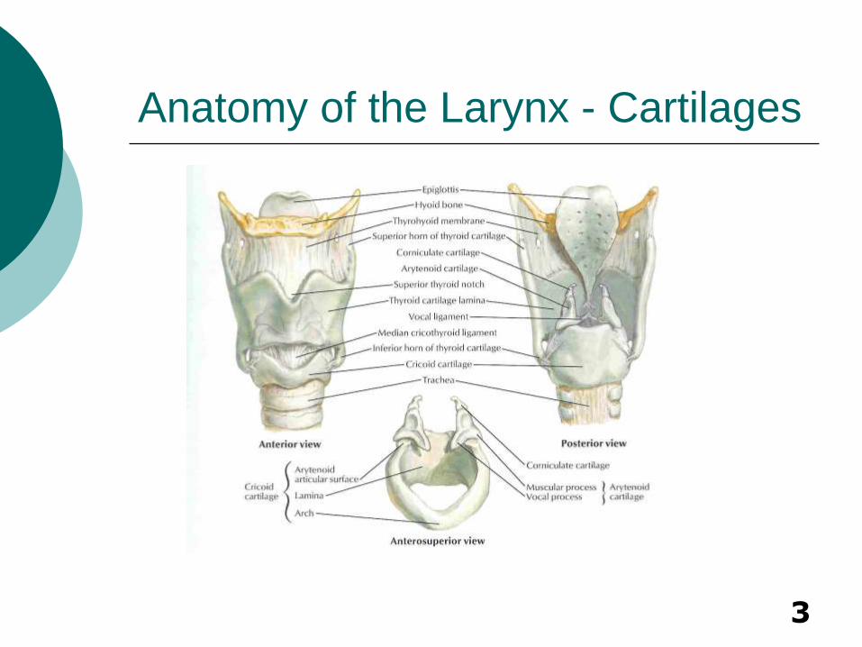
Anatomy of the Larynx - Cartilages
4
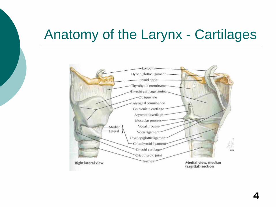
Anatomy of the Larynx - Cartilages
5
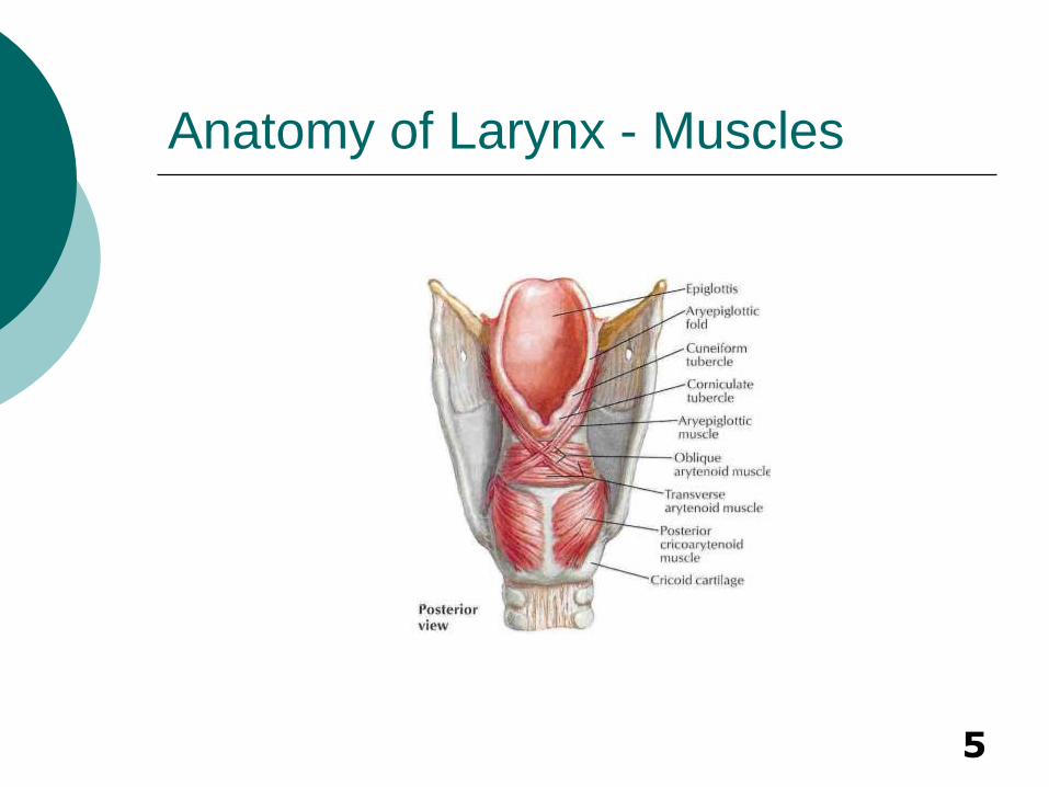
Anatomy of Larynx - Muscles
6
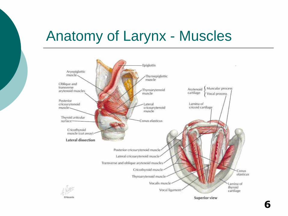
Anatomy of Larynx - Muscles

7
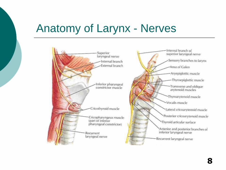
Anatomy of Larynx - Nerves
8
Anatomy of Larynx - Nerves
9
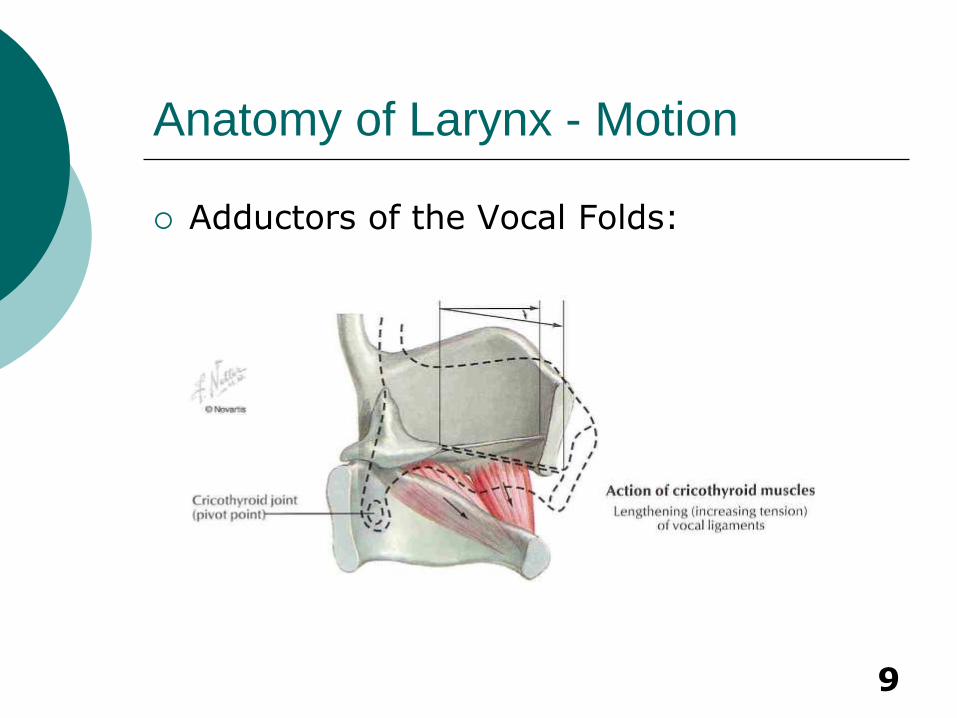
Anatomy of Larynx - Motion
Adductors of the Vocal Folds:
10
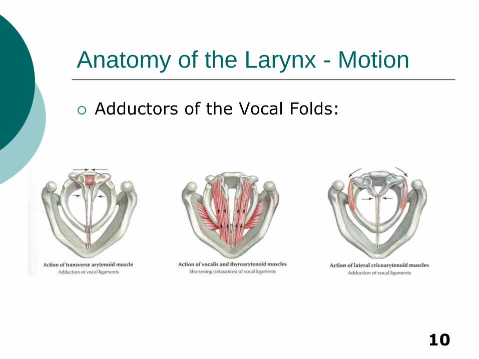
Anatomy of the Larynx - Motion
Adductors of the Vocal Folds:
11
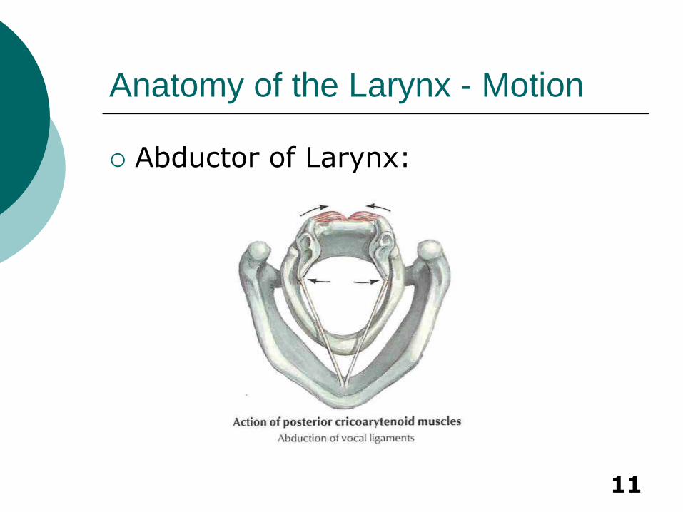
Anatomy of the Larynx - Motion
Abductor of Larynx:
12
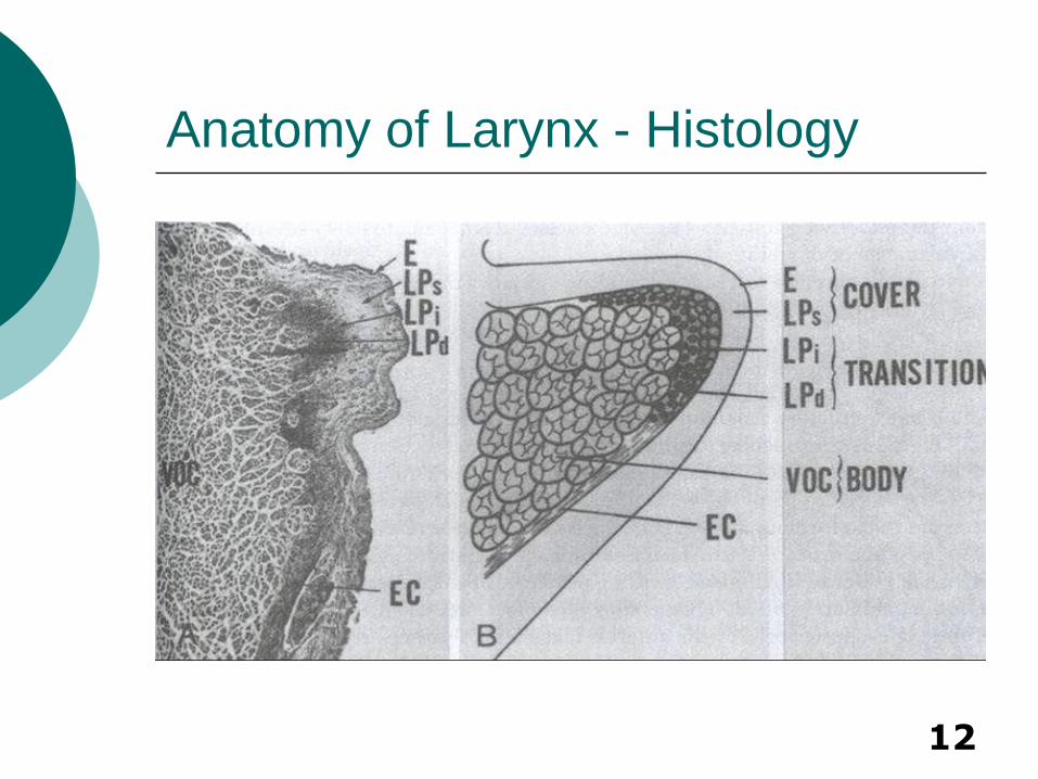
Anatomy of Larynx - Histology
13
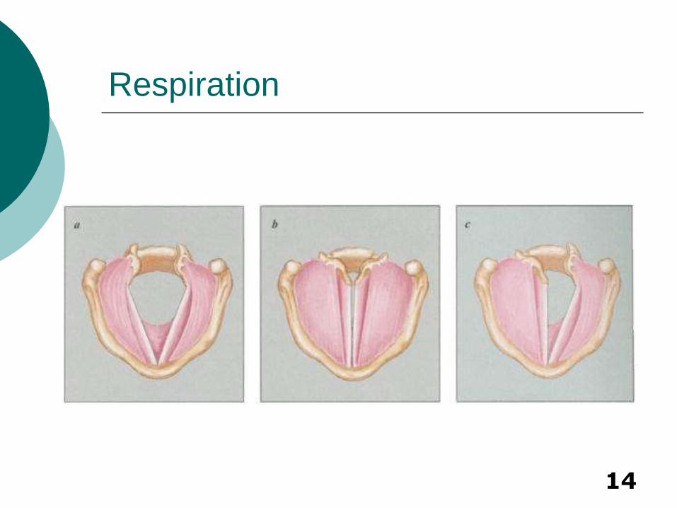
Function of Larynx
Passage for Respiration
Prevents Aspiration
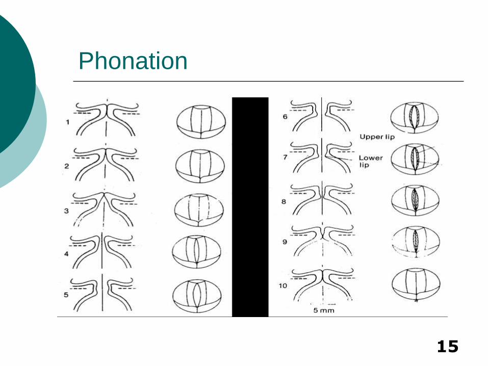
Allows Phonation
Allows Stabilization of Thorax
14
Respiration
15
Phonation
16
Vocal Cord Paralysis
Etiology, Preoperative Evaluation, Treatment
17
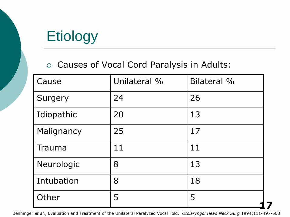
Etiology
Causes of Vocal Cord Paralysis in Adults:
Cause Unilateral % Bilateral %
Surgery 24 26
Idiopathic 20 13
Malignancy 25 17
Trauma 11 11
Neurologic 8 13
Intubation 8 18
Other 5 5
Benninger et al., Evaluation and Treatment of the Unilateral Paralyzed Vocal Fold. Otolaryngol Head Neck Surg 1994;111-497-508
18
Evaluation – Patient History
Alcohol and Tobacco Usage
Voice Abuse
URI and Allergic Rhinitis
Reflux
Neurologic Disorders
History of Trauma or Surgery
Systemic Illness – Rheumatoid
Duration – Affects Prognosis
19
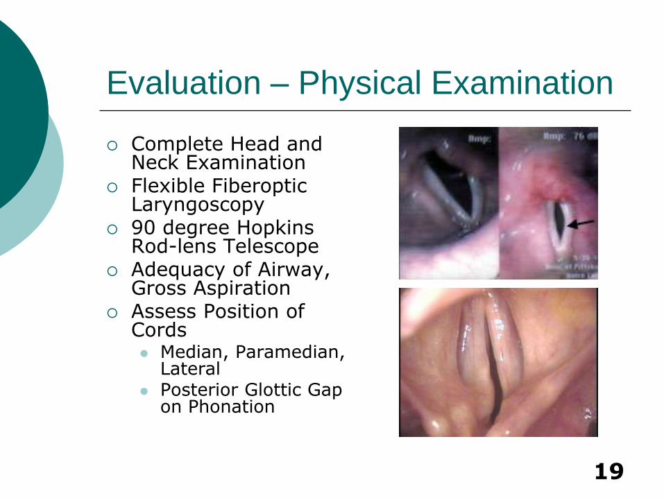
Evaluation – Physical Examination
Complete Head and Neck Examination
Flexible Fiberoptic Laryngoscopy
90 degree Hopkins Rod-lens Telescope
Adequacy of Airway, Gross Aspiration
Assess Position of Cords Median, Paramedian,
Lateral Posterior Glottic Gap
on Phonation
20
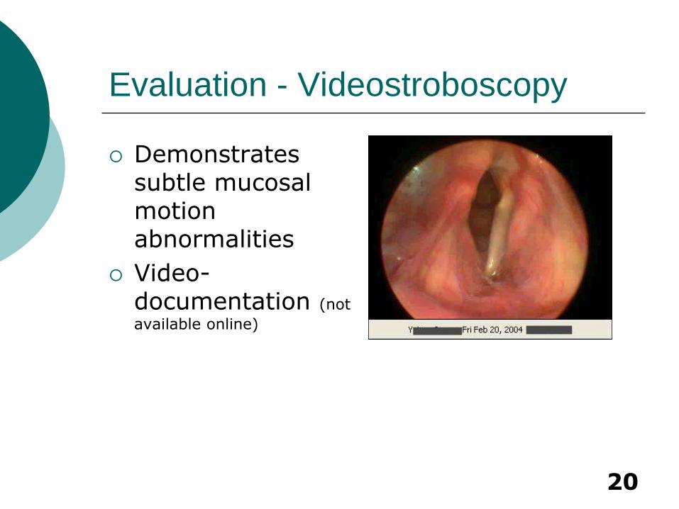
Evaluation - Videostroboscopy
Demonstrates subtle mucosal motion abnormalities
Video-documentation (not
available online)
21
Evaluation - Electromyography
Assesses integrity of laryngeal nerves
Differentiates denervation from mechanical obstruction of vocal cord movement
Electrode in Thyroarytenoid and Cricothyroid
22
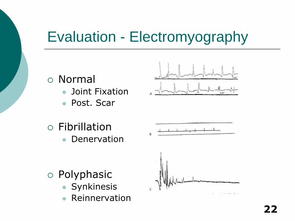
Evaluation - Electromyography
Normal Joint Fixation
Post. Scar
Fibrillation Denervation
Polyphasic Synkinesis
Reinnervation
23
Evaluation - Imaging
Chest X-ray
Screen for intrathoracic lesions
MRI of Brain
Screen for CNS disorders
CT Skull Base to Mediastinum
Direct Laryngoscopy
Palpate arytenoids, especially when no L-EMG
24
Evaluation – Unilateral Paralysis
Preoperative Evaluation
Speech Therapy
Assess patient’s vocal requirements
Do not perform irreversible interventions in patients with possibility of functional return for 6-12 months
Surgery often not necessary in paramedian positioning
25
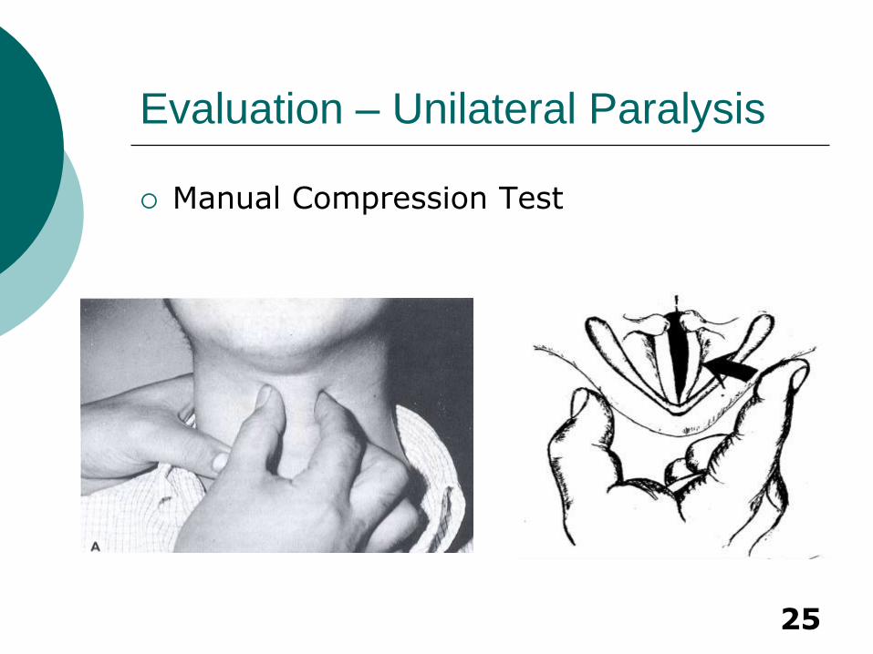
Evaluation – Unilateral Paralysis
Manual Compression Test
26
Evaluation – Unilateral Paralysis
Assess extent of posterior glottic gap
Consider consenting patient for both anterior and posterior medialization procedures
27
Management – Unilateral Paralysis
Type of Anesthesia Local – allows patient to phonate
Careful administration of IV sedation
Internal superior laryngeal nerve block at the thyrohyoid membrane
Glossopharyngeal nerve block at the inferior pole of the tonsils
Flexible endoscope allows visualization
Laryngeal Mask
General
28
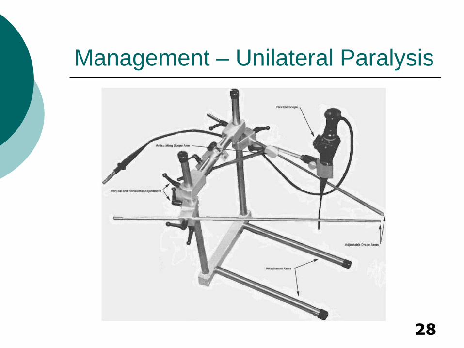
Management – Unilateral Paralysis

29
Management – Unilateral Paralysis
Vocal Cord Injection
Adds fullness to the vocal cord to help it better appose the other side
Injection technique is similar regardless of material used
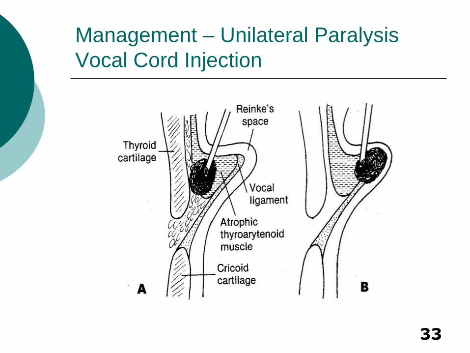
Injection into thyroarytenoid/vocalis
Injection can be done endoscopically or percutaneiously
Poor correction of posterior glottic gap
30
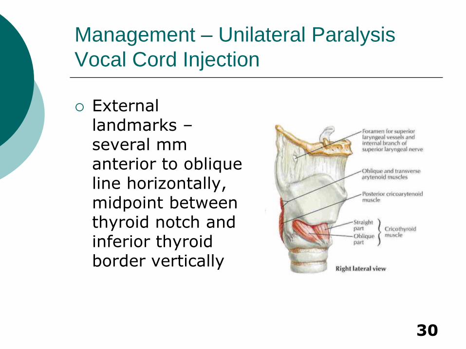
Management – Unilateral Paralysis
Vocal Cord Injection
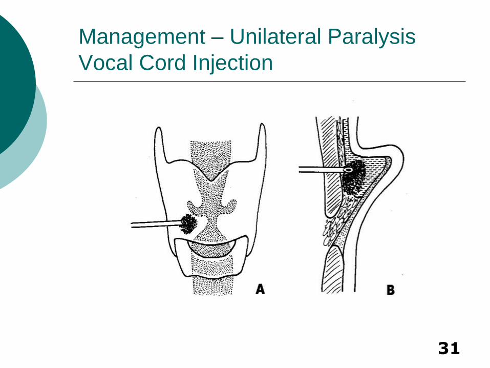
External landmarks – several mm anterior to oblique line horizontally, midpoint between thyroid notch and inferior thyroid border vertically
31
Management – Unilateral Paralysis
Vocal Cord Injection

32
Management – Unilateral Paralysis
Vocal Cord Injection
33
Management – Unilateral Paralysis
Vocal Cord Injection
34
Management – Unilateral Paralysis
Vocal Cord Injection - Materials
Teflon
Fat
Collagen
Autologous Collagen
Homologous Micronized Alloderm (Cymetra)
Heterologous Bovine Collagen (Zyderm
Hyaluronic Acid
Calcium Hydroxyapatite gel (Radiance FN)
Polydimethylsiloxane gel (Bioplastique)
35
Teflon - the first biosynthetic material specifically designed for implantation Advantages
Inexpensive and easily administered
Immediate voice improvement
Disadvantages: Irreversible
Granuloma formation leads to vocal cord stiffening
Migration
Useful mainly in terminal patients
Management – Unilateral Paralysis
Vocal Cord Injection
36
Management – Unilateral Paralysis
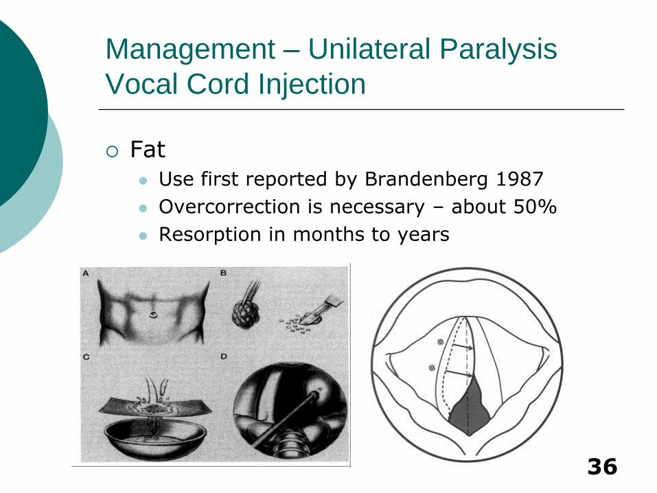
Vocal Cord Injection
Fat
Use first reported by Brandenberg 1987
Overcorrection is necessary – about 50%
Resorption in months to years
37
Management – Unilateral Paralysis
Vocal Cord Injection
Fat Injection
Hsiung et al. divided failures into two categories
Early
failure of fat to soften scarred segments
large glottal gap
large posterior defect
Late
due to absorption of fat
38
Management – Unilateral Paralysis
Vocal Cord Injection
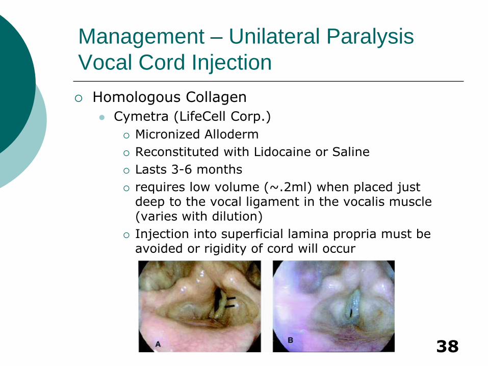
Homologous Collagen
Cymetra (LifeCell Corp.)
Micronized Alloderm
Reconstituted with Lidocaine or Saline
Lasts 3-6 months
requires low volume (~.2ml) when placed just deep to the vocal ligament in the vocalis muscle (varies with dilution)
Injection into superficial lamina propria must be avoided or rigidity of cord will occur
39
Heterologous Collagen
Zyderm
Bovine collagen
May cause immune reaction in 1-2% of cases
Does not last as long as micronized alloderm (Cymetra)
Management – Unilateral Paralysis
Vocal Cord Injection
40
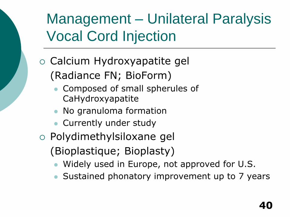
Calcium Hydroxyapatite gel
(Radiance FN; BioForm)
Composed of small spherules of CaHydroxyapatite
No granuloma formation
Currently under study
Polydimethylsiloxane gel
(Bioplastique; Bioplasty)
Widely used in Europe, not approved for U.S.
Sustained phonatory improvement up to 7 years
Management – Unilateral Paralysis
Vocal Cord Injection
41
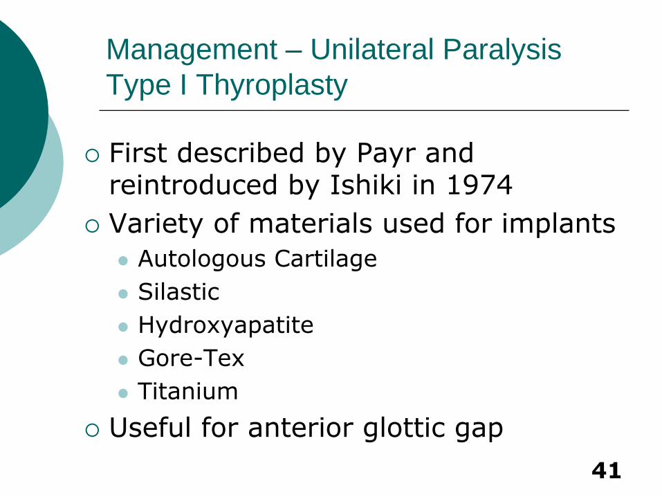
First described by Payr and reintroduced by Ishiki in 1974
Variety of materials used for implants
Autologous Cartilage
Silastic
Hydroxyapatite
Gore-Tex
Titanium
Useful for anterior glottic gap
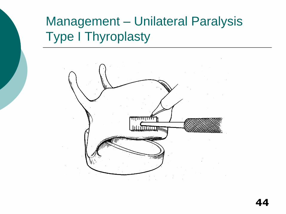
Management – Unilateral Paralysis
Type I Thyroplasty
42
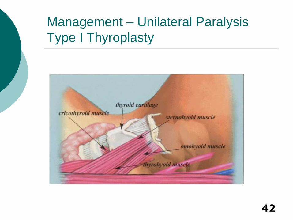
Management – Unilateral Paralysis
Type I Thyroplasty
43
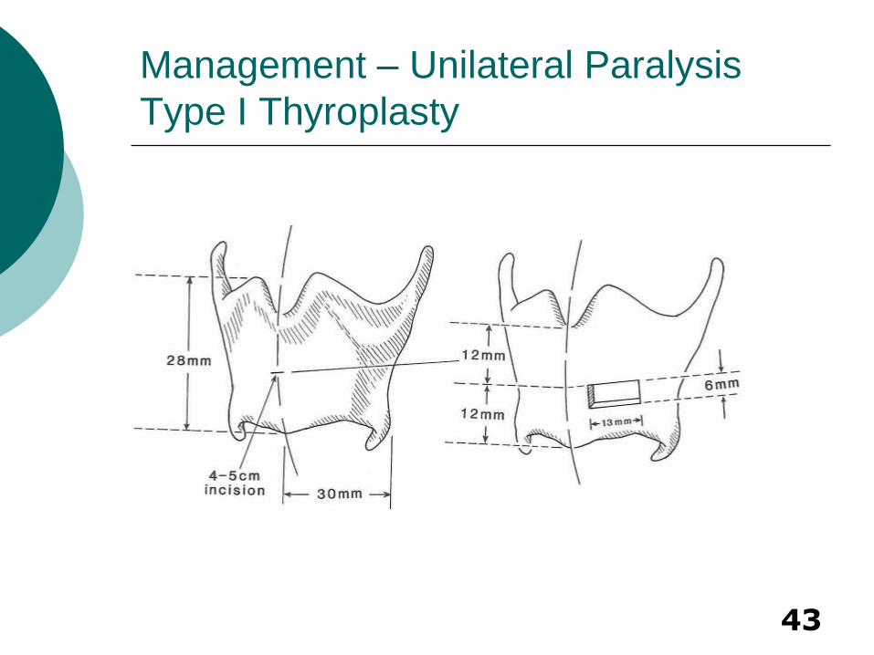
Management – Unilateral Paralysis
Type I Thyroplasty
44
Management – Unilateral Paralysis
Type I Thyroplasty
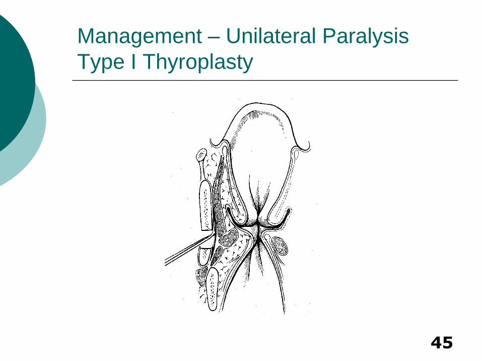
45
Management – Unilateral Paralysis
Type I Thyroplasty
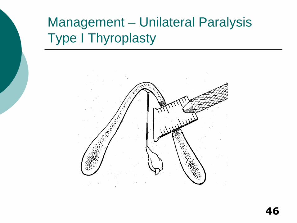
46
Management – Unilateral Paralysis
Type I Thyroplasty
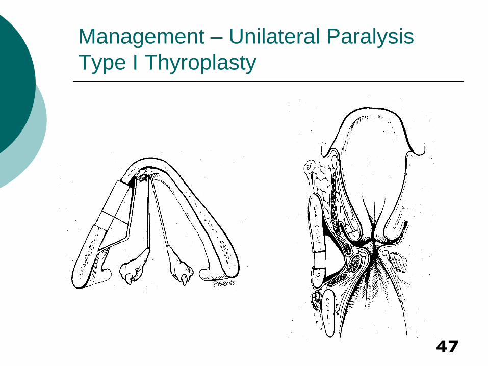
47
Management – Unilateral Paralysis
Type I Thyroplasty
48
Advantages:
Permanent, but surgically reversible
No need to remove implant if vocal function returns
Excellent at closing anterior gap
Disadvantages:
More invasive
Poor closure of posterior glottic gap
Management – Unilateral Paralysis
Type I Thyroplasty
49
Management – Unilateral Paralysis
Type I Thyroplasty – Gore-Tex
Gore-Tex
Homopolymer of polytetrafluoroethylene in minute beads in a fine fiber mesh
Minimal tissue reaction
Cut into long 3mm wide sheet for use
Thyrotomy window drilled to 6-8mm long using a 2mm burr 1cm posterior to midline and 3 or 4mm above lower edge of thyroid
Undermining of perichondrium 4-5mm posterior and inferior to window prior to insertion
Insertion under endoscopic visualization with patient awake

50
Management – Unilateral Paralysis
Type I Thyroplasty – Gore-Tex
51
Complications Extrusion/Displacement (Intraoperative
vs Postop)
Misplacement – most often superior
Infection
Undercorrection – important to overcorrect by 1-2mm
Controversies Location of graft placement
Status of inner perichondrium Many series have shown low extrusion
rate with sacrificed perichondrium
Management – Unilateral Paralysis
Type I Thyroplasty
52
Management – Unilateral Paralysis
Type I Thyroplasty – Variations
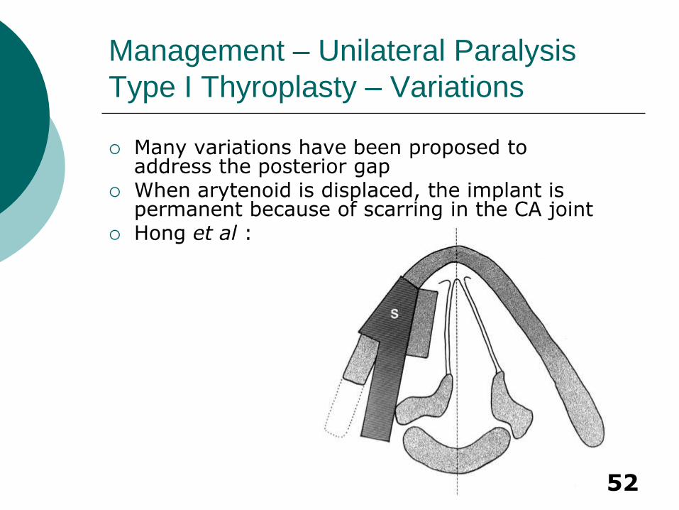
Many variations have been proposed to address the posterior gap
When arytenoid is displaced, the implant is permanent because of scarring in the CA joint
Hong et al :
53
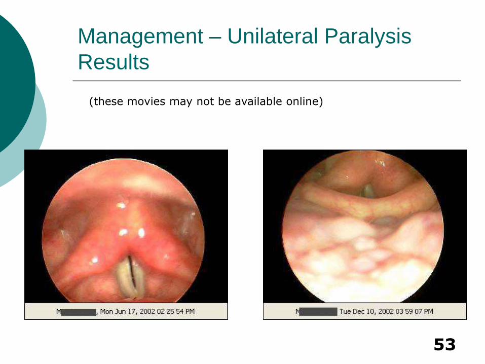
Management – Unilateral Paralysis
Results
(these movies may not be available online)
54
Management – Unilateral Paralysis
Arytenoid Adduction
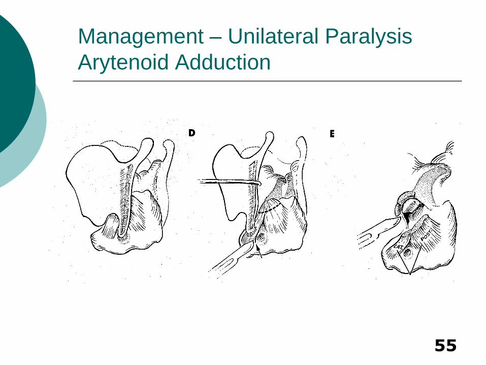
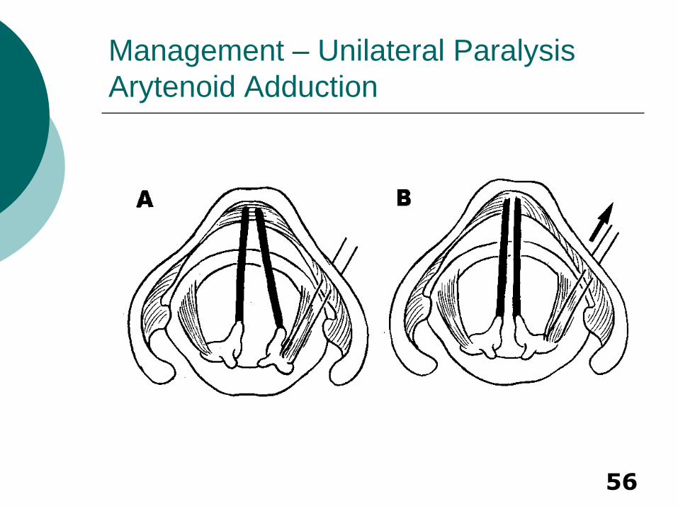
Arytenoid Adduction
First described by Ishiki with modifications by Zeitels and others
Addresses posterior glottic gap by pulling arytenoid into adducted position
Difficult to predict which patients will benefit preoperatively.
Most advocate use in combination with anterior medialization
55
Management – Unilateral Paralysis
Arytenoid Adduction
56
Management – Unilateral Paralysis
Arytenoid Adduction
57
Endoscopic Approaches Suture Placed to Cricoid Cartilage
Simulates action of lateral cricoarytenoid
Zeitels Modification – Arytenopexy Presumably allows a more physiologic
positioning of the arytenoid Involves suturing the arytenoid in a more
posterior and medial position to allow more tension on flaccid cord
Cricothyroid subluxation mimics action of cricothyroid muscle
Modifications should be used selectively
Management – Unilateral Paralysis
Arytenoid Adduction – Modifications
58
Complications
Sutures too tight – may displace arytenoid complex anteriorly, adversely affecting voice
Entry of piriform sinus
Management – Unilateral Paralysis
Arytenoid Adduction
59
Management – Unilateral Paralysis
Reinnervation
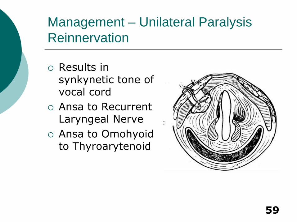
Results in synkynetic tone of vocal cord
Ansa to Recurrent Laryngeal Nerve
Ansa to Omohyoid to Thyroarytenoid
60
Management – Unilateral Paralysis
Reinnervation
Hypoglossal to recurrent laryngeal nerve
Crossed nerve grafts or wire conduction prostheses from one muscle to its paralyzed counterpart are being researched
61
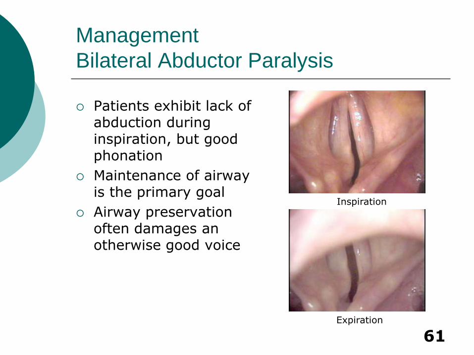
Management
Bilateral Abductor Paralysis
Patients exhibit lack of abduction during inspiration, but good phonation
Maintenance of airway is the primary goal
Airway preservation often damages an otherwise good voice
Expiration
Inspiration
62
Management
Bilateral Abductor Paralysis
Tracheostomy
Gold standard
Most adults will require this
Speaking valves aid in phonation
Laser Cordectomy
Laser Cordotomy
Woodman Arytenoidectomy
63
Bilateral Abductor Paralysis
Phrenic to Posterior Cricoarytenoid anastamosis
Allows abduction during inspiration
Preserves voice when successful
Electrical Pacing
Timed to inspiration with electrode placed on posterior cricoarytenoid
Long-term efficacy not yet shown
64
Bilateral Adductor Paralysis
Patients have good airway with breathy voice
Goal is to prevent aspiration and improve phonation while preserving airway
Aforementioned medialization techniques can be applied
Patients may need tracheostomy if over-medialized
65
Conclusions – Key Points
Anatomy
TVC positioned at about ½ vertical height of the anterior thyroid cartilage and is anterior to the oblique line
Causes of Vocal Cord Paralysis
Iatrogenic (Surgery and intubation #1)
Evaluation
Realize that some function may return with time (6-12 months)
66
Conclusions – Key Points
Management – Unilateral Paralysis
Anterior and Posterior Glottic gap must be addressed
Arytenoid adduction is irreversible
Continued improvement up to 1yr after Type I thyroplasty
Management – Bilateral Paralysis
Preservation of airway is most important goal

67
Vocal Cord Paralysis
Medialization Laryngoplasty
Shashidhar S. Reddy, MD, MPH
Faculty Sponsor: Anna Pou, MD
The University of Texas Medical Branch
Department of Otolaryngology
Grand Rounds Presentation
April 2004