vladimir-prelog-weg 4, ch-8093 zurich, switzerland, e-mail: [email protected] b)...
TRANSCRIPT

Scaffold hopping from synthetic RXR modulators by virtual screening and de novo design
Daniel Merka, Francesca Grisoni
a,b, Lukas Friedrich
a, Elena Gelzinyte
a, Gisbert Schneider
a
a) Department of Chemistry and Applied Biosciences, Swiss Federal Institute of Technology (ETH),
Vladimir-Prelog-Weg 4, CH-8093 Zurich, Switzerland, E-mail: [email protected]
b) Department of Earth and Environmental Sciences, University of Milano-Bicocca, P.za della Scienza,
1, IT-20126 Milan, Italy
- Supporting information -
Table of contents
Supporting figures & tables ............................................................................................................ 2
Materials & Methods ...................................................................................................................... 7
Computational methods .................................................................................................... 7
Chemistry .......................................................................................................................... 9
In vitro pharmacological methods ...................................................................................... 11
NMR spectra & HPLC traces of compounds 24-26 ........................................................................ 12
Supporting references .................................................................................................................... 18
Electronic Supplementary Material (ESI) for MedChemComm.This journal is © The Royal Society of Chemistry 2018
2
Supporting figure & tables
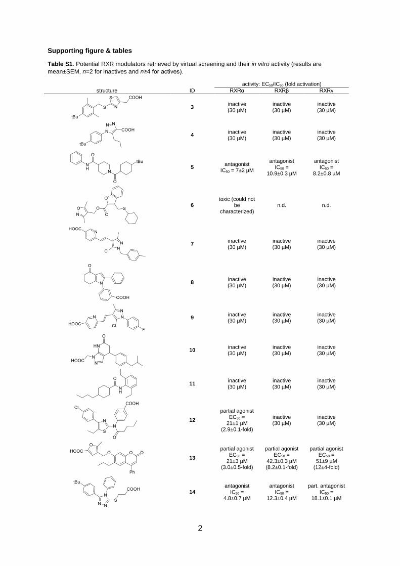
Table S1. Potential RXR modulators retrieved by virtual screening and their in vitro activity (results are
mean±SEM, n=2 for inactives and n≥4 for actives).
activity: EC50/IC50 (fold activation)
structure ID RXRα RXRβ RXRγ
3 inactive (30 µM)
inactive (30 µM)
inactive (30 µM)
4 inactive (30 µM)
inactive (30 µM)
inactive (30 µM)
5 antagonist
IC50 = 7±2 µM
antagonist IC50 =
10.9±0.3 µM
antagonist IC50 =
8.2±0.8 µM
6 toxic (could not
be characterized)
n.d. n.d.
7 inactive (30 µM)
inactive (30 µM)
inactive (30 µM)
8 inactive (30 µM)
inactive (30 µM)
inactive (30 µM)
9 inactive (30 µM)
inactive (30 µM)
inactive (30 µM)
10 inactive (30 µM)
inactive (30 µM)
inactive (30 µM)
11 inactive (30 µM)
inactive (30 µM)
inactive (30 µM)
12
partial agonist EC50 =
21±1 µM (2.9±0.1-fold)
inactive (30 µM)
inactive (30 µM)
13
partial agonist EC50 =
21±3 µM (3.0±0.5-fold)
partial agonist EC50 =
42.3±0.3 µM (8.2±0.1-fold)
partial agonist EC50 =
51±9 µM (12±4-fold)
14 antagonist
IC50 = 4.8±0.7 µM
antagonist IC50 =
12.3±0.4 µM
part. antagonist IC50 =
18.1±0.1 µM
3
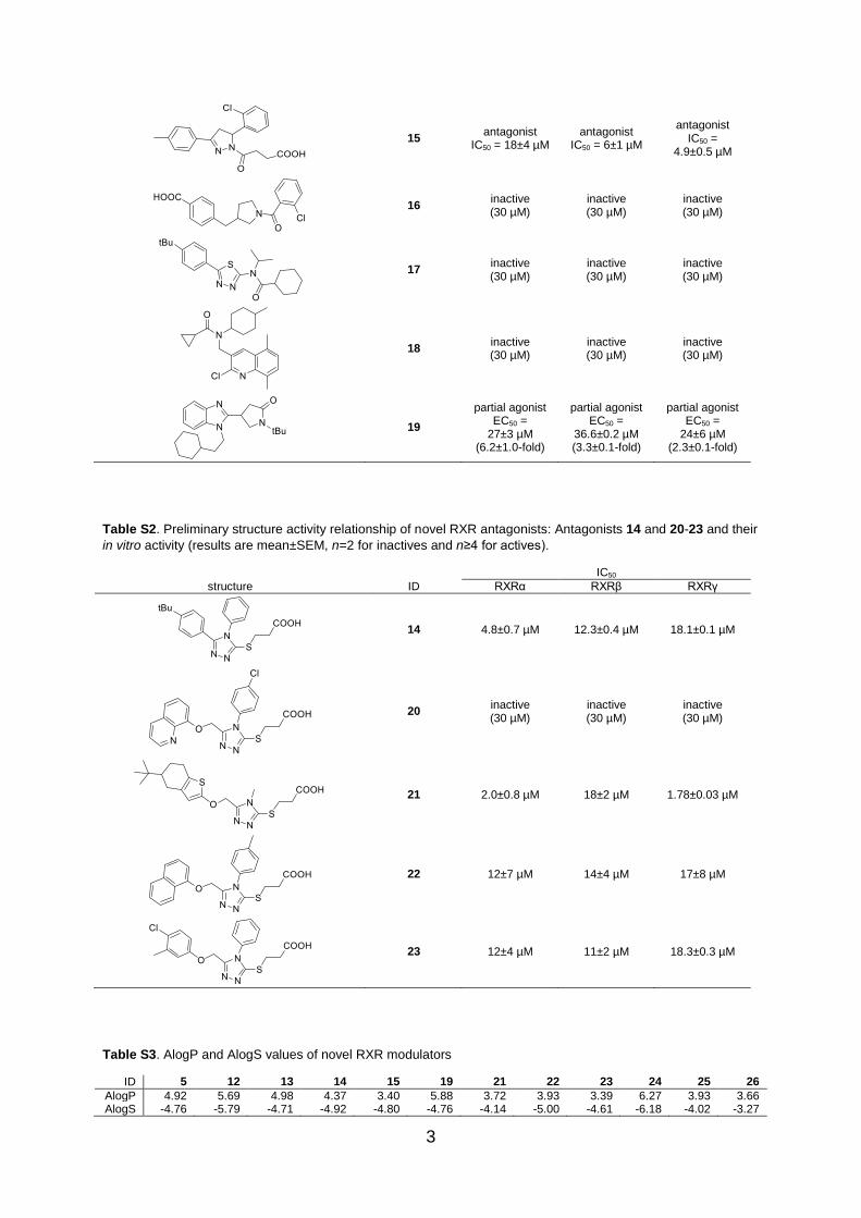
15 antagonist
IC50 = 18±4 µM antagonist
IC50 = 6±1 µM
antagonist IC50 =
4.9±0.5 µM
16 inactive (30 µM)
inactive (30 µM)
inactive (30 µM)
17 inactive (30 µM)
inactive (30 µM)
inactive (30 µM)
18 inactive (30 µM)
inactive (30 µM)
inactive (30 µM)
19
partial agonist EC50 =
27±3 µM (6.2±1.0-fold)
partial agonist EC50 =
36.6±0.2 µM (3.3±0.1-fold)
partial agonist EC50 =
24±6 µM (2.3±0.1-fold)
Table S2. Preliminary structure activity relationship of novel RXR antagonists: Antagonists 14 and 20-23 and their
in vitro activity (results are mean±SEM, n=2 for inactives and n≥4 for actives).
IC50
structure ID RXRα RXRβ RXRγ
14 4.8±0.7 µM 12.3±0.4 µM 18.1±0.1 µM
20 inactive (30 µM)
inactive (30 µM)
inactive (30 µM)
21 2.0±0.8 µM 18±2 µM 1.78±0.03 µM
22 12±7 µM 14±4 µM 17±8 µM
23 12±4 µM 11±2 µM 18.3±0.3 µM
Table S3. AlogP and AlogS values of novel RXR modulators
ID 5 12 13 14 15 19 21 22 23 24 25 26
AlogP 4.92 5.69 4.98 4.37 3.40 5.88 3.72 3.93 3.39 6.27 3.93 3.66 AlogS -4.76 -5.79 -4.71 -4.92 -4.80 -4.76 -4.14 -5.00 -4.61 -6.18 -4.02 -3.27
4
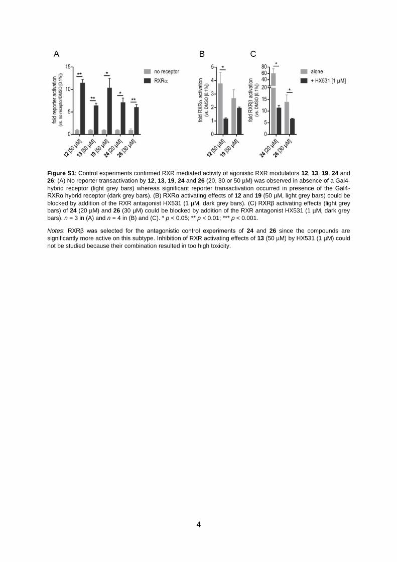
Figure S1: Control experiments confirmed RXR mediated activity of agonistic RXR modulators 12, 13, 19, 24 and
26: (A) No reporter transactivation by 12, 13, 19, 24 and 26 (20, 30 or 50 µM) was observed in absence of a Gal4-
hybrid receptor (light grey bars) whereas significant reporter transactivation occurred in presence of the Gal4-
RXRα hybrid receptor (dark grey bars). (B) RXRα activating effects of 12 and 19 (50 µM, light grey bars) could be
blocked by addition of the RXR antagonist HX531 (1 µM, dark grey bars). (C) RXRβ activating effects (light grey
bars) of 24 (20 µM) and 26 (30 µM) could be blocked by addition of the RXR antagonist HX531 (1 µM, dark grey
bars). n = 3 in (A) and n = 4 in (B) and (C). * p < 0.05; ** p < 0.01; *** p < 0.001.
Notes: RXRβ was selected for the antagonistic control experiments of 24 and 26 since the compounds are
significantly more active on this subtype. Inhibition of RXR activating effects of 13 (50 µM) by HX531 (1 µM) could
not be studied because their combination resulted in too high toxicity.

5
Figure S2. Molecular docking (A-C: RXRα; D: RXRβ; E&F: mRXRα): (A) Partial agonists 12 (orange) and 13
(green) despite their structural diversity form similar binding modes with the RXRα (PDB-ID: 4K4J1) ligand binding
site and neutralize Arg336 as the co-crystallized ligand (purple). (B) The non-acidic modulator 19 (orange) does not
interact with Arg336 and differs from the co-crystallized ligand (purple) in its binding mode. Proposed binding
modes of 24 in RXRα (C) and RXRβ (D) are similar and cannot explain subtype preferential activity. (E)
Antagonists 15 (purple) covers the ligand binding site in mRXRα (PDB-ID: 3A9E2) similar to the co-crystalized
ligand (green) whereas for 14 (orange) a π-stacking contact with Phe318 is suggested but the compound does
not fill the hydrophobic end of the binding pocket. (F) The elongated lipophilic backbone of antagonist 21 (orange)
fills the hydrophobic end of the pocket better than 14 (purple) but lacks π-stacking with Phe318.

6
Figure S3: Diversity of novel RXR modulators: Comparison of scaffolds in newly identified RXR modulators to
ChEMBL annotated RXR agonists and antagonists (IC/EC50 < 50 µM, n = 521) in terms of Jaccard-Tanimoto
distance calculated on the scaffolds’ fragments (Extended Connectivity Fingerprints8, radius = 0 to 4 bonds, bits =
1024): All new RXR modulator chemotypes are markedly different to known modulators.
7
Materials & Methods
Computational methods
Structure preparation
The commercially available screening libraries of ChemBridge, Asinex, Enamine and Specs as well as
reference compounds were washed and protonated with MOE (version 2016.08) at pH7.
Distance calculation and ranking using the CATS3 descriptors
The CATS2 descriptors were calculated for the screening compounds collection and the reference
compounds using in-house software (up to a topological distance of 10 bonds). Euclidean distance
calculations of all screening compounds to the three reference compounds were performed using in-
house software. All screening compounds were then ranked according to their distance to the query
compounds and compounds ranked in the top 100 positions for each query were included. In addition,
compounds were ranked according to the sum of their ranks for individual queries and compounds
ranked in the top 100 positions were considered.
Macromolecular target prediction (SPiDER)4
For the entire screening library, the CATS2 descriptor (see above) and the set of two-dimensional
MOE descriptors (MOE descriptors KNIME node; MOE2016.08; forcefield: MMFF94*) were calculated
and all compounds were individually placed on two pre-trained self-organizing maps for target
prediction (SPiDER). The results were filtered for compounds predicted to be active on retinoid X
receptor with a p-value < 0.05 and compounds were ranked according to their p-value. Compounds
ranked in the top 100 positions were considered.
Virtual screening for antagonists using the CATS descriptors, fingerprints and the LIQUID descriptors5
The CATS (up to a topological distance of 10 bonds) and LIQUID descriptors were calculated using in-
house software for the screening compounds collection after pre-filtering for 1,2,3-substituted five-ring
systems as well as for the reference antagonists. Morgan fingerprints (1024 bits, radius = 0-2) for the
same sets of compounds were calculated using RDKit. Euclidean distance of the pre-filtered screening
compounds to the three reference antagonists for CATS and LIQUID descriptor as well as Tanimoto
similarity for fingerprints were calculated. The screening compounds were then ranked according to
their distance/similarity to each query compound and compounds ranked at least twice in the top 5
positions were included.
De-novo design (Design of Genuine Structures)6
De novo designs were generated by reaction-driven de-novo design using DOGS (Design of Genuine
Structures) with seven RXR agonists from literature or our in-house library as templates. For each
template, one DOGS run was performed (preferences: starting fragments: 100, ISOAK-alpha: 0.40,
including reduced graph abstraction level) and the de-novo designs resulting from each individual run
were ranked according to Euclidean distances calculated on CATS2 and SPIDER p-values as
described above. 24-26 were selected taking into account their design frequency from different
templates, individual ranks and building block availability.
Scaffold diversity analysis
Bemis-Murcko scaffolds7 were computed using RDKit module of python (v 2.7). Extended Connectivity
Fingerprints8 (radius = 0 to 4 atoms, 1024 bit) were computed with Dragon 7
9 using the scaffolds’
SMILES as input. The scaffold diversity between active hits and of ChEMBL annotated actives was
determined using the Jaccard-Tanimoto index calculated on ECFPs, computed in a MATLAB
environment10
.
Molecular docking
The crystal structures of the human retinoid X receptor α (PDB-ID: 4K4J1) and β (PDB-ID: 1H9U
11),
and mouse RXR alpha (PDB code 3A9E2) were prepared with QuickPrep in MOE2016.08. The
molecular structure was protonated at pH 7. Structural issues were corrected (adding hydrogens,
8
capping C terminus and loop from Pro244-Asn262). Water molecules farther away than 4.5 Å from the
receptor or ligand were deleted. The positions of receptor atoms were restrained (force constant = 10,
buffer = 0.25 Å). The position of all atoms farther away than 8 Å from the ligand were fixed, except
hydrogen atoms close to the ligand. The resulting structure was minimized in the AMBER10:EHT
(Extended Hückel Theory) forcefield (termination value = 0.1 kcal mol-1
Å-1
).
Hydrogens were added to all ligand compounds, ligand structures were protonated at pH 7 and all
structures were minimized in the AMBER10:EHT (Extended Hückel Theory) forcefield (termination
value = 0.1 kcal mol-1
Å-1
).
Docking of ligands was executed in MOE2016.08 using the integrated GOLD12
docking program as
placement method. The active site was defined by ligand atoms of the co-crystalized ligand. The
efficiency of the docking calculation was set to “Very Flexible” (200%). For all other GOLD-specific
docking options the default settings were used. The fitness function “GOLDscore” was used as scoring
method. 100 Poses of each ligand were generated. The “Induced Fit” method was selected for the
further refinement of the poses using the standard parameters. The GBVI/WSA dG scoring was
chosen as final scoring function. 10 final poses of each ligand were retained.
Calculation of AlogP and AlogS
AlogP and AlogS values were calculated using the ALOGPS 2.113
online tool.

9
Chemistry
General
All chemicals and solvents were reagent grade and used without further purification, unless specified
otherwise. All reactions were conducted in oven-dried glassware under argon-atmosphere and in
absolute solvents. NMR spectra were recorded on a Bruker AV 400 spectrometer (Bruker Corporation,
Billerica, MA, USA). Chemical shifts (δ) are reported in ppm relative to TMS as reference; approximate
coupling constants (J) are shown in Hertz (Hz). Mass spectra were obtained on an Advion expression
CMS (Advion, Ithaka, NY, USA) equipped with an Advion plate express TLC extractor (Advion) using
electrospray ionization (ESI). High-resolution mass spectra were recorded on a Bruker maXis ESI-Qq-
TOF-MS instrument (Bruker). Compound purity was analyzed on a Varian ProStar HPLC (SpectraLab
Scientific Inc., Markham, ON, Canada) equipped with a MultoHigh100 Phenyl 5 µ 240+4 mm column
(CS-Chromatographie Service GmbH, Langerwehe, Germany) using a gradient (H2O/MeOH
80:20+0.1% formic acid isocratic for 5 min to MeOH+0.1% formic acid after additional 45 min and
MeOH+0.1% formic acid for additional 10 min) at a flow rate of 1 ml/min and UV-detection at 245 nm
and 280 nm. All final compounds for biological evaluation had a purity > 95% (area-under-the-curve for
UV245 and UV280 peaks).
3',5'-Di-tert-butyl-[1,1'-biphenyl]-4-carboxylic acid (24): 1-Bromo-3,5-di-tert-butylbenzene (28, 135 mg,
0.50 mmol, 1.00 eq) and 4-boronobenzoic acid (29, 108 mg, 0.65 mmol, 1.30 eq) were dissolved in a
mixture of toluene (abs., 9 ml) and ethanol (abs., 1 ml), caesium carbonate (488 mg, 1.50 mmol, 3.00
eq) was added and the mixture was stirred at room temperature for 30 min.
Tetrakis(triphenylphosphin)palladium (29 mg, 0.025 mmol, 0.05 eq) was added and the mixture was
stirred under reflux for 6 h. After cooling to room temperature, 25 ml 10% aqueous hydrochloric acid
were added, and the mixture was extracted three times with 25 ml ethyl acetate at a time. The
combined organic layers were dried over magnesium sulfate and the solvents were evaporated in
vacuum. The crude product was purified by column chromatography using hexane/ethyl acetate/acetic
acid (89:9:2) as mobile phase to yield the title compound as colorless solid (64 mg, 41%). 1H NMR
(400 MHz, acetone-d6): δ = 1.26 (s, 18H), 7.42 – 7.45 (m, 3H), 7.64 – 7.71 (m, 2H), 7.95 – 8.01 (m,
2H), 11.09 (s, 1H) ppm. 13
C NMR (101 MHz, acetone-d6): δ = 30.87, 34.69, 121.53, 122.22, 127.18,
130.10, 139.36, 143.42, 146.59, 151.36, 166.61 ppm. MS (ESI-): m/z 309.19 [M-H]-. HRMS (ESI-): m/z
calculated 309.1860 for C21H25O2 found 309.1858 [M-H]-.
3-(5-([1,1'-Biphenyl]-4-yl)oxazol-2-yl)propanoic acid (25): 3-(5-(4-Chlorophenyl)oxazol-2-yl)propanoic
acid (30, 126 mg, 0.50 mmol, 1.00 eq) and phenylboronic acid (31, 79 mg, 0.65 mmol, 1.30 eq) were
dissolved in toluene (abs., 10 ml), caesium carbonate (488 mg, 1.50 mmol, 3.00 eq) was added and
the mixture was stirred at room temperature for 30 min. [1,1′-
bis(diphenylphosphino)ferrocene]dichloropalladium(II) (37 mg, 0.05 mmol, 0.10 eq) was added and the
mixture was stirred under reflux for 24 h. After cooling to room temperature, 25 ml 10% aqueous
hydrochloric acid were added, and the mixture was extracted three times with 25 ml ethyl acetate at a
time. The combined organic layers were dried over magnesium sulfate and the solvents were
evaporated in vacuum. The crude product was purified by column chromatography using methylene
chloride/methanol (96:4) as mobile phase to yield the title compound as colorless solid (45 mg, 31%). 1H NMR (500 MHz, DMSO-d6): δ = 2.77 (t, J = 7.0 Hz, 2H), 3.05 (t, J = 7.0 Hz, 2H), 7.39 (t, J = 7.4 Hz,
1H), 7.50 (dd, J = 10.5, 4.8 Hz, 2H), 7.60 (d, J = 5.4 Hz, 1H), 7.75 – 7.70 (m, 2H), 7.82 – 7.75 (m, 4H),
12.34 (s, 1H) ppm. 13
C NMR (126 MHz, DMSO): δ = 23.51, 30.75, 123.11, 124.73, 127.03, 127.17,
127.72, 128.22, 129.49, 139.74, 140.17, 150.42, 163.59, 173.64. MS (ESI-): m/z 292.21 [M-H]-. HRMS
(MALDI): m/z calculated 294.1133 for C17H16ClN2O2 found 294.1129 [M+H]+.
1-(3-Chlorophenyl)-5-(2,5-dimethyl-1H-pyrrol-1-yl)-1H-pyrazole-4-carbonitrile (26): 5-Amino-1-(3-
chlorophenyl)-1H-pyrazole-4-carbonitrile (32, 109 mg, 0.50 mmol, 1.00 eq) and hexane-2,5-dion (33,
63 mg, 0.55 mmol, 1.10 eq) were dissolved in methylene chloride (10 ml), montmorillonite K10 (0.5 g)
was added and the mixture stirred at room temperature for 15 minutes before the solvent was
evaporated under reduced pressure. The dry powder mixture was transferred to a microwave vial and
irradiated to 90°C for 30 minutes. The powder was then suspended in ethyl acetate (25 ml) and
montmorillonite K10 was filtered off. The resulting solution was washed with 5% aqueous hydrochloric
acid and brine, and dried over magnesium sulfate. The solvent was evaporated under reduced
10
pressure and the crude product was purified by column chromatography using methylene
chloride/methanol (98:2) as mobile phase to yield the title compound as yellow solid (132 mg, 89%). 1H NMR (400 MHz, acetone-d6): δ = 1.84 (s, 6H), 5.89 (s, 2H), 6.87 – 6.96 (m, 2H), 7.29 – 7.38 (m,
2H), 8.28 (s, 1H) ppm. 13
C NMR (101 MHz, acetone-d6): δ = 11.26, 109.50, 111.34, 120.00, 120.15,
121.86, 128.50, 129.08, 131.06, 134.59, 142.76 ppm. MS (ESI+): m/z 297.09 [M+H]+. HRMS (ESI+):
m/z calculated 297.0902 for C16H14ClN4 found 297.0893 [M+H]+.

11
In vitro pharmacological methods
Hybrid reporter gene assays for RXRα, RXRβ and RXRγ
Plasmids: The Gal4-fusion receptor plasmids pFA-CMV-hRXRα-LBD14
, pFA-CMV-hRXRβ-LBD14
and
pFA-CMV-hRXRγ-LBD14
coding for the hinge region and ligand binding domain (LBD) of the canonical
isoform of the respective nuclear receptor have been reported previously. pFR-Luc (Stratagene) was
used as reporter plasmid and pRL-SV40 (Promega) for normalization of transfection efficiency and cell
growth.
Assay procedure: HEK293T cells were grown in DMEM high glucose, supplemented with 10% FCS,
sodium pyruvate (1 mM), penicillin (100 U/mL) and streptomycin (100 μg/mL) at 37 °C and 5% CO2.
The day before transfection, HEK293T cells were seeded in 96-well plates (2.5·104 cells/well). Before
transfection, medium was changed to Opti-MEM without supplements. Transient transfection was
carried out using Lipofectamine LTX reagent (Invitrogen) according to the manufacturer’s protocol with
pFR-Luc (Stratagene), pRL-SV40 (Promega) and pFA-CMV-hNR-LBD. 5 h after transfection, medium
was changed to Opti-MEM supplemented with penicillin (100 U/mL), streptomycin (100 μg/mL), now
additionally containing 0.1% DMSO and the respective test compound or 0.1% DMSO alone as
untreated control. Each concentration was tested in triplicates and each experiment was repeated
independently at least three times. Following overnight (12-14 h) incubation with the test compounds,
cells were assayed for luciferase activity using Dual-Glo™ Luciferase Assay System (Promega)
according to the manufacturer’s protocol. Luminescence was measured with an Infinite M200
luminometer (Tecan Deutschland GmbH). Normalization of transfection efficiency and cell growth was
done by division of firefly luciferase data by renilla luciferase data and multiplying the value by 1000
resulting in relative light units (RLU). Fold activation was obtained by dividing the mean RLU of a test
compound at a respective concentration by the mean RLU of untreated control. Relative activation
was obtained by dividing the fold activation of a test compound at a respective concentration by the
fold activation of the reference agonist bexarotene at 1 µM. All RXR hybrid assays were validated with
the above-mentioned reference agonist which yielded EC50 values in agreement with literature. The
assay was repeated in absence of a hybrid receptor by only transfecting reporter gene construct and
control gene for all agonistic and partial agonistic compounds as control experiment to confirm nuclear
receptor mediated activity.

12
NMR spectra & HPLC traces of compounds 24-26
Compound 24
1H-NMR
13
C-NMR

13
HPLC
245 nm
HPLC 280 nm

14
Compound 25
1H-NMR
13
C-NMR

15
HPLC
245 nm
HPLC 280 nm

16
Compound 26
1H-NMR
13
C-NMR

17
HPLC
245 nm
HPLC 280 nm

18
Supporting References
1 L. J. Boerma, G. Xia, C. Qui, B. D. Cox, M. J. Chalmers, C. D. Smith, S. Lobo-Ruppert, P. R. Griffin, D. D.
Muccio and M. B. Renfrow, J. Biol. Chem., 2014, 289, 814.
2 Y. Sato, N. Ramalanjaona, T. Huet, N. Potier, J. Osz, P. Antony, C. Peluso-Iltis, P. Poussin-
Courmontagne, E. Ennifar, Y. Mély, A. Dejaegere, D. Moras and N. Rochel, PLoS One, 2010, 5, e15119.
3 M. Reutlinger, C. P. Koch, D. Reker, N. Todoroff, P. Schneider, T. Rodrigues and G. Schneider, Mol. Inf.,
2013, 32, 133.
4 D. Reker, T. Rodrigues, P. Schneider and G. Schneider, Proc. Natl. Acad. Sci. U. S. A., 2014, 111, 4067.
5 Y. Tanrikulu, M. Nietert, U. Scheffer, E. Proschak, K. Grabowski, P. Schneider, M. Weidlich, M. Karas, M.
Göbel and G. Schneider, ChemBioChem, 2007, 8, 1932.
6 M. Hartenfeller, H. Zettl, M. Walter, M. Rupp, F. Reisen, E. Proschak, S. Weggen, H. Stark and G.
Schneider, PLoS Comput. Biol., 2012, 8, e1002380.
7 G. W. Bemis and M. A. Murcko, J. Med. Chem., 1996, 39, 2887.
8 D. Rogers and M. Hahn, J. Chem. Inf. Model., 2010, 50, 742.
9 H. kode-solutions.net., Kode srl., Dragon (software for molecular descriptor calculation) version 7.0.6,
2016.
10 MATLAB 2017a, U. S. The MathWorks, Inc., Natick, Massachusetts.
11 J. D. Love, J. T. Gooch, S. Benko, C. Li, L. Nagy, V. K. K. Chatterjee, R. M. Evans and J. W. R. Schwabe,
J. Biol. Chem., 2002, 277, 11385.
12 G. Jones, P. Willett, R. C. Glen, A. R. Leach and R. Taylor, J. Mol. Biol., 1997, 267, 727.
13 I. V Tetko and V. Y. Tanchuk, J. Chem. Inf. Comput. Sci., 2002, 42, 1136.
14 D. Flesch, S.-Y. Cheung, J. Schmidt, M. Gabler, P. Heitel, J. S. Kramer, A. Kaiser, M. Hartmann, M.
Lindner, K. Lüddens-Dämgen, J. Heering, C. Lamers, H. Lüddens, M. Wurglics, E. Proschak, M.
Schubert-Zsilavecz and D. Merk, J. Med. Chem., 2017, 60, 7199.