vitamin a & visual cycle
TRANSCRIPT

VITAMIN A & VISUAL CYCLE
MODERATOR-DR SUDHA SEETHARAM
PRESENTER-DR RAVNEET CHADHA

INTRODUCTION VITAMINS:-
Vitamins may be regarded as organic
compounds required in the diet in small
amounts to perform specific biological
functions for normal maintenance of optimum
growth and health of the organism.


WHAT IS VITAMIN A?
• The term “vitamin A” makes it sound like there is one particular nutrient called “vitamin A”, but this is not true. It is a broad group of related nutrients.
• Vitamin A is a broad term for group of unsaturated nutritional organic compounds, that includes retinol, retinal, retinoic acid, and several provitamin A carotenoids, among which beta-carotene is the most important.

THUS,
VITAMIN A IS AN Essential fat soluble vitamin occuring in the following forms:
Preformed Retinoids (retinal, retinol, retinoic acid)
Found in animal products
Provitamin A Carotenoids
Must be converted to retinoid form
Found in plant products

HISTORY
It is recorded in history that HIPPOCRATES cured night blindness(about 500 B.C)
He prescribed to the patients Ox liver(in honey)which is now known to contain high quantity of vitamin A.
By 1917, Elmer McCollum et al at the University of Wisconsin–Madison, studied the role of fats in the diet and discovered few accessory factors. These "accessory factors" were termed "fat soluble" in 1918 and later "vitamin A" in 1920.

In 1919, Harry Steenbock (University of Wisconsin) proposed a relationship between yellow plant pigments (beta-carotene) and vitamin A.
In 1931, Swiss chemist Paul Karrer described the chemical structure of vitamin A.
Vitamin A was first synthesized in 1947 by two Dutch chemists, David Adriaan van Dorp and Jozef Ferdinand Arens.

Structure of vitamin ANOMENCLATURE
PROVITAMIN A : β-Carotene
VITAMIN A1 : Retinol ( Vitamin A alcohol)
VITAMIN A2 : 3 –Dehydro-retinol
VITAMIN A ALDEHYDE : Retinal
VITAMIN A ACID : Retinoic acid
VITAMIN A ESTER : Retinyl ester
NEO VITAMIN A : Stereoisomer of Vitamin A1, has 70
–80% of biological activity of Vitamin A1.

CHEMISTRY
• Vitamin A is composed of ‘β-IONONE RING’ (CYCLOHEXENYL)
to which ‘POLY ISOPRENOID SIDE CHAIN’ is attached
Polyisoprenoid chain –all trans configuration, contains 4 double bonds, has 2 methyl groups with terminal carbon having ‘R’ group
‘R’ Group –alcohol/aldehyde/acid
β-Ionone ring –contains 1 double bond with 3 methyl groups

Retinol: -(CH2OH)
-found in animal tissues as ‘Retinyl esters’ with long chain fatty acids
•Retinal: -(CHO)
-Aldehyde derived from oxidation of retinol by ‘retinal reductase’ requiring NAD/NADP
-Retinol & Retinal are inter-convertible

•Retinoic acid : -(COOH)
-Acid derived from oxidation of retinal
-Retinoic acid cannot be reduced in body therefore cannot form retinal or retinol.
•β-Carotene :
-Hydrolysed by β-carotene dioxygenase in presence of oxygen & bile salts to two molecules of retinal.

Sources of vitamin A
• Animal : Fish Liver oil, Butter, Milk, Cheese, Egg Yolk
• Plant : All Yellow –Orange –Red –Dark Green fruits & vegetables like Tomatoes, Carrots, Spinach, Papayas, Mangoes, corn, sweet potatoes.

RECOMMENDED DIETARY ALLOWANCE Unit of activity is expressed as ‘RETINAL
EQUIVALENT’ (R.E.) / ‘INTERNATIONAL UNIT’ (I.U.)
1 Retinal Equivalent = 1μg of Retinol OR 6 μg of β-carotene
1 I.U. = 0.3 μg of Retinol OR 0.34 μg of Retinyl acetate OR 0.6 μg of β-carotene
Infants & Children : 400 t0 600 μg/day
Adults (Men & Women) : 600 to 800 μg/day
Pregnancy & Lactation : 1000 to 1200 μg/day

ABSORPTION,TRANSPORT AND STORAGE OF VITAMIN A

VISUAL CYCLE The term “visual cycle” was coined by George Wald in the
mid 1900’s to describe the ability of the eye to “re-cycle” vitamin A for the synthesis of visual pigments(wald,1968)
As originally proposed (Wald,1968),the rod visual cycle requires the involvement of both retina and the retinal pigment epithelium(RPE) in order to properly process the retinal chromophore released from bleached rod pigment(or rhodopsin)

INTRODUCTION The visual cycle is the biological conversion of a photon into
an electrical signal in the retina.
The processing of visual information begins in the retina with the detection of light by photoreceptor cells.
The photoreceptor cells involved in vision are :
1. rods.
2. cones.
Both the rods and cones contain chemicals that decompose on exposure to light and in the process, excite the nerve fibresleading from the eye.
light sensitive chemical in the rods is called rhodopsin and that in the cones is called cone pigments/colour pigments

Anatomy of photoreceptorsRODS:-
Cylindrical stuctures
Length:40-60 microns
Diameter:2 micron
For peripheral vision and scotopic vision
Contain visual purple (Rhodopsin)
120 million
Absent in fovea

Each rod is composed of four structures namely:
1. outer segment
2. Inner segment
3. Cell body
4. Synaptic terminal

Outer segment : Outer segment is cylindrical, transversely striated and contains rhodopsin
The photosensitive pigment rhodopsin is present in membranous discs.
There are about 1000 discs in each rod.
The outer segment of rod cell is constantly renewed by the formation of new discs.(3-4/hr)

Inner segment: connected to outer segment by means of modified cilium.
Contains organelles with large number of mitochondria.
Cell body: also called rod granule, contains the nucleus.
Synaptic terminal: synapses with dendrites of bipolar cells and horizontal cells. Synaptic vesicles present in the synaptic terminal contain the neurotransmitter glutamate.
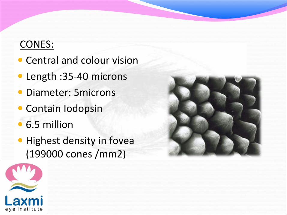
CONES:
Central and colour vision
Length :35-40 microns
Diameter: 5microns
Contain Iodopsin
6.5 million
Highest density in fovea (199000 cones /mm2)

Each cone is composed of four structures namely:
1. outer segment
2. Inner segment
3. Cell body
4. Synaptic terminal

Outer segment: smaller and conical
Contains saccules (infoldingsof cell membrane) counterparts of rod discs.
Renewal of outer segment of cone is a slow process and it differs form that in rods.
.

Inner segment: connected to outer segment with modified cilium. Contain organelles and mitochondria.
Cell body: also called cone granule, possesses the nucleus.
Synaptic terminal: synaptic vesicle present in the synaptic terminal possess the neurotransmitter, glutamate

50.4 Membranuous structures of the outer segments of a rod and cone

Physiology of vision The main mechanisms are:
1. Initiation of vision(phototransduction)
2. Processing and transmission of visual sensations
3. Visual perception
Photochemistry of vision
Will be discussed under the following headings:
1. Rhodopsin-retinal visual cycle in the rods.
• Rhodopsin and its decomposition by light energy.
• Reformation of rhodopsin.
• Role of vitamin A in the formation of rhodopsin.
• Excitation of rod when rhodopsin is activated.
2. Colour vision in the cones.

Chemical basis of visual process
The photopigments present in the rods and cones decompose on exposure to light, in the process, excite the nerve fibers through generation of electrical activity and impulses in the retina.
Photopigments:
Rhodopsin/visual purple present in rods.
Colour pigments/cone pigments(porphyropsin,iodopsinand cyanopsin) present in cones.

Rhodopsin –retinal visual cycle in the rods.
Rhodopsin and its decomposition by light energy:
• The outer segment of the rod that projects into the pigment layer of retina has a concentration of about 40% of light sensitive pigments called Rhodopsin or visual purple.
• Rhodopsin = scotopsin(protein) + retinal(carotenoidprotein).
• Retinal is present in the form of 11-cis retinal known as retinene.
• cis form of retinal is important because only this form can bind with scotopsin to synthesize rhodopsin.

Photochemical changes in rhodopsin:
1.Bleaching of rhodopsin:
When exposed to light, the colour of rhodopsin changes from red to yellow by a process known as bleaching.
Bleaching occurs in a few milliseconds and many unstable intermediates are formed during the process.
2. Reformation of rhodopsin:
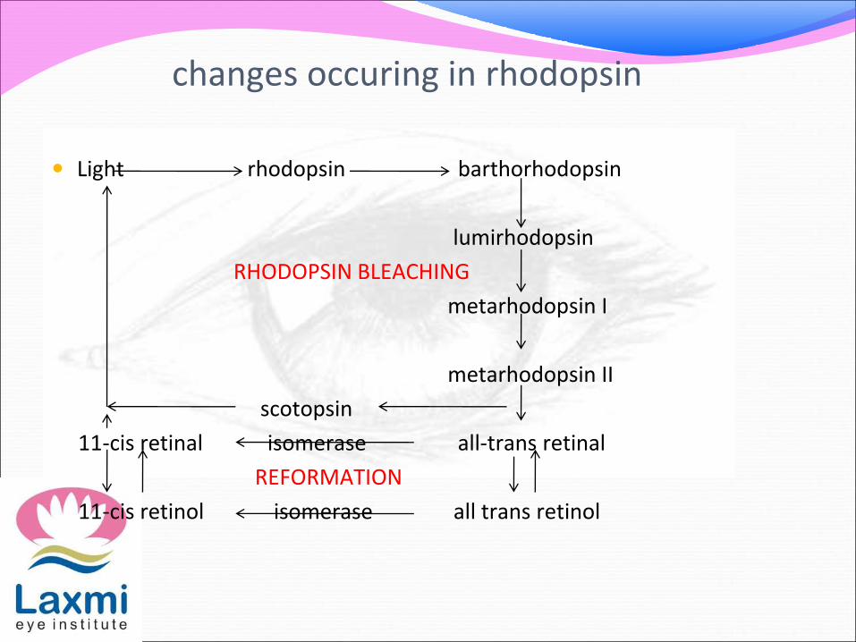
changes occuring in rhodopsin
Light rhodopsin barthorhodopsin
lumirhodopsin
RHODOPSIN BLEACHING
metarhodopsin I
metarhodopsin II
scotopsin
11-cis retinal isomerase all-trans retinal
REFORMATION
11-cis retinol isomerase all trans retinol

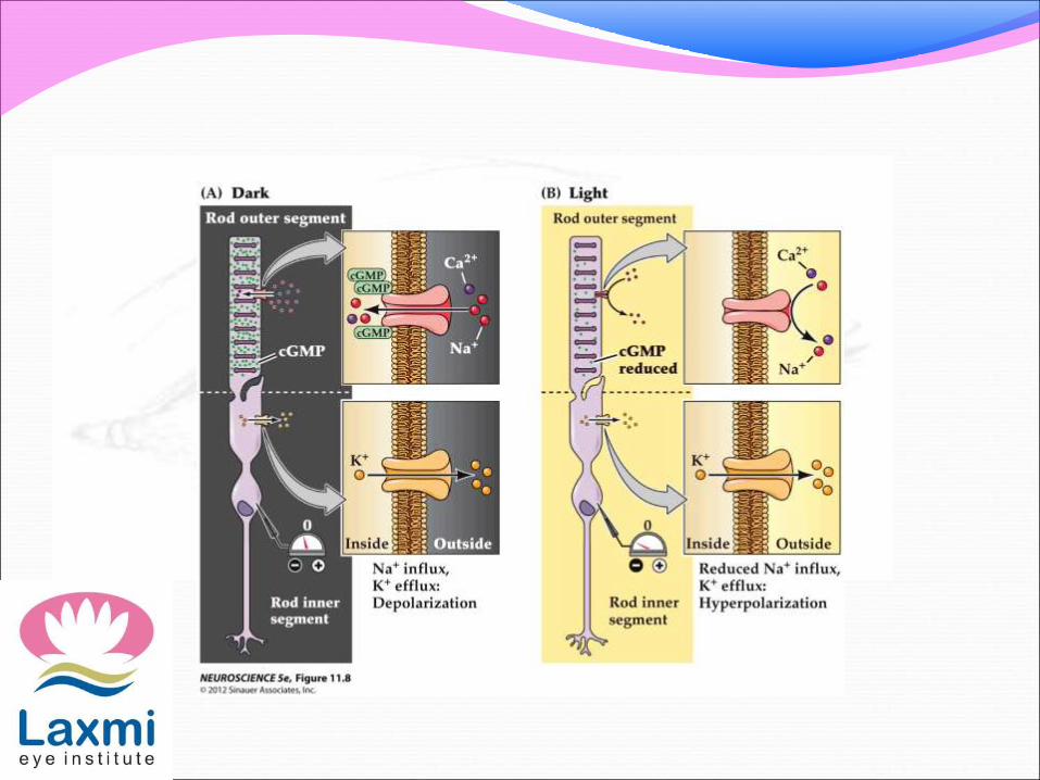
Phototransduction
The transduction of light into a neural signal takes place in the outer segment of a retinal rod or cone photoreceptor

Fig. 50.6 Movement of sodium and potassium ions through the inner and outer segments of the rod


VISUAL CYCLE-COLOUR VISIONCones are specialised in bright & colour vision
Colour vision is governed by 3 colour sensitive pigments :
-Porphyropsin (Red)
-Iodopsin (Green)
-Cyanopsin (Blue)
All these are retinal-opsin complexes
When bright light strikes the retina →one or more of these pigments are bleached, depending on the colour of light →pigment (s) dissociating into All-trans-retinal & Opsin
Differential bleaching
Nerve impulse generated by visual cascade causes perception of specific colour

Receptor potential of the photoreceptors is locally graded potential i.e it does not propagate
The receptor potential does not follow all or none law .
The receptor potential generated in the photoreceptors is transmitted by electronic conduction to the other cells of retina i.e horizontal cells,bipolar cells,amacrine cells and ganglion cells
The ganglion cells transmit the visual signals by means of action potential

FUNCTIONS OF VITAMIN A VISION
GENE TRANSCRIPTION
IMMUNE FUNCTION
EMBRYONIC DEVELOPMENT AND REPRODUCTION
BONE METABOLISM
HAEMATOPOESIS
SKIN AND CELLULAR HEALTH
ANTIOXIDANT ACTIVITY

VITAMIN A DEFICIENCY
Most susceptible populations:
Preschool children with low F&V intake
Urban poor
Older adults
Alcoholism
Liver disease (limits storage)
Fat malabsorption

Vitamin A deficiency may result from :
-Dietary insufficiency of Vitamin A / Precursors
-Interference with absorption from intestines
eg: diarrhoea, malabsorption syndrome, bile salt deficiency
-Defect in the transport due to protein malnutrition –‘Kwashiorkar’
-Defect in the storage due to diseases of liver

Tissues chiefly affected –‘Epithelial’ principally which are not normally keratinised
Includes epithelium of respiratory tract, gastrointestinal tract, genitourinary tract, eye & paraocular glands, salivary glands, accessory glands of tongue & buccal cavity and pancreas
Fundamental change: Metaplasia of normal non-keratinised living cells into keratinising type of epithelium

OCULAR MANIFESTATIONS OF VITAMIN A DEFICIENCY XEROPHTHALMIA
The term xerophthalmia was given by a joint WHO and USAID committee in 1976 to cover all the ocular manifestations of vitamin A deficiency including the structural changes affecting the conjunctiva, cornea and retina and also the biophysical disorders of retinal rods and cones functions.

WHO CLASSIFICATION (1982)XEROPHTHALMIA CLASSIFICATION(modified)
XN Night blindness
X1A Conjunctival xerosis
X1B Bitot’s spots
X2 Corneal xerosis
X3A Corneal ulceration /keratomalacia affecting less than 1/3rd corneal surface
X3B Corneal ulceration /keratomalacia affecting more than 1/3rd corneal surface
XS Corneal scar due to xerophthalmia.
XF Xerophthalmic fundus.

XN :NIGHT BLINDNESS(Nyctalopia)
Earliest symptom of xerophthalmia in children
Diminished visual acuity in ‘dim light’(Insufficient adaptation to darkness)
Defective rhodopsin function.

X1A CONJUNCTIVAL XEROSISCharacterised by:
One or more patches of dry, lustreless,nonwettableconjunctiva.
Interpalpebral conjunctiva(commonly temporal quadrants)
Severe cases involves the entire bulbar conjunctiva.
Desribed as ‘emerging like sand banks at receding tide’when child ceases to cry
Can lead to conjunctival thickening,wrinkling and pigmentation.

X1B BITOT’S SPOTS
Bilateral
Bulbar conjunctiva in the interpalpebral area
Commonly in temporal quadrant.
Triangular greyish/silvery white spots/plaques.
Firmly adherent to conjunctiva
Foamy keratinised epithelium(corynebacterium xerosis)

X2 CORNEAL XEROSIS Dry lustreless appearance of cornea
Earliest change is punctate keratopathy
Begins in the lower nasal quadrant
Bilateral punctate corneal epithelial erosions
Can progress to epithelial defects
Reversible on treatment

X3A & X3B CORNEAL ULCERATION /KERATOMALACIA Stromal defects occur in late stages due to colliquative
necrosis leading to corneal ulceration ,softening (melting) and destruction of cornea(keratomalacia)
Corneal ulcers may be small or large
Stromal defects involving less than 1/3rd cornea usually heal leaving some useful vision
Large stromal defects commonly result in blindness.

Small ulcers
1-3mm
Occur peripherally
Circular
Steep margins and sharply demarcated
Large ulcers
More than 3mm
Occur centrally
Involve entire cornea

XS CORNEAL SCAR Healing of stromal defects results in corneal scarring
Size of the corneal scar depends on the size and density of corneal defect.
XF XEROPHTHALMIC FUNDUS Uncommon in occurance
Typical seed like lesions
Whitish/yellow
Raised
Scattered uniformly over part of fundus
At the level of optic disc.
FFA reveals these dots to be focal retinal pigment epithelial defects

CONTND Rarely these patients can present with scotomas
corresponding to the area of retinal involvement
Respond to vitamin A therapy with scotoma disappearing in 1-2 weeks and retinal lesions fading in 1-4 months.

AGE GROUP DOSE DURATION
1.All patients above one year
2.<1 yr of age or <8 kg weight
3.Women of reproductive age group -less severe- severe
2,00,000 IU
Half the dose i.e1,00,000 IU
10,000 IU2,00,000 IU
Day of presentation, next day and 2-3 weeks later
2 weeks
VITAMIN A THERAPY Treatment schedules apply to all stages of active
xerophthalmia
1. Oral therapy (Recommended)

2. Parenteral therapy: IN CASES OF
-severe disease
-unable to take oral feeds
-Repeated vomiting and diarrhoea
-malabsorption
Intramuscular injections of water miscible vit A preparation
Dose – 1,00,000 IU(Half the oral dose)

Local ocular therapy-
Intense lubrication-instilled every 3-4 hours
Topical retinoic acid
Treatment of keratomalacia and corneal ulcer
Treatment of corneal perforation

PROPHYLAXIS AGAINST XEROPHTHALMIA 1.Short term approach
-Periodic administration of vitamin A supplements
-WHO recommended ,universal distribution schedule of vit A for prevention is as follows:
i) Infants <6months (not being breastfed)—50,000 IU
ii)Infants 6-12 months and any child <8kg – 1,00,000 IU
every 3-6months
iii)Children over 1 year and under 6 years --- 2,00,000 IU orally every 6 months
iv)Lactating mothers – 2,00,000 IU orally once at delivery or
during next 2 months to maintain level of vitamin A in breast milk

PROPHYLAXIS
1.Infants <1 year (not being breastfed)
2.Infants 6-12 months and any child <8kg
3. Children > 1 year and < 6 years
4. Lactating mothers
50,000 IU
1,00,000 IU
2,00,000 IU
2,00,000 IU
Every 3-6 months
Every 6 months
once at delivery or during next 2 months to maintain level of vitamin A in breast milk

ctnd
Under vitamin A supplementation program through Reproductive and child health program(RCH) and now National Rural Health Mission(NRHM)
-- Children between 9 and 36 months of age are to be provided with vitamin A solution every 6 months starting with 1,00,000 IU at 9 months of age along with measles vaccination and subsequently 2,00,000 IU every 6 months till 36 months of age.

2.Medium term approach-
- fortification of food with Vit A
3. Long term approach-
- Promotion of adequate intake of Vit A rich foods in high risk groups particularly preschool aged children on a periodic basis and to mothers within 6-8 weeks after childbirth
- Other measures like nutritional education,socialmarketing,home or community garden programs and measures to improve food security.

HYPERVITAMINOSIS A
Ingestion of large amounts of preformed vitaminA from the diet,supplement intake or medications
Acute:
Single doses of >3,00,000 IU
Headache ,Blurred vision,nausea,vomiting,drowsiness,irritability i.e signs of raised ICP(Benign intracranial hypertension)
Serum vit a values-200-1000 IU/dl

Benign intracranial hypertension Increased intracranial pressure
Idiopathic
Headache (m.c),vomiting,pulsatile tinnitus
Diplopia(compression of 6th nerve)
Rarely compression of 3rd n 4th nerve
Papillaedema
visual field defects
Long standing pappilledema leads to optic atrophy.

Chronic – long-term megadose; possible permanent damage ( >50,000 IU/day for several wks)
Bone and muscle pain
Loss of appetite
Skin disorders
Headache
Dry skin
Hair loss
Increased liver size
-Manifestations reversible when vitamin A discontinued

Thank you