visualization and quantification of the impact of humic
TRANSCRIPT

49
Original ArticleVisualization and Quantification of the Impact of Humic Acid
on Zinc Accumulation in Aquatic Plants Using a Low-Molecular-Weight Fluorescent Probe
Thenuwara Arachchige Omila Kasun Meetiyagoda a, Kabul Fadilah b, Masayori Hagimori c, Mudalige Don Hiranya Jayasanka Senavirathna a, Takeshi Fujino a
a Department of Environmental Science and Technology, Graduate School of Science and Engineering, Saitama University, Saitama, Japan
b Centre for Environment and Sustainability Science, Padjadjaran University, Bandung, Indonesiac Faculty of Pharmaceutical Sciences, Mukogawa Women’s University, Nishinomiya, Japan
ABSTRACTThe main aims of this study were to investigate the impact of humic acid (HA) on zinc (Zn) ac-cumulation in aquatic plants and to study a low-molecular-weight Zn2+-selective fluorescent probe to visualize and quantify the tissue-level Zn concentrations. Ceratophyllum demersum and Aldrovanda vesiculosa were exposed to solutions containing Zn (1 and 3 mg/L), HA (0.5 mg/L), and Zn with HA for nine days. The Zn accumulation (mg/g) in the plants was measured by ICP-OES and we applied a Zn2+-selective fluorescent probe with a low molecular weight to the analysis of Zn in C. demersum plant cells using fluorescence microscopy and ImageJ software. The application of HA reduced the Zn accumulation significantly (p < 0.05) and increased the chlorophyll concentration slightly less signifi-cantly (p > 0.05) in both plants. Results obtained from ImageJ revealed a strong positive correlation between fluorescence intensity and the Zn accumulation in C. demersum (r = 0.988). We showed that the application of HA reduced the Zn accumulation in both plants, and successfully visualized and quantified that a Zn2+-selective fluorescent probe with a low molecular weight can be applied to the diagnosis of Zn osmosis into a cell or tissue on the basis of fluorescence intensity.
Keywords: aquatic plants, humic acid, humic acid-metal complexes, zinc, Zn2+ fluorescent probe
INTRODUCTION
Zinc (Zn) is one of the micronutrients whose presence in the natural ecosystem is essential for the growth of plants. In contrast, high Zn concentrations may have toxic effects on plants [1]. Biomass reduction, growth reduction, alteration in the chloroplast structure, and chlorophyll concentration reduction are some of the toxic effects of Zn on plants [1–3]. The Ministry of the Environment of Japan has established water quality standards for Zn to conserve aquatic life; the concentration of Zn should be less than 0.03 mg/L [4]. According to the United States Environmental Protection Agency (US EPA) [5], the standard level of Zn to protect
freshwater aquatic organisms is 0.12 mg/L. The toxicity of these metals is significantly influenced by physical and chemical water quality parameters such as pH, hardness, temperature, and dissolved organic compounds [6]. The acute toxicity of Zn is constant when the pH value ranges between 8 and 11 [7].
Special attention has been given to naturally occurring dissolved organic compounds such as humic substances be-cause of their potential effect on the bioavailability of toxic metals [8]. Naturally derived organic matter can be consid-ered as natural organic matter (NOM) produced by living or decayed vegetation and microbial decomposition processes [3]. The hydrophilic fraction of NOM is mostly composed
Corresponding author: Takeshi Fujino, E-mail: [email protected]: July 22, 2020, Accepted: December 25, 2020, Published online: April 10, 2021
Open Access
This is an open-access article distributed under the terms of the Creative Commons Attribution Non-Commercial No Deriva-
tives (CC BY-NC-ND) 4.0 License. http://creativecommons.org/licenses/by-nc-nd/4.0/
Journal of Water and Environment Technology, Vol.19, No.2: 49–63, 2021doi: 10.2965/jwet.20-110

Journal of Water and Environment Technology, Vol. 19, No. 2, 2021 50
aliphatic hydrocarbons and nitrogenous compounds, such as carboxylic acids, carbohydrates, and proteins. Hydrophobic NOM primarily consists of humic substances such as hu-mic acid (HA), fulvic acid, and humins, which are rich in aromatic carbon, phenolic structures, and conjugated double bonds [9]. A large proportion of NOM (60–90%) consists of humic substances [10].
The complexation of HA with metal ions is one of its properties. The binding strength between trace metals and HA depends on the nature of the pollutant and the general composition of the aquatic environment [11]; the binding capacity affects the fate of metal ions and plays an impor-tant role in their mobility [12]. Researchers revealed that the carboxylic functional group (COOH) of HA is mainly responsible for metal binding processes [8,13]. Some studies indicate that HA can form complexes with both COOH and OH functional groups [14]. Also, metal complexation with amine and amide groups of HA can occur at low pH values owing to the activity of OH groups [15].
The toxic metals associated with HA can reduce plant uptake by transforming bioavailable fractions of toxic met-als to non-bioavailable fractions [16–18]. Soluble forms of Zn-HA complexes are able to release Zn gradually and which is essential for plant uptake. However, insoluble Zn-HA compounds tend to accumulate, and which may cause a deficiency of micronutrients in the ecosystem [8].
In aquatic ecosystems, plants play an important role in the uptake, accumulation, and recycling of trace metals [4]. Thus, aquatic macrophytes are widely employed in phytore-mediation to remove, transform, or stabilize, various of trace metals in water or bottom sediments [19–21]. Moreover, some species with the ability to accumulate trace metals can be used as tools in the monitoring of trace metals if the metal content in their tissues correlates with the concentration in the environment [8].
The main traditional analytical techniques for measuring the concentrations of metal ions in environmental samples are atomic spectrometry, potentiometry, X-ray, voltamme-try, and nuclear methods [22,23]. Atomic absorption spec-troscopy (AAS), atomic emission spectroscopy (AES), and inductively coupled plasma mass spectrometry (ICP-MS) are advanced techniques commonly used for trace metal analysis [24]. However, these techniques usually require complex sample pre-preparation and expensive instrumenta-tion with trained personnel, and are time-consuming [23,24]. To observe cellular and tissue-level trace metal distributions, electron probe X-ray microanalysis (SEM-EDS) includ-ing transmission electron microscopy (TEM) or scanning
electron microscopy (SEM) coupled with energy-dispersive X-ray spectroscopy (EDS), and other methods such as pro-ton induced X-ray emission (PIXE) and secondary ion mass spectrometry (SIMS) are widely used, but owing to many limitations, these techniques are not suitable for the analysis of trace metals in fresh plant tissue samples [25].
To overcome the above drawbacks, researchers have fo-cused on developing indirect measurement techniques such as the use of fluorescent sensors for the detection of trace metal ions. Because of its high sensitivity, selectivity, repro-ducibility, and rapid real-time monitoring, fluorescence de-tection is one of the most interesting analytical tools among the current sensor approaches [24]. Fluorescent sensors are based on fluorophores such as small organic molecules, quantum dots, or fluorescent proteins. Many fluorescent sensors have been developed recently to clarify the role of Zn2+ in physiology, and Zn2+-selective fluorescent sensors are valuable tools for investigating Zn2+ in living systems [26–28]. Boron-dipyrromethene, coumarin, quinolone, and pyrene are some of the most commonly used Zn fluorescent probes [29,30].
To monitor Zn2+ in living cells, fluorescent sensors should have sufficient solubility in water in addition to sufficient cell permeability. Most fluorescent sensors for Zn2+ typically have large molecular weights due to modifications of the core structures of the probe to achieve a high Zn2+ selectivity [31]. Therefore, the aqueous solubility and cell permeability may be limited because of hydrophobicity [32]. To overcome these problems, a low-molecular-weight (MW = ca. 300), high-affinity Zn2+ fluorescent probe has recently been developed by Hagimori et al. [26] using pyridine–pyridone. Pyridine–pyridine-based sensors exhibit high Zn2+ selectivity (Kd = 7–30 μM), aqueous solubility, and fluorescence [26].
The application of these fluorescent sensors along with fluorescence microscopy to the visualization of trace metals offers an opportunity to address fundamental queries about cellular metal homeostasis [33]. For a specific metal ion of interest, fluorescence microscopy could enable the visualiza-tion of changes in fluorescence while targeting a particular sensor [34,35].
Therefore, it is important to study the applicability of a comprehensive, low-cost, and real-time procedure for the visualization and quantification of Zn accumulation in plant tissues.
In this paper, we study the effect of HA on the level of Zn accumulation in Ceratophyllum demersum and Aldrovanda vesiculosa and the applicability of a low-molecular-weight Zn2+-selective fluorescent probe with fluorescence micros-

Journal of Water and Environment Technology, Vol. 19, No. 2, 2021 51
copy to the visualization and quantification of tissue-level Zn accumulation in C. demersum plant.
MATERIALS AND METHODS
C. demersum and A. vesiculosa aquatic plant species were selected for the study. C. demersum, a submerged, rootless, free-floating aquatic plant, was purchased from an aquarium shop in Saitama City. C. demersum has a higher metal ion uptake ability owing to forked leaves, a thin cuticle, a large aerenchyma area, and large mesophyll cells. Many research-ers have recommended this plant species for use in reme-diation of toxic metals [36,37]. Therefore, we selected C. demersum to study the impact of HA on Zn uptake. Before purchasing, plants were thoroughly observed for any visible symptoms, morphological abnormalities or irregularities.
A. vesiculosa, an aquatic, rootless, free-floating carnivo-rous plant living in swampy waters and one of an endangered species [38], was obtained from the cultures maintained in Faculty of Education, Saitama University. A. vesiculosa nor-mally benefits from animal-derived nutrients to supplement of minerals however it also can uptake available minerals from the medium through their stems and leaves [39]. In this research, A. vesiculosa was selected because it is important to understand the behavior of this carnivorous plant over C. demersum on Zn uptake with the influence of HA.
Before conducting the experiments, the plants were re-tained for a week at room temperature in a 5% Hoagland so-lution to fulfil the nutrient requirement of the aquatic plants and for acclimatization [40]. All chemicals used in the study including HA were purchased from FUJIFILM Wako Pure Chemical Industries, Ltd, Osaka, Japan. A stock solution of HA (1000 ppm) was prepared under alkaline conditions. One gram of HA was dissolved in 125 mL of 1 M NaOH, and then filled up to 1 L with deionized water in a 1 L volumetric flask. The solution was stirred using a magnetic stirrer for 48 hours and it was used to prepare 0.5 mg/L HA solutions after filtering with 0.45 µm Whatman Polytetrafluoroethylene
(PTFE) hydrophobic membrane filters [41].
Experimental designC. demersum and A. vesiculosa were separately exposed to
the different conditions listed in Table 1. Distilled water was used to prepare the following solutions.
All experiments were carried out in triplicates in 1 L glass beakers with a 100 µmol/(m2·s) light intensity and a 12:12 light dark cycle at room temperature during nine days.
For the analyses of chlorophyll and Zn concentrations, a 3 g (wet weight) plant sample was collected from each setup at three-day of intervals during the nine-day experimental period. Morphological toxicity symptoms of plants were observed for each treatment during nine days.
All physicochemical analyses of water samples were car-ried out in triplicates accordance with standard methods for the examination of water and wastewater [42].
Chlorophyll analysesFor the chlorophyll analyses, 100 mg of fresh plant sample
was weighed. The photosynthetic pigments of plant samples were extracted with 80% acetone and the extracted samples were centrifuged at 10,000 × g for 10 min. The absorbance of the supernatant was measured at wavelengths of 645 and 663 nm using a UV-Vis spectrophotometer (UV-1240, Shi-madzu, Kyoto, Japan) [43,44].
Equations (1)–(3) were used to determine chlorophyll concentrations [44].
663 645
mgChlorophyll a 12.7 2.69 mL
A A = −
(1)
645 663
mgChlorophyll b 22.9 4.68 mL
A A = −
(2)
Table 1 Experimental conditions for C. demersum and A. vesiculosa.Beaker No. Experimental condition Abbreviation
1 Without Zn or HA (Distilled water only) Control2 1 mg/L Zn Zn13 3 mg/L Zn Zn34 0.5 mg/L HA HA Only5 1 mg/L Zn with 0.5 mg/L HA Zn1+HA6 3 mg/L Zn with 0.5 mg/L HA Zn3+HA

Journal of Water and Environment Technology, Vol. 19, No. 2, 2021 52
mgTotal Chlorophyll Chlorophyll a Chlorophyll bmL
= +
(3)
A645: absorbance at a wavelength of 645 nmA663: absorbance at a wavelength of 663 nm
Zn analysis of plant samplesFor the analyses of Zn concentrations in plants, a 3 g (wet
weight) plant sample was collected from each setup at three-day of intervals during the nine-day experimental period. Fresh plant samples were washed with 1% HNO3 and deion-ized water to eliminate the impact of adsorbed Zn on plants’ surface. Then plant samples were oven-dried at 80°C for 24 h and 0.2 g of each dried sample was heated in a muffle furnace at 200°C for 1.5 h then at 350°C for 4 h. The ashes of the samples were digested with 10 mL of mixed acid solution (HCl:HNO = 1:1) and the digestion solution was heated to 150°C on an electric hot plate until the liquid had evaporated. The residue was dissolved with 25 mL of 3% HNO3 solu-tion. A similar process was carried out for blank digestion [45]. The prepared solutions were analyzed by inductively coupled plasma optical emission spectrometry (ICP-OES) (Optima 5300 DV, PerkinElmer, Massachusetts, USA).
Preparation of Zn2+-selective fluorescent sensor probe
A novel pyridine–pyridine-based Zn2+ fluorescent probe with a low molecular weight and a high Zn2+ selectivity has been used to examine metals in vivo in cells and subcellular compartments [32]. It successfully tested the probe with mouse macrophage-like and hepatocellular carcinoma cells, and recommended its application in the detection of Zn2+ within living cells.
In this experiment, C. demersum was selected to study the Zn2+ fluorescent probe as it has significant metal ion uptake ability than A. vesiculosa. A plant microtome (MTH-1, NK System, Tokyo, Japan) was used to section the stems of C. demersum plants, and which was carried out in a potato medium. Five milligrams of the original Zn fluorescence sensor powder was diluted in 1.47 mL of dimethyl sulfoxide (DMSO) to obtain a concentration of 10 mM. After section-ing, the stems of the plants were incubated with 1:1 Zn/pyrithione (100 μM) for 15 min at 37°C. The treated cells were again incubated with 1:1 Zn/pyrithione (30 μM) for 30 min at 37°C after washing them with phosphate-buffered saline [32].
Fluorescence intensity analysis of ZnThe incubated cells were imaged using a fluorescence
microscope (BZ-X810, KEYENCE Corp., Osaka, Japan) at 22.2 × magnification. The excitation and emission wave-lengths of the Zn2+ fluorescent probe were 351 nm and 449 nm respectively [32]. The mean fluorescence intensity (MFI) was analyzed using ImageJ software. The region of interest (ROI) was selected to focus on the object, and then the images were processed and the output of the value was used as the MFI [46]. The corrected total cell fluorescence (CTCF) was calculated using the procedure reported by [47].
Statistical analysesStatistical analyses were carried out using IBM SPSS
Statistics 20.0 software. The independent samples t-test was performed to identify the effects of HA on the level of Zn ac-cumulation and chlorophyll in the plants. The average levels of Zn accumulation in the plants were compared over the exposure time by repeated measures ANOVA to identify the effect of exposure time on Zn accumulation. All statistical analyses used a significance level of 5% (p ≤ 0.05).
RESULTS
Effect of HA on Zn accumulation in C. demersumThe average level of Zn accumulation in C. demersum
for different treatments during nine days is shown in Fig. 1. The initial average level of Zn in C. demersum was 0.68 ± 0.08 mg/g dry weight (DW). The highest Zn accumulation in C. demersum was observed in the setup fed with 3 mg/L Zn. It ranged between 1.89 and 2.14 mg/g DW during the nine days with the yellowing of the plant leaves observed after the sixth day. However, the average level of Zn accumula-tion significantly decreased with the application of 0.5 mg/L HA during 1 mg/L Zn treatment (t(4) = 3.918, p = 0.017) and 3 mg/L Zn treatment (t(3) = 6.923, p = 0.006) (Fig. 2), and no toxicity symptoms were observed. However, HA treatment was not observed to have a significant effect on reducing the level of Zn accumulation over exposure time period in the case of feeding with 1 and 3 mg/L Zn (F(2,1) = 2.048, p = 0.443). The lowest Zn accumulation of 0.10 ± 0.03 mg/g DW was reported for C. demersum fed with only 0.5 mg/L HA, and which was lower than that in the control setup.
The initial chlorophyll-a (Chl-a) and chlorophyll-b (Chl-b) of C. demersum were 5.31 ± 1.02 and 2.79 ± 1.20 mg/g fresh weight (FW) respectively. Figure 3 shows the average concentrations of Chl-a and Chl-b in C. demersum after nine days of different treatments. C. demersum plants treated with

Journal of Water and Environment Technology, Vol. 19, No. 2, 2021 53
only HA had the highest chlorophyll concentrations: 7.83 ± 2.27 and 3.39 ± 1.03 mg/g FW for Chl-a and Chl-b, respec-tively. The lowest chlorophyll concentration was observed in the plants with 3 mg/L Zn treatment (Chl-a of 4.09 ± 0.53 and Chl-b of 2.17 ± 0.97 mg/g FW). This indicates that the chlorophyll concentration in C. demersum decreased with in-creasing Zn concentration. However, the results showed that HA has the ability to limit the effect of Zn on C. demersum
plants. The average concentration of Chl-a increased from 5.82 ± 2.12 to 6.57 ± 1.38 mg/g FW when 0.5 mg/L HA was added to 1 mg/L Zn solution. Also, the average concentra-tion of Chl-a increased from 4.09 ± 0.53 to 5.50 ± 1.58 mg/g FW when 0.5 mg/L HA was added to 3 mg/L Zn solution. However, the increases in chlorophyll concentrations at 1 and 3 mg/L Zn with HA treatment were not significant (t(4) = −0.516, p = 0.633 and t(4) = −1.459, p = 0.218).
Fig. 1 Average Zn accumulation in C. demersum for different treatments during nine days. Error bars represent the standard deviation (n = 3).
Fig. 2 Average Zn accumulation in C. demersum for different treatments after nine days. Error bars represent the standard deviation (n = 3). Different superscripts in the bars indicate the significant different at 0.05 significant level for different treatment types.

Journal of Water and Environment Technology, Vol. 19, No. 2, 2021 54
Effect of HA on Zn accumulation in A. vesiculosaThe accumulation of Zn in A. vesiculosa for different
treatments over nine days is shown in Fig. 4. The initial Zn concentration in A. vesiculosa was 0.06 ± 0.02 mg/g DW. The highest average level of Zn accumulation in A. vesicu-losa was observed in the setup fed with 3 mg/L Zn, which was 1.77 ± 0.45 mg/g DW, on the sixth day. However, the Zn
accumulation was decreased to 0.47 ± 0.15 mg/g DW with the addition of 0.5 mg/L HA to 3 mg/L Zn. This reduction of Zn accumulation was significant when compared with that of A. vesiculosa fed with 1 mg/L Zn with HA (t(3) = 4.762, p = 0.018) and 3 mg/L Zn with HA (t(3) = 3.828, p = 0.031) (Fig. 5). However, HA treatment was not observed to have a sig-nificant effect on reducing the level of Zn accumulation in A.
Fig. 3 Effect of HA application on chlorophyll content in C. demersum after nine days of exposure to different treatments. Error bars represent the standard deviation (n = 3). Different superscripts in the bars indicate the significant different at 0.05 significant level separately for Chl-a and Chl-b. The initial Chl-a and Chl-b of C. demersum were 5.31 ± 1.02 and 2.79 ± 1.20 mg/g FW respectively.
Fig. 4 Average Zn accumulation in A. vesiculosa for different treatments over nine days. Error bars represent the standard deviation (n = 3).

Journal of Water and Environment Technology, Vol. 19, No. 2, 2021 55
vesiculosa over exposure time period in the case of feeding with 1 and 3 mg/L Zn (F(2,1) = 0.445, p = 0.728). The lowest Zn accumulation of 0.053 ± 0.01 mg/g DW was observed in the setup fed with only 0.5 mg/L HA on the final day.
The initial Chl-a and Chl-b of A. vesiculosa were 2.81 ± 0.82 and 1.25 ± 0.62 mg/g FW respectively. Figure 6 shows
the concentrations of Chl-a and Chl-b in A. vesiculosa after nine days of different treatments. A. vesiculosa plants treated with only HA had the highest chlorophyll concentrations: 3.79 ± 1.77 and 1.79 ± 0.99 mg/g FW for Chl-a and Chl-b, re-spectively. Also, the treatments with low Zn concentrations showed comparatively high chlorophyll concentrations. The
Fig. 5 Average Zn accumulation in A. vesiculosa for different treatments after nine days. Error bars represent the standard deviation (n = 3). Different superscripts in the bars indicate the significant different at 0.05 significant level for different treat-ment types.
Fig. 6 Effect of HA application on chlorophyll content in A. vesiculosa after nine days of exposure to different treatments. Error bars represent the standard deviation (n = 3). Different superscripts in the bars indicate the significant different at 0.05 significant level separately for Chl-a and Chl-b. The initial Chl-a and Chl-b of A. vesiculosa were 2.81 ± 0.82 and 1.25 ± 0.62 mg/g FW respectively.
Journal of Water and Environment Technology, Vol. 19, No. 2, 2021 56
lowest chlorophyll concentration was observed in A. vesicu-losa with 3 mg/L Zn treatment (Chl-a of 2.50 ± 0.81 and Chl-b of 1.12 ± 0.38 mg/g FW). This reveals that the chlorophyll concentration in A. vesiculosa decreases with increasing Zn concentration. In contrast, chlorophyll values increased with the application of HA with 1 and 3 mg/L Zn, but the results were not significant (t(3) = −1.228, p = 0.427 and t(4) = −0.333, p = 0.768). Also, the chlorophyll concentrations for A. vesiculosa were lower than those for C. demersum subjected to at the same treatments.
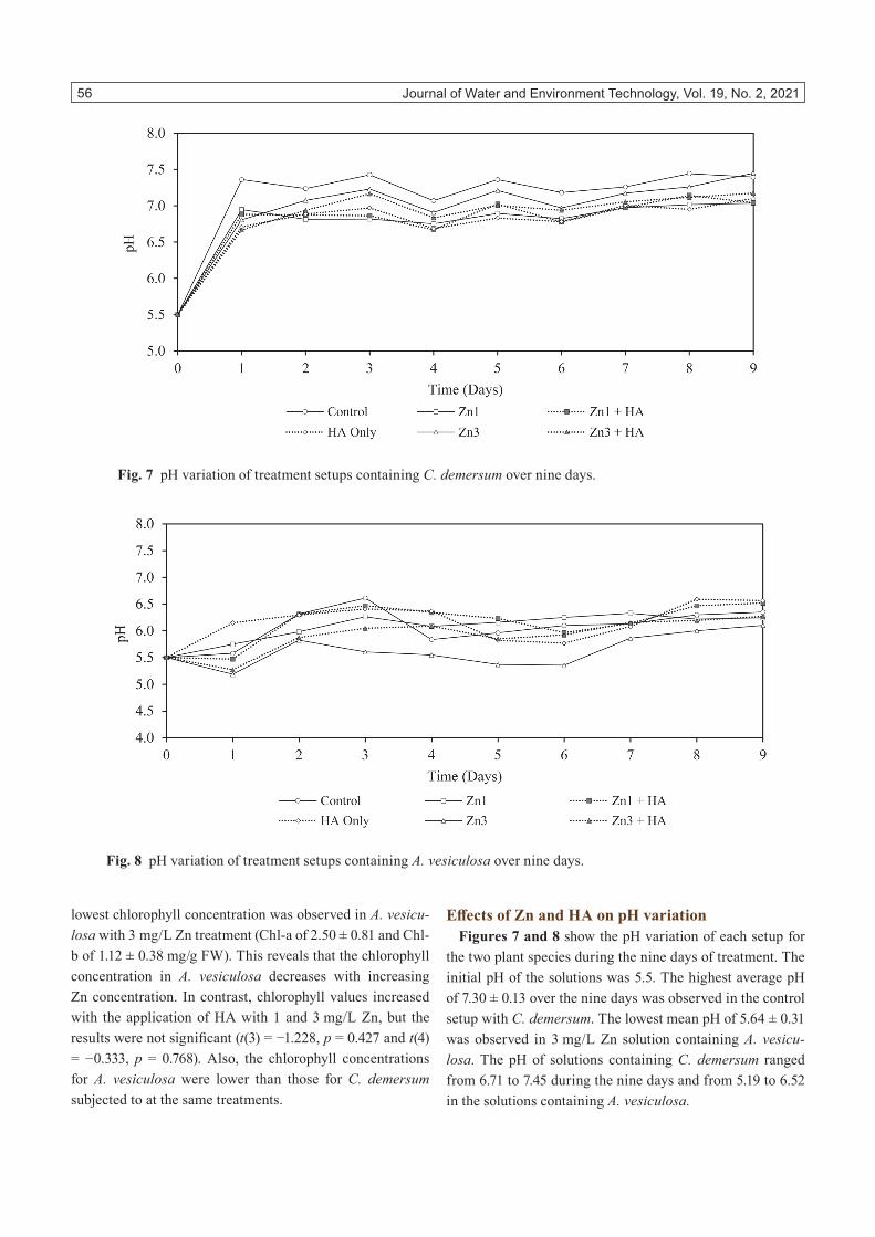
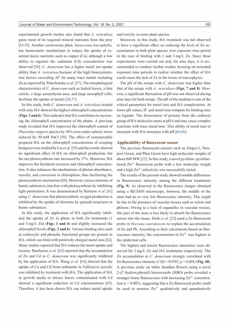
Effects of Zn and HA on pH variationFigures 7 and 8 show the pH variation of each setup for
the two plant species during the nine days of treatment. The initial pH of the solutions was 5.5. The highest average pH of 7.30 ± 0.13 over the nine days was observed in the control setup with C. demersum. The lowest mean pH of 5.64 ± 0.31 was observed in 3 mg/L Zn solution containing A. vesicu-losa. The pH of solutions containing C. demersum ranged from 6.71 to 7.45 during the nine days and from 5.19 to 6.52 in the solutions containing A. vesiculosa.
Fig. 7 pH variation of treatment setups containing C. demersum over nine days.
Fig. 8 pH variation of treatment setups containing A. vesiculosa over nine days.

Journal of Water and Environment Technology, Vol. 19, No. 2, 2021 57
Correlation between Zn fluorescence intensity and Zn accumulation
C. demersum plants were used to study the Zn2+-selective fluorescent probe and determine the Zn fluorescence intensi-ty. Figures 9 (a) and (b) show conventional microscopic and fluorescence cross-sectional images of C. demersum at dif-
ferent treatment stages respectively. The highest fluorescence intensity was observed in the plant tissue sample treated with 3 mg/L Zn. In contrast, the treatment with only HA resulted in the lowest fluorescence intensity. As observed in the fluo-rescence images, Zn was distributed from epidermis cells to the cortex of the stem. No or very low fluorescence intensity
Fig. 9 Cross sections of C. demersum plants subjected to different treatments. (a) Conventional microscopic images and (b) Zn fluorescence images (captured using a BZ-X810 fluorescence microscope at 22.2 × magnification).

Journal of Water and Environment Technology, Vol. 19, No. 2, 2021 58
was observed in the middle of the stem.As shown in Fig. 10, a linear plot was developed using the
average values of the fluorescence intensities against the Zn accumulation in C. demersum plant tissues to determine the slope. The Zn fluorescence intensity increased with level of Zn accumulation in C. demersum. There was a strong posi-tive correlation between fluorescence intensity (A.U.F.) and Zn accumulation in C. demersum(r(4) = 0.988, p < 0.001). The average fluorescence intensity and Zn accumulation values were used to build-up the correlation graph. The limit of detection was 0.64 mg/g DW, was calculated using the following equation (4) [48].
( ) SDLimit of detection LOD 3
S= ×
(4)
SD: standard deviationS: slope between fluorescence intensity and Zn accumulation
DISCUSSION
Impact of HA on Zn accumulation in aquatic plantsPlants can tolerate low concentrations of Zn as it is an es-
sential micronutrient for metabolic activities [1]. Based on initial and final chlorophyll concentrations, we found that the plants fed with 1 mg/L Zn exhibited normal growth similarly to the plants in the control experiment (Figs. 3 and 6). No toxicity symptoms or reductions in chlorophyll concentra-
tions were observed at 1 mg/L Zn. According to Samreen et al. [49], Zn acts as a structural and catalytic component of enzymes, proteins and as co-factor for usual development of photosynthetic pigment biosynthesis. However, excess Zn levels in water can be toxic to aquatic plants [50]. The highest average Zn accumulation after nine days was observed in the plants fed with 3 mg/L Zn. The yellowing of leaves was one of the toxicity symptoms after the sixth day observed in both plants treated with 3 mg/L Zn. In addition, both C. demersum and A. vesiculosa showed the lowest Chl-a and Chl-b concentrations after this treatment (Figs. 3 and 6). This reduction of photosynthetic pigments is occurred owing to the inhibition of the reductive steps in the biosynthetic pathways of photosynthetic pigments as a result of the high redox potential of many trace metals [51]. A similar result was reported by Mishra and Tripathi [52] for water hyacinth (Eichhornia crassipes) treated with chromium (Cr) and Zn solutions with different concentrations. The highest chlo-rophyll concentration of E. crassipes was reported for a Zn concentration of 1 mg/L and the chlorophyll concentration decreased with increasing Zn concentration. Jayasri and Suthindhiran [53] showed that Lemna minor species can tolerate a high Zn concentration of 10 mg/L without showing any toxicity symptoms. However, the Chl-a concentration started to decrease above a Zn concentration of 5 mg/L.
The present study showed that the Zn accumulation is lower in A. vesiculosa than in C. demersum. This is because A. vesiculosa is entirely dependent on the prey, and previous
Fig. 10 Correlation between fluorescence intensity and level of Zn accumulation in C. demersum (The fluorescence inten-sity was calculated using the ImageJ software). Error bars represent the standard deviation (n = 3).
Journal of Water and Environment Technology, Vol. 19, No. 2, 2021 59
experimental growth studies also found that A. vesiculosa gains most of its required mineral nutrients from the prey [54,55]. Another carnivorous plant, Saraccenia leucophylla, has homeostatic mechanisms to reduce the uptake of es-sential micro nutrients such as copper (Cu), although a low ability to regulate the cadmium (Cd) concentration was observed [56]. C. demersum has a higher metal ion uptake ability than A. vesiculosa because of the high bioaccumula-tion factors exceeding 103 for many trace metals including Zn as reported by Polechońska et al. [57]. The morphological characteristics of C. demersum such as forked leaves, a thin cuticle, a large aerenchyma area, and large mesophyll cells, facilitate the uptake of metals [36,37].
In this study, both C. demersum and A. vesiculosa treated with only HA showed the highest chlorophyll concentrations (Figs. 3 and 6). This indicates that HA contributes to increas-ing the chlorophyll concentration of the plants. A previous study revealed that HA improved the chlorophyll content of Phaseolus vulgaris species by 30% even under salinity stress induced by 50 mM NaCl [58]. The effect of commercially prepared HA on the chlorophyll concentration of creeping bentgrass was studied by Liu et al. [59] and the results showed no significant effect of HA on chlorophyll production, but the net photosynthesis rate increased by 17%. Moreover, HA improves the thylakoid structure and chlorophyll concentra-tion. It also enhances the mechanisms of photon absorbance, transfer, and conversion in chloroplasts, thus facilitating the photosynthesis mechanism [60]. However, excess amounts of humic substances interfere with photosynthesis by inhibiting light penetration. It was demonstrated by Reitsem et al. [61] using C. demersum that photosynthetic oxygen production is inhibited by the uptake of electrons by quinoid structures in humic substances.
In this study, the application of HA significantly inhib-ited the uptake of Zn in plants in both Zn treatments (1 and 3 mg/L Zn) (Figs. 1 and 4) and slightly increased the chlorophyll levels (Figs. 3 and 6). Various binding sites such as carboxylic and phenolic functional groups are present in HA, which can bind with positively charged metal ions [62]. Many studies reported that HA reduces the metal uptake and toxicity. Bunluesin et al. [63] reported that the accumulation of Zn and Cd in C. demersum was significantly inhibited by the application of HA. Wang et al. [64] showed that the uptake of Cu and Cd from sediments in Vallisneria spiralis was inhibited by treatment with HA. The application of HA in growth media to lettuce leaves contaminated with Cd showed a significant reduction in Cd concentrations [65]. Therefore, it has been shown HA can reduce metal uptake
and toxicity in some plant species.Moreover, in this study, HA treatment was not observed
to have a significant effect on reducing the level of Zn ac-cumulation in both plant species over exposure time period in the case of feeding with 1 and 3 mg/L Zn. Since these experiments were carried out only for nine days, it is rec-ommended to conduct further studies focusing on extended exposure time periods to realize whether the effect of HA could cause the lack of Zn in the tissue of macrophytes.
The pH of the setups with C. demersum was higher than that of the setups with A. vesiculosa (Figs. 7 and 8). How-ever, a significant fluctuation of pH was not observed during nine days for both setups. The pH of the medium is one of the critical parameters for metal ions and HA complexation. At lower pH values, H+ and metal ions compete for binding sites on ligands. The dissociation of protons from the carboxyl group of HA molecules starts at pH 6 and may cause complex reactions with trace metal ions. This ability of metal ions to associate with HA increases with pH [64,66].
Applicability of fluorescent sensorThe previous fluorescent sensors such as Zinpyr-1, New-
port Green, and Phen Green have high molecular weights of about 800 MW [25]. In this study, a novel pyridine–pyridine-based Zn2+ fluorescent probe with a low molecular weight and a high Zn2+ selectivity was successfully tested.
The results of the present study showed notable differences in fluorescence intensity among the different treatments (Fig. 9). As observed in the fluorescence images obtained using a BZ-X810 microscope, however, the middle of the stem had no or very low fluorescence intensity. This might be due to the presence of vascular tissues such as xylem and phloem. Owing to a lack of organelles in vascular tissues, this part of the stem is less likely to absorb the fluorescence sensor into the tissue. Dinh et al. [25] used a Zn fluorescent probe in Noccaea caerulescens to explain the accumulation of Zn and Pb. According to their calculations based on fluo-rescence intensity, the concentration of Zn2+ was highest in the epidermal cells.
The highest and lowest fluorescence intensities were ob-served for 3 mg/L Zn and HA treatments respectively. The Zn accumulation in C. demersum strongly correlated with Zn fluorescence intensity (r2(4) = 0.9767, p < 0.001) (Fig. 10). A previous study on white dianthus flowers using a novel 2-(2’-hydroxyphenyl) benzoxazole (HBO) probe revealed a stronger linear fluorescence with increasing Zn2+ concentra-tion (r = 0.985), suggesting that a Zn fluorescent probe could be used to monitor Zn2+ qualitatively and quantitatively

Journal of Water and Environment Technology, Vol. 19, No. 2, 2021 60
[67]. Another study carried out with a di (2-picolyl) amine (DPA)-based Zn fluorescent probe using HeLa cells showed a strong positive correlation between fluorescence intensity and Zn2+ concentration (r = 0.996) [68]; the fluorescence intensity and Zn2+ concentration in water samples (lake, river, and distilled water) exhibited good linear properties, indicating that the pyrene-based probe can also be used for determining Zn2+ concentration in water analysis [30]. Therefore, it has been found that the fluorescence intensity positively correlates with the Zn concentration. However, high HA concentrations could cause interferences with the observation and measurement of Zn fluorescence owing to their fluorescence characteristics.
AAS, AES, and ICP-MS are advanced techniques com-monly used for trace metal quantification however these techniques generally require complex sample pre-prepara-tion and expensive instrumentation with trained personnel, and are time-consuming [23,24]. The widely used methods to observe cellular and tissue-level trace metal distributions are SEM-EDS including TEM or SEM coupled with EDS, and other methods such as PIXE and SIMS, are not suitable for the analysis of trace metals in fresh plant tissue samples due to many limitations such as insufficient sensitivity, radiation damages, requirement of pretreatments and high vacuum conditions [25]. Therefore, this Zn2+ fluorescent probe can be developed as a useful tool for determining the Zn accumulation in the tissues of aquatic plants because it has many advantages such as quantifying the level of Zn2+ in specific cell types, ease of use, measuring the tissue-level Zn2+ indirectly without dealing with many chemical analy-ses procedures, freezing or freeze drying like pretreatments, high vacuum conditions, trained personnel are not required. Therefore, this tool has the potential to measure the sensi-tivity of growth at the cellular level Zn compared to water quality environmental standards with respect to Zn response in plants.
CONCLUSIONS
The application of HA significantly inhibited the uptake of Zn in both plants owing to the formation of Zn-HA com-plexes. Accordingly, the chlorophyll concentrations slightly increased and no toxicity symptoms were observed. The Zn fluorescence intensity positively correlated with the Zn ac-cumulation in C. demersum. Consequently, the fluorescent probe can be developed as a useful tool for the diagnosis of Zn osmosis into a plant cell or tissue on the basis of fluores-cence intensity. However, further studies are recommended
to enhance the detection limit and sensitivity of this tool.
ACKNOWLEDGEMENTS
We thank to the anonymous reviewers for improving the manuscript. We also thank Professor Yasuko Kaneko of Saitama University (SU) for providing cultured A.vesiculosa for this study. This study was partially supported by the Research Center for Sustainable Development in East Asia (SU-RCSDEA).
REFERENCES
[1] Chibuike GU, Obiora SC: Heavy metal pol-luted soils: Effect on plants and bioremediation methods. Appl. Environ. Soil Sci., 2014, 1–12, 2014. doi:10.1155/2014/752708
[2] Bonnet M, Camares O, Veisseire P: Effects of zinc and influence of Acremonium lolii on growth parameters, chlorophyll a fluorescence and antioxidant enzyme activities of ryegrass (Lolium perenne L. cv Apollo). J. Exp. Bot., 51(346), 945–953, 2000. PMID:10948221 doi:10.1093/jxb/51.346.945
[3] Ibrahim N, Aziz HA: Trends on natural organic matter in drinking water sources and its treatment. Interna-tional Journal of Scientific Research in Environmental Sciences, 2(3), 94–106, 2014. doi:10.12983/ijsres-2014-p0094-0106
[4] Ministry of the Environment: Environmental Quality Standards for Water Pollution. https://www.env.go.jp/en/water/wq/wp.pdf [accessed in June, 2020]
[5] US EPA: National Recommended Water Quality Cri-teria. United States Environmental Protection Agency, Office of Water, Office of Science and Technology, Washington, DC, USA, 2009.
[6] Ryan AC, Tomasso JR, Klaine SJ: Influence of pH, hardness, dissolved organic carbon concentration, and dissolved organic matter source on the acute toxicity of copper to Daphnia magna in soft waters: implica-tions for the biotic ligand model. Environ. Toxicol. Chem., 28(8), 1663–1670, 2009. PMID:19265455 doi:10.1897/08-361.1
[7] Li XF, Wang PF, Feng CL, Liu DQ, Chen JK, Wu FC: Acute toxicity and hazardous concentrations of zinc to native freshwater organisms under different pH values in China. Bull. Environ. Contam. Toxicol., 103(1), 120–126, 2019. PMID:30250971 doi:10.1007/s00128-018-2441-2

Journal of Water and Environment Technology, Vol. 19, No. 2, 2021 61
[8] Boguta P, Sokołowska Z: Interactions of Zn(II) ions with humic acids isolated from various type of soils. Effect of pH, Zn concentrations and humic acids chemical properties. PLoS One, 11(4), e0153626, 2016. doi:10.1371/journal.pone.0153626
[9] Gaffney JS, Marley NA, Clark SB: Humic and Fulvic Acids, Isolation, Structure, and Environmental Role. American Chemical Society, Washington, DC, USA, 1996. doi:10.1021/bk-1996-0651.fw001
[10] Sachse A, Henrion R, Gelbrecht J, Steinberg CEW: Classification of dissolved organic carbon (DOC) in river systems: Influence of catchment characteristics and autochthonous processes. Org. Geochem., 36(6), 923–935, 2005. doi:10.1016/j.orggeochem.2004.12.008
[11] Misra V, Pandey SD, Viswanathan PN: Environ-mental significance of humic acid in the sequestra-tion of metals. Chem. Ecol., 13(2), 103–112, 1996. doi:10.1080/02757549608035523
[12] Stevenson FJ: Humus Chemistry: Genesis, Composi-tion, Reactions, 2nd Edition. John Wiley & Sons, Hoboken, USA, 1994.
[13] Arifur Rahman M, Abu Hasan M, Rahim A, Shafiqul Alam AM: Characterization of humic acid from the river bottom sediments of Burigonga: Complexation studies of metals with humic acid. J. Anal. Environ. Chem., 11(1), 42–52, 2010.
[14] Pehlivan E, Arslan G: Uptake of metal ions on humic ac-ids. Energy Sources A Recovery Util. Environ. Effects, 28(12), 1099–1112, 2006. doi:10.1080/009083190910451
[15] Abate G, Masini JC: Acid-basic and complexation properties of a sedimentary humic acid. A study on the Barra Bonita reservoir of Tietê river, São Paulo State, Brazil. J. Braz. Chem. Soc., 12(1), 109–116, 2001. doi:10.1590/S0103-50532001000100015
[16] Garcia-Mina JM: Stability, solubility and maximum metal binding capacity in metal–humic complexes involving humic substances extracted from peat and organic compost. Org. Geochem., 37(12), 1960–1972, 2006. doi:10.1016/j.orggeochem.2006.07.027
[17] Rong Q, Zhong K, Huang H, Li C, Zhang C, Nong X: Humic acid reduces the available cadmium, copper, lead, and zinc in soil and their uptake by Tobacco. Appl. Sci. (Basel), 10(3), 1077, 2020. doi:10.3390/app10031077
[18] Vargas C, Pérez-Esteban J, Escolástico C, Masaguer A, Moliner A: Phytoremediation of Cu and Zn by vetiver grass in mine soils amended with humic acids. Envi-ron. Sci. Pollut. Res. Int., 23(13), 13521–13530, 2016. PMID:27030238 doi:10.1007/s11356-016-6430-x
[19] Ali H, Khan E, Sajad MA: Phytoremediation of heavy metals—Concepts and applications. Chemosphere, 91(7), 869–881, 2013. PMID:23466085 doi:10.1016/j.chemosphere.2013.01.075
[20] Xing W, Wu H, Hao B, Liu G: Metal accumulation by submerged macrophytes in eutrophic lakes at the watershed scale. Environ. Sci. Pollut. Res. Int., 20(10), 6999–7008, 2013. PMID:23749202 doi:10.1007/s11356-013-1854-z
[21] Sharma S, Singh B, Manchanda VK: Phytoremedia-tion: role of terrestrial plants and aquatic macrophytes in the remediation of radionuclides and heavy metal contaminated soil and water. Environ. Sci. Pollut. Res. Int., 22(2), 946–962, 2015. PMID:25277712 doi:10.1007/s11356-014-3635-8
[22] Bulska E, Ruszczyńska A: Analytical techniques for trace element determination. Phys. Sci. Rev., 2(5), 1–14, 2017.
[23] Quang DT, Kim JS: Fluoro- and chromogenic chemo-dosimeters for heavy metal ion detection in solution and biospecimens. Chem. Rev., 110(10), 6280–6301, 2010. PMID:20726526 doi:10.1021/cr100154p
[24] Bansod B, Kumar T, Thakur R, Rana S, Singh I: A re-view on various electrochemical techniques for heavy metal ions detection with different sensing platforms. Biosens. Bioelectron., 94(January), 443–455, 2017. PMID:28340464 doi:10.1016/j.bios.2017.03.031
[25] Dinh N, van der Ent A, Mulligan DR, Nguyen AV: Zinc and lead accumulation characteristics and in vivo distribution of Zn2+ in the hyperaccumulator Noccaea caerulescens elucidated with fluorescent probes and laser confocal microscopy. Environ. Exp. Bot., 147(November), 1–12, 2018. doi:10.1016/j.envex-pbot.2017.10.008
[26] Hagimori M, Temma T, Mizuyama N, Uto T, Yama-guchi Y, Tominaga Y, Mukai T, Saji H: A high-affinity fluorescent Zn2+ sensor improved by the suppression of pyridine-pyridone tautomerism and its application in living cells. Sens. Actuators B Chem., 213(July), 45–52, 2015. doi:10.1016/j.snb.2015.02.063

Journal of Water and Environment Technology, Vol. 19, No. 2, 2021 62
[27] Hu Y, Liu Y, Kim G, Jun EJ, Swamy KMK, Kim Y, Kim SJ, Yoon J: Pyrene based fluorescent probes for detecting endogenous zinc ions in live cells. Dyes Pig-ments, 113(February), 372–377, 2015. doi:10.1016/j.dyepig.2014.09.010
[28] Guo Z, Kim GH, Shin I, Yoon J: A cyanine-based fluorescent sensor for detecting endogenous zinc ions in live cells and organisms. Biomaterials, 33(31), 7818–7827, 2012. PMID:22871424 doi:10.1016/j.bio-materials.2012.07.014
[29] Sinclair SA, Sherson SM, Jarvis R, Camakaris J, Cob-bett CS: The use of the zinc-fluorophore, Zinpyr-1, in the study of zinc homeostasis in Arabidopsis roots. New Phytol., 174(1), 39–45, 2007. PMID:17335495 doi:10.1111/j.1469-8137.2007.02030.x
[30] Tang Y, Huang Y, Lu L, Wang C, Sun T, Zhu J, Zhu G, Pan J, Jin Y, Liu A, Wang M: Synthesis of a new pyrene-devived fluorescent probe for the detection of Zn2+. Tetrahedron Lett., 59(44), 3916–3922, 2018. doi:10.1016/j.tetlet.2018.09.038
[31] Hagimori M, Mizuyama N, Yamaguchi Y, Saji H, Tominaga Y: A novel small molecule fluorescent sensor for Zn2+ based on pyridine–pyridone scaffold. Talanta, 83(5), 1730–1735, 2011. PMID:21238776 doi:10.1016/j.talanta.2010.12.003
[32] Hagimori M, Mizuyama N, Tominaga Y, Mukai T, Saji H: A low-molecular-weight fluorescent sensor with Zn2+ dependent bathochromic shift of emission wavelength and its imaging in living cells. Dyes Pig-ments, 113(February), 205–209, 2015. doi:10.1016/j.dyepig.2014.07.032
[33] Timpson P, McGhee EJ, Anderson KI: Imaging molecular dynamics in vivo – from cell biology to animal models. J. Cell Sci., 124(17), 2877–2890, 2011. PMID:21878495 doi:10.1242/jcs.085191
[34] Lichtman JW, Conchello JA: Fluorescence microscopy. Nat. Methods, 2(12), 910–919, 2005. PMID:16299476 doi:10.1038/nmeth817
[35] Carter KP, Young AM, Palmer AE: Fluorescent sen-sors for measuring metal ions in living systems. Chem. Rev., 114(8), 4564–4601, 2014. PMID:24588137 doi:10.1021/cr400546e
[36] Borisova G, Chukina N, Maleva M, Prasad MNV: Ceratophyllum demersum L. and Potamogeton alpinus Balb. from Iset’ river, Ural region, Russia differ in adap-tive strategies to heavy metals exposure--a comparative study. Int. J. Phytoremediation, 16(6), 621–633, 2014. PMID:24912247 doi:10.1080/15226514.2013.803022
[37] Aravind P, Prasad MNV: Cadmium-Zinc interactions in a hydroponic system using Ceratophyllum demer-sum L.: adaptive ecophysiology, biochemistry and molecular toxicology. Braz. J. Plant Physiol., 17(1), 3–20, 2005. doi:10.1590/S1677-04202005000100002
[38] Adamec L: Ecophysiological study of the aquatic carnivorous plant Aldrovanda vesiculosa L. Acta Bot. Gallica, 142(6), 681–684, 1995. doi:10.1080/12538078.1995.10515292
[39] Adlassnig W, Peroutka M, Lambers H, Lichtscheidl IK: The roots of carnivorous plants. Plant and Soil, 274(1), 127–140, 2005. doi:10.1007/s11104-004-2754-2
[40] Hoagland DR, Arnon DI: The Water-Culture Method for Growing Plants without Soil, College of Agric., Univ. of California in Berkeley, California, USA, pp. 1–32, 2nd ed., 1950.
[41] Bazrafshan E, Biglari H, Mahvi AH: Humic acid removal from aqueous environments by electrocoagu-lation process using iron electrodes. E-J. Chem., 9(4), 2453–2461, 2012. doi:10.1155/2012/876739
[42] AWWA-American Water Works Association: Standard Methods for the Examination of Water and Wastewa-ter, 23rd edition. American Public Health Association, Washington DC, USA, 2017.
[43] Hou W, Chen X, Song G, Wang Q, Chi Chang C: Effects of copper and cadmium on heavy metal pol-luted waterbody restoration by duckweed (Lemna minor). Plant Physiol. Biochem., 45(1), 62–69, 2007. PMID:17300947 doi:10.1016/j.plaphy.2006.12.005
[44] Arnon DI: Copper enzymes in isolated chloroplasts. polyphenoloxidase in Beta vulgaris. Plant Physiol., 24(1), 1–15, 1949. PMID:16654194 doi:10.1104/pp.24.1.1
[45] Yang L, Li Y, Xj G, Ma X, Yan Q: Comparison of dry ashing, wet ashing and microwave digestion for deter-mination of trace elements in Periostracum serpentis and Periostracum cicadae by ICP-AES. J. Chil. Chem. Soc., 58(3), 1876–1879, 2013. doi:10.4067/S0717-97072013000300018
[46] Fontenete S, Carvalho D, Lourenço A, Guimarães N, Madureira P, Figueiredo C, Azevedo NF: FISHji: New ImageJ macros for the quantification of fluorescence in epifluorescence images. Biochem. Eng. J., 112(August), 61–69, 2016. doi:10.1016/j.bej.2016.04.001
[47] Fitzpatrick M: Measuring cell fluorescence using Im-ageJ, The Open Lab Book v1.0., 2014. https://theolb.readthedocs.io/en/latest/imaging/measuring-cell-fluorescence-using-imagej.html [accessed in January, 2020].

Journal of Water and Environment Technology, Vol. 19, No. 2, 2021 63
[48] Aziz A, Aboelhasan AE, Sayed MA: A simple fluo-rescent chemosensor for detection of zinc ions in some real samples and intracellular imaging in living cells. J. Braz. Chem. Soc., 31(8), 1635–1647, 2020.
[49] Samreen T, Humaira, Shah HU, Ullah S, Javid M: Zinc effect on growth rate, chlorophyll, protein and mineral contents of hydroponically grown mungbeans plant (Vigna radiata). Arab. J. Chem., 10(May), S1802–S1807, 2017. doi:10.1016/j.arabjc.2013.07.005
[50] Chanu L, Gupta A: Toxicity of zinc on growth of an aquatic macrophyte, Ipomoea aquatica Forsk. Curr. World Environ., 11(1), 218–227, 2016. doi:10.12944/CWE.11.1.27
[51] Chandra R, Kang H: Mixed heavy metal stress on pho-tosynthesis, transpiration rate, and chlorophyll content in poplar hybrids. Forest Sci. Technol., 12(2), 55–61, 2016. doi:10.1080/21580103.2015.1044024
[52] Mishra VK, Tripathi BD: Accumulation of chromium and zinc from aqueous solutions using water hyacinth (Eichhornia crassipes). J. Hazard. Mater., 164(2–3), 1059–1063, 2009. PMID:18938031 doi:10.1016/j.jhazmat.2008.09.020
[53] Jayasri MA, Suthindhiran K: Effect of zinc and lead on the physiological and biochemical properties of aquatic plant Lemna minor: its potential role in phy-toremediation. Appl. Water Sci., 7(3), 1247–1253, 2017. doi:10.1007/s13201-015-0376-x
[54] Adamec L: Seasonal growth dynamics and overwin-tering of the aquatic carnivorous plant Aldrovanda vesiculosa at experimental field sites. Folia Geobot., 34(3), 287–297, 1999. doi:10.1007/BF02912815
[55] Kaminski R: Studies on the ecology of Aldrovanda ve-siculosa LI Ecological differentiation of A. vesiculosa population under the influence of chemical factors in the habitat. Ekol. Pol., 35, 559–590, 1987.
[56] Moody C, Green ID: Assimilation of Cd and Cu by the carnivorous plant Sarracenia leucophylla raf. fed contaminated prey. Environ. Sci. Technol., 44(5), 1610–1616, 2010. PMID:20141102 doi:10.1021/es9019386
[57] Polechońska L, Klink A, Dambiec M, Rudecki A: Eval-uation of Ceratophyllum demersum as the accumulative bioindicator for trace metals. Ecol. Indic., 93(May), 274–281, 2018. doi:10.1016/j.ecolind.2018.05.020
[58] Meganid AS, Al-Zahrani HS, Selim EM: Effect of humic acid application on growth and chlorophyll con-tents of common bean plants (Phaseolus vulgarisL.)under salinity stress conditions. Int. J. Innov. Res. Sci. Eng. Technol., 4(5), 2651–2660, 2015. doi:10.15680/IJIRSET.2015.0405001
[59] Liu C, Cooper RJ, Bowman DC: Humic acid application affects photosynthesis, root development, and nutrient content of creeping bentgrass. HortScience, 33(6), 1023–1025, 1998. doi:10.21273/HORTSCI.33.6.1023
[60] Fan H, Wang X, Sun X, Li Y, Sun X, Zheng C: Ef-fects of humic acid derived from sediments on growth, photosynthesis and chloroplast ultrastructure in chry-santhemum. Sci. Hortic. (Amsterdam), 177(October), 118–123, 2014. doi:10.1016/j.scienta.2014.05.010
[61] Reitsema RE, Meire P, Schoelynck J: The future of freshwater macrophytes in a changing world: dissolved organic carbon quantity and quality and its interactions with macrophytes. Front. Plant Sci., 9(May), 629, 2018. PMID:29868084 doi:10.3389/fpls.2018.00629
[62] Companys E, Puy J, Galceran J: Humic acid complex-ation to Zn and Cd determined with the new electro-analytical technique AGNES. Environ. Chem., 4(5), 347, 2007. doi:10.1071/EN07051
[63] Bunluesin S, Pokethitiyook P, Lanza GR, Tyson JF, Kruatrachue M, Xing B, Upatham S: Influences of cadmium and zinc interaction and humic acid on metal accumulation in Ceratophyllum demersum. Water Air Soil Pollut., 180(1–4), 225–235, 2007. doi:10.1007/s11270-006-9265-0
[64] Wang Q, Li Z, Cheng S, Wu Z: Influence of humic acids on the accumulation of copper and cadmium in Vallisneria spiralis L. from sediment. Environ. Earth Sci., 61(6), 1207–1213, 2010. doi:10.1007/s12665-009-0444-3
[65] Haghighi M, Kafi M, Khoshgoftarmanesh A: Effect of humic acid application on cadmium accumulation by lettuce leaves. J. Plant Nutr., 36(10), 1521–1532, 2013. doi:10.1080/01904167.2013.799182
[66] Pandey AK, Pandey SD, Misra V: Stability constants of metal-humic acid complexes and its role in envi-ronmental detoxification. Ecotoxicol. Environ. Saf., 47(2), 195–200, 2000. PMID:11023698 doi:10.1006/eesa.2000.1947
[67] Shang Y, Zheng S, Tsakama M, Wang M, Chen W: A water-soluble, small molecular fluorescence probe based on 2-(2′-hydroxyphenyl) benzoxazole for Zn2+ in plants. Tetrahedron Lett., 59(45), 4003–4007, 2018. doi:10.1016/j.tetlet.2018.09.057
[68] Yan L, Wen X, Fan Z: A large-Stokes-shift fluorescent probe for Zn2+ based on AIE, and application in live cell imaging. Anal. Bioanal. Chem., 412(6), 1453–1463, 2020. PMID:31901962 doi:10.1007/s00216-019-02378-w