visual outcomes after cataract surgery in diabetic patients: a … · that visual outcomes after...
TRANSCRIPT

November 2017, Vol. 46 No. 11
447
Visual Outcomes after Cataract Surgery in Diabetic Patients: A Meta-Analysis
Dear Editor,Diabetic retinopathy is a common complication of diabetes
which can result in blindness. Studies have generally shown that visual outcomes after cataract surgery in patients with diabetes are worse than in non-diabetics, especially those with diabetic retinopathy.1-3 In patients with diabetic retinopathy, a major cause of unfavourable outcomes after cataract surgery is macular oedema.4 Factors associated with significant macular oedema include the duration of diabetes, glycaemic control, degree of retinopathy, and macular oedema at the time of surgery.4,5 Increased retinopathy progression and increased incidence of macular oedema after cataract surgery have been reported in patients with diabetes,1 while other studies have not reported these findings.6 Some authors suggested that the findings are due to the natural course of diabetes.5,7
To this end, we performed a meta-analysis to determine outcomes of cataract surgery in diabetic patients with diabetic retinopathy as compared to those without retinopathy.
Materials and MethodsMedline, Cochrane, and Google Scholar databases were
searched from inception until January 30, 2015 using combinations of the following keywords: visual outcome, cataract surgery, diabetes, diabetic retinopathy. Inclusion criteria for the meta-analysis were: 1) two-arm studies; 2) one group of eyes that had diabetic retinopathy and another that did not; 3) that both groups of eyes had undergone cataract surgery; 4) quantitative outcomes that had been reported. Single-arm studies and those with patients receiving medical treatment (enoxaparin, ranibizumab, or triamcinolone, etc.) in addition to cataract surgery to prevent macular oedema were excluded. Letters, comments, editorials, case reports, proceedings, and personal communications were also excluded. Studies were identified by the search strategy via two independent reviewers.
Information/data extracted from studies that met the inclusion criteria were: the name of the first author, year of publication, study design, number of participants in each group, participants’ age and gender, presence of retinopathy and the major outcomes. The method described by Hayden et al8 was used to assess the quality of the included studies.
Outcome measures were: 1) percentage of eyes with
postoperative newly developed macular oedema; 2) percent-age of eyes with progressive retinopathy; 3) percentage of eyes with visual acuity reaching 6/12. Odds ratios (ORs) and 95% confidence intervals (95% CIs) were used to represent the effect size of associations between diabetic retinopathy and outcomes. An OR >1 indicated that patients with diabetic retinopathy had greater odds of developing macular oedema and progressive retinopathy compared to those without diabetic retinopathy, and that patients with diabetic retinopathy had greater odds of visual acuity improvement than those without diabetic retinopathy. Heterogeneity among the studies was assessed by the Cochran Q and the I2 statistic. A Q statistic P <0.10 was considered to indicate statistically significant heterogeneity. An I2 statistic ≥50% was considered to indicate large to extreme heterogeneity. Random-effects models were used if heterogeneity was detected (I2 >50% or Q statistics P <0.10). Otherwise, fixed-effects models were used. A two-sided P <0.05 was considered statistically significant. Meta-analysis sensitivity was assessed with the leave-one-out approach. Statistical analyses were performed using Comprehensive Meta-Analysis statistical software, version 2.0 (Biostat, Englewood, NJ, USA).
ResultsA total of 370 articles were identified, and 330 remained
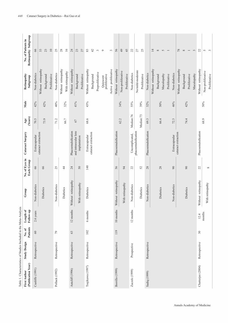
after duplicates were removed. After screening by abstracts and titles, 283 were excluded and the full-texts of 47 articles were reviewed. Thirty-nine articles were excluded for not meeting the inclusion criteria. Ultimately, 8 studies were included in the meta-analysis.1,2,4,9-13 The number of patients in the 8 studies ranged from 30 to 119, and number of eyes ranged from 30 to 180. The mean or median age ranged from 66.4 to 76 years, and the proportion of males ranged from 32% to 79% (Table 1).
There was no evidence of heterogeneity across 3 studies that reported outcomes of newly developed macular oedema. Patients with diabetic retinopathy had higher odds of developing macular oedema than those without diabetic retinopathy (pooled OR = 5.912, 95% CI: 2.723 to 12.839, P <0.001) (Fig. 1a).
There was no evidence of heterogeneity across 5 studies that reported outcomes of progressive retinopathy. Patients
Cataract Surgery in Diabetics—Rui Guo et al
Letter to the Editor
448
Annals Academy of Medicine
Tabl
e 1.
Cha
ract
eris
tics o
f Stu
dies
Incl
uded
in th
e M
eta-
Ana
lysi
s
Firs
t Aut
hor
(Pub
licat
ion
Year
)St
udy
Des
ign
No.
of
Patie
nts
Len
gth
of
Follo
w-u
pG
roup
No.
of E
yes i
n E
ach
Gro
upC
atar
act S
urge
ryA
ge
(Yea
rs)
Mal
eR
etin
opat
hy
Subg
roup
No.
of P
atie
nts i
n R
etin
opat
hy S
ubgr
oup
Cun
liffe
(199
1)R
etro
spec
tive
602.
6 ye
ars
Non
-dia
betic
s
Dia
betic
s
66 66
Extra
caps
ular
ca
tara
ct e
xtra
ctio
n70
.542
%N
on-d
iabe
tics
66
73.9
42%
With
out
retin
opat
hy33
Bac
kgro
und
23
Prol
ifera
tive
10
Polla
ck (1
992)
Ret
rosp
ectiv
e79
Non
-dia
betic
s
Dia
betic
s
25 44
71.2
40%
Non
-dia
betic
s25
66.7
32%
With
out
retin
opat
hy28
With
retin
opat
hy16
Ant
cliff
(199
6)R
etro
spec
tive
6312
mon
ths
With
out
retin
opat
hy
With
retin
opat
hy
24 50
Phac
oem
ulsifi
catio
n an
d in
traoc
ular
lens
im
plan
tatio
n
6741
%W
ithou
t re
tinop
athy
24
Bac
kgro
und
23
Prol
ifera
tive
27
Tsuj
ikaw
a (1
997)
Ret
rosp
ectiv
e10
26
mon
ths
Dia
betic
s14
0Ex
traca
psul
ar
cata
ract
ext
ract
ion
68.6
43%
With
out
retin
opat
hy83
Bac
kgro
und
42
Prep
rolif
erat
ive
3
Qui
esce
nt
prol
ifera
tive
9
Act
ive
pro
lifer
ativ
e3
Bor
rillo
(199
9)R
etro
spec
tive
119
10 m
onth
sW
ithou
t re
tinop
athy
With
retin
opat
hy
56 94
Phac
oem
ulsifi
catio
n62
.234
%W
ithou
t re
tinop
athy
56
Non
-pro
lifer
ativ
e49
Prol
ifera
tive
45
Zacz
ek (1
999)
Pros
pect
ive
12 m
onth
sN
on-d
iabe
tics
22U
ncom
plic
ated
, ph
acoe
mul
sifica
tion
Med
ian:
7655
%N
on-d
iabe
tics
22
Dia
betic
s52
Med
ian:
7339
%N
o/m
ild-m
oder
ate
27
Prol
ifera
tive
25
Sadi
q (1
999)
Ret
rosp
ectiv
eN
on-d
iabe
tics
28Ph
acoe
mul
sifica
tion
69.3
32%
Non
-dia
betic
s
Dia
betic
s28
66.4
50%
With
out
retin
opat
hy14
Bac
kgro
und
8
Mac
ulop
athy
5
Prol
ifera
tive
1
Non
-dia
betic
s90
Extra
caps
ular
ca
tara
ct e
xtra
ctio
n72
.346
%N
on-d
iabe
tics
Dia
betic
s90
74.4
42%
With
out
retin
opat
hy78
Bac
kgro
und
8
Prol
ifera
tive
1
Mac
ulop
athy
3
Cha
tterje
e (2
004)
Ret
rosp
ectiv
e30
12.4
m
onth
sW
ithou
t re
tinop
athy
With
retin
opat
hy
22 8
Phac
oem
ulsifi
catio
n68
.950
%W
ithou
t re
tinop
athy
22
Non
-pro
lifer
ativ
e5
Prol
ifera
tive
3
Cataract Surgery in Diabetics—Rui Guo et al

November 2017, Vol. 46 No. 11
449
Tabl
e 1.
Cha
ract
eris
tics o
f Stu
dies
Incl
uded
in th
e M
eta-
Ana
lysi
s
Firs
t Aut
hor
(Pub
licat
ion
Year
)St
udy
Des
ign
No.
of
Patie
nts
Len
gth
of
Follo
w-u
pG
roup
No.
of E
yes i
n E
ach
Gro
upC
atar
act S
urge
ryA
ge
(Yea
rs)
Mal
eR
etin
opat
hy
Subg
roup
No.
of P
atie
nts i
n R
etin
opat
hy S
ubgr
oup
Cun
liffe
(199
1)R
etro
spec
tive
602.
6 ye
ars
Non
-dia
betic
s
Dia
betic
s
66 66
Extra
caps
ular
ca
tara
ct e
xtra
ctio
n70
.542
%N
on-d
iabe
tics
66
73.9
42%
With
out
retin
opat
hy33
Bac
kgro
und
23
Prol
ifera
tive
10
Polla
ck (1
992)
Ret
rosp
ectiv
e79
Non
-dia
betic
s
Dia
betic
s
25 44
71.2
40%
Non
-dia
betic
s25
66.7
32%
With
out
retin
opat
hy28
With
retin
opat
hy16
Ant
cliff
(199
6)R
etro
spec
tive
6312
mon
ths
With
out
retin
opat
hy
With
retin
opat
hy
24 50
Phac
oem
ulsifi
catio
n an
d in
traoc
ular
lens
im
plan
tatio
n
6741
%W
ithou
t re
tinop
athy
24
Bac
kgro
und
23
Prol
ifera
tive
27
Tsuj
ikaw
a (1
997)
Ret
rosp
ectiv
e10
26
mon
ths
Dia
betic
s14
0Ex
traca
psul
ar
cata
ract
ext
ract
ion
68.6
43%
With
out
retin
opat
hy83
Bac
kgro
und
42
Prep
rolif
erat
ive
3
Qui
esce
nt
prol
ifera
tive
9
Act
ive
pro
lifer
ativ
e3
Bor
rillo
(199
9)R
etro
spec
tive
119
10 m
onth
sW
ithou
t re
tinop
athy
With
retin
opat
hy
56 94
Phac
oem
ulsifi
catio
n62
.234
%W
ithou
t re
tinop
athy
56
Non
-pro
lifer
ativ
e49
Prol
ifera
tive
45
Zacz
ek (1
999)
Pros
pect
ive
12 m
onth
sN
on-d
iabe
tics
22U
ncom
plic
ated
, ph
acoe
mul
sifica
tion
Med
ian:
7655
%N
on-d
iabe
tics
22
Dia
betic
s52
Med
ian:
7339
%N
o/m
ild-m
oder
ate
27
Prol
ifera
tive
25
Sadi
q (1
999)
Ret
rosp
ectiv
eN
on-d
iabe
tics
28Ph
acoe
mul
sifica
tion
69.3
32%
Non
-dia
betic
s
Dia
betic
s28
66.4
50%
With
out
retin
opat
hy14
Bac
kgro
und
8
Mac
ulop
athy
5
Prol
ifera
tive
1
Non
-dia
betic
s90
Extra
caps
ular
ca
tara
ct e
xtra
ctio
n72
.346
%N
on-d
iabe
tics
Dia
betic
s90
74.4
42%
With
out
retin
opat
hy78
Bac
kgro
und
8
Prol
ifera
tive
1
Mac
ulop
athy
3
Cha
tterje
e (2
004)
Ret
rosp
ectiv
e30
12.4
m
onth
sW
ithou
t re
tinop
athy
With
retin
opat
hy
22 8
Phac
oem
ulsifi
catio
n68
.950
%W
ithou
t re
tinop
athy
22
Non
-pro
lifer
ativ
e5
Prol
ifera
tive
3
Fig. 1. Forest plots for association of diabetic retinopathy on (a) newly developed macular oedema, (b) progressive retinopathy and (c) visual acuity reaching 6/12.
with diabetic retinopathy had higher odds of developing progressive retinopathy than those without diabetic retinopathy (pooled OR = 5.282, 95% CI: 3.051 to 9.144, P <0.001) (Fig. 1b).
There was no evidence of heterogeneity across 4 studies that reported visual acuity outcomes. Patients with diabetic retinopathy had lower odds of visual acuity reaching 6/12 than those without retinopathy (pooled OR = 0.217, 95% CI: 0.122 to 0.385, P <0.001) (Fig. 1c).
Sensitivity analyses showed that the magnitude and direction of the outcomes did not change considerably, indicating that there was no single study that had a significant
impact on the pooled results of any of the outcomes (Fig. 2). Quality assessment demonstrated a low risk of bias in both study participation and study attrition. A moderate risk was estimated in prognostic factor measurement, outcome measurement and analysis. All the studies showed a high risk of bias in confounding measurement and account.
DiscussionThis meta-analysis showed that patients with diabetic
retinopathy had higher odds of developing macular oedema, progressive retinopathy, and lower odds of achieving visual acuity of 6/12 after cataract surgery. Only one prior meta-analysis performed in 1995 examined outcomes of
Cataract Surgery in Diabetics—Rui Guo et al

450
Annals Academy of Medicine
Fig. 2. Sensitivity analysis for association of diabetic retinopathy on (a) newly developed macular oedema, (b) progressive retinopathy and (c) visual acuity reaching 6/12.
cataract surgery in patients with diabetes, and the study found that the severity of retinopathy and maculopathy prior to cataract surgery were the major factors affecting postoperative visual acuity.3
Dowler et al14 reported that phacoemulsification was associated with better visual outcomes, less need for capsulotomy, and less inflammation as compared to extracapsular extraction in patients with diabetes. While visual outcomes in diabetic patients with or without minimal retinopathy are similar to those without diabetes, postoperative visual acuity may be less than optimal in patients with significant retinopathy.13 In eyes without macular oedema at the time of surgery, the occurrence of postoperative macular oedema tends to resolve
spontaneously. In cases where macular oedema persists, it may represent the natural course of diabetes rather than the effect of surgery.5 Clinically significant macular oedema at the time of cataract surgery, however, is unlikely to resolve postoperatively.5 Krepler et al7 reported poorer visual outcomes in patients developing macular oedema after phacoemulsification and posterior chamber lens implantation. Eriksson et al15 reported final visual outcomes in eyes with mild to moderate retinopathy without previous macular oedema were as good as that in normal eyes.
Diabetic retinopathy can progress after intracapsular and extracapsular cataract extraction.16 On the other hand, some studies have shown similar results with phacoemulsification, while others have reported no effect on retinopathy as a result
Cataract Surgery in Diabetics—Rui Guo et al

November 2017, Vol. 46 No. 11
451
1Guangdong Eye Institute, Department of Ophthalmology, Guangdong General Hospital, Guangdong Academy of Medical Sciences, People’s Republic of China2Department of Ophthalmology, Guangzhou Hospital of TCM, People’s Republic of China
Address for Correspondence: Dr Yang Xiaohong, Guangdong Eye Institute, Department of Ophthalmology, Guangdong General Hospital, Guangdong Academy of Medical Sciences, 106 Zhongshan Er Road, Guangzhou, 510080, Guangdong, People’s Republic of China. Email: [email protected]
Rui Guo, 1PhD, Xiaohong Yang, 1MD, Xiaoyan Xie, 2MD
of phacoemulsification.7 Differences in outcomes may be attributable to different criteria used to define progressive retinopathy. Two prospective studies of phacoemulsification in patients with diabetes indicated the procedure does not accelerate diabetic retinopathy, and progression is likely the result of the natural course of the diabetes.5
The results of this study must be interpreted with caution due to a number of limitations. The number of studies was only 8 and the total number of patients was small. All 8 studies did not address all outcomes, and all the studies were performed in the 1990s with the exception of one performed in 2004. Data from older studies may not necessarily reflect current outcomes due to advances in surgical techniques. The surgical approach and patient grouping and selection varied between studies, and different surgical approaches are associated with different results. We did not distinguish between proliferative and non-proliferative diabetic retinopathy, or examine confounding factors. Lastly, we did not stratify retinopathy, account for glucose control, or consider prior treatments as the number of studies were so limited.
ConclusionIn conclusion, worse outcomes are seen in diabetic patients
with retinopathy after cataract surgery than in those without retinopathy. The results, however, should be interpreted with caution due to a lack of recent studies.
AcknowledgementsThis study was supported by funding provided by Guangzhou Sci-
Tech Project (No.: Z032012245) and Guangdong Medical Research Fund (No.: A2016202).
REFERENCES1. Antcliff RJ, Poulson A, Flanagan DW. Phacoemulsification in diabetics.
Eye (Lond) 1996;10:737-41.2. Cunliffe IA, Flanagan DW, George ND, Aggarwaal RJ, Moore AT.
Extracapsular cataract surgery with lens implantation in diabetics with and without proliferative retinopathy. Br J Ophthalmol 1991;75:9-12.
3. Dowler JG, Hykin PG, Lightman SL, Hamilton AM. Visual acuity following extracapsular cataract extraction in diabetes: a meta-analysis. Eye (Lond) 1995;9:313-7.
4. Pollack A, Leiba H, Bukelman A, Oliver M. Cystoid macular oedema following cataract extraction in patients with diabetes. Br J Ophthalmol 1992;76:221-4.
5. Squirrell D, Bhola R, Bush J, Winder S, Talbot JF. A prospective, case controlled study of the natural history of diabetic retinopathy and maculopathy after uncomplicated phacoemulsification cataract surgery in patients with type 2 diabetes. Br J Ophthalmol 2002;86:565-71.
6. Wagner T, Knaflic D, Rauber M, Mester U. Influence of cataract surgery on the diabetic eye: a prospective study. Ger J Ophthalmol 1996;5:79-83.
7. Krepler K, Biowski R, Schrey S, Jandrasits K, Wedrich A. Cataract surgery in patients with diabetic retinopathy: visual outcome, progression of diabetic retinopathy, and incidence of diabetic macular oedema. Graefes Arch Clin Exp Ophthalmol 2002;240:735-8.
8. Hayden JA, Côté P, Bombardier C. Evaluation of the quality of prognosis studies in systematic reviews. Ann Intern Med 2006;144:427-37.
9. Borrillo JL, Mittra RA, Dev S, Mieler WF, Pescinski S, Prasad A, et al. Retinopathy progression and visual outcomes after phacoemulsification in patients with diabetes mellitus. Trans Am Ophthalmol Soc 1999;97:435-45.
10. Chatterjee S, Savant VV, Stavrou P. Diabetic retinopathy progression and visual outcome after phacoemulsification in South-Asian and Afro-Caribbean patients with diabetes. Eye (Lond) 2004;18:575-9.
11. Sadiq SA, Sleep T, Amoaku WM. The visual results and changes in retinopathy in diabetic patients following cataract surgery. Eur J Ophthalmol 1999;9:14-20.
12. Tsujikawa A, Otani A, Takanashi T, Ogura Y. Long-term prognosis of extracapsular cataract extraction and intraocular lens implantation in diabetic patients. Jpn J Ophthalmol 1997;41:319-23.
13. Zaczek A, Olivestedt G, Zetterström C. Visual outcome after phacoemulsification and IOL implantation in diabetic patients. Br J Ophthalmol 1999;83:1036-41.
14. Dowler JG, Hykin PG, Hamilton AM. Phacoemulsification versus extracapsular cataract extraction in patients with diabetes. Ophthalmology 2000;107:457-62.
15. Eriksson U, Alm A, Bjärnhall G, Granstam E, Matsson AW. Macular edema and visual outcome following cataract surgery in patients with diabetic retinopathy and controls. Graefes Arch Clin Exp Ophthalmol 2011;249:349-59.
16. Pollack A, Dotan S, Oliver M. Progression of diabetic retinopathy after cataract extraction. Br J Ophthalmol 1991;75:547-51.
Cataract Surgery in Diabetics—Rui Guo et al