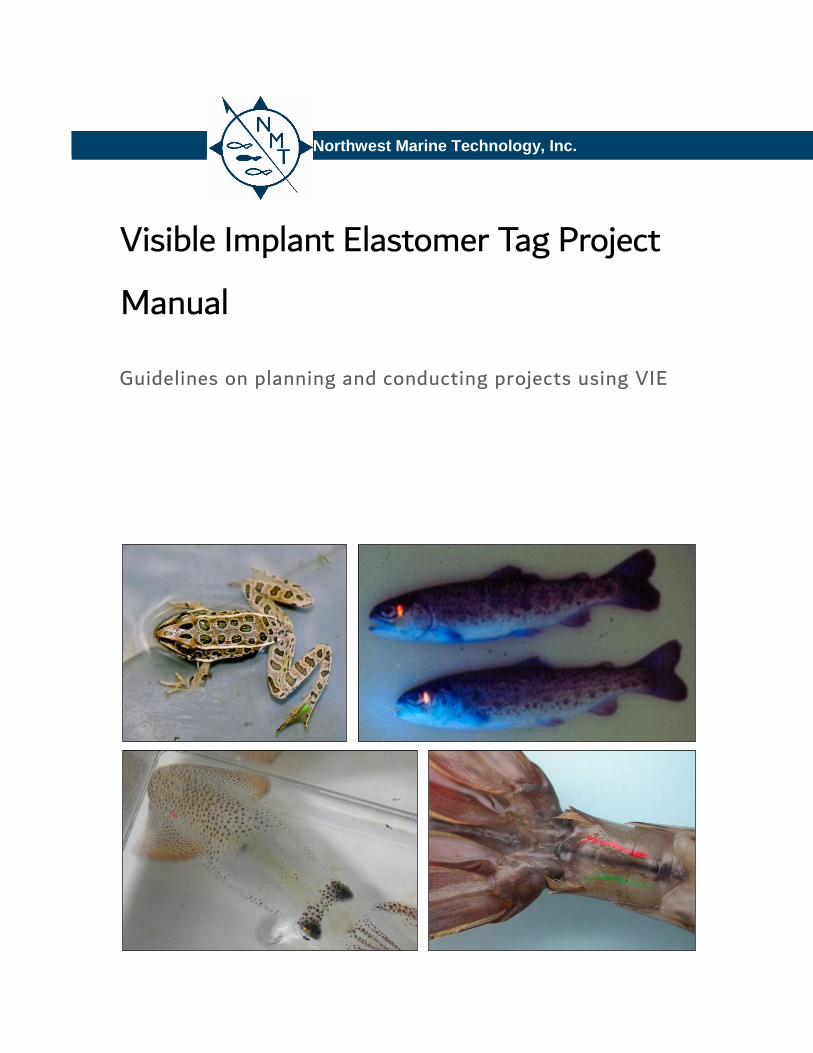
visible implalant elastomer tag project manual
TRANSCRIPT
Northwest Marine Technology, Inc.
Visible Implant Elastomer Tag Project
Manual
Guidelines on planning and conducting projects using VIE

Nov-17 2 GEV
Contents
1 INTRODUCTION 3
1.1 Overview ................................................................................................................................................................................... 3
1.2 Advantages and limitations of the VIE system ...................................................................................................... 4
2 DETAILS OF THE SYSTEM ........................................................................................................................................................ 4
2.1 The material ............................................................................................................................................................................ 4
2.2 The VIE Color Standard ..................................................................................................................................................... 5
2.3 Mixing supplies ...................................................................................................................................................................... 5
2.4 Injection syringes .................................................................................................................................................................. 6
2.5 Manual Elastomer Injector ............................................................................................................................................... 6
2.6 Air Driven Elastomer Injection System ...................................................................................................................... 7
2.7 The VI Light ............................................................................................................................................................................. 8
2.8 Manual Elastomer Injection Kits ................................................................................................................................... 8
3 USING THE SYSTEM ..................................................................................................................................................................... 9
3.1 Color Selection ....................................................................................................................................................................... 9
3.2 Mixing the material .............................................................................................................................................................. 9
3.3 Injecting the tag .................................................................................................................................................................... 9
3.4 Tag location and retention rates. ................................................................................................................................ 10
3.5 Coding capacity of single and multiple tags ........................................................................................................... 11
3.6 How big is a VIE tag? ........................................................................................................................................................ 12
3.7 Tagging very small fish .................................................................................................................................................... 12
3.8 How quickly can fish be tagged with manual VIE kits? .................................................................................... 13
3.9 Marking small numbers of fish ..................................................................................................................................... 14
3.10 Fluorescing and Detecting VIE ............................................................................................................................... 15
3.11 Working underwater ................................................................................................................................................... 16
3.12 The approach to tag detection. .............................................................................................................................. 16
4 SOME SUCCESSFUL APPLICATIONS WITH DIFFERENT SPECIES .................................................................... 17
4.1 Salmonids ............................................................................................................................................................................... 17
4.2 Cyprinids ................................................................................................................................................................................. 19
4.3 Percidae ................................................................................................................................................................................... 20
4.4 Amphibians ............................................................................................................................................................................ 21
4.5 Crustaceans ........................................................................................................................................................................... 23
4.6 Other fish and animal groups ....................................................................................................................................... 25
5 REFERENCES ................................................................................................................................................................................... 27
6 APPENDIX A .................................................................................................................................................................................... 30

Nov-17 3 GEV
1 INTRODUCTION
This document provides information on the
uses and deployment of Northwest Marine
Technology’s (NMT), Inc. Visible Implant
Elastomer (VIE) system and its associated
equipment. It is primarily aimed at new and
potential users, but existing users may also
find it useful, particularly if they are
considering marking new species or working
under different conditions. It is not
intended to replace the instructions issued
with each kit or piece of equipment.
The VIE System was developed by NMT
biologists in the 1990’s while they were
seeking better fish tagging methods than
traditional external tags and fin clips, which
may have adverse impacts on growth,
survival and behavior of fish.
The system was initially used with
salmonids, exploiting an area of transparent
tissue behind the eye. Since then, however,
it has been used on hundreds of species of
fish, amphibians, crustaceans and other
animals, with many body locations being
used.
1.1 Overview
The VIE system provides internal colored
tags that are visible externally, for fish and
other animals that are too small for the
NMT VI Alpha tag, or when batch codes are
sufficient. The system uses a bio-
compatible, two-part, elastomer material.
After mixing, the elastomer is a liquid that
is injected into tissue with a hypodermic
syringe; most species of fish, and many
other animals, have suitable areas of
transparent or translucent tissue. The
curing rate of the VIE tag is temperature
dependent. At warm temperatures it can
cure within hours and at very cold
temperatures it may take days. The VIE
cures into a pliable solid. The VIE tag holds
the pigment in a well-defined tag, without
damaging surrounding tissue. Using
different tag locations, and perhaps two or
more tags on each individual, development
of numerous group or individual codes is
possible. Some of the colored pigments
used are fluorescent, and use of appropriate
lighting can significantly enhance detection
of tags.
The VIE tag lies beneath the skin or deeper
within the tissues, without a permanent
wound or lesion. It has been demonstrated
to have minimal impact upon subsequent
growth and behavior. In contrast,
conventional external tags, attached via

Nov-17 4 GEV
penetration of the skin, cause a wound that
is very slow to heal or may never heal.
1.2 Advantages and limitations of
the VIE system
The advantages and limitations of the
system are summarized in the table below.
Each of these factors is discussed in detail
later in the report.
Advantages of VIE tags
• High retention rates
• Fast to apply
• May be applied to very small fish and
other animals
• Minimal impact on survival, growth and
behavior
• Low capital and material costs make it
viable for small-scale projects
• May be used in large scale projects
quickly tagging many animals using the
Air Driven Elastomer Injection System
• Tags detected visually in ambient light
• Fluorescing the tags with the VI Light
significantly enhances detection
• Tags may be seen in the dark or
underwater by fluorescing with the VI
Light
• Well-established technique with
extensive literature on successful
applications in hundreds of species of
fish, amphibians, crustaceans and other
animals
Limitations of VIE tags
• Limited coding capacity
• Tags may become difficult to detect in
ambient light if growth is considerable
and pigmented tissue is laid down over
the tag
• Tags may not be noticed and reported
by casual observers
2 DETAILS OF THE SYSTEM
2.1 The material
VIE tags are formed from a two-part
mixture. When first mixed, the material is a
viscous liquid. This hardens to a pliable solid
mark that generally retains its structural
integrity as the animal grows; this avoids
the gradual dissipation of pigment that
tends to occur with injections of particulate
material in liquid suspension such as Alcian
Blue.
The rate at which the material hardens, and
thus the length of time that it remains
usable, depends on temperature. At 20oC
this is of the order of 40 minutes; at 0oC it
is many hours.

Nov-17 5 GEV
The material is biocompatible and carries no
known human health hazards. A Material
Data Safety Sheet is in Appendix A.
Ten VIE colors are available. Six (red, pink,
orange, yellow, green, blue) are fluorescent;
the other four (black, white, purple and
brown) are not.
2.2 The VIE Color Standard
The color standard is a small transparent
card with a sample of all ten colors of VIE
that are available. It is supplied with all kits
and extras are available free of charge. It
allows consideration and selection of the
most appropriate color for any particular
application. However, perhaps its greatest
value is a color standard when identifying
tag recoveries, especially when using the VI
Light to fluoresce the material. Customized
color standards can easily be made by the
user by loading small volumes of material of
each color being used onto a transparent
sheet and covering them, when cured, with
transparent tape. This has the advantage
that the volumes loaded onto the
customized standard can be similar in shape
and size to the tags being used in the
particular project. Labeling the samples is
advisable to avoid any risk of confusion
over colors that may appear similar under
certain lighting conditions.
2.3 Mixing supplies
Figure 1: Samples of the ten VIE colors under ambient
light (left) and illuminated by the VI light (right).
Note that only the six fluorescent colors show up
with the VI light, and that the colors, especially
yellow, appear differently.
Figure 2: VIE Color Standard
Figure 3: Mixing and injection supplies for Manual VIE
Kits include mixing cups, stirring sticks, transfer
syringes, and injection needles.

Nov-17 6 GEV
VIE is supplied with enough mixing supplies
for the material involved under most
circumstances. Mixing supplies include
green transfer syringes (without needles)
for transferring the VIE to mixing cups,
wooden stirring sticks, and plastic cups. All
mixing supplies are intended for single use.
2.4 Injection syringes
The mixed VIE is loaded into a BD 0.3 cc
insulin syringes with a 29 g needle for
injecting into the animal. If you have a Trial
Pack, the VIE Tag is injected using the 0.3
cc syringe. All other VIE Kits (except the 6
ml Refill) include a Manual Elastomer
Injector for holding the 0.3 cc syringe for
injecting. All VIE kits contain enough
syringes for all the material supplied but if
additional ones are required NMT can
supply them.
A syringe with a removable needle is used
with the Air Driven Elastomer Injection
System (ADEIS). All orders for use with an
ADEIS include enough syringes for the
quantity of material supplied; additional
supplies are available from NMT.
All syringes are single use and should be
carefully handled and disposed.
2.5 Manual Elastomer Injector
The Manual Injector is a machined plastic
device into which a loaded 0.3 cc injection
syringe is placed. It is designed to make
holding and deployment of the syringe
comfortable, and allows extended use
without fatigue. It allows carefully
controlled pressure to be applied to extrude
the desired volume of material. While it is
perfectly possible to use the bare syringe
for small numbers of tags (for example
while using a Trial Pack for evaluation or
very small projects) we advise the use of
the Manual Elastomer Injector for anything
involving a hundred or more tags.
Figure 4: Manual Elastomer Injector, ready for
tagging.
Nov-17 7 GEV
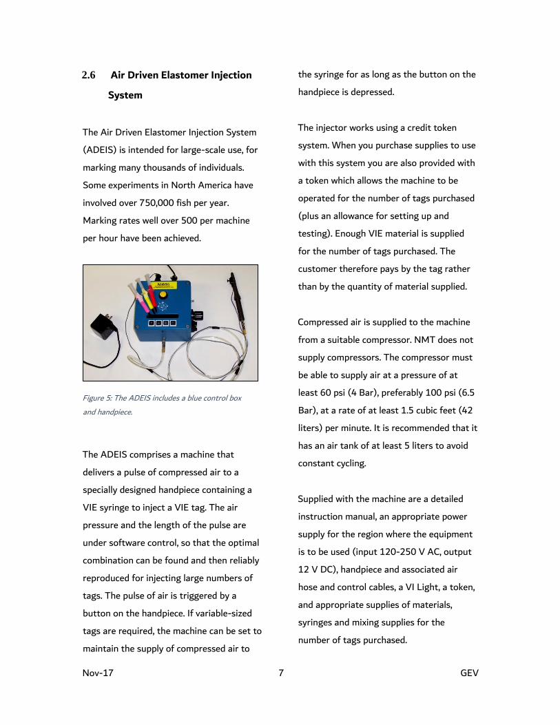
2.6 Air Driven Elastomer Injection
System
The Air Driven Elastomer Injection System
(ADEIS) is intended for large-scale use, for
marking many thousands of individuals.
Some experiments in North America have
involved over 750,000 fish per year.
Marking rates well over 500 per machine
per hour have been achieved.
The ADEIS comprises a machine that
delivers a pulse of compressed air to a
specially designed handpiece containing a
VIE syringe to inject a VIE tag. The air
pressure and the length of the pulse are
under software control, so that the optimal
combination can be found and then reliably
reproduced for injecting large numbers of
tags. The pulse of air is triggered by a
button on the handpiece. If variable-sized
tags are required, the machine can be set to
maintain the supply of compressed air to
the syringe for as long as the button on the
handpiece is depressed.
The injector works using a credit token
system. When you purchase supplies to use
with this system you are also provided with
a token which allows the machine to be
operated for the number of tags purchased
(plus an allowance for setting up and
testing). Enough VIE material is supplied
for the number of tags purchased. The
customer therefore pays by the tag rather
than by the quantity of material supplied.
Compressed air is supplied to the machine
from a suitable compressor. NMT does not
supply compressors. The compressor must
be able to supply air at a pressure of at
least 60 psi (4 Bar), preferably 100 psi (6.5
Bar), at a rate of at least 1.5 cubic feet (42
liters) per minute. It is recommended that it
has an air tank of at least 5 liters to avoid
constant cycling.
Supplied with the machine are a detailed
instruction manual, an appropriate power
supply for the region where the equipment
is to be used (input 120-250 V AC, output
12 V DC), handpiece and associated air
hose and control cables, a VI Light, a token,
and appropriate supplies of materials,
syringes and mixing supplies for the
number of tags purchased.
Figure 5: The ADEIS includes a blue control box
and handpiece.

Nov-17 8 GEV
For more details about the ADEIS please
refer to Air Driven Elastomer Injection
System User’s Manual available on our
website www.nmt.us or contact NMT.
2.7 The VI Light
The VI Light is used to fluoresce the VIE
and VI Alpha Tags. It has a nearly invisible,
regulated, deep-violet beam. Deep violet
(405 nm) is the optimum wavelength for
fluorescing our tags. This causes the
fluorescent VI colors (red, orange, pink,
yellow, green and blue) to fluoresce,
considerably increasing detection and
readability.
The VI Light is different from an ordinary
flashlight. To hold detection efficiency
constant, the VI Light maintains constant
brightness and color right up until its
battery fails completely. It provides a beam
of uniform intensity, then begins flashing
when the batteries near exhaustion.
The VI Light is waterproof to a depth of
150 m so is suitable for underwater studies.
2.8 Manual Elastomer Injection Kits
The VIE in all Manual Elastomer Injection
Kits (except the Trial Pack) is packaged in 3
ml syringes.
The 60 ml Kit includes: 60 ml elastomer
with curing agent - up to ten colors, mixing
supplies, 200 injection syringes, VI Light, 2
Manual Elastomer Injectors, instructions
and field carrying case.
The 24 ml Kit includes: 24 ml elastomer
with curing agent - up to eight colors,
mixing supplies, 60 injection syringes, VI
Light, 1 Manual Elastomer Injector,
instructions, and field carrying case.
6 ml Kit includes: 6 ml elastomer with
curing agent - up to two colors, mixing
supplies, 20 injection syringes, VI Light, 1
Manual Elastomer Injector, instructions,
and field carrying case.
6 ml Refill includes: 6 ml elastomer with
curing agent - up to two colors mixing
supplies, 20 injection syringes, and
instructions.
Figure 6: The VI Light.

Nov-17 9 GEV
Trial Pack of Elastomer includes: 1 ml of
elastomer with curing agent - one color,
mixing supplies, 3 injection syringes, and
instructions. This kit is intended to allow
mixing of two or three batches of material
for tagging small numbers of individuals
and for evaluation of the system.
3 USING THE SYSTEM
Before using the system, we recommend a
review of the available publications on
experience with the same or related species.
NMT maintains a list of up to date
references. If you have questions, please
contact us at [email protected]. We may also
be able to provide advice from our own
experience or guide you to someone else
who may be able to provide information. If
no relevant experience exists elsewhere,
experimentation to determine suitable tag
locations, retention rates and visibility
should be carried out before full-scale
application. While the system works well
with the overwhelming majority of
applications, retention rates can be variable.
3.1 Color Selection
Proper color selection is a vital part of good
experimental design. Your choice depends
on how much contrast you need with the
background pigmentation and how many
different colors you require. Certain color
combinations can be difficult to distinguish.
We do not recommend that green and
yellow be combined in a study because they
are difficult to distinguish when fluoresced
or when placed under pigmented tissue.
The fluorescent colors are highly visible
under ambient light and provide the option
of greatly enhanced tag detection when
fluoresced with the VI Light.
We usually recommend that all options with
the fluorescent colors be exhausted before
using the non-fluorescent colors. The non-
fluorescent colors are most useful where a
high coding requirement demands the use
of the maximum number of colors, or there
is some other specific advantage.
Please contact [email protected] if you would
like assistance.
3.2 Mixing the material
Detailed mixing instructions are provided
with all VIE kits, and are available for
download from our website (www.nmt.us).
3.3 Injecting the tag
To inject a tag, the syringe needle is
inserted into the marking location, and is

Nov-17 10 GEV
slowly withdrawn as the material is injected,
so that a long narrow mark is created. It is
important that the tag created is fully
contained within the target tissue;
extrusion of the material from the needle
must cease before the needle is withdrawn
so that material does not project through
the needle wound, as this is likely to cause
rapid loss of the tag.
In transparent tissue such as the adipose
eyelid of salmonids the VIE tag can be
injected fairly deeply (Figure 7). However, if
the material is being injected into fully or
partly pigmented tissue it is important to
place it just beneath the skin. Frederick
(1997), and Olsen and Vollestad (2001)
describe achieving maximum detectability
by making sure that the syringe needle was
pushed back towards the surface of the skin
after the initial penetration.
3.4 Tag location and retention rates.
Clear tissue, such as behind the eye in
salmonids, is the ideal site. Similar tissue
exists in many other fish families behind
and above the eye. Clear tissue is not
present in all species, but semi-transparent
and translucent tissue may also be suitable
for elastomer implants, especially in smaller
animals. In trials with turbot, VIE tags were
implanted just under the skin in less
pigmented areas. Tagging shrimps in the
last abdominal segment has been very
successful. The base of fins and beneath the
jaw are also good sites in many species. Fin
membrane tissue, in spaces between rays, is
another potential target. Such a technique
offers the potential to develop a variety of
unique codes based upon tags in specific
spaces.
A detailed description of some successful
applications with different species is given
in Section 4; this includes a consideration of
tag locations and retention rates.
Figure 7: Injecting a VIE tag into the clear tissue behind
the eye of a brown trout

Nov-17 11 GEV
3.5 Coding capacity of single and
multiple tags
Although the VIE system was developed as
a batch mark, there is significant scope for
use of different colors, different tag
locations and multiple tags to generate a
significant number of batch marks or even
individual identification (Figure 8). For
example, use of a single tag but using four
colors in five different body locations
immediately gives 20 unique marks.
Using more than one tag greatly increases
the coding capacity, according to the
formula:
No. unique codes = (L!/[L-N]!N!)CN
Where C = number of colors used, L =
number of body locations available, and N is
the number of tags used on each fish.
For example, use of three tags in three body
locations with four colors would give 64
combinations; three tags, four locations and
five colors would give 500. This approach
has been used on sea horses to identify up
to 500 individuals (Dr Keith Martin Smith,
Pers. Comm.), over 1,000 in guppies
Poecilia reticulate (Bryant and Reznick,
2004) and Jung et al. (2000) used three
colors and four body locations to create
255 individual codes in their study of
salamanders. A computer program for
calculating the number of combinations
(NMT VIE color code generator) can be
downloaded from our website www.nmt.us.
One important consideration when using
multiple tags is the scope for confusion if
one or more tags are lost. For this reason,
we recommend that all individuals in an
experiment receive the same number of
tags; then, if a tag is lost, the fish is
correctly recognized as one that has lost a
tag rather than being mistaken for a fish
that started with fewer tags.
If even a low rate of tag loss is likely to be
critical to a project it is worth considering
double marking, placing two tags in
different locations, with a protocol such
that the retention of both or either tag will
allow correct batch identification.
Figure 8: Multiple VIE marks in a 55 mm turbot.
Nov-17 12 GEV
3.6 How big is a VIE tag?
In contrast to conventional tags or Coded
Wire Tags, the size of a VIE tag is
controlled by the user. Very small fish are
likely to require a very small tag, while it
may be desired to put a larger one in larger
fish to aid visibility. Some general guidance
is useful for new users.
The biologists who have used VIE on some
of the smallest fish, 26 mm brown trout
(Olsen and Vollestad, 2001) and 8 mm
damselfish (Frederick,1997) both stated
that the amounts used were “minute”, but
the former reported that the tags made
were 2-3 mm long made with a 29 g
needles. The inside diameter of such
needles is about 0.2 mm, suggesting that
the tags were of the order of 15,000 per ml
of VIE. Dewey and Zigler (1996) reported
tagging around 1000 fish per ml. Willis and
Babcock (1998) used large tags on Pagrus
auratus of the order of 10 mm x 1 mm x 1
mm (127 per ml). We normally advise
assuming 300 to 500 tags per ml of
material for planning purposes, where
efficient use of mixed material can be
achieved.
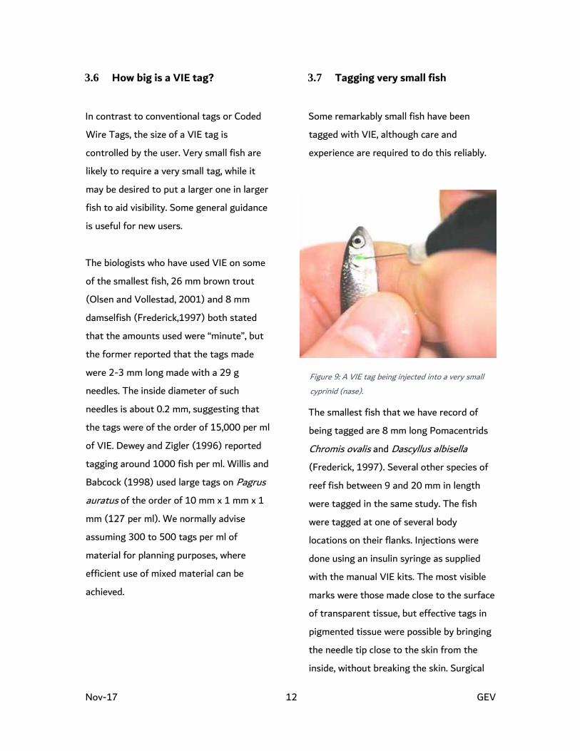
3.7 Tagging very small fish
Some remarkably small fish have been
tagged with VIE, although care and
experience are required to do this reliably.
The smallest fish that we have record of
being tagged are 8 mm long Pomacentrids
Chromis ovalis and Dascyllus albisella
(Frederick, 1997). Several other species of
reef fish between 9 and 20 mm in length
were tagged in the same study. The fish
were tagged at one of several body
locations on their flanks. Injections were
done using an insulin syringe as supplied
with the manual VIE kits. The most visible
marks were those made close to the surface
of transparent tissue, but effective tags in
pigmented tissue were possible by bringing
the needle tip close to the skin from the
inside, without breaking the skin. Surgical
Figure 9: A VIE tag being injected into a very small
cyprinid (nase).

Nov-17 13 GEV
gloves were worn to reduce abrasion of the
very delicate fish. In the field, fish were
tagged under water while being held in a
hand net; “trauma to the fish marked in this
manner appeared to be considerably less
than when they were brought to the surface
and anaesthetized for marking”. Some
mortality was observed with the smallest
fish, mostly within 2 hours of tagging. It
was higher for fish of less than 20 mm than
for larger fish. However, mortality fell
steadily during the project even though the
average size of fish being tagged was
falling; this was ascribed to the operator
gaining experience. “In fact, after
accounting for this learning curve, there
was no significant difference between initial
mortality of individuals marked and that of
the control group”. Tag retention was
virtually 100% for most species over
periods varying from 24 days to 76 days.
The smallest salmonids reported tagged are
brown trout (Salmo trutta) down to 26 mm
(Olsen and Vollestad 2001). Again, the
scientists involved stated how experience
improved the tagging performance. An
insulin syringe was used, with a 29 g needle.
A VIE tag 1-3 mm long was injected
alongside the anal fin, as close to the skin
as possible. No mortality or mark loss
occurred in fish held for 77 days in the
laboratory. At the end of the experiment all
tags were detectable, but two out of 50
required blue light to enhance visibility.
Growth was unaffected. The technique was
then used successfully on a project in small
streams.
3.8 How quickly can fish be tagged
with manual VIE kits?
The rate at which fish can be tagged will
depend upon several factors including
species and size of fish, tag location,
facilities available, and the experience of the
tagger. Tag retention and animal survival is
enhanced with practice and careful tag
placement. Some idea of tagging rates that
have been achieved is useful for project
planning.
Where fish size and species are not limiting
it appears that a rate of about 250-300 VIE
tags per hour is a good rate; actual
examples from the literature include Bailey
et al (1998) (300-400 per hour for juvenile
coho salmon); Astorga et al (2005) (230
per hour for Sparus of 7-18 g); and Dewey
and Zigler (1996) (288 per hour for
Lepomis of 33-133 mm).
Using more than one mark slows the
operation down somewhat; Brennan et al
(2005) reported handling rates for snook of
250-400 per hour for a single tag, and 200-
300 per hour for two tags. Similarly,

Nov-17 14 GEV
tagging very small fish takes much longer;
Olsen and Vollestad (2001) were only able
to process 40 mm trout fry at a rate of
about 60/h.
3.9 Marking small numbers of fish
In some projects, marking small numbers of
fish over an extended time may be required
– for example in a field study where just a
few fish per site are likely to be captured
and marked. As mixed VIE material has
only a limited useful life there is potential
for wastage. We suggest three possible
approaches to addressing this issue.
First, mix the smallest volumes of VIE that
can be achieved. This is generally limited by
the ability to measure the volume of the
hardener which is mixed with ten times the
volume of colored elastomer. Wastage can
be minimized by placing the two
components directly into the barrel of the
injection syringe and mixing them there
with a toothpick. Instructions and hints on
doing this are included in the mixing
instructions which come with each kit, and
which can be found on our website at
www.nmt.us. We have found that as little
as 0.1 ml can be mixed, with care and
practice. Allowing for wastage and dead
space in the syringe needle, this could allow
creation of the order of 20 to 50 tags,
depending on their size.
Second, the life of mixed material can be
extended for many hours or even a few days
by placing it in an ice chest or freezer;
Goldsmith et al (2003) were able to store
mixed material on ice for at least 48 hours.
If possible mixed material to be stored in
this way should not be loaded into the
injection syringe until shortly before it is to
be used as, with prolonged contact, the
rubber part of the plunger in those syringes
may react with the material and prevent
curing. As mixed material stored at minus
20oC remains liquid and can be handled and
manipulated with syringes, storage in the
mixing cup or transfer syringe are viable
options.
Third, the problem of wastage may be
minimized by arranging for any coding
required to be achieved using one color at a
Figure 10: As little as 0.1 ml of VIE can be mixed in the
barrel of the syringe.

Nov-17 15 GEV
time. For example, if several field sites are
involved in a single day it may be feasible to
code them by different body locations of a
single color, using another color for the
same body locations on other days.
3.10 Fluorescing and Detecting VIE
Six of the VIE colors (red, orange, green,
yellow, pink and blue) are fluorescent.
Black, white, purple and brown do not
fluoresce.
NMT’s VI Light has a nearly invisible,
regulated, deep-violet beam. Deep violet
(405 nm) is the optimum wavelength for
fluorescing our tags. Shine the light on the
area where the tag is, or is thought to be.
Don’t try to fluoresce tags in direct
sunlight. Rather, you should work in a little
shade – even the shade of your body is
probably enough for most tags. Very faint
tags are best seen when fluoresced in
darkness.
To maximize tag identification:
• Proper color selection is a vital part
of good experimental design.
• Place tags in clear tissue whenever
possible.
• Train your samplers – let them
practice with the tag colors they
will encounter before they start
collecting data.
• Fluoresce poor or obscured tags
with the VI Light, working out of
direct sunlight.
• Use the VIE Color Standard with
the VI Light to correctly identify
colors. The Color Standard presents
the ten colors on a clear card. The
sampler can place the color sample
beside a tag for comparison, either
under or over the tagged tissue.
Figure 11: VIE is visible in the fin rays in ambient light
(top) but is much easier to see when fluoresced (bottom).

Nov-17 16 GEV
3.11 Working underwater
The simple equipment required for tagging
fish with VIE and for identifying and
observing tagged fish means that VIE tags
and VI Light are well suited for underwater
use by divers or by observation from above
the surface.
Frederick (1997) describes tagging small
reef fish underwater and observing tagged
fish on reefs at depths of up to 10 m. Tags
were clearly visible from 1 m, in clear water
(visibility 15 m), without the need for
additional illumination. However, when
visibility was reduced due to low light, and
at night, fluorescing the tags enhanced
detection and discrimination of tag colors.
Willis and Babcock (1998) made
underwater observations on VIE tagged
snapper on a reef. Visibility was generally
good, with some marks being detectable
from up to 10 m. Bonneau et al. (1995)
also took advantage of the tags’
fluorescence to allow night observations on
small tagged bull trout in streams by divers
or by observers on the bank.
3.12 The approach to tag detection.
A critical factor in the design of tagging
experiments is the manner of recovery and
identification of tagged fish. Workers often
state that returns from fishermen are
critical to the conduct of the project, and
that tags must therefore be large and
colorful. However, depending upon such
returns introduces an unquantifiable and
potentially serious bias to the experiment.
First, large and conspicuous external tags
are likely to influence the survival, growth
and behavior of the fish itself, as discussed
above. Second, fishermen may not notice
even large tags, they may forget or not get
around to making a report of their
recapture, or they may choose not to report
such captures out of apathy, or a
perception that their interests will not be
advanced by doing so. In one study, anglers’
catches were secretly tagged after capture.
In spite of rewards being offered for return
of tags, only 29% of the tags were reported
(Green et al. 1983). Even if all recaptures
are reported, it is difficult to establish the
true size of the “sample” of which the
tagged recaptures formed a part.
A more robust approach from the statistical
viewpoint is for the scientist to scan
samples of fish catches for tagged
individuals. Such sampling can be planned
and appropriately stratified to address
specific questions and to obtain reliable and
unbiased answers. Although this approach
involves an additional phase in the project it
can be highly cost effective; a reasonable

Nov-17 17 GEV
volume of robust data may be much more
valuable than a large volume of possibly
biased data of doubtful validity. The VIE
system is particularly suited to this latter
approach, although they may be noted and
reported by anglers. If dependence upon
angler returns is essential for the project
this can be greatly enhanced by training a
team of interested fishermen to look for
tags and also to maintain a log book of all
fish caught (so that the sample size is
recorded).
A useful discussion of these and other
aspects of fish tagging programs is
provided by Bergman et al. (1992).
4 SOME SUCCESSFUL
APPLICATIONS WITH DIFFERENT
SPECIES
Successful applications of the VIE system
are too numerous to describe in full detail.
Instead, applications with some important
groups of fish, plus amphibians and
crustaceans are discussed in some detail,
together with an extensive list of other
animal families that have been tagged with
VIE. NMT maintains a list of up to date
references. If you have questions, please
contact us at [email protected]
4.1 Salmonids
There have been many published papers
reporting on the use of VIE in salmonids.
This review just deals with a selection of
them, chosen to represent a range of
species and situations.
Bonneau et al (1995) used VIE on cutthroat
trout (Oncorhynchus clarki) and bull trout
(O. confluentus) for behavioral
investigations. Counts of tagged fish were
undertaken both day and night in different
stream reaches using a snorkel diver. The
night-time observations involved the use of
an underwater light. The following body
locations were used as batch marks; top of
the head, post ocular tissue, adipose fin,
dorsal fin, pectoral fin and caudal fin. A
group of 85 fish were retained in captivity
to evaluate tag retention; after 2 months
retention was 100%, and one fish had lost
its tag after 4 months.
Figure 12: VIE tags in juvenile Chinook Salmon.

Nov-17 18 GEV
Adams et al. (2000) undertook a similar
study involving brook trout (Salvelinus
fontinalis) using snorkeling to observe
tagged fish. They were able to tag fish as
small as 50 mm in the adipose eyelid and
lower mandible, and from 75 mm upwards
between the rays of the dorsal and caudal
fins. Overnight losses of tags from samples
of fish retained after tagging were 2-13%
from the adipose eyelid, and 0-27 % from
the fins. These relatively high loss rates
may have been partly due to the small size
of the fish involved.
Walsh and Winkelman (2004) used
VIE in hatchery-reared rainbow trout
(Oncorhynchus mykiss) stocked into
streams. The fish averaged about 250 mm
in length at the time of tagging and 96% of
fish had a tag detectable in ambient light
after six months.
Tagging of very small brown trout (Salmo
trutta) by Olsen and Vollestad has already
been described.
There have been a number of long-term
studies tagging juvenile migratory
salmonids which are then sampled as
adults. One unpublished study was
undertaken with juvenile Chinook salmon
(Oncorhynchus tshawytscha) by the
Washington Department of Fisheries.
Batches of fish were double marked with
Coded Wire Tags (CWT) and red VIE which
was applied using the Air Driven Elastomer
Injection System. The VIE tag was placed in
the post-ocular adipose eyelid. The first
batch were tagged at a length of about 80-
100 mm in late 1992 a few weeks before
release to the wild. On release, a sample
was checked for VIE tags, which indicated
92.1% retention. Of 124 CWT fish
returning to the hatchery in 1994 (as 5-6
kg fish), 107 (86.3%) had observable VIE
tags in ambient light. A year later, 126 out
of 138 (91.3%) returning CWT fish were
found to have a VIE tag. In this case the
tags were fluoresced to enhance detection.
A second group was released in 1994, again
as 80-100 mm juveniles. On release, VIE
tag retention was estimated at 94.6%. Of
1752 CWT fish returning in 1995 (fish of
about 2 kg), 1632 (93.2%) had VIE tags.
These results indicate high retention and
tag detectability between juvenile and adult
salmon, and low rates of loss beyond a few
weeks after tagging.
Hatchery pre-smolts of coho salmon
(Oncorhynchus kisutch) were marked and
released by Bailey et al (1998). About
10,000 fish averaging 108 mm in length
were VIE marked in the adipose eyelid; they
were also tagged with a coded wire tag
Nov-17 19 GEV
(CWT) and adipose clipped to facilitate
recovery. Two groups of 100 fish were
retained for 24 hours, indicating VIE mark
loss rates of about 5%. Returning adults
were sampled in the stocked river, and
heads from ocean catches were obtained
from the CWT sampling program. Most VIE
marks were visible in ambient light, but
detection was improved by fluorescence.
From the double marking it was calculated
that about 73% of the fish that had
received a VIE tag had a detectable tag on
return. The use of a control group of fish
(CWT and Adipose clip only) showed that
VIE tagging had no impact on the survival,
growth or return behavior of the fish. The
authors suggested that the relatively high
loss rates of tags may have been affected
by operator inexperience and a failure of
the material to set properly; small droplets
of uncured VIE were noted when the fish
were released some months after marking.
The formulation of the material has been
improved so that curing problems are now
very unusual.
FitzGerald et al (2004) tagged smolts of
Atlantic salmon (Salmo salar) and then
reared them to maturity in net pens. This
allowed regular observations on the level of
detectable tags as the fish grew. About
9,000 smolts (mean length 213 mm, weight
99.7g) were tagged in the adipose eyelid,
lower jaw or both. The tags were 3-5 mm in
length. Tags in the adipose eyelid were
detectable in ambient light in more than
92% of fish after 17 months (mean length
547 mm, weight 1.7 kg), and those in the
lower jaw at more than 92% at 16 months.
From then onwards the level of tags
detectable in ambient light fell away to
52.2% for adipose eyelid tags at 28
months, and 14.4% for those in the jaw at
28 months. Better detection was possible
with fluorescence (87.8% for adipose
eyelid, 72.2% for jaw) suggesting that the
deterioration was due to the marks being
obscured by growth and pigmented tissue
rather than loss of marks.
In conclusion, VIE tagging of salmonids has
been very successful with good retention
and tag detectability over considerable
periods and through many-fold increases in
weight. Tags can become more difficult to
detect due to growth and development of
pigmented tissue but use of the VI Light to
fluoresce the mark helps considerably.
In no case has a detectable impact upon
survival, growth, or behavior been reported.
4.2 Cyprinids
Haines and Modde (1996) used VIE to mark
small Colorado pikeminnow (Ptychocheilus
lucius). Fish averaged 49.8 mm at time of

Nov-17 20 GEV
marking. VIE was “injected subcutaneously
on the dorsal surface left of the dorsal fin”.
Mortality was less than 1%, and retention
was 85% after 142 days.
Clough (1998) undertook retention trials
on dace (Leuciscus leuciscus) prior to
deploying the system in the field. Thirty-
nine fish of 154-169 mm were tagged with
an anal fin clip and two elastomer tags, one
in the pre-ocular area and one between the
first and second dorsal fin rays. The
detectability rates of the two elastomer
tags after various times are shown in the
table below. Both locations gave good
results but the preocular location would
appear to be the best.
Morgan and Farooqi (1996) used VIE to
tag 79-232 mm barbel (Barbus barbus).
Four tagging sites used: scalp, post-orbital,
base of anal fin and base of caudal fin.
Retention rates after 57 days 82.6%,
44.8%, 82.6% and 91.3% respectively.
There was no impact on growth rate.
4.3 Percidae
Goldsmith et al. (2003) combined colors
and body locations to individually identify
small perch Perca fluviatilis (mean length
88 mm, mean weight 5 g). In a tank trial, 25
fish were tagged with three VIE tags each,
along a horizontal line between the lateral
line and the base of the dorsal fin; using
four colors this allowed a coding capacity of
64, though not all combinations were used.
Retention was 100% after 125 days, and
there was no effect on growth or survival.
Roberts and Angermeir (2004) tagged 40
mm or larger Roanoake darter (Percina
roanoka) and riverweed darter (Etheostoma
podostemone) in a laboratory study. Mark
locations used were mid-ventral, lower
caudal peduncle, upper caudal, peduncle and
middorsal. There was no impact on survival,
and retention rates after 240 days were 90
for Percina and 79% for Etheostoma.
Thompson et al. (2005) compared VIE with
fin clipping as marking methods for
evaluation of stocking with walleye (Sander
vitreum). After tank trials they selected the
ventral surface of the lower jaw as the tag
site, using a 5mm long tag. Tag detection at
the time of release, 14 days after tagging,
was 97%. Recaptures were made over the
five years following release. Overall, VIE
detection rate was calculated at 82.5%, and
no effects on growth rate were observed.
Days since tagging
Tag Location n 0 19 42 292
Dorsal fin 39 100 100 100 83
Pre-ocular 39 100 100 100 93

Nov-17 21 GEV
4.4 Amphibians
Visible Implant Elastomer is the most
widely used alternative to toe clipping for
identifying amphibians. It also allows the
marking of tadpoles with the mark generally
being retained through metamorphosis.
Anholt and Negovetic (1998) tagged 1000
tadpoles of Rana lessonae and R. esculenta.
The tadpoles were anaesthetized and
tagged under a stereo microscope with 6.5
x magnification. Placing a single tag took
about 10 seconds per individual, and two
subcutaneous tag locations were used;
above the musculature of the tail and on
the back. The smallest individuals marked
were 8 mm snout to vent length. Overall
tag retention was 85% after 8 days, but the
authors suggest that losses would have
been less if only the tag location on the
back had been used. Survival was close to
100% after five weeks, during which time
some of the tadpoles had metamorphosed.
Although the tags were obscured by
pigment in the metamorphosed individuals
they were retained and could be recovered
by dissection. The authors concluded that
the consistency and biocompatibility of the
VIE tags allows tagging of small animals,
including larvae, that could not be tagged
using other methods.
Nauwelaerts et al. (2000) tagged 40 adult
Rana esculenta in the transparent tissue
between the toes. Retention was 100%
over eight months.
Three tags per individual were applied to 20
salamanders (Plethodon vehiculum, 36-60
mm snout to vent length) and 12 tree frogs
(Hyla regilla, 16-34 mm SVL) by Davis and
Ovaska (2001). Tag locations were all on
the ventral surface; anterior to the front
leg, posterior to the hind leg at the anterior
end of the vent, and at the posterior end of
the vent in the salamanders; and anterior
end of the thigh, posterior end of thigh, and
mid-calf in the tree frogs. No tagging-
related mortality was noted after 10-11
months. Retention was high; 10.5% of the
Figure 13: This frog, tagged as a tadpole, retained its
VIE marks through metamorphosis. Photo courtesy
of S. Hopkins.
Nov-17 22 GEV
salamanders and 22.2% of the tree frogs
had lost one of their tags at the end of the
experiment, representing 96.5% and
92.6% retention overall for the two species.
Subsequent field trials involved using three
tags per individual on 115 salamanders.
Forty-two were recaptured up to five times,
and 9.5% had lost one tag; this represents
96.8% retention overall.
Binckley et al.(undated) reported on a
project undertaken by Karl Mallory which
involved tagging 421 Pacific giant
salamanders (Dicamptodon tenebrosus)
with VIE in the wild. A total of 55
recaptures were recorded in the following
year, and 63 after two years, indicating high
retention and detectability. Tadpoles were
also tagged; 127 out of a total of 471
individuals were recaptured at least once.
In a novel approach to monitoring the
development of salamander egg masses,
Regester and Woosley (2005) used VIE to
identify and track the egg masses.
Other amphibian species which have been
successfully tagged with VIE include
Ambystoma maculatum, Anolis sagrei,
Ascaphus truei, Plethodon cinereus , Rana
sylvatica, Xenopus tropicalis and many
others.
Many researchers do not anesthetize
amphibians for tagging, while others prefer
the ease of handling that anesthetic
provides. A technique for non-
anaesthetized amphibians has been
developed in which the animal is placed in a
plastic bag with some water and is tagged
through the bag.
Some amphibians lack septa between the
skin and underlying tissue. VIE tags injected
in these animals can therefore migrate from
the original tagging location, making it
impossible to use those tagging locations to
create individual codes. In such cases where
individual identification is needed, we
recommend the use of VI Alpha tags.

Nov-17 23 GEV
4.5 Crustaceans
The first publication on the use of VIE in
crustaceans was Godin et al (1996) who
marked juvenile (mean weight 1.63 g) and
adult (mean weight 38.22 g) shrimps
(Penaeus vannamei). The VIE was injected
into the musculature of the sixth abdominal
segment. After 10 –14 weeks, tag retention
was 99.9% in juveniles and 100% in adults,
though UV light was required to identify
tags in about 9% of the juveniles. The
juveniles had increased in weight to 15-20
g, and had molted 17-23 times by the end
of the experiment. All marks were readily
identified in shrimps tagged as adults
without the use of fluorescent light; the
adults had molted 5-7 times during the
experiment.
Uglem et al. (1996) used VIE placed
beneath the epidermal layer in the abdomen
of juvenile lobsters (Homarus gammarus).
After three molts retention was 100%, and
overall survival 92%.
Jerry et al. (2001) used VIE in the
freshwater crayfish or yabby (Cherax
destructor). They used three sites in
animals averaging 0.9 g in weight; after ten
weeks retention was 94% in the coxa of the
last pair of walking legs, 92% in the 3rd
schlerite of the abdomen, and 82% in the
uropod. The animals had averaged three
molts during the experiment.
No effect on growth and survival over six
months in VIE marked spiny lobsters (Jasus
edwardsii) was noted by Woods and James
(2003). The marks were injected into the
muscle block of the second abdominal
segment of juveniles with a mean weight of
9.6 g. Retention was 100% through the
mean of 1.78 molts per animal, but marks
injected transversely tended to break up
somewhat; those placed longitudinally did
not. The authors suggest that breaking up
was due to the 3mm long tags lay across
Figure 14: VIE Tags in shrimp
Nov-17 24 GEV
the muscle fibers, and recommended
aligning the long axis of the tag with the
fibers.
Davis et al. (2004) compared the
performance of Coded Wire Tags (CWT)
and VIE in small blue crabs (Callinectes
sapidus), and concluded that each had
advantages and limitations which depended
upon animal size and the duration of the
project. The crabs were 6-25 mm carapace
width at the time of tagging, and the VIE
was injected into the upper (basal) segment
of the swimming leg (5th periopod). Tag loss
was 9.2% over 8 days, mainly due to the
shedding of the marked limb. In the longer
term, there was also some loss due to the
tag migrating into the carapace. This was
avoided in slightly larger crabs (30 mm CW)
by placing the material into the distal
segment of the leg. There was no impact of
either tagging method on growth.
Overall, VIE appears to be a very successful
tagging system for crustaceans, with very
high retention rates through multiple molts.

Nov-17 25 GEV
4.6 Other fish and
animal groups
Representatives of the
following families have
been successfully tagged
with VIE. Please contact
NMT [email protected] for
help if you can’t find
references. NMT
maintains an up to date
reference list.
Fish
Acipenseridae - sturgeons
Acanthuridae -
surgeonfishes
Adrianichthyidae –
ricefishes
Anarhichadidae –
wolffishes
Anguillidae – freshwater
eels
Aplocheilidae
Apogonidae –
cardinalfishes
Balitoridae – river loaches
Blenniidae – combtooth
blennies
Carangidae – jacks
Catastomidae - suckers
Centropomidae – snooks
Centrarchidae – sunfishes
Chaenopsidae pike-, tube-
and flagblennies
Chaetodontidae –
butterflyfishes
Chanidae – milkfishes
Characidae
Cichlidae - cichlids
Clupeidae – herrings
Cobitidae - loaches
Cottidae – sculpins
Cyclopteridae -
lumpsuckers
Cyprinidae - carps and
minnows
Cyprinodontidae -
pupfishes
Esocidae - pikes
Eleotridae - sleepers
Engraulidae – anchovies
Fundulidae - topminnows
Gadidae – cod
Galaxiidae - galaxiids
Gasterosteidae -
sticklebacks
Girellidae – nibblers
Gobiidae - gobies
Haemulidae – grunts
Hexagrammidae –
greenlings
Ictaluridae - North
American catfishes
Kuhliidae – flagtails
Labridae – wrasses
Lotidae - lings
Lutjanidae – snappers
Melanotaeniidae –
rainbow fishes
Moronidae – temperate
basses
Mugilidae – mullets
Nemacheilidae – stone
loaches
Nothobranchiidae –
African rivulines
Osmeridae – smelts
Osphronemidae
Paralichthyidae – sand
flounders
Pempheridae
Percichthyidae –
temperate basses
Percidae - perches
Petromyzontidae –
lampreys
Platycephalidae
Pleuronectidae – righteye
flounders
Poecilidae – livebearers
Polynemidae – threadfins
Pomacentridae –
damselfishes
Salmonidae – salmon,
trout, char
Sciaenidae – drums and
croakers
Scophthalmidae – turbots

Nov-17 26 GEV
Scorpaenidae –
scorpionfishes, rockfishes
Serranidae - sea basses
and groupers
Siluridae - sheatfishes
Sparidae – sea breams
and porgies
Syngnathidae – sea
horses and pipefishes
Terapontidae – grunters
or tigerperches
Tripterygiidae - triplefins
Reptiles
Agamidae
Chelydridae
Colubridae
Emydidae – pond turtles
Gekkonidae - geckos
Polychrotidae
Sincidae
Trionychidae
Amphibians
Alytidae
Ambystomatidae – mole
salamanders
Ascaphidae
Bufonidae
Caeciliidae – caecelians
Cryptobranchidae – giant
salamanders
Dicamptodontidae
Dicroglossidae – fork-
tongued frogs
Hylidae – tree frogs
Hyperoliidae
Leptodactylidae
Pelobatidae – spadefoot
toads
Plethodontidae –
terrestrial salamanders
Proteidae
Ranidae – true frogs
Salamandridae
Scaphiopodidae – true
salamanders, newts
Scincidae
Crustaceans
Astacidae
Atyidae
Xiphocarididae
Callianassidae – ghost
shrimps
Cambaridae – crayfishes
Cancridae
Galatheidae – squat
lobsters
Grapsidae – shore, marsh
and talon crabs
Homaridae
Nephropidae – clawed
lobsters
Palaemonidae
Palinuridae – spiny
lobsters
Parastacidae
Penaeidae – penaeid
shrimps
Portunidae – swimming
crabs
Other
Slugs: Arionidae
Echinoderms: Asterinidae,
Stichopodidae
Cephalopods:
Octopodidae, Loliginidae,
Sepiidae
Annelids: Hormogastridae,
Lumbricidae
Elasmobranchs:
Scyliorhinidae
Insects: Buthidae;
Calliphoridae
Mammals: Soricidae –
shrews

Nov-17 27 GEV
5 REFERENCES
Adams, S.B., C.A. Frissell and B.E. Rieman. 2000.
Movements of non-native brook trout in
relation to stream channel slope. Transactions
of the American Fisheries Society 129: 623-638
Anholt, B. R., and Negovetic, S. 1998. Methods
for anaesthetizing and marking larval anurans.
Herpetological Review 29(3):153-154.
Astorga, N., Afonso, J.M., Zamorano, M.J.,
Montero, D., Oliva, V., Fernandez, H. and
Izquierdo, M.S. 2005 Evaluation of Visible
Implant Elastomer tags for tagging juvenile
gilthead bream (Sparus auratus L.); effects on
growth, mortality, handling time and tag loss.
Aquaculture Research 36: 733-738
Bailey, L. L. 2004. Evaluating elastomer marking
and photo identification methods for terrestrial
salamanders: marking effects and observer bias.
Herpetological Review: 35:3841
Bailey, R. E., Irvine, J. R., Dalziel, F. C., and
Nelson, T. C. 1998. Evaluations of visible
implant fluorescent tags for marking coho
salmon smolts. North American Journal of
Fisheries Management 18:191-196
Bergman, P. K., F. Haw, H. L. Blankenship, and R.
M. Buckley. 1992. Perspectives on design, use,
and misuse of fish tags. Fisheries, 11(4):20-25
Binckley, C. A., et al. "Using the Visible Implant
Fluorescent Elastomer (VIE) tagging system to
mark amphibians.". Retrieved 7/12/2010, from
http://www.pwrc.usgs.gov/marking/vie.html
Bonneau, J. L., R. F. Thurow, and D. L.
Scarnecchia. 1995. Capture, marking, and
enumeration of juvenile bull trout and cutthroat
trout in small, low-conductivity streams. North
American Journal of Fisheries Management 15:
563-568
Brennan, N.P., Leber, K.M., Blankenship, H.L.,
Ransier, J.M and DeBruler, R. 2005 An
evaluation of coded wire and elastomer tag
performance in juvenile common snook under
field and laboratory conditions. North American
Journal of Fisheries Management 25: 437-445
Bryant, M. J. and D. Reznick (2004).
Comparative studies of senescence in natural
populations of guppies. American Naturalist
163(1): 55-68.
Clough, S. 1998. Migration and habitat use of
dace (Leuciscus leuciscus(L.)) in an English
chalk stream. PhD thesis, University of St
Andrews
Davis, T.M. and Ovaska, K. 2001. Individual
recognition of amphibians: effects of toe
clipping and fluorescent tagging on the
salamander Plethodon vehiculum. Journal of
Herpetology 35: 217-225
Davis, J. L. D., A. C. Young-Williams, and A. H.
Hines & O. Zmora. 2004 Comparing two types

Nov-17 28 GEV
of internal tags in juvenile blue crabs. Fisheries
Research 67:265-274
Dewey, M. R., and S. J. Zigler 1996. An
evaluation of fluorescent elastomer for marking
bluegill sunfish in experimental studies. The
Progressive Fish Culturist 58: 219-220
Farooqi M.A. & C. E. Morgan. 1996. Elastomer
visible implant (EVI) tag retention and effect of
tagging on the growth and survival of Barbel,
Barbus barbus (L.). Fisheries
Management and Ecology 3:181-183
FitzGerald, J. L., T. F. Sheehan and J. F. Kocik.
2004: Visibility of visual implant elastomer tags
in Atlantic salmon reared for two years in
marine net-pens. North American Journal of
Fisheries Management 24: 222–227
Frederick, J. L. 1997. Post-settlement
movement of coral reef fishes and bias in
survival estimates. Marine Ecology Press Series
150: 65-74
Godin, D. M., W. H. Carr, G. Hagino, F. Segura, J.
N. Sweeney, and L. Blankenship.
1996. Evaluation of a fluorescent elastomer
internal tag in juvenile and adult shrimp
Penaeus vannamei. Aquaculture 139: 243-248
Goldsmith, R. J., Closs, G. P. and Steen, H. 2003.
Evaluation of visible implant elastomer as a
method for individual marking of small perch
and common bully. Journal of Fish Biology 63
(3), 631-636
Green, A. W., G. C. Matlock, and J. E. Weaver.
1983. A method of directly estimating the tag-
reporting rate of anglers. Transcations of the
American Fisheries Society. 112: 412-415
Haines, B. G. and T. Modde. 1996. Evaluation of
marking techniques to estimate population size
and first year survival of Colorado squawfish.
North American Journal of Fisheries
Management 16: 905-912
Hale, R. S. and J. H. Gray. 1998. Retention and
detection of coded wire tags and elastomer tags
in trout. North American Journal of Fisheries
Management 18: 197-201
Jerry, D. R., T. Stewart, I. W. Purvis, L. R. Piper.
2001. Evaluation of visible implant elastomer
and alphanumeric internal tags as a method to
identify juveniles of the freshwater crayfish,
Cherax destructor. Aquaculture 193:149-154
Jung, R. E. S. Droege, J. R. Sauer, and R. B.
Landy. 2000. Evaluation of terrestrial and
streamside salamander monitoring techniques
at Shenandoah National Park.
Environmental Monitoring and Assessment 63:
65-79
Nauwelaerts, S., J. Coeck, and P. Aerts. 2000.
Visible implant elastomers as a method for
marking adult anurans. Histological Review
31(3): 154-155

Nov-17 29 GEV
Olsen, E. M., L. A. Vollestad. 2001. An Evaluation
of Visible Implant Elastomer for Marking Age-0
Brown Trout. North American Journal of
Fisheries Management. 21: 967-970
Regester, K.J.and Woosley, L.B. 2005. Marking
salamander egg masses with Visible
Fluorescent Elastomer: retention time and effect
on embryonic development. The American
Midland Naturalist 153: 52-60
Roberts, J. H. and Angermeier, P. L. 2004. A
comparison of injectable fluorescent marks in
two genera of darters: effects on survival and
retention rates. North American Journal of
Fisheries Management 24: 1017-1024
Thompson, J. M., Hirethota, P. S. and Eggold, B.
T. 2005. A comparison of elastomer marks and
fin clips as marking techniques for walleye.
North American Journal of fisheries
Management, 25: 308-315
Uglem, I., Noess, H., Farestveit, E. and K. E.
Jorstad. 1996. Tagging of juvenile lobsters
(Hommarus gammarus (L.)) with Visible
Implant Fluorescent Elastomer tags.
Aquacultural Engineering 15: 499-501.
Walsh, M. G. and Winkelman, D. L. 2004.
Anchor and Visible Implant Elastomer tag
retention by hatchery rainbow trout stocked
into an Ozark stream. North American Journal
of Fisheries Management 24: 1435-1439
Willis, T. J. and R. C. Babcock. 1998. Retention
and in situ detectability of visible implant
fluorescent elastomer (VIFE) tags in Pagrus
auratus (Sparidae). New Zealand Journal of
Marine and Freshwater Research 32: 247-254
Woods, C. M. C. and P. J. James. 2003.
Evaluation of visible implant fluorescent
elastomer (VIE) as a tagging technique for spiny
lobsters (Jasus edwardsii). Marine and
Freshwater Research 54: 853-858
Woods, C. M. C and K. M. Martin-Smith. 2004.
Visible implant fluorescent elastomer tagging of
the big-bellied seahorse, Hippocampus
abdominalis. Fisheries Research 66: 63-371

Nov-17 30 GEV
6 APPENDIX A
1. PRODUCT AND COMPANY IDENTIFICATION
Northwest Marine Technology, Inc.
P.O. Box 427
Shaw Island, Washington 98286
Emergency Telephone: (360) 468-3375
Customer Service: (360) 468-3375
Trade Name: Visible Implant Elastomer Tag
Chemical Family: Silicone
Other Product Information: The base (Part A) is not a hazardous material as defined in the OSHA Hazard
Communication Standard. The base contains a very small amount (less than 0.1%) of a potentially
hazardous compound, formaldehyde. The maximum possible level of formaldehyde that could be released
into the environment is far below the level allowed by OSHA. The information below applies to the curing
agent (Part B) of the two-part kit. Handle freshly mixed elastomer material as recommended for the curing
agent. After curing, the product is not hazardous. Visible Implant Elastomer Tags are available in various
colors. All colors are equally non-hazardous.
National Fire Protection Association Profile: Health 0 Flammability 1 Instability/Reactivity 1
2. HAZARDS IDENTIFICATION
POTENTIAL HEALTH EFFECTS
Acute Effects
Eye: Direct contact may cause temporary redness and discomfort.
Skin: No significant irritation expected from a single short-term exposure.
Inhalation: No significant effects expected from a single short-term exposure.
Oral: Low ingestion hazard in normal use.
Prolonged/Repeated Exposure Effects
Skin, inhalation, oral: No known applicable information.
Signs and Symptoms of Overexposure
No known applicable information.
Material Safety Data Sheet Visible Implant Elastomer Tags, 10:1 Formulation
Revised 2009/08/15
Nov-17 31 GEV
Medical Conditions Aggravated by Exposure
No known applicable information.
3. COMPOSITION/INFORMATION ON INGREDIENTS
CAS Number Wt % Component Name
68037-59-2 10.0 - 30.0 Dimethyl, methylhydrogen siloxane
The above component is hazardous as defined in 29 CFR 1910.1200.
4. FIRST AID MEASURES
Eye: Immediately flush with water.
Skin, inhalation, oral: No first aid should be needed.
Notes to physician: Treat symptomatically.
5. FIRE FIGHTING MEASURES
Flash point: > 214 °F / > 101.1 °C (Closed Cup)
Autoignition temperature: Not determined.
Flammability limits in air: Not determined.
Extinguishing media: On large fires use AFFF alcohol compatible foam or water spray (fog). On small
fires use AFFF alcohol compatible foam, CO2 or water sprays (fog). Water can be used to cool fire
exposed containers. Do not allow extinguishing medium to contact container contents. Most fire
extinguishing media will cause hydrogen evolution. When the fire is put out, hydrogen may accumulate in
poorly ventilated or confined areas and result in flash fire or explosion if ignited. Foam blankets may also
trap hydrogen or flammable vapors, with the possibility of subsurface explosion.
Unsuitable Extinguishing Media: Dry chemical.
Fire Fighting Measures: Self-contained breathing apparatus and protective clothing should be worn in
fighting large fires involving chemicals. Use water spray to keep fire exposed containers cool. Determine
the need to evacuate or isolate the area according to your local emergency plan.
Unusual Fire Hazards: None.
6. ACCIDENTAL RELEASE MEASURES

Nov-17 32 GEV
Use absorbent material to collect and contain for salvage or disposal.
Waste disposal method: All local, state and federal regulations concerning health and pollution should be reviewed
to determine approved disposal procedures.
7. HANDLING AND STORAGE
Use with adequate ventilation. Avoid eye contact.
Product evolves minute quantities of flammable hydrogen gas which can accumulate. Adequately
ventilate to maintain vapors well below flammability limits and exposure guidelines. Do not repackage.
Do not store in glass containers which may shatter due to pressure build up. Clogged container vents
may increase pressure build up. Keep container closed and store away from water or moisture.
8. EXPOSURE CONTROLS / PERSONAL PROTECTION
Component Exposure Limits: There are no components with workplace exposure limits.
Engineering Controls: Local and general ventilation are recommended.
Personal Protective Equipment for Routine Handling and Spills
Eyes: Use proper protection - safety glasses as a minimum.
Skin: Washing at mealtime and end of shift is adequate.
Suitable Gloves: No special protection needed.
Inhalation: No respiratory protection should be needed.
Precautionary Measures: Avoid eye contact. Use reasonable care.
Comments: When heated above 150˚C (300˚F) in the presence of air, product can form formaldehyde
vapors. Formaldehyde is a potential cancer hazard and a known skin and respiratory sensitizer. Vapors
irritate eyes, nose, and throat. Safe handling conditions may be maintained by keeping vapor conditions
within the OSHA permissible exposure limit for formaldehyde.
9. PHYSICAL AND CHEMICAL PROPERTIES
Odor, appearance, color: little odor, liquid, some color
Specific gravity (at 77 ˚F): 0.972
Vapor pressure: less than 5 mm
Nov-17 33 GEV
Percent volatile by weight (%): less than 5
Solubility in water (%): less than 0.1
10. STABILITY AND REACTIVITY
Chemical Stability: Stable.
Hazardous Polymerization: Hazardous polymerization will not occur.
Conditions to Avoid: None.
Materials to Avoid: Oxidizing material can cause a reaction. Water, alcohols, acidic or basic materials,
and many metals or metallic compounds, when in contact with product, liberate flammable hydrogen gas,
which can form explosive mixtures in air.
Hazardous Decomposition Products: Thermal breakdown of this product during fire or very high heat
conditions may evolve the following decomposition products: Carbon oxides and traces of incompletely
burned carbon compounds. Silicon dioxide. Formaldehyde. Hydrogen.
11. TOXICOLOGICAL INFORMATION/ ECOLOGICAL INFORMATION
No known applicable information.
12. TRANSPORT INFORMATION
DOT Road Shipment Information (49 CFR 172.101): Not subject to DOT.
Ocean Shipment (IMDG): Not subject to IMDG code.
Air Shipment (IATA): Not subject to IATA regulations.
13. REGULATORY INFORMATION
Contents of this MSDS comply with the OSHA Hazard Communication Standard 29 CFR 1910.1200.
TSCA Status: All chemical substances in this material are included on or exempted from listing on the
TSCA Inventory of Chemical Substances.
EPA SARA Title III Chemical Listings
Section 302 Extremely Hazardous Substances (40 CFR 355): None.
Section 304 CERCLA Hazardous Substances (40 CFR 302): None.
Section 311/312 Hazard Class (40 CFR 370): Acute - No; Chronic - No; Fire - No; Pressure - No;
Reactive - Yes

Nov-17 34 GEV
Section 313 Toxic Chemicals (40 CFR 372):
None present or none present in regulated quantities.
14. OTHER INFORMATION
These data are offered in good faith as typical values and not as a product specification. No warranty, expressed or
implied, is hereby made. The recommended industrial hygiene and safe handling procedures are believed to be
generally applicable in the context of the intended use.