viruses & antiviral therapy - part 1
TRANSCRIPT

VIRUSES & ANTIVIRAL THERAPY - PART 1
David J. Maggs BVSc (hons), DACVO Professor, Comparative Ophthalmology
University of California- Davis VM-SRS, 2112 Tupper Hall
Davis, CA 95616 [email protected]
Acknowledgements: The following notes are developed from an original set written by and generously gifted to me by my friend and mentor Mark Nasisse, DVM, DACVO. I have modified and updated them biennially; however, the core content (as well as my fascination with FHV-1) is due entirely to Mark and I am deeply indebted to him. However, any errors are mine and I would be grateful if you brought them to my attention.
INTRODUCTORY VIROLOGY From the perspective of complexity and variability of pathogenic mechanisms, few ocular diseases are more fascinating than those initiated by viral infections. Because most is currently known about ocular diseases due to feline herpesvirus type 1 (FHV-1) and because it utilizes so many of these pathogenic mechanisms, most attention will be paid to this agent and its disease manifestations. A brief discussion of some historic details, general principles of viral pathogenesis, and specific mechanisms of tissue injury known to induce ocular lesions in animals is provided initially.
Vi'rus n. slimy liquid or poison
A LITTLE HISTORY Dorland describes a virus as “a minute infectious agent not resolved in a light microscope, characterized by a lack of independent metabolism and the ability to replicate only within living host cells.” This apparently simple sentence belittles the centuries of scientific endeavor that led up to proving the existence of viruses and establishing those defining characteristics. A brief review of those critical discoveries, especially in context of the understanding (and undoubtedly some ongoing misunderstanding!) we have of viruses today, is fascinating…
“A minute infectious agent not resolved in a light microscope, characterized by a lack of independent metabolism and the ability to replicate only within living host cells”
Dorland’s Medical Dictionary
The relatively small size of viral particles meant that they were not truly “seen” until 1939. It took William Henle - an avante garde scientist and, by the way, the person after whom the Loop of Henle is named - to propose in 1840 that infectious particles too small to see with the light microscope may not only exist but could answer some of the dilemmas with which he was presented. Of course, these were seen as heretical thoughts and not well received! However, not long after this, people with names now familiar to us, such as Pasteur, Koch, and Lister added irrefutable evidence to Henle’s theorem – but for much larger infectious particles - bacteria. Illnesses we now know as caused by viruses remained an enigma for at least another half a century. Around the turn of the 20th century, using what became known as tobacco mosaic virus, a number of researchers proposed the existence of a “filterable infectious agent” that caused discoloration of the commercially important tobacco plant. The finding that this agent could reproduce only in the presence of living tissue as opposed to bacteria, which could replicate in cell free media was a subsequent and equally critical discovery. Still these

proposed agents could not be observed and so were considered to be fluids. Hence the first name for viruses – “contagium vivium fluidum” and the current name “virus” - derived from the Latin word for “slimy liquid or poison”. In 1898, the first animal virus– foot and mouth disease (FMD) virus was discovered. The first human virus – yellow fever virus – was not identified until 1901. However, it was another 40 years before viruses were actually “seen” for the first time - using electron microscopy (1939). And so some of the early mysteries surrounding the existence and character of viruses began to be unraveled… It is tempting to believe that these were all experiments conducted in an earlier time when instrumentation and techniques were “primitive” and that these sorts of misunderstandings were to be expected. That couldn’t happen now, could it?
“PCR made it easier to see that certain people are infected with HIV, and some of those people came down with
symptoms of AIDS. But that doesn't begin even to answer the question, “Does HIV cause AIDS?” The mystery of that damn virus has been generated by the $2 billion a year they spend on it. You take any other virus, and you
spend $2 billion, and you can make up some great mysteries about it too.” Kary Mullis [1993 Nobel Laureate and inventor of the polymerase chain reaction (PCR)]
“It’s the virus – stupid!”
David Ho (Scientific Director, Aaron Diamond AIDS Research Center, NY and Time Magazine’s 1996 Man of the Year)
PATHOGENESIS OF VIRAL INFECTIONS Pathogenesis is the means or mechanism by which a virus causes disruption of function and sometimes death within a cell, (and sometimes a tissue or the whole organism) that it infects. It is very important that we realize this can often not cause signs of disease in animals or sometimes even symptoms in human beings. This will become critical when we discuss feline herpesvirus type 1 (FHV-1) pathogenesis in cats. In other words, there is an extremely delicate balance that exists between health and disease and between viral virulence and host defenses. Disease results when the virus “wins”. However, with many viruses, we must also be open to the fact that excessive host defense mechanisms may not always be an advantage and can cause immunopathology or proliferative disease. Think of feline infectious peritonitis or infectious canine hepatitis (more on them later). Recall that, by definition, for a virus to replicate, it must enter a cell and co-opt some of the host cell’s synthetic machinery and energy sources. This makes specific drug targets and organism clearance more difficult At least a superficial understanding of virus-cell interactions is therefore critical. However, for clinicians, it can sometimes be difficult to understand various cellular and sub-cellular events in virus-cell interactions. One scheme that may assist the clinician involves drawing analogies between similar cellular and “whole host” events (Table 1). Each step in the complex series of events that describes viral entry into, replication within, and release from a cell (or from the host organism) can then be considered a barrier to disease production. When the virus successfully overcomes all barriers or if the host has deficiencies in some barriers, then infection (but not always disease) results. Likewise, each step at the host or cellular level can be seen as a potential target for control or therapeutic measures. Therefore a basic understanding of these steps will assist the clinician in suspecting and diagnosing viral etiologies in their patients, implementing therapeutic and disease control methods, and offering clients an accurate prognosis. Table 1. Comparison of the stages in viral pathogenesis at a “whole animal” and a cellular (and subcellular)
level
Whole Animal Cellular/Subcellular Entry into host Adsorption
Primary replication Penetration Spread throughout host Uncoating Cell and tissue tropism Transcription Host immune responses Translation Secondary replication Replication
Cell injury Assembly Persistence/Latency/Genomic integration Release

At the individual patient-specific and population-specific (think “feline shelter” or “herd health”) levels, there are a number of distinct but very closely related clinical considerations:
• What is/are the route/s of infection? • What are the preferred sites (cells, tissues, organs) of primary/secondary replication? • How does the virus spread within the host (cell to cell, axonal, hematogenous, etc.)? • What are the major means of immunity/defense (humoral, cell-mediated, mucosal, etc.)? • Are some forms of immune response harmful to the host (immunopathology, immunosuppression)? • Does the virus establish persistent or latent infections? For how long? • How does the virus cause cellular injury (dysfunction) or death? • How is the virus excreted or “shed”? • How stable is the virus in the environment? • Etc.
At a cellular level, infection occurs when the viral particle (virion) finds and attaches (adsorbs) to an appropriate cell membrane receptor, penetrates into that cell, exposes (uncoats) its nucleic acids for transcription and protein production, replicates itself, and then assembles new virus particles ready for release from that cell and potential infection of another cell either nearby or at a secondary location. An essential consideration for the clinician is what injurious events occur intracellularly during this reproductive process? Stated in these ways, it becomes more clear that “successful” infection of the whole animal (rather than just a “few” cells) with disease production can be considered uncommon and, when it occurs, a reflection of how well evolved that virus is for infection of that host or how susceptible that host is to infection by that virus.
The critical steps for clinicians are cell entry, viral transcription, and virally mediated pathology because these are what define host susceptibility, viral virulence, nature of the
disease produced, sites of action for potential treatments, and means of control/prevention. Therefore a little time will be spent on these.
Cell Entry Viral entry, beginning with virus-cell receptor binding and continuing with cell membrane penetration, is essential if the virus is to gain access to host cell synthetic machinery. Enveloped viruses uncoat (lose their envelope) during penetration due to fusion of the viral envelope and the cell membrane. It is assumed that viruses have evolved to use cell receptors that are present for other reasons and that this, in part, also determines species specificity and cell tropism. Like all interactions with cell membrane receptors, this is undoubtedly a complex series of physical, electrical, and chemical interactions, and may involve multiple receptors and/or permissive “cofactors”. The old “key and lock” analogy is undoubtedly useful but represents a gross over-simplification of this process. Various host defense mechanisms and potential therapeutic endeavors could target this aspect of host-virus interaction. For example, lactoferrin (present in tears) exerts an antiviral effect against FHV-1 and many other viruses, which is quite distinct from its iron-binding antibacterial effect. Rather, it appears to be due to inhibition of viral adsorption to and subsequent entry into the host cell.1,2 Viral Transcription Transcription of viral DNA utilizes host enzymes (“machinery”) and nucleic acids (“building blocks”). This is why viruses are incapable of independent existence and are obligate intracellular pathogens. Some viruses simultaneously down-regulate host cell transcription to ensure better access to this “machinery” and the “building blocks”. Undoubtedly a multitude of complex and very strictly controlled steps are responsible for the success of this process. Although only a few of these are well defined or understood, some have been useful targets for pharmacological intervention. This is discussed more fully in the later section on antiviral therapies. This intimate and intricate relationship between viral and host cell functions during viral transcription results in antiviral drugs being generally more toxic than most antibacterial drugs because they tend to have “overlapping” negative effects on host machinery as well as the viral machinery they are designed to target.

Virally-mediated Pathology Perhaps of most interest to the clinician are the mechanisms by which viruses cause cell injury or dysfunction (and
ultimately illness within a patient). Figure 1 is intended to summarize the most frequently encountered of these mechanisms. In summary, once a virus enters a cell, one of three initial outcomes is possible. The infection may result in cell lysis (and death). This occurs as a result of successful viral replication and coincides with the release of viral progeny from the host cell and therefore is also referred to as a “productive” infection. This is typical of a primary infection with one of the alphaherpesviruses such as FHV-1 or canine herpes virus (CHV) and it is the cell lysis that causes the corneal and conjunctival ulceration visible clinically. Alternatively, viral replication may occur at an extremely low rate, with no or undetectable release of mature virus particles. In this case cell lysis is unlikely however viral antigen production may continue and “drive”
an immunological reaction that may be equally or more devastating than cytolysis. This is believed to be responsible for some of the more chronic outcomes of alphaherpesvirus infections such as stromal keratitis in cats (and human beings), and is discussed more fully in that section of these notes. The final alternative is that viral replication is stopped completely (i.e., a non-productive infection results). In a non-productive infection, 3 further outcomes are possible: (1) firstly, the cell may eliminate the virus and continue as before, (2) alternatively the cell may eliminate the virus but subsequently die, (3) the third outcome is of most interest pathologically and results when viral DNA is “sequestered” within the cell with either subsequent oncogenic transformation (e.g., FeLV), or reactivation (e.g., FHV-1).
With that brief introduction to how viruses infect (and sometimes injure) host cells (and sometimes tissues and organs), let’s discuss some specific examples of ophthalmic importance. Because FHV-1 is by far and away the most common virus of ocular importance to practicing ophthalmologists and because it can be used as an exemplar of the basic viral principles just introduced. Part I of these notes is devoted to this virus, the disease syndromes it causes, and antiviral therapies directed at it.
FELINE HERPESVIRUS TYPE 1 (FHV-1, Feline Rhinotracheitis Virus) VIRAL FEATURES AND EPIDEMIOLOGY Feline herpesvirus-1 is a DNA virus taxonomically classified in the Herpesviridae family; subfamily Alphaherpesvirinae. The hallmark biological features of the alphaherpesviruses are:-
o Relatively “tight” species specificity o A short reproductive cycle o Rapid spread in culture o Efficient destruction (lysis) of infected cells o Capacity to establish latency (especially in sensory ganglia)
While FHV-1 persists for life in latently infected cats, it is extremely labile in the environment. It survives approximately 12 hours in dry environments and approximately 18 hours in more moist conditions and is susceptible to most disinfectants and to desiccation. With respect to multidose vials in your clinic, it is rapidly killed by fluorescein stain and proparacaine; however FHV-1 could be recovered from eyewash for 5 days.3 Because of this short environmental survival, the major route of infection is via direct transfer of virus-containing macrodroplets between mucosal surfaces (oral, nasal, conjunctival). Of importance to those of us needing to isolate such patients within our hospitals, is the observation that macrodroplets can be sneezed up to 1.3 meters. However it is believed

that cats do not form aerosols of any of the respiratory viruses during normal respiratory movements. Likewise, due to the environmental instability of the virus and its susceptibility to most disinfectants, the importance of fomites (most notably our own hands!) can be limited by practicing adequate hygiene between patients. The structure of FHV-1 is similar to that of other alphaherpesviruses. It consists of 136 kilobase pairs with a long component (104 kb) and short component (30 kb).4-6 The nucleocapsid is 1200 Å in diameter and surrounded by a protein coat (tegument) that is enveloped by glycoproteins. The best studied of all FHV-1-encoded enzymes is thymidine kinase (TK). The FHV-1 TK gene is located approximately 40% along the long unique component of the FHV-1 genome.7 The location (but not the sequence) of the TK gene is reasonably conserved among alphaherpesviruses. Genomic analysis suggests that there is little diversity in the FHV-1 strains on a worldwide basis.8-11 On the basis of restriction analysis, 3 major FHV-1 genotypes appear to exist (C7301, F2 and C7805).12,13 The C7301 appears to be the most common type; represented by 64 of 78 isolates in one study.12 Little variability is seen in pathogenesis of various viral strains and some of this variability may actually represent different host immune responses to viruses of similar pathogenicity.14 The natural host range of FHV-1 appears restricted to domestic and wild felids.9,15-22 This is likely a function of virus entry to the cell and mediated by surface receptor interactions.23,24 An estimated 75-97% of the world’s cat population is seropositive, and the virus is considered to be responsible for 45% of all upper respiratory infections.25-
27 Viral Replication Viral replication occurs primarily within epithelium of the upper respiratory tract and eye; principally the conjunctiva, nasal turbinates, and nasopharynx; with more limited replication in corneal epithelium.28
Like species-specificity, tissue-specificity is likely mediated by virus-host cell surface receptor interactions.23 The initiation of clinical disease is independent of the presence of conjunctival or upper respiratory flora as shown by infection of germfree cats;29 and a study of anaerobic bacterial involvement in ulcerative keratitis failed to show coincident presence of FHV-1 DNA [as assessed by the polymerase chain reaction (PCR)] or cultivable virus [as assessed using virus isolation (VI)]).30 However, the role of secondary bacteria in the severity and progression of ulcerative corneal disease should not be under-estimated. At the cellular level, adsorption occurs between viral surface glycoproteins and specific cell receptor/s. Evidence shows that cell surface receptors containing heparan sulfate are critical for adsorption of FHV-131 and other related alphaherpesviruses. Subsequent events include uncoating, penetration of the viral capsid into the infected cell, release of viral DNA from the capsid, viral gene expression, transcription, synthesis of viral proteins, and either establishment of latency or assembly of viral progeny, and egress from the infected cell. It is during the process of leaving the cell that the viral progeny obtain their envelope and induce cytolysis (and - clinically – corneal and conjunctival erosion/ulceration) At least 17 FHV-1-specific peptides and 7 glycoproteins have been identified.23 Gene expression during FHV-1 replication occurs in 3 phases; immediate early (IE), early, and late. Products of IE genes are important for the regulation of viral reproduction and latency, and are capable of transactivating heterologous viral and cellular proteins. FHV-1 has 3 major immunogenic glycoproteins (gp) that are present predominantly in the viral envelope.32,33 Of these, gD (originally called gp60) is the viral hemagglutinin that elicits HI antibodies.34 In addition to being major targets for neutralizing antibodies, viral glycoproteins, particularly gC, are essential for attachment to cells and subsequent adsorption.35-37 Use of small interfering RNAs targeting gD reveal how critical it is for infectivity.38-40 The article by Maeda et al23 provides a more thorough review of properties and functions of FHV-1 glycoproteins. Incubation and Shedding The incubation period for FHV-1 after initial exposure is 2-10 days but experimentally it has been shown that this and the severity of the disease induced are both dose-dependent. Viral shedding has been observed in the ocular, oropharyngeal and nasal secretions as early as 24 hours after inoculation and can persist continuously for 1-3 weeks. Subsequent, intermittent, shedding is characteristic of the lifelong carrier state.

Latency During acute replication within peripheral epithelial cells, virus particles ascend axons of sensory neurons to the associated ganglia where they establish lifelong latency in the majority (probably about 80%) of cats.41 In the case of ocular, nasal and facial epithelium, the regional sensory ganglion is the trigeminal ganglia.42-47 The definition of latency has evolved as new techniques have become available. Originally, viral latency was described as a period of total absence of clinically or histologically detectable inflammation. Virologically, latency describes a period during which virus cannot be cultured; i.e., a non-productive, non-lytic phase. Molecular biology techniques have permitted investigation of viral transcription, which is extremely limited during latency. The major gene expressed during this period is the “latency-associated transcript” (LAT). FHV-1 LAT has been characterized46 and identified within trigeminal ganglia but not cornea of cats without signs of ocular disease42 suggesting that although virus can be found in normal feline corneas, true corneal latency has not yet been proven. However, the relatively consistent detection of virulent and viable virus in the corneas of normal cats (especially within shelters)48 has important clinical implications for corneal transplantation and diagnostic testing in cats. Periodic reactivation of latent FHV-1 occurs after stress; either physiological (e.g., re-housing, transport, parturition/lactation, etc.), or pharmacological (corticosteroid or epinephrine administration).41,49 Corticosteroid administration has been advocated as a method of detecting carriers in endemic populations.49 The reactivated virus is believed to descend the same sensory nerve axons to reach the peripheral epithelial tissues again; the so-called “round trip theory”. Clearly this provides a highly adapted and successful means of perpetuating an environmentally labile virus within a host population. This viral reactivation may be associated with varying degrees of recrudescent disease; alternatively viral shedding can occur in the absence of clinical signs. Note that the virus is reactivated while the disease is recrudescent. The molecular aspects of reactivation and recrudescence are reviewed by Maes.50 The so-called “round trip” axonal transport theory led to the assumption that viremia is unimportant in the pathogenesis of FHV-1 in cats. However, in other species infected with herpesviruses (and in cats infected with viruses other than FHV-1), viremia allows dissemination of the virus to all organs, and can cause abortion, encephalitis, disseminated intravascular coagulation, and generalized organ failure.51-54 In fact, FHV-1 DNA has been detected in the blood of cats exhibiting clinical signs of herpetic disease55-57 as well as apparently normal cats,57,58 and in distant sites such as bone or tendon in some cats that underwent mucosal infection.59 However, these have not been consistent findings.60 We have examined this in cats undergoing experimental primary infection or natural disease presumed to be due to herpetic reactivation.61 In that study,61 FHV-1 DNA was detected at least once in blood of all cats between 2 and 15 days after inoculation. However, FHV-1 DNA was never detected and live virus was not isolated (cultured) from blood mononuclear cell samples from any adult shelter cat undergoing presumed recrudescent disease. Therefore it appears that a brief period of viremia may be important in the pathogenesis of primary herpetic disease but maybe not during recrudescence.61 Persistent Viral State An additional viral state distinct from latency (without disease), and from primary or recrudescent cytolytic disease has been proposed for HSV-1 and likely is relevant in FHV-1-related disease. This is called a “persistent” state, and mimics some aspects of latency – especially the inability to culture viable (virulent) virus. However, persistent virus is distinct from latent virus because (a) it is associated with (and likely induces) a chronic inflammatory response by the host and (b) because genes in addition to LATs are expressed. It also differs from recrudescent disease because
viral antigens cannot be detected and virus cannot be cultured.62 Persistent HSV-1 has been demonstrated in the cornea, conjunctiva, and eyelid skin of mice infected with HSV-1.62,63 The role that persistent virus may have in chronic immunopathological diseases associated with FHV-1 such as stromal keratitis, chronic conjunctivitis, herpetic dermatitis, and potentially anterior uveitis requires investigation. However, studies utilizing PCR in cats known to be infected with FHV-1 but not showing clinical signs have identified viral DNA in trigeminal ganglia, autonomic ganglia, optic nerves, olfactory bulbs, vestibular ganglia, conjunctiva, and corneas of latently

infected cats.42,43,45,48,64-67 Although this virus may have been latent, gene expression, antigen immunohistochemistry, and histologic responses were not always assessed. In one study, FHV-1 DNA was detected in almost 50% of corneas of cats without clinical evidence of ocular disease; however none contained detectable LATs suggesting that a state other than latency may explain viral presence in the cornea of these cats.42 The subsequent finding of virulent and viable virus in the corneas of normal cats (albeit within a shelter)48 reopens this issue and has important clinical implications for diagnostic testing and corneal transplantation in cats. Figure 2 illustrates and summarizes the viral states associated with latency, and primary, recrudescent, and persistent infections (as well as metaherpetic disease to be discussed in the next section on pathogenic mechanisms). Interactions with Other Agents There appears to be a real but relatively minor relationship between chronic FHV-1 diseases and co-infection with FeLV and FIV.68 Cats with chronic FHV-1 infection are more likely to be infected with FeLV or FIV than normal cats. Experimentally, FIV-infected cats exposed to FHV-1 develop more severe disease, however, the length of the illness or level of FHV-1 shedding is not different.69 In vitro, FHV-1 has been shown to enhance activation of gene sequences of FIV.70,71 Co-infection rates of FHV-1 with other agents of upper (and sometimes lower) respiratory disease such as FCV, Chlamydia felis, Bordetella bronchiseptica, and Mycoplasma spp. range widely,68,72-81 and the likely clinical significance of coinfections is not known. No potential interactions between FHV-1, Bartonella henselae, and Toxoplasma gondii in the induction of uveitis in cats have been detected.82 Immune Responses to FHV-1 Serum neutralizing antibody responses occur to several major viral glycoproteins (60, 68, and 105 KD molecular weight).32,33 Neutralizing titers are usually of low magnitude (< 1:64) after primary infection and may rise with re-exposure or recrudescent disease. However there is a questionable relationship between titer magnitude and disease status, due perhaps in part to the inability of serological methods to discriminate between exposure to vaccine and wild-type virus.25 Similarly, correlation between circulating antibody titer and protection from disease is not clear.83 This may reflect the less major role that systemic humoral immunity is presumed to play relative to that of surface (mucosal) and cell-mediated arms of the immune response.84,85 Humoral immunity to FHV-1 has been shown to diminish the severity of infections, but appears incapable of preventing infection or the establishment of latency.86,87 Following vaccination, detectable titers appear to persist for at least 4 years and to be associated with a 52% reduction in clinical signs upon re-exposure.88 Seroconversion may occur faster in FHV-1-naïve cats receiving a subcutaneously-administered attenuated vaccine than in those receiving a modified-live FVRCP vaccine.89 Vaccines and vaccination are discussed more fully in the section on immunotherapy below. For further insight into the immunologic responses to FHV-1 infection in an in vitro model, see this paper by Nelli et al.90 PATHOGENIC MECHANISMS & CLINCAL SIGNS Pathogenic Mechanisms Herpetic disease in humans (and to a growing extent in cats) has been categorized as resulting from 1 of 3 pathophysiologic mechanisms which are particularly useful because they can be used to guide treatment:
1. Cytolytic disease, where cell rupture occurs as a direct result of viral replication. In this form of disease, virus can be cultured from diseased tissue and antiviral drugs are recommended whereas immunomodulatory therapy is not.
2. Immunopathologic disease, where the host’s reaction to viral antigens or altered auto-antigens is believed
to be the major cause of disease. In this disease subset, virus is less reliably isolated, ulceration is less common, antiviral drugs are typically ineffective when used alone, and concurrent immunomodulatory therapy is often required.
3. Metaherpetic disease, which develops as a result of structural tissue damage (i.e., permanent or semi-
permanent anatomic changes) as a result of cytolytic and/or immunopathologic disease. Traditional antiviral or immunomodulatory therapies alone or together are ineffective, and therapy specific to the anatomic and/or physiologic disruption is required.
Primary Infection & Disease Pathogenesis: Following inoculation onto a tissue surface, FHV-1 replicates in epithelium of nasal mucosa, conjunctiva, tonsil, and turbinates. Tissue damage is due to viral cytolysis. The ability of FHV-1 to induce rapid and severe lysis of susceptible cells presumably is responsible for symblepharon formation in young cats. FHV-1 also replicates to a limited extent in corneal epithelium where it can produce dendritic lesions.91,92 The cause of the branching pattern is unknown, but is considered to be pathognomonic for herpetic infections of all species.91,93 Dendritic corneal lesions occur in a biphasic pattern on days 3 and 12 of primary infection, the latter peak likely reflecting virus released from replication within and rupture of conjunctival epithelium. Little, if any, viral replication occurs within the corneal stroma.92 Clinical Signs: Primary ocular FHV-1 infection is characterized by blepharospasm, conjunctival hyperemia, serous ocular discharge that becomes purulent by day 5-7 of infection, mild to moderate conjunctival swelling, and often conjunctival ulcers. Corneal involvement is not reliable; however some cats develop corneal ulcers which are transiently dendritic at the very earliest phase only. These dendrites quickly coalesce to become geographic ulcers. The ocular signs are seen in association with typical signs of upper respiratory infection. These signs are caused almost exclusively by cytolytic disease, and antiviral drugs are therefore helpful (although not always indicated). The uncomplicated clinical course is typically 10-14 days; however it is critical to realize that almost all cats become latently infected within ganglia for life. Reactivation from latency is likely in at least 50% of cats, sometimes with viral shedding. Reactivation from latency and Recrudescent disease Pathogenesis & Clinical Signs: Despite the number of cats you see in your clinics with recrudescent herpetic disease, this is an epidemiologically uncommon event relative to infection, primary disease, latency, and viral reactivation – all of which occur in the vast majority of cats worldwide. Recrudescent disease occurs following periods of viral reactivation in only some latently infected animals and both disease severity and tissue involvement can range very widely between individuals and even between episodes in the same cat. Recrudescent conjunctivitis is usually milder than in primary disease, but can become chronic and “smoldering”. Although recrudescent conjunctivitis is usually nonulcerative, substantial conjunctival thickening and hyperemia can occur secondary to inflammatory cell infiltration. Corneal disease may involve the corneal epithelium or stroma, and may be ulcerative (due to cytolytic disease) and, as in primary infections, dendritic or geographic ulcers may be seen. Stromal keratitis is also more common in this phase than in the acute disease. Data produced for FHV-1 (like HSV-1) suggest that stromal damage is immunopathological in origin (i.e., immune-mediated but not necessarily autoimmune). Histologic observations from cats with chronic, naturally-occurring stromal keratitis reveal fibrosis, collagen degeneration, and numerous lymphocytes, plasma cells, and macrophages.94 Experimentally, stromal keratitis does not occur during primary infection unless the normal immune response is suppressed by corticosteroids. In this scenario, the development of stromal keratitis is preceded by chronic epithelial ulceration and delayed viral clearance (with prolonged viral shedding), and acquisition of viral antigen by the corneal stroma. The subsequent stromal accumulation of polymorphonuclear cells, and T- and B-lymphocytes, correlates temporally with the return of normal immune function, eventually leading to stromal inflammation and fibrosis. The events surrounding experimental FHV-1 stromal keratitis are most compatible with a delayed type hypersensitivity response (Th1 cell-mediated, macrophage effector cells).94 Despite the intensity of study of HSK in humans and the consensus regarding its immunological mechanism, there is still a degree of controversy regarding the antigen responsible for initiating and maintaining the immunological response. The difficulty isolating virus from the cornea, the clinical response of patients to topical corticosteroid application and their lack of response to antiviral therapy, and the so-called immunologically privileged features unique to the cornea have caused some authors to suggest that auto-immunity is responsible.95,96 The suggested mechanisms are autoantigens (host antigens altered due to viral infection) or molecular mimicry (i.e., viral sensitization of the immune system to extremely similar but unaltered host antigens). By contrast, the detection of persistent virus in these tissues, and the demonstration of similar mechanisms of inflammation in non-corneal tissues such as eyelid skin and conjunctiva have provided evidence that undetectable viral antigens may be responsible.62,63

FHV-1-Associated Disease Syndromes Keratoconjunctivitis Sicca (KCS). In one study, experimental FHV-1 infection was associated with decreased STT results in cats.92 Although the mechanism was not determined, 2 mechanisms were proposed: (i) secretory ductule destruction or obstruction as a result of conjunctivitis, or (ii) destruction of lacrimal acini as a result of viral lacrimal adenitis. More recently, there has been contradictory evidence that FHV-1 infection increases STT values in association with reduced tear film quality; specifically, reduced tear film break-up time (TFBUT), mucin content of the tear film, and conjunctival goblet cell density (GCD).55,97 The change in GCD is an excellent example of metaherpetic disease. Following primary experimental infection in cats, TFBUT and GCD remained abnormal for at least 1 month following infection, despite apparent normalization of clinical and histological examination findings suggesting that FHV-1 infection induces persistent qualitative tear film abnormalities that are not detected without measurement of TFBUT or GCD.97 This suggests that mucinomimetic therapy should continue after apparent clinical recovery from FHV-1 infection and until TFBUT has returned to normal. Despite the remarkable antiviral efficacy of famciclovir, it does not reduce this marked goblet cell depletion suggesting that mucinomimetic therapy should be used in addition to an antiviral drug.98 A recent case report99 also introduces the concept that neurogenic KCS may result from metaherpetic destruction of CN V axons leading to reduced or absent corneal sensitivity, and a consequent inability to reflexively tear in a normal manner. This can be established diagnostically by measurement of a STT during and following stimulation of the olfactory nerve by inhaling 70% alcohol (the nasolacrimal reflex).99,100 Corneal Sequestration. Experimentally, chronic FHV-1 infection can result in corneal sequestration.92 However, the prevalence of detectable FHV-1 in samples collected from cats with sequestra has varied widely in the clinical setting,56,64,65,75,101,102 and the link between FHV-1 and sequestra is not proven to be causative. In the author’s opinion, sequestration is a non-specific response to stromal exposure or damage and FHV-1 is just one possible cause of this. This is borne out by identification of FHV-1 DNA less frequently in sequestra from Persian and Himalayan cats than those from domestic shorthaired cats that had better lid anatomy and function.65 This suggests that other non-viral causes of sequestration are more likely or prevalent in brachycephalic breeds. If this is true then corneal sequestration would represent yet another example of metaherpetic disease. Eosinophilic Keratitis/Conjunctivitis. Clinical studies have suggested a link between FHV-1 infection and eosinophilic keratitis,103 and PCR testing of ocular surface scrapings from cats with cytology-confirmed eosinophilic keratitis has revealed FHV-1 DNA in 55-76%65,104 of cases. By contrast, PCR performed on tears collected using a STT strip failed to detect FHV-1 DNA in 10 cats with cytologically-proven eosinophilic keratitis.105 In one study,104 FHV-1 DNA was more likely to be detected if corneal ulceration preceded the diagnosis of the eosinophilic keratitis. As with corneal sequestra, the role of the virus in the initiation of this disease has not been determined. This appears to be a very specific example of immunopathologic herpetic disease. Interestingly a similar association has not been shown between FHV-1 and feline epitheliotropic mastocytic conjunctivitis.106 Symblepharon. There is little question that symblepharon can be a metaherpetic sequela to severe primary FHV-1 infection. It is commonly seen in young animals, and presumably occurs as a result of widespread epithelial cytolysis with exposure of the conjunctival substantia propria and sometimes also the corneal stroma. It is the author’s belief that FHV-1 is the predominant cause of symblepharon formation in cats and that other infectious agents are unlikely to cause symblepharon formation.106 Uveitis. HSV-1 is a well-documented cause of uveitis in humans.107 Given the shared biological behavior of these 2 alphaherpesviruses, we examined the role of FHV-1 in feline idiopathic uveitis.108 The PCR assay used demonstrated FHV-1 DNA in the aqueous humor of 12/86 cats, all but one of which had uveitis. The same study also used ELISA to examine FHV-1-specific antibody concentrations in aqueous humor and serum. While seropositivity did not vary among cats, intraocular antibody production, as determined by a Goldman-Witmer coefficient (C-value) > 1, was detected only in cats with uveitis. Additionally, a C-value > 8, which is frequently quoted as a more clinically useful indicator of intraocular antibody production, was found only in cats with idiopathic uveitis.108 A subsequent investigation also demonstrated FHV-1 DNA could be detected in the aqueous humor of cats and more often in the blood of cats with uveitis than those without uveitis.57 And an experimental inoculation model revealed that viral DNA could be found within, and live virus isolated from, the uveal tract (and retina) of all 4 cats at 6 and 10 but not 30 days after inoculation.67 Taken together, these data suggest that intraocular FHV-1 infection occurs and that, at least in some cats, stimulates a specific local intraocular antibody response. Because the trigeminal nerve supplies the uveal tract, it is possible that virus may reactivate

spontaneously or via induction and arrive in the uvea (and aqueous humor) via the “round trip theory”, as for surface ocular disease. Viral pathogenic mechanisms similar to those reported in surface disease are therefore plausible explanations for the uveal pathology seen. That is, virally mediated cytolysis and immunopathological responses directed at auto or viral antigens are possible. However, proving a casual association remains difficult. Dermatitis. Periodically, FHV-1 has been identified as a cause of dermatological lesions, particularly those involving facial skin of domestic109-112 and wild17,19 felidae. This is not surprising when one considers the marked epithelial tropism of this virus and the reliability with which HSV-1 causes dermal lesions.63 Unlike for ocular disease where a high rate of viral shedding in normal cats dramatically reduces the diagnostic utility of PCR, FHV-1 PCR appears to be extremely useful for herpetic dermatitis. In one study,112 FHV-1 DNA was detected in all 9 biopsy specimens from 5 cats with herpetic dermatitis but in only 1 of 17 biopsy specimens from the 14 cats with nonherpetic dermatitis, and in none of 21 biopsy specimens from 8 cats without dermatitis. When results of histologic examination were used as the gold standard in this study, sensitivity and specificity of the PCR assay were 100% and 95%, respectively. We concluded that FHV-1 DNA can be detected in the skin of cats with herpetic dermatitis, that the virus may play a causative role in the disease, and that this PCR assay may be useful in confirming a diagnosis of herpetic dermatitis.112 An Italian group using a different PCR assay to assess biopsies from cats with a variety of eosinophilic dermatopathies found FHV-1 DNA in 12/64 biopsies.113 COMPARATIVE SIGNIFICANCE Infection of human beings with herpes simplex virus type 1 (HSV-1) is almost identical in most respects to FHV-1 infection of cats:
• Primary infection manifests by 5 years of age, most commonly producing self-limiting upper respiratory signs.
• Ninety percent of people are seropositive by age 15, and circulating antibodies are found in 95% of adults. • Recovery from primary infection is almost always spontaneous. • In the USA, 30 million people are affected, and 100 million episodes of labial, genital, and ocular infections
occur annually. There are five hundred thousand cases of recurrent HSV keratitis annually, and HSV-1 is the number one infectious cause of corneal blindness in developed countries. (Chlamydia trachomatis is the leading infectious cause of corneal blindness in developing nations).
These similarities mean that much information can be gleaned from one virus and applied to the other. A recent review By Dr. Maes50 and another by Dr. Pennington114 provide valuable summaries. In the case of therapies, the vast majority of measures used for FHV-1 have been “borrowed” from HSV-1. In some situations their efficacy has actually been tested in FHV-1 and sometimes found to be as expected from data generated with HSV-1.115-117 In other cases, these investigations have highlighted the toxicity118 or poor efficacy119 (or both) of certain compounds. This reminds us that knowledge gained from the study and treatment of HSV-1 can improve our approach to cats with FHV-1 only if that information is carefully investigated, thoroughly tested, and judiciously applied.
Whenever we take a drug developed for the treatment of HSV-1 in humans and use it for treating a cat infected with FHV-1 we are making 2 giant leaps of faith unless placebo-
controlled, masked, prospective trials of its safety (for cats) and efficacy (against FHV-1) have been performed.
DIAGNOSIS OF FHV-1 OCULAR DISEASE A major paradox exists with respect to the diagnosis of FHV-1.25 Cats experiencing primary FHV-1 infection shed virus in sufficient quantities that viral detection is relatively easy. However, clinical signs during this phase of infection tend to be characteristic and self-limiting, making definitive diagnosis less necessary. By contrast, during the more chronic FHV-1-associated syndromes, the diversity and ambiguity of clinical signs make viral identification more desirable, especially if specific antiviral therapy is being considered. However, the elusive

nature of the virus in these chronic syndromes makes this difficult. Indeed, the diagnosis of FHV-1 in individual cats represents one of the greatest challenges in the management of chronic FHV-1-related diseases. Although the extreme sensitivity (and specificity) of PCR has improved detection of virus, it has also confirmed that virus can be demonstrated in up to 49% of apparently normal cats.42 This must be considered when interpreting results of diagnostic assays in individual cats with disease. Additionally, we know that HSV-1 (and therefore possibly FHV-1) can be stimulated to reactivate by irritation of the peripheral sensory neurons.120-122 Therefore, it is possible that virus detected at a peripheral site in a diseased animal may be there as a result rather than a cause of the disease being investigated. Finally, PCR assay in common usage can differentiate vaccine from wild-type virus.123 Therefore, whenever virus is detected in a cat with disease, there are at least 4 possible explanations:
1. Its presence is coincidental (i.e., unrelated to the primary disease process) 2. Its presence is a consequence of the primary disease process 3. It is the cause of the primary disease process 4. It is vaccine virus
Whether virus is found or not (i.e. irrespective of the PCR test result), the clinician must still decide whether specific antiviral treatment is warranted. Currently, other than clinical acumen, there is no “test” that will answer that question!
Perhaps one of the best ways to diagnose FHV-1 is to maintain a strong clinical suspicion of its involvement in any cat with ocular surface disease and to be well
aware of its classical clinical features, but to be questioning of its role in that disease process whenever it is detected using any currently available diagnostic
assays. The following discussion highlights suggestive and pathognomonic clinical signs and the role of laboratory assays for FHV-1 diagnosis. Currently-available tests rely on demonstration of an immunological response (usually in serum) to the organism, or detection of whole, cultivable virus by virus isolation (VI), its antigens by immunofluorescent antibody test (IFA), or its DNA by PCR.124 Clinical Signs. Primary ocular FHV-1 infection of the FHV-naïve host is characterized by conjunctival hyperemia (sometimes with conjunctival ulceration), serous ocular discharge that becomes purulent by day 5-7 of infection, mild chemosis, and moderate to marked blepharospasm.92 Primary infection is always associated with typical signs of upper respiratory infection. The uncomplicated clinical course usually is only 10-14 days; however tear film changes may persist longer than 1 month.97 The only pathognomonic clinical sign during primary infection is the presence of dendritic lesions,93 which are inconsistently observed, especially without rose bengal/lissamine green stain. Dendritic corneal ulcers, erosions, or scars are also considered a pathognomonic clinical feature in recrudescent FHV-1 infections. The major causes of feline keratoconjunctivitis are infectious in nature and the major differential diagnoses are FHV-1 and Chlamydia felis (previously Chlamydophila felis and, before that, Chlamydia psittaci). Based upon Koch’s postulates125 and epidemiological studies in naturally infected cats,79 feline calicivirus (FCV) has traditionally been considered an unlikely and minor primary conjunctival pathogen79,125 and is not a recognized corneal pathogen.125 However, cats with marked upper respiratory tract disease due to FCV will sometimes have conjunctivitis as part of that syndrome, especially when they reside in shelters.76 As a result of the difficulty interpreting diagnostic tests for FHV-1, one of the most important steps in the diagnostic process and instead of (in my opinion) requesting laboratory confirmation is “old fashioned” clinical suspicion. Data presented in Table 2 are intended to assist with the distinction of primary infection with C. felis or FHV-1. Differentiation of chronic or recurrent syndromes utilizes the same criteria but is more difficult. Table 2. Clinical signs that may assist with differential diagnosis of the cause of acute keratoconjunctivitis in cats.

Clinical Signs FHV-1 C. felis Conjunctival hyperemia +++ ++ Chemosis + +++ Conjunctival ulceration +/- - Keratitis +/- - Dendrites Pathognomonic -
Respiratory signs/malaise ++ (primary) +/- (reactivation) +/-
Cytology. In humans, the presence of multinucleate epithelial cells is considered compatible with HSV-1 infection. In cats this appears not to be the case. Although intranuclear inclusions are very common during primary FHV-1 infection, they are seen infrequently in clinical cases.68,75,78 The inflammatory cell infiltrate seen in acute FHV-1 infections consists predominantly of neutrophils and is of little diagnostic significance.68 Intranuclear inclusions are extremely helpful in histologic diagnosis of herpetic dermatitis.110-112 Immunofluorescence Antibody Testing. Immunofluorescent antibody (IFA or FA) testing was one of the original methods used to detect FHV-1 in cats.126 Samples for IFA testing can be gathered by conjunctival or corneal scraping or from biopsy sections. Smears should be acetone-fixed to preserve reactive epitopes. The IFA test utilizes an anti-FHV-1 antibody which is directly or indirectly labeled with a fluorescent marker and observed with a fluorescent microscope. Therefore, it is essential to collect samples for IFA testing before the instillation of fluorescein dye, as this will interfere with interpretation of the assay.127 The principal reported limitations of the IFA test for FHV-1 diagnosis include lack of sensitivity92 and, like all other viral detection methods, the number of normal cats which are positive by this assay. In one study, the IFA test was positive in 50% of cats in which acute FHV-1 infection was suspected and 36% in which chronic FHV-1 infection was suspected; however 28% of clinically normal cats were also IFA positive.25 In addition, interpretation of IFA test results requires subjective judgment by the technician and discerning true versus non-specific fluorescence can affect test accuracy. Additionally, because a positive IFA result is dependent upon the identification of a fluorescing cell, sensitivity is greatly diminished unless an adequate number of cells is examined. Finally, a positive IFA test is also dependent upon there being recognizable viral epitopes. In chronic, naturally occurring infections, it is possible that viral epitopes are bound by secretory antibody, and unavailable for binding with the diagnostic antibody. Due to these limitations and the availability of alternate diagnostic methods, IFA testing appears to be of limited clinical application in ophthalmic practice. Virus isolation. Virus isolation (VI) has long been the gold standard for alphaherpesvirus diagnosis. Because the virus replicates so rapidly and produces characteristic cytopathic effects in cell culture, VI is relatively rapid and simple to perform. Although many cell lines may be used, the most common is Crandell-Rees feline kidney (CRFK) cells. Samples should be collected with Dacron swabs with plastic handles since cotton and alginate swabs, as well as wooden swab handles, can be inhibitory to some herpesviruses.128-130 Samples must also be collected before application of fluorescein or rose bengal (and likely lissamine green) stains as vital stains can inhibit viral replication.3 Ideally, samples should be adsorbed onto cells as soon as possible after collection to avoid viral death. If samples cannot be processed immediately, they should be maintained at refrigerator temperature (4oC) pending adsorption. The best approach is to moisten the swab with viral transport media, swab the conjunctival sac aggressively, and break the swab off into a tube containing 1-2 ml of the viral transport media. After vortexing, aliquots of the sample are allowed to adsorb onto confluent layers of CRFK cells, after which the media is replenished, the flask incubated, and observed daily for cytopathic effect. Freezing and re-thawing samples can result in loss of viral titer sufficient to preclude a positive test result. Despite its sensitivity, VI is not routinely advocated for clinical specimens due to the logistical difficulty of transporting and processing samples in a timely fashion. Additionally (and as for other viral detection tests), FHV-1 can be detected in approximately 11%25-24%48 of normal cats and only 0%48-18%25 of cats showing clinical evidence of FHV-1-related disease. Due to the number of normal cats that shed FHV-1, VI is not useful in the diagnosis of individual cats but remains a useful research and epidemiological tool.
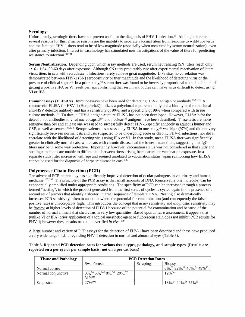
Serology Unfortunately, serologic titers have not proven useful in the diagnosis of FHV-1 infection.25 Although there are several reasons for this, 2 major reasons are the inability to separate vaccinal titers from response to wild-type virus and the fact that FHV-1 titers tend to be of low magnitude (especially when measured by serum neutralization), even after primary infection. Interest in vaccinology has stimulated new investigations of the value of titers for predicting resistance to infection.88,131 Serum Neutralization. Depending upon which assay methods are used, serum neutralizing (SN) titers reach only 1:16 - 1:64, 30-60 days after exposure. Although SN titers predictably rise after experimental reactivation of latent virus, titers in cats with recrudescent infections rarely achieve great magnitude. Likewise, no correlation was demonstrated between FHV-1 (SN) seropositivity or titer magnitude and the likelihood of detecting virus or the presence of clinical signs.25 In a prior study,68 serum titer was found to be inversely proportional to the likelihood of getting a positive IFA or VI result perhaps confirming that serum antibodies can make virus difficult to detect using VI or IFA. Immunoassays (ELISA’s). Immunoassays have been used for detecting HSV-1 antigen or antibody.132,133 A commercial ELISA for HSV-1 (Herpchek®) utilizes a polyclonal capture antibody and a biotinylated monoclonal anti-HSV detector antibody and has a sensitivity of 90%, and a specificity of 99% when compared with tissue culture methods.133 To date, a FHV-1 antigen-capture ELISA has not been developed. However, ELISA’s for the detection of antibodies to viral nucleocapsid134 and nuclear135 antigens have been described. These tests are more sensitive than SN and at least one was used to successfully detect FHV-1-specific antibody in aqueous humor and CSF, as well as serum.108,134 Seroprevalence, as assessed by ELISA in one study,25 was high (97%) and did not vary significantly between normal cats and cats suspected to be undergoing acute or chronic FHV-1 infections; nor did it correlate with the likelihood of detecting virus using IFA or VI. In that study, mean ELISA titer was significantly greater in clinically normal cats, while cats with chronic disease had the lowest mean titers, suggesting that IgG titers may be in some way protective. Importantly however, vaccination status was not considered in that study and serologic methods are unable to differentiate between titers arising from natural or vaccination exposure. In a separate study, titer increased with age and seemed unrelated to vaccination status, again reinforcing how ELISA cannot be used for the diagnosis of herpetic disease in cats.136 Polymerase Chain Reaction (PCR) The advent of PCR technology has significantly improved detection of ocular pathogens in veterinary and human medicine.137,138 The principle of the PCR assay is that small amounts of DNA (conceivably one molecule) can be exponentially amplified under appropriate conditions. The specificity of PCR can be increased through a process termed “nesting”, in which the product generated from the first series of cycles is cycled again in the presence of a second set of primers that identify a shorter, internal sequence of template DNA. Nesting also dramatically increases PCR sensitivity, often to an extent where the potential for contamination (and consequently the false positive rate) is unacceptably high. This introduces the concept that assay sensitivity and diagnostic sensitivity may be inverse at higher levels of detection of FHV-1 because of the potential for contamination and because of the number of normal animals that shed virus in very low quantities. Based upon in vitro assessment, it appears that (unlike VI or IFA) prior application of a topical anesthetic agent or fluorescein stain does not inhibit PCR results for FHV-1; however these results need to be verified in vivo.139 A large number and variety of PCR assays for the detection of FHV-1 have been described and these have produced a very wide range of data regarding FHV-1 detection in normal and abnormal eyes (Table 3). Table 3. Reported PCR detection rates for various tissue types, pathology, and sample types. (Results are reported on a per eye or per sample basis; not on a per cat basis)
Tissue and Pathology PCR Detection Rates Swab/brush Scraping Biopsy Normal cornea 6%,65 32%,48 46%,64 49%42 Normal conjunctiva 3%,74 6%,140 8%,58 20%,72
31%60 12%64
Sequestrum 27%102 18%,64 44%,56 55%65

Feline eosinophilic keratitis/conjunctivitis
55%,104 76%65
Conjunctivitis 10%,78 14%,60 18%,55 24%,140 33%72
54%64
Diseased but not specified 18%,141 25%,76 33%,142 47%,81 58%,79 89%,141 91%143
Although these studies were conducted on different populations with different clinical syndromes in different geographic areas, it is likely that some variation is also due to sampling and testing methodologies that might be controlled for by the clinician collecting and shipping the sample and the laboratory processing and reading out the sample. For example, variability among labs has been shown,144 whereas variation among sampling sites seems less important.79 Minimization of such variation will enable the clinician to better interpret the test result for their individual patient. Not unexpectedly, a major component of variability in PCR test result appears to be attributable to the PCR assay used, with calculated detection rates for the same samples tested with 6 different assays varying from 29-86%, and sample results being concordant in only 9 of 15 conjunctival biopsies.145 The type of sample collected, as well as the way in which the DNA is extracted and the PCR assay is performed also appears to be important.146,147 Although PCR may be used to look for FHV-1 DNA in any biological sample, the test is expected to be more likely to detect virus as more host cells are tested because FHV-1 is an obligate intracellular virus. Therefore, it is expected that biopsy samples are more likely to be positive than samples gathered by surface scraping, which in turn are more likely to be positive than swab samples; however this has not been tested yet. We did however compare cytobrushes and swabs and concluded that a swab was preferable to a cytobrush and that it should be submitted in a dry tube. Once it arrives at the lab, a defined volume (rather than a defined mass) of sample extract should be submitted to PCR.146 By contrast, sample handling and shipping protocols appear to exert less influence on FHV-1 PCR assay results and shipping at ambient temperature (not on ice) seemed appropriate.147 The main “take-home” message is that with shedding of FHV-1 at the peripheral surfaces of up to 50% of normal cats, individual testing seems of little benefit.42 Partly for this reason, quantitative PCR (qPCR) methods for FHV-1 detection have been assessed in experimentally infected animals.148 The authors’ hypothesis was that there is a critical concentration of viral DNA within host tissues that, below which, is likely to represent coincident or consequential virus, and above which is more likely to represent causative virus. The authors demonstrated good correlation between results of conventional methods of quantifying virus (viral titration) and the qPCR. Subsequently, SPF cats experimentally infected for the first time showed gradually reducing viral loads, as has been previously demonstrated using less sensitive techniques, such as VI. Relatively low-cellularity (STT and cytobrush) ocular samples from 20 cats which were demonstrating upper respiratory or ocular signs (keratitis and conjunctivitis) and in which natural FHV-1 infection was suspected then were assessed using the same technology. Reasonable correlation was again demonstrated between viral titer and DNA concentrations and, based on these data, the authors characterized cats into three “stages” of disease. However, another group has since reported the clinical utility of a qPCR method in cats without history of conjunctivitis, cats with presumed herpetic conjunctivitis that had resolved at least 3 weeks prior to testing and those with active presumed herpetic conjunctivitis. They found no difference among the 3 groups in either the number of cats that tested positive or the amount of FHV-1 DNA detected.74 Most recently severity of histologically graded rhinitis was found to be significantly correlated with amount of FHV-1 DNA detected using qPCR;143 however whether these results are repeatable or applicable for ocular signs is not clear. Recombinase polymerase amplification (RPA) is an isothermal gene amplification technology similar to but uses less-complex equipment that is portable and the reaction is completed much faster than PCR. An FHV-1 RPA assay has been performed in vitro and using clinical samples from diseased cats, and the results compared with RT-PCR.149 The detection limit was 102 copies DNA/reaction, the same as that of real time PCR, and the assay did not cross-detect feline panleukopenia virus, feline calicivirus, bovine herpesvirus-1, pseudorabies virus, or Chlamydia psittaci. A total of 120 nasal and conjunctival swabs from cats were then tested, and the results compared with those obtained with real-time PCR. Both assays provided the same testing results in the clinical samples.

TREATMENT OF FHV-1 OCULAR DISEASE Targets for Antiviral Pharmacology. Conceivably any step in the virus replicative process could be a target for antiviral intervention. These include adsorption, penetration, uncoating, early transcription, early translation, replication, late transcription, late translation, virus assembly, and release from the host cell. To date however, most of the effective antiviral therapies that have been developed target viral enzymes and proteins responsible for DNA synthesis. For a review of many available agents and their proposed mechanisms of action see this article by De Clercq,150 and for their specific applications in feline medicine, see the review by Thomasy & Maggs.151 Specific Antiviral Agents and FHV-1 FHV-1 is susceptible to inhibition by all commercially available antiviral ophthalmic medications studied thus far, however their safety in cats is not readily predicted from their behavior in other host species (principally humans), and their efficacy against FHV-1 is not predicted from their efficacy against other viruses (principally HSV-1 and -2). In addition, there are no antiherpetic drugs currently approved in the USA for use against FHV-1 in cats. All those in common use are “borrowed” from the human pharmacy.
Whenever we take a drug developed for the treatment of HSV-1 in humans and use it to treat
a cat infected with FHV-1 we are making 2 giant leaps of faith unless placebo-controlled, masked, prospective trials of its safety (for cats) and efficacy (against FHV-1) have been performed. For these reasons, careful in vitro investigation of efficacy against FHV-1, followed by safety and pharmacokinetic trials, subsequent placebo-controlled efficacy
studies in experimental animals, and finally judicious clinical trials in client-owned animals should always precede widespread clinical use and anecdotal reporting.
Although opinions vary as to when and why antiviral therapy should be instituted in cats believed to be experiencing herpetic diseases, it appears reasonable that antiviral agents should be considered when signs are severe, persistent, or recurrent, particularly when there is corneal involvement, and especially ulceration. Because epithelial replication, latency and reactivation, and persistence are such interdependent and sequential phases of herpetic disease, interruption of any one of them is expected to limit the virus’ abilities to cause subsequent disease. Therefore, aggressive treatment of FHV-1-associated disease may limit disease progression and minimize frequency and severity of recurrences. This seems to be borne out by data that shows a significant direct (positive) correlation between clinical score and viral DNA copy number 30 days after experimental infection, which suggests that the quantity of viral DNA that becomes latently established in the ganglia is related to the severity of the acute clinical infection.67 Some important general concepts about antiviral agents assist with prescribing and expectations of this class of drugs: Because viruses reside intracellularly and utilize host cellular machinery, antiviral agents tend to exhibit
greater host toxicity than do antibacterial drugs. This sometimes limits topical application of these drugs but especially limits their systemic use.
All antiviral agents to date are virostatic; therefore they require replicating virus and must be dosed (orally or topically) relatively frequently.
No antiviral drug has proven antibacterial activity. Antiviral drugs safe in humans are not necessarily safe in cats. Antiviral drugs effective against human herpesviruses are not necessarily effective against FHV-1. Antiviral prodrugs metabolized to their active form by humans are not predictably metabolized by cats. The effect of some antiviral drugs on FHV-1 replication in vitro has been studied and their potency relative to each other and relative to HSV-1 reported (Table 4). Antiviral efficacy is expressed as the IC50 (or the concentration at which viral replication is inhibited by 50% ). Note, therefore, that a lower IC50 equates to a more efficacious antiviral drug.
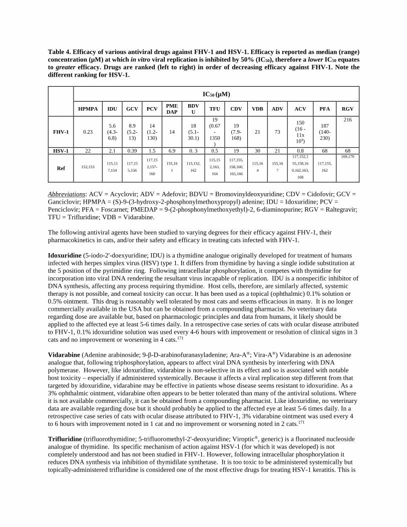
Table 4. Efficacy of various antiviral drugs against FHV-1 and HSV-1. Efficacy is reported as median (range) concentration (µM) at which in vitro viral replication is inhibited by 50% (IC50), therefore a lower IC50 equates to greater efficacy. Drugs are ranked (left to right) in order of decreasing efficacy against FHV-1. Note the different ranking for HSV-1.
Abbreviations: ACV = Acyclovir; ADV = Adefovir; BDVU = Bromovinyldeoxyuridine; CDV = Cidofovir; GCV = Ganciclovir; HPMPA = (S)-9-(3-hydroxy-2-phosphonylmethoxypropyl) adenine; IDU = Idoxuridine; PCV = Penciclovir; PFA = Foscarnet; PMEDAP = 9-(2-phosphonylmethoxyethyl)-2, 6-diaminopurine; RGV = Raltegravir; TFU = Trifluridine; VDB = Vidarabine. The following antiviral agents have been studied to varying degrees for their efficacy against FHV-1, their pharmacokinetics in cats, and/or their safety and efficacy in treating cats infected with FHV-1. Idoxuridine (5-iodo-2'-doexyuridine; IDU) is a thymidine analogue originally developed for treatment of humans infected with herpes simplex virus (HSV) type 1. It differs from thymidine by having a single iodide substitution at the 5 position of the pyrimidine ring. Following intracellular phosphorylation, it competes with thymidine for incorporation into viral DNA rendering the resultant virus incapable of replication. IDU is a nonspecific inhibitor of DNA synthesis, affecting any process requiring thymidine. Host cells, therefore, are similarly affected, systemic therapy is not possible, and corneal toxicity can occur. It has been used as a topical (ophthalmic) 0.1% solution or 0.5% ointment. This drug is reasonably well tolerated by most cats and seems efficacious in many. It is no longer commercially available in the USA but can be obtained from a compounding pharmacist. No veterinary data regarding dose are available but, based on pharmacologic principles and data from humans, it likely should be applied to the affected eye at least 5-6 times daily. In a retrospective case series of cats with ocular disease attributed to FHV-1, 0.1% idoxuridine solution was used every 4-6 hours with improvement or resolution of clinical signs in 3 cats and no improvement or worsening in 4 cats.171 Vidarabine (Adenine arabinoside; 9-β-D-arabinofuranasyladenine; Ara-A®; Vira-A®) Vidarabine is an adenosine analogue that, following triphosphorylation, appears to affect viral DNA synthesis by interfering with DNA polymerase. However, like idoxuridine, vidarabine is non-selective in its effect and so is associated with notable host toxicity – especially if administered systemically. Because it affects a viral replication step different from that targeted by idoxuridine, vidarabine may be effective in patients whose disease seems resistant to idoxuridine. As a 3% ophthalmic ointment, vidarabine often appears to be better tolerated than many of the antiviral solutions. Where it is not available commercially, it can be obtained from a compounding pharmacist. Like idoxuridine, no veterinary data are available regarding dose but it should probably be applied to the affected eye at least 5-6 times daily. In a retrospective case series of cats with ocular disease attributed to FHV-1, 3% vidarabine ointment was used every 4 to 6 hours with improvement noted in 1 cat and no improvement or worsening noted in 2 cats.171 Trifluridine (trifluorothymidine; 5-trifluoromethyl-2'-deoxyuridine; Viroptic®, generic) is a fluorinated nucleoside analogue of thymidine. Its specific mechanism of action against HSV-1 (for which it was developed) is not completely understood and has not been studied in FHV-1. However, following intracellular phosphorylation it reduces DNA synthesis via inhibition of thymidilate synthetase. It is too toxic to be administered systemically but topically-administered trifluridine is considered one of the most effective drugs for treating HSV-1 keratitis. This is
IC50 (µM)
HPMPA IDU GCV PCV PMEDAP
BDVU TFU CDV VDB ADV ACV PFA RGV
FHV-1 0.23 5.6
(4.3-6.8)
8.9 (5.2-13)
14 (1.2-130)
14 18
(5.1-30.1)
19 (0.67
-1350
)
19 (7.9-168)
21 73
150 (16 -11x 104)
187 (140-230)
216
HSV-1 22 2.1 0.39 1.5 6.9 0. 3 0.5 19 30 21 0.8 68 68
Ref 152,153 115,11
7,154 117,15
5,156
117,15
2,157-
160
155,16
1 115,152,
162
115,15
2,163,
164
117,155,
158,160,
165,166
115,16
4 155,16
7
117,152,1
55,158,16
0,162,163,
168
117,155,
162
169,170

in part due to its superior corneal epithelial penetration.172 In vitro, it is also one of the more potent antiviral drugs for FHV-1. It is commercially available in the USA as a 1% ophthalmic solution that based on human recommendations should probably be applied to the affected eye at least 5-6 times daily. Unfortunately, it is expensive, and often causes marked ocular irritation. In a retrospective case series of cats with ocular disease attributed to FHV-1, 1% trifluridine solution was used every 4-8 hours with improvement in 1 cat and no improvement or worsening in 2 cats.171 Acyclovir (9-(2-hydroxyethoxymethy)guanine; ACV; Zovirax®) is the prototype of a group of antiviral drugs known as acyclic nucleoside analogues. Members of this group of antiviral agents all require 3 phosphorylation steps for activation. The first of these steps must be catalyzed by a viral enzyme, thymidine kinase. This fact increases their safety and permits them to be systemically administered to humans. Unfortunately, the activity of the FHV-1 thymidine kinase enzyme on acyclovir is much less than that of the HSV-1-encoded enzyme, which likely explains the relative lack of efficacy of ACV against FHV-1.158,173 The second and third phosphorylation steps must be performed by host enzymes. To my knowledge the affinity of these enzymes for the acyclic nucleoside analogues has not been studied in cats. In addition to relatively low antiviral potency against FHV-1, acyclovir has poor bioavailability and is potentially toxic when systemically administered to cats. Oral administration of 50 mg/kg acyclovir to cats was associated with peak plasma levels of only 33 μM (approximately one third the IC50 for this virus).119 Common signs of toxicity are referable to bone marrow suppression. With the advent of the apparently safer and more effective famciclovir, administration of acyclovir seems more difficult to justify. Acyclovir is also available as an ophthalmic ointment in some countries which, while it may overcome systemic toxicity concerns, need not necessarily increase antiviral efficacy against FHV-1. Regardless, a 0.5% ointment used 5 times daily in naturally infected cats was associated with a median time to resolution of clinical signs of 10 days.163 Cats treated only 3 times daily took approximately twice as long to resolve and did so only once therapy was increased to 5 times daily. Taken together, these data suggest that frequent (at least 5 times daily) topical application of acyclovir may produce concentrations at the corneal surface that exceed the IC50 for this virus but are not associated with clinically appreciable toxicity. There are also in vitro data suggesting that interferon exerts a synergistic effect with acyclovir that could permit an approximately 8-fold reduction in acyclovir dose.174 In vivo investigation and validation of these data are needed. Valacyclovir (L-valine, 2-[(2-amino-1,6-dihydro-6-oxo-9H-purin-9-yl)methoxy]ethyl ester, monochloride; Valtrex®) is another acyclic nucleoside analogue. As a prodrug of acyclovir, it is - in humans and cats - more efficiently absorbed from the gastrointestinal tract than acyclovir is and, following absorption, is converted to acyclovir by a hepatic hydrolase.175 Plasma concentrations of acyclovir that surpass the IC50 for FHV-1 can be achieved after oral administration of this drug. However, in cats experimentally infected with FHV-1, valacyclovir induced fatal hepatic and renal necrosis, along with bone marrow suppression, and did not reduce viral shedding or clinical disease severity.118 Therefore, despite its superior pharmacokinetics, valacyclovir should never be administered to cats. This also reminds us how toxic acyclovir is in cats once a sufficiently high plasma concentration is achieved. Ganciclovir (Cytovene®, Zirgan®) is another acyclic nucleoside analogue that, like acyclovir, requires triphosphorylation to achieve its active form with the first phosphorylation step mediated by viral thymidine kinase. It appears to be approximately 10-fold more effective against FHV-1 than is acyclovir.117 It is available for oral or intravenous administration in humans, where it is associated with more severe neurologic toxicity, neutropenia, and bacterial infections than is acyclovir.176,177 Like acyclovir, a prodrug of ganciclovir (valganciclovir) is available. To my knowledge, the safety and pharmacokinetics of ganciclovir or valganciclovir have not yet been studied in cats. An ophthalmic gel preparation (Zirgan®; 0.15%) is available for human use but is currently very expensive in the USA. However there are anecdotal reports of good efficacy and tolerability in cats with FHV-1 in Europe (where it is cheaper). Given the in vitro efficacy of ganciclovir, this product definitely warrants efficacy and safety trials in cats. However, to the author’s knowledge, neither the safety nor pharmacokinetics of valganciclovir or ganciclovir in any form has been reported in cats. Penciclovir (9-(4-hydroxy-3-hydroxymethylbut-1-yl)guanine; BRL39123; Denavir®, Vectavir®) is a nucleoside deoxyguanosine analogue with a similar mechanism of action to acyclovir and with potent antiviral activity against a number of human herpesviruses. Like acyclovir, it requires viral and cellular phosphorylation but is highly effective against FHV-1 in vitro158-160 and in vivo.98 In a rabbit model of human HSV-1 keratitis, a 3% penciclovir ointment administered once, twice or four times daily decreased epithelial keratitis severity.178 Thus, a topical ophthalmic

penciclovir ointment may be effective in cats with FHV-1 keratitis and/or conjunctivitis, but, to the authors’ knowledge, there are no commercial or compounded preparations available for ophthalmic use. Penciclovir is available as a 1% dermatologic cream for humans, but that should not be applied to the eye. Famciclovir (2-(2-(2-amino-9H-Purin-9-yl)ethyl)-1,3-propanediol diacetate; Famvir®) is a highly bioavailable prodrug of penciclovir, which – once absorbed – is metabolized to penciclovir. In humans this metabolism is complex; requiring di-deacetylation to BRL42359, in the blood, liver, or small intestine, with subsequent oxidation to penciclovir by aldehyde oxidase in the liver.179-181 Neither famciclovir nor BRL42359 has any in vitro antiviral activity against FHV-1,159 therefore complete metabolism to penciclovir is required. However, hepatic aldehyde oxidase activity in cats is about 2% of that seen in humans and lower than in any other species reported to date.182 Famciclovir pharmacokinetics in the cat are extremely complex and nonlinear (i.e., doubling of famciclovir dose does not lead to doubling of plasma penciclovir concentration) presumably due to saturation of the hepatic oxidase.183 As a result very high plasma concentrations of BRL42359 accumulate in the cat.183 Fortunately, this compound demonstrates very little cytotoxicity in vitro.159 Table 5 summarizes the pharmacokinetic data available to date for penciclovir in tears and plasma of cats receiving one of numerous famciclovir dose regimens. Tissue concentration data are not yet available. Table 5. Maximum (Cmax) and minimum plasma and tear penciclovir concentrations and time to plasma and tear Cmax in cats administered a variety of famciclovir doses at various dose frequencies. Abbreviations: BID: twice daily; Cmax: maximum observed drug concentration; Css(min): minimum observed drug concentration during the dosing interval at steady state; FCV: famciclovir; ND: not done; PCV: penciclovir; TID; thrice daily; Tmax: time to Cmax. In addition to these pharmacokinetic data, recommendation of an appropriate famciclovir dose requires: • Knowledge of whether penciclovir concentrations in plasma, tears, or the infected tissues themselves are
most relevant • Selection of an appropriate target penciclovir concentration based on in vitro IC50s (which reportedly range
from 304 to 3500 ng/mL.)117,152,159,160 • Knowledge of whether the targeted IC50 should be exceeded by the trough or the peak penciclovir
concentrations, and for how long. Together, these uncertainties have led to much controversy about the optimum famciclovir dose in cats, with reported doses ranging from 8 mg/kg once daily185 to 140 mg/kg thrice daily.186 The following data are provided to inform dose selection. In the only masked, placebo-controlled efficacy trial to date, cats known to be infected with FHV-1 and given approximately ~90 mg famciclovir/kg thrice daily per os achieved an approximate peak plasma penciclovir concentration of 2100 ng/mL.98 Relative to control cats, treated cats had significantly reduced clinical signs, decreased serum globulin concentrations, reduced histologic evidence of conjunctivitis, decreased viral shedding, and reduced serum FHV-1 titers, as well as increased goblet cell density.98 A subsequent study showed that administration of a single dose of 40 mg/kg to uninfected healthy cats achieved nearly identical plasma penciclovir concentrations to those achieved with a single dose of 90 mg/kg.187 A third study188 revealed that cats receiving 40
FCV dose Dose frequency
Plasma [PCV] (ng/mL)
Plasma PCV Tmax (h)
Tear [PCV] (ng/mL) Tear
PCV Tmax (h) Cmax Css(min) Cmax Css(min)
9-18 mg/kg184
BID 330 64 5.3 ND ND ND TID 680 180 3.8 ND ND ND
30 mg/kg183
BID 2010 345 1.3 305 65 4.7 TID 1755 570 1.7 395 160 2.0
40 mg/kg183
BID 1945 445 2.3 375 55 3.3 TID 2210 790 2.5 750 150 2.6
90 mg/kg183
BID 2720 630 2.7 680 200 2.3 TID 2015 905 2.7 555 275 3.0

mg/kg thrice daily had tear penciclovir concentrations likely to be effective against FHV-1 (using a target IC50 of 304 ng/mL)158 for at least 3 hours after each dose (i.e., for ≥ 9 hours/day). In the most comprehensive pharmacokinetic study to date, healthy cats were administered famciclovir at 30, 40 or 90 mg/kg twice or thrice daily, and plasma and tear famciclovir, BRL42359, and penciclovir concentrations were measured.183 This resulted in the recommendation that cats should receive 90 mg famciclovir/kg twice daily because this regimen achieved comparable plasma and tear penciclovir concentrations to those achieved with 90 mg/kg thrice daily, whereas the lower doses tested did not result in adequate tear penciclovir concentrations, even when administered thrice daily. Perhaps most revealing so far, are data from a retrospective study comparing outcomes when famciclovir was administered thrice daily to cats with presumed herpetic disease at approximately 40 (n = 33 cats) or 90 mg/kg (n = 26 cats).186 Median duration of therapy required for clinical improvement was significantly longer in cats administered 40 versus 90 mg/kg. Furthermore, cats in the 90 mg/kg group showed significantly greater and faster improvement than did cats in the 40 mg/kg group. The reduction in treatment duration with the higher famciclovir dose was estimated to decrease overall client costs due to a reduction in total famciclovir administered (and potentially the number of recheck examinations required). These data, suggest that 90 mg/kg TID is clinically and cost effective. Meanwhile, pharmacokinetic data183 suggest that tear and plasma penciclovir concentrations are similar whether cats receive 90 mg famciclovir /kg 2 or 3 times daily. Taken together, data from these 2 studies183,186 suggest that 90 mg famciclovir/kg twice daily is likely to be effective in treating cats with herpetic disease. These data, combined with those from pharmacokinetic studies suggest that 90 mg/kg twice daily is likely to be effective in treating cats with FHV-1. Adverse events (most commonly gastrointestinal) potentially attributable to famciclovir were reported in 17% of cats receiving 40 or 90 mg famciclovir/kg thrice daily, but the prevalence was not different between the 2 dose groups.186 Assessing all in vivo tolerance data for famciclovir, this drug appears to be markedly safer than acyclovir119 or valacyclovir118 - the only other systemic antiviral drugs to be orally administered to cats.98,118,119,183-188However, patients administered famciclovir should be closely monitored, and assessment of a complete blood count, serum biochemistry panel and urinalysis should be considered in cats with known intercurrent concurrent disease or cats expected to receive famciclovir for long periods. As in humans,189 reduction in dose frequency should be considered in cats with renal insufficiency. Data to date reveal that administration of famciclovir to cats in shelters is not successful.190 Cidofovir is a cytosine analogue that requires 2 host-mediated phosphorylation steps but does not require virally-mediated phosphorylation. Its safety arises from its relatively high affinity for viral DNA polymerase compared with human DNA polymerase.191 It is available in injectable form in the United States and is administered intravenously or intravitreally to humans infected with herpesviruses; principally cytomegalovirus. This drug has also been topically applied as a 0.5% or 1.0% solution in experimental animal models of human herpetic keratoconjunctivitis and found to be equally effective when administered only twice daily as trifluridine was when administered 4-9 times daily.178,192 This is believed to be due to the long tissue half-lives of the metabolites of this drug.193 The efficacy of a 0.5% solution compounded in methylcellulose artificial tears and applied topically twice daily to cats experimentally infected with FHV-1 was associated with reduced viral shedding and clinical disease in a prospective, masked placebo-controlled study.194 There are occasional reports of its experimental topical use in humans being associated with stenosis of the nasolacrimal drainage system components,195,196 and it is not commercially available as an ophthalmic agent in humans. Therefore, although the in vitro and short-term in vivo efficacy of cidofovir against FHV-1 are proven, cats should be monitored for nasolacrimal cicatrization. Unlike most compounded agents, there are also some excellent data on the efficacy over time and using various storage conditions for cidofovir. Cidofovir 0.5% compounded in normal saline (not methylcellulose as was studied in vivo) retained efficacy under all combinations of storage conditions tested: in plastic or glass; refrigerated (4C), or frozen at -20C or -80C; and for 1, 2, 4, or 6 months.197 Importantly, safety was not studied. Lysine Lysine is perhaps the best studied and yet maybe one of the more controversial of all of the other compounds with proven or putative antiviral efficacy against FHV-1 in cats. As with the antiviral drugs, initial interest arose from in vitro data and clinical trials in humans. Lysine’s antiviral effect is believed to arise because arginine is an essential amino acid for FHV-1198 and HSV-1199,200 replication, and assumes that lysine antagonizes arginine availability to or utilization by these viruses during protein synthesis. This was hypothesized to affect protein synthesis of the virus more than the host because viral proteins had a higher arginine-to-lysine content than did human (and feline) proteins.201 And recent analysis suggests that the difference in feline versus FHV-1 protein amino acid content is minimal.202 This may explain why in vitro replication of HSV-1 199,200 and FHV-1198 is notably suppressed in the

presence of markedly elevated lysine concentrations in combination with exceptionally low arginine concentrations, but that this is not borne out with more physiologic amino acid concentrations.203 Alternate mechanisms may explain the suppression of viral replication attributable to lysine supplementation. Firstly, lysine and arginine competitively inhibit the transport of each other, suggesting that these amino acids share a common transport system.204 Secondly, lysine induces arginase, an enzyme that causes the catabolic degradation of arginine.205,206 This enzyme could be responsible for a further increase in the intracellular lysine-to-arginine ratio. In vivo data in cats are also contradictory. Stiles, et al began by demonstrating the safety and efficacy of oral administration of lysine to cats prior to primary exposure to FHV-1.207 Cats receiving 500 mg L-lysine every 12 hours per os (beginning 6 hours prior to experimental inoculation with FHV-1) exhibited less severe conjunctivitis than cats receiving placebo. However, viral shedding, as determined by VI, did not differ between groups. Subsequently, we demonstrated that oral lysine supplementation helps to prevent viral reactivation and/or shedding in latently infected cats.208 Despite significant elevations in plasma lysine concentration, no change in plasma arginine concentration or any ill effects attributable to lysine administration were observed in either study (other than perhaps some mild and reversible GI disturbance in Stiles’ study). I am aware of only one study in which bolus administration of lysine has been tested in naturally infected cats.209 This study examined the effects of lysine in 144 cats residing in a humane shelter. Cats received oral boluses of 250 mg (kittens) or 500 mg (adult cats) of lysine once daily for the duration of their stay at the shelter and outcomes were compared with those of an untreated control group. No significant treatment effect was detected on the incidence of infectious upper respiratory disease (IURD), the need for antimicrobial treatment for IURD, or the interval from admission to onset of IURD. However it was not determined if and to what extent these cats were shedding or infected with FHV-1 (or other pathogens). Bolus administration of lysine is sometimes impractical, for several reasons. Cats frequently require very protracted therapy (life-long in some cases), and often live in multicat environments in which all cats should be treated. In some multicat situations, twice-daily oral administration techniques may stimulate further viral reactivation through stress or facilitate transfer of infectious organisms among cats by operators. Thus, we studied the safety and efficacy of incorporating lysine into cat food. Results of an initial safety trial were encouraging.210 Cats fed a diet supplemented with up to 8.6% (dry matter) lysine showed no signs of toxicity, had normal plasma arginine concentrations, and had normal food intake. Mean plasma lysine concentration of these cats was increased to levels similar to that achieved with bolus administration. In a subsequent study,211 25 cats with enzootic upper respiratory tract disease were fed a diet supplemented to 5.1% lysine while 25 cats remained on a basal ration (~1% lysine). The study was conducted for 52 days at the beginning of which cats were subjected to rehousing stress (which is known to cause viral reactivation). Ironically, food (and therefore lysine) intake decreased coincident with peak disease and viral presence. As a result, cats did not receive lysine at the very time they needed it most. Perhaps partly because of this, disease in cats fed the supplemented ration was more severe than that in cats fed the basal diet. In addition, viral shedding was more frequent in cats receiving the supplemented diet. To further assess these unexpected outcomes, we performed a similarly designed experiment212 in a local human shelter with a more consistent “background” level of stress and with greater numbers enrolled compared to the initial rehousing study.211 We enrolled 261 cats; each for 4 weeks. Despite plasma lysine concentration in treated cats being greater than that in control cats, again more treated cats than control cats developed moderate to severe disease and shed FHV-1 DNA at certain points throughout the study.212 In summary, there is considerable variability among these studies, especially with respect to methodology, study population, and dose and method of lysine administration. However, taken together, data from these studies suggest that lysine is safe when orally administered to cats and, provided that it is administered as a bolus, may reduce viral shedding in latently infected cats and clinical signs in cats undergoing primary exposure to the virus. However, the stress of bolus administration in shelter situations may well negate its effects and data do not support dietary supplementation. Unfortunately, no clinical trials have been conducted in the very group in which this drug is commonly used - client-owned cats with recurrent herpetic disease. Currently, I let clients help me decide whether they feel a trial of lysine in their cat was effective. I always administer L-lysine at 500 mg twice daily, and ensure that clients do not add it to food for animals to “graze” over an extended period. I also make owners aware that this is usually only an adjunctive or palliative therapy and that administration of antiviral drugs may also be necessary to gain better control of signs. Unlike the protocol for HSV-1-infected humans, owners of cats receiving lysine for FHV-1 should not be advised to restrict their cat’s arginine intake. Feeding a single arginine-deficient meal to a cat can induce fatal encephalopathy.213 By adopting a common protocol of administering lysine to client-owned cats, it is hoped that well designed, double-masked studies in clinically relevant populations will be forthcoming. Such data would be especially relevant if generated in populations with variable vaccination history, intercurrent disease,

physiologic stresses, and high turnover of cats of diverse genetic composition and with varied exposure to infectious diseases. Lambda-carrageenan Lambda-carrageenan is a red seaweed extract containing sulfated polysaccharides with demonstrated antiviral activity against numerous enveloped viruses including HSV-1. Activity of λ-carrageenan against FHV-1 replication in vitro and in vaccinated cats exposed for the first time to wild type FHV-1 has been examined in a placebo-controlled masked study.214 λ-carrageenan demonstrated some antiviral activity in vitro when used prior to but not following viral adsorption. Safety/tolerance trials revealed no adverse effects in uninfected cats when 1 drop of a 250 µg/mL (or less concentrated) solution was applied topically 4 times per day. Efficacy was tested using 1 drop of a 250 µg/mL λ-carrageenan solution applied before and after infection (n = 6 cats) or after infection only (n = 6 cats). The drug did not reduce clinical signs, regardless of whether it was applied prior to and after infection or only after infection and a significant reduction in virus isolation was noted only on day 21 following inoculation. Other plant extracts with antiviral activity have also been studied in vitro.215 Leflunomide Leflunomide is an immunosuppressive agent that appears to have some antiviral effects against human herpesviruses including HSV-1.216 Its effects against FHV-1 in vitro have been reported.217 Simultaneous inoculation of CRFK cells with virus and treatment with leflunomide was associated with a significant and dose-dependent reduction in plaque number and - at higher concentrations - viral load. However (curiously) viral load was not reduced in a dose dependent manner at very low concentrations of this drug. At higher concentrations, some cytotoxicity was observed. Electron microscopy revealed an apparent failure in viral assembly, especially of the tegument and external membrane, which may indicate the mode of action of this drug.217 Lactoferrin Lactoferrin is a mammalian iron-binding glycoprotein that has antibacterial, antifungal, antiprotozoal, and antiviral properties. It is produced by mucosal epithelial cells of many mammalian species and is present in secretions such as tears, saliva, seminal and vaginal fluids, and in low concentrations in plasma. Breast milk, especially colostrum, is a major source of lactoferrin. Lactoferrin has been demonstrated to exert a very potent antiviral effect against FHV-1 replication in vitro.2 Further analysis of the data in this study reveals that lactoferrin appears to inhibit FHV-1 adsorption to the cell surface and/or penetration of the virus into the cell. At a molecular level, this may occur due to interaction of lactoferrin with receptors on the cell surface or by direct neutralization or inhibition of the viral particle. Probiotics To date, I am aware of one prospective, placebo-controlled study of probiotic use for FHV-1 infection in cats.218 Cats were experimentally infected with FHV-1 for another study,194 and then administered as a dietary supplement an immune-enhancing probiotic (Enterococcus faecium strain SF68) or placebo. All cats in both groups showed such minimal evidence of disease that a statistically significant difference between groups could not be demonstrated. Pheromones/Stress Likewise, I am aware of one prospective, placebo-controlled study of pheromone use for FHV-1 infection in cats.219 Cats were experimentally infected with FHV-1 for another study, and then (8 weeks later) placed in a room with a diffuser distributing a pheromone (Feliway®) or placebo, while simultaneously subjected to changes in housing and groupings that are known stressors. At various time points the pheromone treated group had lower stress scores, and sneezing relative to the placebo-treated group. Suggesting that stress and herpetic reactivation with recrudescent disease may have been reduced by the use of the pheromone. Very few cats shed viral DNA so this could not be evaluated. Small (or short) interfering RNAs (siRNAs) Small (or short) interfering RNAs (siRNAs) are short (about 20 nucleotide), double-stranded sections of RNA designed to transfect a cell and interfere with (“knockdown”) expression of specific gene(s). To overcome the short-lived effect of transfection of native siRNAs, they can be incorporated into plasmids and thus extend their longevity,

especially within rapidly dividing cells. Three studies have now been published investigating the effects of these agents on FHV-1.38-40 An initial in vitro study of siRNAs that targeted the glycoprotein and DNA polymerase genes of FHV-1 showed that the siRNAs directed against the FHV-1 glycoprotein D (gD) gene had some antiviral effect and warranted further study.38 A subsequent in vitro study assessed efficacy of some additional siRNAs alone or in combination and revealed that combined use of siRNAs targeting the FHV-1 DNA polymerase and gD genes was effective.39 However, intracellular delivery of these agents is essential but proving complex. While delivery agents that facilitate delivery of siRNAs into corneal cells in vitro have been developed and these were nontoxic in vitro and nonirritating when applied topically to normal cats’ eyes, thus far they have failed to deliver the siRNAs into corneal cells following topical application in vivo, perhaps due to rapid removal from the ocular surface by tears.40 Anti-inflammatory therapy The use of anti-inflammatory drugs in the management of FHV-1 infections remains controversial.220 However, a return to basic virology and a review of the literature makes some comments possible. Recall that FHV-1 produces disease by (at least) two very different mechanisms that require markedly different (almost opposite) therapeutic approaches. Cytolytic infection represents active viral replication and is often ulcerative. Immunomodulation at this point is almost certainly contraindicated. By contrast, immunopathological (or immune-mediated) injury (such as feline eosinophilic keratitis) is mediated by host inflammatory responses “driven” by persistent viral and/or auto antigens.
The important therapeutic implication is that the immune-mediated disease may involve no or low-grade viral replication and so antiviral agents may not be effective as sole therapeutic agents. However, anti-inflammatory therapy may be indicated (presumably in addition to antiviral agents) for the treatment of immunopathological herpetic diseases.
Corticosteroids. Systemic administration of corticosteroids is a well-established and reliable means of inducing viral reactivation from latency.49 This must be considered whenever these drugs are considered in the clinical management of other disease in FHV-1-infected cats. The ability of locally-administered corticosteroids to exacerbate self-limiting primary conjunctival infection and to sometimes induce chronic herpetic keratitis has also been well established.92 Complications seen in corticosteroid treated eyes included deeper and more persistent corneal ulcers, corneal edema, corneal vascularization, sequestrum or band keratopathy formation, and protracted viral shedding. Topical corticosteroids are therefore contraindicated in primary ocular FHV-1 infection. The specific adverse effects of corticosteroids in ocular viral infection are diverse and include inhibition of RNA and DNA, suppression of protein synthesis, decreased antigen presentation by T cells, decreased production of IL2, and impairment of macrophage and cellar immune functions. Therefore, from a basic virological standpoint, corticosteroid administration is difficult to support if active viral replication is occurring. However, because corneal opacification in chronic stromal keratitis occurs secondary to an exuberant immune response, corticosteroids may be indicated in such cases. It would seem prudent, however, to always treat concurrently with a topical antiviral. NSAIDs and Cyclosporine. The potential complications from using corticosteroids have prompted interest in the use of non-steroidal anti-inflammatory drugs (NSAIDs) for managing the inflammatory effects of ocular FHV-1 infection. Although flurbiprofen has been shown to exert some in vitro antiviral effect,221 HSV-1 clinical trials have demonstrated equivocal efficacy. In fact, in rabbits, flurbiprofen has been shown to exacerbate viral keratitis.222 Due to its relatively specific T-cell suppression, cyclosporine therapy has been investigated but has produced equivocal results. Although cyclosporine is capable of suppressing inflammatory events operative in viral stromal keratitis, this drug is also capable of impairing viral clearance from the eye, and beneficial immune responses. In vitro, cyclosporine exerts a dose dependent effect on HSV-1 replication.223 In some experimental model systems, however, cyclosporine therapy resulted in more severe and persistent keratitis.224 In clinical trials, cyclosporine and trifluridine were used in combination to treat HSK in humans with good results,225 and cyclosporine was used (often along with other therapies) for cats with eosinophilic keratitis, typically with good results.226 Other than that, use of NSAIDs and cyclosporine in chronic feline herpetic disease has been inadequately studied and I am unaware of any studies examining the effects of tacrolimus on ocular herpetic infections in any species. This suggests that use of these agents should, as a minimum, be restricted by the same principles that govern the use of corticosteroids in HSK. A study assessing orally administered cyclosporine failed to show a marked deleterious effect in cats previously infected with FHV-1.227

Immunotherapy A commercial hyperimmune serum containing antibodies against FCV, FHV-1, and feline panleukopenia virus is available in Europe for treatment of cats with upper respiratory disease and has been assessed in a randomized, placebo-controlled, double-masked clinical trial.228 Forty two cats received hyperimmune serum (n = 22) or saline (n = 20) for 3 consecutive days as well as symptomatic treatment. The serum was administered subcutaneously once daily and topically into eyes, nostrils, and mouth every 8 h. Clinical signs were recorded daily for 8 days and again on day 21. Clinical signs in both groups improved significantly over time (P < 0.001) with treated cats improving earlier (Day 3) than placebo-treated cats (Day 7). In a separate study,229 2 days post inoculation with FHV-1, 9 cats received placebo (n=3), or 10 (n=3) or 30 (n=3) mg/kg of chimeric antibodies developed against FHV-1 in hybridoma cells. Treated cats at both doses showed remarkable reduction in clinical signs. Pre-treatment of cats 15 days before inoculation had similar results.230 Meanwhile, polyprenyl immunostimulant (PI), an immunomodulatory veterinary biologic, when administered orally at 5 mg/kg (10× therapeutic dose) and at the same time as experimental FHV-1 inoculation of SPF cats was associated with significantly reduced clinical scores relative to placebo-treated cats and was suggested to be safe in cats as young as 8 weeks of age based on data from a field safety study involving 390 owned cats in 10 states.231 Interferons The interferons (IFNs) are a group of cytokines that have diverse immunological and antiviral functions. The IFNs are divided into 4 groups; α, β, γ, and ω interferons, and numerous subtypes. Viral infection stimulates cells to secrete IFN into the extracellular space where it binds to specific receptors on neighboring cells and, through mechanisms not fully understood, prevents or limits the spread of infection (i.e., it is not virucidal). Knowledge and consideration of this mode of action allows the clinician to set and maintain reasonable expectations of how effectively the IFNs may work when used therapeutically, and in which patients with which stage of disease they might be expected to be most effective.
Although IFNs may play important physiological roles in the control of viral infections, in vitro and clinical trials investigating potential therapeutic applications have produced conflicting and not solidly supportive results. In in vitro studies, 1 x 10⁵ - 5 x 10⁵ IU/ml of recombinant human IFNα or recombinant feline IFNω significantly reduced FHV-1 titer and/or cytopathic effect while not producing any detectable cytotoxic changes in the feline corneal cell line232 or CRFK cells233 on which the virus was grown. At higher concentrations, the effect of the recombinant feline IFNω was greater than that of IFNα.233 In a separate in vitro study, notable synergistic activity against FHV-1 was demonstrated when 10 - 62.5 µg/mL of acyclovir was combined with 10 or 100 IU/mL of human recombinant IFNα. The combined use of the 2 compounds did not cause increased cytotoxicity but permitted a nearly eightfold reduction in the dose of acyclovir required to achieve maximal inhibition of FHV-1. Significant synergistic interactions resulted when the IFNα was given before or after infection at the lower doses of acyclovir; however pretreatment was more effective.174 To my knowledge, there are relatively few peer-reviewed, placebo-controlled, prospective clinical trials of IFN administration to SPF cats experimentally infected with FHV-1 (the gold standard for testing efficacy of the other antiviral products described here). One study utilized 10,000 IU of recombinant feline IFNω administered topically (OU) q 12 hours and 2,000 IU administered PO q 24 hours.234 Based on data generated in studies utilizing other viruses and knowledge of the mode of action of IFNs, IFN administration in this feline study234 was initiated 2 days prior to viral inoculation but was not continued after inoculation. No beneficial effects were shown. The effects of very high-dose systemic administration of IFNα prior to experimental FHV-1 infection have also been studied; 108 IU/kg were administered subcutaneously BID on two consecutive days prior to inoculation.235 Although disease was not prevented, cumulative clinical scores were lower for cats treated with IFNα. An abstract has also been presented detailing preliminary low-dose oral data.236 Cats received 1, 5 or 25 IU/cat IFNα PO q 24 hrs 24 and 48 hours after inoculation. Scores for disease severity were significantly lower in cats receiving 5 or 25 units than in control cats. A study of 16 cats naturally infected with FeLV, FIV, or both and living in a shelter assessed the effect of recombinant feline IFNω therapy in a non placebo-controlled trial. The treatment protocol involved 3 cycles of injections at Days 0, 14 and 60; each treatment cycle consisted of 5 subcutaneous injections of 1 MU/kg once daily for 5 days. Although cats tended to shed less FHV-1 by Day 60 than on admission, it is unclear if this was due to IFN administration. Finally, I am aware of two prospective, randomized, placebo-controlled, double-masked clinical

trials. In the first study,237 37 cats were randomly assigned to receive placebo or recombinant feline IFNω (2.5 MU/kg) subcutaneously, followed by 0.5 MU topically at 8-h intervals via the conjunctiva, intranasally, and orally for 21 days. All cats received additional treatment with antibiotics, expectorants, and inhalation of nebulized physiological saline with camomile. Clinical signs were evaluated over 42 days. All cats demonstrated improvement in clinical signs during the course of the study, with no significant difference in any of the assessed variables when comparing the two groups. In the second study,238 15 cats were treated twice daily with oral “low doses” of interleukin (IL)-12 and IFN-gamma for 6 months, and 10 cats were treated with placebo. Unfortunately, it is not clear from the manuscript what exact dose was administered. All cats were FHV-1 positive by PCR, but were FIV- and FeLV-negative. At 1, 6 and 12 months after the beginning of treatment, clinical assessment, PCR, and serum biochemistry were performed. In 12/15 treated cats there was significant improvement in clinical signs and the PCR became negative. Placebo-treated cats showed clinical improvement (3/10) or worsened (7/10, and all remained PCR-positive. Biochemistry values were normal in both groups. The relative lack of effect of IFN against FHV-1 (especially when used alone) may be, at least in part, explained by demonstration by Tian et al that FHV-1 has surprising IFN-blocking activity that increases virulence and facilitates trigeminal invasion.239 Given the lack of rigorous supporting data to date, further research is necessary before use of this group of compounds can be recommended, especially in the more chronic or recrudescent clinical syndromes in those client-owned cats commonly seen by ophthalmologists. Two additional areas of controversy exist regarding IFN administration. Firstly, the stability of INFα once thawed appears to be very limited and should be addressed with respect to dispensing practices for this drug. There has also been some concern raised regarding the efficacy of this drug when given orally. Since IFN is a simple peptide, it is presumably digested by gastric peptidases. However, the proposed route of absorption following oral administration is across the oropharyngeal mucosa, from where it is believed to be active in surrounding and distant lymphoid tissue. The dissemination of IFN activity is believed to occur via direct cell-to-cell transfer of the antiviral state. This provides a mechanism whereby low concentrations of IFN are able to produce potent antiviral activity even at sites distant from the route of entry, and are amplified via a cascade effect.240 Vaccination Vaccination plays a critical role in reduction of clinical signs attributable to FHV-1 in immunologically naïve animals. However, its role in treatment of FHV-1 infection remains controversial. A number of the basic virological issues already introduced, along with data from duration of immunity (DOI) studies bear particular relevance to discussions regarding the benefits and risks of FHV-1 vaccination. Because FHV-1 generally causes self-limiting disease with high morbidity but rare mortality, the benefits of vaccination must outweigh this relatively low risk.
The perfect vaccine for FHV-1 would prevent clinical evidence of disease and block establishment of latency in cats challenged with wild-type virus for a long period following vaccination. Additionally, it would do this without causing adverse side effects and without establishing latency and being shed itself. Currently, such a vaccine does not exist.
Vaccination Route, Immunology, and Safety. Typical vaccination strategies for FHV-1 have included use of modified-live, inactivated, or genetically modified virus administered by injection (subcutaneously or intramuscularly) or topically (conjunctivally and/or intranasally). (By the way, some topical vaccines were sometimes mislabeled as “intraocular” vaccines!) Topically administered vaccines appear to have certain merit in the prevention of FHV-1 because the virus gains entry, replicates, causes pathology, and recurs at mucosal sites and because mucosal associated lymphoid tissue (MALT) of the conjunctiva and respiratory tract fulfills distinct and frequently advantageous immunological functions (Table 6).
Table 6. Comparison of the mucosal and systemic routes of FHV-1 vaccination.
Feature Vaccine Route Mucosal Systemic
Onset of immunity Rapid Slower Post-vaccinal sarcoma Not recognized Possible Post vaccinal disease 2-4% Rare
Potential for spread of vaccine virus Possible Unlikely

Generation of mucosal immunity + ? Generation of systemic immunity + +
Anamnestic response demonstrated ? + Mucosal vaccination induces systemic immunity and mucosal immunity both at the site of inoculation and at distant mucosal sites. The onset of mucosal immunity is relatively rapid, with development of partial immunity within 2 days and complete immunity in approximately 4 days.241 In addition, it is suggested that maternal immunity is less likely to interfere with mucosal vaccination than systemic vaccination allowing relatively early vaccination of kittens believed to be at risk. When compared with injectable vaccines, mucosal vaccination is believed to be associated with a higher but infrequent (1.6-4.3%) rate of usually mild side effects. Additionally, mucosal administration of vaccines would not be expected to induce postvaccinal sarcoma formation, whereas the development of postvaccinal sarcomas that are spatially and temporally related to trivalent (panleucopenia/ calicivirus/ herpesvirus) vaccine administration has been reported.242 There may also be some additive effect from simultaneous mucosal and subcutaneous vaccine administration relative to subcutaneous administration alone.243 Recent data suggest a more rapid effect than has traditionally been attributed to just one subcutaneous injection of a modified live or an inactivated FVRCP vaccine with decreased clinical signs in cats challenged just 7 days later. Finally, there is some early evidence that cats vaccinated with a modified live intranasal FHV-1/feline calicivirus vaccine developed nonspecific protection against subsequent challenge with the completely unrelated pathogen – Bordetella bronchiseptica.244 Vaccination and Latency. Although, some vaccines have been suggested to decrease viral load within the trigeminal ganglia,86,245 a vaccine that prevents disease or establishment of latency has not been developed. In fact, there are some disturbing data that suggest some vaccines may themselves become latent within and reactivate from the ganglia.246 Cats in this study were vaccinated topically with a commercially-available, temperature-sensitive mutant virus and monitored for FHV-1 shedding using VI and PCR for 13 weeks. During this period, half were challenged with a field strain of FHV-1 at 28 days post vaccination, and viral reactivation was induced in all cats at 56 days post vaccination. All cats shed the vaccine virus from oropharyngeal or conjunctival tissues at multiple times following vaccination; most for the duration of the experiment (13 weeks). Vaccination did not significantly reduce the amount of virus shed following challenge with wild type virus, and vaccine virus comprised 32% of all virus shed. Vaccination also did not significantly reduce the amount of virus shed following reactivation. At necropsy, vaccine virus was identified in the same tissues and at the same frequency as wild type virus. Persistent or latent virus was detected in neuronal (trigeminal ganglia, optic nerve, olfactory bulb) and extra-neuronal (cornea, nasal turbinates, and tonsil) sites. Taken together, these data suggest that this intranasal vaccination did reduce clinical evidence of disease but did not limit establishment of latency, reactivation from latency, or viral shedding following challenge. The finding that vaccine virus itself establishes latency and is shed from conjunctival fornices and the oropharynx suggests that it could be a source of antigen and thereby cause or exacerbate chronic herpetic disease, infect in-contact cats, or be detected by diagnostic assays and alter their interpretation.123 Another pilot study assessed FHV-1 shedding 7 days prior to and 28 days following intranasal vaccination and found it to be infrequent during both time periods.247 Vaccination and Disease Severity. Given, the questionable ability of vaccination to act as an epidemiological control tool via inhibition of latency, the major role for FHV-1 vaccination appears to be diminishing disease severity. Most vaccine studies have tended to emphasize the dramatic reduction in severity of clinical disease when animals are challenged at varying periods after vaccination. A study of duration of immunity for FHV-1 vaccination administered subcutaneously to young kittens provides further interesting information.88 While antibody titers appeared to wane rapidly between 6 and 7.5 years post vaccination, cats showed a rapid and exaggerated anamnestic humoral response upon challenge - even in the absence of a detectable pre-challenge titer. Even 7.5 years after vaccination, clinical evidence of disease following challenge was reduced by approximately half and fatalities were completely prevented. Importantly though from an epidemiological standpoint, vaccination did not reduce the amount or duration of virus shed following challenge. I am unaware of any reports demonstrating that vaccination is beneficial in cats that are already infected with FHV-1; however this is an area of growing interest, especially in light of early evidence that intranasal vaccines may infer resistance of a nonspecific nature.244 Vaccination as a Therapy in the Face of Disease The traditional role of vaccination has been one of prevention and not therapy. In fact, some have advocated against vaccination in the face of disease. More recently, there is a growing body of evidence suggesting that vaccination

may have some role in the treatment of some diseases. For example, in one clinical trial248 conducted in a shelter housing cats with chronic (3-4 weeks) signs suspected to be due to FHV-1 or FCV. Thirteen cats received 10,000 U/kg of a commercially available formulation of human interferon-α2b administered subcutaneously once daily for 14 days. The second group of 12 cats received a single dose of a FHV-1 and FCV intranasal vaccine. All cats receiving the vaccine but only 62% of cats receiving IFN had a clinically apparent response. Given the ease of a single intranasal vaccine application versus daily SC injections of IFN, the dramatically different response rates, and the other known advantages of vaccination, this therapy deserves more investigation. REFERENCES 1. Marchetti M, Longhi C, Conte MP, Pisani S, Valenti P, Seganti L. Lactoferrin inhibits herpes simplex virus
type 1 adsorption to Vero cells. Antiviral Res. 1996;29(2-3):221-231. 2. Beaumont SL, Maggs DJ, Clarke HE. Effects of bovine lactoferrin on in vitro replication of feline
herpesvirus. Vet Ophthalmol. 2003;6(3):245-250. 3. Storey ES, Gerding PA, Scherba G, Schaeffer DJ. Survival of equine herpesvirus-4, feline herpesvirus-1, and
feline calicivirus in multidose ophthalmic solutions. Vet Ophthalmol. 2002;5(4):263-267. 4. Grail A, Harbour DA, Chia W. Restriction endonuclease mapping of the genome of feline herpesvirus type
1. Arch.Virol. 1991;116(1-4):209-220. 5. Rota PA, Maes RK, Ruyechan WT. Physical characterization of the genome of feline herpesvirus-1. Virology.
1986;154(1):168-179. 6. Tai SH, Niikura M, Cheng HH, Kruger JM, Wise AG, Maes RK. Complete genomic sequence and an
infectious BAC clone of feline herpesvirus-1 (FHV-1). Virology. 2010;401(2):215-227. 7. Nunberg JH, Wright DK, Cole GE, et al. Identification of the thymidine kinase gene of feline herpesvirus:
use of degenerate oligonucleotides in the polymerase chain reaction to isolate herpesvirus gene homologs. J Virol. 1989;63(8):3240-3249.
8. Horimoto T, Limcumpao JA, Xuan X, et al. Heterogeneity of feline herpesvirus type 1 strains. Arch.Virol. 1992;126(1-4):283-292.
9. Scherba G, Hajjar AM, Pernikoff DS, et al. Comparison of a cheetah herpesvirus isolate to feline herpesvirus type 1. Arch.Virol. 1988;100(1-2):89-97.
10. Lewin AC, Kolb AW, McLellan GJ, et al. Genomic, Recombinational and Phylogenetic Characterization of Global Feline Herpesvirus 1 Isolates. Virology. 2018;518:385-397.
11. Vaz PK, Job N, Horsington J, et al. Low genetic diversity among historical and contemporary clinical isolates of felid herpesvirus 1. BMC Genomics. 2016;17:704.
12. Maeda K, Kawaguchi Y, Ono M, Tajima T, Mikami T. Restriction endonuclease analysis of field isolates of feline herpesvirus type 1 and identification of heterogeneous regions. J Clin.Microbiol. 1995;33(1):217-221.
13. Maeda K, Kawaguchi Y, Ono M, Tajima T, Mikami T. Comparisons among feline herpesvirus type 1 isolates by immunoblot analysis. J Vet Med Sci. 1995;57(1):147-150.
14. Hamano M, Maeda K, Mizukoshi F, et al. Experimental infection of recent field isolates of feline herpesvirus type 1. J Vet Med.Sci. 2003;65(8):939-943.
15. Swanson WF, Maggs DJ, Clarke HE, et al. Assessment of viral presence in semen and reproductive function of frozen-thawed spermatozoa from Pallas' cats (Otocolobus manul) infected with feline herpesvirus. J Zoo.Wildl.Med. 2006;37(3):336-346.
16. Shibly S, Schmidt P, Robert N, Walzer C, Url A. Immunohistochemical screening for viral agents in cheetahs (Acinonyx jubatus) with myelopathy. Vet Rec. 2006;159(17):557-561.
17. Junge RE, Miller RE, Boever WJ, Scherba G, Sundberg J. Persistent cutaneous ulcers associated with feline herpesvirus type 1 infection in a cheetah. J.Am.Vet Med.Assoc. 1991;198(6):1057-1058.
18. van Vuuren M, Goosen T, Rogers P. Feline herpesvirus infection in a group of semi-captive cheetahs. J.S.Afr.Vet.Assoc. 1999;70(3):132-134.
19. Munson L, Wack R, Duncan M, et al. Chronic eosinophilic dermatitis associated with persistent feline herpes virus infection in cheetahs (Acinonyx jubatus). Vet Pathol. 2004;41(2):170-176.
20. Furtado MM, Taniwaki SA, de Barros IN, et al. Molecular detection of viral agents in free-ranging and captive neotropical felids in Brazil. J Vet Diagn Invest. 2017;29(5):660-668.
21. Chaber AL, Cozzi G, Broekhuis F, Hartley R, J WM. Serosurvey for Selected Viral Pathogens among Sympatric Species of the African Large Predator Guild in Northern Botswana. J Wildl Dis. 2017;53(1):170-175.
22. Carver S, Bevins SN, Lappin MR, et al. Pathogen exposure varies widely among sympatric populations of

wild and domestic felids across the United States. Ecol Appl. 2016;26(2):367-381. 23. Maeda K, Horimoto T, Mikami T. Properties and functions of feline herpesvirus type 1 glycoproteins. J Vet
Med Sci. 1998;60(8):881-888. 24. Arii J, Kato K, Kawaguchi Y, Tohya Y, Akashi H. Analysis of herpesvirus host specificity determinants using
herpesvirus genomes as bacterial artificial chromosomes. Microbiology and Immunology. 2009;53(8):433-441.
25. Maggs DJ, Lappin MR, Reif JS, et al. Evaluation of serologic and viral detection methods for diagnosing feline herpesvirus-1 infection in cats with acute respiratory tract or chronic ocular disease. Journal of the American Veterinary Medical Association. 1999;214(4):502-507.
26. Ellis TM. Feline respiratory virus carriers in clinically healthy cats. Aust.Vet.J. 1981;57(3):115-118. 27. Studdert MJ, Martin MC. Virus diseases of the respiratory tract of cats. 1. Isolation of feline rhinotracheitis
virus. Aust.Vet J. 1970;46(3):99-104. 28. Li Y, Van Cleemput J, Qiu Y, Reddy VR, Mateusen B, Nauwynck HJ. Ex vivo modeling of feline herpesvirus
replication in ocular and respiratory mucosae, the primary targets of infection. Virus Res. 2015;210:227-231. 29. Hoover EA, Rohovsky MW, Griesemer RA. Experimental feline viral rhinotracheitis in the germfree cat.
Am J Pathol. 1970;58(2):269-282. 30. Ledbetter EC, Scarlett JM. Isolation of obligate anaerobic bacteria from ulcerative keratitis in domestic
animals. Vet Ophthalmol. 2008;11(2):114-122. 31. Maeda K, Yokoyama N, Fujita K, Maejima M, Mikami T. Heparin-binding activity of feline herpesvirus type
1 glycoproteins. Virus Res. 1997;52(2):169-176. 32. Maes RK, Fritsch SL, Herr LL, Rota PA. Immunogenic proteins of feline rhinotracheitis virus. J.Virol.
1984;51(1):259-262. 33. Burgener DC, Maes RK. Glycoprotein-specific immune responses in cats after exposure to feline
herpesvirus-1. Am.J.Vet.Res. 1988;49(10):1673-1676. 34. Maeda K, Kawaguchi Y, Ono M, et al. A gD homologous gene of feline herpesvirus type 1 encodes a
hemagglutinin (gp60). Virology. 1994;202(2):1034-1038. 35. Spatz SJ, Rota PA, Maes RK. Identification of the feline herpesvirus type 1 (FHV-1) genes encoding
glycoproteins G, D, I and E: expression of FHV-1 glycoprotein D in vaccinia and raccoon poxviruses. J.Gen.Virol. 1994;75 ( Pt 6):1235-1244.
36. Maeda K, Horimoto T, Norimine J, et al. Identification and nucleotide sequence of a gene in feline herpesvirus type 1 homologous to the herpes simplex virus gene encoding the glycoprotein B. Arch.Virol. 1992;127(1-4):387-397.
37. Svennerholm B, Jeansson S, Vahlne A, Lycke E. Involvement of glycoprotein C (gC) in adsorption of herpes simplex virus type 1 (HSV-1) to the cell. Arch.Virol. 1991;120(3-4):273-279.
38. Wilkes RP, Kania SA. Use of interfering RNAs targeted against feline herpesvirus 1 glycoprotein D for inhibition of feline herpesvirus 1 infection of feline kidney cells. American journal of veterinary research. 2009;70(8):1018-1025.
39. Wilkes RP, Kania SA. Evaluation of the effects of small interfering RNAs on in vitro replication of feline herpesvirus-1. American journal of veterinary research. 2010;71(6):655-663.
40. Wilkes RP, Ward DA, Newkirk KM, Adams JK, Kania SA. Evaluation of delivery agents used for introduction of small interfering RNAs into feline corneal cells. American journal of veterinary research. 2013;74(2):243-247.
41. Gaskell RM, Povey RC. Experimental induction of feline viral rhinotracheitis virus re-excretion in FVR-recovered cats. Vet Rec. 1977;100(7):128-133.
42. Townsend WM, Stiles J, Guptill-Yoran L, Krohne SG. Development of a reverse transcriptase-polymerase chain reaction assay to detect feline herpesvirus-1 latency-associated transcripts in the trigeminal ganglia and corneas of cats that did not have clinical signs of ocular disease. American journal of veterinary research. 2004;65(3):314-319.
43. Weigler BJ, Babineau CA, Sherry B, Nasisse MP. High sensitivity polymerase chain reaction assay for active and latent feline herpesvirus-1 infections in domestic cats. Vet Rec. 1997;140(13):335-338.
44. Gaskell RM, Dennis PE, Goddard LE, Cocker FM, Wills JM. Isolation of felid herpesvirus I from the trigeminal ganglia of latently infected cats. J.Gen.Virol. 1985;66 ( Pt 2):391-394.
45. Reubel GH, Ramos RA, Hickman MA, Rimstad E, Hoffmann DE, Pedersen NC. Detection of active and latent feline herpesvirus 1 infections using the polymerase chain reaction. Arch.Virol. 1993;132(3-4):409-420.
46. Ohmura Y, Ono E, Matsuura T, Kida H, Shimizu Y. Detection of feline herpesvirus 1 transcripts in trigeminal ganglia of latently infected cats. Arch.Virol. 1993;129(1-4):341-347.
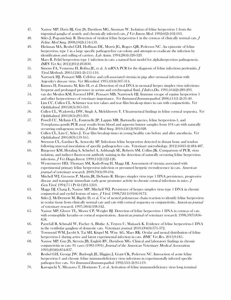
47. Nasisse MP, Davis BJ, Guy JS, Davidson MG, Sussman W. Isolation of feline herpesvirus 1 from the trigeminal ganglia of acutely and chronically infected cats. J Vet Intern Med. 1992;6(2):102-103.
48. Stiles J, Pogranichniy R. Detection of virulent feline herpesvirus-1 in the corneas of clinically normal cats. J Feline Med Surg. 2008;10(2):154-159.
49. Hickman MA, Reubel GH, Hoffman DE, Morris JG, Rogers QR, Pedersen NC. An epizootic of feline herpesvirus, type 1 in a large specific pathogen-free cat colony and attempts to eradicate the infection by identification and culling of carriers. Lab Anim. 1994;28(4):320-329.
50. Maes R. Felid herpesvirus type 1 infection in cats: a natural host model for alphaherpesvirus pathogenesis. ISRN Vet Sci. 2012;2012:495830.
51. Simons FA, Vennema H, Rofina JE, et al. A mRNA PCR for the diagnosis of feline infectious peritonitis. J Virol.Methods. 2005;124(1-2):111-116.
52. Nawynck HJ, Pensaert MB. Cell-free and cell-associated viremia in pigs after oronasal infection with Aujeszky's disease virus. Vet Microbiol. 1995;43(4):307-314.
53. Kimura H, Futamura M, Kito H, et al. Detection of viral DNA in neonatal herpes simplex virus infections: frequent and prolonged presence in serum and cerebrospinal fluid. J Infect.Dis. 1991;164(2):289-293.
54. van der Meulen KM, Favoreel HW, Pensaert MB, Nauwynck HJ. Immune escape of equine herpesvirus 1 and other herpesviruses of veterinary importance. Vet Immunol.Immunopathol. 2006;111(1-2):31-40.
55. Lim CC, Cullen CL. Schirmer tear test values and tear film break-up times in cats with conjunctivitis. Vet Ophthalmol. 2005;8(5):305-310.
56. Cullen CL, Wadowska DW, Singh A, Melekhovets Y. Ultrastructural findings in feline corneal sequestra. Vet Ophthalmol. 2005;8(5):295-303.
57. Powell CC, McInnis CL, Fontenelle JP, Lappin MR. Bartonella species, feline herpesvirus-1, and Toxoplasma gondii PCR assay results from blood and aqueous humor samples from 104 cats with naturally occurring endogenous uveitis. J Feline Med Surg. 2010;12(12):923-928.
58. Cullen CL, Lim C, Sykes J. Tear film breakup times in young healthy cats before and after anesthesia. Vet Ophthalmol. 2005;8(3):159-165.
59. Swenson CL, Gardner K, Arnoczky SP. Infectious feline herpesvirus detected in distant bone and tendon following mucosal inoculation of specific pathogen-free cats. Veterinary microbiology. 2012;160(3-4):484-487.
60. Burgesser KM, Hotaling S, Schiebel A, Ashbaugh SE, Roberts SM, Collins JK. Comparison of PCR, virus isolation, and indirect fluorescent antibody staining in the detection of naturally occurring feline herpesvirus infections. J Vet Diagn.Invest. 1999;11(2):122-126.
61. Westermeyer HD, Thomasy SM, Kado-Fong H, Maggs DJ. Assessment of viremia associated with experimental primary feline herpesvirus infection or presumed herpetic recrudescence in cats. American journal of veterinary research. 2009;70(1):99-104.
62. Mitchell WJ, Gressens P, Martin JR, DeSanto R. Herpes simplex virus type 1 DNA persistence, progressive disease and transgenic immediate early gene promoter activity in chronic corneal infections in mice. J Gen.Virol. 1994;75 ( Pt 6):1201-1210.
63. Maggs DJ, Chang E, Nasisse MP, Mitchell WJ. Persistence of herpes simplex virus type 1 DNA in chronic conjunctival and eyelid lesions of mice. J Virol. 1998;72(11):9166-9172.
64. Stiles J, McDermott M, Bigsby D, et al. Use of nested polymerase chain reaction to identify feline herpesvirus in ocular tissue from clinically normal cats and cats with corneal sequestra or conjunctivitis. American journal of veterinary research. 1997;58(4):338-342.
65. Nasisse MP, Glover TL, Moore CP, Weigler BJ. Detection of feline herpesvirus 1 DNA in corneas of cats with eosinophilic keratitis or corneal sequestration. American journal of veterinary research. 1998;59(7):856-858.
66. Parzefall B, Schmahl W, Fischer A, Blutke A, Truyen U, Matiasek K. Evidence of feline herpesvirus-1 DNA in the vestibular ganglion of domestic cats. Veterinary journal. 2010;184(3):371-372.
67. Townsend WM, Jacobi S, Tai SH, Kiupel M, Wise AG, Maes RK. Ocular and neural distribution of feline herpesvirus-1 during active and latent experimental infection in cats. BMC Vet Res. 2013;9:185.
68. Nasisse MP, Guy JS, Stevens JB, English RV, Davidson MG. Clinical and laboratory findings in chronic conjunctivitis in cats: 91 cases (1983-1991). Journal of the American Veterinary Medical Association. 1993;203(6):834-837.
69. Reubel GH, George JW, Barlough JE, Higgins J, Grant CK, Pedersen NC. Interaction of acute feline herpesvirus-1 and chronic feline immunodeficiency virus infections in experimentally infected specific pathogen free cats. Vet Immunol.Immunopathol. 1992;35(1-2):95-119.
70. Kawaguchi Y, Miyazawa T, Horimoto T, et al. Activation of feline immunodeficiency virus long terminal
repeat by feline herpesvirus type 1. Virology. 1991;184(1):449-454. 71. Kawaguchi Y, Norimine J, Miyazawa T, Kai C, Mikami T. Sequences within the feline immunodeficiency
virus long terminal repeat that regulate gene expression and respond to activation by feline herpesvirus type 1. Virology. 1992;190(1):465-468.
72. Rampazzo A, Appino S, Pregel P, Tarducci A, Zini E, Biolatti B. Prevalence of Chlamydophila felis and feline herpesvirus 1 in cats with conjunctivitis in northern Italy. J Vet Intern.Med. 2003;17(6):799-807.
73. Cai Y, Fukushi H, Koyasu S, Kuroda E, Yamaguchi T, Hirai K. An etiological investigation of domestic cats with conjunctivitis and upper respiratory tract disease in Japan. J Vet Med Sci. 2002;64(3):215-219.
74. Low HC, Powell CC, Veir JK, Hawley JR, Lappin MR. Prevalence of feline herpesvirus 1, Chlamydophila felis, and Mycoplasma spp DNA in conjunctival cells collected from cats with and without conjunctivitis. American journal of veterinary research. 2007;68(6):643-648.
75. Volopich S, Benetka V, Schwendenwein I, Mostl K, Sommerfeld-Stur I, Nell B. Cytologic findings, and feline herpesvirus DNA and Chlamydophila felis antigen detection rates in normal cats and cats with conjunctival and corneal lesions. Vet Ophthalmol. 2005;8(1):25-32.
76. Gerriets W, Joy N, Huebner-Guthardt J, Eule JC. Feline calicivirus: a neglected cause of feline ocular surface infections? Veterinary ophthalmology. 2012;15(3):172-179.
77. Wieliczko AK, Ploneczka-Janeczko K. Feline herpesvirus 1 and Chlamydophila felis prevalence in cats with chronic conjunctivitis. Pol J Vet Sci. 2010;13(2):381-383.
78. Hillstrom A, Tvedten H, Kallberg M, Hanas S, Lindhe A, Holst BS. Evaluation of cytologic findings in feline conjunctivitis. Vet Clin Pathol. 2012.
79. Schulz C, Hartmann K, Mueller RS, Helps C, Schulz BS. Sampling sites for detection of feline herpesvirus-1, feline calicivirus and Chlamydia felis in cats with feline upper respiratory tract disease. J Feline Med Surg. 2015;17(12):1012-1019.
80. Litster A, Wu CC, Leutenegger CM. Detection of feline upper respiratory tract disease pathogens using a commercially available real-time PCR test. Veterinary journal. 2015;206(2):149-153.
81. von Bomhard W, Polkinghorne A, Lu ZH, et al. Detection of novel chlamydiae in cats with ocular disease. American journal of veterinary research. 2003;64(11):1421-1428.
82. Powell CC, Kordick DL, Lappin MR. Inoculation with Bartonella henselae followed by feline herpesvirus 1 fails to activate ocular toxoplasmosis in chronically infected cats. J Feline Med Surg. 2002;4(2):107-110.
83. Johnson RP, Povey RC. Vaccination against feline viral rhinotracheitis in kittens with maternally derived feline viral rhinotracheitis antibodies. J.Am.Vet Med.Assoc. 1985;186(2):149-152.
84. Tham KM, Studdert MJ. Clinical and immunological responses of cats to feline herpesvirus type 1 infection. Vet Rec. 1987;120(14):321-326.
85. Tham KM, Studdert MJ. Antibody and cell-mediated immune responses to feline herpesvirus 1 following inactivated vaccine and challenge. Zentralbl.Veterinarmed.[B]. 1987;34(8):585-597.
86. Orr CM, Gaskell CJ, Gaskell RM. Interaction of an intranasal combined feline viral rhinotracheitis, feline calicivirus vaccine and the FVR carrier state. Vet Rec. 1980;106(8):164-166.
87. Orr CM, Gaskell CJ, Gaskell RM. Interaction of a combined feline viral rhinotracheitis-feline calicivirus vaccine and the FVR carrier state. Vet.Rec. 1978;103(10):200-202.
88. Scott FW, Geissinger CM. Long-term immunity in cats vaccinated with an inactivated trivalent vaccine. Am.J.Vet.Res. 1999;60(5):652-658.
89. Lappin MR. Feline panleukopenia virus, feline herpesvirus-1 and feline calicivirus antibody responses in seronegative specific pathogen-free kittens after parenteral administration of an inactivated FVRCP vaccine or a modified live FVRCP vaccine. J Feline Med Surg. 2012;14(2):161-164.
90. Nelli RK, Maes R, Kiupel M, Hussey GS. Use of a feline respiratory epithelial cell culture system grown at the air-liquid interface to characterize the innate immune response following feline herpesvirus 1 infection. Virus Res. 2016;214:39-48.
91. Nasisse MP. Feline herpesvirus ocular disease. Vet Clin.North Am.Small Anim Pract. 1990;20(3):667-680. 92. Nasisse MP, Guy JS, Davidson MG, Sussman WA, Fairley NM. Experimental ocular herpesvirus infection in
the cat. Sites of virus replication, clinical features and effects of corticosteroid administration. Investigative Ophthalmology and Visual Science. 1989;30(8):1758-1768.
93. Roberts SR, Dawson CR, Coleman V, Togni B. Dendritic keratitis in a cat. J.Am.Vet Med.Assoc. 1972;161(3):285-289.
94. Nasisse MP, English RV, Tompkins MB, Guy JS, Sussman W. Immunologic, histologic, and virologic features of herpesvirus-induced stromal keratitis in cats. American journal of veterinary research. 1995;56(1):51-55.

95. Avery AC, Zhao ZS, Rodriguez A, et al. Resistance to herpes stromal keratitis conferred by an IgG2a-derived peptide. Nature. 1995;376(6539):431-434.
96. Deshpande SP, Zheng M, Lee S, Rouse BT. Mechanisms of pathogenesis in herpetic immunoinflammatory ocular lesions. Vet Microbiol. 2002;86(1-2):17-26.
97. Lim CC, Reilly CM, Thomasy SM, Kass PH, Maggs DJ. Effects of feline herpesvirus type 1 on tear film break-up time, Schirmer tear test results, and conjunctival goblet cell density in experimentally infected cats. American journal of veterinary research. 2009;70(3):394-403.
98. Thomasy SM, Lim CC, Reilly CM, Kass PH, Lappin MR, Maggs DJ. Evaluation of orally administered famciclovir in cats experimentally infected with feline herpesvirus type-1. American journal of veterinary research. 2011;72(1):85-95.
99. Sebbag L, Pesavento PA, Carrasco SE, Reilly CM, Maggs DJ. Feline dry eye syndrome of presumed neurogenic origin: a case report. JFMS Open Rep. 2018;4(1):2055116917746786.
100. Zilstorff-Pedersen K. Quantitative Measurements of the Nasolacrimal Reflex. Arch Otolaryngol. 1965;81:457-462.
101. Andrew SE, Tou S, Brooks DE. Corneoconjunctival transposition for the treatment of feline corneal sequestra: a retrospective study of 17 cases (1990-1998). Vet Ophthalmol. 2001;4(2):107-111.
102. Grahn BH, Sisler S, Storey E. Qualitative tear film and conjunctival goblet cell assessment of cats with corneal sequestra. Vet Ophthalmol. 2005;8(3):167-170.
103. Morgan R, Abrams K, Kern T. Feline Eosinophilic Keratitis: A Retrospective Study of 54 Cases: (1989-1994). Veterinary and Comparative Ophthalmology. 1996;6(2):131-134.
104. Dean E, Meunier V. Feline eosinophilic keratoconjunctivitis: a retrospective study of 45 cases (56 eyes). J Feline Med Surg. 2013;15(8):661-666.
105. Allgoewer I, Schaffer EH, Stockhaus C, Vogtlin A. Feline eosinophilic conjunctivitis. Vet Ophthalmol. 2001;4(1):69-74.
106. Beckwith-Cohen B, Dubielzig RR, Maggs DJ, Teixeira LB. Feline Epitheliotropic Mastocytic Conjunctivitis in 15 Cats. Vet Pathol. 2017;54(1):141-146.
107. Liesegang TJ. A community study of ocular herpes simplex. Curr Eye Res 1991;10 Suppl:111-115. 108. Maggs DJ, Lappin MR, Nasisse MP. Detection of feline herpesvirus-specific antibodies and DNA in aqueous
humor from cats with or without uveitis. American journal of veterinary research. 1999;60(8):932-936. 109. Suchy A, Bauder B, Gelbmann W, Lohr CV, Teifke JP, Weissenbock H. Diagnosis of feline herpesvirus
infection by immunohistochemistry, polymerase chain reaction, and in situ hybridization. J Vet Diagn.Invest. 2000;12(2):186-191.
110. Hargis AM, Ginn PE. Feline herpesvirus 1-associated facial and nasal dermatitis and stomatitis in domestic cats. Vet Clin.North Am Small Anim Pract. 1999;29(6):1281-1290.
111. Hargis AM, Ginn PE, Mansell JE, Garber RL. Ulcerative facial and nasal dermatitis and stomatitis in cats associated with feline herpesvirus 1. Veterinary Dermatology. 1999;10:267-274.
112. Holland JL, Outerbridge CA, Affolter VK, Maggs DJ. Detection of feline herpesvirus 1 DNA in skin biopsy specimens from cats with or without dermatitis. Journal of the American Veterinary Medical Association. 2006;229(9):1442-1446.
113. Persico P, Roccabianca P, Corona A, Vercelli A, Cornegliani L. Detection of feline herpes virus 1 via polymerase chain reaction and immunohistochemistry in cats with ulcerative facial dermatitis, eosinophilic granuloma complex reaction patterns and mosquito bite hypersensitivity. Vet Dermatol. 2011;22(6):521-527.
114. Pennington MR, Ledbetter EC, Van de Walle GR. New Paradigms for the Study of Ocular Alphaherpesvirus Infections: Insights into the Use of Non-Traditional Host Model Systems. Viruses. 2017;9(11).
115. Nasisse MP, Guy JS, Davidson MG, Sussman W, De Clercq E. In vitro susceptibility of feline herpesvirus-1 to vidarabine, idoxuridine, trifluridine, acyclovir, or bromovinyldeoxyuridine. American journal of veterinary research. 1989;50(1):158-160.
116. Maggs DJ, Collins BK, Thorne JG, Nasisse MP. Effects of L-lysine and L-arginine on in vitro replication of feline herpesvirus type-1. Am.J.Vet.Res. 2000;61(12):1474-1478.
117. Maggs DJ, Clarke HE. In vitro efficacy of ganciclovir, cidofovir, penciclovir, foscarnet, idoxuridine, and acyclovir against feline herpesvirus type-1. American journal of veterinary research. 2004;65(4):399-403.
118. Nasisse MP, Dorman DC, Jamison KC, Weigler BJ, Hawkins EC, Stevens JB. Effects of valacyclovir in cats infected with feline herpesvirus 1. American journal of veterinary research. 1997;58(10):1141-1144.
119. Owens JG, Nasisse MP, Tadepalli SM, Dorman DC. Pharmacokinetics of acyclovir in the cat. J Vet Pharmacol Ther. 1996;19(6):488-490.
120. Anderson WA, Margruder B, Kilbourne ED. Induced reactivation of herpes simplex virus in healed rabbit

corneal lesions. Proc.Soc.Exp.Biol.Med. 1961;107:628-632. 121. Hill TJ, Blyth WA, Harbour DA. Trauma to the skin causes recurrence of herpes simplex in the mouse. J
Gen.Virol. 1978;39(1):21-28. 122. Harbour DA, Hill TJ, Blyth WA. Recurrent herpes simplex in the mouse: inflammation in the skin and
activation of virus in the ganglia following peripheral stimulation. J Gen.Virol. 1983;64 (Pt 7):1491-1498. 123. Maggs DJ, Clarke HE. Relative sensitivity of polymerase chain reaction assays used for detection of feline
herpesvirus type 1 DNA in clinical samples and commercial vaccines. American journal of veterinary research. 2005;66(9):1550-1555.
124. Nasisse MP, Weigler BJ. The diagnosis of ocular feline herpesvirus infection. Vet.Comp.Ophthalmol. 1997;7(1):44-51.
125. Hoover EA, Kahn DE. Experimentally induced feline calicivirus infection: clinical signs and lesions. J.Am.Vet Med.Assoc. 1975;166(5):463-468.
126. Carlson JH, Scott FW. An immunofluorescence diagnostic test for feline viral rhinotracheitis. Am.J.Vet.Res. 1978;39(3):465-467.
127. da Silva Curiel JMA, Nasisse MP, Hook RR, Wilson HH, Collins BK, Mandell CP. Topical fluorescein dye: effects on immunofluorescent antibody test for feline herpesvirus keratoconjunctivitis. Prog.Vet.Comp.Ophthalmol. 1991;1(2):99-104.
128. Johnson C, Johnson F. Effect of Sampling Swab Materials on Detection of Herpes Simplex Virus. 1995. 129. Crane LR, Gutterman PA, Chapel T, Lerner AM. Incubation of swab materials with herpes simplex virus. J
Infect.Dis. 1980;141(4):531. 130. Hanson BR, Schipper IA. Effects of swab materials on infectious bovine rhinotracheitis virus. American
journal of veterinary research. 1976;37(6):707-708. 131. Lappin MR, Andrews J, Simpson D, Jensen WA. Use of serologic tests to predict resistance to feline
herpesvirus 1, feline calicivirus, and feline parvovirus infection in cats. Journal of the American Veterinary Medical Association. 2002;220(1):38-42.
132. Dunkel EC, Pavan-Langston D, Fitzpatrick K, Cukor G. Rapid detection of herpes simplex virus (HSV) antigen in human ocular infections. Curr.Eye Res. 1988;7(7):661-666.
133. Wu TC, Zaza S, Callaway J. Evaluation of the Du Pont HERPCHEK herpes simplex virus antigen test with clinical specimens. J.Clin.Microbiol. 1989;27(8):1903-1905.
134. Dawson DA, Carman J, Collins J, Hill S, Lappin MR. Enzyme-linked immunosorbent assay for detection of feline herpesvirus 1 IgG in serum, aqueous humor, and cerebrospinal fluid. J.Vet.Diagn.Invest. 1998;10(4):315-319.
135. Satoh Y, Iizuka K, Fukuyama M, et al. An enzyme-linked immunosorbent assay using nuclear antigen for detection of feline herpesvirus 1 antibody. J Vet Diagn.Invest. 1999;11(4):334-340.
136. Munks MW, Montoya AM, Pywell CM, et al. The domestic cat antibody response to feline herpesvirus-1 increases with age. Vet Immunol Immunopathol. 2017;188:65-70.
137. Adleberg JM, Wittwer C. Use of the polymerase chain reaction in the diagnosis of ocular disease. Curr.Opin.Ophthalmol. 1995;6(3):80-85.
138. Pfeffer M, Wiedmann M, Batt CA. Applications of DNA amplification techniques in veterinary diagnostics. Vet.Res.Commun. 1995;19(5):375-407.
139. Segarra S, Papasouliotis K, Helps C. The in vitro effects of proxymetacaine, fluorescein, and fusidic acid on real-time PCR assays used for the diagnosis of Feline herpesvirus 1 and Chlamydophila felis infections. Veterinary ophthalmology. 2011;14 Suppl 1:5-8.
140. Fernandez M, Manzanilla EG, Lloret A, Leon M, Thibault JC. Prevalence of feline herpesvirus-1, feline calicivirus, Chlamydophila felis and Mycoplasma felis DNA and associated risk factors in cats in Spain with upper respiratory tract disease, conjunctivitis and/or gingivostomatitis. J Feline Med Surg. 2017;19(4):461-469.
141. Hara M, Fukuyama M, Suzuki Y, et al. Detection of feline herpesvirus 1 DNA by the nested polymerase chain reaction. Vet Microbiol. 1996;48(3-4):345-352.
142. Polak KC, Levy JK, Crawford PC, Leutenegger CM, Moriello KA. Infectious diseases in large-scale cat hoarding investigations. Veterinary journal. 2014;201(2):189-195.
143. Burns RE, Wagner DC, Leutenegger CM, Pesavento PA. Histologic and molecular correlation in shelter cats with acute upper respiratory infection. Journal of Clinical Microbiology. 2011;49(7):2454-2460.
144. Sandmeyer LS, Waldner CL, Bauer BS, Wen X, Bienzle D. Comparison of polymerase chain reaction tests for diagnosis of feline herpesvirus, Chlamydophila felis, and Mycoplasma spp. infection in cats with ocular disease in Canada. Canadian Veterinary Journal. 2010;51(6):629-633.

145. Maggs DJ, Clarke HE, Smith SJ. Comparison of 6 PCR protocols for detection of feline herpesvirus (FHV-1) DNA in clinical samples and commercial vaccines. 2004/10/20/.
146. Westermeyer HD, Kado-Fong H, Maggs DJ. Effects of sampling instrument and processing technique on DNA yield and detection rate for feline herpesvirus-1 via polymerase chain reaction assay. American journal of veterinary research. 2008;69(6):811-817.
147. Clarke HE, Kado-Fong H, Maggs DJ. Effects of temperature and time in transit on polymerase chain reaction detection of feline herpesvirus DNA. J Vet Diagn.Invest. 2006;18(4):388-391.
148. Vogtlin A, Fraefel C, Albini S, et al. Quantification of Feline Herpesvirus 1 DNA in Ocular Fluid Samples of Clinically Diseased Cats by Real-Time TaqMan PCR. J Clin.Microbiol. 2002;40(2):519-523.
149. Wang J, Liu L, Wang J, Sun X, Yuan W. Recombinase Polymerase Amplification Assay-A Simple, Fast and Cost-Effective Alternative to Real Time PCR for Specific Detection of Feline Herpesvirus-1. PLoS One. 2017;12(1):e0166903.
150. De Clercq E. Antivirals: past, present and future. Biochem Pharmacol. 2013;85(6):727-744. 151. Thomasy SM, Maggs DJ. A review of antiviral drugs and other compounds with activity against feline
herpesvirus type 1. Veterinary ophthalmology. 2016;19 Suppl 1:119-130. 152. Williams DL, Fitzmaurice T, Lay L, et al. Efficacy of antiviral agents in feline herpetic keratitis: results of an
in vitro study. Curr Eye Res. 2004;29(2-3):215-218. 153. Andrei G, Snoeck R, Goubau P, Desmyter J, De Clercq E. Comparative activity of various compounds
against clinical strains of herpes simplex virus. Eur J Clin Microbiol Infect Dis. 1992;11(2):143-151. 154. Babiuk LA, Meldrum B, Gupta VS, Rouse BT. Comparison of the antiviral effects of 5-methoxymethyl-
deoxyuridine with 5-iododeoxyuridine, cytosine arabinoside, and adenine arabinoside. Antimicrob.Agents Chemother. 1975;8(6):643-650.
155. van der Meulen K, Garre B, Croubels S, Nauwynck H. In vitro comparison of antiviral drugs against feline herpesvirus 1. BMC Vet Res. 2006;2:13.
156. Chen ZJ, Liu WG, Song JZ. [Experimental studies of 9-(1, 3-dihydroxy-2-propoxymethyl)-guanine(DHPG) against herpes simplex virus]. Yao Xue Xue Bao. 1989;24(5):331-334.
157. Lauter CB, Bailey EJ, Lerner AM. Microbiologic assays and neurological toxicity during use of adenine arabinoside in humans. The Journal of infectious diseases. 1976;134(1):75-79.
158. Hussein IT, Menashy RV, Field HJ. Penciclovir is a potent inhibitor of feline herpesvirus-1 with susceptibility determined at the level of virus-encoded thymidine kinase. Antiviral Res. 2008;78(3):268-274.
159. Groth AD, Contreras MT, Kado-Fong HK, Nguyen KQ, Thomasy SM, Maggs DJ. In vitro cytotoxicity and antiviral efficacy against feline herpesvirus type 1 of famciclovir and its metabolites. Veterinary ophthalmology. 2014;17(4):268-274.
160. Hussein IT, Field HJ. Development of a quantitative real-time TaqMan PCR assay for testing the susceptibility of feline herpesvirus-1 to antiviral compounds. Journal of Virological Methods. 2008;152(1-2):85-90.
161. De Clercq E, Sakuma T, Baba M, et al. Antiviral activity of phosphonylmethoxyalkyl derivatives of purine and pyrimidines. Antiviral Res. 1987;8(5-6):261-272.
162. Gadler H, Larsson A, Solver E. Nucleic acid hybridization, a method to determine effects of antiviral compounds on herpes simplex virus type 1 DNA synthesis. Antiviral Res. 1984;4(1-2):63-70.
163. Williams DL, Robinson JC, Lay E, Field H. Efficacy of topical aciclovir for the treatment of feline herpetic keratitis: results of a prospective clinical trial and data from in vitro investigations. Veterinary Record. 2005;157(9):254-257.
164. Ayisi NK, Gupta SV, Babiuk LA. Combination chemotherapy: interaction of 5-methoxymethyldeoxyuridine with trifluorothymidine, phosphonoformate and acycloguanosine against herpes simplex viruses. Antiviral Res. 1985;5(1):13-27.
165. Sandmeyer LS, Keller CB, Bienzle D. Effects of cidofovir on cell death and replication of feline herpesvirus-1 in cultured feline corneal epithelial cells. American journal of veterinary research. 2005;66(2):217-222.
166. Bronson JJ, Ghazzouli I, Hitchcock MJ, Webb RR, 2nd, Martin JC. Synthesis and antiviral activity of the nucleotide analogue (S)-1-[3-hydroxy-2-(phosphonylmethoxy)propyl]cytosine. J Med Chem. 1989;32(7):1457-1463.
167. Bestman-Smith J, Boivin G. Herpes simplex virus isolates with reduced adefovir susceptibility selected in vivo by foscarnet therapy. J Med Virol. 2002;67(1):88-91.
168. Collins P. The spectrum of antiviral activities of acyclovir in vitro and in vivo. J Antimicrob.Chemother. 1983;12 Suppl B:19-27.
169. Pennington MR, Fort MW, Ledbetter EC, Van de Walle GR. A novel corneal explant model system to

evaluate antiviral drugs against feline herpesvirus type 1 (FHV-1). J Gen Virol. 2016;97(6):1414-1425. 170. Zhou B, Yang K, Wills E, Tang L, Baines JD. A mutation in the DNA polymerase accessory factor of herpes
simplex virus 1 restores viral DNA replication in the presence of raltegravir. J Virol. 2014;88(19):11121-11129.
171. Stiles J. Treatment of cats with ocular disease attributable to herpesvirus infection: 17 cases (1983-1993). Journal of the American Veterinary Medical Association. 1995;207(5):599-603.
172. O'Brien WJ, Edelhauser HF. The corneal penetration of trifluorothymidine, adenine arabinoside, and idoxuridine: a comparative study. Invest Ophthalmol.Vis.Sci. 1977;16(12):1093-1103.
173. Hussein IT, Miguel RN, Tiley LS, Field HJ. Substrate specificity and molecular modelling of the feline herpesvirus-1 thymidine kinase. Archives of Virology. 2008;153(3):495-505.
174. Weiss RC. Synergistic antiviral activities of acyclovir and recombinant human leukocyte (alpha) interferon on feline herpesvirus replication. American journal of veterinary research. 1989;50(10):1672-1677.
175. de Miranda P, Burnette TC. Metabolic fate and pharmacokinetics of the acyclovir prodrug valaciclovir in cynomolgus monkeys. Drug Metab Dispos. 1994;22(1):55-59.
176. Ernst ME, Franey RJ. Acyclovir- and ganciclovir-induced neurotoxicity. Ann Pharmacother. 1998;32(1):111-113.
177. Burns LJ, Miller W, Kandaswamy C, et al. Randomized clinical trial of ganciclovir vs acyclovir for prevention of cytomegalovirus antigenemia after allogeneic transplantation. Bone Marrow Transplant. 2002;30(12):945-951.
178. Kaufman HE, Varnell ED, Thompson HW. Trifluridine, cidofovir, and penciclovir in the treatment of experimental herpetic keratitis. Arch.Ophthalmol. 1998;116(6):777-780.
179. Clarke SE, Harrell AW, Chenery RJ. Role of aldehyde oxidase in the in vitro conversion of famciclovir to penciclovir in human liver. Drug Metab Dispos. 1995;23(2):251-254.
180. Vere Hodge RA, Sutton D, Boyd MR, Harnden MR, Jarvest RL. Selection of an oral prodrug (BRL 42810; famciclovir) for the antiherpesvirus agent BRL 39123 [9-(4-hydroxy-3-hydroxymethylbut-l-yl)guanine; penciclovir]. Antimicrob.Agents Chemother. 1989;33(10):1765-1773.
181. Filer CW, Allen GD, Brown TA, et al. Metabolic and pharmacokinetic studies following oral administration of 14C-famciclovir to healthy subjects. Xenobiotica. 1994;24(4):357-368.
182. Dick RA, Kanne DB, Casida JE. Identification of aldehyde oxidase as the neonicotinoid nitroreductase. Chemical Research in Toxicology. 2005;18(2):317-323.
183. Sebbag L, Thomasy SM, Woodward AP, Knych HK, Maggs DJ. Pharmacokinetic modeling of penciclovir and BRL42359 in the plasma and tears of healthy cats to optimize dosage recommendations for oral administration of famciclovir. American journal of veterinary research. 2016;77(8):833-845.
184. Thomasy SM, Maggs DJ, Moulin NK, Stanley SD. Pharmacokinetics and safety of penciclovir following oral administration of famciclovir to cats. American journal of veterinary research. 2007;68(11):1252-1258.
185. Malik R, Lessels NS, Webb S, et al. Treatment of feline herpesvirus-1 associated disease in cats with famciclovir and related drugs. J Feline Med Surg. 2009;11(1):40-48.
186. Thomasy SM, Shull O, Outerbridge CA, et al. Oral administration of famciclovir for treatment of spontaneous ocular, respiratory, or dermatologic disease attributed to feline herpesvirus type 1: 59 cases (2006-2013). Journal of the American Veterinary Medical Association. 2016;249(5):526-538.
187. Thomasy SM, Whittem T, Bales JL, Ferrone M, Stanley SD, Maggs DJ. Pharmacokinetics of penciclovir in healthy cats following oral administration of famciclovir or intravenous infusion of penciclovir. American journal of veterinary research. 2012;73(7):1092-1099.
188. Thomasy SM, Covert JC, Stanley SD, Maggs DJ. Pharmacokinetics of famciclovir and penciclovir in tears following oral administration of famciclovir to cats: a pilot study. Veterinary ophthalmology. 2012;15(5):299-306.
189. Boike SC, Pue MA, Freed MI, et al. Pharmacokinetics of famciclovir in subjects with varying degrees of renal impairment. Clin Pharmacol Ther. 1994;55(4):418-426.
190. Litster AL, Lohr BR, Bukowy RA, Thomasy SM, Maggs DJ. Clinical and antiviral effect of a single oral dose of famciclovir administered to cats at intake to a shelter. Veterinary journal. 2015;203(2):199-204.
191. Neyts J, Snoeck R, Schols D, Balzarini J, De Clercq E. Selective inhibition of human cytomegalovirus DNA synthesis by (S)-1-(3-hydroxy-2-phosphonylmethoxypropyl)cytosine [(S)-HPMPC] and 9-(1,3-dihydroxy-2-propoxymethyl)guanine (DHPG). Virology. 1990;179(1):41-50.
192. Romanowski EG, Bartels SP, Gordon YJ. Comparative antiviral efficacies of cidofovir, trifluridine, and acyclovir in the HSV-1 rabbit keratitis model. Invest Ophthalmol.Vis.Sci. 1999;40(2):378-384.
193. Neyts J, Snoeck R, Balzarini J, De Clercq E. Particular characteristics of the anti-human cytomegalovirus
activity of (S)-1-(3-hydroxy-2-phosphonylmethoxypropyl)cytosine (HPMPC) in vitro. Antiviral Res. 1991;16(1):41-52.
194. Fontenelle JP, Powell CC, Veir JK, Radecki SV, Lappin MR. Effect of topical ophthalmic application of cidofovir on experimentally induced primary ocular feline herpesvirus-1 infection in cats. American journal of veterinary research. 2008;69(2):289-293.
195. Sherman MD, Feldman KA, Farahmand SM, Margolis TP. Treatment of conjunctival squamous cell carcinoma with topical cidofovir. Am J Ophthalmol. 2002;134(3):432-433.
196. Gordon YJ, Romanowski EG, Araullo-Cruz T. Topical HPMPC inhibits adenovirus type 5 in the New Zealand rabbit ocular replication model. Invest Ophthalmol.Vis.Sci. 1994;35(12):4135-4143.
197. Stiles J, Gwin W, Pogranichniy R. Stability of 0.5% cidofovir stored under various conditions for up to 6 months. Veterinary ophthalmology. 2010;13(4):275-277.
198. Maggs DJ, Collins BK, Thorne JG, Nasisse MP. Effects of L-lysine and L-arginine on in vitro replication of feline herpesvirus type-1. Am J Vet Res. 2000;61(12):1474-1478.
199. Tankersley RW, Jr. Amino Acid Requirements of Herpes Simplex Virus in Human Cells. Journal of bacteriology. 1964;87:609-613.
200. Griffith RS, DeLong DC, Nelson JD. Relation of arginine-lysine antagonism to herpes simplex growth in tissue culture. Chemotherapy. 1981;27(3):209-213.
201. Kaplan AS, Shimono H, Ben-Porat T. Synthesis of proteins in cells infected with herpesvirus. 3. Relative amino acid content of various proteins formed after infection. Virology. 1970;40(1):90-101.
202. Bol S, Bunnik EM. Lysine supplementation is not effective for the prevention or treatment of feline herpesvirus 1 infection in cats: a systematic review. BMC Vet Res. 2015;11(1):284.
203. Cave NJ, Dennis K, Gopakumar G, Dunowska M. Effects of physiologic concentrations of l-lysine on in vitro replication of feline herpesvirus 1. American journal of veterinary research. 2014;75(6):572-580.
204. Banos G, Daniel PM, Pratt OE. Saturation of a shared mechanism which transports L-arginine and L-lysine into the brain of the living rat. J Physiol (Lond). 1974;236(1):29-41.
205. Stutz MW, Savage JE, O'Dell BL. Relation of dietary cations to arginine-lysine antagonism and free amino acid patterns in chicks. J Nutr. 1971;101(3):377-384.
206. Hunter A, Downs CE. The inhibition of arginase by amino acids. J Biol.Chem. 1945;157 427-446. 207. Stiles J, Townsend WM, Rogers QR, Krohne SG. Effect of oral administration of L-lysine on conjunctivitis
caused by feline herpesvirus in cats. American journal of veterinary research. 2002;63(1):99-103. 208. Maggs DJ, Nasisse MP, Kass PH. Efficacy of oral supplementation with L-lysine in cats latently infected with
feline herpesvirus. American journal of veterinary research. 2003;64(1):37-42. 209. Rees TM, Lubinski JL. Oral supplementation with L-lysine did not prevent upper respiratory infection in a
shelter population of cats. J Feline Med Surg. 2008;10(5):510-513. 210. Fascetti AJ, Maggs DJ, Kanchuk ML, Clarke HE, Rogers QR. Excess dietary lysine does not cause lysine-
arginine antagonism in adult cats. J.Nutr. 2004;134(8 Suppl):2042S-2045S. 211. Maggs DJ, Sykes JE, Clarke HE, et al. Effects of dietary lysine supplementation in cats with enzootic upper
respiratory disease. J Fel Med Surg. 2007;9(2):97-108. 212. Drazenovich TL, Fascetti AJ, Westermeyer HD, et al. Effects of dietary lysine supplementation on upper
respiratory and ocular disease and detection of infectious organisms in cats within an animal shelter. American journal of veterinary research. 2009;70(11):1391-1400.
213. Morris JG, Rogers QR. Ammonia intoxication in the near-adult cat as a result of a dietary deficiency of arginine. Science. 1978;199(4327):431-432.
214. Stiles J, Guptill-Yoran L, Moore GE, Pogranichniy RM. Effects of lambda-carrageenan on in vitro replication of feline herpesvirus and on experimentally induced herpetic conjunctivitis in cats. Invest Ophthalmol.Vis.Sci. 2008;49(4):1496-1501.
215. Bagla VP, McGaw LJ, Eloff JN. The antiviral activity of six South African plants traditionally used against infections in ethnoveterinary medicine. Veterinary microbiology. 2012;155(2-4):198-206.
216. Knight DA, Hejmanowski AQ, Dierksheide JE, Williams JW, Chong AS, Waldman WJ. Inhibition of herpes simplex virus type 1 by the experimental immunosuppressive agent leflunomide. Transplantation. 2001;71(1):170-174.
217. Williams CR, Sykes JE, Mehl M, et al. In vitro effects of the active metabolite of leflunomide, A77 1726, on feline herpesvirus-1. American journal of veterinary research. 2007;68(9):1010-1015.
218. Lappin MR, Veir JK, Satyaraj E, Czarnecki-Maulden G. Pilot study to evaluate the effect of oral supplementation of Enterococcus faecium SF68 on cats with latent feline herpesvirus 1. J Feline Med Surg. 2009;11(8):650-654.

219. Contreras ET, Hodgkins E, Tynes V, Beck A, Olea-Popelka F, Lappin MR. Effect of a Pheromone on Stress-Associated Reactivation of Feline Herpesvirus-1 in Experimentally Inoculated Kittens. J Vet Intern Med. 2018;32(1):406-417.
220. Nasisse MP. Issues in Ophthalmology: Anti-Inflammatory Therapy in Herpesvirus Keratoconjunctivitis. Progress in Veterinary and Comparative Ophthalmology. 1990;1(1):63-65.
221. Hendricks RL, Barfknecht CF, Schoenwald RD, Epstein RJ, Sugar J. The effect of flurbiprofen on herpes simplex virus type 1 stromal keratitis in mice. Invest Ophthalmol.Vis.Sci. 1990;31(8):1503-1511.
222. Trousdale MD, Dunkel EC, Nesburn AB. Effect of flurbiprofen on herpes simplex keratitis in rabbits. Invest Ophthalmol.Vis.Sci. 1980;19(3):267-270.
223. Vahlne A, Larsson PA, Horal P, et al. Inhibition of herpes simplex virus production in vitro by cyclosporin A. Arch.Virol. 1992;122(1-2):61-75.
224. Meyers-Elliott RH, Chitjian PA, Billups CB. Effects of cyclosporine A on clinical and immunological parameters in herpes simplex keratitis. Invest Ophthalmol.Vis.Sci. 1987;28(7):1170-1180.
225. Gunduz K, Ozdemir O. Topical cyclosporin as an adjunct to topical acyclovir treatment in herpetic stromal keratitis. Ophthalmic Res. 1997;29(6):405-408.
226. Spiess AK, Sapienza JS, Mayordomo A. Treatment of proliferative feline eosinophilic keratitis with topical 1.5% cyclosporine: 35 cases. Veterinary ophthalmology. 2009;12(2):132-137.
227. Lappin MR, Roycroft LM. Effect of ciclosporin and methylprednisolone acetate on cats previously infected with feline herpesvirus 1. J Feline Med Surg. 2015;17(4):353-358.
228. Friedl Y, Schulz B, Knebl A, Helps C, Truyen U, Hartmann K. Efficacy of passively transferred antibodies in cats with acute viral upper respiratory tract infection. Veterinary journal. 2014;201(3):316-321.
229. Umehashi M, Imamura T, Akiyama S, Kimachi K, Tokiyoshi S, Mikami T. Post-exposure Treatment of Cats with Mouse-Cat Chimeric Antibodies against Feline Herpesvirus Type 1 and Feline Calicivirus. J Vet Med Sci. 2002;64(11):1017-1021.
230. Umehashi M, Imamura T, Akiyama S, et al. Pre-exposure treatment of cats with anti-FHV-1 and anti-FCV mouse-cat chimeric antibodies. J.Vet Med.Sci. 2003;65(5):563-566.
231. Legendre AM, Kuritz T, Heidel RE, Baylor VM. Polyprenyl Immunostimulant in Feline Rhinotracheitis: Randomized Placebo-Controlled Experimental and Field Safety Studies. Front Vet Sci. 2017;4:24.
232. Sandmeyer LS, Keller CB, Bienzle D. Effects of interferon-alpha on cytopathic changes and titers for feline herpesvirus-1 in primary cultures of feline corneal epithelial cells. American journal of veterinary research. 2005;66(2):210-216.
233. Siebeck N, Hurley DJ, Garcia M, et al. Effects of human recombinant alpha-2b interferon and feline recombinant omega interferon on in vitro replication of feline herpesvirus-1. American journal of veterinary research. 2006;67(8):1406-1411.
234. Haid C, Kaps S, Gonczi E, et al. Pretreatment with feline interferon omega and the course of subsequent infection with feline herpesvirus in cats. Vet Ophthalmol. 2007;10(5):278-284.
235. Cocker FM, Howard PE, Harbour DA. Effect of human alpha-hybrid interferon on the course of feline viral rhinotracheitis. Vet Rec. 1987;120(16):391-393.
236. Nasisse MP, Halenda RM, Luo H. Efficacy of low dose oral, natural human interferon alpha (nHuIFN ) in acute feline herpesvirus 1 (FHV-1) infection, a preliminary dose determination trial. 1996/10/21/; Wailea, HI.
237. Ballin AC, Schulz B, Helps C, Sauter-Louis C, Mueller RS, Hartmann K. Limited efficacy of topical recombinant feline interferon-omega for treatment of cats with acute upper respiratory viral disease. Veterinary journal. 2014;202(3):466-470.
238. Fiorito F, Cantiello A, Granato GE, et al. Clinical improvement in feline herpesvirus 1 infected cats by oral low dose of interleukin-12 plus interferon-gamma. Comp Immunol Microbiol Infect Dis. 2016;48:41-47.
239. Tian J, Liu Y, Liu X, Sun X, Zhang J, Qu L. Feline herpesvirus 1 (FHV-1) US3 blocks type I IFN signal pathway by targeting IRF3 dimerization in a kinase-independent manner. J Virol. 2018.
240. Weigent DA, Langford MP, Stanton GJ, Blalock JE. Interferon-induced transfer of viral resistance by human B and T lymphocytes. Cell Immunol. 1984;87(2):678-683.
241. Cocker FM, Gaskell RM, Newby TJ, Gaskell CJ, Stokes CR, Bourne FJ. Efficacy of early (48 and 96 hour) protection against feline viral rhinotracheitis following intranasal vaccination with a live temperature sensitive mutant. Vet Rec. 1984;114(14):353-354.
242. Burton G, Mason KV. Do postvaccinal sarcomas occur in Australian cats? Aust.Vet J. 1997;75(2):102-106. 243. Reagan KL, Hawley JR, Lappin MR. Concurrent administration of an intranasal vaccine containing feline
herpesvirus-1 (FHV-1) with a parenteral vaccine containing FHV-1 is superior to parenteral vaccination alone

in an acute FHV-1 challenge model. Veterinary journal. 2014;201(2):202-206. 244. Bradley A, Kinyon J, Frana T, Bolte D, Hyatt DR, Lappin MR. Efficacy of intranasal administration of a
modified live feline herpesvirus 1 and feline calicivirus vaccine against disease caused by Bordetella bronchiseptica after experimental challenge. J Vet Intern Med. 2012;26(5):1121-1125.
245. Sussman MD, Maes RK, Kruger JM. Vaccination of cats for feline rhinotracheitis results in a quantitative reduction of virulent feline herpesvirus-1 latency load after challenge. Virology. 1997;228(2):379-382.
246. Weigler BJ, Guy JS, Nasisse MP, Hancock SI, Sherry B. Effect of a live attenuated intranasal vaccine on latency and shedding of feline herpesvirus 1 in domestic cats. Arch.Virol. 1997;142(12):2389-2400.
247. Ruch-Gallie RA, Veir JK, Hawley JR, Lappin MR. Results of molecular diagnostic assays targeting feline herpesvirus-1 and feline calicivirus in adult cats administered modified live vaccines. J Feline Med Surg. 2011;13(8):541-545.
248. Fenimore A, Carter K, Fankhauser J, Hawley JR, Lappin MR. Evaluation of intranasal vaccine administration and high-dose interferon- alpha2b therapy for treatment of chronic upper respiratory tract infections in shelter cats. J Feline Med Surg. 2016;18(8):603-611.