viral pathogens john scott meschke office: suite 2338, 4225 roosevelt phone: 206-221-5470 email:...
Post on 21-Dec-2015
214 views
TRANSCRIPT

Viral Pathogens
John Scott Meschke
Office: Suite 2338, 4225 Roosevelt
Phone: 206-221-5470
Email: [email protected]
Viruses
• What is a Virus
• Composition– Genome– Capsid– Envelope
• Taxonomy
• Examples of Environmentally Transmitted Viruses
Viruses (Encyclopedia Britannica)
..infectious agents of small size and simple composition that can multiply only in living cells of animals, plants and bacteria. Viruses are obligate parasites that are metabolically inert when they are outside their hosts. They all rely, to varying extents, on the metabolic processes of their hosts to reproduce themselves. The viral diseases we see are due to the effects of this interaction between the virus and its host cell (and/or the host’s response to this interaction).

Recognition of viruses
How long viruses have been within our midst?
1500 BC: Leg deformities indicative of poliomyelitis, pock marks indicative of smallpox.
During the 1800's, all infectious agents were considered to be viruses until Koch developed pure culture techniques which allowed the separation and growth of bacteria. In the late 1800's: Bacteria were purified and established as disease causing agents. It then became possible to distinguish them from the "filterable agents", those able to pass through special filters designed to prevent the passage of bacteria. The first viruses described were foot and mouth disease (a picornavirus), 1898, Yellow fever (a flavivirus), 1900, Rous sarcoma virus (an oncogenic retrovirus), 1906.
"Virus" is from the Greek meaning for "poison" and was initially described by Edward Jenner in 1796.

Viral diseases have played a major role in human history over whatever time scale we choose to explore:
Over the past 1000 years: Smallpox and measles were brought to North and South America by early European explorers/conquerers. These diseases, for which the native American populations had no acquired partial immunity, killed large fractions of the populations, and were a major factor in the decimation of these societies.
Over the past 100 years: A newly emerged strain of influenza killed 20 million people in 1918-1919 in the immediate aftermath of World War I. A decade later, polio became one of the most feared infections of children and young adults (including Franklin D. Roosevelt, the U.S. President throughout the Depression and World War II).
As the century entered its final 20 years, a new ~100% lethal virus, HIV, spread rapidly around the world via body fluid transmission.

Over the past 10 years: As the global HIV epidemic continues, sporadic cases and outbreaks in humans of some non-human host viruses such as Ebola and Hanta raise the concern about future epidemics by other viruses in the new century.
Four Corners Virus (Hanta)
Ebola virus

Viruses• smallest (0.02-0.3 micrometers diameter)• simplest (nucleic acid + protein coat (+ lipoprotein envelope) )• spherical (icosahedral) or rod-shaped (helical) • no biological activity outside of host cells/or host organisms
– obligate intracellular parasites; recruit host cell to make new viruses, often destroying the cell
• non-enveloped viruses are most persistent in the environment– protein coat confers stability
• enteric and respiratory viruses are most important for environmental health – transmitted by direct and indirect contact, fecally contaminated
water, food, fomites and air.

Viral Composition
Nucleic acid: •DNA or RNA•single or double-stranded •1 or several segments •Capsid (protein coat):• multiple copies of 1 or more proteins in an arrayEnvelope:•lipid bilayer membrane + glycoproteins) •typically acquired from host cell membranes

Virus Composition
• nucleic acid:– RNA or DNA– double- or single-stranded– one piece or multiple,
genetically distinct pieces • represent separate genes• some have multiple copies
of same gene– linear, circular or
circular+supercoiled

Proteins produced by viruses
• Structural proteins
• Non-structural proteins

Virion
capsomeres
Nucleocapsid(a nucleocapsid without a genome is a capsid)
envelopedvirus
envelope
a virion
nucleocapsid

Some viral shapes
adenovirus
morbillivirus
parvoviruspapillomavirus
100 nm1 nm = 1 millionth of a mm100 nm = 1 ten thousandth of a mm

Some viral shapes
herpesvirus
parainfluenzavirus
influenzavirus
poxvirus
1 nm = 1 millionth of a mm100 nm = 1 ten thousandth of a mm
100 nm
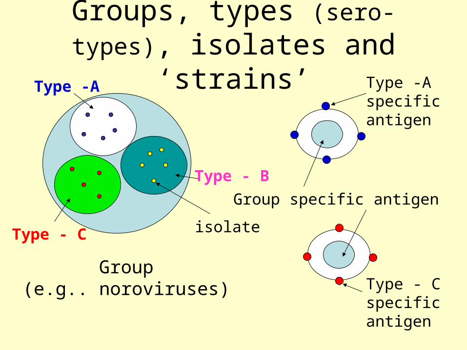
Groups, types (sero-types), isolates and ‘strains’
Group(e.g.. noroviruses)
Type -A
Type - B
Type - C
Group specific antigen
Type -A specificantigen
Type - C specificantigen
isolate

Errors in replication lead to “quasispecies”
persistentinfection
mixture of variant viruses(quasispecies)

GIII.1_JenaGIII.2_CH126
GI.3_Desert Shield
GI.7_WinchesterGI.8_Boxer
GI.4_ChibaGI.5_Musgrove
GI.2_Southampton
GI.6_Hesse
GI.1_Norwalk
GV.1_MurineGIV.1_Alphatron
GII.15_J23GII.4_Bristol
GII.11_SW918
GII.8_AmsterdamGII.9_Virginia Beach
GII.14_M7
GII.7_Leeds
GII.12_WortleyGII.1_Hawaii
GII.16_TiffinGII.2_MelkshamGII.5_Hillingdon
GII.10_ErfurtGII.13_Fayetteville
GII.17_CS-E1
GII.3_TorontoGII.6_Seacroft
.10
GVGIV
GIII
GII
GI
Clusters differ by ≥ 20% amino acid pairwise distance
Genogroups differ by 44-55% amino acid pairwise distance
Genotypes: ex. 29 Clusters of Noroviruses

A General Caution!
Molecular sub-typing is a bit like the parable of the blind men and the elephant – you can get an entirely different picture of what you’re dealing with depending on which part of the beast you’re examining!

Release of virus
Release by lysis of cell(cytopathic)
or by budding (withoutdeath of cell, non-cytopathic)

Incubation period
incubation period - time between infection and the appearance of clinical signs
infection

Patterns of disease
acute
recurrent
chronic
slow
clinical signsvirus shedding
virus difficult to detect

Virus Infections: Some Important Viruses Cause Localized Infections and Others Systemic Infections
Enteric Viruses:• Localized:
– caliciviruses– astroviruses– adenoviruses– rotaviruses
• Generalized:– enteroviruses– hepatitis A and E viruses
Respiratory Viruses:• Localized:
– rhinoviruses– coronaviruses– Orthomyxoviruses(Flu)– paramyxoviruses
• Generalized:– herpesviruses– measles– mumps

How are viruses classified ?
Hierarchical virus classification: (order) family - subfamily - genus - species - strain/type
All families have the suffix viridae, e.g.:
* Poxviridae * Herpesviridae * Parvoviridae * Retroviridae
Genera have the suffix virus. Within the Picornaviridae there are 5 genera:
* enterovirus (alimentary tract), species e.g. poliovirus 1, 2, 3 * cardiovirus (neurotropic), species e.g. mengovirus * rhinovirus (nasopharyngeal region), species e.g. Rhinovirus 1a * apthovirus (cloven footed animals ), species e.g. FMDV-C * hepatovirus (liver), species e.g. Hepatitis A virus

Virus naming and classificationUsually based on data available at the time of
discovery:
i Disease they are associated with, e.g.:Poxvirus, Hepatitis virus, HIV, measles virus
ii Cytopathology they cause, e.g.:Respiratory Syncytial virus, Cytomegalovirus
iii Site of isolation, e.g.:Adenovirus, Enterovirus, Rhinovirus
iv Places discovered or people that discovered them, e.g.:Epstein-Barr virus, Rift Valley Fever
v Biochemical features, e.g.:Retrovirus, Picornavirus, Hepadnavirus

These naming conventions can lead to confusion later, e.g., viral hepatitis is caused by at least 6 different viruses
DD
““Infectious”Infectious”
““Serum”Serum”
Viral Viral hepatitishepatitis
AA
NANBNANB
BB
EntericallyEntericallytransmittedtransmitted
ParenterallyParenterallytransmittedtransmitted
EE
CC
F, G,F, G,? Other *? Other *
* 10-20% of cases of presumed viral hepatitis are still not accounted for

Thus,
Different viruses can cause (nearly) the same symptoms. e.g., the hepatitis viruses
However, different members of the same group can cause different symptoms. e.g., the herpes viruses

Virus Classification is now based principally on analysis of the particle:
Morphology:by electron microscopy
Serology: antigenic cross-reactivity
Genetic material: form of nucleic acid
ssDNA (+ or - strand) dsDNA
ssRNA (+ or - strand) dsRNA
segmented RNA genetic organization sequence homology
DNA sequence Hybridization
Rotavirus

Some Important Human Enteric VirusesViruses/Groups Animal Hosts?
Enteroviruses: no(polios, echos*, coxsackies*, etc.)
Hepatitis A virus no (primates)Hepatitis E virus pigs, rats, othersReoviruses yesRotaviruses yes**Adenoviruses* yes**Caliciviruses (Noroviruses)*: maybe**
Norwalk, Snow Mountain, etc.Astroviruses Unknown*On EPA’s candidate contaminants list (CCL) for drinking water.**humans & animals usually infected by different ones; but perhaps not always.

Viral Gastroenteritis
• It is thought that viruses are responsible for up to 3/4 of all infective diarrhoeas.
• Viral gastroenteritis is the second most common viral illness after upper respiratory tract infection.
• In developing countries, viral gastroenteritis is a major killer of infants who are undernourished. Rotaviruses are responsible for half a million deaths a year.
• Many different types of viruses are found in the gut but
only some are associated with gastroenteritis

Enteroviruses
• Icosahedral shape• ~27-30 nm diameter• single-stranded +sense RNA
– about 7,500 nucleotides• icosahedral protein coat (capsid)
– 4 capsid proteins: VP1, VP2, VP3, VP4 (all cleaved from VP0)
• >71 antigenically distinct human types– polioviruses (3 types)– coxsackie B viruses (6 types)– coxsackie A viruses (23 types)– echoviruses (31 types)
• distinct animal enteroviruses• Cause enteric illness• Some cause respiratory illness

Enteroviruses• Enteroviruses are a genus of the picornavirus family which
replicate mainly in the gut.• Single stranded naked RNA virus with icosahedral symmetry• Unlike rhinoviruses, they are stable in acid pH• capsid has 60 copies each of 4 proteins, VP1, VP2, VP3 and
VP4 arranged with icosahedral symmetry around a positive sense genome. At least 71 serotypes are known: divided into 5 groups
– Polioviruses
– Coxsackie A viruses
– Coxsackie B viruses
– Echoviruses
– Enteroviruses (more recently, new enteroviruses subtype have been allocated sequential numbers (68-71))

Diseases associated with Enteroviruses
Syndrome Polio Cox A Cox B Echo Paralytic disease + + + + Meningitis-encephalitis + + + + Carditis + + + + Neonatal disease - - + + Pleurodynia - - + - Herpangina - + - - Rash disease - + + + Haemorr. conjunctivitis - + - - Respiratory infections + + + + Undifferentiated fever + + + + Diabetes/pancreatitis - - + -

Reovirus and Rotaviruses• ~spherical; icosahedral• ~75-80 nm diameter• double-layered capsid• nucleic acid:
– double-stranded RNA– 11 segments rota)– 10 segments (reo)– electropherotypes
• 7 Groups– Subgroups, serotypes
• Cause enteric illness– Group A most important in humans
(children)– Group C causes sporadic illness– Group B has caused large outbreaks
(adults), rare

Rotaviruses
• Naked double stranded RNA viruses, 80 nm in diameter
• also found in other mammals and birds, causing diarrhoea
• account for 50-80% of all cases of viral gastroenteritis
• usually endemic, but responsible for occasional outbreaks
• causes disease in all age groups but most severe symptoms in neonates and young children. Asymptomatic infections common in adults and older children. Symptomatic infections again common in people over 60
• up to 30% mortality rate in malnourished children, responsible for up to half a million deaths per year

Rotaviruses
• 80% of the population have antibody against rotavirus by the
age of 3
• more frequent during the winter
• faecal-oral spread. ? respiratory droplets
• 24-48 hr incubation period followed by an abrupt onset of
vomiting and diarrhoea, a low grade fever may be present.
• diagnosed by electron microscopy or by the detection of
rotavirus antigens in faeces by ELISA or other assays.
• Live attenuated vaccines now available for use in children

•Icosahedral• “structured”; cup-like surface morphology•30-35 nm diameter•ss(+) RNA, ~7.7 KB•1 major capsid polypeptide, ~60 kD•minor protein, ~30 kD•3 major HuCV groups
•G 1 and 2; “Sapporo-like”•Genetically diverse/variable
•No culture (except in humans)•Distinct animal caliciviruses
•some genetically similar to human caliciviruses•cross-species transmission?
Caliciviruses: Noroviruses and Sappoviruses

Noroviruses (Norwalk-like)
• always associated with epidemic outbreaks of gastroenteritis, adults more commonly affected than children
• associated with consumption of shellfish and other contaminated foods. Aerosol spread possible as well as faecal-oral spread
• Also named "winter vomiting disease", with vomiting being the prominent symptom, diarrhoea usually mild
• Antibodies acquired later in life, in the US, only 50% of adults are seropositive by the age of 50.
• diagnosis is made by electron microscopy and by PCR.

•In the US:
• 12.7 million cases
•International:
• 200 million cases
•12.7 million cases x 4 episodes/day x 3 days duration x 200ml/episode = 3.6x10¹¹ ml of diarrhea or 105 million gallons or 6 minutes of flow over Niagra Falls
Just How Much Diarrhea is That?

Sapoviruses
• associated mainly with epidemic outbreaks of gastroenteritis, although occasionally responsible for endemic cases
• like Norwalk type viruses, vomiting is the prominent feature of disease
• majority of children have antibodies against caliciviruses by the age of three.
• diagnosed by electron microscopy only, often difficult to diagnose because of small size.

Astroviruses• Small RNA viruses, named because
of star-shaped surface morphology, 28 nm in diameter
• associated with cases of endemic gastroenteritis, usually in young children and neonates. Can cause occasional outbreaks.
• responsible for up to 10% of cases of gastroenteritis
• similar disease to rota and adenoviruses
• most people have antibodies by the age of three.
• diagnosed by electron microscopy only, often very difficult because of small size

ADENOVIRUSES:• icosahedral• ~80 nm diameter• double-stranded, linear DNA• protein coat contains at least 10
proteins– Hexons, pentons, minor
polypeptides– attachment fibers with knobs
• At least 41 human adenoviruses– types 1-39 mostly respiratory
• but fecally shed– types 40 and 41 are enteric
• Often the most prevalent viruses in treated sewage– resistance to treatment?
• Distinct animal adenoviruses

Enteric Adenoviruses
• associated with cases of endemic gastroenteritis, usually in young children and neonates. Can cause occasional outbreaks.
• possibly the second most common viral cause of gastroenteritis (7-15% of all endemic cases)
• similar disease to rotaviruses• most people have antibodies against enteric
adenoviruses by the age of three• diagnosed by electron microscopy or by the
detection of adenovirus antigens in faeces by ELISA or other assays.

Other Clinical Syndromes
1. Pharyngitis 1, 2, 3, 5, 7 2. Pharyngoconjunctival fever 3, 7 3. Acute respiratory disease of recruits 4, 7, 14, 21 4. Pneumonia 1, 2, 3, 7 5. Follicular conjunctivitis 3, 4, 11 6. Epidemic keratoconjunctivitis 8, 19, 37 7. Petussis-like syndrome 5 8. Acute haemorrhaghic cystitis 11, 21 9. Acute infantile gastroenteritis 40, 41 10. Intussusception 1, 2, 5 11. severe disease in AIDS and other immunocompromized patients 5, 34, 35 12. Meningitis 3, 7
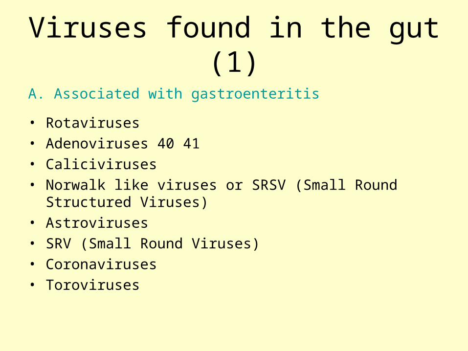
Viruses found in the gut (1)
A. Associated with gastroenteritis
• Rotaviruses• Adenoviruses 40 41• Caliciviruses• Norwalk like viruses or SRSV (Small Round Structured
Viruses)• Astroviruses• SRV (Small Round Viruses)• Coronaviruses• Toroviruses

Viruses found in the gut (2)B. Found in the gut, not normally associated with gastroenteritis• Polio• Coxsackie A• Coxsackie B• Echo• Enteroviruses 68-71• Hepatitis A• Hepatitis E• Adenoviruses 1-39• Reoviruses
C. Found in the gut as opportunistic infection • CMV• HSV• VZV• HIV

Viruses Associated with Respiratory Infections
Syndrome Commonly Associated Viruses
Less Commonly Associated Viruses
Colds Rhinoviruses, Coronaviruses
Influenza and parainfluenza viruses, enteroviruses, adenoviruses
Influenza Influenza viruses Parainfluenza viruses, adenoviruses
Croup Parainfluenza viruses Influenza virus, RSV, adenoviruses
Bronchiolitis RSV Influenza and parainfluenza viruses, adenoviruses
Bronchopneumonia Influenza virus, RSV, Adenoviruses
Parainfluenza viruses, measles, VZV, CMV

Common Cold Viruses
• Common colds account for one-third to one-half of all acute respiratory infections in humans
• Rhinoviruses are responsible for 30-50% of common colds, coronaviruses 10-30%
• The rest are due to adenoviruses, enteroviruses, RSV, influenza, and parainfluenza viruses, which may cause symptoms indistinguishable to those of rhinoviruses and coronaviruses

Rhinoviruses
• Spherical– 27-30 nm diamter
• +ssRNA genome– ~7.2kb
• Nonenveloped, icosahedral capsid
• 50% of Common Colds• 105 serotypes

Coronaviruses
• Irregularly shaped– 60-220 nm diameter
• +ssRNA genome (27-31 kb)• Enveloped particles with
loosely wound nucleocapsid– characteristic “club-shaped”
peplomers
• 10 % of Common colds

Some other coronaviruses
• Transmissible gastroenteritis and respiratory disease in pigs
• Infectious bronchitis in poultry
• Feline enteric coronavirus (FEC) and infectious peritonitis (FIP)
• SARS coronavirus

Severe Acute Respiratory Syndrome
(SARS)

Influenza Viruses• Pleomorphic, spherical
filamentous forms occur– 50-120 nm diameter, or 20
nm diameter and 200-300 nm long
• Segmented, linear -ssRNA genome– 7 to 8 segments
• Enveloped, filamentous nucleocapsids– Envelope is lipid bilayer with
~500 spikes for attachment• Hemagglutinin• Neuraminidase
• Causes “the flu”

Influenza A Virus
• Undergoes antigenic shifts and antigenic drifts with the haemagglutinin and neuraminidase proteins.
• Antigenic shifts of the haemagglutinin results in pandemics. Antigenic drifts in the H and N proteins result in epidemics.
• Usually causes a mild febrile illness
• Death may result from complications such as viral/bacterial pneumonia


The big pandemic of 1918
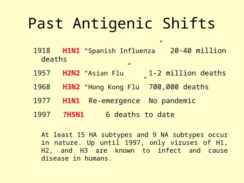
Past Antigenic Shifts
1918 H1N1 “Spanish Influenza” 20-40 million deaths
1957 H2N2 “Asian Flu” 1-2 million deaths
1968 H3N2 “Hong Kong Flu” 700,000 deaths
1977 H1N1 Re-emergence No pandemic
1997 ?H5N1 6 deaths to date
At least 15 HA subtypes and 9 NA subtypes occur in nature. Up until 1997, only viruses of H1, H2, and H3 are known to infect and cause disease in humans.

Avian to human transfer
• Hong Kong 1997, H5N1- 18 cases, 6 fatal• Hong Kong, 1999, H9N2 - 2 cases• Hong Kong Feb 2003, H5N1 - 2 cases, 1
fatal• Hong Kong Dec 2003, H9N2 - 1 case• Netherlands Feb 2003, H7N7 - 83 cases,
1 fatal (vet)• Asia - 2003-2005, H5N1

Factors that sustain epizootics/epidemics
• Antigenic drift
• Reassortment and antigenic shift
• Short term immunity
• Cross species transfer
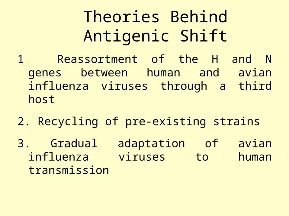
Theories Behind Antigenic Shift
1.Reassortment of the H and N genes between human and avian influenza viruses through a third host
2. Recycling of pre-existing strains
3. Gradual adaptation of avian influenza viruses to human transmission

Reassortment
Avian H3 Human H2
Human H3
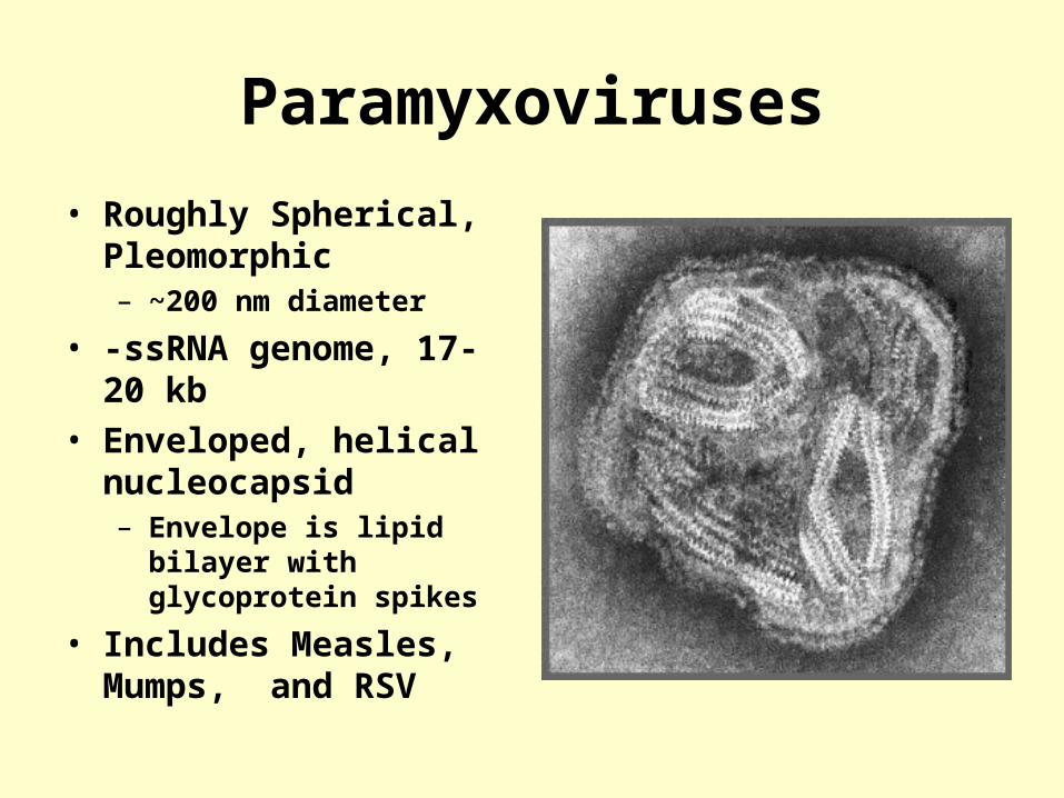
Paramyxoviruses
• Roughly Spherical, Pleomorphic– ~200 nm diameter
• -ssRNA genome, 17-20 kb• Enveloped, helical
nucleocapsid– Envelope is lipid bilayer with
glycoprotein spikes
• Includes Measles, Mumps, and RSV

Respiratory Syncytial Virus (RSV)
• ssRNA eveloped virus• belong to the genus Pneumovirus of the Paramyxovirus
family• Considerable strain variation exists, may be classified
into subgroups A and B by monoclonal sera• Both subgroups circulate in the community at any one
time.• Causes a sizable epidemic each year.

Clinical Manifestations
• Most common cause of severe lower respiratory tract disease in infants, responsible for 50-90% of Bronchiolitis and 5-40% of Bronchopneumonia
• Other manifestations include croup (10% of all cases).
• In older children and adults, the symptoms are much milder: it may cause a corza-like illness or bronchitis.

Parainfluenza Virus
• ssRNA virus• enveloped, pleomorphic
morphology• 5 serotypes: 1, 2, 3, 4a
and 4b• No common group
antigen• Closely related to Mumps
virus
(Linda Stannard, University of Cape Town, S.A.)
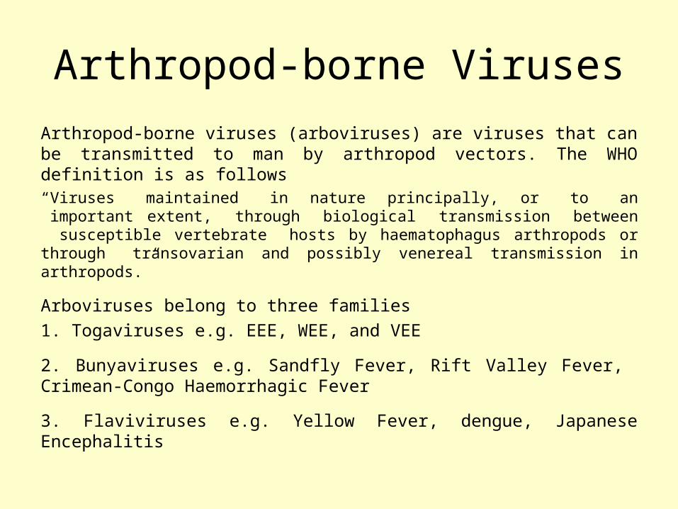
Arthropod-borne Viruses
Arthropod-borne viruses (arboviruses) are viruses that can be transmitted to man by arthropod vectors. The WHO definition is as follows“Viruses maintained in nature principally, or to an important extent, through biological transmission between susceptible vertebrate hosts by haematophagus arthropods or through transovarian and possibly venereal transmission in arthropods.”
Arboviruses belong to three families
1. Togaviruses e.g. EEE, WEE, and VEE
2. Bunyaviruses e.g. Sandfly Fever, Rift Valley Fever, Crimean-Congo Haemorrhagic Fever
3. Flaviviruses e.g. Yellow Fever, dengue, Japanese Encephalitis

Man-Arthropod-Man Cycle
e.g. dengue, urban yellow fever. Reservoir may be in either man or arthropod vector. In the latter transovarial transmission may take place.

Animal-Arthropod-Man Cycle
e.g. Japanese encephalitis, EEE, WEE, jungle yellow fever. The reservoir is in an animal. The virus is maintained in nature in a transmission cycle involving the arthropod vector and animal. Man becomes infected incidentally.

Arthopod Vectors
MosquitoesJapanese encephalitis, dengue, yellow fever, St. Louis encephalitis, EEE, WEE, VEE etc.
TicksCrimean-Congo haemorrhagic fever, various tick-borne encephalides etc.
SandfliesSicilian sandfly fever, Rift valley fever

Animal Reservoirs
In many cases, the actual reservoir is not known. The following animals are implicated as reservoirs
Birds Japanese encephalitis, St Louis encephalitis, EEE, WEE
Pigs Japanese encephalitis
Monkeys yellow fever
Rodents VEE, Russian Spring-Summer encephalitis

Diseases Caused
Fever and rash - this is usually a non-specific illness resembling a number of other viral illnesses such as influenza, rubella, and enterovirus infections. The patients may go on to develop encephalitis or haemorrhagic fever
Encephalitis - e.g. EEE, WEE, St Louis encephalitis, Japanese encephalitis
Haemorrhagic fever - e.g. yellow fever, dengue, Crimean-Congo haemorrhagic fever

Dengue
• Dengue is the biggest arbovirus problem in the world today with over 2 million cases per year. Dengue is found in SE Asia, Africa and the Caribbean and S America.
• Flavivirus, 4 serotypes, transmitted by Aedes mosquitoes which reside in waterfilled containers.
• Human infections arise from a human-mosquitoe-human cycle
• Classically, dengue presents with a high fever, lymphadenopathy, myalgia, bone and joint pains, headache, and a maculopapular rash.
• Severe cases may present with haemorrhagic fever and shock with a mortality of 5-10%. (Dengue haemorrhagic fever or Dengue shock syndrome.)

Distribution of Dengue

http://www.cdc.gov/ncidod/dvbid/westnile/virus.htm
• 40-60 nm• enveloped• +ve sense ssRNA• polyprotein,
processed by proteolysis
Flaviviridae

Flaviviridae
Pestiviruses
FlavivirusesHepatitis C virus
BVD, Hog cholera, Border disease
Japanese encephalitisSt. Louis encephalitisDengueWest Nile virus
(arthropods, biological vectors)

West Nile OutbreaksWest Nile Outbreaks
• Israel - 1951-1954, 1957• South Africa - 1974• Romania – 1996• Italy - 1998• Russia - 1999 (human)• Israel – 1998, 2000 (human)• France (Rhine delta) - 2000 (equine)• United States –1999-2004 (equine, human)• Canada - 2001-2004 (equine, human)

springearly summer
return from south
overwinteror
eggs
amplification in birds
late summer andfall
dead-end hosts

Zoonotic Viruses
• Zoonoses are diseases of vertebrate animals that can be transmitted to man: either directly or indirectly through an insect vector.
• When an insect vector is involved, the disease is also known as an arboviral disease.
• However, not all arboviral diseases are zoonosis: where the transmission cycle takes place exclusively between insect vector and human e.g. dengue and urban yellow fever.
• Examples of viral zoonoses that can be transmitted to man directly include rabies, hantaviruses, lassa and ebola fevers.

Rabies Virus
• member of the Lyassavirus of the Rhabdoviridae
• ssRNA enveloped virus, characteristic bullet-shaped appearance with 6-7 nm spike projections.
• virion 130-240nm * 80nm
• -ve stranded RNA codes for 5 proteins; G, M, N, L, S
• Exceedingly wide range of hosts
• There are 5 other members of Lyassavirus : Mokola, Lagosbat, Duvenhage, EBL-1, and EBL-2
• Duvenhage and EBL-2 have been associated with human rabies.

Rabies Virus
Structure of rabies virus (Source: CDC)
Rabies virus particles
Epidemiology
• Rabies is a zoonosis which is prevalent in wildlife. The main animals involved differs from continent to continent.
• Europe fox, bats• Middle East wolf, dog • Asia dog• Africa dog, mongoose, antelope• N America foxes, skunks, raccoons,
insectivorous bats• S America dog, vampire bats