viral immunology 2011.ppt - home | howard university€¢association of viral infection with cancer...
TRANSCRIPT

2/28/11 VIRAL IMMUNOLOGYSergei Nekhai Ph DSergei Nekhai, Ph.D.
Objectives:Objectives:
• Overview of immune system
• Innate immune response
•Adaprive immunity
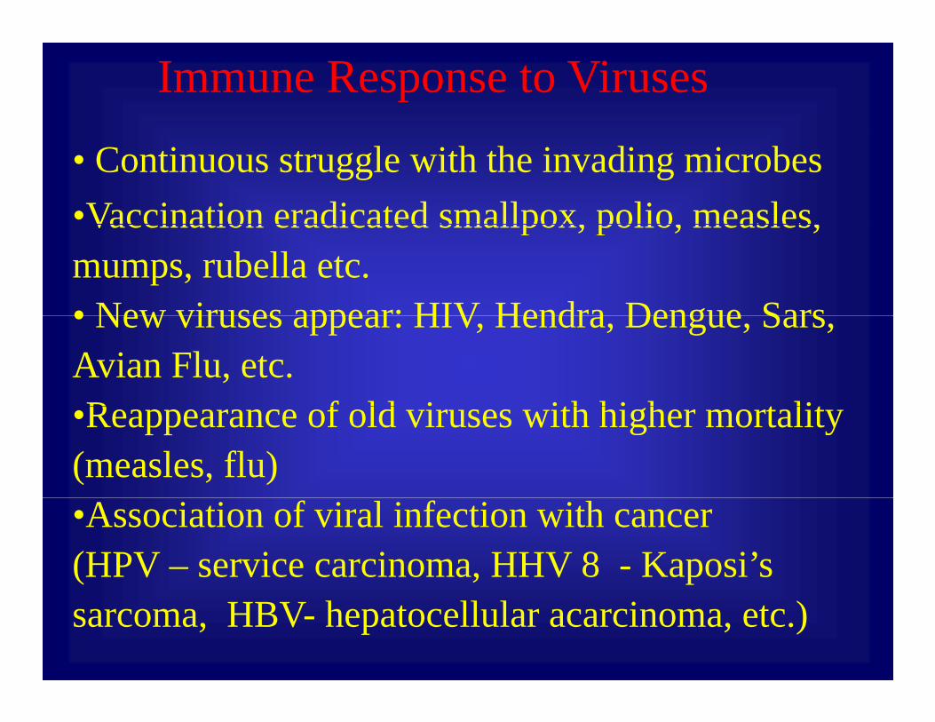
Immune Response to Viruses
• Continuous struggle with the invading microbes•Vaccination eradicated smallpox, polio, measles,Vaccination eradicated smallpox, polio, measles, mumps, rubella etc.• New viruses appear: HIV Hendra Dengue Sars• New viruses appear: HIV, Hendra, Dengue, Sars, Avian Flu, etc.R f ld i ith hi h t lit•Reappearance of old viruses with higher mortality
(measles, flu)•Association of viral infection with cancer (HPV – service carcinoma, HHV 8 - Kaposi’ssarcoma, HBV- hepatocellular acarcinoma, etc.)

Immune Response – cont.
• Effector function•Carried by cells (natural killer (NK) T cells) – “cellular”•Carried by cells (natural killer (NK), T cells) – cellular
immunity
•Fluid-born – “humoral” immunity (antibodies, chemokines,
cytokines, complement, etc)
•Antigen Specificity•Antigen-specific (adaptive) – has memory
•Non-antigen specific (innate) – no memory

The Immune SystemParasite in red blood cellBacteria
SARS virus FungusSARS virus Fungus

Markers of Self
Muscle cellEpithelialcell
Leukocyte
Nervecell
Class I MHC self-marker protein

Markers of Non-Self
EpitopeA ti
Bacteria SARS virus
Epitope
Antibody
Antigen
Non-self leukocyteNon-self nerve cell Non self leukocyte
EpitopeAntigen
Non self nerve cell
Epitope Class I MHC protein
Antigen
Antibody
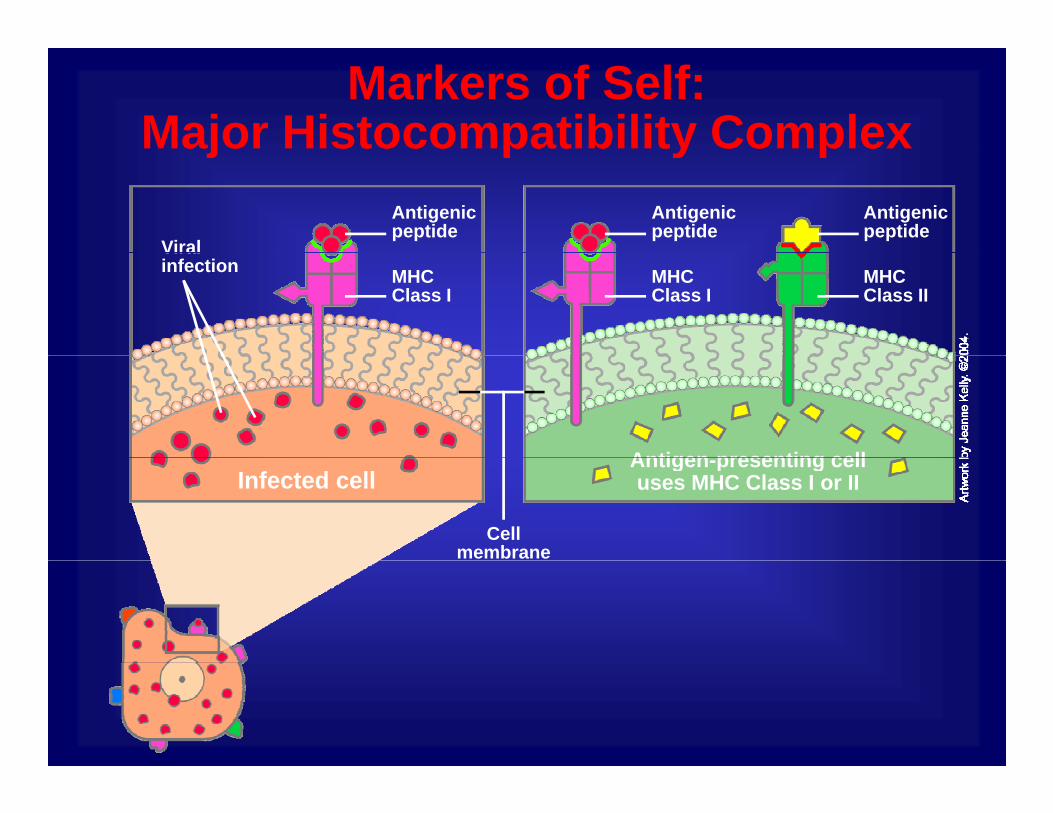
Markers of Self:Major Histocompatibility Complexj p y p
Antigenic peptide
Antigenic peptide
Viral
Antigenic peptide
MHC Class II
Viral infection MHC
Class IMHC Class I
Antigen presenting cellAntigen-presenting cell uses MHC Class I or II
Cell membrane
Infected cell

Endogenous antigen processing: MHC class I peptide Endogenous antigen processing: MHC class I peptide presentationpresentation
• Intracellular proteins of host and virus are marked for degradation by ubiquitination and are degraded by the Proteasome.Proteasome.
• The resulting viral peptides are transported into the ER lumen by the Tap1 Tap2 heterodimeric transporterlumen by the Tap1-Tap2 heterodimeric transporter.
• In the ER lumen, viral peptides associate with newly synthesized MHC class I molecules.
• MHC class I-peptide complex is transported to the cellMHC class I peptide complex is transported to the cell surface via the golgi compartments.
• On the cell surface the MHC class I peptide complex• On the cell surface, the MHC class I-peptide complex interacts with the T- cell receptor of a Tc cell carrying the CD8 coreceptor.

Endogenous antigen processing: MHC class I peptide presentationEndogenous antigen processing: MHC class I peptide presentation
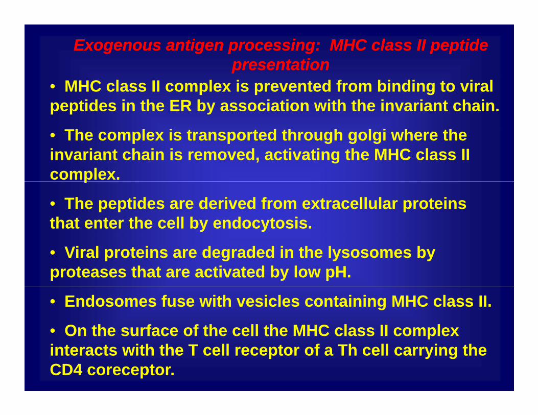
Exogenous antigen processing: MHC class II peptide Exogenous antigen processing: MHC class II peptide presentationpresentation
• MHC class II complex is prevented from binding to viral peptides in the ER by association with the invariant chain.
Th l i t t d th h l i h th• The complex is transported through golgi where the invariant chain is removed, activating the MHC class II complex.p
• The peptides are derived from extracellular proteins that enter the cell by endocytosis.
• Viral proteins are degraded in the lysosomes by proteases that are activated by low pH.
• Endosomes fuse with vesicles containing MHC class II.
• On the surface of the cell the MHC class II complex pinteracts with the T cell receptor of a Th cell carrying the CD4 coreceptor.

Exogenous antigen processing: MHC class II peptide Exogenous antigen processing: MHC class II peptide presentationpresentation
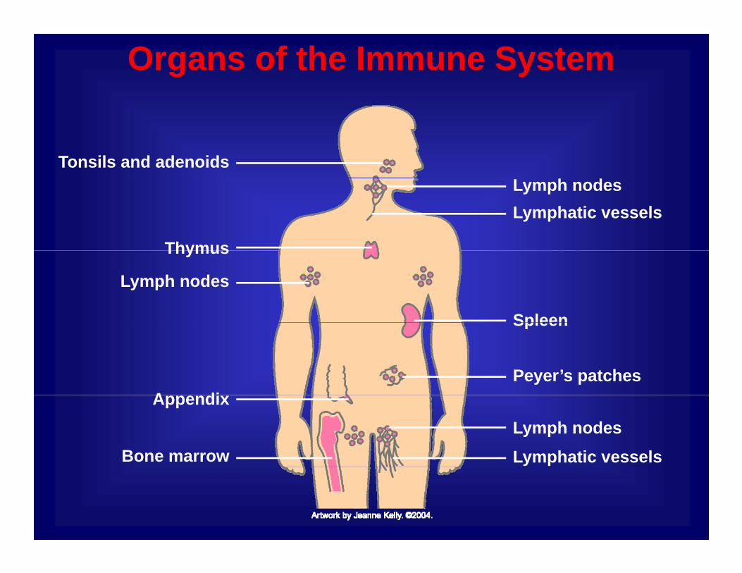
Organs of the Immune System
Tonsils and adenoids
Thymus
Lymphatic vesselsLymph nodes
Lymph nodes
Thymus
Spleen
A diPeyer’s patches
Spleen
Bone marrow
Appendix
Lymphatic vesselsLymph nodes

ANATOMY OF THE IMMUNE SYSTEM• Thymus – glandular organ near the heart – where T cells learn
Sergei Nekhai:
y g gtheir jobs
• Bone marrow blood-producing tissue located inside certain• Bone marrow – blood-producing tissue located inside certain bones– blood stem cells give rise to all of the different types of blood cells
• Spleen – serves as a filter for the blood– removes old and damaged red blood cells
f– removes infectious agents and uses them to activate cells called lymphocytes
• Lymph nodes – small organs that filter out dead cells, antigens, and other “stuff” to present to lymphocytes
• Lymphatic vessels – collect fluid (lymph) that has “leaked” out from the blood into the tissues and returns it to circulation

Lymph Node
Incoming lymphatic vessel
Germinal center
ParacortexFollicle
CortexMedulla
VeinOutgoing lymphatic vessel
ArteryArtery
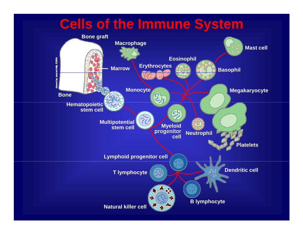
Cells of the Immune SystemBone graft
MacrophageMacrophage
ErythrocytesEosinophil
Mast cell
BasophilMarrow
MegakaryocyteMonocyteBone
Multipotentialstem cell
Hematopoieticstem cell
N t hilMyeloid
progenitor
Platelets
Neutrophil
Lymphoid progenitor cell
progenitor cell
T lymphocyte Dendritic cell
Natural killer cellB lymphocyte
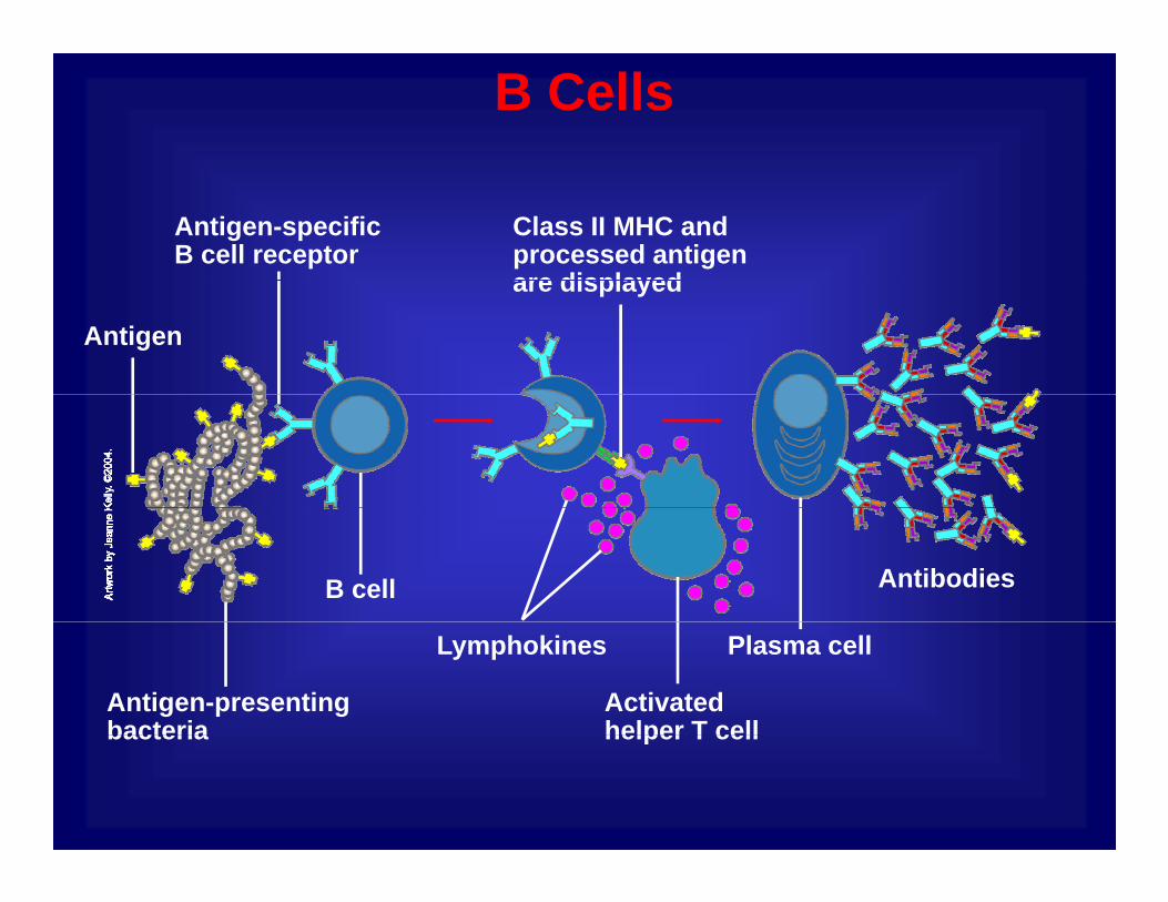
B Cells
Class II MHC and processed antigen are displayed
Antigen-specific B cell receptor
are displayed
Antigen
AntibodiesB cell
Plasma cell
Antigen-presenting bacteria
Activated helper T cell
Lymphokines
bacteria helper T cell
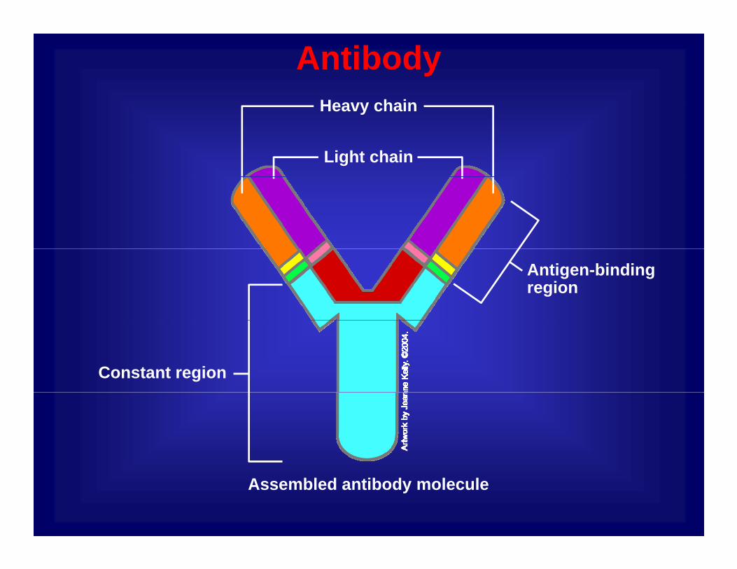
Antibody Heavy chainHeavy chain
Light chain
Antigen-bindingregion
Constant region
Assembled antibody molecule
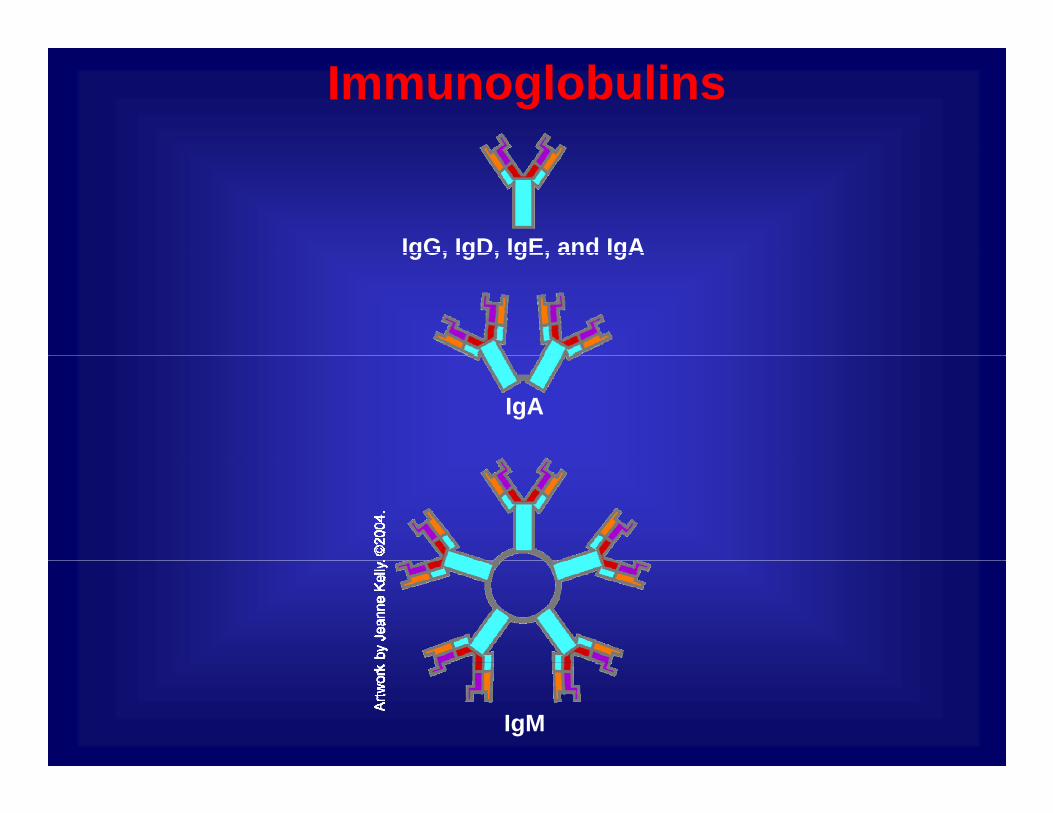
Immunoglobulins
IgG, IgD, IgE, and IgAIgG, IgD, IgE, and IgA
IgA
IgM

Type Number of ag binding sites
Site of action Functions
sites
IgG 2 •Blood•Tissue fluid
•Increase macrophage activity
•CAN CROSS PLACENTA
•Antitoxins•Agglutination
IgM 10 •Blood AgglutinationIgM 10 Blood•Tissue fluid
Agglutination
IgA 2 or 4 •Secretions (saliva tears •Stop bacteriaIgA 2 or 4 •Secretions (saliva, tears, small intestine, vaginal, prostate, nasal, breast milk)
•Stop bacteria adhering to host cells•Prevents bacteria forming colonies on gmucous membranes
IgE 2 Tissues •Activate mast cellsgHISTAMINE
•Worm response

T Cells Resting cytotoxic T cellResting helper T cell
Activated killer cellActivated helper T cellp
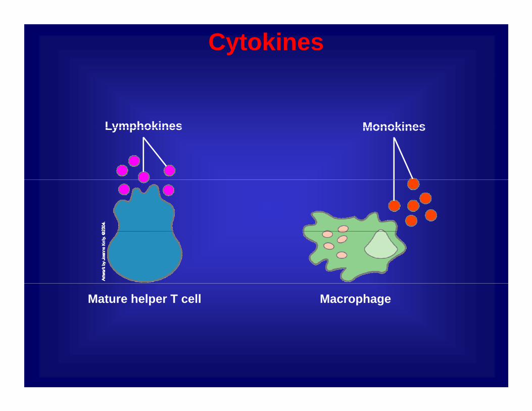
Cytokines
MonokinesLymphokines MonokinesLymphokines
Mature helper T cell Macrophage
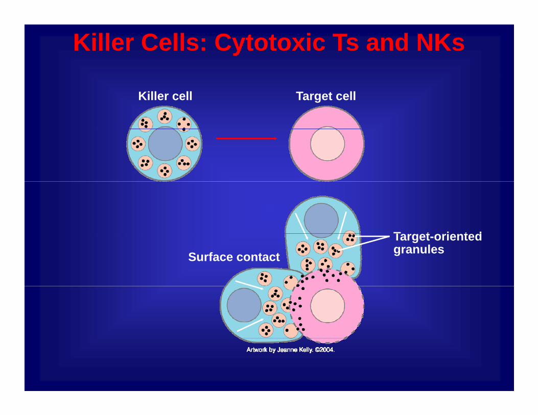
Killer Cells: Cytotoxic Ts and NKs
Killer cell Target cell
T t i t dTarget-oriented granulesSurface contact

Phagocytes and Their Relatives
Monocyte
Eosinophil
Dendritic cell
Mast cell
Macrophage
Neutrophil
Basophil
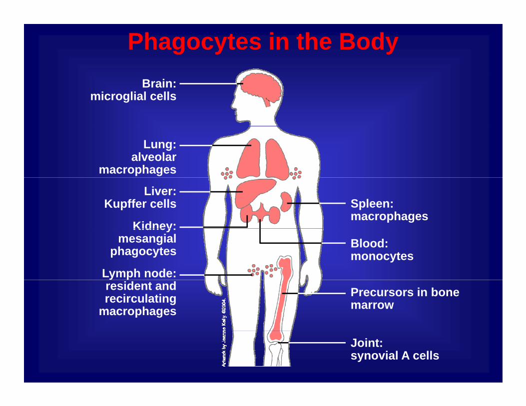
Phagocytes in the Body Brain:
microglial cells
Lung:alveolar
macrophages
Kidney:
Spleen: macrophages
Liver: Kupffer cells
Lymph node:
Blood: monocytes
Kidney:mesangial
phagocytes
Precursors in bone marrow
resident and recirculating
macrophages
Joint:synovial A cells
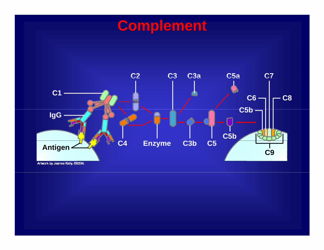
Complement
C2 C3aC3 C7C5aC2 C3aC3
C1 C8
C7
C6C5b
C5a
E C5C3bC4
IgG C5b
C5b
C9Enzyme C5C3bC4Antigen

Mounting an Immune Response
Complement
T cell
Lymphokines
B cellAntibodies
T cellMacrophage
VirusKiller cell

YOUR ACTIVE IMMUNE DEFENSES
Innate Immunity- invariant (generalized)
Adaptive Immunity- variable (custom)a a t (ge e a ed)
- early, limited specificity- the first line of defense
( )- later, highly specific- ‘‘remembers’’ infection
The innate immune response:The innate immune response:C b ti t d idl d f ti ithi h• Can be activated rapidly and functions within hours
of a viral infection.
• Continued activity is damaging to the host• Continued activity is damaging to the host.
• Considerable interplay occurs between the adaptive and innate immune defensesand innate immune defenses.
Important components are:
t ki-cytokines
-complement
-collectins
-natural killer (NK) cells

Initiation of Immune Responses
Field’s Virology, Fifth Edition

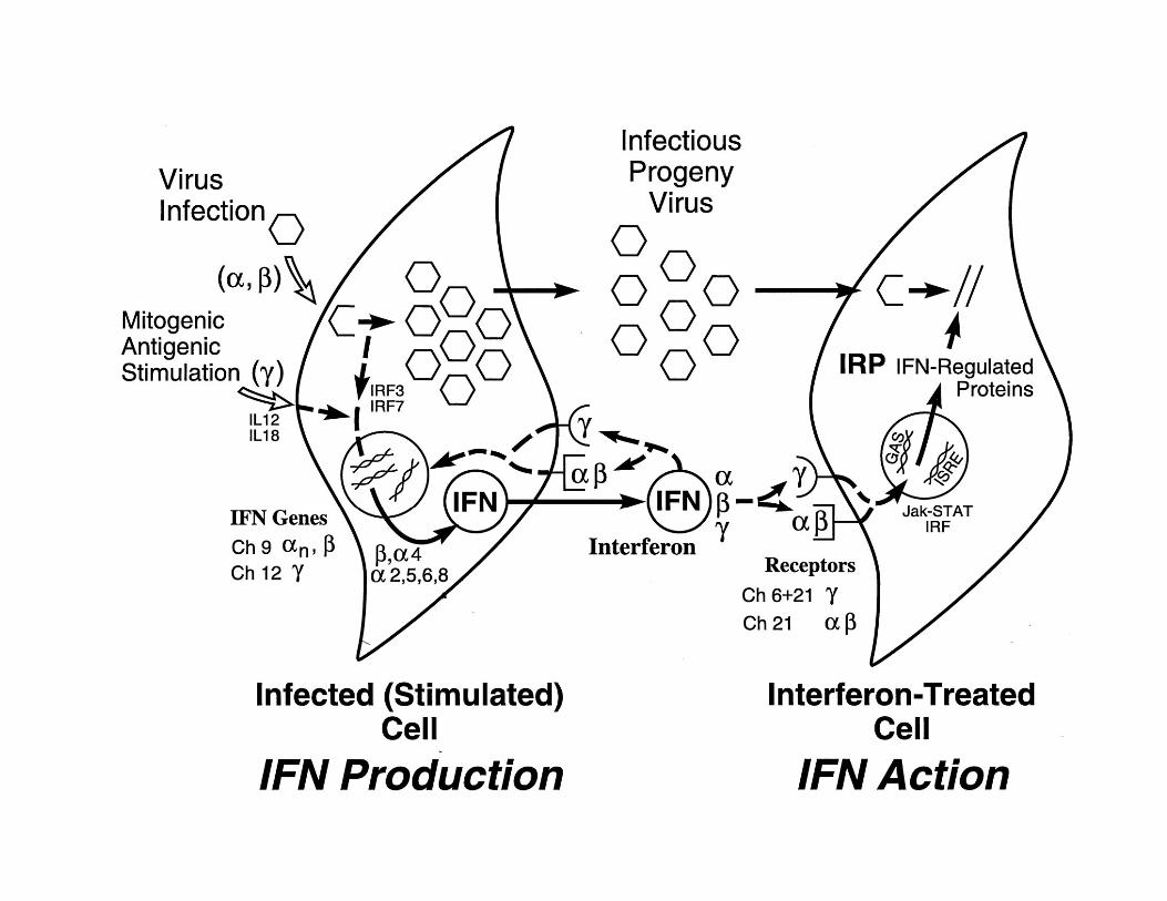
Activation of INFs and Cytokines by Viral Infection
Field’s Virology, Fifth Edition
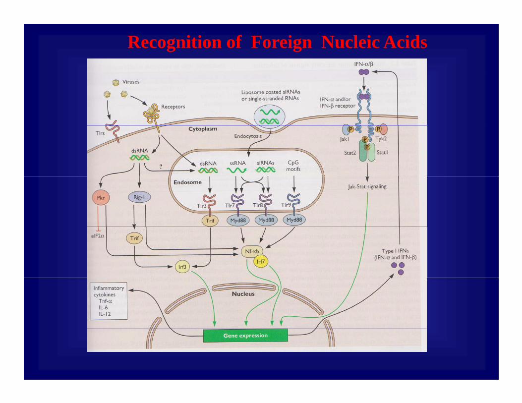
Recognition of Foreign Nucleic Acids
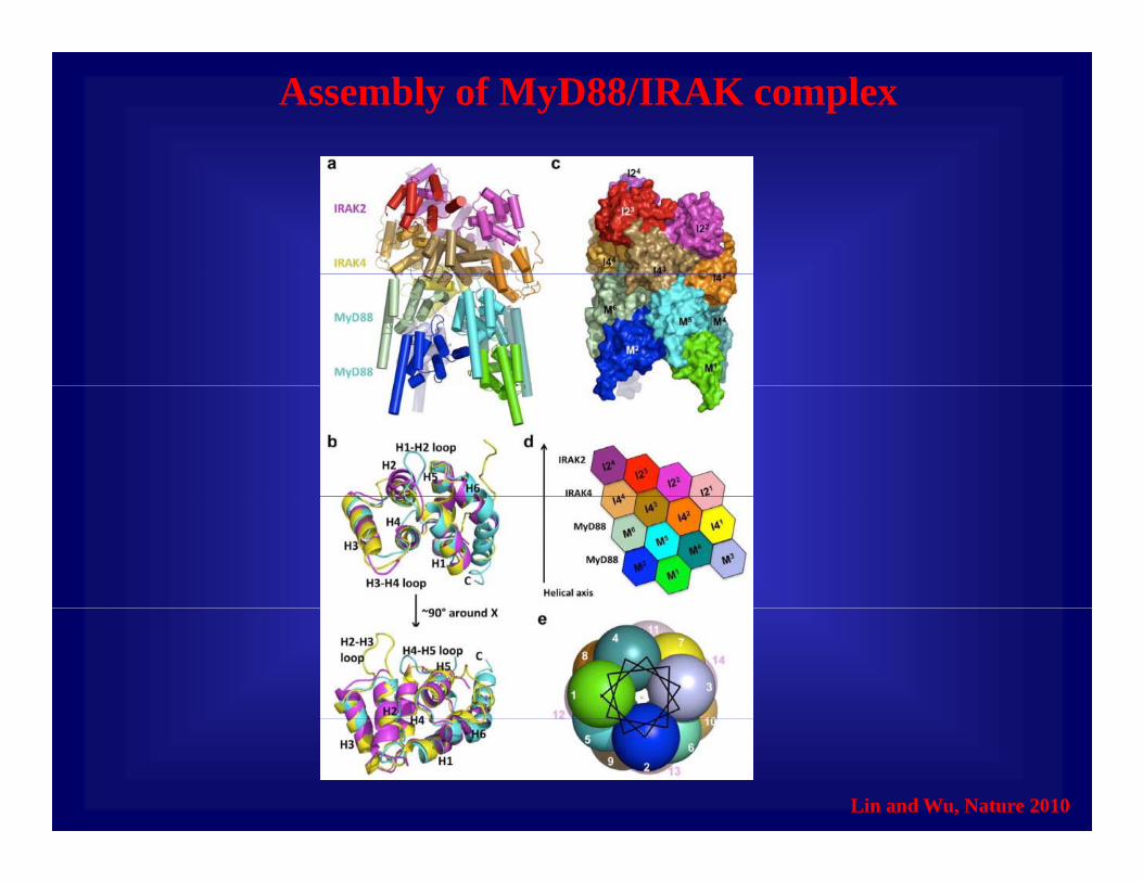
Assembly of MyD88/IRAK complex
Lin and Wu, Nature 2010
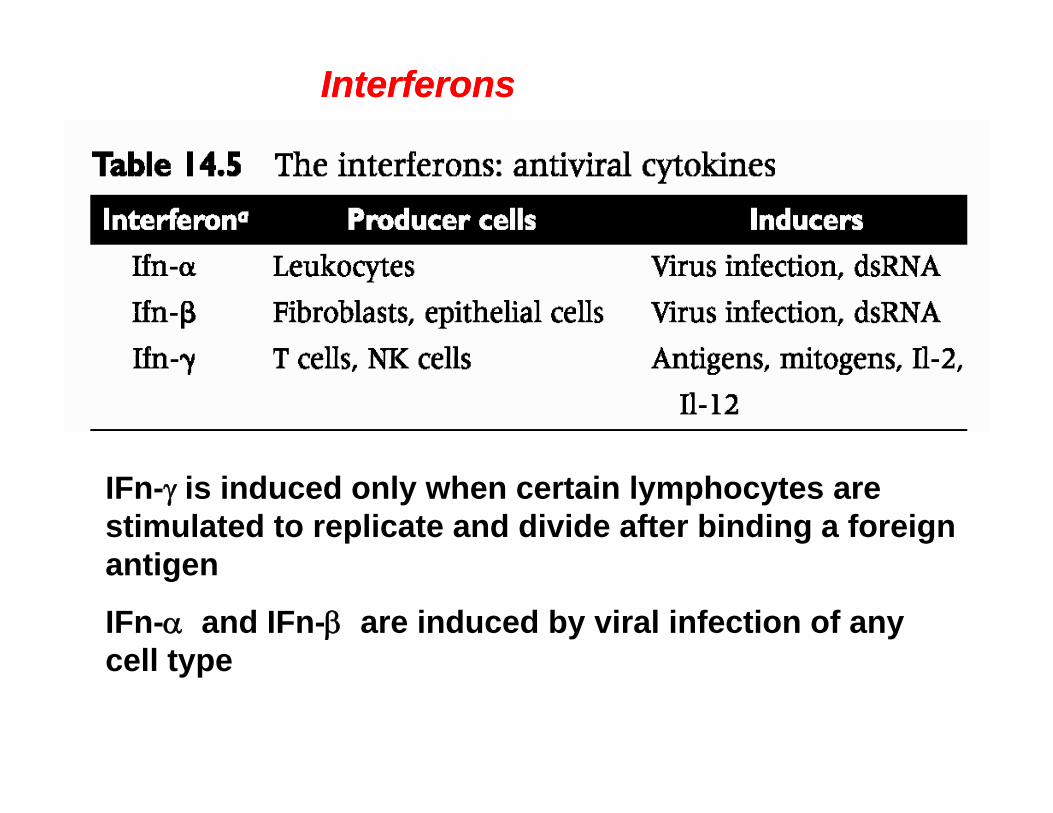
InterferonsInterferons
IFn-γ is induced only when certain lymphocytes are stimulated to replicate and divide after binding a foreign antigenantigen
IFn-α and IFn-β are induced by viral infection of any cell typecell type
• IFN is induced by accumulation of double stranded InterferonsInterferonsy
RNA (dsRNA).
• IFN induces gene expression at the transcriptional level after binding to specific cell surface receptors.
• A cell that is bound to interferon and responds to it i i ti i l t tis in an antiviral state.
• IFN induces expression of more that 100 genes, products of many of these genes possess broadproducts of many of these genes possess broad spectrum antiviral activity.
• They lead to cell death by apoptosis or programmed They lead to cell death by apoptosis or programmed cells death, limiting cell to cell spread of virus.
• Production of large amounts if IFN causes common gsymptoms such as fever, chills, nausea, etc.

Interferon induced antiviral responses:Interferon induced antiviral responses:• Both viral and cellular protein synthesis stops in IFN treated cells.
• This is dues to two cellular proteins, ds-RNA activated protein kinase (PKR) and ribonuclease L (RNase L).
• PKR is a serine/threonine kinase that has antiviral properties, as well as antiproliferative and antitumor functions.
• Activated PKR phosphorylates the alpha subunit of the translation initiation factor eIF2, inhibiting translation.
• RNase L is a nuclease that can degrade cellular and viral RNA; its concentration increases after Ifn treatment.
RN L t ti i 10 1 000 f ld ft
Interferon induced antiviral responses:Interferon induced antiviral responses:• RNase L concentration increases 10-1,000 fold after Ifn treatment, but is inactive unless 2’-5’-oligo(A) synthetase is produced.
• 2’-5’-oligo(A) synthetase produces 2’, 5’ oligomers of adenylic acid, only when activated by dsRNA.
• These poly(A) oligomers then activate RNase L, which degrades all host and viral mRNA in the cell.
• RNase L participates not only in Ifn-mediated antiviral defense, but also in apoptosis.
• Ifn is a broad spectrum, highly effective antiviral agent. However, viruses have developed numerous mechanisms for inhibiting interferon action.mechanisms for inhibiting interferon action.

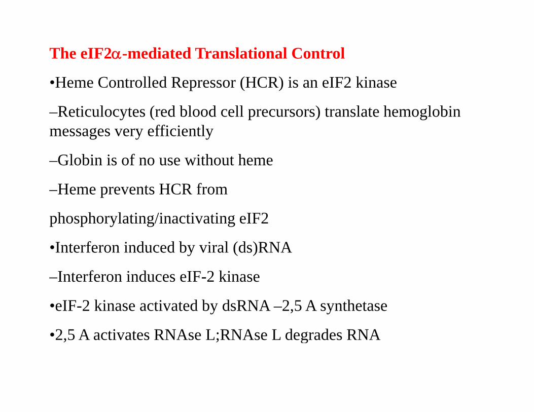
The eIF2α-mediated Translational Control
•Heme Controlled Repressor (HCR) is an eIF2 kinase
–Reticulocytes (red blood cell precursors) translate hemoglobin ffi i tlmessages very efficiently
–Globin is of no use without heme
–Heme prevents HCR from
phosphorylating/inactivating eIF2
•Interferon induced by viral (ds)RNA
–Interferon induces eIF-2 kinase
•eIF-2 kinase activated by dsRNA –2,5 A synthetase
•2 5 A activates RNAse L;RNAse L degrades RNA•2,5 A activates RNAse L;RNAse L degrades RNA

Heme Regulates the Activity of Heme-regulated Kinase
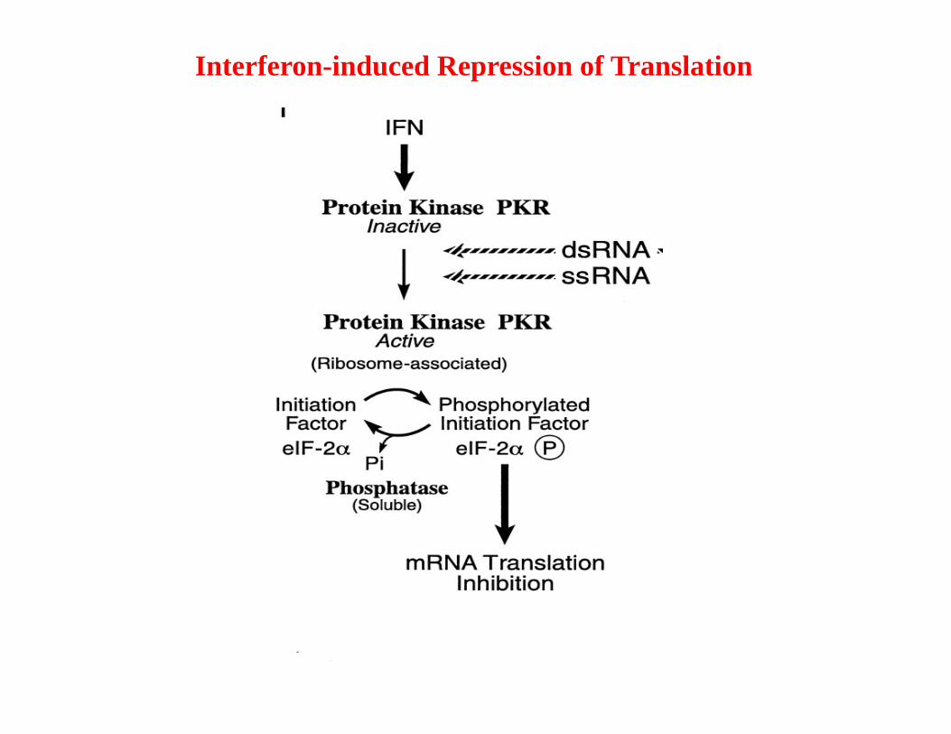
Interferon-induced Repression of Translation

Heme Regulates the Activity of Heme-regulated Kinase
J.J. Chen

Iron Transport in Humans
Drakesmith and Prentice, 2008
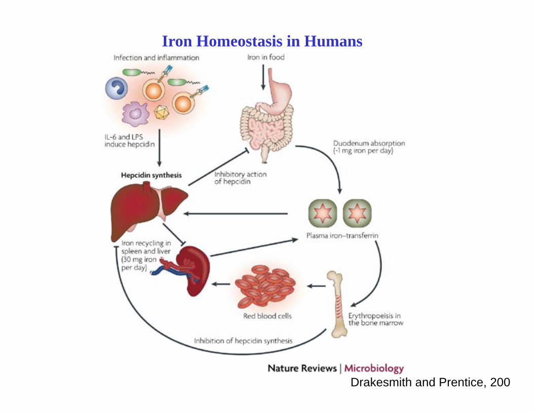
Iron Homeostasis in Humans
Drakesmith and Prentice, 200
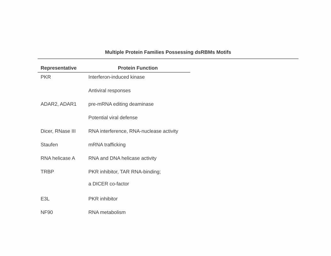
Multiple Protein Families Possessing dsRBMs Motifs
Representative Protein Function
PKR Interferon-induced kinase
Antiviral responses
ADAR2, ADAR1 pre-mRNA editing deaminase
Potential viral defense
Dicer, RNase III RNA interference, RNA-nuclease activity
Staufen mRNA traffickingStaufen mRNA trafficking
RNA helicase A RNA and DNA helicase activity
TRBP PKR inhibitor, TAR RNA-binding;
a DICER co-factor
E3L PKR inhibitor
NF90 RNA metabolism

Other Intrinic Antivirail ResponcesA t h F i f i li d b•Autophagy Formation of specialized membrane
compartmetnts related to lysosomes•Epigenetic silencing Defence against DNA containing viruses, formation hromatin structure•RNA silencing Ssequence-specific RNA degradationdegradation•Cytosine Deamination (APOBEC) C’s to U’s convertionconvertion •TRIM Proteins Targeting capsid protein by TRM5a proteinTRM5a protein•Tetherin Inability of the virions to bud

Micro RNA
•Founding members of miRNAs, 22 nt and 61 nt RNAs coded by C.elegans Lin-1 gene complementary to 3’UTR of Lin-14 gene that blocked translation of Lin-14
• Control of cell proliferaton, cell death and fat metabolism in flies
M d l ti f h t i ti li diff ti ti i l•Modulation of hematopoietic lineage differentiation in mammals
•Leaf and flower development in plants
•Majority of miRNAs are transcribed independently
•Some (quarter) miRNA are derived from intronesSo e (qua e ) N a e de ved o o es
•miRNA are conserved

Formation of RISCs and Other Silencing Complexes
Pratt and McRae, JBC, 2009

Examples of Metazoan miRNAs

Maturation of miRNA
Plants Metazoa AnimalsCleavege with Drosha
AdditionalCleavage with Dicerwith Dicer
Complex withRISC (RNA-inducedSilencing complex)g p )

Actions of Small Silencing RNAs
mRNA celavage Translational repression Transcriptional silencingmRNA celavage Translational repression Transcriptional silencing
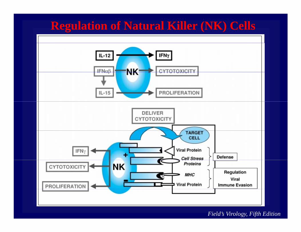
Regulation of Natural Killer (NK) Cells
Field’s Virology, Fifth Edition
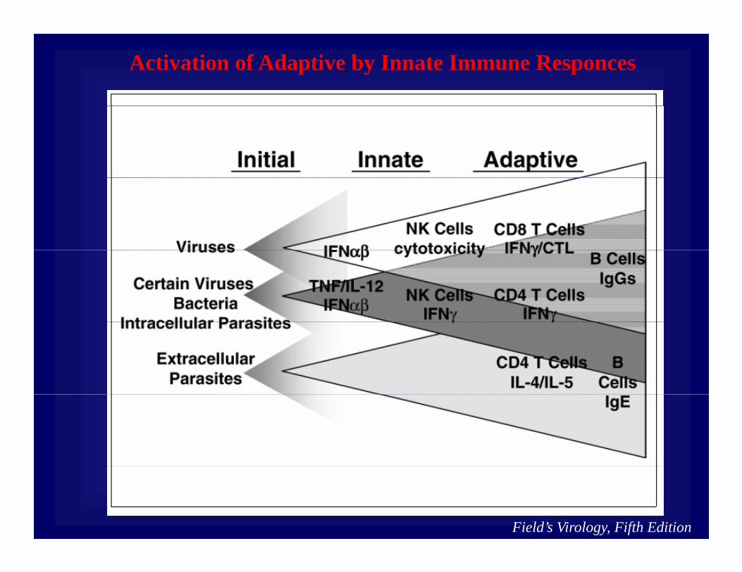
Activation of Adaptive by Innate Immune Responces
Field’s Virology, Fifth Edition

Cytokine-mediated Immune Responces
Field’s Virology, Fifth Edition

The adaptive immune response:The adaptive immune response:
• Humoral response
Consists of lymphocytes of the B-cell lineage
Interaction of a specific receptor on precursor B lymphocytes with antigens promotes differentiation i t tib d ti ll ( l ll )into antibody secreting cells (plasma cells).
• Cell-mediated response
Consists of lymphocytes of the T-cell lineage
Cytotoxic T cells (Tc cells) and T-helper cells (Th cells) are the key effectors of this response.

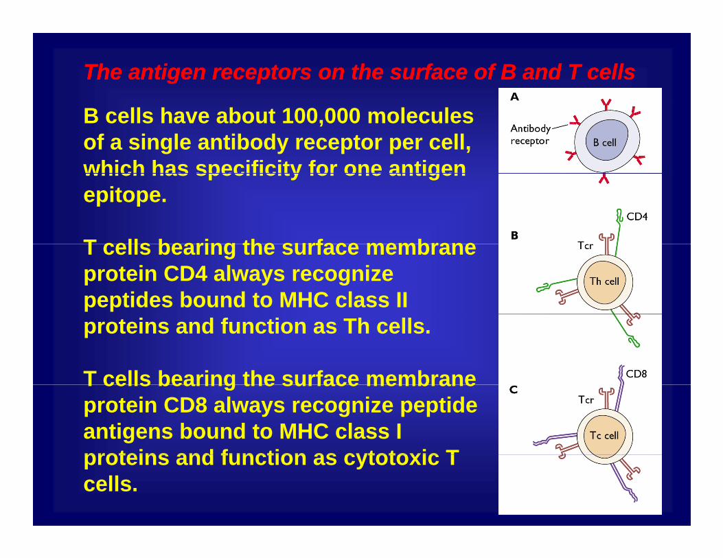
The antigen receptors on the surface of B and T cellsThe antigen receptors on the surface of B and T cells
B cells have about 100,000 molecules of a single antibody receptor per cell, which has specificity for one antigenwhich has specificity for one antigen epitope.
T cells bearing the surface membraneT cells bearing the surface membrane protein CD4 always recognize peptides bound to MHC class II proteins and function as Th cells.
T cells bearing the surface membraneT cells bearing the surface membrane protein CD8 always recognize peptide antigens bound to MHC class I proteins and function as cytotoxic Tproteins and function as cytotoxic T cells.

Antibody Activities in Viral Infection
Field’s Virology, Fifth Edition

Maturation of CD4+ and CD8+ T cells
Field’s Virology, Fifth Edition
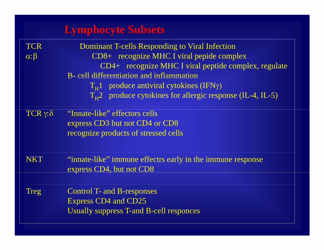
Lymphocyte SubsetsTCR Dominant T cells Responding to Viral InfectionTCR α:β
Dominant T-cells Responding to Viral InfectionCD8+ recognize MHC I viral pepide complex
CD4+ recognize MHC I viral peptide complex, regulate B- cell differentiation and inflammationB cell differentiation and inflammation
TH1 produce antiviral cytokines (IFNγ)TH2 produce cytokines for allergic response (IL-4, IL-5)
TCR γ:δ “Innate-like” effectors cellsexpress CD3 but not CD4 or CD8recognize products of stressed cells
NKT “innate-like” immune effectrs early in the immune responseexpress CD4, but not CD8p ,
Treg Control T- and B-responsesExpress CD4 and CD25pUsually suppress T-and B-cell responces
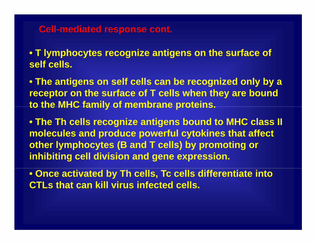
Cell-mediated response cont.
• T lymphocytes recognize antigens on the surface of self cells.
• The antigens on self cells can be recognized only by a receptor on the surface of T cells when they are bound to the MHC family of membrane proteins.to the MHC family of membrane proteins.
• The Th cells recognize antigens bound to MHC class II molecules and produce powerful cytokines that affect p p yother lymphocytes (B and T cells) by promoting or inhibiting cell division and gene expression.
• Once activated by Th cells, Tc cells differentiate into CTLs that can kill virus infected cells.
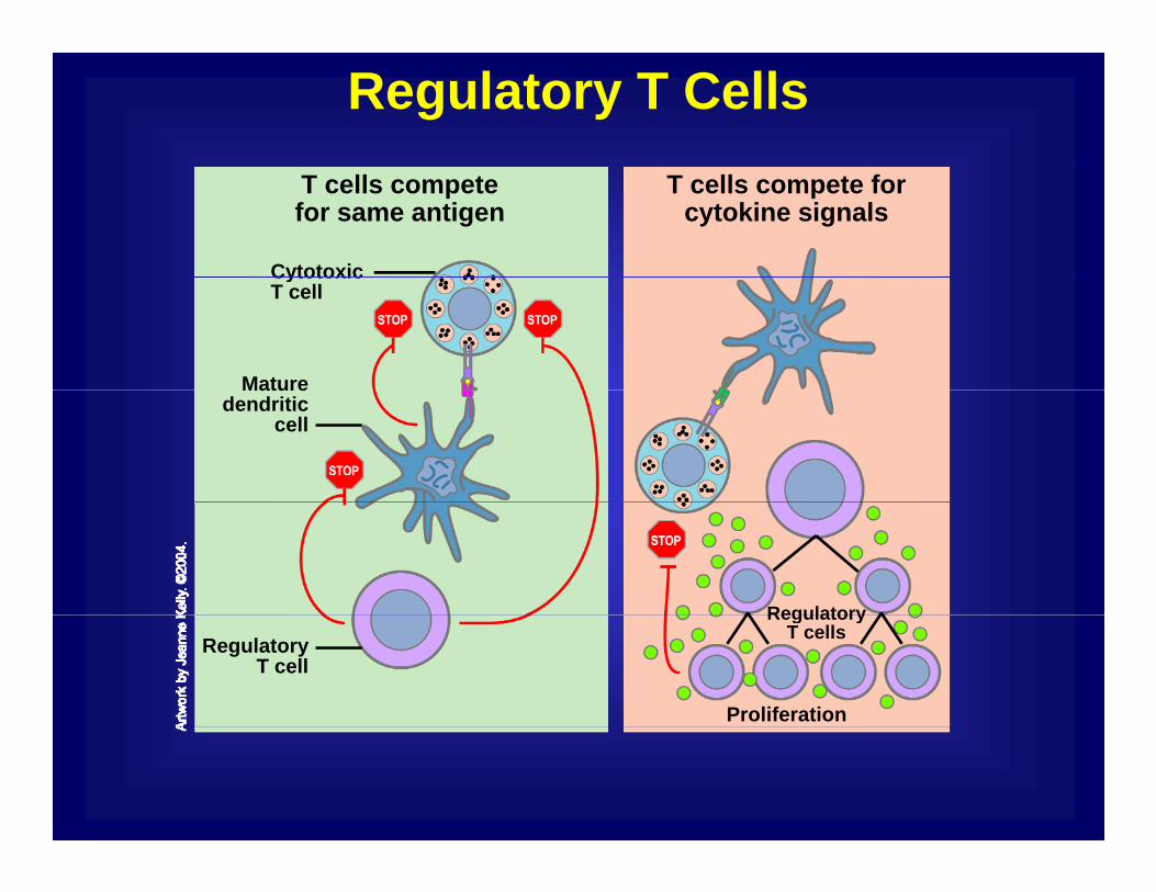
Regulatory T CellsT cells compete for
cytokine signalsT cells competefor same antigen
Cytotoxic
Mature
CytotoxicT cell
Mature dendritic
cell
RegulatoryRegulatory T cellsRegulatory
T cell
Proliferation