view - open access lmu
TRANSCRIPT

Cryptic Species in Tropic Sands - Interactive 3D Anatomy,Molecular Phylogeny and Evolution of MeiofaunalPseudunelidae (Gastropoda, Acochlidia)Timea P. Neusser1,2*, Katharina M. Jorger1,2, Michael Schrodl1,2
1 Bavarian State Collection of Zoology, Munchen, Germany, 2 Department Biology I of the Ludwig-Maximilians-Universitat Munchen, Planegg-Martinsried, Germany
Abstract
Background: Towards realistic estimations of the diversity of marine animals, tiny meiofaunal species usually areunderrepresented. Since the biological species concept is hardly applicable on exotic and elusive animals, it is even moreimportant to apply a morphospecies concept on the best level of information possible, using accurate and efficientmethodology such as 3D modelling from histological sections. Molecular approaches such as sequence analyses may revealfurther, cryptic species. This is the first case study on meiofaunal gastropods to test diversity estimations from traditionaltaxonomy against results from modern microanatomical methodology and molecular systematics.
Results: The examined meiofaunal Pseudunela specimens from several Indo-Pacific islands cannot be distinguished byexternal features. Their 3D microanatomy shows differences in the organ systems and allows for taxonomic separation insome cases. Additional molecular analyses based on partial mitochondrial cytochrome c oxidase subunit I (COI) and 16SrRNA markers revealed considerable genetic structure that is largely congruent with anatomical or geographical patterns.Two new species (Pseudunela viatoris and P. marteli spp. nov.) are formally described integrating morphological and geneticanalyses. Phylogenetic analysis using partial 16S rRNA, COI and the nuclear 18S rRNA markers shows a clade ofPseudunelidae species as the sister group to limnic Acochlidiidae. Within Pseudunela, two subtypes of complex excretorysystems occur. A complex kidney already evolved in the ancestor of Hedylopsacea. Several habitat shifts occurred duringhedylopsacean evolution.
Conclusions: Cryptic species occur in tropical meiofaunal Pseudunela gastropods, and likely in other meiofaunal groups withpoor dispersal abilities, boosting current diversity estimations. Only a combined 3D microanatomical and molecularapproach revealed actual species diversity within Pseudunela reliably. Such integrative methods are recommended for alltaxonomic approaches and biodiversity surveys on soft-bodied and small-sized invertebrates. With increasing taxonsampling and details studied, the evolution of acochlidian panpulmonates is even more complex than expected.
Citation: Neusser TP, Jorger KM, Schrodl M (2011) Cryptic Species in Tropic Sands - Interactive 3D Anatomy, Molecular Phylogeny and Evolution of MeiofaunalPseudunelidae (Gastropoda, Acochlidia). PLoS ONE 6(8): e23313. doi:10.1371/journal.pone.0023313
Editor: Roland G. Roberts, Public Library of Science, United Kingdom
Received November 5, 2010; Accepted July 13, 2011; Published August 31, 2011
Copyright: � 2011 Neusser et al. This is an open-access article distributed under the terms of the Creative Commons Attribution License, which permitsunrestricted use, distribution, and reproduction in any medium, provided the original author and source are credited.
Funding: This study benefited from financial support by the German Research Foundation (www.dfg.de) (SCHR 667/4-2, 3, 4 to MS). Molecular studies weresupported by the VW-Stiftung (http://www.volkswagenstiftung.de) (grant to KMJ). The funders had no role in study design, data collection and analysis, decisionto publish, or preparation of the manuscript.
Competing Interests: The authors have declared that no competing interests exist.
* E-mail: [email protected]
Introduction
The study of cryptic species, i.e. two or more distinct species
classified as a single species due to the lack of morphological
differences, augmented during the last 20 years [1]. There is a
consensus about the importance of our knowledge of cryptic
diversity for, amongst others, animal diversity estimations,
biological control, natural resource protection and conservation
(e.g. [1,2]). However, the distribution of cryptic species among
metazoan taxa and biogeographical regions is discussed contro-
versially. Whereas Bickford et al. [1] proposed a non-random
distribution across taxa and biomes, Pfenninger & Schwenk [3]
suggested an almost even distribution among the major metazoan
taxa and biogeographical regions. Trontelj & Fiser [2] emphasised
that regularities of the cryptic diversity probably will be discovered
only by means of genus- or species-level studies.
One area with an unexpectedly high level of cryptic speciation is
the Antarctic Ocean. Molecular studies revealed flocks of cryptic
rather than single widespread and variable species throughout
benthic invertebrate groups examined, e.g. in crinoids, pycnogo-
nids, crustaceans and molluscs [4,5,6,7]. Many, but not all of those
organisms from high geographic latitudes are brooders or direct
developers with low dispersal abilities, such as the nudibranch
gastropod Doris kerguelenensis (Bergh, 1884) which ultimately was
shown to have undergone an explosive cryptic radiation in the
Southern Ocean [6]. According to Thorson’s rule, direct developers
in benthic organisms such as most molluscs are considered as scarce
in subtropical or tropical waters [8]. Exceptions are members of
taxa living in the mesopsammon which generally are assumed to be
direct developers [9] or, as in case of acochlidian panpulmonate
gastropods, may have planktonic larvae which remain in the
interstitial spaces [10]. Thus, it can be assumed that their dispersal
PLoS ONE | www.plosone.org 1 August 2011 | Volume 6 | Issue 8 | e23313

ability in the larval stage is very low. Also, meiofaunal acochlidian
gastropods appear to occur in coastal sands only, i.e. postlarval
stages have virtually no potential for active migration or forming
continuous populations across deeper waters. Given this level of
supposed immobility and habitat restrictions as opposed to the vast
coasts of the world’s oceans and innumerable, highly isolated
archipelagos and off-shore reefs we should expect that there are
plenty of narrow ranged rather than a few wide-ranged acochlidian
species. However, based on morphology, only 28 valid species, 20 of
them mesopsammic, were described globally. Several of these
species such as Microhedyle remanei (Marcus, 1953) were considered to
be widespread throughout Western Atlantic warm water sands, i.e.
in Brazil, Colombia and Bermuda [11,12,13,14], and Pseudunela
cornuta (Challis, 1970) was recorded to occur on the Solomon Islands
(Melanesia) and near Hong Kong (South China Sea) [15,16].
Recently, both species were re-described in considerable anatomical
and histological detail [14,17]. However, until now, applying
morphospecies concepts on tiny meiofaunal gastropods has never
been tested by molecular analyses.
During several expeditions to different Indo-Pacific archipelagos
and islands, specimens of the genus Pseudunela have been collected
and preserved for comparative structural and molecular investi-
gation. Externally, they show variation regarding the colour of the
digestive gland shining through the epidermis and the external
identification of the eyes, but both features do not allow an
unambiguous discrimination from the well-described P. cornuta
from the Solomon Islands. Within the Hedylopsacea the marine
and brackish genus Pseudunela possesses a key position as sister
group to the limnic Acochlidiidae [18]. For a better understanding
of the invasion of freshwater systems and the evolution of involved
organ systems in Acochlidia, it was thus indispensable to assess the
organ and species diversity within Pseudunela, as well as their
phylogeny and directions of evolution. Pseudunela cornuta from the
Solomon Islands was first described by Challis [15]. Recently,
these original data were complemented and corrected by Neusser
et al. [17] including an interactive 3D-reconstruction. Hughes [16]
reported of a second record of P. cornuta from Hong Kong.
However, her species description is very brief and vague, so that a
recollection at the same locality and a detailed re-description of
this species is essential before including it in our comparative study
of Pseudunela. The same situation applies to the description of
Pseudunela eirene Wawra, 1988 [19] which needs a revision as well.
The present study gives an extensive anatomical description of
all Pseudunela specimens available to us, including interactive 3D-
reconstructions of Pseudunela viatoris sp. nov. from Fiji. Another new
species involved is described in the same detail in the present study
and is briefly compared with P. viatoris sp. nov.. The genetic
diversity within Pseudunela is assessed using partial mitochondrial
cytochrome c oxidase subunit I (COI) gene, which was proposed as
standard DNA barcoding marker [20,21,22], and partial 16S
rRNA gene sequences. The origin and the phylogenetic
relationships of Pseudunela species are reconstructed by additionally
using the nuclear 18S rRNA marker. The largely cryptic radiation
of the different Pseudunela species is discussed. A possible scenario
on the evolution of the excretory system in Acochlidia is given.
Methods
Sampling and semithin sectioningSpecimens of different Pseudunela species were collected during
expeditions to various Indo-Pacific Islands, namely Fiji, Indonesia,
Solomon Islands and Vanuatu. They were extracted from sand
samples according to Schrodl [23] and subsequently relaxed by a
solution of isotonic MgCl2. Some specimens were preserved in 4%
glutardialdehyde in 0.2 M sodium cacodylate buffer (0.1 M NaCl
and 0.35 M sucrose, pH 7.2), followed by post-fixation in buffered
1% OsO4 for 1.5 h in the dark. The specimens were decalcified in
1% ascorbic acid overnight and dehydrated in an acetone series (30,
50, 70, 90, 100%). For semithin sectioning specimens were
embedded in Spurr’s low viscosity resin [24]. Several series of
ribboned serial semithin sections of 1.5 mm thickness were prepared
using a diamond knife (Histo Jumbo, Diatome, Biel, Switzerland)
and contact cement on the lower cutting edge to form ribbons [25].
Sections finally were stained with methylene-azure II [26] and were
deposited at the Mollusca Department, Bavarian State Collection of
Zoology (ZSM), Munich, Germany. A list of the material examined
including the museum numbers is shown in Table 1.
3D reconstructionDigital photographs of every slice were taken with a CCD
microscope camera (Spot Insight, Diagnostic Instruments, Sterling
Heights, USA) mounted on a DMB-RBE microscope (Leica
Microsystems, Wetzlar, Germany). Images were converted to 8bit
greyscale format, contrast enhanced and unsharp masked with
standard image editing software. A detailed computer-based 3D-
reconstruction of all major organ systems was conducted with the
software AMIRA 5.2 (Visage Imaging GmbH, Berlin, Germany)
following basically the procedure explained by Ruthensteiner [25]. The
presented 3D-reconstruction is based on series Nu ZSM 20080492.
Interactive 3D-modelThe interactive 3D-model for the supporting information was
prepared according to Ruthensteiner & Heß [27], but using
different software, i.e. the 3D tools of Deep Exploration 5.5 (Right
Hemisphere EMEA, Germany) and Adobe Acrobat 9.0 Profession-
al Extended (Adobe Systems GmbH, Germany). The reconstructed
surfaces were saved as *.obj format in Amira and one by one opened
in Deep Exploration. The display settings were adjusted (solid, no
grid, CAD optimized illumination, smoothing 180u) and each
surface was reduced to 10–30%. The surfaces were saved as *.u3d
format. Finally, a complex *.u3d model including all surfaces was
generated. For that purpose each surface was given a name and
colour and the model was set up using the function ‘merge file’. The
surfaces were arranged according to organ systems using the
function ‘create group’. The *.u3d model was imported in a pdf in
Adobe Acrobat 9.0 Professional Extended and different views of the
organ systems were prefabricated to standard views allowing the
reader to get rapidly a general idea of the model. The 3D-model is
accessible by clicking onto the figure in the supporting information
figure S1 (Adobe Reader Version 7 or higher required).
Analysis by scanning electron microscopy (SEM)Specimens preserved in 75% and 96% EtOH were used for the
examination of the radulae by SEM. They were macerated in 10%
KOH overnight to separate the radula from the surrounding
tissue. Remaining tissue was manually removed with fine
dissection pins. The radulae were mounted on specimen stubs,
sputter coated with gold for 135 sec. (SEM-Coating-System,
Polaron) and analysed using a LEO 1430 VP (Leo Elektronen-
mikroskopie GmbH, Oberkochen, Germany) at 15 kV.
DNA extraction, polymerase chain reaction andsequencing
DNA was extracted from entire specimens using QIAGEN
DNeasy Tissue Kit according to the manufacture’s instructions.
Three different gene regions were amplified: approximately
650 bp of the mitochondrial cytochrome c oxidase subunit I
Cryptic Species of Meiofaunal Pseudunelidae
PLoS ONE | www.plosone.org 2 August 2011 | Volume 6 | Issue 8 | e23313

(COI) gene; partial mitochondrial 16S rRNA gene sequence
(around 420 bp) and approximately 1800 bp of the nuclear 18S
rRNA gene (for PCR protocols and primers used see Table 2).
Successful PCR products were cleaned up using ExoSapIT (USB,
Affymetrix, Inc.). Cycle sequencing and the sequencing reaction
was performed by the sequencing service of the Department of
Biology Genomic Service Unit (GSU) of the Ludwig-Maximilians-
University Munich using Big Dye 3.1 kit and an ABI 3730
capillary sequencer. All fragments were sequenced in both
directions using the PCR primers as specified in Table 2.
For 16S rRNA gene and COI one to three individual(s) of each
Pseudunela species were sequenced and analysed, for 18S rRNA gene
and outgroup species only one specimen was analysed. Outgroup
sequences were retrieved from GenBank (see Table 1) and selected
based on the latest phylogenetic hypotheses of the Acochlidia [18,28].
All sequences generated within this study are deposited to GenBank
and DNA aliquots are stored at DNAbank at the ZSM (http://www.
dnabank-network.org) (see Table 1 for accession numbers).
Sequence alignment and phylogenetic analysesAll sequences generated were checked for contaminations with
BLAST searches [29], implemented in the GenBank database.
Sequences were edited using BioEdit 7.0.9 and Sequencher 4.8 (Gene
Codes Corporation). The alignment was performed with MAFFT v6
[30] using the default settings. The alignment of the protein-coding
COI data was corrected manually according to amino acids. Poorly
Table 1. Material examined in the present study.
Species Locality
Museum
N6
Pre-parationtype
Accessionnumber of DNAvoucher (ZSM) GenBank Accession N6
COI 16S 18S
Pseudunela viatorissp. nov.
Fiji, Viti Levu, Laucala Bay,Nukumbutho Island
20080492 sections
20080493 sections
20062048 SEM
20080020 mol AB34404247 JF819766 JF819741 JF819751
20080021 mol AB34404265 JF819767 JF819742 -
20080057 mol AB34404281 JF819768 JF819743 -
Pseudunela viatorissp. nov.
Indonesia, bay of GiliLawa Laut Island
20090422 sections
20090423 sections
20071120 SEM
20071120 mol AB34404285 JF819769 JF819744 JF819752
20070953 mol AB34404276 JF819770 JF819745 -
Pseudunela martelisp. nov.
Solomon Islands, Guadalcanal,Honiara, beach of ‘‘Art Gallery’’
20071851 sections
20071864 sections
20071865 sections
20071826 SEM
20080022 mol AB34404252 JF819771 JF819746 JF819753
20080023 mol AB34404298 JF819772 - -
20080024 mol AB34404218 JF819773 JF819747 -
Pseudunela marteli sp. nov. Vanuatu, Oyster Island 20071061 sections
20090416 sections
20080105 SEM
20080393 GenBank AB35081809 HQ168456 HQ168418 HQ168431
Pseudunela cornuta Solomon Islands,Guadalcanal, Komimbo Bay
20071809 mol AB34404215 JF819774 JF819748 JF819754
Pseudunela espiritusanta Vanuatu, Espiritu Santo 20080117 mol AB34404289 JF819775 JF819749 JF819755
20071118 mol AB34404210 JF819776 JF819750 -
Hedylopsis ballantinei Egypt, Dahab, Red Sea 20090244 GenBank AB34858170 HQ168454 HQ168416 HQ168429
Strubellia paradoxa Indonesia, Ambon, MalukuUtara
193944 (Natural HistoryMuseum, Berlin)
GenBank AB34858174 HQ168457 HQ168419 HQ168432
Acochlidium fijiense Fiji, Viti Levu, Lami River 20080063 GenBank AB34404244 HQ168458 HQ168420 HQ168433
Microhedyle glandulifera Croatia, Istria, Kap Kamenjak 20081019 GenBank AB35081799 HQ168461 HQ168424 HQ168437
Aitengidae sp. Japan, Okinawa, Miyako Island - GenBank - HQ168453 HQ168415 HQ168428
Museums numbers refer to the Bavarian State Collection of Zoology, Germany (ZSM), if not indicated otherwise; GenBank, molecular data retrieved from GenBank;mol, molecular data generated within this study; sections, semithin serial sections for histology; SEM, scanning electron microscopy.doi:10.1371/journal.pone.0023313.t001
Cryptic Species of Meiofaunal Pseudunelidae
PLoS ONE | www.plosone.org 3 August 2011 | Volume 6 | Issue 8 | e23313

aligned positions and divergent regions in the 18S rRNA gene and
16S rRNA gene alignment were excluded using the standard options
for a less stringent selection in Gblocks [31].
The combined data set comprised of the 18S, 16S and COI was
subject to phylogenetic analyses using maximum likelihood in
RAxML 7.0.4 [32]. Data were analysed in four partitions (18S;
16S; COI 1st and 2nd codon position and 3rd separately) under the
G+C+I model selected with jModeltest [33]. The microhedylacean
Microhedyle glandulifera was defined as outgroup, following recent
phylogenetic approaches based on morphology [18] and molec-
ular data [28]. The program parameters were adapted to the
alignment as described in the manual (‘‘hard and slow way’’ – with
ten parsimony starting trees and six different rate categories).
Additionally 200 multiple interferences were executed on the
alignment and 1000 bootstrap replicates were generated.
For species delineation based on our molecular dataset, we
additionally used Species Identifier (obtained from TaxonDNA
[34]) to group sequences into clusters based on pairwise distances
of both mitochondrial markers (testing thresholds from 1–10%)
and to evaluate intra- and interspecific variation. Haplotype
networks of Pseudunela based on the partial mitochondrial COI
sequences were inferred using statistical parsimony as implement-
ed in TCS 1.21 [35] under the default settings (95% confidence
criterion) for both mitochondrial markers. Using a maximum
likelihood approach, the general mixed Yule-coalescent (GMYC)
model is able to discriminate between population and speciation
patterns based on a phylogenetic tree (for detailed description of
the methodology see [36,37]). We performed GMYC using the R
package SPLITS (http://r-forge.r-project.org/projects/splits/).
The input tree was generated with RAxML 7.0.4 [32] as described
above, based on the concatenated mitochondrial dataset
(COI+16S). Our RAxML tree was converted into an ultrametric
tree using the package ‘ape’ in R (chronopl function [38]) and an
analysis allowing multiple thresholds [36] was performed.
Nomenclatural actsThe electronic version of this document does not represent a
published work according to the International Code of Zoological
Nomenclature (ICZN), and hence the nomenclatural acts contained
in the electronic version are not available under that Code from the
electronic edition. Therefore, a separate edition of this document
was produced by a method that assures numerous identical and
durable copies, and those copies were simultaneously obtainable
(from the publication date noted on the first page of this article) for
the purpose of providing a public and permanent scientific record,
in accordance with Article 8.1 of the Code. The separate print-only
edition is available on request from PLoS by sending a request to
PLoS ONE, Public Library of Science, 1160 Battery Street, Suite
100, San Francisco, CA 94111, USA along with a check for $10 (to
cover printing and postage) payable to ‘‘Public Library of Science’’.
In addition, this published work and the nomenclatural acts it
contains have been registered in ZooBank, the proposed online
registration system for the ICZN. The ZooBank LSIDs (Life
Science Identifiers) can be resolved and the associated information
viewed through any standard web browser by appending the
LSID to the prefix ‘‘http://zoobank.org/’’. The LSID for this
publication is: urn:lsid:zoobank.org:pub:08C58B19-13BC-45CE-
AEF5-BD1D508A1C10.
The online version of this work is archived via PubMed Central
and LOCKSS and also available at http://www.zsm.mwn.de/
mol/pub_schroedl.htm.
Results
Species description of Pseudunela viatoris sp. nov. fromFiji and Indonesia
Systematics. Family PSEUDUNELIDAE Rankin, 1979
Genus Pseudunela Salvini-Plawen, 1973
Pseudunela viatoris sp. nov.
urn:lsid:zoobank.org:act: 9A559BA2-4EEE-4F3B-A1D2-
A72ECB92096B.
TYPE MATERIAL—Holotype: ZSM Mol 20061954, stored in
75% EtOH; collected in Fiji, Viti Levu, Laucala Bay, Nukum-
butho Island. GPS: 18u10.479S, 178u28.349E. Paratypes: ZSM
Mol 20061945, 20 specimens stored in 75% EtOH; all paratypes
collected together with holotype.
Table 2. Primer sequences and PCR protocols used for each of the amplified gene regions.
Gene region Primer Sequence 59 - 39 Reference PCR program
18S 18A1 CCT ACT TCT GGT TGA TCC TGC CAG T [70] 98uC 30 sec (98uC 5 sec, 48–65uC 5 sec,72uC 20–25 sec)628–40, 72uC 60 sec(Phire polymerase, New England Biolabs)
700R CGC GGC TGC TGG CAC CAG AC [71]
470F CAG CAG GCA CGC AAA TTA CCC [71]
1500R CAT CTA GGG CAT CAC AGA CC [71]
1155F CTG AAA CTT AAA GGA ATT GAC GG [71]
1800 TAA TGA TCC TTC CGC AGG TT [70]
16S 16S-H CGC CTG TTT ATC AAA AAC AT [72] 98uC 30 sec (98uC 5 sec, 48–55uC 5 sec,72uC 25 sec)635–40, 72uC 60 sec(Phire polymerase, New England Biolabs)
16S-R CCG GTC TGA ACT CAG ATC ACG T [72]
16Sf-50 GGC CGC AGT ACC TTG ACT GT present study
16Sr-380 TCC ACC ATC GAG GTC ACA AG present study
COI LCO1490 GGT CAA CAA ATC ATA AAG ATA TTG G [73] 94uC 3 min (94uC 60 sec, 48–52uC 60 sec,72uC 90 sec)635–40, 72uC 3 min(Taq polymerase, Sigma)
HCO2198 TAA ACT TCA GGG TGA CCA AAA AAT CA [73]
doi:10.1371/journal.pone.0023313.t002
Cryptic Species of Meiofaunal Pseudunelidae
PLoS ONE | www.plosone.org 4 August 2011 | Volume 6 | Issue 8 | e23313

ETYMOLOGY—Pseudunela viatoris sp. nov. is named after the latin
word ‘‘viator’’ (engl. pilgrim/voyager) according to its supposed
ability to travel over long distances.
DISTRIBUTION—Known from Viti Levu, Fiji and Gili Lawa
Laut, Indonesia.
In addition to the 3D plates please see also the supporting
information (Fig S1): Interactive 3D-model of Pseudunela viatoris sp.
nov. from Fiji.
External morphology. The body of Pseudunela viatoris sp. nov.
is divided into an anterior head-foot complex (hf) and a posterior
elongated visceral hump (vh) (Fig. 1A). The paired labial tentacles
(lt) are broad at the base and taper to the end. The rhinophores
(rh) are tapered and shorter and thinner than the labial tentacles
(Fig. 1A). The densely ciliated foot (f) is as broad as the anterior
head-foot complex and extends about one third of the elongated
visceral hump (Fig. 1B). The heart bulb (hb) (Fig. 1A) is visible
externally in the anterior part of the visceral hump on the right
body side. Subepidermal, needle-shaped calcareous spicules are
sparsely distributed in the cephalic tentacles, the foot and the
visceral hump; in the anterior part of the latter they are larger than
in the posterior part. The body colour is whitish translucent, the
digestive gland (dg) (Fig. 1A) is brownish coloured (in specimens
from Indonesia: orange-brownish (Fig. 2A)) shining through
the epidermis. Epidermal glands (eg) (Fig. 3E) are distributed
Figure 1. Photograph of a living specimen and 3D reconstruction of P. viatoris sp. nov. from Fiji. A: external morphology of a livingspecimen (body size 3 mm), dorsal view. B: general anatomy, right view. C: CNS, left view. D: CNS, dorsal view. E: digestive system with CNS, rightview. Abbreviations: alg, albumen gland; apg, anterior pedal gland; bf, basal finger; bg, buccal ganglion; cg, cerebral ganglion; cns, central nervoussystem; dg, digestive gland; ey, eye; f, foot; gog, gastro-oesophageal ganglion; hb, heart bulb; hf, head-foot complex; hn, Hancock’s nerve; ho,Hancock’s organ; i, intestine; k, kidney; lt, labial tentacle; ltn, labial tentacle nerve; oe, oesophagus; og, optic ganglion; on, optic nerve; osg,osphradial ganglion; ot, oral tube; otg, oral tube gland; ov, ovotestis; p, penis; pag, parietal ganglion; pc, pericardium; pg, pedal ganglion; ph,pharynx; plg, pleural ganglion; pr, prostate; r, radula; rh, rhinophore; rhg, rhinophoral ganglion; rhn, rhinophoral nerve; s, statocyst; sgd, salivarygland duct; sgl, salivary gland; subg, subintestinal ganglion; supg, supraintestinal ganglion; vd, vas deferens; vg, visceral ganglion; vh, visceralhump; arrowhead, common opening of digestive and excretory systems. The interactive 3D-model of P. viatoris sp. nov. can be accessed byclicking onto the figure in the supporting information figure S1 (Adobe Reader Version 7 or higher required). Rotate model by dragging with leftmouse button pressed, shift model: same action+ctrl (or change default action for left mouse button), zoom: use mouse wheel. Select or deselect (orchange transparency of) components in the model tree, switch between prefab views or change surface visualization (e.g. lightning, render mode,crop etc.).doi:10.1371/journal.pone.0023313.g001
Cryptic Species of Meiofaunal Pseudunelidae
PLoS ONE | www.plosone.org 5 August 2011 | Volume 6 | Issue 8 | e23313

Figure 2. Photograph of a living specimen and histological cross-sections of P. viatoris sp. nov. from Indonesia. A: external morphologyof a living specimen (body size 3 mm). B: unpigmented eye. C: pigmented eye. Abbreviations: cg, cerebral ganglion; dg, digestive gland; ey, eye; hb,heart bulb; lt, labial tentacle; on, optic nerve; rh, rhinophore; vh, visceral hump.doi:10.1371/journal.pone.0023313.g002
Figure 3. Histological cross-sections of P. viatoris sp. nov. from Fiji. A: anterior pedal gland and ganglia. B: circulatory and excretory systems.C: common opening of digestive and excretory systems. D: penial stylet and prostate. E: basal finger and pharynx. F: ampulla and ovotestis.Abbreviations: am, ampulla; apg, anterior pedal gland; bc, bursa copulatrix; bf, basal finger; bg, buccal ganglion; bs, bursa stalk; cg, cerebralganglion; dg, digestive gland; ed, ejaculatory duct; eg, epidermal gland; f, foot; i, intestine; k, kidney; kn, narrow lumen of kidney; kw, wide lumen ofkidney; meg, membrane gland; mo, mouth opening; nd, nephroduct; oe, oesophagus; osg, osphradial ganglion; ov, ovotestis; pag, parietalganglion; pc, pericardium; pg, pedal ganglion; pgl, pedal gland; ph, pharynx; plg, pleural ganglion; ppd, paraprostatic duct; ppr, paraprostate; pr,prostate; ps, penial sheath; pst, penial stylet; r, radula; rpd, renopericardioduct; sgd, salivary gland duct; sgl, salivary gland; st, stylet of basal finger;supg, supraintestinal ganglion; v, ventricle; vdp, posterior-leading vas deferens; *, pre-ampullary gonoduct; **, post-ampullary gonoduct;arrowhead, common opening of digestive and excretory systems.doi:10.1371/journal.pone.0023313.g003
Cryptic Species of Meiofaunal Pseudunelidae
PLoS ONE | www.plosone.org 6 August 2011 | Volume 6 | Issue 8 | e23313

particularly over the visceral hump. The body size of living
specimens is about 3 mm. Whereas eyes are not visible externally
in specimens from Fiji (Fig. 1A), eyes (ey) are weakly visible in
some specimens from Indonesia (Fig. 2A).
Microanatomy: Central nervous system (CNS). The
euthyneurous CNS of Pseudunela viatoris sp. nov. consists of the
paired cerebral (cg), rhinophoral (rhg), optic (og), pedal (pg),
pleural (plg), buccal (bg) and gastro-oesophageal ganglia (gog) and
three distinct ganglia on the visceral nerve cord, plus an osphradial
ganglion (osg) (Fig. 4). All ganglia excluding the buccal and gastro-
oesophageal ganglia are located pre-pharyngeally (Fig. 1E). The
cerebral, pedal and pleural ganglia are linked by short connectives
forming the pre-pharyngeal nerve ring. The strong labiotentacular
nerve (ltn) (Figs. 1C, D; 4) emerges from the cerebral ganglion
innervating the labial tentacle. A rhinophoral ganglion (Figs. 1 D;
4) is connected anterodorsally to each cerebral ganglion by a short,
single cerebro-rhinophoral connective. A nerve arises from the
rhinophoral ganglion and bifurcates at its base. The rhinophoral
nerve (rhn) (Figs. 1C, D; 4) innervates the rhinophore and the
Hancock’s nerve (hn) (Figs. 1C; 4) extends to the paired Hancock’s
organ (ho) (Figs. 1C, D; 4). The latter is a ciliated groove just
behind the rhinophore. An optic ganglion (Figs. 1C, D; 4) is
connected laterally to each cerebral ganglion by a thin nerve. The
optic nerve (on) (Figs. 1C; 4) emerges from the optic ganglion
innervating the unpigmented eye (ey) (Figs. 1C, D; 4) of 30–
35 mm. In specimens from Indonesia unpigmented (Fig. 2B) and
pigmented (Fig. 2C) eyes are present. Precerebral accessory
ganglia are absent. The pedal commissure is slightly longer than
the cerebral commissure. A statocyst (Figs. 1C; 4) is attached
dorsally to each pedal ganglion. The pleural ganglia (Figs. 1C, D;
4) are connected by very short connectives to the visceral nerve
cord, thus the latter is arranged anterior to the pharynx. There are
three separate ganglia on the visceral nerve cord: the left parietal
ganglion (pag), the fused subintestinal/visceral ganglion (subg+vg)
and the fused right parietal/supraintestinal ganglion (pag+supg)
(Figs. 1C, D; 4). Only the subintestinal/visceral-parietal/sup-
raintestinal connective is long. An osphradial ganglion (Figs. 1C,
D; 3A; 4) is connected to the fused parietal/supraintestinal
ganglion. No histologically differentiated osphradium could be
detected. The buccal ganglia (Figs. 1E; 3E; 4) are located posterior
to the pharynx and the short buccal commissure runs ventrally to
the oesophagus. A small gastro-oesophageal ganglion (Figs. 1E; 4)
is connected dorsally to each buccal ganglion.
Microanatomy: Digestive system. The mouth opening
(mo) (Fig. 3A) is situated ventrally between the labial tentacles. The
paired anterior pedal glands (apg) (Figs. 1E; 3A) discharge
ventrally of the mouth opening to the exterior. The oral tube
(ot) (Fig. 1E) is long and flanked by paired oral tube glands (otg)
(Fig. 1E) which discharge in its anterior part. The hook-shaped
radula (r) (Figs. 1E; 3E) is approx. 180 mm long and embedded
within the muscular pharynx (ph) (Figs. 1E; 3E). The radula
formula is 44–5061.1.2 with 32–37 teeth on the upper ramus and
12–17 teeth on the lower one. The triangular rhachidian tooth
(Fig. 5B) bears one projecting central cusp (cc) with 3–4 lateral
denticles (d) on each side. The first pair of lateral denticles shows
almost the same size as the central cusp, the other denticles are
smaller. The left lateral tooth (ltl) (Fig. 5A, D) is plate-like and has
a well-developed, pointed denticle on their anterior margin and a
prominent notch (n) on the posterior one, in which the denticle of
the anterior lateral tooth matches. The right lateral teeth (ltr)
(Fig. 5A, C) consist of two plates; the first inner one shows also a
denticle on its anterior margin and a small emargination (Fig. 5C)
next to the notch, the second outer lateral tooth lacks any denticle.
The inner margins of the first lateral plates are always rounded;
the outer margin of the left lateral tooth is rounded as well,
whereas strait in the right lateral tooth. In the specimens from
Indonesia the rhachidian tooth shows 2–4 denticles per side. The
presence or absence of a second lateral tooth on the right side
cannot be confirmed here; however, there is an emargination
present and the outer margin of the first right lateral tooth is strait
as in the Fijian specimens. These features may indicate a second
lateral tooth in the specimen from Indonesia, as well. Jaws are
absent. The oesophagus (oe) (Figs. 1E; 3D, E) is long and ciliated.
In the anterior part one pair of large salivary glands (sgl) (Figs. 1E;
3C, D) is connected via salivary gland ducts (sgd) (Figs. 1E; 3E).
The sac-like digestive gland (dg) (Figs. 1E; 3F) extends to the
posterior end of the visceral hump (Fig. 1A, B). The intestine (i)
(Figs. 1E; 3C) is densely ciliated and short. It receives the
nephroduct (nd) before opening as a common duct (Figs. 3C; 6B)
ventrolaterally on the right side of the visceral hump and posterior
to the female gonopore to the exterior.
Microanatomy: Circulatory and excretory systems. The
circulatory and excretory systems are situated at the beginning of
the visceral hump at the right side of the body (Fig. 1B). The
circulatory system comprises a thin-walled pericardium (pc)
(Figs. 6A, B; 7) surrounding a large one-chambered heart (v)
(Figs. 3B; 7). The aorta could not be detected. The reno-
pericardioduct (rpd) (Figs. 3B; 6A; 7) is a well-developed, densely
ciliated funnel. The kidney (k) is an elongated sac (Fig. 1B) that
extends over the anterior half of the visceral hump. Internally it is
subdivided into two histologically distinct sections: a narrow lumen
Figure 4. CNS of P. viatoris sp. nov. from Fiji (schematicoverview, dorsal view). Abbreviations: bg, buccal ganglion; cg,cerebral ganglion; ey, eye; gog, gastro-oesophageal ganglion; hn,Hancock’s nerve; ho, Hancock’s organ; ltn, labial tentacle nerve; og,optic ganglion; on, optic nerve; osg, osphradial ganglion; pag, parietalganglion; pg, pedal ganglion; plg, pleural ganglion; rhg, rhinophoralganglion; rhn, rhinophoral nerve; s, statocyst; subg, subintestinalganglion; supg, supraintestinal ganglion; vg, visceral ganglion; vn,visceral nerve. Not to scale.doi:10.1371/journal.pone.0023313.g004
Cryptic Species of Meiofaunal Pseudunelidae
PLoS ONE | www.plosone.org 7 August 2011 | Volume 6 | Issue 8 | e23313

Figure 6. 3D reconstruction of the excretory and reproductive systems of P. viatoris sp. nov. from Fiji. A: circulatory and excretorysystems, left view. B: circulatory and excretory systems, right view. C: complete reproductive system, left view. D: nidamental glands and spermstoring receptacles, right view. E: anterior male copulatory organs, right view. F: penis and basal finger, left view. Abbreviations: alg, albumen gland;am, ampulla; bc, bursa copulatrix; bf, basal finger; bs, bursa stalk; ed, ejaculatory duct; fgo, female gonopore; i, intestine; kn, narrow lumen ofkidney; kw, wide lumen of kidney; meg, membrane gland; mgo, male gonopore; mug, mucus gland; nd, nephroduct; od, oviduct; ov, ovotestis; p,penis; pc, pericardium; ppd, paraprostatic duct; ppr, paraprostate; pr, prostate; ps, penial sheath; pst, penial stylet; rpd, renopericardioduct; st,stylet of basal finger; vd, vas deferens; vdp, posterior-leading vas deferens; arrowhead, common opening of digestive and excretory systems.doi:10.1371/journal.pone.0023313.g006
Figure 5. SEM micrographs of the radula of P. viatoris sp. nov. from Fiji. A: row of radular teeth. B: rhachidian tooth. C: right lateral teeth. D:left lateral tooth. Abbreviations: cc, central cusp; d, denticle; ltl, left lateral tooth; ltr1, first right lateral tooth; ltr2, second right lateral tooth; n,notch; rh, rhachidian tooth; 1,2,3, lateral denticle on rhachidian tooth; arrowhead, emargination.doi:10.1371/journal.pone.0023313.g005
Cryptic Species of Meiofaunal Pseudunelidae
PLoS ONE | www.plosone.org 8 August 2011 | Volume 6 | Issue 8 | e23313

(kn) bordered by tissue with small vacuoles, and a wide lumen (kw)
limited by tissue with large vacuoles (Figs. 3B; 6A, B; 7). The
renopericardioduct connects to the excretory system in the
anterior part of the kidney to its narrow lumen (Fig. 3B). The
latter joins the wide lumen in the posterior part of the kidney
(Fig. 7). The transition of the kidney and the nephroduct is narrow
and ciliated. The nephroduct (Figs. 6A, B; 7) is short and empties
into the distal part of the intestine just before the opening to the
exterior (Figs. 3C; 7).
Microanatomy: Reproductive system. The terminology
used below follows basically Ghiselin [39], Klussmann-Kolb [40]
and Haase & Wawra [41].
Specimens of Pseudunela viatoris sp. nov. have a hermaphroditic
and special androdiaulic reproductive system. The sac-like
ovotestis (ov) (Figs. 1B; 6C; 8) extends over the half of the visceral
hump and is separated into follicles (Fig. 3F). No yolky oocytes are
developed in the examined specimen. Anterior to the ovotestis
there is a tubular ampulla (am) (Figs. 3F; 6C, D; 8) filled with
autosperm lying in disorder. Sperm heads are short (Fig. 3F). A
receptaculum seminis (rs) is absent or not developed in the
examined specimen. Three nidamental glands (Figs. 6C, D; 8) can
be distinguished from proximal to distal: the sac-like blue-stained
albumen gland (alg), the tubular purple-stained membrane gland
(meg) and the sac-like purple-stained mucus gland (mug). The
distal part of the mucus gland runs to the right side of the body
where the hermaphroditic duct bifurcates into the vas deferens (vd)
and the highly undulated oviduct (od) (Figs. 6D; 8). The bursa stalk
(bs) (Figs. 3C; 6D; 8) connects to the large bursa copulatrix (bc)
(Figs. 3D; 6D; 8) the content of which is stained dark blue. The
oviduct and the bursa stalk join to a common duct just before
opening through the female gonopore (fgo) (Figs. 6D; 8) laterally at
the right side of the visceral hump to the exterior. The female
gonopore is situated considerably anterior to the common opening
of the digestive and the excretory systems. The internal vas
deferens (Fig. 8) extends subepidermally up to the right rhinophore
connecting the posterior reproductive system to the anterior male
copulatory organs (Fig. 6E). The posterior-leading vas deferens
(vdp) (Figs. 6E; 8) joins the tubular prostate gland (pr) (Figs. 3D;
6E; 8). The long, coiled and muscular ejaculatory duct (ed)
(Figs. 3D; 6E, F) arises from the prostate and discharges at the top
of the penis (p) through a hollow penial stylet (pst) (Figs. 3D; 6F; 8)
of approx. 70 mm length (125 mm in a specimen from Indonesia).
The blind ending and highly coiled glandular paraprostate (ppr)
(Figs. 3D; 6E; 8) is longer and thinner than the prostate. The
paraprostatic duct (ppd) (Figs. 3C, D; 6E, F) connects the
paraprostate with the muscular basal finger (bf) (Fig. 6E, F), which
is united to the penial muscle mass at its base. It enters the basal
finger approx. in the upper half of the muscle (Fig. 6F) and
discharges terminally via a hollow curved stylet (st) (Figs. 3E; 6F; 8)
of about 200 mm length (30 mm in a specimen from Indonesia).
Both stylets can be somewhat retracted into the muscles. Parts of
the penis and the basal finger are surrounded by a thin-walled
penial sheath (ps) (Figs. 3D; 6F; 8).
Note: Morse [42] reported on a Pseudunela species from Fiji.
However, at present stage of knowledge we would not like to assign
her specimens to our species P. viatoris sp. nov. from Fiji. Due to a
different collecting site in Morse [42] we cannot exclude that there
are two different Pseudunela species on different Fijian islands. On
the Solomon Islands we found two distinct species on the same
island, at neighbouring beaches. Furthermore, Morse’s drawing
([42] fig. 4A) indicates the presence of externally visible eyes which
is definitely not applicable for our species. Nevertheless, there are
pigmented and externally visible eyes in at least one specimen of P.
viatoris sp. nov. from Indonesia, but our molecular results show
great similarities even on the fast evolving mitochondrial markers,
despite of the large geographic distance.
Species description of Pseudunela marteli sp. nov. fromthe Solomon Islands and Vanuatu
Systematics. Pseudunela marteli sp. nov.
urn:lsid:zoobank.org:act:77053243-8F24-4ED9-89DC-D5665814E750
TYPE MATERIAL—Holotype: ZSM Mol 20071803, stored in
99% EtOH; collected in Solomon Islands, Guadalcanal, Honiara,
beach of ‘‘Art Gallery’’. Paratypes: ZSM Mol 20090418, two
specimens stored in 99% EtOH; ZSM Mol 20071851 (one seri-
ally sectioned specimen); all paratypes collected together with
holotype.
ETYMOLOGY—Pseudunela marteli sp. nov. with its large heart-bulb,
is named in honour of our big-hearted friend and colleague Martin
‘‘Martl’’ Heß.
DISTRIBUTION—Known from Guadalcanal, Solomon Islands
and Oyster Island, Vanuatu.
Figure 7. Circulatory and excretory systems of P. viatoris sp. nov. from Fiji (schematic drawing, right view). Abbreviations: dg, digestivegland; i, intestine; kn, narrow lumen of kidney; kw, wide lumen of kidney; nd, nephroduct; oe, oesophagus; pc, pericardium; rpd,renopericardioduct; v, ventricle; *, common opening of excretory and digestive systems. Drawing not to scale.doi:10.1371/journal.pone.0023313.g007
Cryptic Species of Meiofaunal Pseudunelidae
PLoS ONE | www.plosone.org 9 August 2011 | Volume 6 | Issue 8 | e23313

Species diagnosis. External morphology and anatomy as in
P. viatoris sp. nov. from Fiji.
Exceptions. Colour of digestive gland greenish or orange-
brownish (Fig. 9A); eyes (30–35 mm) pigmented (Fig. 9B) and well
visible externally (Fig. 9A); foot length up to half of the visceral
hump (Fig. 9A); subepidermal spicules more abundant in cephalic
tentacles, foot and visceral hump. The radula formula is 57–
5961.1.?; rhachidian tooth with 3–4 denticles per side. The hollow
curved penial stylet measures 130 mm in length, the stylet of basal
finger is 30 mm long. The ampulla is sac-like; allosperm receptacles
are absent in the examined specimen. The albumen and mucus
glands are tubular; the membrane gland is sac-like.
Note: Specimens of P. marteli sp. nov. collected in Vanuatu
(Fig. 10) differ from those collected on the Solomon Islands in some
details: the pigmented eyes are slightly smaller (25–30 mm) and only
weakly visible externally (Fig. 10A); subepidermal spicules are
situated additionally around the CNS (Fig. 10D); the hollow curved
penial stylet is longer measuring 180–200 mm in length; the ampulla
(Fig. 10F) is tubular; the albumen and the mucus glands (Fig. 10E)
are sac-like, the membrane gland (Fig. 10F) is tubular. Based on
these anatomical differences both populations could, however, not
satisfyingly be delimited due to potential intraspecific variation (see
discussion). Future comparative analyses dedicated to evaluate the
degree of intraspecific variation might, however, lead to a
delineation of both populations.
Molecular resultsThe result of the maximum likelihood analysis of the concatenated
dataset analysed in four partitions is shown in Fig. 11. The genus
Pseudunela results monophyletic, but with low support (bootstrap value
(BS) 56%). The sister group relationship of Pseudunela (i.e. Pseudune-
lidae) with limnic Acochlidiidae is well supported (BS 91%). The
internal phylogeny of Pseudunela is fully resolved, but the sister group
relationships within the genus do not gather support. All morpholog-
ically defined Pseudunela lineages are recovered as monophyletic. The
topological species delimitation based on the available molecular
dataset (combining nuclear and mitochondrial markers) results in four
different clades within the genus Pseudunela, supporting the morpho-
logical descriptions of P. viatoris and P. marteli spp. nov..
Pairwise genetic differences and values of intraspecific variation
were generated based on partial mitochondrial COI and 16S rRNA
using Species Identifier. The largest variation within the different
populations of Pseudunela species is relatively low (0.15–0.45% on
partial COI and 0.0–0.69% on partial 16S rRNA). The largest
intraspecific uncorrected p-distances among P. viatoris sp. nov. are
1.67% on COI and 1.39% on 16S rRNA (n = 5), in P. marteli sp.
nov. the largest distance between individuals of Solomon Island and
Vanuatu populations is comparably high with 5.49% on COI and
3.24% on 16S rRNA. Between species, the smallest interspecific
distances within Pseudunela were considerably larger with 14.04–
16.48% on COI and 8.82–14.85% on 16S rRNA; smallest
interspecific distances occurred between the morphologically clearly
distinct P. espiritusanta and P. marteli sp. nov. (see Tables 3, 4, 5, 6).
Figure 9. Photograph of a living specimen and histological cross-section of P. marteli sp. nov. (Solomon Islands). A: externalmorphology of a living specimen (body size 3 mm). B: pigmented eye. Abbreviations: ey, eye; f, foot; hb, heart bulb; lt, labial tentacle; ltn, labialtentacle nerve; on, optic nerve; rh, rhinophore; rhg, rhinophoral ganglion; vh, visceral hump.doi:10.1371/journal.pone.0023313.g009
Figure 8. Reproductive system of P. viatoris sp. nov. from Fiji(schematic drawing, dorsal view). Abbreviations: alg, albumengland; am, ampulla; bc, bursa copulatrix; bf, basal finger; bs, bursastalk; ed, ejaculatory duct; fgo, female gonopore; meg, membranegland; mgo, male gonopore; mug, mucus gland; od, oviduct; ov,ovotestis; p, penis; ppd, paraprostatic duct; ppr, paraprostate; pr,prostate; ps, penial sheath; pst, penial stylet; st, stylet of basal finger;vd, vas deferens; vdp, posterior-leading vas deferens. Not to scale.doi:10.1371/journal.pone.0023313.g008
Cryptic Species of Meiofaunal Pseudunelidae
PLoS ONE | www.plosone.org 10 August 2011 | Volume 6 | Issue 8 | e23313

Statistical parsimony analyses in TCS 1.21 of each mitochon-
drial marker (COI and 16S rRNA) congruently produce
unconnected haplotype networks (not shown) for each of the
herein morphologically defined Pseudunela species (i.e. P. cornuta, P.
espiritusanta, P. viatoris sp. nov. (uniting populations from Fiji and
Indonesia) and P. marteli sp. nov.). Moreover, the haplotype of P.
marteli sp. nov. from Vanuatu is unconnected to the haplotypes
from the Solomon population in both markers.
As an additional method of species delineation we applied
GMYC to our molecular dataset, using a RAxML starting tree
generated from the concatenated mitochondrial dataset (COI+16S).
Under the multiple threshold option, GMYC recovers four entities,
representing the above morphologically distinguished species: P.
cornuta, P. espiritusanta, P. marteli sp. nov. and P. viatoris sp. nov.
Discussion
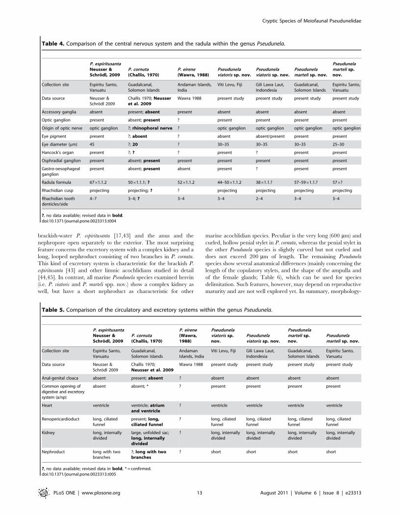
Morphology-based taxonomyThe Pseudunela specimens from different Indo-Pacific islands
examined herein are compared according to their external
morphology, microanatomy, and molecular markers. Externally,
only the larger, recently discovered Pseudunela espiritusanta from
Vanuatu [43] can be clearly distinguished from congeners by its
much larger body size, the foot width and the shape of the visceral
hump, as well as its unique brackish-water habitat (Table 3). In
contrast, the herein examined, fully marine Pseudunela species all
resemble externally P. cornuta from the Solomon Islands which was
recently re-examined by Neusser et al. [17]. The body size and
colour, the foot length and width, as well as the presence of
subepidermal spicules do not differ between the species (Table 3).
Only the visibility of the eyes through the body integument greatly
varies among - and partly within - the marine Pseudunela species. In
contrast to external features, our detailed anatomical examinations
enable the discrimination of P. cornuta from the remaining marine
Pseudunela species. Differences are related to all organ systems
(Tables 4, 5, 6). The eyes are unpigmented and considerably
smaller in P. cornuta than in the other Pseudunela species and they
are not innervated by the optic ganglion, but the optic nerve
emerges from the rhinophoral nerve [17]. The common opening
of the excretory and digestive systems is absent in P. cornuta and the
Figure 10. Histological cross-sections of P. marteli sp. nov. from Vanuatu. A: external morphology of a living specimen (body size 3 mm). B:Hancock’s organ and eye. C: Hancock’s organ. D: spicule cavities. E: albumen and mucus glands. F: ampulla and membrane gland. G: oocytes andspermatocytes. Abbreviations: alg, albumen gland; am, ampulla; cg, cerebral ganglion; dg, digestive gland; ey, eye; f, foot; ho, Hancock’s organ; k,kidney; lt, labial tentacle; ltn, labial tentacle nerve; meg, membrane gland; mug, mucus gland; oo, oocyte; ot, oral tube; ov, ovotestis; pg, pedalganglion; rh, rhinophore; rhg, rhinophoral ganglion; sc, spermatocytes; sp, spicule cavity; vd, vas deferens; vh, visceral hump.doi:10.1371/journal.pone.0023313.g010
Cryptic Species of Meiofaunal Pseudunelidae
PLoS ONE | www.plosone.org 11 August 2011 | Volume 6 | Issue 8 | e23313
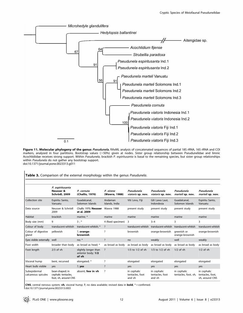
Figure 11. Molecular phylogeny of the genus Pseudunela. RAxML analysis of concatenated sequences of partial 18S rRNA, 16S rRNA and COImarkers, analysed in four partitions. Bootstrap values (.50%) given at nodes. Sister group relationship between Pseudunelidae and limnicAcochlidiidae receives strong support. Within Pseudunela, brackish P. espiritusanta is basal to the remaining species, but sister group relationshipswithin Pseudunela do not gather any bootstrap support.doi:10.1371/journal.pone.0023313.g011
Table 3. Comparison of the external morphology within the genus Pseudunela.
P. espiritusantaNeusser &Schrodl, 2009
P. cornuta(Challis, 1970)
P. eirene(Wawra, 1988)
Pseudunelaviatoris sp. nov.
Pseudunelaviatoris sp. nov.
Pseudunelamarteli sp. nov.
Pseudunelamarteli sp. nov.
Collection site Espiritu Santo,Vanuatu
Guadalcanal,Solomon Islands
AndamanIslands, India
Viti Levu, Fiji Gili Lawa Laut,Indondesia
Guadalcanal,Solomon Islands
Espiritu Santo,Vanuatu
Data source Neusser & Schrodl2009
Challis 1970; Neusseret al. 2009
Wawra 1988 present study present study present study present study
Habitat brackish marine; * marine marine marine marine marine
Body size (mm) 9 3 ; * 4 (fixed specimen) 3 3–4 3 3
Colour of body translucent-whitish translucent-whitish; * ? translucent-whitish translucent-whitish translucent-whitish translucent-whitish
Colour of digestivegland
yellowish ?; orange-brownish
? brownish orange-brownish greenish ororange-brownish
orange-brownish
Eyes visible externally well no; * ? no weakly well weakly
Foot width broader than body as broad as head; * as broad as body as broad as body as broad as body as broad as body as broad as body
Foot length 2/3 of vh slightly longer thananterior body; 1/2of vh
? 1/3 to 1/2 of vh 1/3 to 1/2 of vh 1/2 of vh 1/2 of vh
Visceral hump bent, recurved elongated; * ? elongated elongated elongated elongated
Heart bulb visible yes ?; yes ? yes yes yes yes
Subepidermalcalcareous spicules
bean-shaped; incephalic tentacles,foot, vh, around CNS
absent; few in vh ? in cephalictentacles, footand vh
in cephalictentacles, footand vh
in cephalictentacles, foot, vh,
in cephalictentacles, foot,vh, around CNS
CNS, central nervous system; vh, visceral hump; ?, no data available; revised data in bold, * = confirmed.doi:10.1371/journal.pone.0023313.t003
Cryptic Species of Meiofaunal Pseudunelidae
PLoS ONE | www.plosone.org 12 August 2011 | Volume 6 | Issue 8 | e23313
brackish-water P. espiritusanta [17,43] and the anus and the
nephropore open separately to the exterior. The most surprising
feature concerns the excretory system with a complex kidney and a
long, looped nephroduct consisting of two branches in P. cornuta.
This kind of excretory system is characteristic for the brackish P.
espiritusanta [43] and other limnic acochlidians studied in detail
[44,45]. In contrast, all marine Pseudunela species examined herein
(i.e. P. viatoris and P. marteli spp. nov.) show a complex kidney as
well, but have a short nephroduct as characteristic for other
marine acochlidian species. Peculiar is the very long (600 mm) and
curled, hollow penial stylet in P. cornuta, whereas the penial stylet in
the other Pseudunela species is slightly curved but not curled and
does not exceed 200 mm of length. The remaining Pseudunela
species show several anatomical differences (mainly concerning the
length of the copulatory stylets, and the shape of the ampulla and
of the female glands; Table 6), which can be used for species
delimitation. Such features, however, may depend on reproductive
maturity and are not well explored yet. In summary, morphology-
Table 5. Comparison of the circulatory and excretory systems within the genus Pseudunela.
P. espiritusantaNeusser &Schrodl, 2009
P. cornuta(Challis, 1970)
P. eirene(Wawra,1988)
Pseudunelaviatoris sp.nov.
Pseudunelaviatoris sp. nov.
Pseudunelamarteli sp.nov.
Pseudunelamarteli sp. nov.
Collection site Espiritu Santo,Vanuatu
Guadalcanal,Solomon Islands
AndamanIslands, India
Viti Levu, Fiji Gili Lawa Laut,Indondesia
Guadalcanal,Solomon Islands
Espiritu Santo,Vanuatu
Data source Neusser &Schrodl 2009
Challis 1970;Neusser et al. 2009
Wawra 1988 present study present study present study present study
Anal-genital cloaca absent present; absent ? absent absent absent absent
Common opening ofdigestive and excretorysystem (a/np)
absent absent; * ? present present present present
Heart ventricle ventricle; atriumand ventricle
? ventricle ventricle ventricle ventricle
Renopericardioduct long, ciliatedfunnel
present; long,ciliated funnel
? long, ciliatedfunnel
long, ciliatedfunnel
long, ciliatedfunnel
long, ciliatedfunnel
Kidney long, internallydivided
large, unfolded sac;long, internallydivided
? long, internallydivided
long, internallydivided
long, internallydivided
long, internallydivided
Nephroduct long with twobranches
?; long with twobranches
? short short short short
?, no data available; revised data in bold, * = confirmed.doi:10.1371/journal.pone.0023313.t005
Table 4. Comparison of the central nervous system and the radula within the genus Pseudunela.
P. espiritusantaNeusser &Schrodl, 2009
P. cornuta(Challis, 1970)
P. eirene(Wawra, 1988)
Pseudunelaviatoris sp. nov.
Pseudunelaviatoris sp. nov.
Pseudunelamarteli sp. nov.
Pseudunelamarteli sp.nov.
Collection site Espiritu Santo,Vanuatu
Guadalcanal,Solomon Islands
Andaman Islands,India
Viti Levu, Fiji Gili Lawa Laut,Indondesia
Guadalcanal,Solomon Islands
Espiritu Santo,Vanuatu
Data source Neusser &Schrodl 2009
Challis 1970; Neusseret al. 2009
Wawra 1988 present study present study present study present study
Accessory ganglia absent present; absent present absent absent absent absent
Optic ganglion present absent; present ? present present present present
Origin of optic nerve optic ganglion ?; rhinophoral nerve ? optic ganglion optic ganglion optic ganglion optic ganglion
Eye pigment present ?; absent ? absent absent/present present present
Eye diameter (mm) 45 ?; 20 ? 30–35 30–35 30–35 25–30
Hancock’s organ present ?; ? ? present ? present present
Osphradial ganglion present absent; present present present present present present
Gastro-oesophagealganglion
present absent; present absent present ? present present
Radula formula 6761.1.2 5061.1.1; ? 5261.1.2 44–5061.1.2 3861.1.? 57–5961.1.? 576?
Rhachidian cusp projecting projecting; ? ? projecting projecting projecting projecting
Rhachidian toothdenticles/side
4–7 3–4; ? 3–4 3–4 2–4 3–4 3–4
?, no data available; revised data in bold.doi:10.1371/journal.pone.0023313.t004
Cryptic Species of Meiofaunal Pseudunelidae
PLoS ONE | www.plosone.org 13 August 2011 | Volume 6 | Issue 8 | e23313

based taxonomy and even sophisticated 3D modelling of
anatomical details as applied herein can only reveal parts of the
actual species diversity of Pseudunela unambiguously; diagnosable
microanatomical units found need to be tested by molecular
phylogenetic analyses.
Cryptic species?The present molecular dataset is limited due to the low amount
of individuals sampled, thus not allowing population genetic
approaches and in depth comparison between intraspecific versus
interspecific variation justifying molecularly based species delin-
eation. Still, there are several lines of evidence supporting the
defined microanatomical units as genetically separated partially
cryptic lineages: 1) our maximum likelihood analyses based on a
concatenated molecular dataset (combining nuclear and mito-
chondrial markers) recovers all microanatomical units as mono-
phyla (Fig. 11). In our phylogenetic hypothesis P. cornuta separates
cryptic P. marteli sp. nov. and P. viatoris sp. nov. 2) In contrast to
earlier approaches relying on thresholds of divergence for the
barcoding marker COI in molluscs [6,21,46], several recent
studies showed that there is no universal threshold and that rates of
intraspecific variation can outnumber supposedly ‘high’ rates of
interspecific variation [34,47]. Our limited dataset shows low rates
of intraspecific variation, even when comparing far distant
populations of P. viatoris sp. nov. from Fiji and Indonesia (n = 5;
largest p-distance: 1.67% on partial COI, 1.39% on 16S rRNA).
Then again interspecific variation among the microanatomically
defined units is comparably high (14.04–16.48% on COI and
8.82–14.85% on 16S RNA) and the distances between the
morphologically cryptic species are in the same range as to the
morphologically clearly distinct P. espiritusanta. 3) In addition to
ML tree-based methods and the comparison of pairwise distances,
we generated haplotype networks applying 95% parsimony
criterion, which resulted in unconnected haplotype networks for
the described microanatomical units on both markers. Addition-
ally, the P. marteli sp. nov. from Vanuatu (n = 1) is unconnected to
the haplotype network of P. marteli sp. nov. from the Solomon
Islands (n = 3) on both mitochondrial markers. 4) GMYC recovers
all four microanatomical units; however, the performance and
accuracy of GMYC to our knowledge has never been tested on
such a small dataset, as ours. These independent molecular
approaches are in congruence with our microanatomical units and
thus, in our opinion, justify a separation in two formal new species.
There are several microanatomical differences between the two
populations of P. marteli sp. nov. (e.g. size of eyes, length of penial
stylet, see Tables 4, 5, 6), but intraspecific variation of these
characters cannot be evaluated at present stage of knowledge and
results from molecular data are incongruent (e.g. unconnected
haplotype networks vs. one entity in GMYC). Moreover, the
genetic distance between the two populations is low compared to
the distances present in the closely related Pseudunela species. More
data is needed to evaluate intraspecific variation and test
conspecifity of the two P. marteli populations. Within specimens
of Pseudunela viatoris sp. nov. from Fiji and Indonesia there are slight
differences concerning the eye visibility and the length of stylets on
the penial papilla, while stylets on the basal finger are remarkably
different-sized. Specimens from Indonesia and Fiji cluster on
different clades (Fig. 11). However, the genetic similarity between
these specimens is very high (approx. 98–99% on COI and 16S
rRNA) and intrapopulation variation is low. Thus, we do not
consider these lineages to be specifically distinct, despite the distant
geographic localities. More specimens are needed to explore
morphological variability and genetic structure of these popula-
tions.
We conclude that we discovered morphologically cryptic species
within the genus Pseudunela. External morphological, microana-
tomical and genetic evidences for recognizing species are
congruent, and a combined approach of 3D-microanatomy and
Table 6. Comparison of the reproductive system within the genus Pseudunela.
P. espiritusantaNeusser &Schrodl, 2009
P. cornuta(Challis, 1970)
P. eirene(Wawra,1988)
Pseudunelaviatoris sp.nov.
Pseudunelaviatoris sp.nov.
Pseudunelamarteli sp.nov.
Pseudunelamarteli sp.nov.
Collection site Espiritu Santo,Vanuatu
Guadalcanal,Solomon Islands
AndamanIslands, India
Viti Levu, Fiji Gili Lawa Laut,Indondesia
Guadalcanal,Solomon Islands
Espiritu Santo,Vanuatu
Data source Neusser &Schrodl 2009
Challis 1970; Neusseret al. 2009
Wawra 1988 present study present study present study present study
Hollow curved penialstylet (mm)
80 100 ; 600(coiled 1.5 spirals)
200 70 125 130 180–200
Solid basal thorn (mm) absent absent; * 30 absent absent absent absent
Hollow curved stylet onbasal finger (mm)
340 absent; 110 ? 200 30 30 30
Glands associated withcopulatory organs
prostate,paraprostate
prostate, penialgland; prostate,paraprostate
? prostate,paraprostate
prostate,paraprostate
prostate,paraprostate
prostate,paraprostate
Yolky oocytes developed present present; * ? absent ? absent present
Ampulla sac-like ?; sac-like ? tubular ? sac-like tubular
Receptaculum seminis present ?; present ? absent ? absent absent
Bursa copulatrix present present; * ? present ? absent absent
Albumen gland tubular ?; tubular ? sac-like ? tubular sac-like
Membrane gland tubular ?; tubular tubular sac-like tubular
Mucus gland sac-like sac-like ? sac-like ? tubular sac-like
?, no data available; revised data in bold, * = confirmed.doi:10.1371/journal.pone.0023313.t006
Cryptic Species of Meiofaunal Pseudunelidae
PLoS ONE | www.plosone.org 14 August 2011 | Volume 6 | Issue 8 | e23313

genetic markers can reliably distinguish and delineate all of the
four species. Surprisingly, far distant geographic populations of
specimens with slightly differing anatomy and presumably poor
dispersive ability do not necessarily indicate different species, as
revealed by highly similar mitochondrial sequences in P. viatoris sp.
nov.. An integrative taxonomic approach combining morpholog-
ical, 3D-microanatomical and molecular markers, like demon-
strated here for Pseudunela species, thus is a powerful tool to
independent structural or genetic approaches.
Overall, our results might be indicative for a still unknown
diversity within mesopsammic gastropods. Recent studies on
cryptic speciation within Meiofauna across taxa, has often revealed
formerly considered wide-spread or even cosmopolitan species as
flock of cryptic species (e.g. in proseriate flatworms [48,49],
polychaete annelids [50,51] and gastrotrichs [52,53]). Leading to
the assumption that especially within this habitat, which is
generally known for taxa with low dispersal abilities, there might
be a high degree of cryptic speciation and the contribution of
Meiofauna to marine biodiversity might be currently seriously
underestimated [49]. However, some studies supported the
presence of truly amphi-atlantic or cosmopolitan meiofaunal taxa,
with the distribution and genetic interaction across Oceans in the
absence of pelagic larvae still to be explained [50,54].
DistributionThe distribution of the four different Pseudunela species (P. eirene
from Andaman Islands is not considered in this discussion as there
exist only inadequate data and no material is available for detailed
study) on the Indo-Pacific islands raises questions: 1) How can two
different, genetically isolated Pseudunela species inhabit nearby
beaches on one island with continuous coastline and 2) how can
we explain the occurrence of P. viatoris sp. nov. on two far distant
islands?
Considering that all Hedylopsacea occur in warm or tropical
waters (except of Hedylopsis spiculifera, which inhabits temperate
waters), we can assume that the common ancestor of the
Pseudunelidae and Acochlidiidae s.l. has its origin in warm
tropical waters as well. Recently, Jorger et al. [28] calibrated a
molecular clock estimating divergence times for shell-less, and
hence fossil-lacking Heterobranchia. In this study the origin of
Acochlidia was estimated to the Mesozoic Triassic or Jurassic.
According to the authors, the major diversification of Acochlidia
took place in Jurassic, but the split between Pseudunelidae and
Acochlidiidae was estimated to the Palaeogene. Even though this is
a very rough estimation, it indicates that the diversification and
distribution of the genus Pseudunela might have started over 35
mya, a long timeframe for a long-distance distribution, even for
marine meiofaunal acochlidian species, which are regarded as
poor dispersers. The hedylopsacean species Pseudunela cornuta [17]
and P. marteli sp. nov. from Vanuatu, as well as the micro-
hedylacean species, such as Microhedyle remanei (Marcus, 1953), M.
nahantensis (Doe, 1974), Parhedyle cryptophthalma (Westheide &
Wawra, 1974) and Asperspina murmanica (Kudinskaya & Minichev,
1978) [14,55,56,57] have only a small number of large, yolky
oocytes indicating a low reproductive output and a lecithotrophic
development within a capsule rather than a planktotrophic larval
development [10,57]. Therefore, the distribution of larval and
adult stages is expected only within a small radius step by step.
Natural disasters (such as volcanic eruptions, earthquakes, heavy
storms or erosion) or settlement by humans may disturb or even
destroy sandy beaches [42]. This might result in genetically
isolated populations or even local extinctions, which can explain
the co-occurrence of two distinct Pseudunela species on nearby
beaches. Another explication may be the adaptation to diverse,
but subtle ecological conditions in the habitat, such as different
currents, grain size, freshwater influx or food resources, which
finally might result in separation of species.
The extensive distribution of P. viatoris sp. nov. is surprising. Due
to aforementioned reasons a distribution of larvae via water
currents is not likely. An accidentally distribution of different
ontogenetic stages after heavy (sub-)tropical storms is not very
probable due to the large distances. We cannot exclude a man-
made dispersal, where small patches of sand of neighboured
populations were displaced e.g. by ships. More likely, however,
there exist intermediate populations between those from Fiji and
Indonesia that have not been discovered yet – or already got
extinct. Missing intermediates and restricted gene flow across these
stepping stones might also explain the slight anatomical differences
between the Fijian specimens and those from Indonesia, such as
the variation in the length of the copulatory stylets or the
pigmentation of the eye. Possibly, small genetic distances observed
between these distant populations also may reflect a stage of
ongoing allopatric speciation. Finally, another aspect should be
considered: juveniles of the amphidromous nerite snail Neritina
asperulata Recluz, 1842 show a ‘‘hitchhiking’’ behaviour by
attaching to the shell of the congeneric N. pulligera Linnaeus,
1758. In this way young specimens travel upstream for growth and
reproduction [58]. We can imagine that eggs and accordingly
larval or adult acochlidians stick to e.g. benthic living organisms
when the living conditions in the sand are changing for the worse
and thus, may be displaced into another habitat [45].
Phylogeny and evolutionOur molecular analysis (see Fig. 11) shows the marine and
brackish-water Pseudunela as the sister group to the limnic
Acochlidiidae s.l. and supports herein the results of recent
morphological analysis [18] and previous molecular analysis
[28]. Again, Aitengidae sp. clusters within the Hedylopsacea, as
sister to Pseudunelidae plus Acochlidiidae [59]. The relationships
between the Pseudunela species are fully resolved but with no robust
support. As suspected by Neusser & Schrodl [43], the brackish
Pseudunela espiritusanta from Vanuatu is the most basal Pseudunela
species forming the sister group to all marine and temporary
brackish Pseudunela species. The fully marine P. marteli sp. nov. from
the Solomon Islands and Vanuatu form the sister group to the
temporary brackish P. cornuta (also from the Solomon Islands) and
the marine P. viatoris sp. nov. from Fiji and Indonesia. This tree
topology (Fig. 11), however, does not clearly support previous
ideas [18], i.e. that evolution within acochlidians was directed
from marine to limnic habitats, possibly via brackish water.
Instead, the ancestor of Pseudunela plus Acochlidiidae might have
been already limnic or brackish water associated, with marine
species evolving secondarily within Pseudunela.
To visualise patterns and reconstruct evolution in a more
comprehensive context, habitats were plotted on a consensus tree
(Fig. 12) combining all relevant acochlidian clades from morphol-
ogy-based and molecular analyses. While the ancestral acochlidian
[28] and all microhedylacean species are marine, the Hedylopsa-
cea clade includes a mosaic of limnic, marine and brackish water
associated taxa, implying several independent incidents of habitat
shifts from marine to limnic and brackish water systems and/or
vice versa. In contrast to previous assumptions [17,18], the
hedylopsacean ancestor could have been either still marine or
already limnic.
In order to decide on a preferred scenario, we explored different
characteristics and organ systems that are most closely linked to
osmolarity changes. The first one is the body volume as a whole.
Since all acochlidians, including all marine species and the basal
Cryptic Species of Meiofaunal Pseudunelidae
PLoS ONE | www.plosone.org 15 August 2011 | Volume 6 | Issue 8 | e23313

limnic Tantulum elegans are small sized meiofaunal forms, there is no
doubt that the large adult size of limnic, benthic Acochlidiidae is
an adaptive apomorphy of this clade. The brackish water
Pseudunela espiritusanta that is no more mesopsammic but living
under stones either independently increased to an intermediate
size or, alternatively, the common ancestor of Pseudunela plus
Acochlidiidae already was large, with secondary reduction in
mesopsammic Pseudunela species. Summing up, increasing body
size alone may be advantageous but not strictly necessary for
acochlidians invading freshwater or brackish water systems.
The second feature that is crucial for dealing with osmotic stress,
especially in small species and juveniles, is the excretory system.
Neusser & Schrodl [43] emphasised that the acochlidian excretory
system varies considerably between marine and limnic species. The
different types are illustrated in Fig. 12 and, based on our results,
mapped on the consensus tree. All microhedylacean Acochlidia
known in detail (e.g. Microhedyle remanei, Pontohedyle milaschewitchii
(Kowalevsky, 1901) or Asperspina murmanica) have a quite simple
excretory system of type I consisting of a small, sac-like kidney and a
short nephroduct (Fig. 12) [14,55,60]. This simple type of sac-like
kidney corresponds to almost all marine euthyneurans, including
marine Panpulmonata, such as Siphonarioidea [61], the sacoglos-
san Platyhedyle [62], Amphiboloidea [63], and marine eupulmonates.
In contrast, the acochlidian excretory system type II comprises a
complex, internally divided kidney with a narrow and a wide lumen.
All fully marine hedylopsacean species (such as the newly described
Pseudunela species) have an excretory system of type II (Fig. 12), i.e.
with a complex kidney, and with a short nephroduct (type IIa).
Hedylopsis ballantinei Sommerfeldt & Schrodl, 2005 was described
with a long, sac-like kidney and a nephropore opening into a mantle
cavity [64,65]. However, a brief re-examination of the original
sections revealed this species to possess a complex, internally divided
kidney (own unpubl. data). The most complex excretory system type
IIb consists of a large, divided kidney as in type IIa, and additionally
a long looped nephroduct with two branches. This type is present in
all limnic acochlidian species, i.e. the small Caribbean limnic
Tantulum elegans [66] and the large Indo-Pacific Acochlidiidae [44],
in the brackish Pseudunela espiritusanta [43] and the at least temporary
Figure 12. Evolution of excretory systems and habitat in acochlidian lineages. The habitat of the different acochlidian lineages and theirtypes of excretory systems are plotted on a consensus tree (topology combined from Schrodl & Neusser [18] and molecular results herein; theenigmatic Aitengidae are not shown due to the uncertain position within Hedylopsacea and the different and special excretory system [59]). WhileMicrohedylacea present a simple excretory system with a small, sac-like kidney (type I), hedylopsacean taxa evolved a complex excretory system witha large, internally divided kidney (type II): type IIa is characterised by a short nephroduct, type IIb by a long, looped nephroduct. The complex kidneyalready evolved in the ancestor of the Hedylopsacea. The mosaic-like distribution of habitat and excretory system types within Hedylopsacea impliesan evolutionary scenario with multiple habitat shifts and adaptations. Abbreviations: ao, aorta; h, heart; k, kidney; kn, narrow lumen of kidney; kw,wide lumen of kidney; nd, nephroduct; ndd, dorsal branch of nephroduct; ndv, ventral branch of nephroduct; pc, pericardium. Not to scale.doi:10.1371/journal.pone.0023313.g012
Cryptic Species of Meiofaunal Pseudunelidae
PLoS ONE | www.plosone.org 16 August 2011 | Volume 6 | Issue 8 | e23313

brackish P. cornuta [17]. Thus, the type of the excretory system in
acochlidians is not strictly correlated with the habitat in acochlidian
species: marine acochlidian species have either a type I or IIa
excretory system with a simple or a complex kidney, respectivly.
Interestingly, all (marine) microhedylacean species have the
simple, supposedly ancestral type I system. In contrast, all
hedylopsacean species have the complex type II excretory system,
even the marine species. We therefore conclude that the ancestral
hedylopsacean species already had a complex kidney, which is an
apomorphy of the clade. The presence of complex kidneys can be
seen as a preadaptation to brackish water or limnic life, or much
more likely, evolved as an adaptation to invading such habitats.
Thus, considering evidence from excretory systems, we favour a
scenario with hedylopsaceans originating in a freshwater, or at
least freshwater influenced, habitat.
Considering the still poorly known and enigmatic Aitengidae
[59] aberrant amphibious hedylopsacean offshoot (Fig. 11) would
fit with and further extend the ecological tolerance and
evolutionary plasticity observed within the hedylopsacean lineage.
Finally, the question arises if the complex type II kidney has
already evolved in the – then supposedly brackish water or even
limnic - ancestor of the Acochlidia. A recent multi-locus molecular
study including six out of seven acochlidian families in a
comprehensive euthyneuran taxon sampling [28] fundamentally
changed our understanding of euthyneuran systematics. Surpris-
ingly, this study confirms the Acochlidia in a well-supported
(pan)pulmonate rather than opisthobranch relationship, as sister of
basally still marine Eupulmonata. However, there is an alternative,
though less likely topology suggesting that Acochlidia are the sister
of – limnic – Hygrophila. In this scenario, a common ancestor
could have been limnic as well, with a simple or complex kidney as
both conditions occur apparently among different hygrophilan
subgroups [61,67,68].
ConclusionsOur study on mesopsammic Acochlidia testing the power of
traditional taxonomy (i.e. examination of the external morphology
and the radula) against results from in-depth micro-anatomical
and molecular data clearly shows: 1) Traditional taxonomy fails to
reveal the cryptic diversity within the genus Pseudunela in tropical
sands, and thus is likely to generally underestimate biodiversity of
meiofaunal invertebrates; 2) labour intensive and sophisticated
3D-modelling of micro-morphology is more suitable to delineate
species, i.e. diagnosable units within Pseudunela are congruent with
genetic lineages, and show relatively high genetic divergence; 3)
only the combined evidence of microanatomical and molecular
data enabled us to uncover and describe the full range of cryptic
speciation in our material; low genetic distances of anatomically
distinguishable genetic lineages of P. viatoris sp. nov. suggest there
could be some gene flow between geographically distant
populations, preventing us from establishing separate species; 4)
patterns of distribution of Pseudunela species are discovered that
cannot, however, be satisfyingly explained in the absence of sound
biological knowledge on tiny meiofaunal species. We thus agree
with Cook et al. [69] and advocate that taxonomy should integrate
and consider all relevant types of data. Our exploration of the
genus Pseudunela in older studies [17,43] and herein also showed
considerable ecological and structural diversity, i.e. of fully marine
species, and those steadily or temporarily exposed to freshwater,
having complex excretory systems. The combination of molecular
phylogenetic and detailed micromorphological studies will shed
further light on the origin of acochlidians, their much more
frequent than expected habitat shifts, and their evolutionary
adaptations to an extraordinarily wide range of completely
different habitats.
Supporting Information
Figure S1 Interactive 3D-model of Pseudunela viatorissp. nov. from Fiji. To activate the 3D-model of P. viatoris sp.
nov. for interactive manipulation click into figure. Rotate model
by dragging with left mouse button pressed, shift model: same
action+ctrl (or change default action for left mouse button), zoom:
use mouse wheel. Select or deselect (or change transparency of)
components in the model tree, switch between prefab views or
change surface visualization (e.g. lightning, render mode, crop
etc.). Interactive manipulation requires Adobe Reader 7 or higher.
(PDF)
Acknowledgments
TPN is grateful to Dr. Philippe Bouchet (Museum National d’Histoire
Naturelle, Paris, France) for the opportunity to join the ‘‘Mission MNHN/
PNI/IRD Santo 2006’’ to Vanuatu. The SANTO 2006 Expedition was
organized by Museum National d’Histoire Naturelle, Paris, Pro Natura
International (PNI), and Institut de Recherche pour le Developpement
(IRD). It operated under a permit granted to Philippe Bouchet (MNHN) by
the Environment Unit of the Government of Vanuatu. The Marine
Biodiversity part of the expedition, a part of Census of Marine Life’s
CReefs programme, was specifically funded by grants from the Total
Foundation and the Sloan Foundation. Dr. Fontje Kaligis (University of
Sam Ratulangi, Manado, Indonesia) is thanked for the availability of
material from Indonesia and Dr. Yasunori Kano (University of Tokyo,
Japan) for support and company during the expedition to the Solomon
Islands. Johnson Seeto (University of the South Pacific, Suva, Fiji) is
thanked for kind support. 3D reconstruction was facilitated by the
GeoBioCenter/LMU Munchen, Germany. Dr. Martin Heß (LMU) is
thanked for assistance in preparing the interactive 3D-model. We would
like to express our gratitude to three anonymous reviewers for valuable
comments on the manuscript.
Author Contributions
Conceived and designed the experiments: TPN KJM MS. Performed the
experiments: TPN KJM. Analyzed the data: TPN KJM. Collected and
contributed materials: TPN KJM MS. Realised morphological analyses
and interactive 3D-model: TPN. Carried out molecular studies: KJM.
Planned and supervised the study: MS. Read, approved and wrote the final
manuscript: TPN KMJ MS.
References
1. Bickford D, Lohman DJ, Sodhi NS, Ng PKL, Meier R, et al. (2007) Cryptic
species as a window on diversity and conservation. Trends Ecol Evol 22: 148–155.
2. Trontelj P, Fiser C (2009) Cryptic species diversity should not be trivialised. Syst
Biodiver 7: 1–3.
3. Pfenninger M, Schwenk K (2007) Cryptic animal species are homogeneously
distributed among taxa and biogeographical regions. BMC Evol Biol 7: 6.
4. Krabbe K, Leese F, Mayer C, Tollrian R, Held C (2010) Cryptic mitochondrial
lineages in the widespread pycnogonid Colossendeis megalonyx Hoek, 1881 from
Antarctic and Subantarctic waters. Polar Biol 33: 281–292.
5. Raupach MJ, Wagele JW (2006) Distinguishing cryptic species in Antarctic
Asellota (Crustacea : Isopoda) - a preliminary study of mitochondrial DNA in
Acanthaspidia drygalskii. Antarctic Sci 18: 191–198.
6. Wilson NG, Schrodl M, Halanych KM (2009) Ocean barriers and glaciation:
evidence for explosive radiation of mitochondrial lineages in the Antarctic sea
slug Doris kerguelenensis (Mollusca, Nudibranchia). Mol Ecol 18: 965–984.
7. Wilson NG, Hunter RL, Lockhart SJ, Halanych KM (2007) Multiple lineages
and absence of panmixia in the ‘‘circumpolar’’ crinoid Promachocrinus kerguelensis
from the Atlantic sector of the Antarctica. Mar Biol 152: 895–904.
Cryptic Species of Meiofaunal Pseudunelidae
PLoS ONE | www.plosone.org 17 August 2011 | Volume 6 | Issue 8 | e23313

8. Mileikovsky SA (1971) Types of larval development in marine bottominvertebrates, their distribution and ecological significance: re-evaluation. Mar
Biol 10: 193–213.
9. Swedmark B (1964) The interstitial fauna of marine sand. Biol Rev 39: 1–42.
10. Swedmark B (1968) The biology of interstitial Mollusca. Symp Zool Soc Lond
22: 135–149.
11. Kirsteuer E (1973) Occurrence of the interstitial Opisthobranch Unela remanei
Marcus, in the Caribbean Sea. Mitt Inst Colombo-Aleman Invest Cient 7:41–46.
12. Marcus E (1954) Uber Philinoglossacea und Acochlidiacea. Kiel Meeresforsch
10: 215–223.
13. Marcus E (1953) Three brazilian sand-Opisthobranchia. Bol Fac Filos Ci Letr
Univ Sao Paulo, Zool 164: 165–203.
14. Neusser TP, Heß M, Haszprunar G, Schrodl M (2006) Computer-based three-dimensional reconstruction of the anatomy of Microhedyle remanei (Marcus, 1953),
an interstitial acochlidian gastropod from Bermuda. J Morphol 267: 231–247.
15. Challis DA (1970) Hedylopsis cornuta and Microhedyle verrucosa, two new
Acochlidiacea (Mollusca: Opisthobranchia) from the Solomon Islands Protec-torate. T Royal Soc NZ (BiolSci) 12: 29–40.
16. Hughes HPI (1991) Sand-dwelling opisthobranchs from Hong-Kong.J Molluscan Stud 57: 425–431.
17. Neusser TP, Heß M, Schrodl M (2009) Tiny but complex - interactive 3D
visualization of the interstitial acochlidian gastropod Pseudunela cornuta (Challis,
1970). Front Zool 6: 20.
18. Schrodl M, Neusser TP (2010) Towards a phylogeny and evolution ofAcochlidia (Mollusca: Gastropoda: Opisthobranchia). Zool J Linn Soc 158:
124–154.
19. Wawra E (1988) Sand-Opisthobranchia aus dem Golf von Bengalen. Ann
Nathist Mus Wien Ser B Bot Zool 90: 427–431.
20. Hebert PDN, Cywinska A, Ball SL, DeWaard JR (2003) Biological identifica-
tions through DNA barcodes. Proc R Soc Lond B Biol Sci 270: 313–321.
21. Hebert PDN, Ratnasingham S, deWaard JR (2003) Barcoding animal life:cytochrome c oxidase subunit 1 divergences among closely related species.
Proc R Soc Lond B Biol Sci 270: S96–S99.
22. Raupach MJ, Astrin JJ, Hannig K, Peters MK, Stoeckle MY, et al. (2010)
Molecular species identification of Central European ground beetles (Coleop-tera: Carabidae) using nuclear rDNA expansion segments and DNA barcodes.
Front Zool 7: 26.
23. Schrodl M (2006) Techniques for collecting interstitial opisthobranchs.
Available: http://www.seaslugforum.net/factsheet/inteextr.
24. Spurr AR (1969) A low-viscosity epoxy resin embedding medium for electronmicroscopy. J Ultra Res 26: 31–43.
25. Ruthensteiner B (2008) Soft Part 3D visualization by serial sectioning andcomputer reconstruction. Zoosymposia 1: 63–100.
26. Richardson KC, Jarett L, Finke EH (1960) Embedding in epoxy resins for
ultrathin sectioning in electron microscopy. Stain Technology 35: 313–323.
27. Ruthensteiner B, Heß M (2008) Embedding 3D models of biological specimens
in PDF publications. Microsc Res Tech 71: 778–786.
28. Jorger K, Stoger I, Kano Y, Fukuda H, Schrodl M (2010) On the origin of
Acochlidia and other enigmatic euthyneuran gastropods, with implications forthe systematics of Heterobranchia. BMC Evol Biol 10: 323.
29. Altschul SF, Gish W, Miller W, Myers EW, Lipman DJ (1990) Basic local
alignment search tool. J Mol Biol 215: 403–410.
30. Katoh K, Kuma K, Toh H, Miyata T (2005) MAFFT version 5: improvement in
accuracy of multiple sequence alignment. Nucleic Acids Res 33: 511–518.
31. Talavera G, Castresana J (2007) Improvement of phylogenies after removing
divergent and ambiguously aligned blocks from protein sequence alignments.Syst Biol 56: 564–577.
32. Stamatakis A (2006) RAxML-VI-HPC: maximum likelihood-based phylogenetic
analyses with thousands of taxa and mixed models. Bioinformatics 22:
2688–2690.
33. Posada D (2008) jModelTest: Phylogenetic model averaging. Mol Biol Evol 25:1253–1256.
34. Meier R, Shiyang K, Vaidya G, Ng PKL (2006) DNA barcoding and taxonomyin diptera: A tale of high intraspecific variability and low identification success.
Syst Biol 55: 715–728.
35. Clement M, Posada D, Crandall KA (2000) TCS: a computer program toestimate gene genealogies. Mol Ecol 9: 1657–1659.
36. Monaghan MT, Wild R, Elliot M, Fujisawa T, Balke M, et al. (2009)Accelerated species inventory on Madagascar using coalescent-based models of
species delineation. Syst Biol 58: 298–311.
37. Pons J, Barraclough TG, Gomez-Zurita J, Cardoso A, Duran DP, et al. (2006)
Sequence-based species delimitation for the DNA taxonomy of undescribedinsects. Syst Biol 55: 595–609.
38. Sanderson MJ (2002) Estimating absolute rates of molecular evolution and
divergence times: A penalized likelihood approach. Mol Biol Evol 19: 101–109.
39. Ghiselin MT (1966) Reproductive function and the phylogeny of opisthobranch
gastropods. Malacologia 3: 327–378.
40. Klussmann-Kolb A (2001) Comparative investigation of the genital systems in
the Opisthobranchia (Mollusca, Gastropoda) with special emphasis on thenidamental glandular system. Zoomorphology 120: 215–235.
41. Haase M, Wawra E (1996) The genital system of Acochlidium fijiensis
(Opisthobranchia: Acochlidioidea) and its inferred function. Malacologia 38:
143–151.
42. Morse MP (1987) Distribution and ecological adaptations of interstitial molluscs
in Fiji. Am Malacol Bull 5: 281–286.
43. Neusser TP, Schrodl M (2009) Between Vanuatu tides: 3D anatomical
reconstruction of a new brackish water acochlidian gastropod from Espiritu
Santo. Zoosystema 31: 453–469.
44. Brenzinger B, Neusser TP, Glaubrecht M, Haszprunar G, Schrodl M (2011)
Redescription and three-dimensional reconstruction of the limnic acochlidian
gastropod Strubellia paradoxa (Strubell, 1892) (Gastropoda: Euthyneura) from
Ambon, Indonesia. J Nat Hist 45: 183–209.
45. Brenzinger B, Neusser TP, Jorger KM, Schrodl M Integrating 3D-micromor-
phology and molecules: natural history of the Pacific freshwater slug genus Strubellia
Odhner, 1937 (Heterobranchia, Acochlidia, Acochlidiidae). J Moll Stud, In press.
46. Puillandre N, Cruaud C, Kantor YI (2010) Cryptic species in Gemmuloborsonia
(Gastropoda: Conoidea). J Moll Stud 76: 11–23.
47. Boyer SL, Baker JM, Giribet G (2007) Deep genetic divergences in Aoraki
denticulata (Arachnida, Opiliones, Cyphophthalmi): a widespread ‘mite harvest-
man’ defies DNA taxonomy. Mol Ecol 16: 4999–5016.
48. Casu M, Curini-Galletti M (2004) Sibling species in interstitial flatworms: a case
study using Monocelis lineata (Proseriata: Monocelididae). Mar Biol 145: 669–679.
49. Casu M, Lai T, Sanna D, Cossu P, Curini-Galletti M (2009) An integrative
approach to the taxonomy of the pigmented European Pseudomonocelis Meixner,
1943 (Platyhelminthes: Proseriata). Biol J Linn Soc 98: 907–922.
50. Schmidt H, Westheide W (2000) Are the meiofaunal polychaetes Hesionides
arenaria and Stygocapitella subterranea true cosmopolitan species? — results of
RAPD-PCR investigations. Zool Scr 29: 17–27.
51. Schmidt H, Westheide W (1999) Genetic relationships (RAPD-PCR) between
geographically separated populations of the ‘‘cosmopolitan’’ interstitial poly-
chaete Hesionides gohari (Hesionidae) and the evolutionary origin of the freshwater
species Hesionides riegerorum. Biol Bull 196: 216–226.
52. Leasi F, Todaro M (2009) Meiofaunal cryptic species revealed by confocal
microscopy: the case of Xenotrichula intermedia (Gastrotricha). Mar Biol 156:
1335–1346.
53. Todaro MA, Fleeger JW, Hu YP, Hrincevich AW, Foltz DW (1996) Are
meiofaunal species cosmopolitan? Morphological and molecular analysis of
Xenotrichula intermedia (Gastrotricha: Chaetonotida). Mar Biol 125: 735–742.
54. Westheide W, Hass-Cordes E, Krabusch M, Muller MCM (2003) Ctenodrilus
serratus (Polychaeta : Ctenodrilidae) is a truly amphi-Atlantic meiofauna species -
evidence from molecular data. Mar Biol 142: 637–642.
55. Neusser TP, Martynov AV, Schrodl M (2009) Heartless and primitive? 3D
reconstruction of the polar acochlidian gastropod Asperspina murmanica. Acta Zool
(Stockh) 90: 228–245.
56. Westheide W, Wawra E (1974) Organisation, Systematik und Biologie von
Microhedyle cryptophthalma nov. spec. (Gastropoda, Opisthobranchia) aus dem
Brandungsstrand des Mittelmeeres. Helgol wissen Meeres 26: 27–41.
57. Morse MP (1994) Current knowledge of reproductive biology in two taxa of
interstitial molluscs (class Gastropoda: order Acochlidiacea and class Aplaco-
phora: order Neomeniomorpha). In: Wilson WH, Stricker SA, Shinn GL, eds.
Reproduction and development of marine invertebrates John Hopkins
University Press. pp 195–205.
58. Kano Y (2009) Hitchhiking behaviour in the obligatory upstream migration of
amphidromous snails. Biol Lett 5: 465–468.
59. Neusser TP, Fukuda H, Jorger KM, Kano Y, Schrodl M Sacoglossa or
Acochlidia? 3D-reconstruction, molecular phylogeny and evolution of Aiteng ater
and Aiteng mysticus n. sp. (Aitengidae, Gastropoda). J Molluscan Stud, In press.
60. Jorger KM, Neusser TP, Haszprunar G, Schrodl M (2008) Undersized and
underestimated: 3D-visualization of the Mediterranean interstitial acochlidian
gastropod Pontohedyle milaschewitchii (Kowalevsky, 1901). Org Divers Evol 8:
194–214.
61. Hubendick B (1978) Systematics and comparative morphology of the
Basommatophora. In: Fretter V, Peake P, eds. Pulmonates. London/New
York/San Francisco: Academic Press. pp 1–47.
62. Ruckert I, Altnoder A, Schrodl M (2008) Computer-based 3D anatomical
reconstruction and systematic placement of the mesopsammic gastropod
Platyhedyle denudata Salvini-Plawen, 1973 (Opisthobranchia, Sacoglossa). Org
Divers Evol 8: 358–367.
63. Golding RE, Ponder WF, Byrne M (2007) Taxonomy and anatomy of
Amphiboloidea (Gastropoda: Heterobranchia: Archaeopulmonata). Zootaxa
1476: 1–50.
64. Sommerfeldt N, Schrodl M (2005) Microanatomy of Hedylopsis ballantinei, a new
interstitial acochlidian gastropod from the Red Sea, and its significance for
phylogeny. J Molluscan Stud 71: 153–165.
65. Fahrner A, Haszprunar G (2002) Microanatomy, ultrastructure, and systematic
significance of the excretory system and mantle cavity of an acochlidian
gastropod (Opisthobranchia). J Molluscan Stud 68: 87–94.
66. Neusser TP, Schrodl M (2007) Tantulum elegans reloaded: a computer-based 3D-
visualization of the anatomy of a Caribbean freshwater acochlidian gastropod.
Invertebr Biol 126: 18–39.
67. Bekius R (1971) The circulatory system of Lymnaea stagnalis (L.). Neth J Zool 22:
1–58.
68. Little C (1983) The colonisation of land: Origins and adaptations of terrestrial
animals. Cambridge: Cambridge University Press. 290 p.
69. Cook LG, Edwards RD, Crisp MD, Hardy NB (2010) Need morphology always
be required for new species descriptions? Invertebr Syst 24: 322–326.
Cryptic Species of Meiofaunal Pseudunelidae
PLoS ONE | www.plosone.org 18 August 2011 | Volume 6 | Issue 8 | e23313

70. Wollscheid E, Wagele H (1999) Initial results on the molecular phylogeny of the
nudibranchia (Gastropoda, Opisthobranchia) based on 18S rDNA data. Mol
Phylogenet Evol 13: 215–226.
71. Vonnemann V, Schrodl M, Klussmann-Kolb A, Wagele H (2005) Reconstruc-
tion of the phylogeny of the Opisthobranchia (Mollusca: Gastropoda) by means
of 18S and 28S rRNA gene sequences. J Molluscan Stud 71: 113–125.
72. Simon C, Frati F, Beckenbach A, Crespi B, Liu H, et al. (1994) Evolution,
weighting, and phylogenetic utility of mitochondrial gene sequences and acompliation of conserved polymerase chain reaction primers. Ann Entomol Soc
Am 87: 651–701.
73. Folmer O, Black M, Hoeh W, Lutz R, Vrijenhoek R (1994) DNA primers foramplification of mitochondrial cytochrome c oxidase subunit I from diverse
metazoan invertebrates. Mol Mar Biol Biotech 3: 294–299.
Cryptic Species of Meiofaunal Pseudunelidae
PLoS ONE | www.plosone.org 19 August 2011 | Volume 6 | Issue 8 | e23313