view - european heart journal supplements
TRANSCRIPT
Introduction
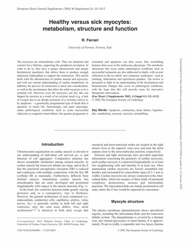
Ultrastructural organization in cardiac muscle is relevant toour understanding of individual cell survival per se andfunction of cell aggregates. Comparative anatomy hasshown remarkable similarities among striated muscles. Incardiac muscle the transverse tubules are large, and at the Zline the junctional sarcoplasmic reticulum (SR) is reducedand continuous with multiple connections with the free SR(corbular SR in mammals). Furthermore, different fromskeletal muscle, mammalian cardiac muscle hasmitochondria that are more developed and positionedlongitudinally with respect to the muscle structure (Fig. 1).
In the heart, the ventricles function under greatly varyingpressures and, as a consequence, vary in thickness.However, the general architecture of each component (i.e.endocardium, conduction cells, capillaries, arteries, veins,nerves, etc.) is generally similar in both left and rightventricles; only the total mass differs. Also, atrialarchitecture[1,2] is identical in both atria except that
sinoatrial and atrioventricular nodes are located in the rightatrium close to the superior vena cava, and near the atrialseptum close to the atrioventricular junction, respectively.
Electron and light microscopy have provided importantinformation concerning the geometry of cardiac myocytes;each cardiac myocyte is connected longitudinally to at leasttwo neighbouring cells and laterally to at least one. Mostmammalian cardiac myocytes are loosely assembled intobundles and surrounded by extracellular space (0·2–1 µm inwidth). Cardiac myocytes are always connected at the inter-calated disks, which are mosaics of three kinds of junctionalcomplexes: desmosomes, nexuses and intermediatejunctions. The intercalated disks are mainly positioned at cellends, where the Z line would be expected in a sarcomere.
Myocyte structure
The plasma membrane (plasmalemma) shows specializedregions, including the intercalated disks and the transversetubular system. The plasmalemma is covered by a laminarcoat, also termed glycocalyx or basal lamina. It is approxi-mately 50 nm in width, is separable into two layers (lamina
European Heart Journal Supplements (2002) 4 (Supplement G), G1–G12
1520-765X/02/0G0001 + 12 $35.00/0 © 2002 The European Society of Cardiology
Healthy versus sick myocytes:metabolism, structure and function
R. Ferrari
University of Ferrara, Ferrara, Italy
The myocytes are extraordinary cells. They are immortal andcontract for a lifetime, supporting the peripheral circulation. Inorder to do so, they have a unique ultrastructure and uniquebiochemical machinery that allows them to produce enoughadenosine triphosphate to support the contraction. This articledeals with the ultrastructure of cardiac muscle and myocytes,and with our current understanding of cardiac metabolism. Inaddition, the process of contraction is taken into consideration,as well as the mechanisms that allow the adult myocyte to be aterminal cell. However, even the myocytes can die; this canhappen by necrosis as a result of an external insult (e.g. a lackof oxygen due to an abrupt occlusion of a coronary artery) orby apoptosis – a genetically programmed type of death that isoperative in foetal life. Interestingly, and quite amazingly,under pathological conditions such as acute myocardialinfarction or congestive heart failure, this genetic programme is
reinstated and apoptosis can then occur, thus resemblingfeatures that occur in the embryonic phenotype. The metabolicalterations that occur under pathological conditions such asmyocardial ischaemia are also addressed in depth, with severalreferences to the so-called ‘new ischaemic syndromes’, such asstunning, hibernation and reperfusion paradox. The review ispresented in light of an understanding of the biochemical andbiomolecular changes that occur in pathological conditions,with the hope that this will provide room for innovativetherapeutic intervention.(Eur Heart J Supplements 2002; 4 (Suppl G): G1–G12)© 2002 The European Society of Cardiology
Key Words: Apoptosis, contraction, heart failure, hypertro-phy, metabolism, myocyte, necrosis, remodelling.
Correspondence: Prof. Roberto Ferrari, Chair of Cardiology,University of Ferrara, Corso Giovecca, 203, 44100 Ferrara, Italy.
Dow
nloaded from https://academ
ic.oup.com/eurheartjsupp/article/4/suppl_G
/G1/379260 by guest on 13 January 2022

densa and lamina lucida) and may trap several ions,including calcium. The sarcolemma – a term that isspecifically used in muscle cells – is composed of plasma-lemma, glycocalyx and collagen. The plasmalemma isanchored to the intracellular cytoskeleton by anchor fibresthat are approximately 10 nm in diameter, and these aremainly located at Z lines where they form grooves and thetransverse tubules emanate, so as to give cardiac myocytesa scalloped surface. The transverse tubules are tubularextensions of the plasmalemma into the interior ofmammalian cardiac myocytes. In most mammals theydevelop during the first 6–8 weeks after birth, whereas inothers they are present at birth. The ratio of the totalplasmalemma surface area to the total cell volume appearsto be constant in cardiac muscle and correlates well withheart rate. One of the functions of transverse tubules is thepropagation of the action potential. The area of existingtransverse tubules increases with hypertrophy.
The cardiac SR is a tubular network that surrounds themyofibrils and has the function of sequestering calciumfrom the cytosol for muscle relaxation via a calcium pump(sarco(endo)plasmic reticulum Ca2+-ATPase [SERCA2a])in order to take up calcium, and calsequestrin allows highaccumulation of the ion in the SR. The SR network has twomain components: junctional SR (proper junctional SR,extended SR and corbular SR) and free SR (Z retes, M retes,Z tubules and longitudinal SR). The close associationbetween two subsarcolemmal cisternae of junctional SR andone transverse tubule in cardiac muscle is called a ‘triad’(Fig. 2). Junctional SR stores and releases calcium[3,4].
The contractile material in cardiac myocytes is arrangedin a complex structure called ‘Felderstruktur’, containingcytoplasm, mitochondria and other intracellular organelles,including the cytoskeleton. The myofibrils are assembledinto small repeating units called sarcomeres, which stretchbetween two Z lines and represent the structural base forcontraction. The length of a sarcomere, although dependingon the state of contraction, is approximately 2 µm. Thestriations are produced by the thin actin filaments, forming
the light I bands (isotropic in polarized light), and by thethick filaments of myosin, forming the dark A bands(anisotropic in polarized light). The A band has a constantwidth of 1·65 µm. Analogous to skeletal muscle, and in agree-ment with the sliding filament theory, actin and myosinoverlap in tandem during contraction, activated by calciumbinding to another myofilament, namely troponin C.
Among the intracellular organelles, mitochondriacomprise a large proportion of the total amount in cardiacmyocytes. They are the site of the Krebs cycle metabolism,and are therefore deputed to energy production. They arecomposed of an inner membrane that is folded into the so-called cristae, which are separated by the matrix spacecontaining a variety of soluble enzymes and cofactors.Mitochondria are surrounded by an outer membrane, whereenzymes are involved in substrate and high energyphosphate transport (Fig. 3).
Finally, the cytoskeleton can be viewed as an intracellularscaffold, having two purposes: first, to stabilize thetopography of intracellular components; and second, tocontrol cell size and shape. The former is important forbiochemical processes and the latter is crucial in definingsurface to volume ratios, which influence electricalproperties of excitable cells. Interestingly, alteration inventricular size and shape (remodelling), due to patho-logical conditions, has negative prognostic impact.
G2 R. Ferrari
Eur Heart J Supplements, Vol. 4 (Suppl G) November 2002
Figure 1 Contractile material of mammalian cardiacmuscle in longitudinal section. Mitochondria arepositioned along the muscle structure.
Figure 2 The sarcoplasmic reticulum forms an intricatenetwork around the contractile material. Longitudinallyorientated tubules make contact with the junctionalsarcoplasmic reticulum, which, together with thetransverse tubules, forms triads.
Dow
nloaded from https://academ
ic.oup.com/eurheartjsupp/article/4/suppl_G
/G1/379260 by guest on 13 January 2022

Cardiac cell metabolism
The heart is a rhythmically contracting organ, andcontinuously needs oxygen and metabolites such as glucoseand/or free fatty acids to produce energy (i.e. adenosinetrisphosphate [ATP] and creatine phosphate) and so maintainthe cardiac cycle (i.e. systole and diastole). However, theneed for oxygen is the conditioning factor for heart function.Intracellular oxygen reserve is enough to keep the heartbeating only for a few seconds. The carbohydrates or thelipid store can support contraction for at least an hour.
Therefore, heart metabolism is aerobic and closelydependent on oxygen availability, as confirmed by theabundance of mitochondria (30% of total volume; Fig. 1)and myoglobin. The contractile process or, more precisely,myosin ATPase activity represents more than 75% ofmyocardial energy requirements, the remainder beingcovered mostly by ion homeostasis, which in turn isessential for the cardiac contraction cycle; the high energyrequirement is almost exclusively covered by mitochondrialoxidative phosphorylation. This leads to high sensitivity ofmyocardial cells to oxygen deficiency, and mitochondriafunction probably plays a central role in the molecularevents that lead to the tissue damage that occurs underconditions of oxygen deprivation.
In the heart, mitochondria play two main roles that areessential to cell survival: synthesis of ATP and maintenanceof calcium homeostasis. These two processes are driven bythe same energy source, namely the hydrogenelectrochemical gradient (∆H+), which is generated byelectron transport along the inner mitochondrialmembrane[5]. Under aerobic physiological conditions, mito-chondria do not contribute to the beat-to-beat regulation ofcytosolic calcium, although a calcium transient in the
mitochondrial matrix has been described. A molar increasein mitochondrial calcium concentration stimulates theKrebs cycle and reduces nicotinamide adenine dinucleotide(NADH) redox potential, and therefore stimulates ATPsynthesis, allowing a perfect match between increase incontraction and increase in energy availability to support it.
As stated in Mitchell’s chemiosmotic hypothesis[5],mitochondria energy conservation is mediated by theformation of a hydrogen electrochemical gradient (∆H+),which may be utilized as an energy source for ATPsynthesis, via the F1, F0 ATPase, and for ion and metabolitetransport via a specific system.
Mitochondria may directly participate in calciumhomeostasis by means of separate influx and efflux path-ways that are located within the inner membrane[6,7]. Thetotal mitochondrial capacity for calcium accumulation isseveral times greater than that of the SR, suggesting a keymitochondrial role in calcium homeostasis only underpathological conditions when cytosolic calcium concen-tration increases abnormally.
Several conditions may cause a calcium-dependentincrease in mitochondrial permeability to ions and solutes[6].As a result, there is uncoupling of oxidative phosphory-lation and matrix swelling.
Excitation–contraction coupling
Calcium is essential in cardiac electrical conduction and isa direct activator of the myofilaments[8]. During the actionpotential, calcium enters the cell through depolarization-activated channels (L-type calcium channels ordihydropyridine receptors [DHPRs]) in the sarcolemma.The inward calcium current generated contributes to theaction potential plateau (Fig. 4). Intracellular calcium entrytriggers calcium release from the SR, allowing (through therise in free cytosolic calcium concentration) binding to themyofilament protein troponin C, and so starting contraction.During relaxation, free intracellular calcium must declineand dissociate from troponin C. The physiologicalcontraction generates both isometric force (ventricularpressure) and rapid shortening (to expel blood). There aretwo main ways to change the strength of cardiac musclecontraction: by altering the amplitude or duration of thecalcium transients; and by altering the sensitivity of themyofilaments to calcium. Stretching enhances myofilamentcalcium sensitivity; this is because filling of the heart withblood results in stronger contraction due to stimulation ofthe actin–myosin interaction[9]. This represents an importantautoregulatory mechanism by which the heart adjusts toaltered diastolic filling (the classic Frank–Starlingresponse). On the other hand, myofilament calciumsensitivity is reduced by acidosis and by high levels ofphosphate and magnesium, which occur during ischaemia.
Although contractile strength corresponds with calciumtransients, there is a dynamic interplay between calcium andmyofilaments during excitation–contraction coupling.
In order to relax, calcium must be removed from thecytosol to lower intracellular calcium concentration. In
Healthy versus sick myocytes G3
Eur Heart J Supplements, Vol. 4 (Suppl G) November 2002
Figure 3 Mitochondrion showing the outer and innermembranes, as well as the large number of tightlypacked cristae.
Dow
nloaded from https://academ
ic.oup.com/eurheartjsupp/article/4/suppl_G
/G1/379260 by guest on 13 January 2022
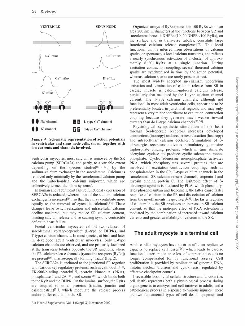
ventricular myocytes, most calcium is removed by the SRcalcium pump (SERCA2a) and partly, to a variable extentdepending on the species studied[8,10–13], by thesodium–calcium exchanger in the sarcolemma. Calcium isremoved only minimally by the sarcolemmal calcium pumpand the mitochondrial calcium uniporter, which arecollectively termed the ‘slow systems’.
In human and rabbit heart failure functional expression ofSERCA2a is reduced, whereas that of the sodium–calciumexchanger is increased[14], so that they may contribute moreequally to the removal of cytosolic calcium[8,15]. Thesechanges leave twitch relaxation and intracellular calciumdecline unaltered, but may reduce SR calcium content,limiting calcium release and so causing systolic contractiledeficit in heart failure.
Foetal ventricular myocytes exhibit two classes ofsarcolemmal voltage-dependent (L-type or DHPRs, andT-type) calcium channels. In most species, at birth and laterin developed adult ventricular myocytes, only L-typecalcium channels are observed, and are primarily localizedat the transverse tubules opposite the SR junctions, wherethe SR calcium release channels (ryanodine receptors [RyRs])are present[16], macroscopically forming ‘triads’ (Fig. 2).
The SERCA2a is anchored to the junctional SR togetherwith various key regulatory proteins, such as calmodulin[17],FK-506-binding protein[18], protein kinase A (PKA),phosphatase 1 and 2A [19], and sorcin[20], which binds bothto the RyR and the DHPR. On the luminal surface, the RyRsare coupled to other proteins (triadin, junctin andcalsequestrin)[21], which modulate the release processand/or buffer calcium in the SR.
Organized arrays of RyRs (more than 100 RyRs within anarea 200 nm in diameter) at the junctions between SR andsarcolemma beneath DHPRs (10–20 DHPRs/100 RyRs), onthe surface and in transverse tubules, constitute largefunctional calcium release complexes[22]. This localfunctional unit is inferred from observations of calciumsparks, or spontaneous local calcium transients, and reflectsa nearly synchronous activation of a cluster of approxi-mately 6–20 RyRs at a single junction. Duringexcitation–contraction coupling, several thousand calciumsparks are synchronized in time by the action potential,whereas calcium sparks are rarely present at rest.
The most widely accepted mechanism underlyingactivation and termination of calcium release from SR incardiac muscle is calcium-induced calcium release,particularly that mediated by the L-type calcium channelcurrent. The T-type calcium channels, although notfunctional in most adult ventricular cells, appear not to bepreferentially located in junctional regions, and may onlyrepresent a very minor contributor to excitation–contractioncoupling because they generate much weaker inwardcurrents than do L-type calcium channels[23,24].
Physiological sympathetic stimulation of the heartthrough β-adrenergic receptors increases developedcontractions (inotropy) and accelerates relaxation (lusitropy)and intracellular calcium declines. Stimulation of β-adrenergic receptors activates stimulatory guanosinetriphosphate binding proteins, which in turn stimulateadenylate cyclase to produce cyclic adenosine mono-phosphate. Cyclic adenosine monophosphate activatesPKA, which phosphorylates several proteins that areinvolved in excitation–contraction coupling, such asphospholamban in the SR, L-type calcium channels in thesarcolemma, SR calcium release channels, troponin I andmyosin binding protein C. The lusitropic effect of β-adrenergic agonists is mediated by PKA, which phosphory-lates phospholamban and troponin I; the latter cause fasterreuptake of calcium in the SR and dissociation of calciumfrom the myofilaments, respectively[25]. The faster reuptakeof calcium into the SR produces an increase in SR calciumcontent, so that the inotropic effect of PKA activation ismediated by the combination of increased inward calciumcurrents and greater availability of calcium in the SR.
The adult myocyte is a terminal cell
Adult cardiac myocytes have no or insufficient replicativecapacity to replace cell losses[26], which leads to cardiacfunctional deterioration once loss of contractile tissue is nolonger compensated for by functional reserve. Cellproliferation is provided by replication of genomic DNA,mitotic nuclear division and cytokinesis, regulated byeffective checkpoint controls.
Irreversible loss of vital cellular structure and function (i.e.cell death) represents both a physiological process duringorganogenesis in embryos and cell turnover in adults, and apathological process in response to various injuries. Thereare two fundamental types of cell death: apoptosis and
G4 R. Ferrari
Eur Heart J Supplements, Vol. 4 (Suppl G) November 2002
Figure 4 Schematic representation of action potentialsin ventricular and sinus node cells, shown together withion currents and channels involved.
VENTRICLE SINUS NODE
Na influx+ Ca influx++
Ca influx++ K efflux+
K efflux+
L L
L L
T T
T T
Na channel+
K channel+
L-type Ca channel++
T-type Ca channel++
K+ K+Na+ Ca++ Ca++
Dow
nloaded from https://academ
ic.oup.com/eurheartjsupp/article/4/suppl_G
/G1/379260 by guest on 13 January 2022

necrosis. Both necrosis and apoptosis may contributeindependently to myocyte cell death after infarction.
Apoptosis
During the normal development of vertebrates andinvertebrates, multifocal single cell death is constantlyobserved and designated apoptosis, which means ‘fallingoff’[27,28]. Apoptosis has been suggested to have acomplementary but opposite role to that of mitosis in normalhomeostasis. Also, it is considered to be the major processresponsible for cell death not only in various physiologicalevents, such as embryonic tissue remodelling, adult cellturnover and differentiation[27–30], but also duringpathological conditions such as viral hepatitis, death oftumour cells and graft-versus-host disease[31]. Cellproliferation encompasses replication of genomic DNA,mitotic nuclear division and cytokinesis, which are regulatedby checkpoint controls. Apoptosis was originally defined asan energy-dependent form of cell death, characterized bydistinct phases of ultrastructural morphological features.
Morphologically, apoptosis is characterized by nuclearand cytoplasmic condensation of single parenchymal cells(shrinkage), followed by loss of the nuclear membrane,fragmentation of the nuclear chromatin and subsequentformation of multiple fragments of condensed nuclearmaterial and cytoplasm[27,28], called apoptotic bodies. Theseapoptotic bodies are rapidly phagocytosed by neighbouringcells[32,33]. Therefore, flogosis does not occur. The initiationphase of apoptosis is characterized by activation and denovo synthesis of endonucleases. Several interventions (e.g.catecholamines, atrial natriuretic peptide, angiotensin II andstretch) have been shown to induce apoptosis in culturedmyocytes, but their role in the clinical setting has not yetbeen proven. Alternatively, intracellular alterationsfollowing ischaemic and/or reperfusion injury, such as anexcess of nitric oxide, may be involved. The pro-apoptoticstimuli, via two major pathways (the mitochondrial and thedeath receptor pathway), lead to activation of upstreamfollowed by downstream caspases[32,34,35], which cleave anincompletely characterized number of proteins, such asnuclear proteins, proteins that are involved in signaltransduction and cytoskeleton proteins. The mitochondrialpathway involves release of cytochrome c, apoptosis-inducing factor and probably other factors into the cytosol.Cytochrome c release appears to depend on the opening ofthe mitochondrial permeability pore, which is associatedwith a breakdown of the electrochemical gradient (∆Ψ) onthe inner membrane of mitochondria. Cytochrome c, whencomplexed with apoptotic protease factor 1, activates pro-caspase 9. The death receptor pathway, instead, involvesbinding of Fas ligand and tumour necrosis factor-alpha totheir receptors[36], thus activating pro-caspase 8. Activationof both pro-caspases 8 and 9 in turn leads to activation ofcaspase 3, whose target is DNA fragmentation, which canbe detected using the TUNEL (terminal transferasemediated DNA nick-end labelling) technique on lightmicroscopy[37,38].
Our understanding of the mechanisms of apoptosis incardiac myocytes may provide new strategies to preventmyocyte loss. Although the baseline rate of apoptosis isreported to be in the range 5–10% in cultured myocytes, theimpact of some stimuli may result in rates as high as75%[39,40]. Acute and progressive myocyte loss leads tocardiac functional deterioration, because myocytes have noor at least insufficient replicative capacity to replace celllosses[26].
Necrosis
Cell death in response to an overwhelming insult is referredto as necrosis, and is characterized by the sum ofmorphological and metabolic changes that accompany orfollow irreversible cell injury in living organisms. The basicpattern of pathological cell death that develops in responseto a variety of severe injury, such as ischaemia, hypoxia,chemical toxins, infections and trauma[41], is coagulationnecrosis, which is represented by denaturation andcoagulation of cellular proteins. Colliquative necrosis ischaracterized by tissue that rapidly liquefies due to itsprotein-poor nature. Necrosis often involves cell swellingrather than cell shrinkage, as occurs in apoptosis. Cellularfragmentation represents a late phase that results from thedegradative changes of autolysis, from activation andrelease of lysosomal enzymes, and of heterolysis, which iscaused by the actions of inflammatory cells invading thenecrotic tissue following cell death.
The evolution from reversible to irreversible injury innecrosis involves progressive derangements in energy andsubstrate metabolism, most importantly involving alteredhigh energy phosphate metabolism and progressivereduction in the cellular content of ATP[42]. Potentiallyreversible changes include condensation and clumping ofnuclear chromatin and intracellular oedema manifested byswelling of various organelles (endoplasmic reticulum,mitochondria, lysosomes and other vesicles), subsurfacecell blebbing and general cell swelling. Irreversible injuryis associated with additional morphological features(advanced changes in nuclear chromatin, mitochondriallesions with electron dense calcium phosphate deposits,breaks in the plasma membrane and organellarmembranes)[28,43].
Cardiac pathological conditions
In recent years, several studies have established thatapoptosis, also called ‘programmed cell death’, occurs invarious cardiac pathologies, including ischaemia andreperfusion injury, myocardial infarction and heart failure.It is known that the pathophysiology of ischaemic heartdisease and congestive heart failure is multifactorial. At thelevel of the myocyte, contractile dysfunction, due to alteredcalcium handling, impaired excitation–contractioncoupling, electrical instability and myocyte loss, is
Healthy versus sick myocytes G5
Eur Heart J Supplements, Vol. 4 (Suppl G) November 2002
Dow
nloaded from https://academ
ic.oup.com/eurheartjsupp/article/4/suppl_G
/G1/379260 by guest on 13 January 2022

believed to contribute to disease initiation and progression.At the clinical level several mechanisms have beenconsidered, including impaired excitation–contractioncoupling, altered neurohumoral balance, calciumhomeostasis and extracellular matrix (i.e. connectivetissue) composition, to which cardiomyocyte apoptosismust be added[44].
In myocardial infarction, apoptosis is believed toresponsible for nearly all cell death. Apoptosis has beenobserved in the core and border region of the ischaemicarea, and in viable myocardium[32]. Apoptosis is alsothought to accompany ventricular dysfunction in heartfailure. TUNEL-positive cardiac myocytes have been foundin the hearts of patients with ischaemic and idiopathicdilated cardiomyopathy[44–46], with higher incidence ofTUNEL-positive cells in the infarct border zones ofischaemic cardiomyopathy, indicating the possibility thatischaemia and increased stretch are involved[34]. Stretch isalways present in ventricular overload and dilatation, whichare associated with left ventricular hypertrophy.
The metabolic approach to ischaemic heart disease
Myocardial ischaemia is defined as an imbalance betweenfractional uptake of oxygen and the rate of cellularoxidation in the heart. This condition may have severalpotential outcomes. First, when ischaemia is of shortduration, there is no major molecular damage and atransient post-ischaemic ventricular dysfunction occurs onreperfusion, a condition termed ‘stunned myocardium’.Second, when ischaemia is prolonged and severe,irreversible damage occurs, with no recovery in contractilefunction upon reperfusion, and necrosis inevitably develops.Finally, when ischaemia is less severe but still prolonged,the myocytes may remain viable but exhibit depressedcontractile function. Under the latter condition, termed‘hibernating myocardium’, reperfusion is able to restorecontractility. The key transition might occur within minutesfrom onset of ischaemia, or take up to several hoursdepending on a multitude of factors (the underlyingmetabolic rate probably being the most important). For theclinical cardiologist, this is determined by the extent ofresidual flow, the underlying heart rate, the degree ofhaemodynamic change (such as an increase in pre- andafter-load wall stress), and the effects of any accompanyingneuroendocrine activation. This physiological ischaemia,characterized by down-regulation of contraction in theabsence of molecular changes, can also be considered aconservative adaptive response by the myocyte that down-regulates its contraction independently of extra cardiacsignals and, in so doing, reduces its energy needs in anattempt to maintain viability.
In contrast, in biochemical ischaemia[47], possibly inresponse to a series of complex and dominantly extra-cardiacneurohormone signals (activated to ensure the maintenanceof pump function and cardiac output), the myocyte will, athigh cost, succumb to a series of cellular mechanisms to
maintain contractile function, despite impairment in oxygensupply. As a consequence, the supply of energy fails tomatch consumption, and intracellular equilibrium (steady-state metabolism) is sacrificed, initiating a cascade ofincreasingly severe metabolic perturbations. The cell willthen become ‘metabolically distressed’ and, unlessinterrupted by early reperfusion, biochemical ischaemia willinevitably progress toward cell death.
From ischaemia to cell death
As shown in Fig. 5, mitochondria are the organelles that aremost likely to be involved in the transition from reversibleischaemia to cell death. This is perhaps not surprisingbecause these organelles play a fundamental role in cellularenergy production (the ATP turnover of the humanmyocardium exceeds 30 kg . day – 1) and in maintainingintracellular ionic homeostasis, which is the other keyprocess that is threatened by ischaemia.
Our understanding of the complexities of ischaemia andtissue injury is further complicated by the need to reperfusethe tissue to determine whether ischaemic damage isreversible or irreversible. Some, but not all, investigatorsbelieve that reperfusion itself might be detrimental and caninflict injury over and above that attributable toischaemia[48]. Other investigators, however, question theexistence of reperfusion-induced injury[49]. Ischaemia is nota static condition, and reperfusion is a part of a continuumof coronary artery disease. Such reperfusion might occur atdifferent times during transition from angina to myocardialinfarction, and may have several different outcomes,including early or delayed recovery (stunning), somerecovery (hibernation) or no recovery at all.
Physiological and biochemical ischaemia
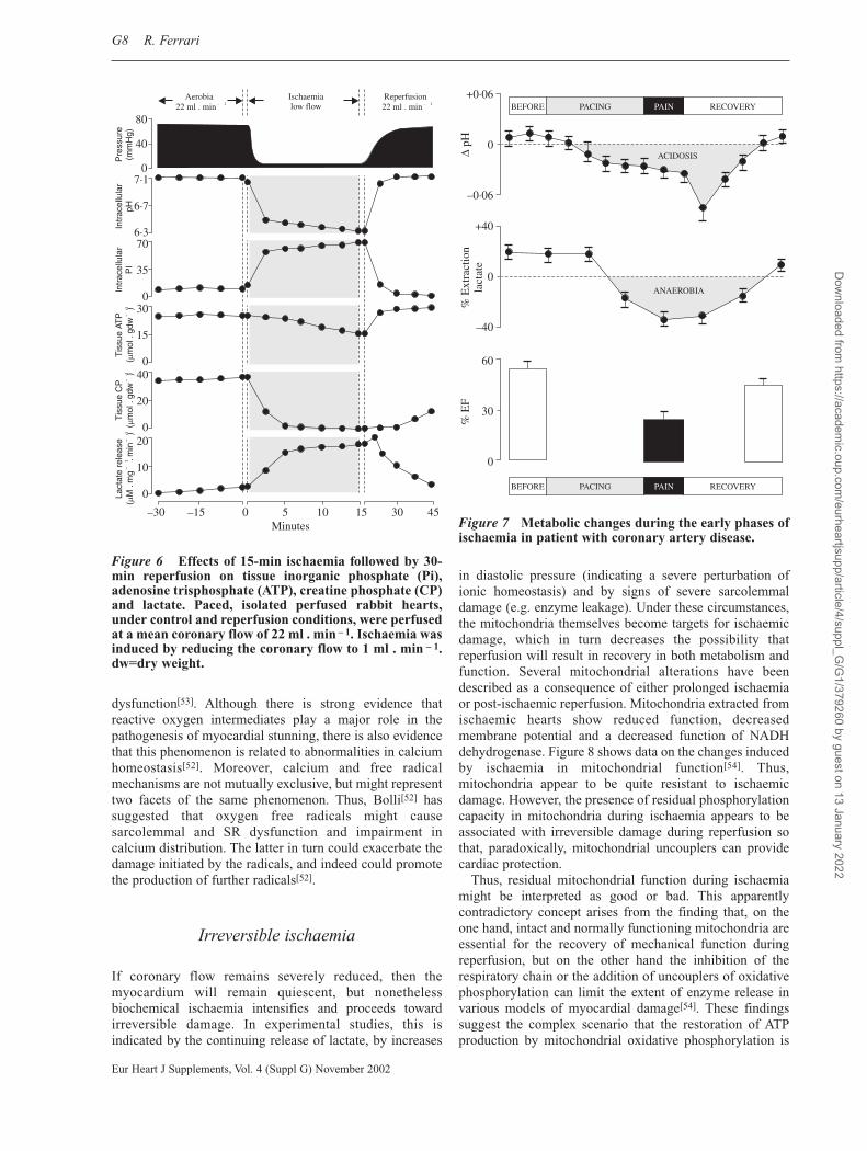
During short periods of ischaemia (e.g. in angina) there is aperfect match between biochemical and mechanicalactivity; this maintains viability. As shown in Fig. 6,restriction of coronary flow results in a rapid down-regulation of contraction and eventually quiescence. This isdue to the effects of intracellular acidosis, which developswithin seconds of induction of ischaemia and reducescalcium movements within the sarcolemma, SR andmyofilaments[50]. Shortly after this, the energy charge of themyocyte is reduced, and creatine phosphate declines fasterand to a greater extent than does ATP. Anaerobicmetabolism, as shown by lactate release in the coronaryeffluent, develops and contributes to the formation oflimited amounts of ATP by oxygen-independent, substrate-level phosphorylation. Taken together, these findingssuggest the occurrence of biochemical as well asphysiological ischaemia. Both down-regulation incontraction (and therefore in ATP consumption) andincreased anaerobic ATP production explain why the fall intissue ATP after the onset of ischaemia is not immediate.
G6 R. Ferrari
Eur Heart J Supplements, Vol. 4 (Suppl G) November 2002
Dow
nloaded from https://academ
ic.oup.com/eurheartjsupp/article/4/suppl_G
/G1/379260 by guest on 13 January 2022

The availability of this residual energy supply is essential tomaintain cellular viability. Reperfusion at this stage resultsin a recovery of high energy phosphate production, which inturn indicates that the mitochondria are still functionallyintact and capable of normal aerobic metabolism; this islinked to a recovery of mechanical function that might beeither immediate or somewhat delayed (Fig. 6).
This sequence of metabolic and functional events is notrestricted to experimental models but also occurs at theclinical level, for example during angina induced by atrialpacing. Figure 7 shows that in patients with coronary arterydisease and angina, an increasing heart rate (and thereforeincreasing energy requirements of the heart to the extentthat they are no longer met by supply) results in a reductionin coronary sinus pH, indicating the occurrence ofmyocardial acidosis. This is followed by an increase incoronary sinus lactate (indicative of the development ofanaerobic metabolism) and a down-regulation of regionalcontraction (revealed by a reduction in the ejection fraction,suggesting systolic dysfunction). All these biochemical andmechanical events precede the occurrence of angina. Oncethe heart is returned to its basal level and ischaemiatherefore no longer persists, coronary sinus pH and lactaterelease return to normal values and left ventricular systolicfunction improves. However, the functional recovery is notimmediate because of the presence of stunning. Clearly,under these circumstances viability is maintained, althoughevidence of the ischaemic insult persists as long as therecovery of function fails to match the recovery ofmetabolism[51].
Stunning
There is now convincing evidence that the myocardiumreperfused after a short period of ischaemia is characterizedby a variety of unfavourable, but non-lethal, cellular changesthat, given sufficient time, will return to normal. The mostprominent of these changes is myocardial stunning, which isthe prolonged contractile dysfunction that occurs duringreperfusion despite the absence of irreversible damage[51,52].The duration of dysfunction greatly exceeds that of theantecedent ischaemia. However, by definition, this form ofinjury is fully reversible, provided sufficient time is allowed.Interventions such as the use of inotropic agents can overridestunning, and other interventions such as antioxidants canprevent its occurrence[52].
A number of candidate mechanisms for stunning havebeen investigated; these include an impaired ability toresynthesize high energy phosphates, functional sympatheticdenervation, heterogeneous impairment of regionalperfusion, abnormal electrical activation, loss of creatinekinase activity, damage to the collagen matrix, leucocyteactivation and decreased sensitivity of myofilaments tocalcium. However, the two most plausible mechanisms relateto free radical induced injury during the early moments ofreperfusion and impaired calcium homeostasis.
Numerous studies suggest that oxygen-derived freeradicals contribute to post-ischaemic dysfunction. Electronmagnetic resonance spectroscopy has demonstrated theformation of free radicals in the stunned myocardiumdirectly, as well as the attenuation of contractile
Healthy versus sick myocytes G7
Eur Heart J Supplements, Vol. 4 (Suppl G) November 2002
Figure 5 Possible outcomes of myocardial ischaemia.
Left ventricular dysfunction• Systolic• Diastolic
IrreversibleReversible
Duration and severityof ischaemia
Degree of mitochondrialdamage
Functional recovery
Delayed(stunning)
Persistent(hibernation)
Immediate Reperfusion damage
Functional deterioration
• Angina
• Angioplasty
• Short ischaemia
• Thrombolysis
• Infarction
• Surgery
• Acute ischaemia
• Hypoperfusion
• Viability
• Further necrosis
• Ventricular expansion
• Remodelling – heart failure
Mitochondrial function maintainedor slightly altered
Mitochondrial function alteredCa ; O ATP& & '2 + –
2
O2O2 ATP ATP
Dow
nloaded from https://academ
ic.oup.com/eurheartjsupp/article/4/suppl_G
/G1/379260 by guest on 13 January 2022
dysfunction[53]. Although there is strong evidence thatreactive oxygen intermediates play a major role in thepathogenesis of myocardial stunning, there is also evidencethat this phenomenon is related to abnormalities in calciumhomeostasis[52]. Moreover, calcium and free radicalmechanisms are not mutually exclusive, but might representtwo facets of the same phenomenon. Thus, Bolli[52] hassuggested that oxygen free radicals might causesarcolemmal and SR dysfunction and impairment incalcium distribution. The latter in turn could exacerbate thedamage initiated by the radicals, and indeed could promotethe production of further radicals[52].
Irreversible ischaemia
If coronary flow remains severely reduced, then themyocardium will remain quiescent, but nonethelessbiochemical ischaemia intensifies and proceeds towardirreversible damage. In experimental studies, this isindicated by the continuing release of lactate, by increases
in diastolic pressure (indicating a severe perturbation ofionic homeostasis) and by signs of severe sarcolemmaldamage (e.g. enzyme leakage). Under these circumstances,the mitochondria themselves become targets for ischaemicdamage, which in turn decreases the possibility thatreperfusion will result in recovery in both metabolism andfunction. Several mitochondrial alterations have beendescribed as a consequence of either prolonged ischaemiaor post-ischaemic reperfusion. Mitochondria extracted fromischaemic hearts show reduced function, decreasedmembrane potential and a decreased function of NADHdehydrogenase. Figure 8 shows data on the changes inducedby ischaemia in mitochondrial function[54]. Thus,mitochondria appear to be quite resistant to ischaemicdamage. However, the presence of residual phosphorylationcapacity in mitochondria during ischaemia appears to beassociated with irreversible damage during reperfusion sothat, paradoxically, mitochondrial uncouplers can providecardiac protection.
Thus, residual mitochondrial function during ischaemiamight be interpreted as good or bad. This apparentlycontradictory concept arises from the finding that, on theone hand, intact and normally functioning mitochondria areessential for the recovery of mechanical function duringreperfusion, but on the other hand the inhibition of therespiratory chain or the addition of uncouplers of oxidativephosphorylation can limit the extent of enzyme release invarious models of myocardial damage[54]. These findingssuggest the complex scenario that the restoration of ATPproduction by mitochondrial oxidative phosphorylation is
G8 R. Ferrari
Eur Heart J Supplements, Vol. 4 (Suppl G) November 2002
Figure 6 Effects of 15-min ischaemia followed by 30-min reperfusion on tissue inorganic phosphate (Pi),adenosine trisphosphate (ATP), creatine phosphate (CP)and lactate. Paced, isolated perfused rabbit hearts,under control and reperfusion conditions, were perfusedat a mean coronary flow of 22 ml . min – 1. Ischaemia wasinduced by reducing the coronary flow to 1 ml . min – 1.dw=dry weight.
7·1
6·7
6·370
35
030
15
040
20
020
10
0
80
40
0
Lact
ate
rele
ase
(M
.m
g
.min
)
%–
1
–
1
Tis
sue
CP
(m
ol .
gdw
)
%–
1
Tis
sue
ATP
(m
ol .
gdw
)
%–
1
Intr
acel
lula
rP
IIn
trac
ellu
lar
pHP
ress
ure
(mm
Hg)
–30 –15 0 5 10 15 30 45Minutes
Aerobia22 ml . min – 1
Ischaemialow flow
Reperfusion22 ml . min – 1
Figure 7 Metabolic changes during the early phases ofischaemia in patient with coronary artery disease.
+0·06
0
–0·06
+40
0
–40
60
30
0%
EF
% E
xtra
ctio
nla
ctat
e(
pH
BEFORE PACING PAIN RECOVERY
BEFORE PACING PAIN RECOVERY
ACIDOSIS
ANAEROBIA
Dow
nloaded from https://academ
ic.oup.com/eurheartjsupp/article/4/suppl_G
/G1/379260 by guest on 13 January 2022

essential for cell recovery but, at the same time,mitochondrial activity can contribute to those processes thatproduce cell necrosis.
The reperfusion paradox
Several factors contribute to the immediate reperfusioninjury that can occur when supply of oxygen is restored toseverely ischaemic tissue. All this can be considered aparadox within a paradox. The two most important factorsare re-energization and rapid normalization in pH. Theseevents are not independent, but are synergistic.
More than two decades ago, Hearse et al.[55]
demonstrated that reoxygenation after prolonged oxygendepletion triggered sudden and major cellular injury, asindicated by massive enzyme leakage, sarcolemmaldisruption and hyper-contracture. It then became apparentthat re-energization sets in motion a series of paradoxes.
First, recovery of energy production reactivates the SRcalcium pump, leading to excess sequestration of calciumoften exceeding the capacity of the SR, which initiates acycle of continuous release and uptake of calcium[56].Second, the consequent excess of cytosolic calcium is takenup by the re-energized mitochondria at the expense of ATPproduction[54]. Finally, the resupply of energy to themyofibrillar elements in the presence of excess cytosoliccalcium leads to uncontrolled, excessive force generationand hyper-contraction.
In addition, upon reperfusion, interstitial pH is rapidlynormalized by the washout of protons, and a gradient isgenerated between the extracellular space and the cytosolthat still contains a high concentration of protons. This, inturn, activates proton-extruding mechanisms, namelysodium–hydrogen exchange and sodium–bicarbonatecotransport[57]. Activation of sodium–hydrogen exchangefollows, causing a net influx of sodium into the cytosol.Depending on the ability of the sodium pump to remove thisexcess sodium, there might be a secondary activation of the
Healthy versus sick myocytes G9
Eur Heart J Supplements, Vol. 4 (Suppl G) November 2002
Figure 8 Mitochondrial function in paced, isolated and perfused rabbit hearts. Under control and reperfusionconditions, the hearts were perfused at a mean coronary flow of 22 ml . min – 1. Ischaemia was induced by reducingcoronary flow to 1 ml . min – 1. (a) Typical example of a left ventricular (LV) pressure tracing from a heart subjectedto an ischaemia and reperfusion protocol. (b, c) Typical examples of isolated mitochondrial tracings for oxygenconsumption and adenosine trisphosphate (ATP) production. The mitochondria were isolated from hearts that hadbeen aerobic for 30 min; ischaemic for 30, 60 and 90 min; and reperfused for 30 min. The numerical values reportedin the oxygen consumption tracing represent rates (nmol oxygen/mg protein per min) consumed by the isolatedmitochondria during states III and IV of respiration. Glutamate was used as the respiratory substrate.
400
300
200
100
0
AT
P pr
oduc
tion
(nm
ol .
mg
prot
ein
)–
1O
xyge
n co
nsum
ptio
n(n
mol
. m
g pr
otei
n )–
1LV
pre
ssur
e(m
mH
g)
400
200
80
0
–30 0 30 60 90 120Minutes of perfusion
(a)
(b)
(c)
AEROBIA REPERFUSIONISCHAEMIA
0 20 40 60 0 20 40 60 0 20 40 60 0 20 40 60 0 20 40 60Seconds
0 20 40 60 0 20 40 60 0 20 40 60 0 20 40 60 0 20 40 60Seconds
ADP
250
12·54
ADP
249
12·39
ADP
200
13·01
ADP
175
16·90
ADP
9417·20
Dow
nloaded from https://academ
ic.oup.com/eurheartjsupp/article/4/suppl_G
/G1/379260 by guest on 13 January 2022

sodium–calcium exchange mechanism which, underconditions of intracellular sodium overload, will transportsodium in the outward direction and calcium in the inwarddirection; this uncoupled mechanism will then furtherexacerbate the pre-existing calcium overload[58]. Moreover,the sodium overload will act to increase osmotic gradientswith consequent cellular uptake of water, stretching anddamaging the sarcolemma and further disrupting ionichomeostasis[59]; intracellular acidosis, which down-regulates myofibrillar activity, is rapidly normalized,allowing the myofibrils in the presence of excessive calciumand low ATP to hyper-contract. This whole series ofcomplex and interacting mechanisms explains themitochondrial paradox that characterizes reperfusion.
Hibernation
The term ‘hibernation’ has been narrowed from zoology andimplies an adaptive reduction in energy use through reducedactivity in the presence of a reduced energy supply. In thecontext of coronary artery disease, myocardial hibernationwas originally seen as a chronic, adaptive reduction ofmyocardial contractile function in response to a reduction inmyocardial blood flow. It was also viewed as a condition inwhich there would be a complete recovery of contractilefunction upon restoration of flow. Thus, in the concept ofmyocardial hibernation, the observed chronic reduction inmyocardial contractile function is not regarded as the resultof a persistent energy deficit, but instead as a regulatoryevent that acts to avoid an ongoing energy deficit, therebymaintaining myocardial integrity and viability.
Interestingly, the concept of myocardial hibernation doesnot originate in the laboratory, but is entirely founded onclinical grounds. In the early 1980s, Rahimtoola[60], byreviewing the results of coronary bypass surgery trials,identified a subset of patients with coronary artery diseaseand chronic left ventricular dysfunction who improved uponrevascularization. Whereas the original idea of an adaptivereduction in contractile function in response to a reductionin blood flow was straightforward and simple, the conceptof chronic yet reversible contractile dysfunction in patientswith coronary artery disease was not recognized, and wasseen as enormously complex and controversial.
The introduction of the concept of hibernation haschallenged the traditional view that the extent of chroniccontractile dysfunction necessarily reflects the amount ofinfarcted tissue. In hibernation, the preservation of viabilityrather than the occurrence of necrosis accounts for theobserved reduction in function. In view of the preservedviability of the tissue, hibernation is a key factor in assessingthe potential benefit from reperfusion/revascularization.Hibernating myocardium must be recognized and identifiedby appropriate diagnostic procedures, and requiresappropriate decisions by the cardiologist responsible for theselection of patients who will benefit from the interventionalreperfusion or surgical revascularization. Obviously,hibernation is only one of several important aspects thatmust be considered in this patient selection, and many
patients with coronary artery disease and no evidence ofhibernating myocardium will also benefit.
A hibernation-like metabolic adaptation to a severe,sustained, low-flow ischaemia has been reported in studieswith isolated buffer-perfused rabbit hearts in which therewas a preceding short episode (10-min) of zero-flowischaemia. The rapid decline in contractile function(physiological ischaemia) during the brief episode of zero-flow ischaemia was accompanied by a greater decrease ininterstitial[50] and intracellular[61] pH, and the contractilequiescence was attributed to a faster development ofmyocardial acidosis. During low-flow perfusion there wasno lactate release, suggesting that biochemical ischaemiadid not occur. During reperfusion following sustainedischaemia, only a transient creatine kinase leakage occurredin the hearts with preceding zero-flow ischaemia. Thus, theestablishment of this experimental form of myocardialhibernation requires an initial period of zero-flowischaemia, during which rapid decreases in interstitial andintracellular pH trigger the decrease in contractile functionand thereby facilitate restoration of the balance betweenenergy supply and energy demand. Other experimentalstudies in pigs attribute a potentially important role to theinitial stimulus of severe ischaemia in critically triggeringthe development of a protective state, with preservedviability during a subsequent period of sustainedischaemia[62,63]. Whether or not such an initialstimulus/trigger of severe ischaemia represents a mandatorylink between hibernation and ischaemic pre-conditioning isunclear at present[64], but it supports the hypothesis thathibernating myocardium, at least most of the time, mightnot be biochemically but physiologically ischaemic[65].
References[1] Bossen E, Sommer JR, Waugh RA. Comparative stereology of the
mouse and finch left ventricle. Tissue Cell 1978; 10: 773–84.[2] Bossen EH, Sommer JR, Waugh RA. Comparative stereology of
mouse atria. Tissue Cell 1981; 13: 71–7.[3] Somlyo AP, Somlyo AV, Gonzales-Serratos H, Shuman H,
McLennan G. The sarcoplasmic reticulum and its composition inresting and in contracting muscle. In: Muscle Contraction. ItsRegulatory Mechanisms. Tokyo, Japan Scientific Societies Press;Berlin, Springer-Verlag: 421–33.
[4] Wendt-Gallitelli MF, Wolburg H. Electron probe microanalysis offrozen dried sections of heart muscle prepared using low temper-ature techniques. Scan Electron Microsc 1981; II: 455–62.
[5] Mitchell P. Chemiosmotic Coupling in Oxidative and Photo-synthetic Phosphorylation. Bodminster, Glynn Res Ltd., 1966.
[6] Gunter TE, Pfeiffer DR. Mechanisms by which mitochondriatransport calcium. Am J Physiol 1990; 258: C755–86.
[7] Hansford RG. Dehydrogenase activation by Ca++ in cells andtissues. J Bioenerg Biomembr 1991; 23: 823–54.
[8] Bers DM. Excitation-Contraction Coupling and Cardiac ContractileForce, 2nd ed. Dordrecht, The Netherlands, Kluwer Academic, 2001.
[9] Fukuda N, Sasaki D, Ishiwata S, Kurihara S. Length dpendence oftension generation in rat skinned cardiac muscle. Circ Res 2001;104: 1639–45.
[10] Bassani JWM, Bassani RA, Bers DM. Relaxation in rabbit and ratcardiac cells: species-dependent differences in cellular mecha-nisms. J Physiol 1994; 476: 279–93.
[11] Brandes R, Bers DM. Intracellular Ca2+ increases the mitochon-drial NADH concentration during elevated work in intact cardiacmuscle. Circ Res 1997; 80: 82–7.
G10 R. Ferrari
Eur Heart J Supplements, Vol. 4 (Suppl G) November 2002
Dow
nloaded from https://academ
ic.oup.com/eurheartjsupp/article/4/suppl_G
/G1/379260 by guest on 13 January 2022

[12] Hove-Madsen L, Bers DM. Sarcoplasmic reticulum Ca2+ uptakeand thapsigargin sensitivity in permeabilized rabbit and ratventricular myocytes. Circ Res 1993; 73: 820–8.
[13] Li L, Chu G, Kranias EG, Bers DM. Cardiac myocyte calciumtransport in phopholamban knockout mouse: relaxation andendogenous CaMKII effects. Am J Physiol 1998; 274: H1335–47.
[14] Hasenfuss G. Alterations of calcium-regulatory proteins in heartfailure. Cardiovasc Res 1998; 37: 279–89.
[15] Pogwizd SM, Schlotthauer K, Li L, Yuan W, Bers DM.Arrhythmogenesis and contractile dysfunction in heart failure:roles of sodium–calcium exchange, inward rectifier potassiumcurrent and residual β-adrenergic responsiveness. Circ Res 2001;88: 1159–67.
[16] Scriven DRL, Dan P, Moore EDW. Distribution of proteins impli-cated in excitation-contraction coupling in rat ventricularmyocytes. Biophys J 2000; 79: 2682–91.
[17] Fruen BR, Bardy JM, Byrem TM, Strasburg GM, Louis CF.Differential Ca2+ sensitivity of skeletal and cardiac muscleryanodine receptors in the presence of calmodulin. Am J Physiol2000; 279: C724–33.
[18] Marx SO, Reiken S, Hisamatsu Y, et al. PKA phosphorylationdissociates FKBP12.6 from the calcium release channel(ryanodine receptor): defective regulation in failing hearts. Cell2000; 101: 365–76.
[19] Marx SO, Reiken S, Hisamatsu Y, et al. Phosphorylation-depend-ent regulation of ryanodine receptors: a novel role forleucine/isoleucine zippers. J Cell Biol 2001; 153: 699–708.
[20] Meyers MB, Puri TS, Chien AJ, et al. Sorcin associates with thepore-forming subunit of voltage-dependent L-type Ca2+ channels.J Biol Chem 1998; 273: 18930–5.
[21] Zhang L, Kelley J, Schmeisser G, Kobayashi YM, Jones LR.Complex formation between junctin, triadin, calsequestrin, and theryanodine receptor. Proteins of the cardiac junctional sarcoplasmicreticulum membrane. J Biol Chem 1997; 272: 23389–97.
[22] Franzini-Armstrong C, Protasi F, Ramesh V. Shape, size, anddistribution of Ca2+ release units and couplons in skeletal andcardiac muscles. Biophys J 1999; 77: 1528–39.
[23] Sipido KR, Carmeliet E, van de Werf F. T-type Ca2+ current as atrigger for Ca2+ release from the sarcoplasmic reticulum inguinea-pig ventricular myocytes. J Physiol 1998; 508: 439–51.
[24] Zhou ZF, January CT. Both T- and L-type Ca2+ channels can con-tribute to excitation-contraction coupling in cardiac Purkinjecells. Biophys J 1998; 74: 1830–9.
[25] Li L, DeSantiago J, Chu G, Kranias EG, Bers DM.Phosphorylation of phospholamban and troponin I in β-adrenergic-induced acceleration of cardiac relaxation. Am J Physiol 2000;278: H769–79.
[26] Soonpaa MH, Field LJ. Survey of studies examining mammaliancardiomyocyte DNA synthesis. Circ Res 1998; 83: 15–26.
[27] Kerr JFR, Wyllie AH, Currie AR. Apoptosis: a basic biologicalphenomenon with wide-ranging implications in tissue kinetics. Br J Cancer 1972; 26: 239–57.
[28] Searle J, Kerr JFR, Bishop CJ. Necrosis and apoptosis: distinctmodes of cell death with fundamentally different significance.Pathol Annu 1982; 17: 229–59.
[29] Alles A, Alley K, Barrett JC, et al. Apoptosis: a general comment.FASEB J 1991; 5: 2127–8.
[30] Gerschenson LE, Rotello RJ. Apoptosis: a different type of celldeath. FASEB J 1992; 6: 2450–5.
[31] Eigenbrodt ML, Eigenbrodt EH, Thiele DL. Histologic similarityof murine colonic graft-versus-host disease (GVHD) to humancolonic GVHD and inflammatory bowel disease. Am J Pathol1990; 137: 1065–76.
[32] Borgers M, Voipio-Pulkki LM, Izumo S. Spotlight on: apoptosis.Cardiovasc Res 2000; 45: 525–804.
[33] Majno G, Joris I. Apoptosis, oncosis and necrosis: an overview ofcell death. Am J Pathol 1995; 146: 3–15.
[34] Haunstetter A, Izumo S. Apoptosis: basic mechanisms and impli-cations for cardiovascular disease. Circulation 1998; 82: 1111–29.
[35] Wolf BB, Green DR. Suicidal tendencies: apoptotic cell death bycaspase family proteinases. J Biol Chem 1999; 274: 20049–52.
[36] Nagata S. Fas ligand-induced apoptosis. Annu Rev Genet 1999;33: 29–55.
[37] Onoh M, Takemura G, Ohno H, et al. ‘Apoptotic’ myocytes in theinfarct area in rabbit hearts may be oncotic myocytes with DNAfragmentation: analysis by immunogold electron microscopycombined with in situ end labeling. Circulation 1998; 98:1422–30.
[38] Shaper J, Elsasser A, Kostin S. The role of cell death in heartfailure. Circulation 1999; 85: 867–9.
[39] Aikawa R, Komuro I, Yamazaki T, et al. Oxidative stress activatesextracellular signal-regulated kinases through src and ras incultured cardiac myocytes of neonatal rats. J Clin Invest 1997;100: 1813–21.
[40] Long X, Boluyt MO, Hipolito ML, et al. p53 and the hypoxia-induced apoptosis of cultured neonatal rat cardiac myocytes. J Clin Invest 1997; 99: 2635–43.
[41] Farber JL. Biology of disease: membrane injury and calciumhomeostasis in the pathogenesis of coagulative necrosis. LabInvest 1982; 47: 114–23.
[42] Hillis LD, Braunwald E. Myocardial ischemia. N Engl J Med1977; 296: 971–8, 1034–41, 1093–6.
[43] Trump BF, Valigorsky JM, Dees JH, et al. Cellular change inhuman disease: a new method of pathological analysis. HumPathol 1973; 4: 89–100.
[44] Sabbah HN. Apoptotic cell death in heart failure. Cardiovasc Res2000; 45: 704–12.
[45] Olivetti G, Abbi R, Quaini F, et al. Apoptosis in the failing humanheart. N Engl J Med 1997; 336: 1131–41.
[46] Guerra S, Leri A, Wang X, et al. Myocyte death in the failinghuman heart is gender dependent. Circ Res 1999; 85: 856–66.
[47] Hearse DJ. Myocardial ischaemia: can we agree on a definitionfor the 21st century? Cardiovasc Res 1994; 28: 1737–44.
[48] Opie LH. Reperfusion injury and its pharmacologic modification.Circulation 1989; 80: 1049–62.
[49] Ferrari R, Hearse DJ. Reperfusion injury: does it exist and does ithave clinical relevance? J Thromb Thrombolysis 1997; 4: 25–34.
[50] Ferrari R, Cargnoni A, Bernocchi P, et al. Metabolic adaptationduring a sequence of no-flow and low-flow ischaemia: a possibletrigger for hibernation. Circulation 1996; 94: 2587–96.
[51] Braunwald E, Kloner RA. The stunned myocardium: prolonged,post-ischaemic ventricular dysfunction. Circulation 1982; 66:1146–9.
[52] Bolli R. Mechanism of myocardial ‘stunning’. Circulation 1990;82: 723–38.
[53] Bolli R, Jeroudi MO, Patel BS, et al. Marked reduction of freeradical generation and contractile dysfunction by antioxidanttherapy begun at the time of reperfusion: evidence that myo-cardial ‘stunning’ is a manifestation of reperfusion injury. CircRes 1989; 65: 607–22.
[54] Ferrari R, Pedersini P, Bongrazio M, et al. Mitochondrial energyproduction and cation control in myocardial ischaemia and reper-fusion. Basic Res Cardiol 1993; 88: 495–512.
[55] Hearse DJ, Humphrey SM, Chain EB. Abrupt reoxygenation ofthe anoxic potassium-arrested perfused rat heart: a study ofmyocardial enzyme release. J Mol Cell Cardiol 1973; 5: 395–407.
[56] Siegmund B, Zude R, Piper HM. Recovery of anoxic-reoxygenated cardiomyocytes from severe Ca2+ overload. Am JPhysiol 1992; 63: H1262–9.
[57] Piper HM, Balser C, Ladilov YV, et al. The role of Na+/H+
exchange in ischaemia-reperfusion. Basic Res Cardiol 1996; 91:191–202.
[58] Ladilov YV, Siegmund B, Piper HM. Protection of thereoxygenated cardiomyocyte against hypercontracture by inhibi-tion of Na+/H+ exchange. Am J Physiol 1995; 268: H1531–9.
[59] Ruiz-Meana M, Garcia-Dorado D, Gonzales MA, et al. Effect ofosmotic stress on sarcolemmal integrity of isolated cardio-myocytes following transient metabolic inhibition. CardiovascRes 1995; 30: 64–9.
[60] Rahimtoola SH. A perspective on the three large multicenterrandomized clinical trials of coronary bypass surgery for chronicstable angina. Circulation 1983; 72: V123–35.
[61] van Binsbergen XA, van Emous JG, Ferari R, et al. Metabolic andfunctional consequences of successive no-flow and sustained low-flow ischaemia: a 31P MRS study on rat hearts. J Mol Cell Cardiol1996; 28: 2373–81.
Healthy versus sick myocytes G11
Eur Heart J Supplements, Vol. 4 (Suppl G) November 2002
Dow
nloaded from https://academ
ic.oup.com/eurheartjsupp/article/4/suppl_G
/G1/379260 by guest on 13 January 2022

[62] Schulz R, Post H, Sakka S, et al. Intra-ischemic preconditioning:increased tolerance to sustained low-flow ischemia by a briefepisode of no-flow ischemia without intermittent reperfusion.Circ Res 1995; 76: 942–50.
[63] Koning MMG, Simonis LAJ, de Zeeuw S, et al. Ischaemicpreconditioning by partial occlusion without intermittentreperfusion. Cardiovasc Res 1991; 28: 1146–51.
[64] Schulz R, Heusch G. Ischaemic preconditioning and myocardialhibernation: is there a common mechanism? Basic Res Cardiol1996; 91: 50–2.
[65] Heusch G, Ferrari R, Hearse DJ, et al. Myocardial hibernation:questions and controversies. Cardiovasc Res 1997; 36: 301–9.
G12 R. Ferrari
Eur Heart J Supplements, Vol. 4 (Suppl G) November 2002
Dow
nloaded from https://academ
ic.oup.com/eurheartjsupp/article/4/suppl_G
/G1/379260 by guest on 13 January 2022