view article online / journal homepage / table of contents...
TRANSCRIPT
Organic &BiomolecularChemistry
Dynamic Article Links
Cite this: Org. Biomol. Chem., 2012, 10, 8654
www.rsc.org/obc PAPER
1,8-Naphthyridine-2,7-diamine: a potential universal reader of Watson–Crickbase pairs for DNA sequencing by electron tunneling†
Feng Liang, Stuart Lindsay* and Peiming Zhang*
Received 2nd August 2012, Accepted 13th September 2012DOI: 10.1039/c2ob26529j
With the aid of Density Functional Theory (DFT), we designed 1,8-naphthyridine-2,7-diamine as arecognition molecule to read DNA base pairs for genomic sequencing by electron tunneling. NMRstudies show that it can form stable triplets with both A : T and G : C base pairs through hydrogenbonding. Our results suggest that the naphthyridine molecule should be able to function as a universalbase pair reader in a tunneling gap, generating distinguishable signatures under electrical bias for each ofDNA base pairs.
Introduction
Next generation DNA sequencing (NGS) has revolutionizedmany aspects of biological science, ranging from human diseaseanalyses1–5 to drug discovery6 to environmental monitoring.7
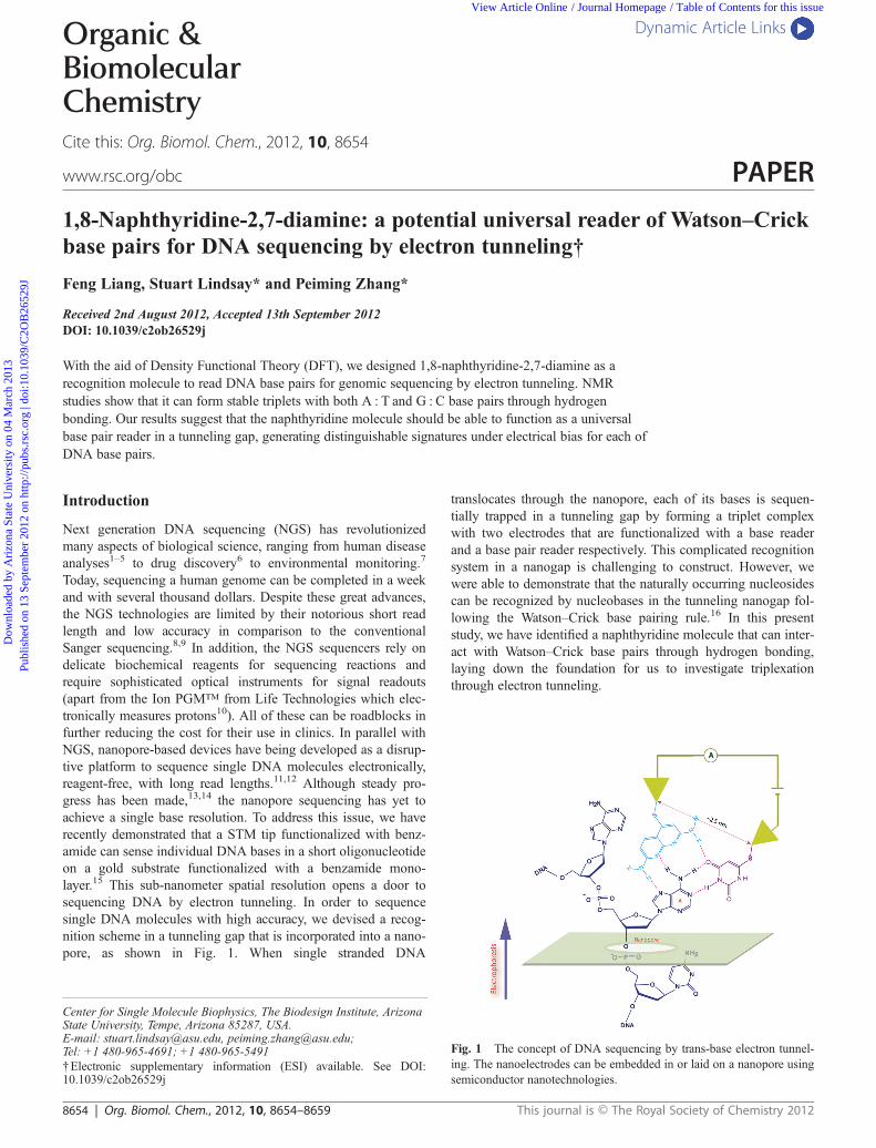
Today, sequencing a human genome can be completed in a weekand with several thousand dollars. Despite these great advances,the NGS technologies are limited by their notorious short readlength and low accuracy in comparison to the conventionalSanger sequencing.8,9 In addition, the NGS sequencers rely ondelicate biochemical reagents for sequencing reactions andrequire sophisticated optical instruments for signal readouts(apart from the Ion PGM™ from Life Technologies which elec-tronically measures protons10). All of these can be roadblocks infurther reducing the cost for their use in clinics. In parallel withNGS, nanopore-based devices have being developed as a disrup-tive platform to sequence single DNA molecules electronically,reagent-free, with long read lengths.11,12 Although steady pro-gress has been made,13,14 the nanopore sequencing has yet toachieve a single base resolution. To address this issue, we haverecently demonstrated that a STM tip functionalized with benz-amide can sense individual DNA bases in a short oligonucleotideon a gold substrate functionalized with a benzamide mono-layer.15 This sub-nanometer spatial resolution opens a door tosequencing DNA by electron tunneling. In order to sequencesingle DNA molecules with high accuracy, we devised a recog-nition scheme in a tunneling gap that is incorporated into a nano-pore, as shown in Fig. 1. When single stranded DNA
translocates through the nanopore, each of its bases is sequen-tially trapped in a tunneling gap by forming a triplet complexwith two electrodes that are functionalized with a base readerand a base pair reader respectively. This complicated recognitionsystem in a nanogap is challenging to construct. However, wewere able to demonstrate that the naturally occurring nucleosidescan be recognized by nucleobases in the tunneling nanogap fol-lowing the Watson–Crick base pairing rule.16 In this presentstudy, we have identified a naphthyridine molecule that can inter-act with Watson–Crick base pairs through hydrogen bonding,laying down the foundation for us to investigate triplexationthrough electron tunneling.
Fig. 1 The concept of DNA sequencing by trans-base electron tunnel-ing. The nanoelectrodes can be embedded in or laid on a nanopore usingsemiconductor nanotechnologies.
†Electronic supplementary information (ESI) available. See DOI:10.1039/c2ob26529j
Center for Single Molecule Biophysics, The Biodesign Institute, ArizonaState University, Tempe, Arizona 85287, USA.E-mail: [email protected], [email protected];Tel: +1 480-965-4691; +1 480-965-5491
8654 | Org. Biomol. Chem., 2012, 10, 8654–8659 This journal is © The Royal Society of Chemistry 2012
Dow
nloa
ded
by A
rizo
na S
tate
Uni
vers
ity o
n 04
Mar
ch 2
013
Publ
ishe
d on
13
Sept
embe
r 20
12 o
n ht
tp://
pubs
.rsc
.org
| do
i:10.
1039
/C2O
B26
529J
View Article Online / Journal Homepage / Table of Contents for this issue

Results and discussion
We chose 1,8-naphthyridine-2,7-diamine (designated as N) as acandidate to read the base pairs. There are varied structures thathave been investigated as motifs of triplex forming oligonucleo-tides (TFO) to recognize the Watson–Crick base pairs in doublestranded DNA.17 Two typical examples are shown in Fig. 2a,which exhibit fair affinity and selectivity to the DNA base pairswhen incorporated into TFO.18,19 One common feature of thesestructures is that they are configured either by connecting twoaromatic rings together through a free rotating sigma bond or bymodifying the DNA base with a functional tail to match thehydrogen bonding patterns of the Watson–Crick base pairs in themajor groove side. However, such flexibility results in a loss ofentropy when a hydrogen bonded complex is formed becausethe freely rotating bonds become fixed. This would not be suit-able for our recognition scheme in which the hydrogen bondingmay be a dominating force for formation of a stable triplet andthe important base stacking interactions may be not as effectiveas that in the double stranded DNA. We postulate that a rigid andplanar scaffold with the right geometry will have an advantagein this regard. Furthermore, if a tandem hydrogen bonding sitearray is constructed along one edge of the scaffold without anyC–H interruption, it should increase the hydrogen bonding coope-rativity. 1,8-Naphthyridine-2,7-diamine is a molecule that con-tains an aromatic plane composed of two fused pyridyl rings andtwo amines aligned with the ring nitrogen atoms to form anarray of four hydrogen bonding sites (see Fig. 1). It has beenexploited as a moiety to create new base pairs in DNA.20,21 Inaddition, the amine derivatives of naphthyridine form stablecomplexes with guanine22,23 and deaza guanine,24 and havebeen used as a fluorescent dye to stain nucleoli in the nucleus ofMDCK-cells.25 However, the interactions of 1,8-naphthyridine-2,7-diamine with the DNA base pairs have not been explored.With the aid of Density Functional Theory (DFT), we first
scrutinized the structural fitness through computer modeling. Asillustrated in Fig. 2b, 1,8-naphthyridine-2,7-diamine can formhydrogen bonded complexes of N–T : A and N–C : G with theWatson–Crick base pairs from the major groove sides. In thecomputer simulation, a methyl group was placed at the 3-posi-tion of N as a prospective site for attachment, and all the sugarsconnected to DNA bases were substituted with methyl groups inorder to reduce the computing time. Note that the methyl substi-tution would not exert a game changing influence on our com-puting results since the hydrogen bonding interactions take placeon the opposite sides of the sugars. The DFT calculations showthat there is a gain of ∼15 kcal mol−1 in energy when N hydrogen-bonds to either T : A or C : G base pairs in a vacuum, whichis slightly higher than the hydrogen-bonding energy of the T : AWatson–Crick base pair (Table S1 in ESI†). The distancebetween the two amino nitrogen atoms (∼6.86 Å) of N is fairlymatched to that from N-7 of adenine to O-4 of thymine(∼6.26 Å) in the T : A base pair, resulting in formation of a goodfit N–T : A triplet. In the N–C : G triplet, N is twisted out of theC : G base pair plane due to a steric hindrance between the2-position amine of N and the 5-position hydrogen of cytosine.The DFT solvation calculation indicates that the triplets areslightly less stable in DMSO, a solvent that has a dielectric con-stant (ε = 46.7 D) comparable to one in the major groove ofDNA (ε = 55 D).26 We believe that the hydrogen bonding inter-actions should prevail in the nanogap, which has a local environ-ment less hydrophilic than the bulk aqueous solution especiallywhen it is functionalized with organic molecules.
Following the computer modeling studies, we investigated thehydrogen bonding interactions of N with the DNA base pairsformed by individual nucleosides in solutions using NMR tech-niques. To adequately dissolve these entities into chloroform (acommonly used solvent for hydrogen bonding studies), N wasconverted to an amine-acetylated derivative (Nac), and thehydroxyl groups of the naturally occurring nucleosides were sily-lated with tert-butyldimethylsilyl chloride (designated as dA,dC, dG, dT, and dU). Our primary focus was on the hydrogenbonding of Nac with the A : T base pair because the computermodeling showed a good fit between these two entities. First, anNMR spectrum of a mixture of dA and dT in deuterated chloro-form (in a 1 : 1 molar ratio) was recorded to confirm the basepairing (upper spectrum in Fig. 3a). It shows that the iminoproton peak of dT has not only shifted downfield but also splitinto two with an integration ratio of 1 : 2, compared to that (δH =9.8 ppm) in a dT only solution. This indicates that there weretwo different hydrogen-bonding interactions involved betweendA and dT. By means of 1H–1H NOESY NMR (Fig. S1 inESI†), we found that the peak at 11.8 ppm cross-talks to the HA2
peak of dA, and the one at 11.5 ppm cross-talks to the HA8 peakof dA. The NMR data can be best explained by coexistence ofboth Hoogsteen (HG) and Watson–Crick (WC) base pairs inequilibrium as delineated in Scheme 1. The HG base pair hasbeen observed in crystals of alkylated nucleobase complexes byX-ray diffraction,27,28 and in a dA : dU (silylated 2′-deoxyuri-dine) solution by NMR.29 It could even exist with a 1% prob-ability in DNA.30,31 The early calculations predicted that the HGbase pair would be slightly more stable than the WC basepair.32,33 Our data show that the HG base pair is a preferred formin the chloroform solution. Thus, we believe that the HG base
Fig. 2 (a) DNA base pair recognition molecules for TFO and theirhydrogen bonding interactions with DNA base pairs; (b) DFT models ofthe complexes of 1,8-naphthyridine-2,7-diamine with the T : A and C : Gbase pairs.
This journal is © The Royal Society of Chemistry 2012 Org. Biomol. Chem., 2012, 10, 8654–8659 | 8655
Dow
nloa
ded
by A
rizo
na S
tate
Uni
vers
ity o
n 04
Mar
ch 2
013
Publ
ishe
d on
13
Sept
embe
r 20
12 o
n ht
tp://
pubs
.rsc
.org
| do
i:10.
1039
/C2O
B26
529J
View Article Online

pairing is intrinsic at least to A and T bases. This may be one ofreasons why it can occur even in DNA double helices where thegeometry is constrained to favor the WC base pairing. Comparedto the WC base pair, the HG base pair has a shorter distancefrom N9 of dA to N1 of dT.34 As a result, the HG base pair maynot fit well with N to form a stable triplex.
When mixing Nac with dA and dT in a 1 : 1 : 1 ratio, a singleimino proton peak was only observed in the NMR spectrum(lower spectrum in Fig. 3a). The variable concentration NMRshowed that amide protons of Nac and amino protons of theadenine were also involved in the hydrogen-bonding interactions(Fig. S2 in ESI†). Formation of a Nac
–dA : dT triplet wasconfirmed by 1H–1H NOESY NMR. In the 2D NOESY spec-trum (Fig. 3b), we only observed the cross peaks from the dTimino proton (HT3) correlating with HA6 and HA2 of dA (seeFig. 3c for designation of each proton), implying that only theWC base pair was formed in the complex. Furthermore, both Hi
and He of Nac are correlated to HA8 of dA, and the Hi is corre-
lated to HT6 of dT as well. These NMR data allow us to sketch aconnection among the three molecules shown in Fig. 3c. Due to
crowding in the region of the methyl groups at the dT end of thecomplex, we cannot unambiguously assign the HT5–Hi crosspeak. The DFT modelling shows that the amide ends of Nac arepushed out of the dA : dT base pair plane in the triplet becauseof the steric hindrance between the acetyl groups of Nac and themethyl group of thymine and the HA8 of dA (see Fig. 4 for thetriplet conformation from computer modeling). This may explainwhy Hi is correlated with HT6 in the NOESY NMR. We noticedthat the two amide protons of Nac appeared as a single peak inthe NMR spectrum of the complex. They were split into twobroad peaks when the temperature was lowered to −55 °C in an800 MHz NMR spectrometer (Fig. S3 in ESI†), indicating thatthese two protons were in a fast exchange within the NMR time-frame at room temperature. In contrast, guanine and cytosineonly form a stable WC base pair under the same conditions forthe A : T base pair. The formation of a triplet between Nac andthe dG : dC base pair was confirmed by 2D NOESY NMR(Fig. S4 in ESI†).
Based on the principle of complexation-induced chemicalshifts (CIS),35–38 we have determined association constants ofNac with DNA bases and base pairs by NMR titration (seeTable 1). The silylated nucleosides (dA, dC, dG, dT, and dU)were used as either titrants or substrates, and Nac was only usedas a substrate due to its limited solubility in chloroform. In atypical NMR titration experiment, a titrant was incrementallyadded to a substrate solution, and a proton NMR spectrum wasrecorded following each addition. In general, protons directlyinvolved in hydrogen bonding exhibited downfield chemicalshifts, resulting in positive CIS values. The proton we closelymonitored in the NMR titration is listed in parentheses under the
Fig. 3 (a) 1H NMR spectra of a mixture of dA and dT in a ratio of 1 : 1 and a mixture of dA, dT and Nac in a ratio of 1 : 1 : 1 at room temperature;(b) 2D 1H–1H NOESY NMR spectra of a mixture of dA, dT and Nac in a 1 : 1 : 1 ratio and proton correlation assignments. (c) A schematic of molecu-lar connections in the dA, dT and Nac complex.
Scheme 1
8656 | Org. Biomol. Chem., 2012, 10, 8654–8659 This journal is © The Royal Society of Chemistry 2012
Dow
nloa
ded
by A
rizo
na S
tate
Uni
vers
ity o
n 04
Mar
ch 2
013
Publ
ishe
d on
13
Sept
embe
r 20
12 o
n ht
tp://
pubs
.rsc
.org
| do
i:10.
1039
/C2O
B26
529J
View Article Online

substrate in Table 1. First, the complexing stoichiometriesbetween titrants and substrates were determined using the moleratio plot.39,40 We found that all of these complexes were fairlyclose to a 1 : 1 binding mode (Fig. S5 in ESI†). The associationconstants (Kass) were then derived from curve fitting datasets ofchemical shift vs. concentration in HypNMR 2008, a program toanalyze NMR titration data. All of our NMR data were best fit toa 1 : 1 binding isotherm. We tested the reproducibility of ourexperimental process by performing the reverse titration. Forexample, titrating dA with dT yielded a virtually identical resultas that from titrating dT with dA. The Kass value (∼40 M−1) ofthe dA : dT base pairing we determined is close to that reportedin the literature.41,42 As shown in Table 1, Nac was titrated with aseries of individual nucleosides and their mixtures. Clearly, itformed a more stable complex with dC : dG than with dA : dT.Nonetheless, Kass of N
ac complexing to dA : dT is comparable tothat for the dA : dT base pairing. This Kass value, may underesti-mate the actual stability of Nac complexing to dA : dT becausethe dA and dT mixture mainly exists in a HG base pairing formin chloroform so that there is a free energy penalty to convert theHG base pair to the WC base pair for formation of the Nac
–
dA : dT triplet. When titrating a mixture of Nac and dT with dAor a mixture of Nac and dAwith dT, the Kass values (∼120 M−1)derived from monitoring HA6 and HT3 in these two mixed sub-strates are about three times higher than that of the dA : dT basepairing. This indicates that Nac could stabilize the dA : dT basepair. It has been known that the G–C base pair is very stable in a
nonpolar solvent, such as chloroform (Kass = ∼104–5 M−1).41,43
A 1 : 1 mixture of dG and dC is often treated as a single com-ponent in NMR titration experiments.44–47 Titrating Nac withdG : dC yielded a relatively stable complex with an associationconstant of Kass ∼ 467 M−1. However, Nac prevents dG frombase pairing with dC because it forms a very stable complexwith dG (Kass ∼ 3990 M−1). As a result, when titrating a Nac anddG mixture with dC, a positive CIS on the imino proton of dGwas obtained, indicating that there was a hydrogen bondinginteraction between dG and dC, but the resultant Kass value(∼710 M−1) is significantly smaller than one of the normaldG : dC base pairing. Thus, we have to follow an appropriateroute to assemble a Nac
–dG : dC triplet. We also notice that the5-methyl group of dT did not cause any significant steric hin-drance to the formation of an Nac–dT : dA triplet because there isa negligible difference in Kass between Nac–dT : dA and Nac–
dU : dA.To interpret the NMR data, we constructed molecular models
for the complexes of Nac with the nucleoside pairs (Fig. 4),which were optimized by DFT calculations using B3LYP incombination with 6-31G* basis sets. As shown in Fig. 4, Nac
forms the hydrogen bonded triplets with the Watson–Crick basepairs from the major groove side. The DFT calculation indicatesthat Nac–dC : dG is more stable than Nac–dT : dA and Nac–
dU : dA in terms of their complexing energies (ΔETot in Table 2).This matches the results from the NMR titrations. The higherstability of the Nac
–dC : dG triplet may be attributed to the
Fig. 4 DFT models of hydrogen bonded triplets of Nac with dT : dA (A), dU : dA (B), and dC : dG (C) calculated by B3LYP in combination with6-31G* basis sets in a vacuum.
Table 1 Complexation induced chemical shift (CIS, ppm) and association constants (Kass, M−1) of Nac with individual nucleosides and nucleoside
pairs derived from curve fitting of NMR titration data
Substrate
Titrant
dA dC dG dT dU dA : dT dA : dU dG : dC
Nac (He) CISa 2.1 ± 0.4 0.9 ± 0.1 1.1 ± 0.0 3.5 ± 0.4 2.0 ± 0.1 1.5 ± 0.0 1.5 ± 0.1 0.7 ± 0.0Kass 6.3 ± 1.4 53 ± 9 3889 ± 833 18 ± 3 31 ± 3 59 ± 1 62 ± 11 525 ± 166
dG-Nac (HG1) CIS 1.2 ± 0.0Kass 710 ± 151
dA-Nac (HA6) CIS 0.8 ± 0.0Kass 115 ± 11
dT-Nac (HT3) CIS 0.4 ± 0.0Kass 126 ± 4
aCIS = δmax − δinitial in ppm, which was determined from the fit titration curve.
This journal is © The Royal Society of Chemistry 2012 Org. Biomol. Chem., 2012, 10, 8654–8659 | 8657
Dow
nloa
ded
by A
rizo
na S
tate
Uni
vers
ity o
n 04
Mar
ch 2
013
Publ
ishe
d on
13
Sept
embe
r 20
12 o
n ht
tp://
pubs
.rsc
.org
| do
i:10.
1039
/C2O
B26
529J
View Article Online

strong dG : dC base pair since the calculated hydrogen bondingenergy of Nac with the dG : dC pair is close to those of Nac withthe dU : dA and dT : dA pairs.
Conclusions
Our DFT calculations and NMR studies reveal that 1,8-naphthy-ridine-2,7-diamine can form hydrogen bonded triplets with bothA : T and G : C Watson–Crick base pairs, which are as stable asthe A : T base pair or more so. We have found that the naphthy-ridine molecule has a number of unique features: it tends tostabilize the A : T Watson–Crick base pair, block the A : THoogsteen base pairing, and form a stable complex with guanineto prevent the G : C base pairing. Due to differences in theirstructures, these triplet complexes should create different path-ways for electron tunneling, resulting in distinguishable electricalsignals for readout of the DNA base pairs in the tunneling gap,which makes it a universal base pair reader. We are developingchemistry to attach the naphthyridine molecules to the metalelectrodes for the tunneling measurements.
Experimental section
General information
Proton NMR (1H) spectra were recorded at 400 MHz on a Varian400 MHz spectrometer, and carbon NMR (13C) spectra wererecorded at 100 MHz on a Varian 400 MHz spectrometer.HRMS spectra were recorded using the atmospheric pressurechemical ionization (APCI) technique. Flash chromatographywas performed using automated flash chromatography (TeledyneIsco, Inc. CombiFlash Rf). All reagents were obtained fromcommercial suppliers unless otherwise stated. Where necessary,organic solvents were routinely dried and/or distilled prior to useand stored over molecular sieves under nitrogen. All reactionsrequiring anhydrous conditions were performed under a nitrogenatmosphere.
Synthesis of N,N′-(1,8-naphthyridine-2,7-diyl)diacetamide (Nac)
A mixture of 1,8-naphthyridine-2,7-diamine48 (280 mg,1.75 mmol) in acetic anhydride (3 mL) was heated at reflux for2.5 h. After cooling, the excess solvent was removed, and theresidue was purified by flash chromatography (on silica gel witha gradient of dichloromethane–methanol from 100 : 0 to100 : 10) to give 180 mg (42%) of the product as a yellow
powder. 1H NMR (400 MHz, DMSO-d6): δ 2.16 (s, 6H), 8.21(d, J = 9.2 Hz, 2H), 8.26 (d, J = 9.2 Hz, 2H), 10.77 (s, 2H);13C NMR (100 MHz, DMSO-d6): δ 24.7, 113.3, 117.7, 139.2,154.3, 154.8, 170.5. HRMS (APCI+): found, 245.1038 (calcdfor C12H13N4O2, 245.1043).
Silylation of nucleosides
All the nucleosides were silylated with tert-butyldimethylsilylchloride following our reported method.49
Computation
DFT calculations were performed using the program Spartan’10for Windows, Wavefunction Inc. Individual 2D chemical struc-tures were drawn in ChemBioDraw Ultra 11 and exported toSpartan’10 to generate the respective 3D structures, from whichhydrogen bonded complexes were constructed. The geometry ofeach complex was first subjected to energy minimization usingthe built-in MMFF94s molecular mechanics, and then calculatedusing B3LYP/6-311++G (2df, 2p) in a vacuum. All of the calcu-lations were successfully converged and no BSSE correctionswere carried out for such a large basis set. Following completionof the calculation in a vacuum, the complex was solvated withDMSO using B3LYP/6-31G** based on a SM8 model.50
1H NMR binding studies
Proton NMR (1H) spectra were recorded at 400 MHz on a Varian400 MHz, 500 MHz on a Varian 500 MHz or 800 MHz on aVarian 800 MHz spectrometer. All 1H NMR chemical shiftswere referenced to the residual non-deuterated solvent peak as7.26 ppm in chloroform-d (CDCl3). 2D NOE spectra wererecorded at 400 MHz on a Bruker 400 MHz spectrometer. ForVT NMR, temperature was calibrated with a standard of 100%CH3OH and regulated to an accuracy of ±0.1 °C by a EurothermVariable Temperature Unit on the Bruker NMR or a HighlandTechnologies Temperature unit on the Varian NMR System.Temperatures below zero Celsius were achieved with a LiquidNitrogen Heat Exchanger on the Bruker and FTS CoolingSystem (Stone Ridge, New York) on the Varian. CDCl3 was pur-chased from Sigma-Aldrich, used as received without furtherpurification. Volumetric flasks and syringes for preparing thestock solutions were rinsed with CDCl3 and dried under vacuumprior to use. For NMR titration, samples were prepared fromstock solutions, transferred to NMR tubes using a syringe, anddiluted following the method in the previous report.51 Associ-ation constants reported were averages of two or more repeatsand were derived from fitting NMR titration data to a 1 : 1binding isotherm using the HypNMR program.
Acknowledgements
This project is funded by the DNA sequencing technologyprogram of the National Institute of Human Genome Research(HG004378). We thank Dr Brian Cherry of the magnetic reson-ance research center at ASU for his technical support and infor-mative discussion during the NMR experiments.
Table 2 DFT energies (kcal mol−1) of the Nac triplets
ΔETot ΔE(Nac)
Nac–dT : dA −39.3 −22.4Nac–dU : dA −40.6 −22.8Nac–dC : dG −54.5 −20.7
ΔETot: complexing energy calculated from energy of (complex −individual monomers constituting the complex). ΔE (Nac): hydrogenbonding energy of Nac calculated from energy of (complex − Nac −nucleoside pair).
8658 | Org. Biomol. Chem., 2012, 10, 8654–8659 This journal is © The Royal Society of Chemistry 2012
Dow
nloa
ded
by A
rizo
na S
tate
Uni
vers
ity o
n 04
Mar
ch 2
013
Publ
ishe
d on
13
Sept
embe
r 20
12 o
n ht
tp://
pubs
.rsc
.org
| do
i:10.
1039
/C2O
B26
529J
View Article Online
Notes and references
1 J. Bras, R. Guerreiro and J. Hardy, Nat. Rev. Neurosci., 2012, 13, 453–464.
2 M. Meyerson, S. Gabriel and G. Getz, Nat. Rev. Genet., 2010, 11, 685–696.
3 M. R. Nelson, D. Wegmann, M. G. Ehm, D. Kessner, P. St Jean,C. Verzilli, J. Shen, Z. Tang, S. A. Bacanu, D. Fraser, L. Warren,J. Aponte, M. Zawistowski, X. Liu, H. Zhang, Y. Zhang, J. Li, Y. Li,L. Li, P. Woollard, S. Topp, M. D. Hall, K. Nangle, J. Wang,G. Abecasis, L. R. Cardon, S. Zollner, J. C. Whittaker, S. L. Chissoe,J. Novembre and V. Mooser, Science, 2012, 337, 100–104.
4 G. E. Palomaki, C. Deciu, E. M. Kloza, G. M. Lambert-Messerlian,J. E. Haddow, L. M. Neveux, M. Ehrich, D. van den Boom,A. T. Bombard, W. W. Grody, S. F. Nelson and J. A. Canick, Genet.Med., 2012, 14, 296–305.
5 L. Ding, T. J. Ley, D. E. Larson, C. A. Miller, D. C. Koboldt, J. S. Welch,J. K. Ritchey, M. A. Young, T. Lamprecht, M. D. McLellan,J. F. McMichael, J. W. Wallis, C. Lu, D. Shen, C. C. Harris,D. J. Dooling, R. S. Fulton, L. L. Fulton, K. Chen, H. Schmidt,J. Kalicki-Veizer, V. J. Magrini, L. Cook, S. D. McGrath, T. L. Vickery,M. C. Wendl, S. Heath, M. A. Watson, D. C. Link, M. H. Tomasson,W. D. Shannon, J. E. Payton, S. Kulkarni, P. Westervelt, M. J. Walter,T. A. Graubert, E. R. Mardis, R. K. Wilson and J. F. DiPersio, Nature,2012, 481, 506–510.
6 P. M. Woollard, N. A. Mehta, J. J. Vamathevan, S. Van Horn,B. K. Bonde and D. J. Dow, Drug Discovery Today, 2011, 16, 512–519.
7 M. Hajibabaei, S. Shokralla, X. Zhou, G. A. Singer and D. J. Baird,PLoS One, 2011, 6, e17497.
8 J. Kuczynski, C. L. Lauber, W. A. Walters, L. W. Parfrey, J. C. Clemente,D. Gevers and R. Knight, Nat. Rev. Genet., 2012, 13, 47–58.
9 O. Morozova and M. A. Marra, Genomics, 2008, 92, 255–264.10 J. M. Rothberg, W. Hinz, T. M. Rearick, J. Schultz, W. Mileski,
M. Davey, J. H. Leamon, K. Johnson, M. J. Milgrew, M. Edwards,J. Hoon, J. F. Simons, D. Marran, J. W. Myers, J. F. Davidson,A. Branting, J. R. Nobile, B. P. Puc, D. Light, T. A. Clark, M. Huber,J. T. Branciforte, I. B. Stoner, S. E. Cawley, M. Lyons, Y. Fu, N. Homer,M. Sedova, X. Miao, B. Reed, J. Sabina, E. Feierstein, M. Schorn,M. Alanjary, E. Dimalanta, D. Dressman, R. Kasinskas, T. Sokolsky,J. A. Fidanza, E. Namsaraev, K. J. McKernan, A. Williams, G. T. Rothand J. Bustillo, Nature, 2011, 475, 348–352.
11 M. Zwolak and M. D. Ventra, Rev. Mod. Phys., 2008, 80, 141–165.12 D. Branton, D. W. Deamer, A. Marziali, H. Bayley, S. A. Benner,
T. Butler, M. D. Ventra, S. Garaj, A. Hibbs, X. Huang, S. B. Jovanovich,P. S. Krstic, S. Lindsay, X. S. Ling, C. H. Mastrangelo, A. Meller,J. S. Oliver, Y. V. Pershin, J. M. Ramsey, R. Riehn, G. V. Soni, V. Tabard-Cossa, M. Wanunu, M. Wiggin and J. A. Schloss, Nat. Biotechnol., 2008,26, 1146–1153.
13 E. A. Manrao, I. M. Derrington, A. H. Laszlo, K. W. Langford,M. K. Hopper, N. Gillgren, M. Pavlenok, M. Niederweis andJ. H. Gundlach, Nat. Biotechnol., 2012, 30, 349–353.
14 G. M. Cherf, K. R. Lieberman, H. Rashid, C. E. Lam, K. Karplus andM. Akeson, Nat. Biotechnol., 2012, 30, 344–348.
15 S. Huang, J. He, S. Chang, P. Zhang, F. Liang, S. Li, M. Tuchband,A. Fuhrman, R. Ros and S. Lindsay, Nat. Nanotechnol., 2010, 5, 868–873.
16 S. Chang, J. He, A. Kibel, M. Lee, O. Sankey, P. Zhang and S. Lindsay,Nat. Nanotechnol., 2009, 4, 297–301.
17 Y. Hari, S. Obika and T. Imanishi, Eur. J. Org. Chem., 2012, 2875–2887.
18 D. A. Rusling, V. E. C. Powers, R. T. Ranasinghe, Y. Wang,S. D. Osborne, T. Brown and K. R. Fox, Nucleic Acids Res., 2005, 33,3025–3032.
19 A. Semenyuk, E. Darian, J. Liu, A. Majumdar, B. Cuenoud, P. S. Miller,A. D. MacKerell Jr. and M. M. Seidman, Biochemistry, 2010, 49, 7867–7878.
20 K. Kuramoto, N. Tarashima, Y. Hirama, Y. Kikuchi, N. Minakawa andA. Matsuda, Chem. Commun., 2011, 47, 10818–10820.
21 S. Ogata, K. Kuramoto, N. Inoue, N. Minakawa and A. Matsuda, NucleicAcids Symp. Ser., 2006, 50, 153–154.
22 Q. Gao, H. Satake, Q. Dai, K. Ono, S. Nishizawa and N. Teramae,Nucleic Acids Symp. Ser., 2005, 49, 219–220.
23 K. Nakatani, S. Sando, H. Kumasawa, J. Kikuchi and I. Saito, J. Am.Chem. Soc., 2001, 123, 12650–12657.
24 Y. Li, T. Park, J. K. Quansah and S. C. Zimmerman, J. Am. Chem. Soc.,2011, 133, 17118–17121.
25 C. Hoock, J. Reichert and M. Schmidtke, Molecules, 1999, 4, 264–271.26 D. A. Barawkar and K. N. Ganesh, Nucleic Acids Res., 1995, 23, 159–
164.27 F. S. Mathews and A. Rich, J. Mol. Biol., 1964, 8, 89–95.28 K. Hoogsteen, Acta Crystallogr., 1959, 12, 822–823.29 A. Dunger, H.-H. Limbach and K. Weisz, J. Am. Chem. Soc., 2000, 122,
10109–10114.30 E. N. Nikolova, F. L. Gottardo and H. M. Al-Hashimi, J. Am. Chem.
Soc., 2012, 134, 3667–3670.31 E. N. Nikolova, E. Kim, A. A. Wise, P. J. O’Brien, I. Andricioaei and
H. M. Al-Hashimi, Nature, 2011, 470, 498–502.32 I. R. Could and P. A. Kollman, J. Am. Chem. Soc., 1994, 116, 2493–
2499.33 K. I. Trollape, I. R. Gould and I. H. Hillier, Chem. Phys. Lett., 1993, 209,
113–116.34 B. Honig and R. Rohs, Nature, 2010, 470, 472–473.35 M. J. Packer, C. Zonta and C. A. Hunter, J. Magn. Reson., 2003, 162,
102–112.36 V. Rudiger and H.-J. Schneider, Chem.–Eur. J., 2000, 6, 3771–3776.37 C. A. Hunter and M. J. Packer, Chem.–Eur. J., 1999, 5, 1891–1897.38 D. H. Brouwer, S. Alavi and J. A. Ripmeester, Phys. Chem. Chem. Phys.,
2008, 10, 3857–3860.39 C. D. Chriswell and A. A. Schilt, Anal. Chem., 1975, 47, 1623–1629.40 K. A. Connors, Binding Constants, John Wiley & Sons, New York,
Chichester, Brisbabe, Toroto, Singapore, 1987.41 J. Sartorius and H.-J. Schneider, Chem.–Eur. J., 1996, 2, 1446–1452.42 T. Caruso, A. Capobianco and A. Peluso, J. Am. Chem. Soc., 2007, 129,
15347–11535.43 Y. Kyogoku, R. C. Lord and A. Rich, Biochim. Biophys. Acta, Nucleic
Acids Protein Synth., 1969, 179, 10–17.44 E. Mertz, S. Mattei and S. C. Zimmerman, Org. Lett., 2000, 2, 2931–
2934.45 W. Wang, M. G. M. Purwanto and K. Weisz, Org. Biomol. Chem., 2004,
2, 1194–1198.46 Z. Xiao and K. Weisz, J. Phys. Org. Chem., 2007, 20, 771–777.47 S. C. Zimmerman and P. Schmitt, J. Am. Chem. Soc., 1995, 117, 10769–
10770.48 P. S. Corbin, S. C. Zimmerman, P. A. Thiessen, N. A. Hawryluk and
T. J. Murray, J. Am. Chem. Soc., 2001, 123, 10475–10488.49 S. Chang, S. Huang, J. He, F. Liang, P. Zhang, S. Li, X. Chen, O. Sankey
and S. Lindsay, Nano Lett., 2010, 10, 1070–1075.50 A. V. Marenich, R. M. Olson, C. P. Kelly, C. J. Cramer and D. G. Truhlar,
J. Chem. Theory Comput., 2007, 3, 2011–2033.51 F. Liang, S. Li, S. Lindsay and P. Zhang, Chem.–Eur. J., 2012, 18, 5998–
6007.
This journal is © The Royal Society of Chemistry 2012 Org. Biomol. Chem., 2012, 10, 8654–8659 | 8659
Dow
nloa
ded
by A
rizo
na S
tate
Uni
vers
ity o
n 04
Mar
ch 2
013
Publ
ishe
d on
13
Sept
embe
r 20
12 o
n ht
tp://
pubs
.rsc
.org
| do
i:10.
1039
/C2O
B26
529J
View Article Online