view article online chemcommdownload.xuebalib.com/3x63a7wzxoti.pdf2n (2n = 68 and 80). synthesis of...
TRANSCRIPT

This is an Accepted Manuscript, which has been through the Royal Society of Chemistry peer review process and has been accepted for publication.
Accepted Manuscripts are published online shortly after acceptance, before technical editing, formatting and proof reading. Using this free service, authors can make their results available to the community, in citable form, before we publish the edited article. We will replace this Accepted Manuscript with the edited and formatted Advance Article as soon as it is available.
You can find more information about Accepted Manuscripts in the author guidelines.
Please note that technical editing may introduce minor changes to the text and/or graphics, which may alter content. The journal’s standard Terms & Conditions and the ethical guidelines, outlined in our author and reviewer resource centre, still apply. In no event shall the Royal Society of Chemistry be held responsible for any errors or omissions in this Accepted Manuscript or any consequences arising from the use of any information it contains.
Accepted Manuscript
rsc.li/chemcomm
ChemCommChemical Communicationswww.rsc.org/chemcomm
ISSN 1359-7345
COMMUNICATIONMarilyn M. Olmstead, Alan L. Balch, Josep M. Poblet, Luis Echegoyen et al. Reactivity diff erences of Sc
3N@C
2n (2n = 68 and 80). Synthesis of the
fi rst methanofullerene derivatives of Sc3N@D
5h-C
80
Volume 52 Number 1 4 January 2016 Pages 1–216
ChemCommChemical Communications
View Article OnlineView Journal
This article can be cited before page numbers have been issued, to do this please use: Q. Zhao, Z. Zhang
and Y. Tang, Chem. Commun., 2017, DOI: 10.1039/C7CC04293K.

Journal Name
COMMUNICATION
This journal is © The Royal Society of Chemistry 20xx J. Name., 2013, 00, 1-3 | 1
Please do not adjust margins
Please do not adjust margins
Received 00th January 20xx,
Accepted 00th January 20xx
DOI: 10.1039/x0xx00000x
www.rsc.org/
A New Conjugated Polymers-Based Combination Probe for ATP
Detection Using Multisite-Binding and FRET Strategy
Qi Zhao, Ziqi Zhang, and Yanli Tang*
A new conjugated polymers -based ratiometric combination probe
was constructed for adenosine triphosphate detection by taking
advantage of multisite-binding and fluorescence resonance energy
transfer strategy. The method is rapid and highly selective, which
can clearly discriminate ATP from persistent interferents such as
ADP, AMP, other nucleoside polyphosphates and nucleobases.
The nucleoside polyphosphates (NPPs) that construct the
basic genetic framework are essential and ubiquitous in almost
all the cellular events.1 Particularly, ATP can transport chemical
energy for metabolism, and is usually referred to as “the
primary energy currency’’ of intracellular energy transfer.2
Fluctuation in the ATP level has been found to cause many
clinical diseases, for instance, Parkinson's disease and various
malignant tumors.2b, 3
In addition, ATP concentration as a key
indicator has been widely used to monitor cell viability, food
quality, and environment.4 Therefore, to accurately detect and
quantify ATP is an important goal for both biochemical and
clinical applications.
The classical ATP detection methods including HPLC, NMR
spectroscopy and capillary electrophoresis have been used for
decades of years. However, compared with preferable
fluorescence assay, these traditional methods usually are short
of high sensitivity and selectivity, spatiotemporal resolution
and satisfactory compatibility for visible and real-time imaging
in biological system.5 A commercialized ‘‘luciferin-luciferase
bioluminescence assay’’ 6 for ATP sensing is sensitive but must
use unstable and costly luciferin and luciferase, which limits its
widespread use.4 Recently, several small molecule or aptamer-
based fluorescent ATP probes have been developed.7 Small-
molecule probes are mainly based on the electrostatic
interaction between negatively charged phosphates of ATP
and positively charged recognition groups, however, selectivity
of these probes is not satisfying due to the interference from
NPPs or negatively charged biomolecules.8 Compared to small-
molecule probes, aptamer-based probes are generally more
selective and can be more readily and economically
synthesized on a large scale. 9 However, the non-specific
binding between aptamer and fluorophore may cause higher
background. 10
Besides, most of the aptamer-based methods
cannot distinguish adenosine from ATP. 11
Recently, a few
probes with more than one recognition site had been
developed to sense ATP in living cells. However, they worked
in a high ATP concentration at a range of 0.1-10 mM.3, 12
In recent years, owing to their strong light-harvesting and
signal-amplification properties, conjugated polymers (CPs)
have attracted much attention in cell imaging, in vivo tumor
targeting, selective recognition of biomolecules, drug carriers,
and medical diagnostics/therapeutics.13
Recently, some CPs-
based ATP sensors, especially polythiophenes (PT) based
sensors, have been reported based on electrostatic
interactions between cationic probes and anionic phosphates
of ATP.7g, 13c, 14
Nevertheless, the electrostatic attraction is
nonspecific. In addition, some of these probes utilized a
fluorescence “turn-off” strategy or monitored ATP according
to maximal absorption wavelength red-shift, which could
suffer from the background interference or be less sensitive.
Herein, we designed and synthesized two conjugated
polymers including phenylboronic acid (PBA)-modified PPE-
PBA and quaternary ammonium-modified PFP-NMe3+, which
were used as a new combination probe to sense ATP. The new
neutral PPE-PBA can recognize ribose of ATP by covalent bond.
Whereas, PFP-NMe3+ can interact with the negatively charged
phosphates of ATP by electrostatic interaction. Owing to the
dramatical overlap between emission spectrum of PFP-NMe3+
and absorption spectrum of PPE-PBA, the efficient FRET will
occur when ATP is present and brings two CPs close by multi
recognition sites. The new CPs-based combination probe
based on multisite binding and FRET strategy could sensitively
and selectively distinguish ATP from interferents, such as
adenosine, ADP, AMP, NNPs and saccharides, etc..
Page 1 of 5 ChemComm
Che
mC
omm
Acc
epte
dM
anus
crip
t
Publ
ishe
d on
31
July
201
7. D
ownl
oade
d by
Uni
vers
ity o
f N
ewca
stle
on
31/0
7/20
17 1
1:59
:34.
View Article OnlineDOI: 10.1039/C7CC04293K

COMMUNICATION Journal Name
2 | J. Name., 2012, 00, 1-3 This journal is © The Royal Society of Chemistry 20xx
Please do not adjust margins
Please do not adjust margins
Scheme 1. Synthesis of PPE-PBA: (I) I2, H5IO6, MeOH, 70 oC, 4 h; (II) BBr3,
CH2Cl2, -78 oC to r.t., 14 h; (III) TsCl, pyridine, CH2Cl2, 0
oC to r.t., overnight;
(IV) K2CO3, acetone, DMF, 58 oC, overnight; (V) TSMA, Pd(PPh3)2Cl2/CuI,
triethylamine, THF, r.t., 4 h; (VI) K2CO3, MeOH/THF, r.t. 5 h; (VII) ethyl 4-
bromobutyrate, K2CO3, DMF, 100 oC, 1 h; (VIII) I2, KIO3, con. H2SO4, AcOH,
120 o
C, 3 h; (IX) Pd(PPh3)4/CuI, morpholine; (X) 3-aminophenylboronic acid,
EDC/HOBt, triethylamine, DMF, 0 oC to r.t., overnight.
PFP-NMe3+ is commonly used as energy donor owing to its
ideal fluorescence quantum yield in biosensors, which is
prepared according to the previously reported procedure.14a
The PPE-PBA is selected as the energy acceptor owing to its
absorbance spectrum possibly overlaping the emission
spectrum of PFP-NMe3+. The synthetic route for the PPE-PBA
and its monomers and precursor are outlined in Scheme 1. 14c-e,
15 The PEG groups are introduced to the side chains of
polymers to further improve the water-solubility and reduce
nonspecific interactions and undesirable aggregation.13c
To
build a binding site between neutral poly (phenylene
ethynylene) and ATP, the carboxyl groups are doped into side
chains at a 50% molar ratio to afford the precursor polymer
PPE-COOH firstly. Then, the 3-aminophenylboric acid, which
acts as the ATP binding site, can covalently linked to the PPE-
COOH by coupling amino groups with carboxyl groups to
create the target PPE-PBA. According to the integral ratio from
the 1H- NMR spectrum of PPE-PBA, one can estimate around
25 % of carboxyl groups are modified with PBA groups.
The working principle of the probe is schematically
represented in Scheme 2. When ATP is added into the probe
solution, PPE-PBA can recognize the ribose of ATP by a
covalent binding manner, whereas PFP-NMe3+ can keep close
to the phosphate group of ATP by electrostatic interactions.
Finally, a PFP-NMe3+/ATP/ PPE-PBA complex is achieved, which
shortens the distance of CPs and enhances FRET efficiency.
The absorption and fluorescence spectra of PFP-NMe3+ and
PPE-PBA in water are shown in Fig. 1a. The fluorescence
emission spectrum of PFP-NMe3+ matches well with the
absorption spectrum of PPE-PBA under excitation at 380 nm,
making the FRET possibly occurs. 16
Note that upon binding
with ATP, the fluorescence intensity (420 nm) of PFP-NMe3+
remarkably decreases and the fluorescence intensity (496 nm)
of PPE-PBA increases (Fig. 1b), which demonstrates the
Scheme 2. Mechanism of the CPs-based combination probe for sensing ATP.
efficient FRET from PFP-NMe3+ to PPE-PBA occurs in virtue of
ATP-bridged linking. Corresponding fluorescent photographs of
polymers in the absence and presence of ATP were shown in
Fig. S1, which is consistent with the fluorescence emission. To
prove the FRET process, the emission lifetimes (τ) and the
quantum yields (ΦF) for the donor and acceptor were
measured and summarized in Table S1. The ΦF of PFP-NMe3+
decreased dramatically meanwhile that of PPE-PBA increased
from 2.26% to 5.40% in the presence of ATP. As the energy
donor, PFP-NMe3+ showed a shorter τ after mixing with PPE-
PBA and ATP.17
These data indicated that FRET occurs between
PFP-NMe3+ and PPE-PBA. Our probe system applies a multisite-
binding strategy, which allows a more efficient and selective
sensor for the detection of ATP.
To further prove the recognition of ATP by the combination
probe, the size distribution measurement was conducted by
dynamic light scattering (DLS). Because conjugated polymers
are amphiphilic macromolecules, they inevitably form very
loose aggregations in aqueous solution. As shown in Fig. 1c,
PFP-NMe3+, PPE-PBA and combination probe have average
hydrodynamic diameters of 170.9, 105.8 and 142.5 nm,
respectively. Upon adding ATP in the combination probe
solution, the PFP-NMe3+/ATP/PPE-PBA complex shows an
average hydrodynamic diameter of 887.0 nm, which means
the combination probe recognizes the ATP and form bigger
aggregation by virtue of ATP-bridged linking. These results
indicate that ATP induces the formation of PFP-NMe3+/ATP
PPE-PBA complex and activates FRET between the two CPs.
Considering that the CPs molar ratio is crucial for the
fluorescence intensity ratio as well as detection sensitivity, we
investigated the fluorescence ratio of PPE-PBA to PFP-NMe3+
(R (I496 nm/I420 nm)) under different CPs molar ratios (PFP-NMe3+/
PPE-PBA). Herein, the CPs molar ratio of 2:1, 3:1, 1:1 and 1:2
are studied. As shown in Fig. S2a, when the CPs ratio is 2:1, the
best fluorescence enhancement is obtained, which exhibits
lower background signal and most sensitivity compared with
others. Therefore, 2:1 as the optimized CPs molar ratio is used
for the following studies.
It is noted that the pH value of the buffer strongly influences the formation of covalent bonding between PBA and 1,2 -
Page 2 of 5ChemComm
Che
mC
omm
Acc
epte
dM
anus
crip
t
Publ
ishe
d on
31
July
201
7. D
ownl
oade
d by
Uni
vers
ity o
f N
ewca
stle
on
31/0
7/20
17 1
1:59
:34.
View Article OnlineDOI: 10.1039/C7CC04293K
Journal Name COMMUNICATION
This journal is © The Royal Society of Chemistry 20xx J. Name., 2013, 00, 1-3 | 3
Please do not adjust margins
Please do not adjust margins
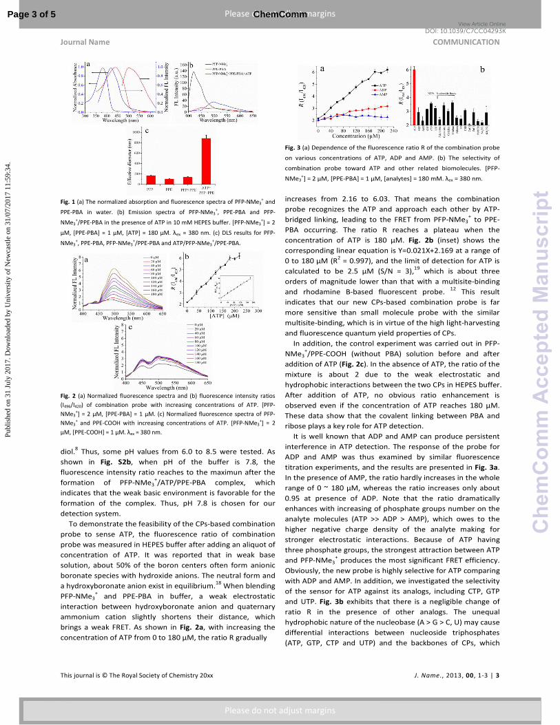
Fig. 1 (a) The normalized absorption and fluorescence spectra of PFP-NMe3+ and
PPE-PBA in water. (b) Emission spectra of PFP-NMe3+, PPE-PBA and PFP-
NMe3+/PPE-PBA in the presence of ATP in 10 mM HEPES buffer. [PFP-NMe3
+] = 2
μM, [PPE-PBA] = 1 μM, [ATP] = 180 μM. λex = 380 nm. (c) DLS results for PFP-
NMe3+, PPE-PBA, PFP-NMe3
+/PPE-PBA and ATP/PFP-NMe3
+/PPE-PBA.
Fig. 2 (a) Normalized fluorescence spectra and (b) fluorescence intensity ratios
(I496/I420) of combination probe with increasing concentrations of ATP. [PFP-
NMe3+] = 2 μM, [PPE-PBA] = 1 μM. (c) Normalized fluorescence spectra of PFP-
NMe3+ and PPE-COOH with increasing concentrations of ATP. [PFP-NMe3
+] = 2
μM, [PPE-COOH] = 1 μM. λex = 380 nm.
diol.8 Thus, some pH values from 6.0 to 8.5 were tested. As
shown in Fig. S2b, when pH of the buffer is 7.8, the
fluorescence intensity ratio reaches to the maximun after the
formation of PFP-NMe3+/ATP/PPE-PBA complex, which
indicates that the weak basic environment is favorable for the
formation of the complex. Thus, pH 7.8 is chosen for our
detection system.
To demonstrate the feasibility of the CPs-based combination
probe to sense ATP, the fluorescence ratio of combination
probe was measured in HEPES buffer after adding an aliquot of
concentration of ATP. It was reported that in weak base
solution, about 50% of the boron centers often form anionic
boronate species with hydroxide anions. The neutral form and
a hydroxyboronate anion exist in equilibrium.18
When blending
PFP-NMe3+ and PPE-PBA in buffer, a weak electrostatic
interaction between hydroxyboronate anion and quaternary
ammonium cation slightly shortens their distance, which
brings a weak FRET. As shown in Fig. 2a, with increasing the
concentration of ATP from 0 to 180 μM, the ratio R gradually
Fig. 3 (a) Dependence of the fluorescence ratio R of the combination probe
on various concentrations of ATP, ADP and AMP. (b) The selectivity of
combination probe toward ATP and other related biomolecules. [PFP-
NMe3+] = 2 μM, [PPE-PBA] = 1 μM, [analytes] = 180 mΜ. λex = 380 nm.
increases from 2.16 to 6.03. That means the combination
probe recognizes the ATP and approach each other by ATP-
bridged linking, leading to the FRET from PFP-NMe3+ to PPE-
PBA occurring. The ratio R reaches a plateau when the
concentration of ATP is 180 μM. Fig. 2b (inset) shows the
corresponding linear equation is Y=0.021X+2.169 at a range of
0 to 180 μM (R2 = 0.997), and the limit of detection for ATP is
calculated to be 2.5 μM (S/N = 3),19
which is about three
orders of magnitude lower than that with a multisite-binding
and rhodamine B-based fluorescent probe. 12
This result
indicates that our new CPs-based combination probe is far
more sensitive than small molecule probe with the similar
multisite-binding, which is in virtue of the high light-harvesting
and fluorescence quantum yield properties of CPs.
In addition, the control experiment was carried out in PFP-
NMe3+/PPE-COOH (without PBA) solution before and after
addition of ATP (Fig. 2c). In the absence of ATP, the ratio of the
mixture is about 2 due to the weak electrostatic and
hydrophobic interactions between the two CPs in HEPES buffer.
After addition of ATP, no obvious ratio enhancement is
observed even if the concentration of ATP reaches 180 μM.
These data show that the covalent linking between PBA and
ribose plays a key role for ATP detection.
It is well known that ADP and AMP can produce persistent
interference in ATP detection. The response of the probe for
ADP and AMP was thus examined by similar fluorescence
titration experiments, and the results are presented in Fig. 3a.
In the presence of AMP, the ratio hardly increases in the whole
range of 0 ~ 180 μM, whereas the ratio increases only about
0.95 at presence of ADP. Note that the ratio dramatically
enhances with increasing of phosphate groups number on the
analyte molecules (ATP >> ADP > AMP), which owes to the
higher negative charge density of the analyte making for
stronger electrostatic interactions. Because of ATP having
three phosphate groups, the strongest attraction between ATP
and PFP-NMe3+ produces the most significant FRET efficiency.
Obviously, the new probe is highly selective for ATP comparing
with ADP and AMP. In addition, we investigated the selectivity
of the sensor for ATP against its analogs, including CTP, GTP
and UTP. Fig. 3b exhibits that there is a negligible change of
ratio R in the presence of other analogs. The unequal
hydrophobic nature of the nucleobase (A > G > C, U) may cause
differential interactions between nucleoside triphosphates
(ATP, GTP, CTP and UTP) and the backbones of CPs, which
Page 3 of 5 ChemComm
Che
mC
omm
Acc
epte
dM
anus
crip
t
Publ
ishe
d on
31
July
201
7. D
ownl
oade
d by
Uni
vers
ity o
f N
ewca
stle
on
31/0
7/20
17 1
1:59
:34.
View Article OnlineDOI: 10.1039/C7CC04293K

COMMUNICATION Journal Name
4 | J. Name., 2012, 00, 1-3 This journal is © The Royal Society of Chemistry 20xx
Please do not adjust margins
Please do not adjust margins
makes for the best selectivity for ATP.20
Besides, the selective
recognition of the probe to ATP may also be improved by a
more effective π-π stacking interaction between the
backbones of CP and adenine due to matched spatial
orientations.3 We also investigated the response of the new
probe for nucleoside bases, such as adenosine, guanosine,
cytidine and uridine. The ratio only increases a little no matter
adding A, G, C or U to the mixture. In addition, PPi, ribose,
various anions and bio-related metal ions were also chosen to
examine the selectivity of the new probe. No obvious
enhancement of ratio R is observed no matter whichever is
present. Furthermore, we studied the interference from
saccharides 21
, such as glucose, fructose and mannose (Fig. S3).
These saccharides do not make obvious interference on ATP
detection. These results demonstrate that the novel approach
exhibits excellent selectivity for ATP detection. Importantly,
both the electrostatic interactions and covalent binding play a
significant and synergetic role in specifically sensing ATP. The
two are independent, which illustrates the high selectivity of
the new probe.
Finally, we compared the performance of our new method
with other methods reported recently in literatures. As shown
in Table S2, although most of the reported ways showed lower
LOD than that of our approach, they presented limited
selectivity because they cannot distinguish NPPs and/or
nucleotide bases. Taking advantage of the multisite-binding
strategy, our CPs-based combination probe greatly improves
the selectivity of ATP sensor. Although the multi-site sensors
reported by Chang’ group and Li’ group presented a good
selectivity, they are less sensitive.3, 8
Therefore, our new
method demonstrates an obvious advantage for ATP detection
with high selectivity and good sensitivity.
In summary, we have designed a novel CPs-based
combination probe for ATP detection based on multisite-
binding and FRET mechanism. The cationic conjugated polymer
PFP-NMe3+ functions as the energy donor while a PBA and PEG
modified conjugated polymer PPE-PBA acts as the energy
acceptor. When ATP was introduced, strong electrostatic
attraction and covalent binding between the CPs and ATP
functioned cooperatively to narrow the distance between two
CPs, which brings the strong FRET from donor to acceptor. The
significant enhancement of fluorescence ratio can be used to
quantify the concentration of ATP. Our new method
demonstrated a sensitive and highly selective detection for
ATP by taking advantage of high light-harvesting and strong
fluorescence properties of CPs and CPs-based FRET. This study
should provide a novel strategy for designing composite CPs-
based ratiometric probes to detect chemical and biological
analytes via multisite interactions.
This work is financially supported from the National Natural
Science Foundation of China (Grants 21675106), Natural
Science Basic Research Plan in Shaanxi Province of China
(Grant 2017JM2019), the 111 Project (B14041).
Notes and references
1 P. P. Neelakandan, M. Hariharan and D. Ramaiah, J. Am. Chem. Soc.,
2006, 128, 11334-11335.
2 (a) T. Tsuyama, J. I. Kishikawa, Y. W. Han, Y. Harada, A. Tsubouchi, H.
Noji, A. Kakizuka, K. Yokoyama, T. Uemura and H. Imamura, Anal. Chem.,
2013, 85, 7889-7896. (b) C. H. Ma, C. S. Lin, Y. R. Wang and X. Chen, Trac-
Trends Anal. Chem., 2016, 77, 226-241.
3 K. Y. Tan, C. Y. Li, Y. F. Li, J. J. Fei, B. Yang, Y. J. Fu and F. Li, Anal. Chem.,
2017, 89, 1749-1756.
4 C. Lin, Z. Cai, Y. Wang, Z. Zhu, C. J. Yang and X. Chen, Anal. Chem., 2014,
86, 6758-6762.
5 (a) I. S. Kucherenko, D. Y. Didukh, O. O. Sodatkin and A. P. Soldatkin,
Anal. Chem., 2014, 86, 5455-5462. (b) M. Li, J. Zhang, S. Suri, L. J. Sooter,
D. Ma and N. Wu, Anal. Chem., 2012, 84, 2837-2842. (c) E. Gout, F.
Rebeille, R. Douce and R. Bligny, Proc. Natl. Acad. Sci. U.S.A., 2014, 111,
E4560-4567. (d) D. A. Middleton, E. Hughes and M. Esmann, Angew.
Chem., Int. Ed., 2011, 50, 7041-7044. (e) X. Liu, F. Wang, R. Aizen, O.
Yehezkeli and I. Willner, J. Am. Chem. Soc., 2013, 135, 11832-11839. (f) X.
Chen, X. Tian, I. Shin and J. Yoon, Chem. Soc. Rev., 2011, 40, 4783-4804.
6 W. D. McElroy, Proc. Natl. Acad. Sci. U.S.A., 1947, 33, 342-345.
7 (a) A. Ojida, I. Takashima, T. Kohira, H. Nonaka and I. Hamachi, J. Am.
Chem. Soc., 2008, 130, 12095-12101. (b) Z. Xu, N. J. Singh, J. Lim, J. Pan,
H. N. Kim, S. Park, K. S. Kim and J. Yoon, J. Am. Chem. Soc., 2009, 131,
15528-15533. (c) A. J. Moro, P. J. Cywinski, S. Korsten and G. J. Mohr,
Chem. Commun., 2010, 46, 1085-1087. (d) A. S. Rao, D. Kim, H. Nam, H.
Jo, K. H. Kim, C. Ban and K. H. Ahn, Chem. Commun., 2012, 48, 3206-
3208. (e) H. Zhang, X. Ma, K. T. Nguyen and Y. Zhao, ACS Nano, 2013, 7,
7853-7863. (f) X. Li, X. Guo, L. Cao, Z. Xun, S. Wang, S. Li, Y. Li and G.
Yang, Angew. Chem., Int. Ed., 2014, 53, 7809-7813. (g) Z. Chen, P. Wu, R.
Cong, N. Xu, Y. Tan, C. Tan and Y. Jiang, ACS Appl. Mater. Interfaces,
2016, 8, 3567-3574.
8 L. Wang, L. Yuan, X. Zeng, J. Peng, Y. Ni, J. C. Er, W. Xu, B. K. Agrawalla, D.
Su, B. Kim and Y. T. Chang, Angew. Chem., Int. Ed., 2016, 55, 1773-1776.
9 H. Z. He, V. P. Ma, K. H. Leung, D. S. Chan, H. Yang, Z. Cheng, C. H. Leung
and D. L. Ma, Analyst, 2012, 137, 1538-1540.
10 Y. Wei, Y. Chen, H. Li, S. Shuang, C. Dong and G. Wang, Biosens.
Bioelectron., 2015, 63, 311-316.
11 M. Zhao, L. Liao, M. Wu, Y. Lin, X. Xiao and C. Nie, Biosens. Bioelectron.,
2012, 34, 106-111.
12 L. Wang, L. Yuan, X. Zeng, J. J. Peng, Y. Ni, J. C. Er, W. Xu, B. K. Agrawalla,
D. D. Su, B. Kim and Y. T. Chang, Angew. Chem., Int. Ed., 2016, 55, 1773-
1776.
13 (a) C. Zhu, L. Liu, Q. Yang, F. Lv and S. Wang, Chem. Rev., 2012, 112, 4687-
4735. (b) L. Feng, C. Zhu, H. Yuan, L. Liu, F. Lv and S. Wang, Chem. Soc.
Rev., 2013, 42, 6620-6633. (c) Q. L. Cui, Y. Yang, C. Yao, R. H. Liu and L. D.
Li, ACS Appl. Mater. Interfaces, 2016, 8, 35578-35586.
14 (a) B. Liu, B. S. Gaylord, S. Wang and G. C. Bazan, J. Am. Chem. Soc., 2003,
125, 6705-6714. (b) T. Hong, T. Wang, P. Guo, X. Xing, F. Ding, Y. Chen, J.
Wu, J. Ma, F. Wu and X. Zhou, Anal. Chem., 2013, 85, 10797-10802. (c) L.
R. Swem, D. L. Swem, C. T. O'Loughlin, R. Gatmaitan, B. X. Zhao, S. M.
Ulrich and B. L. Bassler, Mol. Cell, 2009, 35, 143-153. (d) C. Li, M. Numata,
M. Takeuchi and S. Shinkai, Angew. Chem., Int. Ed., 2005, 44, 6371-6374.
(e) D. Cheng, Y. Li, J. Wang, Y. Sun, L. Jin, C. Li and Y. Lu, Chem. Commun.,
2015, 51, 8544-8546.
15 J. K. Lee, Y. H. Jung, J. B. Tok and Z. Bao, ACS Nano, 2011, 5, 2067-2074.
16 (a) H. Morawetz, Science, 1988, 240, 172-176. (b) N. Q. An, Q. Zhang, J.
Wang, C. Liu, L. Q. Shi, L. H. Liu, L. D. Deng and Y. Lu, Polym. Chem., 2017,
8, 1138-1145. (c) G. H. Aryal, L. M. Huang and K. W. Hunter, RSC Adv.,
2016, 6, 76448-76452.
17 Q. Cui, X. Wang, Y. Yang, S. Li, L. Li, and S. Wang, Chem. Mater., 2016, 28,
4661–4669.
18 W. L. Brooks and B. S. Sumerlin, Chem. Rev., 2016, 116, 1375-1397.
19 Y. Wu, F. Xiao, Z. Wu and R. Yu, Anal. Chem., 2017, 89, 2852-2858.
20 D. Maity, M. Li, M. Ehlers and C. Schmuck, Chem. Commun., 2016, 53,
208-211.
21 (a) J. Li, L. L. Liu, P. G. Wang, Y. Yang and J. B. Zheng, Sensor. Actuat. B-
Chem., 2014, 198, 219-224. (b) Y. Egawa, T. Seki, S. Takahashi and J. Anzai,
Mat. Sci. Eng. C-Mater., 2011, 31, 1257–1264. (c) S. Takahashi and J.
Anzai, Langmuir, 2005, 21, 5102-5107.
Page 4 of 5ChemComm
Che
mC
omm
Acc
epte
dM
anus
crip
t
Publ
ishe
d on
31
July
201
7. D
ownl
oade
d by
Uni
vers
ity o
f N
ewca
stle
on
31/0
7/20
17 1
1:59
:34.
View Article OnlineDOI: 10.1039/C7CC04293K

Journal Name COMMUNICATION
This journal is © The Royal Society of Chemistry 20xx J. Name., 2013, 00, 1-3 | 5
Please do not adjust margins
Please do not adjust margins
Table of Contents
A new conjugated polymers -based ratiometric combination probe was constructed for adenosine triphosphate detection by taking advantage of multisite-binding and FRET strategy
Page 5 of 5 ChemComm
Che
mC
omm
Acc
epte
dM
anus
crip
t
Publ
ishe
d on
31
July
201
7. D
ownl
oade
d by
Uni
vers
ity o
f N
ewca
stle
on
31/0
7/20
17 1
1:59
:34.
View Article OnlineDOI: 10.1039/C7CC04293K

本文献由“学霸图书馆-文献云下载”收集自网络,仅供学习交流使用。
学霸图书馆(www.xuebalib.com)是一个“整合众多图书馆数据库资源,
提供一站式文献检索和下载服务”的24 小时在线不限IP
图书馆。
图书馆致力于便利、促进学习与科研,提供最强文献下载服务。
图书馆导航:
图书馆首页 文献云下载 图书馆入口 外文数据库大全 疑难文献辅助工具