video digitizer analysis of birefringence along...
TRANSCRIPT
J. Cell Sci. 65, 41-60 (1984) 41Printed in Great Britain © The Company of Biologists Limited 1984
VIDEO DIGITIZER ANALYSIS OF BIREFRINGENCEALONG THE LENGTHS OF SINGLE CHROMOSOMALSPINDLE FIBRES
II. CRANE-FLY SPERMATOCYTE CHROMOSOMAL SPINDLEFIBRES ARE NOT TEMPERATURE-LABILE
CATHERINE J. SCHAAP AND ARTHUR FORERBiology Department, York University, 4700 Keele Street, Doumsview, Ontario,Canada M3J IP3
SUMMARY
Retardations were measured along the lengths of single chromosomal spindle fibres, frommetaphase through anaphase, from video-taped images of crane-fly spermatocytes incubated atvarious temperatures (4— 30 °C). These measurements were made using a video digitizer interfacedto a microcomputer. Over most of the range of temperatures at which normal anaphase movementoccurs the chromosomal spindle fibres are not temperature-labile. The non-specific and continuousfibre birefringence is temperature-labile, however. The data are discussed with respect to the'dynamic equilibrium' model of anaphase chromosome movement. We conclude that, since singlechromosomal fibre birefringence is not temperature-labile over most of the range of temperaturesat which normal anaphase chromosome movement occurs, these data do not support the dynamicequilibrium model of anaphase chromosome movement.
INTRODUCTION
The 'dynamic equilibrium' model of anaphase chromosome movement was basedon measurements of spindle birefringence (retardation) in metaphase-arrestedoocytes of Chaetopteruspergamentaceons (Inou6, 1952a,6, 1959, 1964). As the tem-perature was changed the spindle retardation changed to an 'equilibrium retardation'specific for that temperature. The idea that there is a temperature-sensitive dynamicequilibrium that governs the concentration of oriented material in spindle fibres(Inoue', 1959, 1964) is based on the assumption that retardation reflects the concentra-tion of oriented material. These retardation data were criticized as applying only tometaphase-arrested spindles (Forer, 1969), but subsequent data have shown thatactive (non-arrested) metaphase spindles are also temperature-sensitive and thus thatthe concept of dynamic equilibrium is generally applicable (e.g. see Stephens, 1973;Fuseler, 1973, 19756; Salmon, 1975a,6).
The dynamic equilibrium concept was developed into a 'depolymerization' modelfor anaphase chromosome movement (Inoue, 1964, 1976, 1981, 1982; Inou6 & Sato,1967; Inoue' & Ritter, 1975). The model proposes that since the polymeric com-ponents of spindle fibres are in dynamic equilibrium with monomers, chromosomemovements occur by shifting the equilibrium so that the polymers depolymerize. Iffibres depolymerize along their lengths (Inou6, 1964), at the kinetochores (Gruzdev,

42 C. J. Schaap and A. Forer
1972) or at the poles (Inou6 & Ritter, 1975), while remaining attached to thekinetochore, polymer depolymerization could pull chromosomes poleward. Thisdepolymerization model for force production is based on the effects of temperatureon the dynamic equilibrium. By assuming that the measured retardation is propor-tional to the concentration of oriented polymer (microtubules), the entropy, enthalpyand free-energy changes for the temperature-sensitive polymerization reaction werecalculated from data on spindle birefringence versus temperature (e.g. see Inoue",Fuseler, Salmon & Ellis, 1975; Inou<§ & Ritter, 1975). The calculated values for thechange in free energy were used to calculate the force that could be derived from thedepolymerization of microtubules (Inou6 & Ritter, 1975). These calculations sugges-ted that the depolymerization reaction could indeed produce enough force to movechromosomes.
There are several methodological problems involved in using the data in this way,however. The measurements were only semi-quantitative; they were made visuallyand at one spot in the spindle. That one spot is not homogeneous: it consists of bothchromosomal spindle fibres (force transmitters for chromosome-to-pole movement;e.g. see Nicklas, 1975; Begg & Ellis, 1979) and continuous spindle fibres. In additionthe data used for calculating the thermodynamic parameters do not necessarily reflectthe concentrations of the oriented material, since the measurements did not takespindle diameter changes into account (discussed by Forer, 1976). We have overcomethese problems by using cells in which single chromosomal spindle fibres can bestudied. Images of these cells were recorded on video tape and objective values ofbirefringence were extracted from along the lengths of individual spindle fibres usinga video digitizer interfaced to a computer (described by Schaap & Forer, 1983). Inthis paper we show that individual chromosomal spindle fibres are not in atemperature-sensitive equilibrium over most of their physiological range of tem-perature: the birefringence along the lengths of chromosomal spindle fibres does notchange with temperature over this range. Continuous fibre birefringence, however,is sensitive to temperature. Thus the concept of a temperature-sensitive dynamicequilibrium does not apply to the force-transmitting chromosomal spindle fibres,except at very low temperatures where movement is either extremely slow or does notoccur.
MATERIALS AND METHODS
Spermatocyte cultures from laboratory-reared Nephrotoma suturalis (Loew) and Nephrotomaferruginea (Fabricius), crane flies, were prepared as described by Schaap & Forer (1979, 1983) andForer (1982). The experimental techniques are described in detail in the preceding paper (Schaap& Forer, 1983), but also are described briefly here. The temperature of the spermatocyte culturewas controlled by a temperature-control slide in which the coolant flowed directly under the cover-slip on which the cells were situated. Cells were video-taped at the experimental temperature, usingpolarizing optics. (The photographs in this paper were taken from 'stop frame' video images usinga 35 mm camera mounted on a tripod.) In most experiments suitable flat, late metaphase cells werefound and were then taken to the experimental temperature by allowing coolant to enter thetemperature-control slide for at least 5 min prior to the cell entering anaphase. For some experiments(the 'step-up' experiments), the cells were taken from room temperature (RT, which is 22-24°C)to about 5 °C (in one jump) and then gradually taken back to about 20 °C in a step-wise fashion (by
Temperature effects on spindle fibres 43
increasing the temperature of the coolant) over a period of 1-3 h. For the initial 'jump' down, coolantfrom the circulating water bath was allowed to enter the tubing and consequently the temperature-control slide. The cells were at about 10°C in l-2min and at about 6°C in about 5 min; however,the remaining drop to 4 or 5 °C sometimes took an additional 5 min. The cells were maintained atthe cold temperature for 10-30 min. The temperature was then increased in a step-wise fashion bychanging the setting on the water-bath thermostat by several degrees. The temperature at the slidewas monitored using a surface thermistor. Anaphase was usually in progress by the time the cellswere back to 20 °C.
The video tape recorder (VTR) that we use has an automatic gain circuit (AGC), the effects ofwhich we described previously (Schaap & Forer, 1983); as noted in the figure legends, some of thedata were taken with the AGC 'on' and some with the AGC 'off (discussed by Schaap & Forer,1983).
Some living spermatocytes were embedded in a fibrin clot (Forer, 1972), at room temperature,and fixed by the addition of 2% glutaraldehyde at room temperature prior to observation (detaileddescription of the technique will be given elsewhere). Subsequent temperature treatment wasidentical to that used for living cells.
Retardations along the lengths of single chromosomal spindle fibres were obtained at various timesfrom single video fields using the previously described video digitizer system (Schaap & Forer,1983). Single chromosomal spindle fibres in the video field were selected and scanned on severalsuccessive video fields. The intensity data were stored on floppy diskettes by the microcomputer.The computer subsequently used a calibration curve to convert the intensities to retardations. Thesystem and method of measuring are described in detail in the preceding paper (Schaap & Forer,1983).
RESULTS
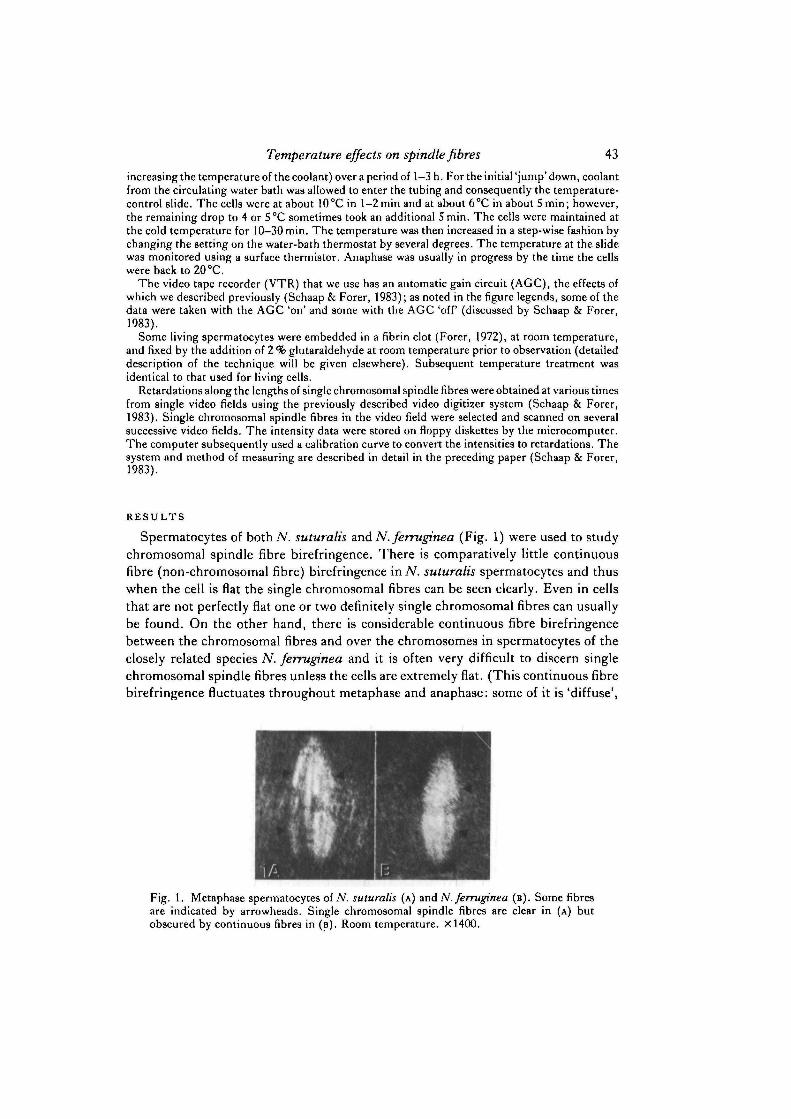
Spermatocytes of both N. suturalis and N.ferruginea (Fig. 1) were used to studychromosomal spindle fibre birefringence. There is comparatively little continuousfibre (non-chromosomal fibre) birefringence in N. suturalis spermatocytes and thuswhen the cell is flat the single chromosomal fibres can be seen clearly. Even in cellsthat are not perfectly flat one or two definitely single chromosomal fibres can usuallybe found. On the other hand, there is considerable continuous fibre birefringencebetween the chromosomal fibres and over the chromosomes in spermatocytes of theclosely related species N. ferruginea and it is often very difficult to discern singlechromosomal spindle fibres unless the cells are extremely flat. (This continuous fibrebirefringence fluctuates throughout metaphase and anaphase: some of it is 'diffuse',
Fig. 1. Metaphase spermatocytes of A', suturalis (A) and N'. ferruginea (B). Some fibresare indicated by arrowheads. Single chromosomal spindle fibres are clear in (A) butobscured by continuous fibres in (B). Room temperature. X1400.

44 C. jf. Schaap and A. Forer
while some of it is more localized and 'fibrous'.) For this reason most of thechromosomal spindle fibre retardations that were quantified were from N. suturalisspermatocytes. The shapes of the retardation profiles (retardation versus position)were the same for chromosomal spindle fibres in both species.
The effect of temperature on the birefringence along the lengths of singlechromosomal fibres was studied over a temperature range from 4°C to 30 °C. Thephysiological range of temperatures at which divisions occur normally is about 6 °Cto 28°C for N. ferruginea and about 8°C to 30°C for/V. suturalis. (In N. ferrugineaspermatocytes there are abnormal divisions at 30 °C; below 6°C there is either nomovement, it is too slow to measure, or it is delayed beyond the limits of the ex-perimenter's patience. In N. suturalis spermatocytes anaphase movements at tem-peratures above 30 °C or below 8°C have not been studied. We expect that 30 °C isnear the upper limit for normal N. suturalis spermatocytes because the two species arevery similar and because the animal colonies do not survive when laboratory con-ditions are above 27 °C for extended periods (Schaap & Forer, 1979; unpublishedobservations).)
We first studied cells that were taken to the experimental temperature shortlybefore anaphase and kept at that temperature from metaphase through late anaphase.Over the physiological range of temperatures (at which normal movement occurs)there was very little (if any) effect of temperature on retardation: when averageretardations at the kinetochores of spindle fibres in both metaphase and anaphase ofTV. suturalis spermatocytes are plotted against temperature there is very little effectof temperature between 10 °C and 25 °C (Fig. 2). (To obtain the data given in Fig.2, chromosomal spindle fibre retardations were calculated at several times from latemetaphase through anaphase; the kinetochore retardations in this figure are averagekinetochore retardations of several repeat measurements at the same time and arerepresentative of the retardations at that stage. High or low retardations resultingfrom jumps in birefringence (Schaap & Forer, 1983) were not used for theseaverages.) It is relevant to point out that the effects of temperature on the birefrin-gence at the kinetochore reflect effects of temperature on the birefringence of the restof the fibre (Schaap & Forer, 1983). Similar data forN. ferruginea spermatocytes andsome second meiotic divisions are given in Table 1. Though chromosomal fibrebirefringence does not change much between 10 °C and 25 °C, it does change outsidethis range (Fig. 2).
Whereas there seems to be little change in chromosomal spindle fibre birefringence
Fig. 2. Effect of temperature on the retardation at the kinetochore (N. suturalis) duringlate metaphase (A) and early anaphase (B). A representative kinetochore retardation valuewas taken at each of these times and averaged with those from other fibres. The averagesand their standard deviations (vertical bars) are graphed. The number below the error barsrepresents the number of fibres used in the average while the number in parenthesisrepresents the number of cells. Data were grouped together so that the temperatures are±1 deg. C. These cells were subjected to a constant temperature after the shift from thenormal environmental temperature (22-24°C). (A) and ( • ) were calculated from datataken with the AGC on or off, respectively.

Temperature effects on spindle fibres 45
o
•521
5 bo
s £
aE0
in
I 4 <£ in
-J 1 1 1 1 1 i I I I
(UJU) uoj

46 C. J. Schaap and A. Forer
Table 1. Kinetochore birefringences
Temp. Retardation ( ± S . D . ) No. of No. of(°C) (nm) fibres cells
MetaphaseN. ferrugittea
Early anaphaseN. ferruginea
N. suturalis
10RTRT
10RTRT15
Ml(±0-25)l-38(±0-20)l-33(±0-26)*
l-27(±0-21)l-55(±0-22)l-33(±0-29)»l-43(±0-23)*t
162825
1914248
364
4341
The retardations are given as average kinetochore retardations (and standard deviations) of thenumber of fibres measured. These data were not included in Fig. 2. All the data were recorded withthe ACC 'off except for those marked | . The average retardations marked • are from second meioticdivision cells and includes data from both sex-chromosomal and autosomal spindle fibres as the twotypes of chromosomes could not be distinguished between.
with temperature, within most of the physiological range of temperatures, it is dif-ficult to decide from the data (Fig. 2) whether there might indeed be a small changein birefringence with temperature: there are large spreads in the values of birefrin-gence for fibres at any given temperature, as indicated by the large standard devia-tions. The comparisons are further complicated by the fact that the birefringence ofan individual fibre can vary by as much as 25 or 30 % during metaphase and anaphase(Schaap & Forer, 1983) and by the fact that the data are derived from differentnumbers of fibres from different cells. Use of Student's /-test indicates that there ismost likely no effect of temperature, but the results are ambiguous. To decide howmuch the birefringence of individual chromosomal spindle fibres might vary withtemperature, we studied individual spindle fibres as the temperatures were changed.
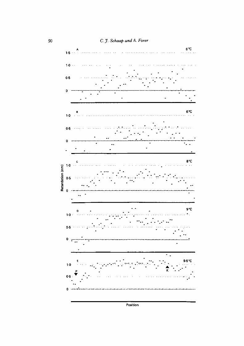
We studied individual ./V. suturalis and N. ferruginea metaphase spermatocytes thatwere rapidly taken from room temperature to 4°C or 5 °C; then the temperature wasincreased gradually ('step-up' experiments) to about 20°C (Figs 3, 4, 5). (Spindlefibre retardations do not change through metaphase and anaphase (Schaap & Forer,1983), and thus any one chromosomal spindle fibre should be of constant birefrin-gence except for the effects of temperature and random jumps.) Figs 6, 7, 8 illustrateretardations along the lengths of three fibres treated in step-up experiments. At 4°Cor 5°C the chromosomal spindle fibres were usually only barely visible (this wasvariable, however), so their retardation profiles were very noisy. The birefringencesof individual chromosomal spindle fibres recovered rapidly by 7 °C or 8 °C and wereback to room temperature levels by about 10 °C. Above 10 °C and up to at least about22 °C there was no further change in chromosomal spindle fibre birefringence in the18 N. suturalis or 20 N. ferruginea spermatocyte chromosomal spindle fibres (from4 cells of each species), although, as with cells held at a constant temperature (Schaap& Forer, 1983), fluctuations in birefringence sometimes occurred.

Temperature effects on spindle fibres 47
Fig. 3. A N. suturalis spermatocyte at 5°C (A), 10°C (B) and 12°C (c). X1400.
Fig. 4. A N. suturalis spermatocyte at 5°C (A), 10°C (B), 14°C (C) and 17-5°C (D) .X1400.
Fig. 5. A N.ferruginea spermatocyte at room temperature (A), 4-5°C (B), 7°C (C, D),1 0 - 5 ° C ( E ) . X1400.
Glutaraldehyde-fixed cells were studied to see if the effect of temperature onchromosomal spindle fibre birefringence was somehow an artefact of our analysisprocedure. Two cells were analysed in detail (11 chromosomal spindle fibres); thecells at room temperature were fixed with glutaraldehyde at room temperature and thefixed cells were then placed at different temperatures. In both cells chromosomalspindle fibre birefringence was about the same as that in living cells and birefringence

48 C. jf. Schaap and A. Forer
10 A ^ ^ v £ 4-5°C
0-5
1-0
co
11 0-5
0
0-5
1-5 10°C
19-5°C
1-0 . . . • • • .*.:
Position
Fig. 6. Step-up experiment retardation profiles of a chromosomal spindle fibre of a N.suturalis spermatocyte. The temperature causes the fibre to be shorter at low temperatures(compare 4- 5 °C and 10 °C). P and K in this and subsequent figures represent the pole andkinetochore regions, respectively. Each + in these and subsequent fibre profiles representsthe retardation data obtained from one pixel. Depending on the angle of the fibre on theTV monitor, there are five to nine pixels per [un (see Schaap & Forer, 1983a). A.Metaphase; B, very early anaphase; c, anaphase. AGC off.
did not change at all when individual fibres were studied at various temperaturesbetween 4 and 22 °C (Fig. 9). Thus our technique would seem to work, and effectsof temperature on chromosomal fibre birefringence in vivo reflect changes in spindlefibres and not artefacts of the technique.
The effects of temperature on interzonal birefringence were studied to see if thetemperature sensitivity of spindle birefringence observed by other workers (e.g. seeInoue\ 1952a, 1959, 1964; Fuseler, 1973, 19756; Stephens, 1973; Salmon, 1975a)
1-5
10
0-5
0-5
Temperature effects on spindle fibres 49
RT
6°CB -
0-5 •*• •• • • * . . . . — • • • • • • •
n°cc
1-0 . . •• • • •
Position
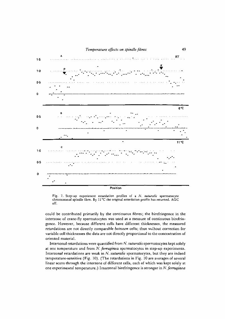
Fig. 7. Step-up experiment retardation profiles of a N. suturalis spermatocytechromosomal spindle fibre. By 11°C the original retardation profile has returned. AGCoff.
could be contributed primarily by the continuous fibres; the birefringence in theinterzone of crane-fly spermatocytes was used as a measure of continuous birefrin-gence. However, because different cells have different thicknesses, the measuredretardations are not directly comparable between cells; thus wilhout correction forvariable cell thicknesses the data are not directly proportional to the concentration oforiented material.
Interzonal retardations were quantified fromN. suturalis spermatocytes kept solelyat one temperature and from N. ferruginea spermatocytes in step-up experiments.Interzonal retardations are weak in N. suturalis spermatocytes, but they are indeedtemperature-sensitive (Fig. 10). (The retardations in Fig. 10 are averages of severallinear scans through the interzone of different cells, each of which was kept solely atone experimental temperature.) Interzonal birefringence is stronger in N. ferruginea
50 C. J. Schaap and A. Forer
A 6°C1-5
10
0-5
B 6°C1-0
0-5
0 —
c 8°C•^1-0
c
•s
IDC
D
vo
0-5
E • • * ••„ • % 9-5°C
P K • • • .
0-5
0
Position

Temperature effects on spindle fibres 51
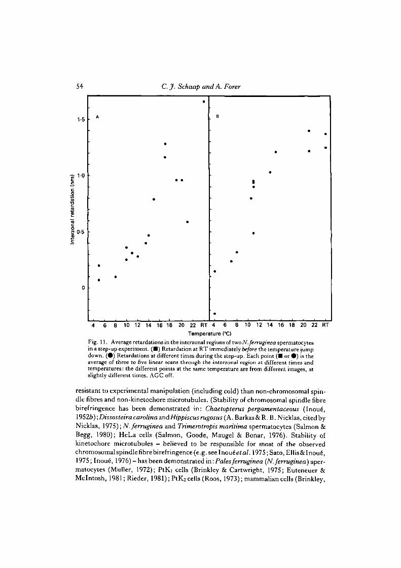
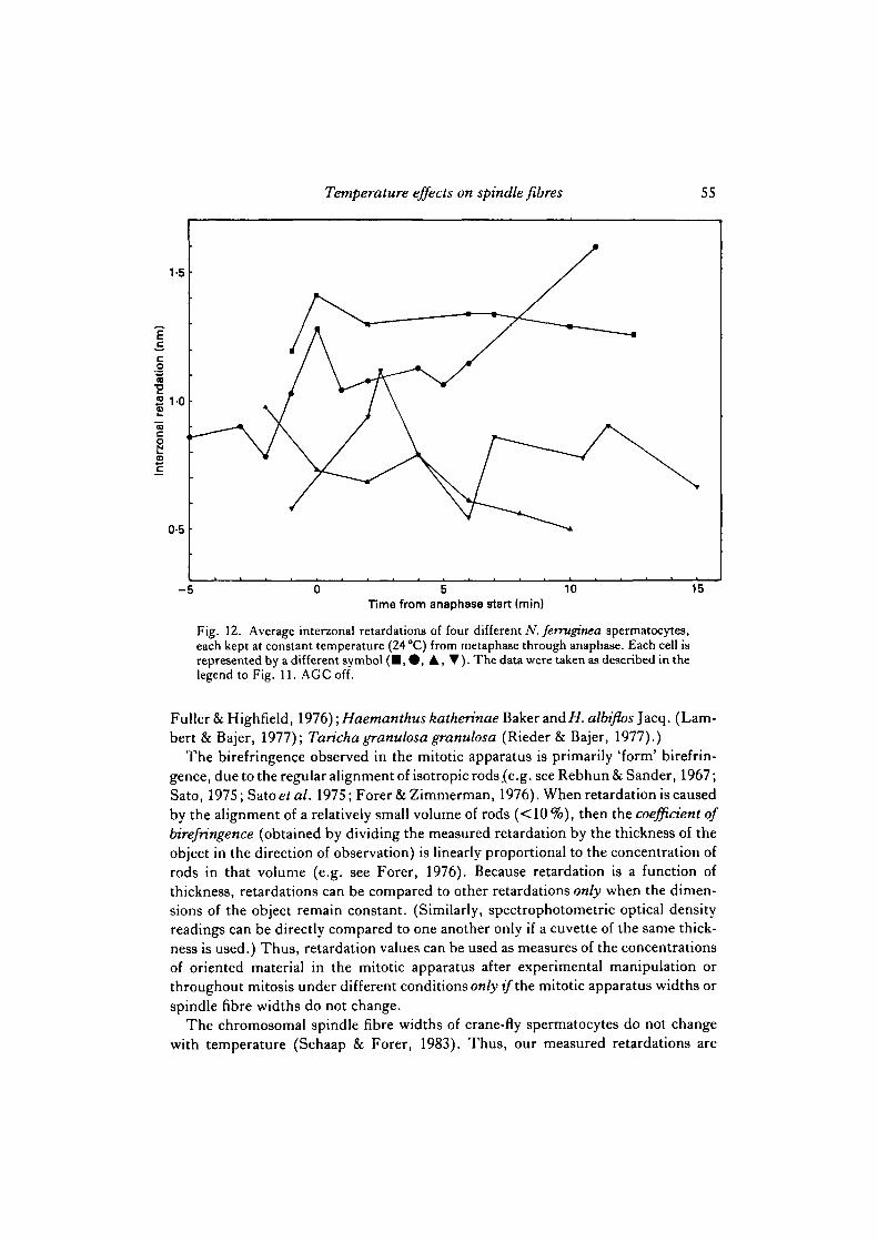
spermatocytes than in N. suturalis spermatocytes; thus temperature sensitivity ofnon-chromosomal fibre birefringence should be readily observable in N. ferrugineaspermatocytes. (Indeed metaphase cells with barely distinguishable chromosomalfibres at room temperature have single fibres at lower temperatures, when the non-chromosomal fibre birefringence is almost eliminated (Fig. 5), as also reported bySalmon & Begg (1980).) Data on interzonal retardations for two cells are given in Fig.11, from which it is concluded that interzonal birefringence is clearly temperature-dependent. Since any one cell maintains approximately the same width until at leastmid-anaphase, and the measurements are always made through the same part of thecell, interzonal retardations at different temperatures within the same cell are compar-able, although the exact relationship to concentration of oriented material (coefficientof birefringence) is unknown because the thickness of the cell is unknown. Progressionthrough metaphase and anaphase alone cannot account for the observed retardationchanges because, though there may be considerable fluctuations in birefringence inthe interzone during anaphase, there is no predictable increase or decrease in inter-zonal retardation at constant temperatures (Fig. 12). Thus, unlike chromosomalspindle fibres, continuous spindle fibres (interzonal fibres) are in a temperature-sensitive dynamic equilibrium throughout most of the physiological range of tem-peratures, in both species. Qualitative observations suggest that astral birefringencealso varies with temperature in the same way as interzonal birefringence (i.e., asterdiameter and birefringence decrease as the temperature decreases).
Spindle pole-to-pole distances were altered by temperature. In some of the step-upexperiments cells were filmed prior to the initial drop in temperature. In six out ofseven such cells the pole-to-pole distances decreased about 20 % during the initialtemperature drop. (These were five N. ferruginea and two N. suturalis sper-matocytes.) In the seventh cell (fromN. suturalis) the pole-to-pole distance decreasedby about 10 %. The spindles increased in length as temperature was stepped up andgenerally reached their original lengths by the time that the cells were at 10 °C. (Theincreases in retardation usually started before the increase in length.) Decreases inpole-to-pole distances due to large drops in temperature were noticed previously ingrasshopper spermatocytes (Barkas & Nicklas as cited by Nicklas, 1975) andmetaphase-arrested Chaetopterus oocytes (Inou6, 1952a), although they were notseen in crane-fly spermatocytes by Salmon & Begg (1980). In the crane-fly sper-matocytes we studied, the shortening of the pole-to-pole distances occurred when theretardations of the chromosomal spindle fibres became temperature-labile.
The retardation profiles of individual spindle fibres were studied during the step-up experiments to see if we could determine a 'growth end' for a chromosomalspindle fibre. That is, if the birefringent material is added primarily at one end ofa spindle fibre, as in current models of spindle microtubule organization (e.g. seeBergen & Borisy, 1980; Margolis, Wilson & Kiefer, 1978), then birefringence might
Fig. 8. Step-up experiment retardation profiles of a N. ferruginea spermatocytechromosomal spindle fibre showing the build up of retardation after cold treatment. AGCoff.


Temperature effects on spindle fibres 53
20Temperature (°C)
Fig. 10. Average interzonal retardations of different N. suturalis spermatocytes duringearly anaphase. Each point represents one cell kept at that temperature throughoutanaphase. AGC on.
be expected to grow from one end. We were unable to detect such local changes inbirefringence, but rather we saw only increases along the entire length of thechromosomal spindle fibres (e.g. see Fig. 8). Perhaps such local changes could bedetected with better time resolution (e.g., using the video tape recorder at 60frames/s instead of the 1 frame/s we used) or by using a TV camera that would giveless electronic noise in the recorded image.
DISCUSSION
Inou6's pioneering work with the polarizing microscope established without doubtthe existence of fibrous elements in the mitotic apparatus of living cells (Inou6,1952a,6). These fibres can be visualized because they are composed of orientedmaterial and thus are birefringent.
Spindle birefringence was shown to be labile to low temperatures (e.g. see Inoue\1952a, 1959, 1964). In this paper we have shown that over most of the temperaturerange at which normal movement occurs chromosomal spindle fibres are not labile tolowered temperatures, but that the continuous (interzonal) fibres are. Others have alsoshown that chromosomal spindle fibres and kinetochore microtubules tend to be more
Fig. 9. Retardation profiles of a glutaraldehyde-fixed N. suturalis spermatocytechromosomal spindle fibre at different temperatures. The small variations are probablydue to the scans being of slightly different parts of the spindle fibre (see Schaap & Forer,1983). AGC off.
54
1-5
| 1 0
coco•2
reta
mc§0-5o1:
0
A
•
*
C. J. Schaap and A. Forer
m
•
•
B
•
i*
•
•
4 6 8 10 12 14 16 18 20 22 RT 4 6 8 10 12 14 16 18 20 22 RT
Temperature (°C)
Fig. 11. Average retardations in the interzonal regions of two N.ferruginea spermatocytesin a step-up experiment. (•) Retardation at RT immediately before the temperature jumpdown. (•) Retardations at different times during the step-up. Each point ( • or • ) is theaverage of three to five linear scans through the interzonal region at different times andtemperatures: the different points at the same temperature are from different images, atslightly different times. AGC off.
resistant to experimental manipulation (including cold) than non-chromosomal spin-dle fibres and non-kinetochore microtubules. (Stability of chromosomal spindle fibrebirefringence has been demonstrated in: Chaetopterus pergamentaceous (Inou6,19526) ;DissosteiraCarolina andHippiscus rugosus (A. Barkas & R. B. Nicklas, cited byNicklas, 1975); N.ferruginea and Trimerotropis maritima spermatocytes (Salmon &Begg, 1980); HeLa cells (Salmon, Goode, Maugel & Bonar, 1976). Stability ofkinetochore microtubules — believed to be responsible for most of the observedchromosomalspindlefibrebirefringence(e.g.seelnou6e/a/. 1975; Sato, Ellis&Inoue",1975; Inoue", 1976) - has been demonstrated in: Pales ferruginea (N.ferruginea) sper-matocytes (Muller, 1972); PtKi cells (Brinkley & Cartwright, 1975; Euteneuer &Mclntosh, 1981; Rieder, 1981); PtK2 cells (Roos, 1973); mammalian cells (Brinkley,
Temperature effects on spindle fibres 55
Time from anaphase start (min)
Fig. 12. Average interzonal retardations of four different N. ferruginea spermatocytes,each kept at constant temperature (24 °C) from metaphase through anaphase. Each cell isrepresented by a different symbol ( • , • , A , • ) . The data were taken as described in thelegend to Fig. 11. AGC off.
Fuller & Highfield, 1976); Haemanthus katherinae Baker and//, albiflos Jacq. (Lam-bert & Bajer, 1977); Taricha granulosa granulosa (Rieder & Bajer, 1977).)
The birefringence observed in the mitotic apparatus is primarily 'form' birefrin-gence, due to the regular alignment of isotropic rods (e.g. see Rebhun & Sander, 1967;Sato, 1975; Sato et al. 1975; Forer & Zimmerman, 1976). When retardation is causedby the alignment of a relatively small volume of rods (<10 %), then the coefficient ofbirefringence (obtained by dividing the measured retardation by the thickness of theobject in the direction of observation) is linearly proportional to the concentration ofrods in that volume (e.g. see Forer, 1976). Because retardation is a function ofthickness, retardations can be compared to other retardations only when the dimen-sions of the object remain constant. (Similarly, spectrophotometric optical densityreadings can be directly compared to one another only if a cuvette of the same thick-ness is used.) Thus, retardation values can be used as measures of the concentrationsof oriented material in the mitotic apparatus after experimental manipulation orthroughout mitosis under different conditions only if the mitotic apparatus widths orspindle fibre widths do not change.
The chromosomal spindle fibre widths of crane-fly spermatocytes do not changewith temperature (Schaap & Forer, 1983). Thus, our measured retardations are
56 C. jf. Schaap and A. Forer
proportional to coefficients of birefringence and to concentrations of orientedmaterial. Since chromosomal spindle fibre retardation is not affected by temperatureover most of the temperature range at which normal chromosome movement canoccur (i.e. between 10 °C and 25 °C), then there is no change in the concentration oforiented material in the spindle fibres over this range. Thus the oriented material inthe chromosomal spindle fibres is not in a temperature-sensitive dynamic equilibriumover most of the physiological temperature range. Since chromosomal spindle fibrestransmit the force for chromosome-to-pole movement (e.g. see Nicklas, 1975; Begg& Ellis, 1979), and since the data on 'spindle fibre' lability do not apply tochromosomal spindle fibres, there is no experimental basis for the interpretation thatchromosomal spindle fibre organization is regulated by a temperature-sensitivedynamic equilibrium or that chromosomal spindle fibre depolymerization causesanaphase chromosome-to-pole movement. In contrast to the chromosomal spindlefibres, the continuous spindle fibre birefringence (as measured in the interzone) istemperature-sensitive. (Salmon & Begg (1980) also found that N. ferruginea sper-matocyte continuous fibres were temperature-sensitive whereas chromosomal spindlefibres were stable at low temperatures.) We suggest, therefore, that the results ofprevious workers (e.g. see Inou6, 1952a, 1959, 1964; Fuseler, 1973, 19756; Stephens,1973; Salmon, 1975a) on the lability to decreased temperature of overall spindlebirefringence pertains primarily to continuous fibres and not to chromosomal spindlefibres. Thus although the dynamic equilibrium model could apply to continuousspindle fibres, and perhaps also to spindle elongation and shortening, it cannot applyto the chromosome-to-pole movement that is due to the temperature-insensitivechromosomal spindle fibres. This suggestion can be tested in the following way.
Data from the temperature sensitivity of spindle birefringence have been used tocalculate the change in free energy, entropy and enthalpy associated with spindlepolymerization (e.g. see Inou6 et al. 1975). If, as we argue, this is primarily due tothe polymerization of continuous fibres, then calculations of the same parametersfrom our interzonal birefringence data should give values similar to those reported forwhole spindles. The thermodynamic parameters were calculated by Inou6 and co-workers on the assumption that the birefringence values are direct measures of theconcentration of oriented microtubules and that monomer subunits are in anequilibrium with the polymers so that:
In this equation A) is the maximum concentration of orientable polymer (maximumretardation), B is the actual concentration of oriented polymer (measured retarda-tion), andAo—B is the remaining monomer (e.g. see Inoue\ 1959, 1964; Inou6& Sato,1967; Stephens, 1973; Fuseler, 1973, 19756; Salmon, 1975a,b). Average spindleretardations are used as measures of B, while the assymptote of the retardation versustemperature curve is used to estimate A>. Van't Hoff plots (of log (B/(A0—B)) versus1/K) and the van't Hoff equation are used to calculate the changes in entropy,enthalpy and free energy during the polymerization reaction; the basic procedure forthe calculations was outlined by Inoue" et al. (1975, p. 735). For our calculations we

Temperature effects on spindle fibres 57
Table 2. Thermodyanamic parameters calculated from measured retardations
Fibre type and AH to AS ACspecies (kcal/mol) (°C) (e-u.) (kcal/mol)
ContinuousN.ferruginea 33-7 17-7 1161 -0-9 Data Fig. 11A
44'2 10-8 155-6 -2-2 Data Fig. 11BN.suturalis 26-6 190 91-0 -0-6 Data Fig. 10
ChromosomalN. suturalis* 99 6 354 —6-5 Representative data from
step-up experiments
AW is the enthalpy change in kilocalories; U> is the temperature at which AG = 0; AS is theentropy change in entropy units; A C is the free energy change under standard temperature andpressure conditions.
• T h e following retardations were used in the calculations: A>, 1-1 nm; 10°C, l-0nm; 5°C,0-3 nm.
used the interzonal birefringence versus temperature data of the two N.ferruginea cellsgiven in Fig. 11, and we used the pooled data iromN. suturalis cells given in Fig. 10.Because the assymptote of the increasing retardations was not clear, the maximumretardation was used to representj40 • The results of our calculations are given in Table2, together with estimates of these parameters for the polymerization of a chromosomalfibre based on the temperature sensitivity of these fibres below 10 °C, as determined bystep-up experiments. (Since exact kinetochore positions were often difficult to deter-mine at the lower temperatures an approximate kinetochore retardation was used.)
The values we calculated for the various thermodynamic parameters can be com-pared with the values in the literature. Inoue" & Ritter (1975) reviewed these data andstated that the free energy change for the polymerization reaction is about —0-7kcal/mol (of subunit polymerized), that the reaction is endothermic (enthalpy changeis generally about 30—40 kcal/mol) and that the reaction is entropy driven (entropychange is about 100—200 e.u.). (Compilations of the previously calculated thermo-dynamic parameters have been published by Fuseler (1973), Stephens (1973) andSalmon (1975a).) The thermodynamic parameters we calculated for interzonalbirefringence are right within the ranges of the previous values but those we calculatedfor chromosomal spindle fibres below 10 °C are not. This supports our interpretationthat the previously published parameters pertain primarily to non-chromosomalspindle-fibre birefringence. Thus these parameters cannot be used to describe reac-tion equilibria for the oriented material of the chromosomal spindle fibres.
The data presented here emphasize that one cannot apply the retardation measure-ments of one area of the spindle to the dynamics of chromosomal spindle fibres.Birefringence measurements not specifically made on single chromosomal spindlefibres include both chromosomal and continuous fibre types and do not reflect thedifferential sensitivities of the fibres to experimental manipulation. From our data onthe different temperature sensitivities of chromosomal and non-chromosomal spindle
3 CEL65

58 C. J. Schaap and A. Forer
fibres, it seems clear that changes in average retardations cannot be used to reflectchanges in concentrations of oriented material in both fibre types. Thus birefringencemeasurements of one single spot in a non-homogeneous spindle can give an erroneouspicture even for the mixed population.
Other erroneous conclusions can be drawn from measurements of one spot in aspindle, even if the spot contains only one fibre type. We have shown (Schaap & Forer,1983) that there is no decay in the birefringence of a crane-fly spermatocytechromosomal spindle fibre during anaphase. We argued that the temperature-sensitive birefringence decay seen by Fuseler (1973, 1975a,6) was probably anartefact due to the fact that birefringence was measured a few ^m in front of thepoleward-moving chromosomes. The temperature sensitivity of the anaphasebirefringence decay is due to the fact that chromosome-to-pole velocity istemperature-sensitive (e.g. see Fuseler, 1973, 19756; Schaap & Forer, 1979) and thespot at which the birefringence is measured moves towards the pole faster at highertemperatures than at lower temperatures.
The birefringence of individual chromosomal spindle fibres does not change withtemperature over most of the physiological range of temperatures. Chromosome-to-pole velocity does indeed change with temperature, however: chromosome-to-polemovement is faster at higher temperatures than at lower temperatures (Schaap &Forer, 1979). Thus, since chromosomal fibre birefringence is constant throughoutanaphase and at different temperatures, there is no correlation between thetemperature-dependent chromosome-to-pole velocity and the temperature-independent chromosomal spindle fibre birefringence. The only measured birefrin-gence that changes with temperature in crane-fly spermatocytes is the interzonalbirefringence: compare Figs 10 and 11 (on interzonal birefringence) in this paper withfig. 5 (on chromosome-to-pole velocity) of Schaap & Forer (1979).
A final point concerns the identity of material giving rise to spindle birefringence.Chromosomal spindle fibre birefringence is sensitive to temperatures below 10 °C.Although chromosome movement was not detected below 6°C (Schaap & Forer,1979), there is still a 25 % or so 'remnant' of the original chromosomal spindle fibrebirefringence until at least 4°C is reached. Most of the spindle microtubules havedisappeared at this temperature (Schaap & Forer, unpublished). Areas of reducedbirefringence produced by ultraviolet microbeam irradiation of single chromosomalspindle fibres have 30—40% of the original birefringence (Sillers & Forer, 1983).'Residual' birefringence that was about 30—40 % of the original birefringence was seenin isolated sea-urchin mitotic apparatuses after the disappearance of the microtubules(Goldman & Rebhun, 1969; discussed by Forer, 1976). These residual birefringencevalues may reflect the presence of a spindle fibre component that contributes birefrin-gence but has temperature and ultraviolet sensitivities different from that of the bulkof the birefringent material, the microtubules.
We thank Fred E. D. B. Maskinen and L. Rotgans for assisting with the computing; R. J. Planckfor help with software modifications; and the following for financial assistance: Atkinson CharitableFoundation, J. P. Bickell Foundation, W. Garfield Weston Foundation and the Natural Sciencesand Engineering Research Council of Canada.
Temperature effects on spindle fibres 59
This work was submitted to the Faculty of Graduate Studies of York University by C.J.S. inpartial fulfilment of the requirements for the degree of Doctor of Philosophy.
REFERENCES
BEGG, D. A. & ELLIS, G. W. (1979). Micromanipulation studies of chromosome movement. II.Birefringent chromosomal fibers and the mechanical attachment of chromosomes to the spindle.J. Cell Biol. 82, 542-554.
BERGEN, L. G. & BORISY, G. G. (1980). Head-to-tail polymerization of microtubules in vitro.Electron microscope analysis of seeded assembly. J. Cell Biol. 84, 141-150.
BRINKLEY, B. R. & CARTWRIGHT, J. JR (1975). Cold-labile and cold-stable microtubules in themitotic spindle of mammalian cells. Ann. N.Y. Acad. Sci. 253, 428-439.
BRINKLEY, B. R., FULLER, G. M. &HIGHFIELD, D. P. (1976). Tubulin antibodies for microtubulesin dividing and non-dividing mammalian cells. In Cell Motility, book A: Motility, Muscle andNon-muscle Cells (ed. R. Goldman, T. Pollard & J. Rosenbaum), pp. 435-456. New York: ColdSpring Harbor Laboratory.
EUTENEUER, U. & MCINTOSH, J. R. (1981). Structural polarity of kinetochore microtubules inPtK, cells. J. Cell Biol. 89, 338-345.
FORER, A. (1969). Chromosome movements during cell division. In Handbook of Molecular Cytol-ogy (ed. A. Lima-de-Faria), pp. 553-601. Amsterdam: North-Holland.
FORER, A. (1972). A method for making preparations of living crane fly spermatocytes for studywith light microscopy followed by electron microscopy. Cytobiologie 6, 403-409.
FORER, A. (1976). Actin filaments and birefringent spindle fibers during chromosome movements.In Cell Motility, book C: Microtubules and Related Proteins (ed. R. Goldman, T. Pollard & J.Rosenbaum), pp. 1273-1293. New York: Cold Spring Harbor Laboratory.
FORER, A. (1982). Crane fly spermatocytes and spermatids: A system for studying cytoskeletalcomponents. In Methods in Cell Biology, vol. 25 (ed. L. Wilson), pp. 227-252. New York:Academic Press.
FORER, A. & ZIMMERMAN, A. M. (1976). Spindle birefringence of isolated mitotic apparatusanalysed by pressure treatment. J . Cell Sci. 20, 309-327.
FUSELER, J. W. (1973). The effect of temperature on chromosome movement and the assembly-disassembly process of birefringent spindle fibres in actively dividing plant and animal cells. PhDthesis, University of Pennsylvania.
FUSELER, J. W. (1975a). Mitosis in Tilia americana endosperm. J. Cell Biol. 64, 159-171.FUSELER, J. W. (19756). Temperature dependence of anaphase chromosome velocity and
microtubule depolymerization.^- Cell Biol. 67, 789-800.GOLDMAN, R. D. & REBHUN, L. I. (1969). The structure and some properties of the isolated
mitotic apparatus. J . Cell Sci. 4, 179-209.GRUZDEV, A. D. (1972). Critical review of some hypotheses concerning anaphase chromosome
movements (in Russian). Tsitologiya 14, 141-149 (English translation: NRC Technical transla-tion no. 1758, National Research Council of Canada, Ottawa).
INOUE, S. (1952a). Effects of temperature on the birefringence of the mitotic spindle. Biol. Bull.mar. biol. Lab., Woods Hole 103, 316.
INOUE, S. (19526). The effect of colchicine on the microscopic and submicroscopic structure of themitotic spindle. Expl Cell Res. (suppl.) 2, 305-318.
INOUE, S. (1959). Motility of cilia and the mechanism of mitosis. Rev. mod. Phys. 31, 402-408.INOUE, S. (1964). Organization and function of the mitotic spindle. In Primitive Motile Systems
in Cell Biology (ed. R. D. Allen & N. Kamiya), pp. 549-598. New York: Academic Press.INOUE, S. (1976). Chromosome movement by reversible assembly of microtubules. In Cell Motil-
ity, book C: Microtubules and Related Proteins (ed. R. Goldman, T. Pollard &J. Rosenbaum),pp. 1317-1328. New York: Cold Spring Harbor Laboratory.
INOUE, S. (1981). Cell division and the mitotic spindle. J . Cell Biol. 91, 131s-147s.INOUE, S. (1982). The role of self-assembly in the generation of biologic form. In Developmental
Order: Its Origin and Regulation (ed. S. Subtelny & P. B. Green), pp. 35-76. New York: AlanR. Liss.
INOUE, S., FUSELER, J., SALMON, E. D. & ELLIS, G. W. (1975). Functional organization of mitoticmicrotubules: Physical chemistry of the in vivo equilibrium system. Biophys.jf. 15, 725-744.

60 C. J. Schaap and A. Forer
INOUE, S. & RITTER, H. JR (1975). Dynamics of mitotic spindle organization and function. InMolecules and Cell Movement (ed. S. Inou6 & R. E. Stephens), pp. 3-30. New York: RavenPress.
INOUE, S. &SATO, H. (1967). Cell motility by labile association of molecules: The nature of mitoticspindle fibers and their role in chromosome movement. J. gen. Physiol. 50, 259-292.
LAMBERT, A. M. & BAJER, A. S. (1977). Microtubule distribution and reversible arrest ofchromosome movements induced by low temperature. Cytobiologie 15, 1-23.
MARGOUS, R. L., WILSON, L. & KIEFER, B. I. (1978). Mitotic mechanism based on intrinsicmicrotubule behaviour. Nature, Land. 272, 450—452.
MULLER, W. (1972). Elektronenmikroskopische Untersuchungen zum Formwechsel derkinetochoren wahrend der Spermatocytenteilungen. Chromosoma 38, 139-172.
NICKLAS, R. B. (1975). Chromosome movement: current models and experiments on living cells.In Molecules and Cell Movement (ed. S. Inoufi&R. E. Stephens), pp. 97-117. New York: RavenPress.
REBHUN, L. I. & SANDER, G. (1967). Infrastructure and birefringence of the isolated mitoticapparatus of marine eggs. J. Cell Biol. 34, 859-883.
RIEDER, C. L. (1981). The structure of the cold-stable kinetochore fiber in metaphase PtKi cells.Chromosoma 84, 145-158.
RIEDER, C. L. & BAJER, A. S. (1977). Effect of elevated temperatures on spindle microtubules andchromosome movements in cultured newt lung cells. Cytobios 18, 201-234.
Roos, U.-P. (1973). Light and electron microscopy of rat kangaroo cells in mitosis. 1. Formationand breakdown of the mitotic apparatus. Chromosoma 40, 43-82.
SALMON, E. D. (1975a). Pressure-induced depolymerization of spindle microtubules. II. Thermo-dynamics of in vivo spindle assembly../. Cell Biol. 66, 114—127.
SALMON, E. D. (19756). Spindle microtubules: thermodynamics of in vivo assembly and role inchromosome movement. Ann. N.Y. Acad. Sci. 253, 383-406.
SALMON, E. D. & BEGG, D. A. (1980). Functional implications of cold-stable microtubules inkinetochore fibers of insect spermatocytes during anaphase. J. Cell Biol. 85, 853-865.
SALMON, E. D., GOODE, D., MAUGEL, T. K. & BONAK, D. B. (1976). Pressure-induced
depolymerization of spindle microtubules. III. Differential stability in HeLa cells. J. Cell Biol.69, 443-454.
SATO, H. (1975). The mitotic spindle. In Aging Gametes (ed. R. J. Blandau), pp. 19-49. Basel:S. Karger A.G.
SATO, H., ELLIS, G. W. & INOUE, S. (1975). Microtubular origin of mitotic spindle form birefrin-gence: Demonstration of the applicability of Wiener's equation..?. Cell Biol. 67, 501-517.
SCHAAP, C. J. & FOREH, A. (1979). Temperature effects on anaphase chromosome movement inthe spermatocytes of two species of crane flies (Nephrotoma suturalis Loew and Nephrotomaferruginea Fabricius). J . Cell Sci. 39, 29-52.
SCHAAP, C. J. & FORER, A. (1983). Video digitizer analysis of birefringence along the lengths ofsingle chromosomal spindle fibres. I. Description of the system and general results.,7. Cell Sci.65, 21-40.
SILLERS, P. J. & FORER, A. (1983). Action spectrum for changes in spindle fibre birefringence afterultraviolet microbeam irradiations of single chromosomal spindle fibres in crane fly sper-matocytes. J. Cell Sci. 62, 1-25.
STEPHENS, R. E. (1973). A thermodynamic analysis of mitotic spindle equilibrium at activemetaphase. J . Cell Biol. 57, 133-147.
{Received 24 February 1983-Accepted, in revised form, 18 July 1983)