veterinary immunology and...
TRANSCRIPT

R
Ai
EMG
ARRA
KITCMKD
1
dii
CLt
M
0h
Veterinary Immunology and Immunopathology 157 (2014) 42– 48
Contents lists available at ScienceDirect
Veterinary Immunology and Immunopathology
j ourna l ho me pag e: www.elsev ier .com/ locate /vet imm
esearch paper
llergen-induced production of IL-31 by canine Th2 cells anddentification of immune, skin, and neuronal target cells�
rin E. McCandless, Catherine A. Rugg, Gregory J. Fici, James E. Messamore,ichelle M. Aleo, Andrea J. Gonzales ∗
lobal Therapeutics Research, Zoetis Inc., Kalamazoo, MI, USA
a r t i c l e i n f o
rticle history:eceived 18 June 2013eceived in revised form 24 October 2013ccepted 28 October 2013
eywords:L-31h2anineacrophages
eratinocytesorsal root ganglia
a b s t r a c t
The canine cytokine IL-31 induces pruritus in dogs and can be detected in dogs with atopicdermatitis; however very little is understood around its interactions with specific caninecells. We hypothesize that IL-31 is involved in the progression of allergic skin disease bycoordinating the interaction between the immune system with skin and neuronal systems.The goal of the following work was to identify cells that produce IL-31 as well as cellsthat may respond to this cytokine. Peripheral blood mononuclear cells (PBMCs) were col-lected from naïve and house dust mite (HDM) allergen-sensitized beagle dogs and usedfor ex vivo characterization of cytokine production assessed using ELISpot and quantitativeimmunoassay. Sensitization to HDM allergen induced a T-helper type 2 (Th2) cell phenotypecharacterized by an increase in the production of IL-4 protein. Interestingly, repeated aller-gen challenge over time also resulted in an increase in IFN-�. Further evaluation showedthat co-stimulation of Th2 polarized cells with antigen and the bacterial component Staphy-lococcus enterotoxin B (SEB) produced higher levels of IL-31 compared to either stimulantalone. Production of IL-31 when PBMCs were stimulated by T cell mitogens suggests T cellsas a source of IL-31. Quantitative real-time PCR was utilized to determine expression of the
IL-31 receptor alpha chain in canine cell lines and tissue. Canine monocytic cells, keratino-cytes, and dorsal root ganglia were shown to express the IL-31 receptor alpha chain mRNA.In a multifaceted disease such as canine atopic dermatitis, the combination of Th2 polariza-tion and microbial presence may lead to IL-31 mediated effects driving inflammation andpruritus by immune cells, keratinocytes, and direct neuronal stimulation.©
. Introduction
Canine atopic dermatitis (AD) is a common allergic skin
isease characterized by pruritus and inflammation. ADs genetically pre-disposed and results from dysfunctionn the skin and immune system (DeBoer, 2004). Skin
� This is an open-access article distributed under the terms of thereative Commons Attribution-NonCommercial-No Derivative Worksicense, which permits non-commercial use, distribution, and reproduc-ion in any medium, provided the original author and source are credited.∗ Corresponding author at: Zoetis Inc., 333 Portage Street, Kalamazoo,I 49007, USA. Tel.: +1 269 833 4146.
E-mail address: [email protected] (A.J. Gonzales).
165-2427/$ – see front matter © 2013 The Authors. Published by Elsevier B.V. Attp://dx.doi.org/10.1016/j.vetimm.2013.10.017
2013 The Authors. Published by Elsevier B.V. All rights reserved.
abnormalities in the ultrastructure, stratum corneum, andbarrier proteins have been identified in dogs with clinicaldisease (Olivry, 2011). Imbalance in the immune responsecharacterized by increased levels of T helper type 2 (Th2)cytokines including IL-4, IL-5, and IL-13 (Hayashiya et al.,2002; Maeda et al., 2009; Nuttall et al., 2002b; Schlotteret al., 2011) is also associated with AD. The role for theTh1 cytokine IFN-� has been less clear although it hasbeen detected in both lesional skin and peripheral bloodmononuclear cells (PBMCs) from dogs with AD (Nuttall
et al., 2002a; Stehle et al., 2010). Interaction betweenthe skin and immune system are highlighted in studiesdemonstrating that canine keratinocytes can respond toboth cytokine and allergen challenge with the productionll rights reserved.

nology a
E.E. McCandless et al. / Veterinary Immuof inflammatory mediators such as GM-CSF (Kimura et al.,2012; Maeda et al., 2009).
IL-31 is a relatively novel cytokine that when over-expressed in transgenic mice leads to the recapitulation ofmany of the hallmark signs of AD including inflammatoryinfiltrate in the skin and pruritus (Dillon et al., 2004). Inhuman AD, IL-31 levels correlate with disease severity andhave been shown to be increased in both the skin and serum(Ezzat et al., 2011; Kim et al., 2011b), and reports suggestthat Th2 cells and skin-homing T cells are sources of IL-31 (Bilsborough et al., 2006; Gutzmer et al., 2009). Initialefforts to clone canine IL-31 resulted in the detection of IL-31 mRNA in mitogen-activated PBMCs and various tissues(Mizuno et al., 2009). Recently canine IL-31 was shown torapidly induce pruritus when directly administered to dogs(Gonzales et al., 2013). In addition IL-31 was detected inthe serum of dogs with atopic dermatitis but not in healthycontrols (Gonzales et al., 2013).
IL-31 receptors have been detected on a variety ofcell types including primary human CD1c(+) cells, humanmonocyte-derived dentritic cells, human macrophages,keratinocytes, eosinophils, mast cells, bronchial epithelia,colonic subepithelial myofibroblasts, and human as well asrat dorsal root ganglion tissue (Cornelissen et al., 2012).Some investigators have also reported that IL-31 receptorexpression is induced by the pro-inflammatory cytokineIFN-� (Diveu et al., 2003; Heise et al., 2009; Kasraie et al.,2011), suggesting that the IL-31 cytokine and its receptorsystem may provide positive feedback to drive inflam-mation in non-immune tissues to lead to conditions likeinflammatory skin disease.
The objective of the following work was to test thehypothesis that IL-31 is involved in the propagation ofatopic disease in dogs via coordinating an interactionbetween the immune system, skin and neuronal systems.To address this, we investigated the polarization of immunecells before and after allergen sensitization as well as dur-ing allergen re-challenge over time and characterized IL-31production. In order to determine cell types that may betargets of IL-31 activity, we tested monocytic cells, kerati-nocytes, and neuronal tissue for mRNA expression of theIL-31 receptor alpha chain.
2. Materials and methods
2.1. Animals
All animal procedures were approved by the Insti-tutional Animal Care and Use Committee (Zoetis, Inc.,Kalamazoo, MI, USA) and were performed in compliancewith the Animal Welfare Act, Regulations, 9 CFR Parts1, 2 and 3, and with the Guide for the Care and Useof Laboratory Animals Eighth Edition, issued by the USInstitute for Laboratory Animal Research Commission ofLife Sciences (National Academies Press, Washington, DC,2011). Purpose-bred beagle dogs (Marshall BioResources,North Rose, NY, USA) were used for all experimental
work. Samples were collected from normal or house dustmite (HDM)-sensitized dogs. In brief, HDM-sensitizationconsisted of a series of three injections of antigen (10 �g,Greer Laboratories, Lenoir, NC, USA) and Rehydragelnd Immunopathology 157 (2014) 42– 48 43
(0.1 mL, Reheis Inc, Berkley Heights, NJ, USA) at two weekintervals. The same allergen formulation was used for anyre-exposures subsequent to the initial sensitization. Sensi-tization was confirmed by intradermal skin testing. Wholeblood samples were collected by venopuncture. Dorsal rootganglia (DRG) tissue samples were collected from non-HDM-sensitized dogs that were euthanized for purposesunrelated to this study. Immediately post-mortem, tissuewas snap-frozen and stored at −80 ◦C until RNA processingwas completed at a later time.
2.2. Cell isolation, culture, and treatment
Fresh whole blood samples were collected into lithiumheparin tubes, diluted with PBS and overlayed onto pre-pared AccuSpin tubes (Sigma–Aldrich, St. Louis, MO, USA)for PBMC isolation. Samples were spun at 800 × g for30 min at room temperature with no brake, collected,washed twice and resuspended in culture media. Mono-cytic cells (DH82, ATCC, Manassas, VA, USA) were grown inMEM-based media containing 15% FBS. Canine progenitorepithelial keratinocytes (CPEKs, ZenBio, Research TrianglePark, NC, USA) were grown in Epidermal Keratinocyte-based media (ZenBio, Research Triangle Park, NC, USA)containing 10% FBS. Cytokines were purchased (canineIFN-�, R&D Systems, Minneapolis, MN, USA) or generatedby our group (Gonzales et al., 2013) (canine IL-31). Formitogen stimulation experiments, 50 ng/mL phorbol 12-myristate 13-acetate (PMA, Sigma–Aldrich, St. Louis, MO,USA) plus 2 �M ionomycine (Sigma–Aldrich, St. Louis, MO,USA), 100 ng/mL lipopolysaccharide (LPS, Sigma–Aldrich,St. Louis, MO, USA) or 5 �g/mL concanavalin A (ConA,Sigma–Aldrich, St. Louis, MO, USA) were incubated withPBMCs overnight at 37 ◦C. The following day supernatantswere collected and assessed for cytokine production.
2.3. ELISpot
PBMCs isolated from HDM-sensitized dogs were incu-bated on pre-coated IFN-� and IL-4 ELISpot plates (R&DSystems, Minneapolis, MN, USA) in AIM-V AlbuMax serumfree medium (Invitrogen, Grand Island, NY, USA) togetherwith 10 �g/mL HDM antigen (Greer Laboratories, Lenoir,NC, USA) for 24 h at 37 ◦C then removed by washing.Biotinylated detection antibody was added and plates incu-bated overnight at 4 ◦C. Alkaline phosphatase conjugatedstreptavidin was then added followed by substrate. Thenumber of IFN-� or IL-4 secreting cells (spots) was quanti-fied using an AID ELISpot reader (Autoimmun DiagnostikaGmbH, Strassberg, Germany).
2.4. IL-31
Isolated PBMCs were resuspended in RPMI-1640 com-plete media containing 10% FBS and cultured in thepresence of HDM antigen at 10 �g/mL and Staphylococcusenterotoxin B (SEB) (Sigma–Aldrich, St. Louis, MO, USA)
at 0.10 �g/mL for 96 hours at 37 ◦C. A GyroLab sandwichimmunoassay was used to quantitate canine IL-31 levelsin cell culture media supernatant as previously described(Gonzales et al., 2013). For IL-31R� chain Q-RT-PCR, RNA
44 E.E. McCandless et al. / Veterinary Immunology a
Table 1Primer and probe sequences for of IL-31R� and GAPDH (control).
Gene Reagent 5′–3′ sequence
IL-31RA
Forward primer ATGGATGCTCCTTCTACTCTGTAAACTReverse primer CAGGAAATGTTCTCAGGCTTAGCProbe AGCCTGGCAGTTCT
wcf3UgqWaT2iarBhtbaI
3
3p
a
FR(p
GAPDH Forward primer GGCACAGTCAAGGCTGAGAACReverse primer CCAGCATCACCCCATTTGATProbe TCCAGGAGCGAGATC
as isolated using the RNeasy Mini Kit (Qiagen, Valen-ia, CA, USA) according to manufacturer’s instructionsrom cells treated with 10 ng/mL IFN-� (to up-regulate IL-1 receptor expression, R&D Systems Minneapolis, MN,SA) and untreated controls or from frozen dorsal rootanglia disrupted using a mortar and pestle. RNA wasuantitated using a Nano Drop 8000 (Thermo Scientific,altham, MA, USA) and quality/integrity was assess using
2100 Bioanalyzer (Agilent Technologies, Fort Worth,X, USA). PCR reactions were run in a total volume of0 �L using 500 ng of RNA with the SuperScript III Plat-
num One-Step (Invitrogen, Grand Island, NY, USA) systemccording to manufacturer’s instructions. Samples wereun on a 7900HT Fast Real-Time PCR System (Appliediosystems, Foster City, CA, USA) and normalized to theousekeeping gene GAPDH (Saliaris et al., 2007). Rela-ive quantitation was calculated by standard method. Inrief, the values used to express fold increase are gener-ted by (2(̂-treatment �Ct − control �Ct)) where �Ct = CtL-31RA − Ct GAPDH (Table 1).
. Results
.1. Allergen sensitization induces a Th2 polarized
henotypePBMCs from allergen-sensitized dogs were collectednd frozen at both pre- and post-HDM sensitization for
ig. 1. ELISpot analysis of IFN-� and IL-4 production by PBMCs isolated from dogsepresentative images from ELISpot wells from pre- (week −1) and post- (week
week −1 and day 0) and after (weeks 5 and 7) sensitization each from a single inost- (week 5) sensitization where n = 30.
nd Immunopathology 157 (2014) 42– 48
simultaneous analysis of cytokine production in an ELISpotassay using IFN-� protein as an indicator of Th1 polariza-tion and IL-4 protein as an indicator of Th2 polarization. Thedramatic increase in HDM-induced IL-4 production (Fig. 1Aand B) suggests that sensitization with HDM induces a Th2phenotype. The alteration in polarization was very consis-tent (Fig. 1C and Supplementary Table 1) and repeatable intwo entirely separate experiments.
Supplementary material related to this article can befound, in the online version, at http://dx.doi.org/10.1016/j.vetimm.2013.10.017.
3.2. Polarization of cells changes upon re-exposure toallergen
In order to further characterize the polarization ofPBMCs collected from dogs chronically sensitized to aller-gen, dogs were initially sensitized to allergen with threeinjections two weeks apart and then re-exposed to aller-gen three more times at approximately 1 month intervalsafter the initial sensitization regime (Fig. 2, arrows indicateallergen exposure). After re-exposure, IL-4 remained ele-vated and IFN-� expression increased. Dogs then remainedunchallenged, and cytokine production was continuallymeasured. As time from challenge increased, the responseto allergen stimulation decreased for both IL-4 and IFN-�(Fig. 2 days 201 and 242). As may be expected basedon previous experimental data and clinical presentation,re-challenge with allergen induced increased cytokine pro-duction including IL-4 and interestingly IFN-� as well (Fig. 2day 291).
3.3. Th2 polarized cells produce IL-31 in response toHDM and SEB
In order to identify which cell types may be capable ofIL-31 production, naïve PBMCs were treated with a vari-ety of immune stimulating agents including LPS, ConA, and
pre- and post-HDM allergen sensitization showing Th2-polarization. (A)5) sensitization are shown. (B) Analysis at two independent times before
dividual is shown. (C) Average cytokine production pre- (week −1) and
E.E. McCandless et al. / Veterinary Immunology a
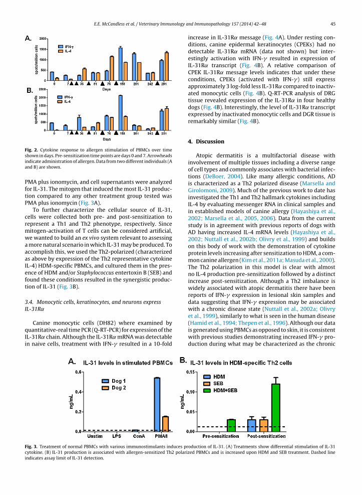
Fig. 2. Cytokine response to allergen stimulation of PBMCs over time
shown in days. Pre-sensitization time points are days 0 and 7. Arrowheadsindicate administration of allergen. Data from two different individuals (Aand B) are shown.PMA plus ionomycin, and cell supernatants were analyzedfor IL-31. The mitogen that induced the most IL-31 produc-tion compared to any other treatment group tested wasPMA plus ionomycin (Fig. 3A).
To further characterize the cellular source of IL-31,cells were collected both pre- and post-sensitization torepresent a Th1 and Th2 phenotype, respectively. Sincemitogen-activation of T cells can be considered artificial,we wanted to build an ex vivo system relevant to assessinga more natural scenario in which IL-31 may be produced. Toaccomplish this, we used the Th2-polarized (characterizedas above by expression of the Th2 representative cytokineIL-4) HDM-specific PBMCs, and cultured them in the pres-ence of HDM and/or Staphylococcus entertoxin B (SEB) andfound these conditions resulted in the synergistic produc-tion of IL-31 (Fig. 3B).
3.4. Monocytic cells, keratinocytes, and neurons expressIL-31R˛
Canine monocytic cells (DH82) where examined byquantitative-real time PCR (Q-RT-PCR) for expression of theIL-31R ̨ chain. Although the IL-31R ̨ mRNA was detectablein naive cells, treatment with IFN-� resulted in a 10-fold
Fig. 3. Treatment of normal PBMCs with various immunostimulants induces prcytokine. (B) IL-31 production is associated with allergen-sensitized Th2 polarizindicates assay limit of IL-31 detection.
nd Immunopathology 157 (2014) 42– 48 45
increase in IL-31R ̨ message (Fig. 4A). Under resting con-ditions, canine epidermal keratinocytes (CPEKs) had nodetectable IL-31R ̨ mRNA (data not shown) but inter-estingly activation with IFN-� resulted in expression ofIL-31R ̨ transcript (Fig. 4B). A relative comparison ofCPEK IL-31R ̨ message levels indicates that under theseconditions, CPEKs (activated with IFN-�) still expressapproximately 3 log-fold less IL-31R ̨ compared to inactiv-ated monocytic cells (Fig. 4B). Q-RT-PCR analysis of DRGtissue revealed expression of the IL-31R ̨ in four healthydogs (Fig. 4B). Interestingly, the level of IL-31R ̨ transcriptexpressed by inactivated monocytic cells and DGR tissue isremarkably similar (Fig. 4B).
4. Discussion
Atopic dermatitis is a multifactorial disease withinvolvement of multiple tissues including a diverse rangeof cell types and commonly associates with bacterial infec-tions (DeBoer, 2004). Like many allergic conditions, ADis characterized as a Th2 polarized disease (Marsella andGirolomoni, 2009). Much of the previous work to date hasinvestigated the Th1 and Th2 hallmark cytokines includingIL-4 by evaluating messenger RNA in clinical samples andin established models of canine allergy (Hayashiya et al.,2002; Marsella et al., 2005, 2006). Data from the currentstudy is in agreement with previous reports of dogs withAD having increased IL-4 mRNA levels (Hayashiya et al.,2002; Nuttall et al., 2002b; Olivry et al., 1999) and buildson this body of work with the demonstration of cytokineprotein levels increasing after sensitization to HDM, a com-mon canine allergen (Kim et al., 2011a; Masuda et al., 2000).The Th2 polarization in this model is clear with almostno IL-4 production pre-sensitization followed by a distinctincrease post-sensitization. Although a Th2 imbalance iswidely associated with atopic dermatitis there have beenreports of IFN-� expression in lesional skin samples anddata suggesting that IFN-� expression may be associatedwith a chronic disease state (Nuttall et al., 2002a; Olivryet al., 1999), similarly to what is seen in the human disease
(Hamid et al., 1994; Thepen et al., 1996). Although our datais generated using PBMCs as opposed to skin, it is consistentwith previous studies demonstrating increased IFN-� pro-duction during what may be characterized as the chronicoduction of IL-31. (A) Treatments show differential stimulation of IL-31ed PBMCs and is increased upon HDM and SEB treatment. Dashed line

46 E.E. McCandless et al. / Veterinary Immunology and Immunopathology 157 (2014) 42– 48
F . (A) IL-3w genitor
r
ptisd
damcoHartphtswtraop(sP22Ia
awori(wtc
ig. 4. Fold increase in IL-31 receptor expression as assessed by Q-RT-PCRith IFN-�. (B) Relative expression of IL-31R� mRNA in keratinocyte pro
oot ganglia (DRG) tissue compared to untreated DH82s where n = 3.
hase of the disease model. These results suggest that inhis model there is a clear Th2 response to allergen but alsonvolvement of Th1 cytokines and supports the hypothe-is that IFN-� is associated with a chronic disease state inogs.
Based on the ability of PMA to activate protein kinase Cownstream of the T cell receptor (Berry et al., 1989; Chatiland Geha, 1988) and the lack of IL-31 production from theonocyte-activator LPS, the data in this study suggests that
anine T cells are a source of IL-31. Under more physi-logically relevant conditions using PBMCs isolated fromDM-sensitized dogs, we have seen that IL-31 is producedfter exposure of Th2 polarized cells to allergen and bacte-ial endotoxin. The polarization status of the cells appearso be an important characteristic associated with IL-31roduction as cells collected from dogs pre-sensitizationave significantly reduced cytokine production comparedo post-sensitization. To confirm polarized T cells as theource of IL-31, cell-type specific characterization alongith isolation or intracellular labeling techniques within
he polarized PBMCs cultures would be needed. Concur-ent staphylococcal infections are common in dogs withtopic dermatitis (DeBoer and Marsella, 2001) and col-nization with Staphylococcus pseudintermedius is morerevalent in dogs with AD compared to healthy controlsFazakerley et al., 2009). Staphylococcus aureus has beenhown to promote the production of Th2 cytokines byBMCs from humans with AD (Dillon et al., 2004; Lin et al.,011) including IL-31 (Niebuhr et al., 2011; Sonkoly et al.,006). Importantly, recent studies have demonstrated that
L-31 is detected in dogs with AD but not non-diseasednimals (Gonzales et al., 2013).
Based on the IFN-� production we saw in the char-cterization of the HDM allergen model over time, weere interested in further investigating the involvement
f this Th1 cytokine in a classically Th2 disease. The IL-31eceptor has been characterized as heterodimic consist-ng of the OSMR chain and the IL-31 receptor alpha chain
IL-31R˛) (Dillon et al., 2004). Studies have shown thathile both receptor chains are required to induce signaling,he IL-31R˛ has the predominant interaction with theytokine (Diveu et al., 2004). Data from the current study
1R� mRNA in monocytic cells (DH82) at six and 24 hours post-treatmentcells (CPEK) treated with IFN-� (24 hours) and RNA isolated from dorsal
indicate that although the production of IL-31 came frompolarized cultures exhibiting a Th2-cell phenotype, theability to respond to the cytokine, i.e. receptor expres-sion, may depend at least in part on Th1 cytokine presencesince IFN-� was required to induce IL-31R˛ gene expres-sion in cells. Additionally our results showed that IFN-�increased expression of the IL-31R˛, which is in agreementwith previous findings that IFN-� can increase expres-sion if the IL-31 receptor in human monocytes (Diveuet al., 2003) and skin-resident cells including keratino-cytes (Heise et al., 2009; Kasraie et al., 2011). Recent datafrom our group confirms that IFN-� induced the pres-ence of functional IL-31 receptor on canine monocyticcells by causing IL-31-mediated phosphorylation of STAT3and ERK1/2 (Gonzales et al., 2013). Although receptorexpression implicates both monocytes and keratinocytesas having the potential ability to respond to IL-31, addi-tional studies are needed to confirm and identify thephysiological effect.
IL-31R ̨ mRNA was also detected in DRG tissue. DRGare neuronal cells involved in the transition of the pru-ritic signal to the spinal cord (Buddenkotte and Steinhoff,2010) and have been shown to express relatively high lev-els of classically hematopoietic receptors including the H4histamine receptor (Strakhova et al., 2009) as well as theIL-31 receptor (Bando et al., 2006). These data imply thatneurons may be able to directly respond to IL-31. There isevidence to support an IL-31 mediated direct effect on neu-ronal cells as exogenous administration of IL-31 inducespruritus in dogs within two hours (Gonzales et al., 2013).Although reports have shown upregulation of cytokinesand other inflammatory mediators post-treatment withIL-31 (Cornelissen et al., 2012; Zhang et al., 2008), phys-iological effects seen in such a short time frame may be theresult of direct neuronal action.
Taken together, these data suggest a dual-involvementof both Th1 and Th2 cytokines in responses in canineallergy. An important role for IL-31 in pruritus has been
suggested in human AD (Ezzat et al., 2011; Raap et al., 2008;Sonkoly et al., 2006) and most recently characterized incanine AD (Gonzales et al., 2013). Expression of the IL-31receptor on not only monocytic cells, but also keratinocytes
nology a
E.E. McCandless et al. / Veterinary Immuand DRG tissue suggest that this cytokine may mediate dia-log between the immune system and the peripheral organsites. The results of the current study identify stimulatedTh2 polarized cells as a source of canine IL-31 and suggestdisease-relevant tissues as the target. This data builds ongrowing understanding of the mechanism of action of IL-31in canine AD and highlights the complexity and breadth ofaction that this cytokine may have in orchestrating cellu-lar changes within the animal and inducing clinical signsassociated with allergic skin disease in dogs.
Conflict of interest
All authors are employees of Zoetis Inc.
Sources of funding
Support for this work was provided by Zoetis Inc.
Acknowledgements
We would like to thank Chris Chio, Steve Dunham, andGary Bammert for their help in designing and develop-ing reagents used. Much appreciation goes to Dr. DuncanMwangi and Dr. Sharath Rai for their contributions towarddesign of assays developed. Additionally we would like tothank Bill Humphrey for his work with the animal model,Grace Kiel and Adena Leibbrand for technical support, andDr. Mary Pat Gorman and Dr. Candace Sousa for criticalreview of the manuscript.
References
Bando, T., Morikawa, Y., Komori, T., Senba, E., 2006. Complete overlap ofinterleukin-31 receptor A and oncostatin M receptor beta in the adultdorsal root ganglia with distinct developmental expression patterns.Neuroscience 142, 1263–1271.
Berry, N., Ase, K., Kikkawa, U., Kishimoto, A., Nishizuka, Y., 1989. HumanT cell activation by phorbol esters and diacylglycerol analogues. J.Immunol. 143, 1407–1413.
Bilsborough, J., Leung, D.Y., Maurer, M., Howell, M., Boguniewicz, M., Yao,L., Storey, H., LeCiel, C., Harder, B., Gross, J.A., 2006. IL-31 is asso-ciated with cutaneous lymphocyte antigen-positive skin homing Tcells in patients with atopic dermatitis. J. Allergy Clin. Immunol. 117,418–425.
Buddenkotte, J., Steinhoff, M., 2010. Pathophysiology and therapy of pru-ritus in allergic and atopic diseases. Allergy 65, 805–821.
Chatila, T.A., Geha, R.S., 1988. Phosphorylation of T cell membrane proteinsby activators of protein kinase C. J. Immunol. 140, 4308–4314.
Cornelissen, C., Luscher-Firzlaff, J., Baron, J.M., Luscher, B., 2012. Signalingby IL-31 and functional consequences. Eur. J. Cell Biol. 91, 552–566.
DeBoer, D.J., 2004. Canine atopic dermatitis: new targets, new therapies.J. Nutr. 134, 2056S–2061S.
DeBoer, D.J., Marsella, R., 2001. The ACVD task force on canine atopicdermatitis (XII): the relationship of cutaneous infections to the patho-genesis and clinical course of canine atopic dermatitis. Vet. Immunol.Immunopathol. 81, 239–249.
Dillon, S.R., Sprecher, C., Hammond, A., Bilsborough, J., Rosenfeld-Franklin,M., Presnell, S.R., Haugen, H.S., Maurer, M., Harder, B., Johnston, J., Bort,S., Mudri, S., Kuijper, J.L., Bukowski, T., Shea, P., Dong, D.L., Dasovich,M., Grant, F.J., Lockwood, L., Levin, S.D., LeCiel, C., Waggie, K., Day, H.,Topouzis, S., Kramer, J., Kuestner, R., Chen, Z., Foster, D., Parrish-Novak,J., Gross, J.A., 2004. Interleukin 31, a cytokine produced by activated Tcells, induces dermatitis in mice. Nat. Immunol. 5, 752–760.
Diveu, C., Lelievre, E., Perret, D., Lak-Hal, A.H., Froger, J., Guillet, C., Cheva-lier, S., Rousseau, F., Wesa, A., Preisser, L., Chabbert, M., Gauchat, J.F.,Galy, A., Gascan, H., Morel, A., 2003. GPL, a novel cytokine receptorrelated to GP130 and leukemia inhibitory factor receptor. J. Biol. Chem.278, 49850–49859.
nd Immunopathology 157 (2014) 42– 48 47
Diveu, C., Lak-Hal, A.H., Froger, J., Ravon, E., Grimaud, L., Barbier, F.,Hermann, J., Gascan, H., Chevalier, S., 2004. Predominant expres-sion of the long isoform of GP130-like (GPL) receptor is required forinterleukin-31 signaling. Eur. Cytokine Netw. 15, 291–302.
Ezzat, M.H., Hasan, Z.E., Shaheen, K.Y., 2011. Serum measurement ofinterleukin-31 (IL-31) in paediatric atopic dermatitis: elevated lev-els correlate with severity scoring. J. Eur. Acad. Dermatol. Venereol.25, 334–339.
Fazakerley, J., Nuttall, T., Sales, D., Schmidt, V., Carter, S.D., Hart, C.A.,McEwan, N.A., 2009. Staphylococcal colonization of mucosal andlesional skin sites in atopic and healthy dogs. Vet. Dermatol. 20,179–184.
Gonzales, A.J., Humphrey, W.R., Messamore, J.E., Fleck, T.J., Fici, G.J., Shelly,J.A., Teel, J.F., Bammert, G.F., Dunham, S.A., Fuller, T.E., McCall, R.B.,2013. Interleukin-31: its role in canine pruritus and naturally occur-ring canine atopic dermatitis. Vet. Dermatol. 24, 48–53, e11-42.
Gutzmer, R., Mommert, S., Gschwandtner, M., Zwingmann, K., Stark, H.,Werfel, T., 2009. The histamine H4 receptor is functionally expressedon T(H)2 cells. J. Allergy Clin. Immunol. 123, 619–625.
Hamid, Q., Boguniewicz, M., Leung, D.Y., 1994. Differential in situ cytokinegene expression in acute versus chronic atopic dermatitis. J. Clin.Invest. 94, 870–876.
Hayashiya, S., Tani, K., Morimoto, M., Hayashi, T., Hayasaki, M., Nomura,T., Une, S., Nakaichi, M., Taura, Y., 2002. Expression of T helper 1and T helper 2 cytokine mRNAs in freshly isolated peripheral bloodmononuclear cells from dogs with atopic dermatitis. J. Vet. Med. APhysiol. Pathol. Clin. Med. 49, 27–31.
Heise, R., Neis, M.M., Marquardt, Y., Joussen, S., Heinrich, P.C., Merk, H.F.,Hermanns, H.M., Baron, J.M., 2009. IL-31 receptor alpha expressionin epidermal keratinocytes is modulated by cell differentiation andinterferon gamma. J. Invest. Dermatol. 129, 240–243.
Kasraie, S., Niebuhr, M., Baumert, K., Werfel, T., 2011. Functional effects ofinterleukin 31 in human primary keratinocytes. Allergy 66, 845–852.
Kim, H.J., Kang, M.H., Park, H.M., 2011a. Common allergens of atopic der-matitis in dogs: comparative findings based on intradermal tests. J.Vet. Sci. 12, 287–290.
Kim, S., Kim, H.J., Yang, H.S., Kim, E., Huh, I.S., Yang, J.M., 2011b. IL-31 serumprotein and tissue mRNA levels in patients with atopic dermatitis. Ann.Dermatol. 23, 468–473.
Kimura, T., Sekido, M., Chimura, N., Shibata, S., Kondo, N., Kamishina, H.,Kamishina, H., Maeda, S., 2012. Production of GM-CSF mediated bycysteine protease of Der f 1 in canine keratinocytes. J. Vet. Med. Sci..
Lin, Y.T., Wang, C.T., Chao, P.S., Lee, J.H., Wang, L.C., Yu, H.H., Yang, Y.H.,Chiang, B.L., 2011. Skin-homing CD4+ Foxp3+ T cells exert Th2-likefunction after staphylococcal superantigen stimulation in atopic der-matitis patients. Clin. Exp. Allergy 41, 516–525.
Maeda, S., Maeda, S., Shibata, S., Chimura, N., Fukata, T., 2009. House dustmite major allergen Der f 1 enhances proinflammatory cytokine andchemokine gene expression in a cell line of canine epidermal kerati-nocytes. Vet. Immunol. Immunopathol. 131, 298–302.
Marsella, R., Girolomoni, G., 2009. Canine models of atopic dermati-tis: a useful tool with untapped potential. J. Invest. Dermatol. 129,2351–2357.
Marsella, R., Nicklin, C., Lopez, J., 2005. Atopy patch test reactions inhigh-IgE beagles to different sources and concentrations of house dustmites. Vet. Dermatol. 16, 308–314.
Marsella, R., Olivry, T., Nicklin, C., Lopez, J., 2006. Pilot investigation of amodel for canine atopic dermatitis: environmental house dust mitechallenge of high-IgE-producing beagles, mite hypersensitive dogswith atopic dermatitis and normal dogs. Vet. Dermatol. 17, 24–35.
Masuda, K., Sakaguchi, M., Fujiwara, S., Kurata, K., Yamashita, K., Odagiri,T., Nakao, Y., Matsuki, N., Ono, K., Watari, T., Hasegawa, A., Tsujimoto,H., 2000. Positive reactions to common allergens in 42 atopic dogs inJapan. Vet. Immunol. Immunopathol. 73, 193–204.
Mizuno, T., Kanbayashi, S., Okawa, T., Maeda, S., Okuda, M., 2009. Molec-ular cloning of canine interleukin-31 and its expression in varioustissues. Vet. Immunol. Immunopathol. 131, 140–143.
Niebuhr, M., Mamerow, D., Heratizadeh, A., Satzger, I., Werfel, T., 2011.Staphylococcal alpha-toxin induces a higher T cell proliferation andinterleukin-31 in atopic dermatitis. Int. Arch. Allergy Immunol. 156,412–415.
Nuttall, T.J., Knight, P.A., McAleese, S.M., Lamb, J.R., Hill, P.B., 2002a. Expres-sion of Th1, Th2 and immunosuppressive cytokine gene transcripts incanine atopic dermatitis. Clin. Exp. Allergy 32, 789–795.
Nuttall, T.J., Knight, P.A., McAleese, S.M., Lamb, J.R., Hill, P.B., 2002b.T-helper 1, T-helper 2 and immunosuppressive cytokines in canineatopic dermatitis. Vet. Immunol. Immunopathol. 87, 379–384.
Olivry, T., 2011. Is the skin barrier abnormal in dogs with atopic dermati-tis? Vet. Immunol. Immunopathol. 144, 11–16.

4 ology a
O
R
S
S
S
8 E.E. McCandless et al. / Veterinary Immun
livry, T., Dean, G.A., Tompkins, M.B., Dow, J.L., Moore, P.F., 1999. Towarda canine model of atopic dermatitis: amplification of cytokine-genetranscripts in the skin of atopic dogs. Exp. Dermatol. 8, 204–211.
aap, U., Wichmann, K., Bruder, M., Stander, S., Wedi, B., Kapp, A., Wer-fel, T., 2008. Correlation of IL-31 serum levels with severity of atopicdermatitis. J. Allergy Clin. Immunol. 122, 421–423.
aliaris, A.P., Amado, L.C., Minhas, K.M., Schuleri, K.H., Lehrke, S., St John,M., Fitton, T., Barreiro, C., Berry, C., Zheng, M., Kozielski, K., Eneboe,V., Brawn, J., Hare, J.M., 2007. Chronic allopurinol administrationameliorates maladaptive alterations in Ca2+ cycling proteins and beta-adrenergic hyporesponsiveness in heart failure. Am. J. Physiol. HeartCirc. Physiol. 292, H1328–H1335.
chlotter, Y.M., Rutten, V.P., Riemers, F.M., Knol, E.F., Willemse, T., 2011.
Lesional skin in atopic dogs shows a mixed Type-1 and Type-2 immuneresponsiveness. Vet. Immunol. Immunopathol. 143, 20–26.onkoly, E., Muller, A., Lauerma, A.I., Pivarcsi, A., Soto, H., Kemeny, L.,Alenius, H., Dieu-Nosjean, M.C., Meller, S., Rieker, J., Steinhoff, M., Hoff-mann, T.K., Ruzicka, T., Zlotnik, A., Homey, B., 2006. IL-31: a new link
nd Immunopathology 157 (2014) 42– 48
between T cells and pruritus in atopic skin inflammation. J. AllergyClin. Immunol. 117, 411–417.
Stehle, M.E., Hanczaruk, M., Schwarz, S.C., Gobel, T.W., Mueller, R.S., 2010.Effects of polyunsaturated fatty acids on isolated canine peripheralblood mononuclear cells and cytokine expression (IL-4, IFN-gamma,TGF-beta) in healthy and atopic dogs. Vet. Dermatol. 21, 112–117.
Strakhova, M.I., Nikkel, A.L., Manelli, A.M., Hsieh, G.C., Esbenshade, T.A.,Brioni, J.D., Bitner, R.S., 2009. Localization of histamine H4 receptorsin the central nervous system of human and rat. Brain Res. 1250,41–48.
Thepen, T., Langeveld-Wildschut, E.G., Bihari, I.C., van Wichen, D.F., vanReijsen, F.C., Mudde, G.C., Bruijnzeel-Koomen, C.A., 1996. Biphasicresponse against aeroallergen in atopic dermatitis showing a switch
from an initial TH2 response to a TH1 response in situ: an immuno-cytochemical study. J. Allergy Clin. Immunol. 97, 828–837.Zhang, Q., Putheti, P., Zhou, Q., Liu, Q., Gao, W., 2008. Structures and bio-logical functions of IL-31 and IL-31 receptors. Cytokine Growth FactorRev. 19, 347–356.