veterinary aspects of the turkey breeding. - vfu · poultry. morbidity is low to moderate and...
TRANSCRIPT
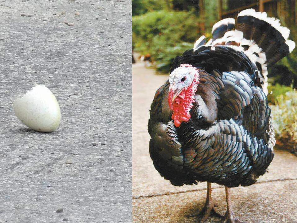
Veterinary aspects of the turkey breeding.
Summer term 2010


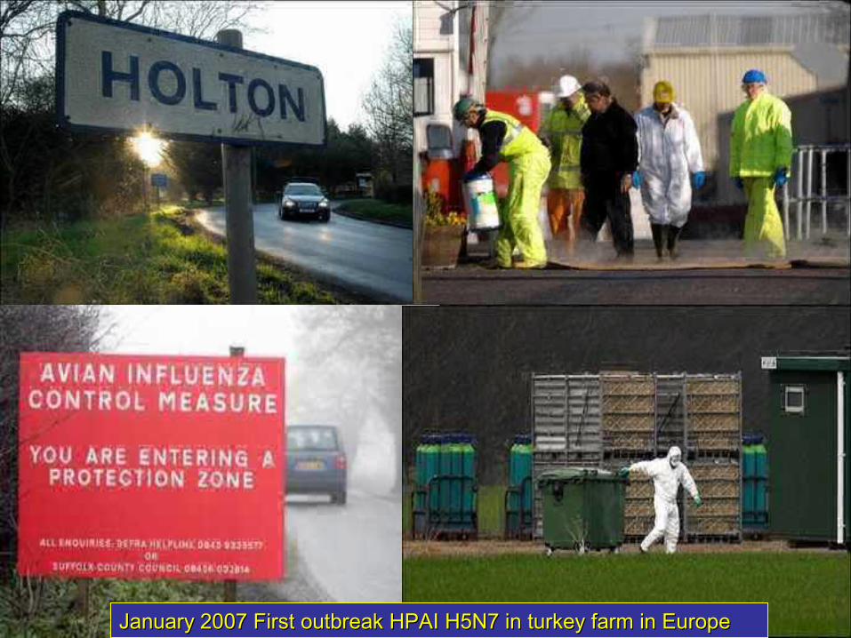
January 2007 First outbreak HPAI H5N7 in turkey farm in EuropeJanuary 2007 First outbreak HPAI H5N7 in turkey farm in Europe
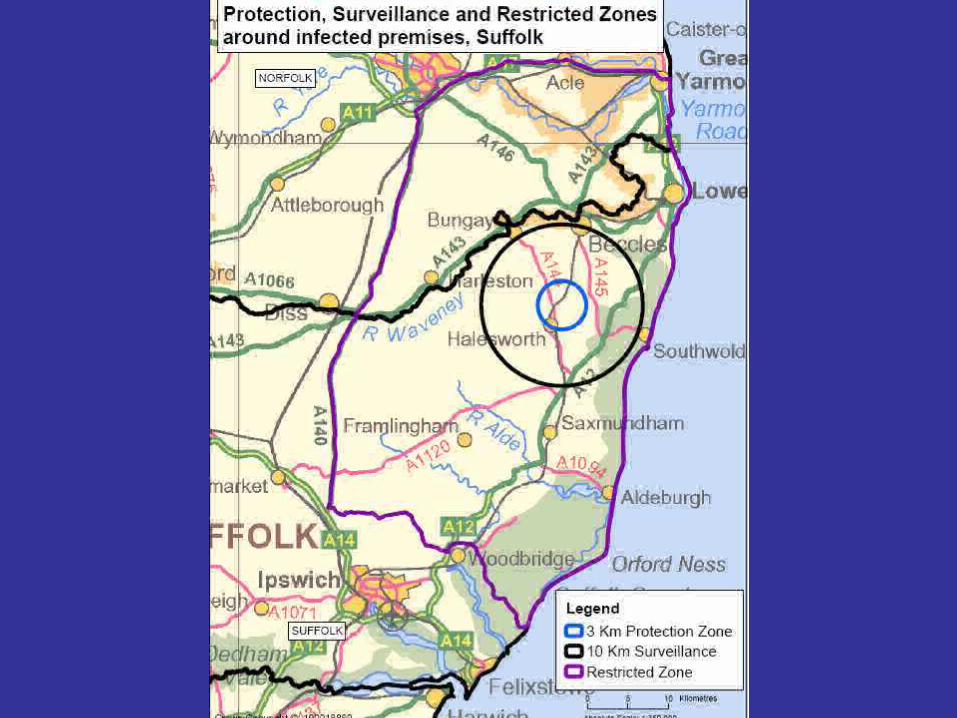

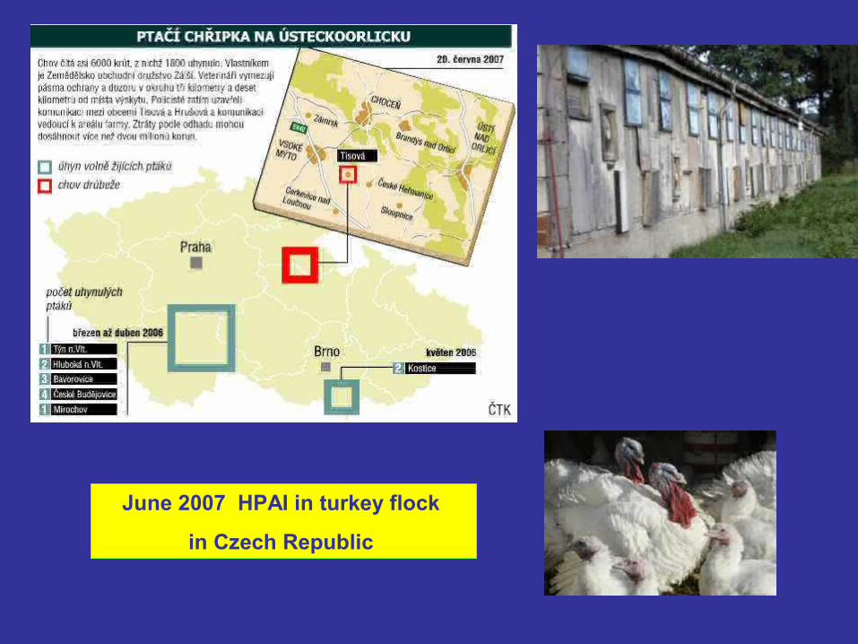
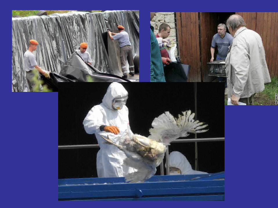
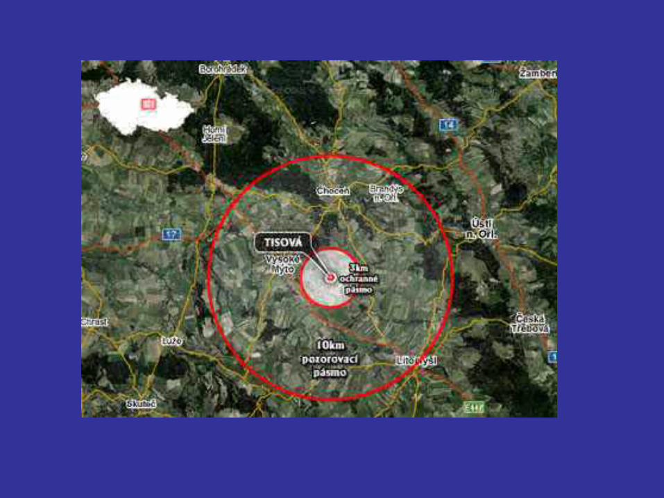
June 2007 HPAI in turkey flock
in Czech Republic
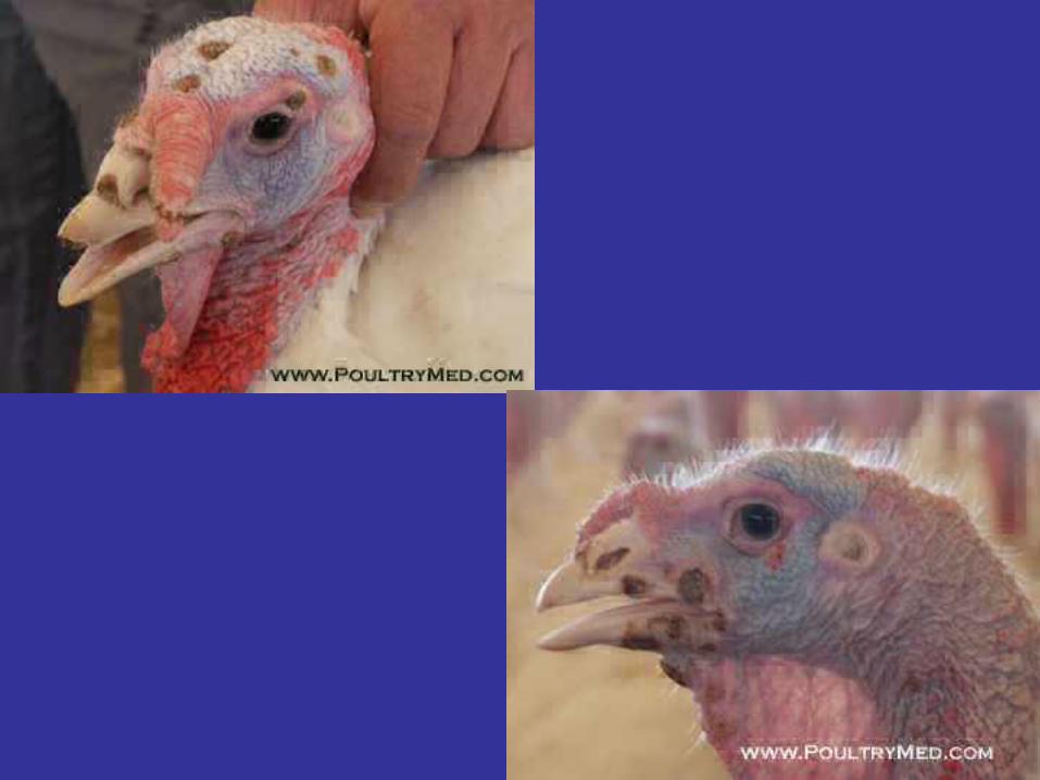
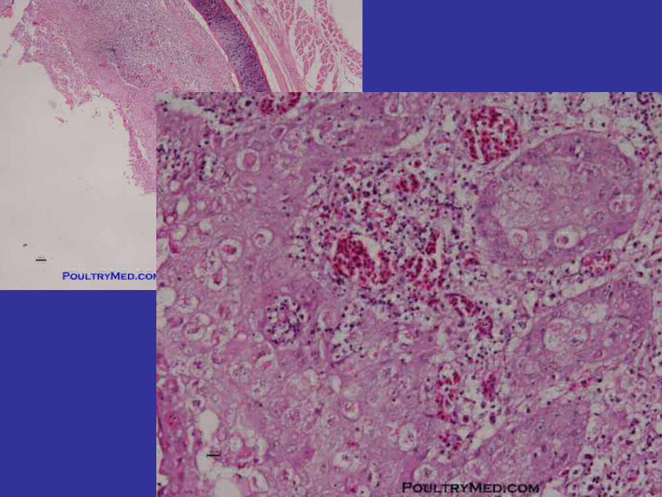
Fowl Pox
Turkeypox virus• Possibility of transmission of poxvirus by
insemination• Cutaneous form• Diphtheric (wet) form
• Prevention by vaccination
Hemorrhagic Enteritis of Turkeys
Hemorrhagic enteritis is an acute GI disorder affecting young turkeys. In its most severe form, it is characterized by depression, bloody droppings, and substantial mortality.
The etiologic agent is a nonenveloped, icosahedral DNA virus, 70-90 nm in diameter. It is a member of the family Adenoviridae and has recently been assigned to the new genus Siadenovirus .
Hemorrhagic Enteritis of Turkeys
The usual route of infection is oral, and virus is often introduced onto previously uninfected premises via personnel or equipment contaminated with infectious feces. Turkey poults <4 wk of age are resistant to infection due to age-related resistance or, more commonly, the presence of maternal antibody.
Hemorrhagic Enteritis of Turkeys
• In commercial operations, hemorrhagic enteritis typically affects
turkeys 6-12 wk of age.
• In outbreaks involving highly virulent pathotypes, clinical signs
can include depression, pallor, and bloody droppings.
• Acute mortality ranges from <1% to 60% with an average of 10-15%
over a 2-wk period.
• Birds that survive the acute phase experience a transient
immunosuppression related to the lymphotrophic, lymphocytopathic
nature of the virus.
Hemorrhagic Enteritis of Turkeys
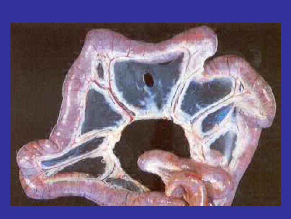
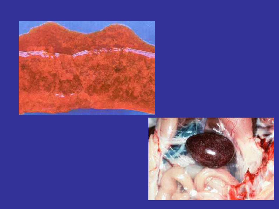
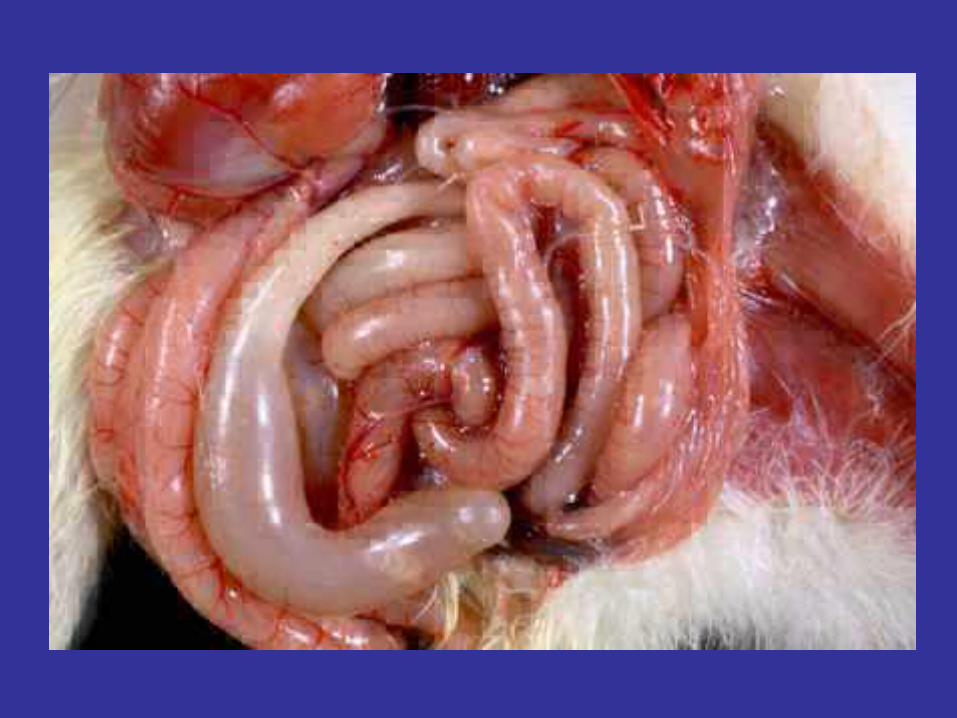
Necropsy of moribund or dead birds infected
with hemorrhagic enteritis virus reveals gross
congestion and intraluminal hemorrhage in the
proximal small intestine. The spleen is usually
enlarged, friable, and mottled, except in birds
that have hemorrhaged extensively.
Hemorrhagic Enteritis of Turkeys
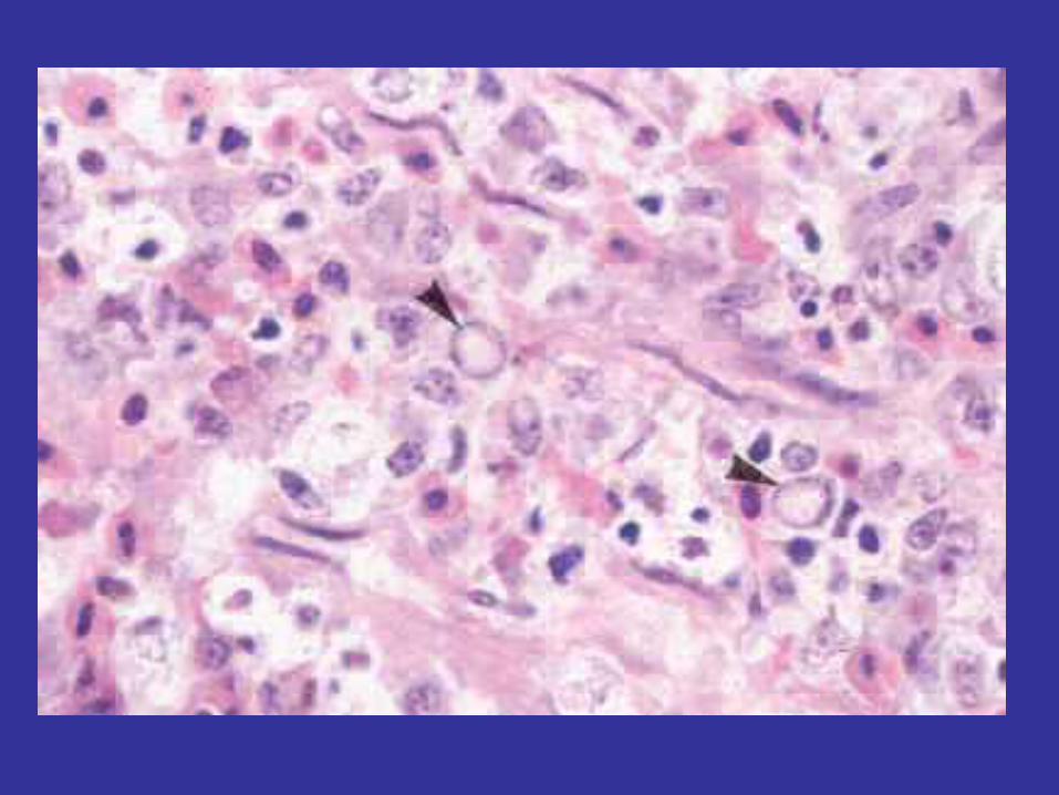
Histopathologic changes in the duodenum include congestion, hemorrhage, and necrosis of the intestinal epithelium. This lesion in particular is thought to be the result of a virally induced, cytokine-mediated anaphylactic reaction, with the GI tract being considered the target shock organ in the turkey. Basophilic intranuclear inclusions can be found in lymphocytes and macrophages in a variety of tissues but predominantly in the spleen where lymphoreticular hyperplasia and lymphoid necrosis are noted.

Trasmisible enteritis of turkey
Etiologyturkey
coronavirus
– is not related with infection bronchitis virus
ID : 1-5 dní
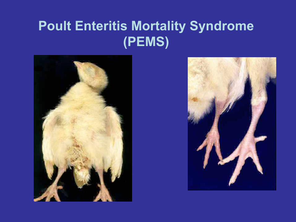
PEMS and Spiking Mortality of Turkeys
Poult Enteritis and Mortality Syndrome (PEMS) was first
identified in high density turkey producing areas of the
South Eastern USA in 1991.
It is an infectious and transmissible cause of sudden
increases in mortality in turkeys between 7 and 28 days
of age. A less acute form of the disease appears to
produce more of a lingering mortality.
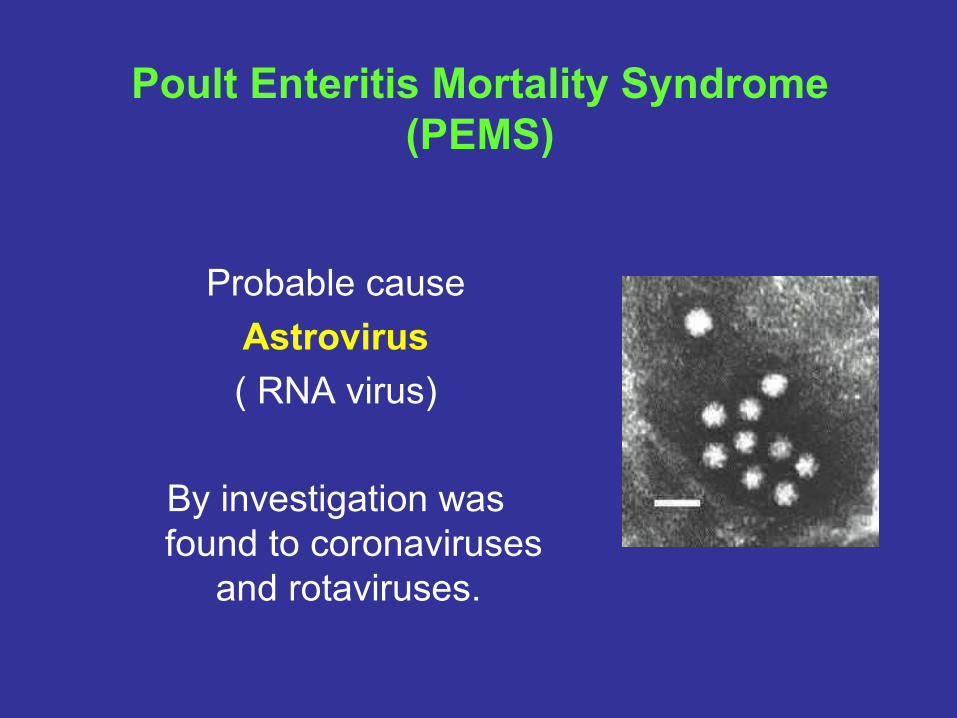
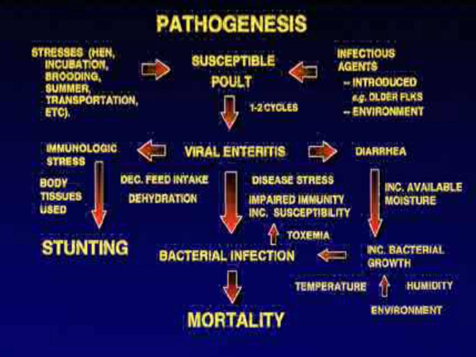
Poult Enteritis Mortality Syndrome (PEMS)
Probable causeAstrovirus( RNA virus)
By investigation was found to coronaviruses
and rotaviruses.
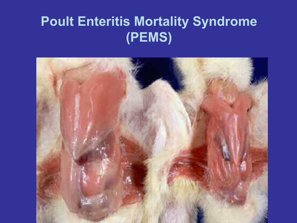
PEMS and Spiking Mortality of Turkeys
In both cases mortality can be high and can be associated with a
marked depression in growth. A range of viruses have been isolated
from affected flocks. To replicate the condition in full it appears to be
necessary to include bacteria in the inoculum. This syndrome is
clearly distinguished from typical viral enteritis in young turkeys
because of the high mortality and severe growth depression.
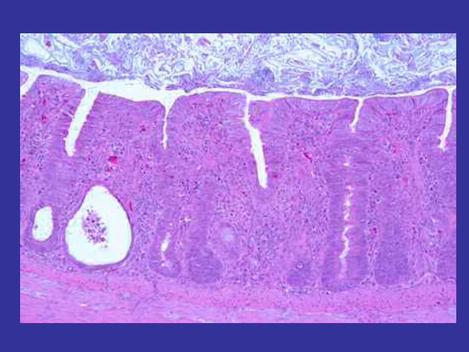
Poult Enteritis Mortality Syndrome (PEMS)
Poult Enteritis Mortality Syndrome (PEMS)
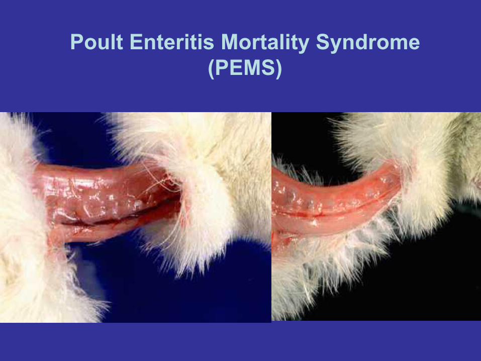
Poult Enteritis Mortality Syndrome (PEMS)

Poult Enteritis Mortality Syndrome (PEMS)

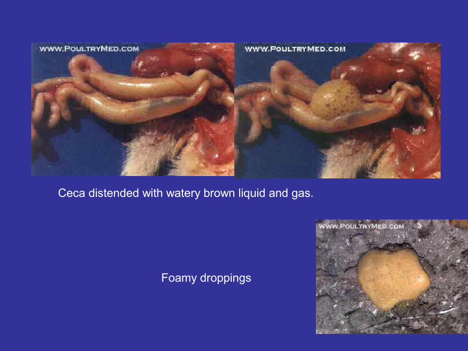
Ceca distended with watery brown liquid and gas.
Foamy droppings
Turkey Rhinotracheitis
A disease of turkeys caused by the viruses of the
Avipneumovirus genus, family Paramyxoviridae.
Morbidity is 10-100% and mortality 1- 30%.
Rapid transmission occurs laterally, possibly involving
fomites; vertical transmission is uncertain, maternal
antibody may protect.
Turkey Rhinotracheitis
Signs• Decreased appetite, weight gain and feed efficiency. • Loss of voice. • Ocular and nasal discharge. • Conjunctivitis. • Snick. • Dyspnoea. • Sinusitis.
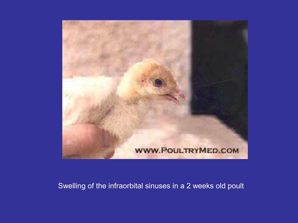
Swelling of the infraorbital sinuses in a 2 weeks old poult
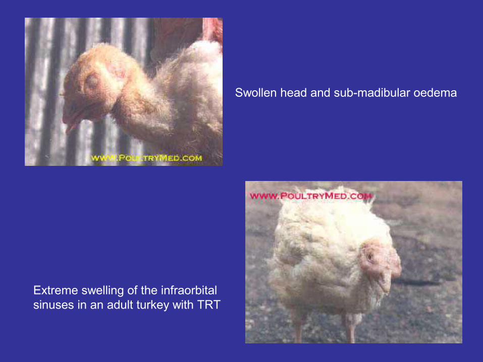
Swollen head and sub-madibular oedema
Extreme swelling of the infraorbital sinuses in an adult turkey with TRT
Turkey Rhinotracheitis
Post - mortem lesions
• Serous rhinitis and tracheitis, sometimes pus in
bronchi.
• If there is secondary E. coli infection then
pneumonia, airsacculitis and perihepatitis.
Turkey Rhinotracheitis
Diagnosis• Clinical signs, serology (using an Elisa test to
demonstrate rising titre), isolation and identification of the ciliostatic virus.
Treatment• Antibiotics are not very effective, control
respiratory stressors, chlorinate drinking water. Prevention• All-in/all-out production. Vaccination at day-old
seems to be most effective.
Israel Turkey Meningoencephalitis
• Turkey meningoencephalitis (TME) is a viral neuroparalytic disease of turkeys, which can lead to mortality, usually of about 15-30%.
• The clinical signs include incoordination, unwillingness to move and extended or twisted necks.
• The causative agent is a virus belongs to the family: Flaviviridae
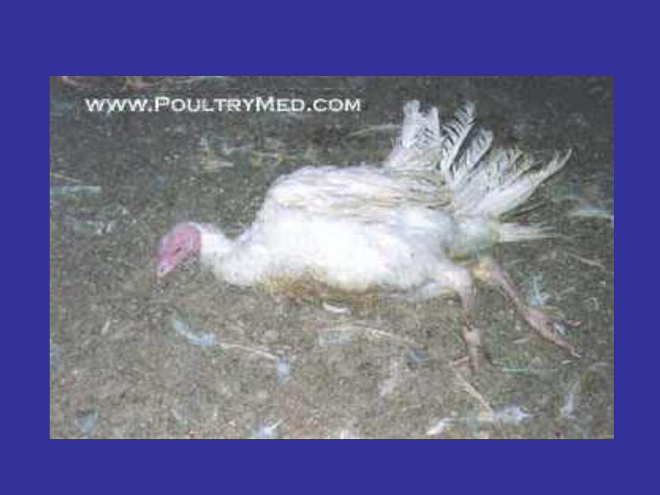
Infectious Sinusitis - Turkeys
A slow onset chronic respiratory disease of turkeys often with severe sinusitis and associated with Mycoplasma gallisepticum infection. It is seen worldwide, though in many countries this infection is now rare in commercial poultry. Morbidity is low to moderate and mortality low.
The route of infection is via the conjunctiva or upper respiratory tract with an incubation period of 6-10 days. Transmission may be transovarian, or by direct contact with birds, exudates, aerosols, and fomites. Recovered birds remain infected for life; subsequent stress may cause recurrence of disease.
Infectious Sinusitis - Turkeys
Signs• Coughing. • Nasal and ocular discharge. • Swollen sinuses. • Slow growth. • Leg problems. • Stunting. • Inappetance.

Infectious Sinusitis - Turkeys
Swelling of the paranasal sinuses

Infectious Sinusitis - Turkeys
Post-mortem lesions• Swollen infraorbital sinuses, often with
inspissated pus.
• Airsacculitis.
• Pericarditis.
• Perihepatitis.

Infectious Sinusitis - TurkeysDiagnosis• Lesions, serology, isolation and identification of
organism, demonstration of specific DNA (commercial kit available). Serology: serum agglutination is the standard screening test, suspect reactions are examined further by heat inactivation and/or dilution. HI may also be used.
• Suspect flocks should be re-sampled after 2-3 weeks. Some inactivated vaccines induce 'false positives' in serological testing. PCR is possible if it is urgent to determine the flock status.
• Differentiate from viral respiratory disease, especially Turkey Rhinotracheitis.
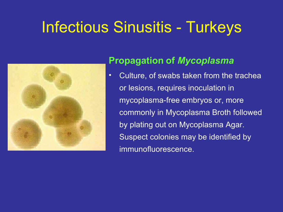
Infectious Sinusitis - Turkeys
Propagation of Mycoplasma• Culture, of swabs taken from the trachea
or lesions, requires inoculation in mycoplasma-free embryos or, more commonly in Mycoplasma Broth followed by plating out on Mycoplasma Agar. Suspect colonies may be identified by immunofluorescence.
Infectious Sinusitis - Turkeys
Treatment• Tilmicosin, tylosin, spiramycin, tetracyclines,
fluoroquinolones. Effort should be made to reduce dust and secondary infections.
Prevention• Eradication of this infection has been the central
objective of official poultry health programmes in most countries. These are based on purchase of uninfected poults, all-in/all-out production, and biosecurity.
In some circumstances preventative medication of known infected flocks may be of benefit.
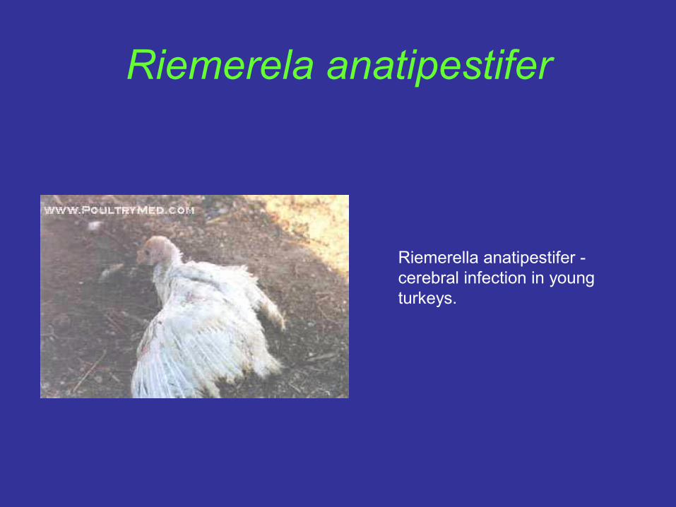
Riemerela anatipestifer
Riemerella anatipestifer - cerebral infection in young turkeys.
Arizona infection, Arizonosis
• Caused by the bacterium Salmonella arizonae. It affects
turkeys, mainly in North America, and is not present in
the UK turkey population.
• Mortality is 10-50% in young birds, older birds are
asymptomatic carriers. Transmission is vertical,
transovarian, and also horizontal, through faecal
contamination of environment, feed etc, from long-term
intestinal carriers, rodents, reptiles.
Arizona infection, Arizonosis
Signs• Dejection. • Inappetance. • Diarrhoea. • Vent-pasting. • Nervous signs. • Paralysis. • Blindness, cloudiness in eye. • Huddling near heat.
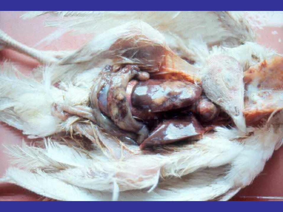
Arizona infection, Arizonosis
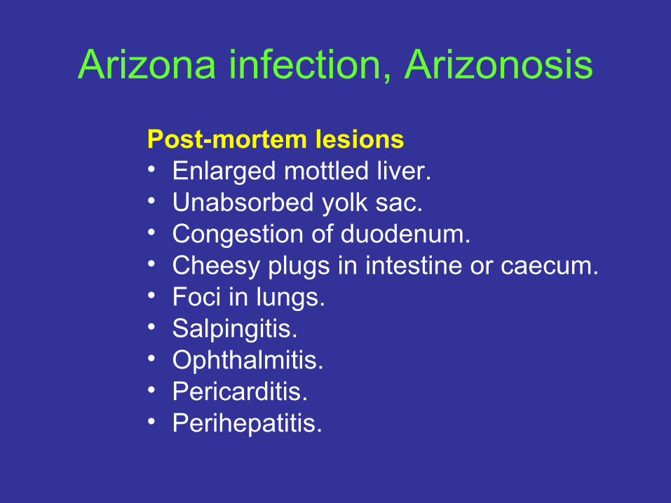
Post-mortem lesions• Enlarged mottled liver. • Unabsorbed yolk sac. • Congestion of duodenum. • Cheesy plugs in intestine or caecum. • Foci in lungs. • Salpingitis. • Ophthalmitis. • Pericarditis. • Perihepatitis.

Arizona infection, ArizonosisDiagnosis• Isolation and identification, methods as per Salmonella
spp. Differentiate from salmonellosis, coli-septicaemia. Treatment• Injection of streptomycin, spectinomycin, or gentamycin
at the hatchery is used in some countries. Formerly in-feed medication with nitrofurans was also used.
Prevention• Eradicate from breeder population, fumigation of
hatching eggs, good nest and hatchery hygiene, inject eggs or poults with antibiotics, monitor sensitivity.
Erysipelas A sudden onset infection with the bacterium Erysipelothrix insidiosa (E. rhusiopathiae) seen in turkeys and increasingly in free-range chickens, rarely in geese, ducks, pheasants. It is also seen in some mammals. It may be transmitted by faecal carriers for 41 days, in soil, water, fishmeal and semen and by cannibalism.
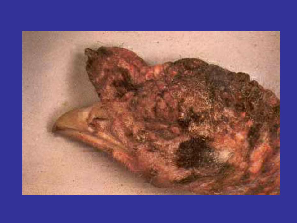
Erysipelas• Signs
– Inappetance. – Depression. – Sleepiness. – Swollen snood. – May be diarrhoea and respiratory signs. – Perineal congestion. – Chronic scabby skin, especially snood. – Sudden death.
Erysipelas
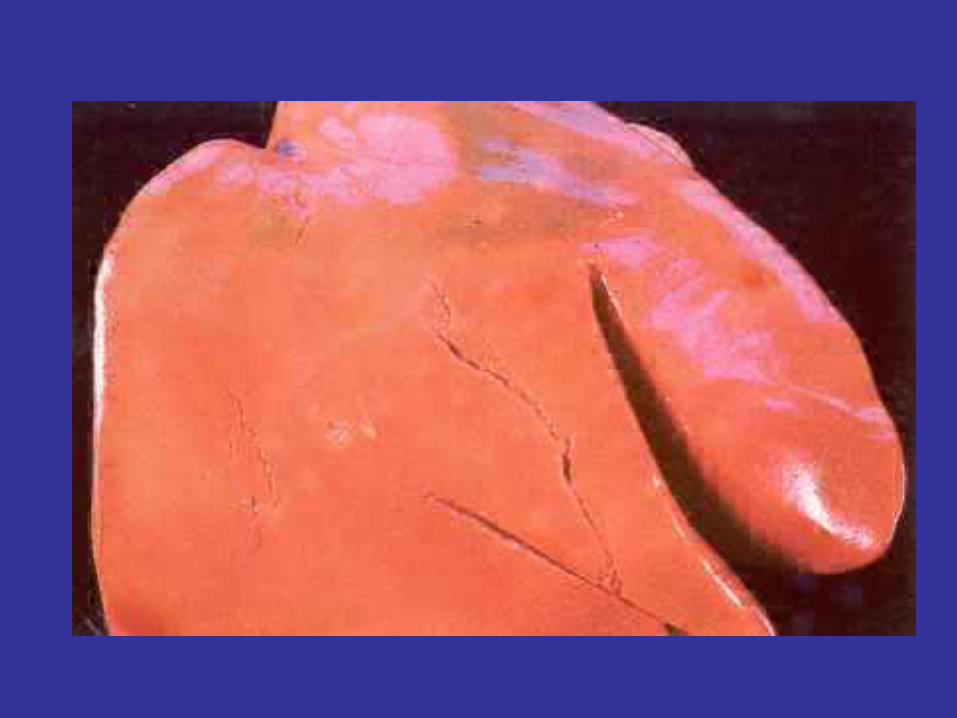
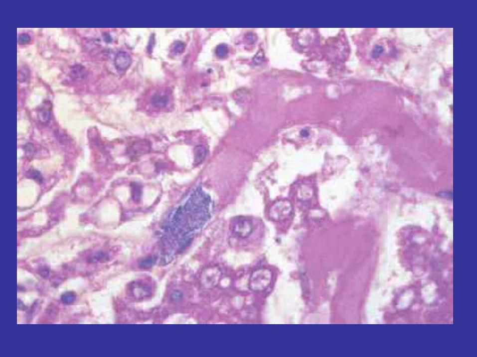
Post-mortem lesions• Carcase congestion. • Liver, kidney, spleen swollen. • Haemorrhages in fat, muscle, epicardium. • Marked catarrhal enteritis. • Joint lesions. • Endocarditis.
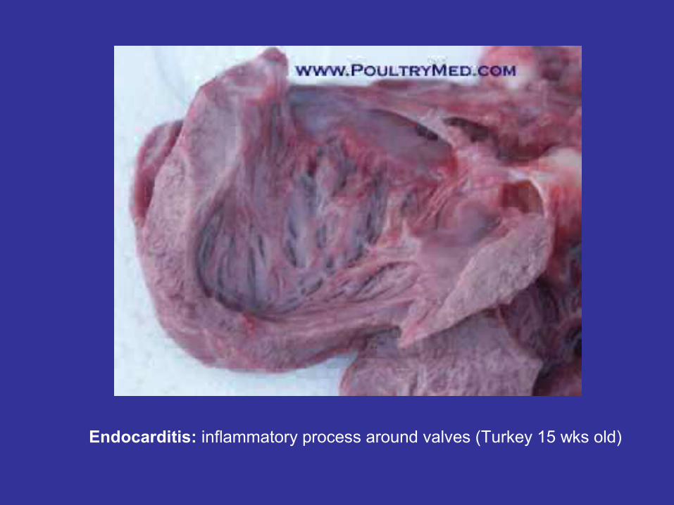
Endocarditis: inflammatory process around valves (Turkey 15 wks old)
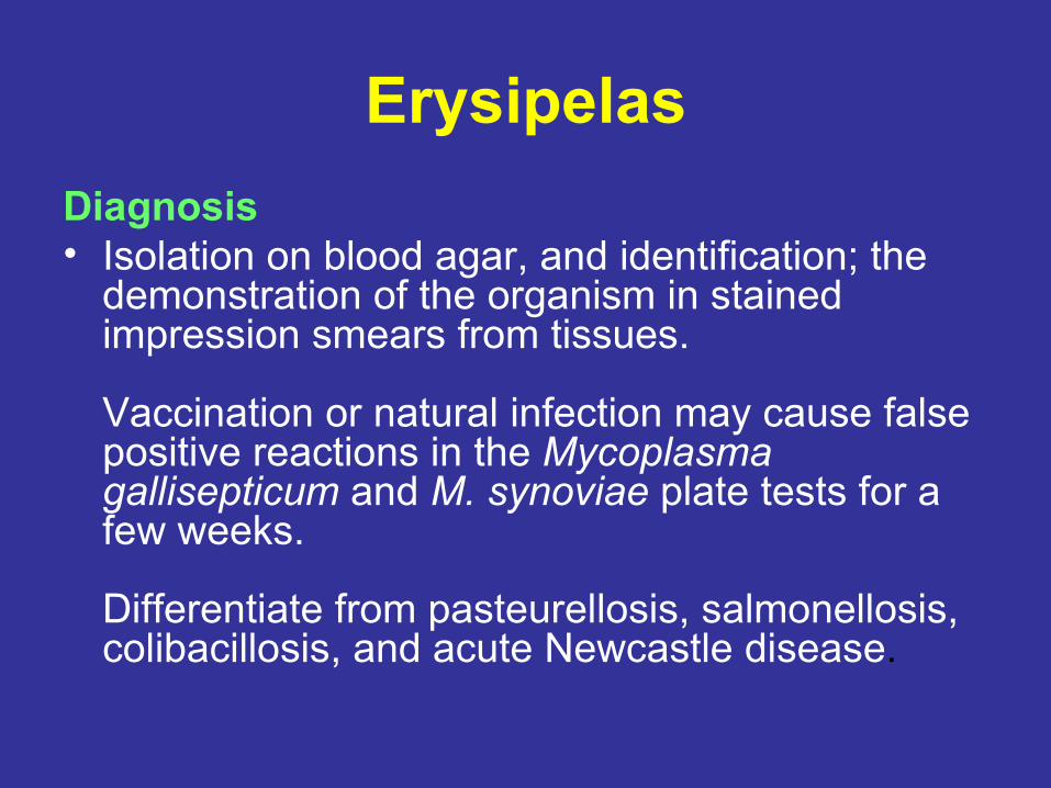
ErysipelasDiagnosis• Isolation on blood agar, and identification; the
demonstration of the organism in stained impression smears from tissues.
Vaccination or natural infection may cause false positive reactions in the Mycoplasma gallisepticum and M. synoviae plate tests for a few weeks.
Differentiate from pasteurellosis, salmonellosis, colibacillosis, and acute Newcastle disease.
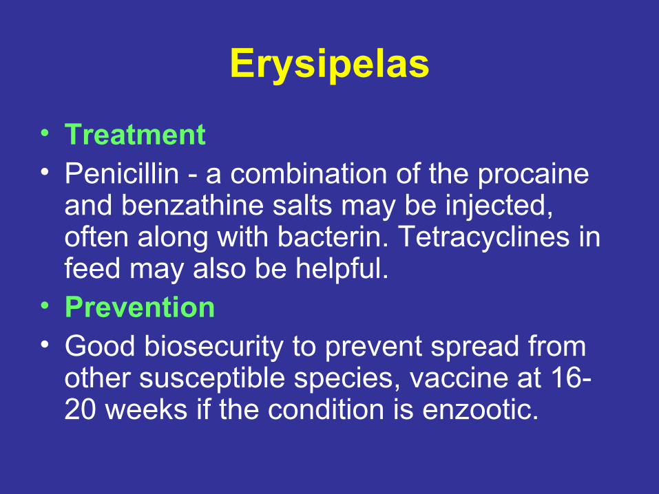
Erysipelas• Treatment• Penicillin - a combination of the procaine
and benzathine salts may be injected, often along with bacterin. Tetracyclines in feed may also be helpful.
• Prevention• Good biosecurity to prevent spread from
other susceptible species, vaccine at 16-20 weeks if the condition is enzootic.
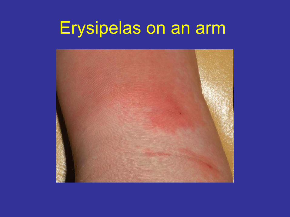
Erysipelas on an arm
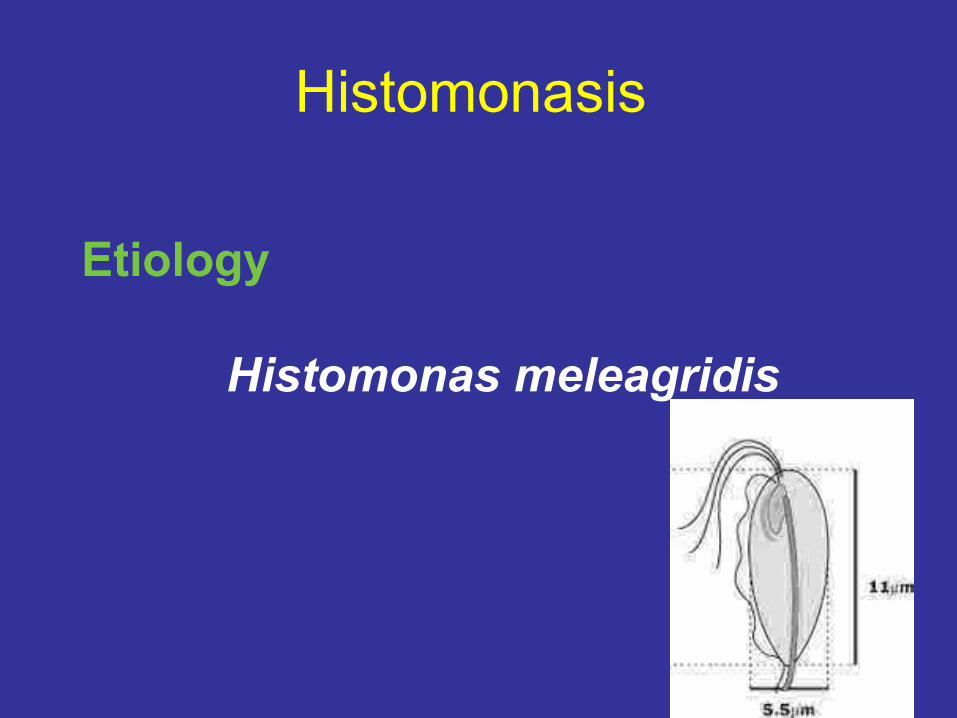
Histomonasis
Etiology
Histomonas meleagridis
Histomonasis
• Species affected: Mostly turkeys, occasionally in chicken, quail, pheasants, grouse, chukar partridges, guinea fowl.
• Effects: Enterohepatitis has a 7-12 day incubation period. Morbidity (to 100%) occurs, a cyanotic head and bloody caecal diarrhoea. In turkeys, signs include drowsiness, drooping of wings, stilted gait, closed eyes, head down and anorexia. Mortality occurs up to 100%.
Histomonasis
• It is called blackhead because birds may have a dark discoloured head. Young turkeys, chickens, quail, pheasants, grouse, chukar partridges and guinea fowl are susceptible to this acute to chronic disease. Enterohepatitis is caused by Histomonas meleagridis, a highly pleomorphic amoeboid protozoa with a stout flagellum and pseudopodia.
Histomonasis
Mode of transmission• Chicken caecal worm (Heterakis gallinae) engulfs and
packages Histomonas oocysts in its egg.
• Earthworms also consume Histomonas oocysts. Birds become infected by eating caecal or earthworms or caeca worm egg, which contains oocysts. Oocysts can also develop (sporulate) in worms and histomonads enter the tissues of the worm.
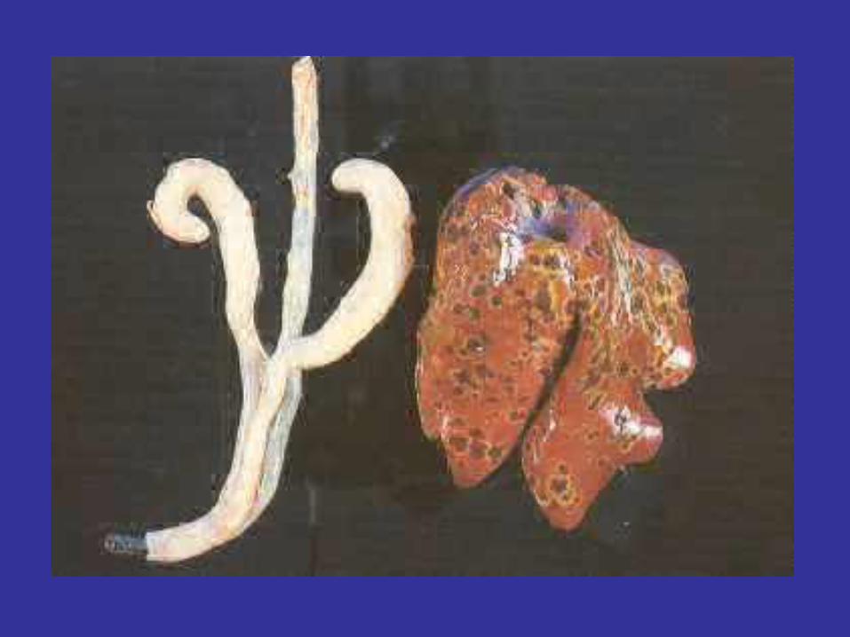
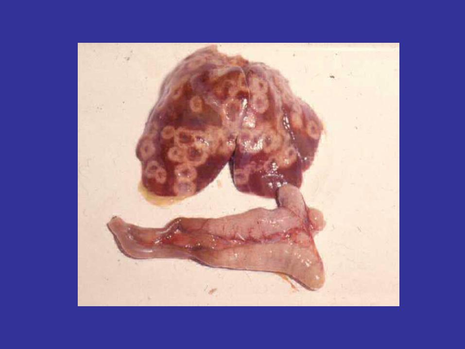
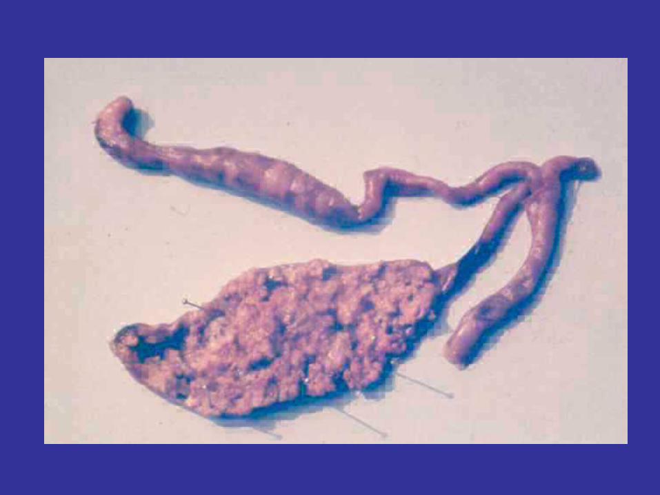
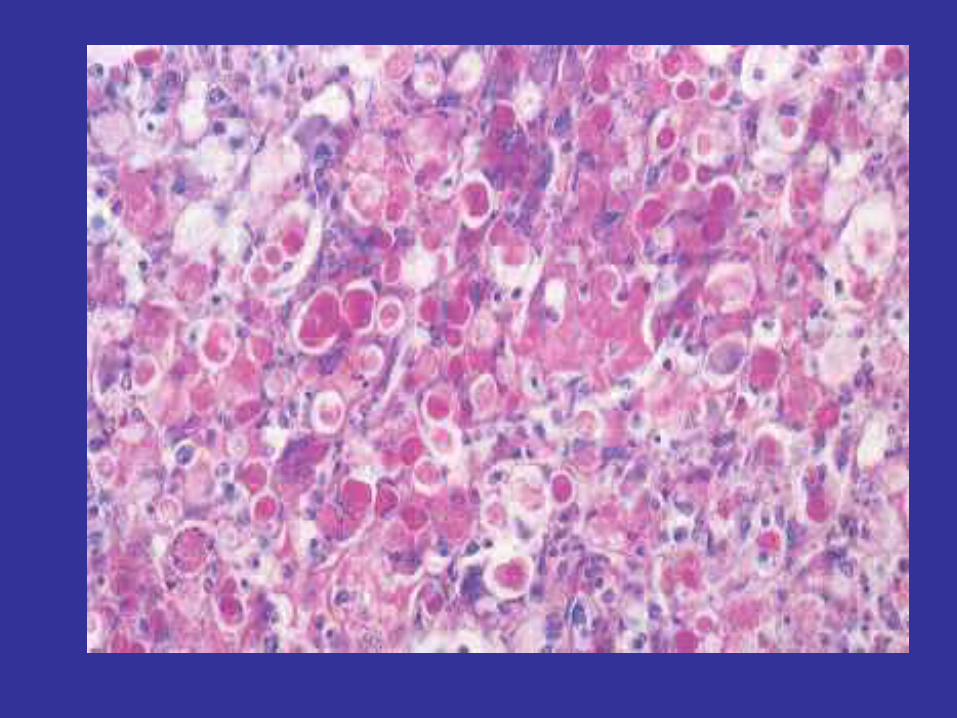
Histomonasis
Postmortem lesions
The caeca have an ulcerated (cheesy core) or
haemorrhagic exudates.
Crater-like liver lesions (bulls eye) and an enlarged
green coloured liver can also be seen.

HistomonasisTreatment and control:• Prevention• Enterohepatitis is controlled by controlling the spread of
the helminths, which spread disease. This is done by changing the litter or using antihelmintic drugs in the feed or water. Chickens and turkeys should always be kept separate to prevent introduction of caecal worms.
• Treatment• Dimetridazol (0.015%), Carbasone (0.025%),
Ipronidazole (0.00625%), Nitarsone (0.01875%) or Furazolidone (0.011%), are effective drugs, though not licensed in Western Europe.
Coccidiosis of Turkeys
• Infection of turkeys with Eimeria spp.
• This disease is not very common in commercially reared
turkeys though most turkey growers receive preventative
medication for at least part of their lives.
• Five species of Eimeria have been identified that cause
lesions in turkeys, of which two are associated with
significant disease effects.
Coccidiosis of Turkeys
• E. meleagrimitisE. meleagrimitis affects the upper small intestine
• E. adenoidesE. adenoides affects the caecae and rectum.
• E. gallopavonis lower small intestine rectum and caecae • E. meleagridis lower small intestine rectum and caecae
• E. dispersa in the small intestine
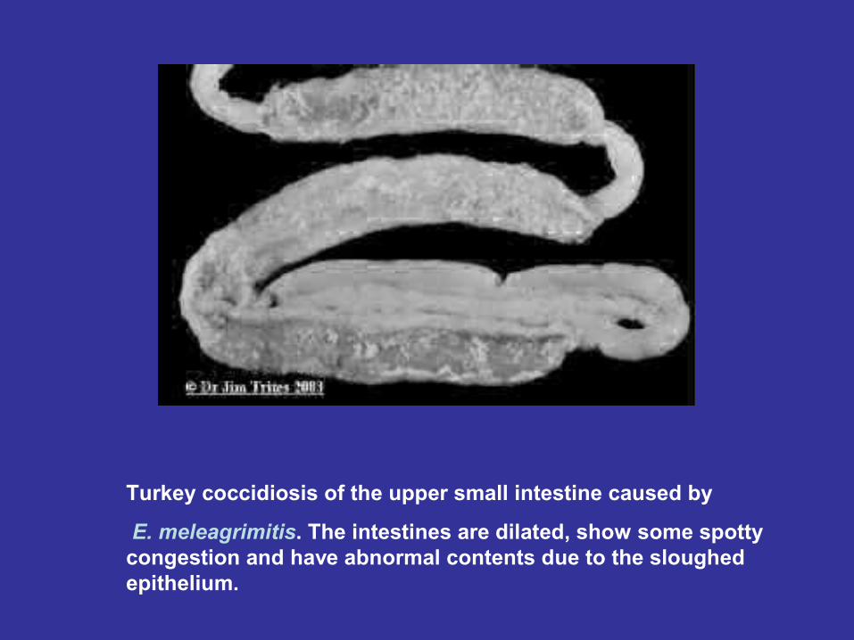
Turkey coccidiosis of the upper small intestine caused by
E. meleagrimitis. The intestines are dilated, show some spotty congestion and have abnormal contents due to the sloughed epithelium.
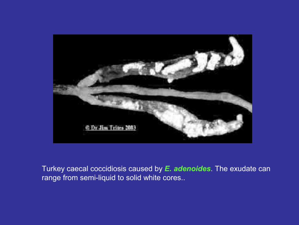
Turkey caecal coccidiosis caused by E. adenoides. The exudate can range from semi-liquid to solid white cores..