vertebral osteomyelitis associated with vre empyema...
TRANSCRIPT
Vertebral Osteomyelitis
Associated with VRE Empyema
Following Stabilization of
Unexplained Compression Fracture
Kaley
Tash, HMS3Gillian Lieberman, MD
Clinical Presentation•
45yoF c h/o
alcoholism, cirrhosis, hepatic
encephalopathy, pancreatitis, diabetes mellitus, gastroesophageal
reflux, recently
admitted to outside hospital for EtOH detoxification.
•
Returned from rehab with new acute back pain, bilateral leg spasms. No reported history of recent trauma.
•
Transferred to BIDMC following chest CT.
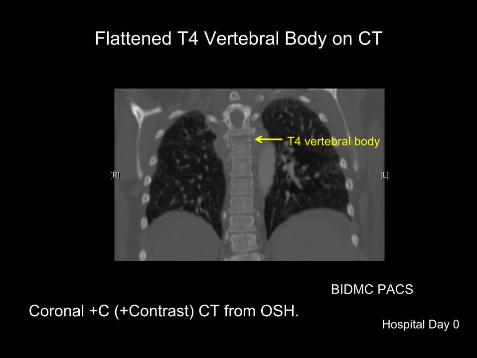
Coronal +C (+Contrast) CT from OSH.BIDMC PACS
Flattened T4 Vertebral Body on CT
Hospital Day 0
T4 vertebral body
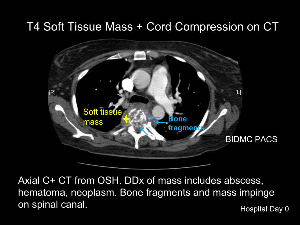
Axial C+ CT from OSH. DDx
of mass includes abscess, hematoma, neoplasm. Bone fragments and mass impinge on spinal canal.
BIDMC PACS
T4 Soft Tissue Mass + Cord Compression on CT
Soft tissue mass Bone
fragments
Hospital Day 0
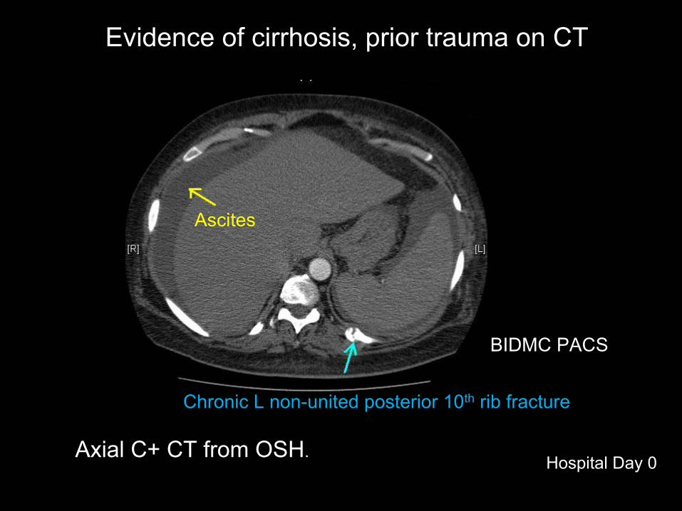
Evidence of cirrhosis, prior trauma on CT
BIDMC PACS
Ascites
Chronic L non-united posterior 10th
rib fracture
Hospital Day 0Axial C+ CT from OSH.

Pursued further imaging at BIDMC to guide likely surgical management.
MR uninterpretable
due to motion, so CT repeated.
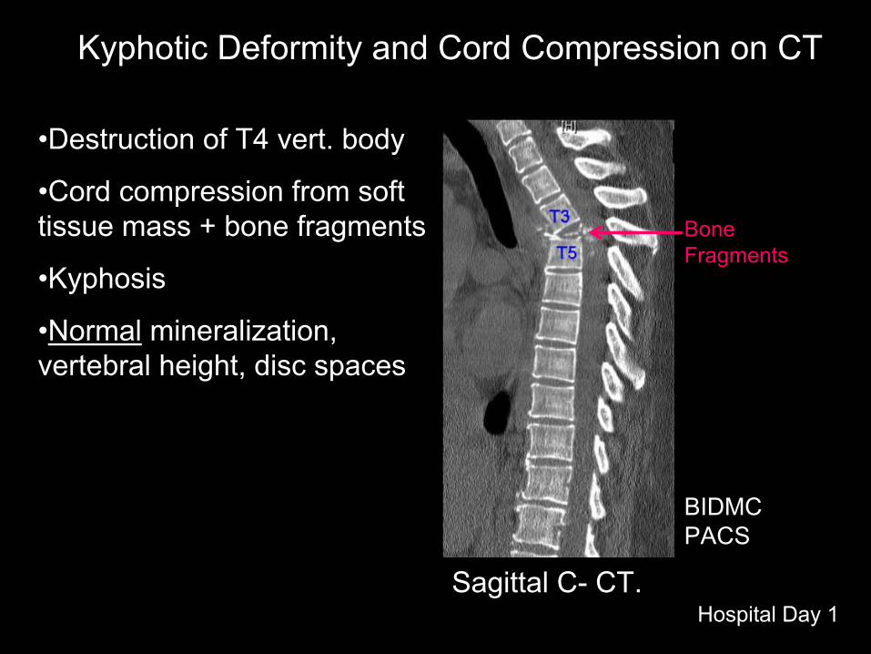
•Destruction of T4 vert. body
•Cord compression from soft tissue mass + bone fragments
•Kyphosis
•Normal
mineralization, vertebral height, disc spaces•.
BIDMC PACS
Kyphotic
Deformity and Cord Compression on CT
Hospital Day 1
Bone Fragments
Sagittal
C-
CT.
What destroyedT4?•
DDx: osteomyelitis, neoplasm, occult trauma (e.g., seizure*)
•
Blood cultures positive for Corynebacterium
in 1 of 4 bottles, suggestive of possible osteomyelitis.
•
Lack of diffuse spine disease and no recent trauma history raised strong suspicion for osteomyelitis, including Pott’s
disease.
•*Aboukasm AG and Smith BJ. 1997
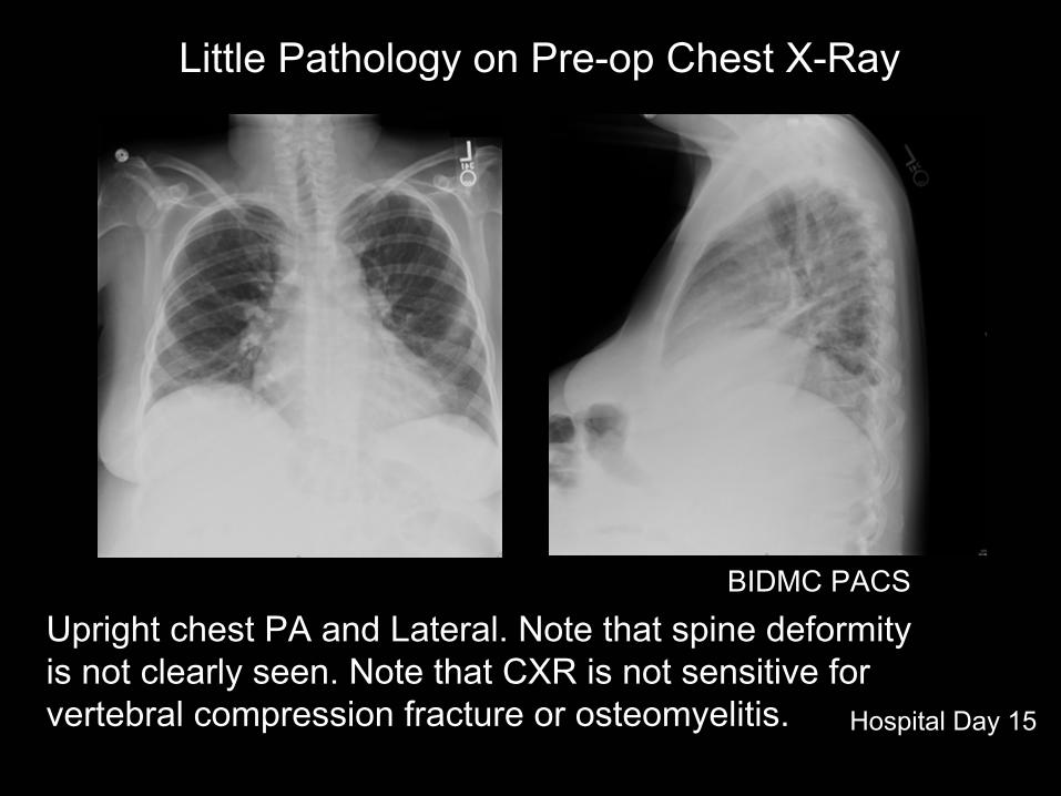
Upright chest PA and Lateral. Note that spine deformity is not clearly seen. Note that CXR is not sensitive for vertebral compression fracture or osteomyelitis.
BIDMC PACS
Little Pathology on Pre-op Chest X-Ray
Hospital Day 15
Anesthesia unable to ventilate pt in prone position with safe pressures, likely due to ascites
pushing against diaphragms.
Surgery rescheduled using anterior approach.
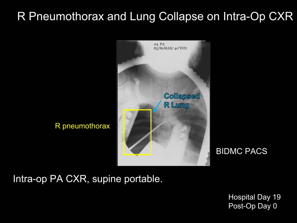
Intra-op PA CXR, supine portable.
BIDMC PACS
R Pneumothorax
and Lung Collapse on Intra-Op CXR
Hospital Day 19Post-Op Day 0
R pneumothorax
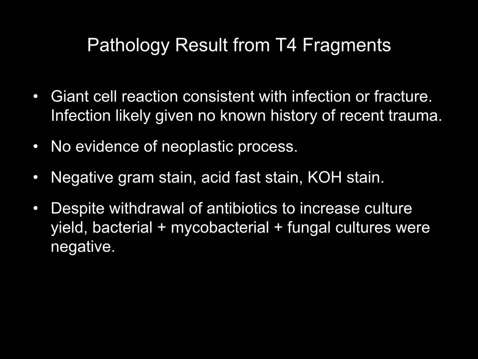
Pathology Result from T4 Fragments
•
Giant cell reaction consistent with infection or fracture. Infection likely given no known history of recent trauma.
•
No evidence of neoplastic
process.
•
Negative gram stain, acid fast stain, KOH stain.
•
Despite withdrawal of antibiotics to increase culture yield, bacterial + mycobacterial
+ fungal cultures were
negative.

R pleural fluid marginatingmediastinum
Decreased R lung volume
BIDMC PACS Hospital Day 19Post-Op Day 0
R Pleural Effusion and Pneumothorax on Post-Operative CXR
R pleural fluid
Ptx
Supine AP Portable CXR.

BIDMC PACS
Correction of Kyphotic
Deformity, Bone Fragments on CT
Hospital Day 19Post-Op Day 0
BIDMC PACS
Hospital Day 1
Bone Fragments
New Hardware
Sagittal
C-
CT.
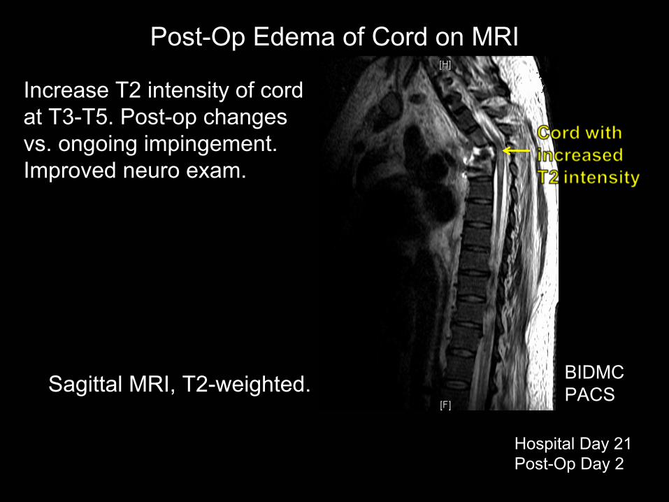
Increase T2 intensity of cord at T3-T5. Post-op changes vs. ongoing impingement. Improved neuro
exam.
Sagittal
MRI, T2-weighted. BIDMC PACS
Post-Op Edema of Cord on MRI
Hospital Day 21Post-Op Day 2

Patient experienced hypoxia while recovering from procedure and still required supplemental oxygen at post-
operative day 10. Team ordered CXR.
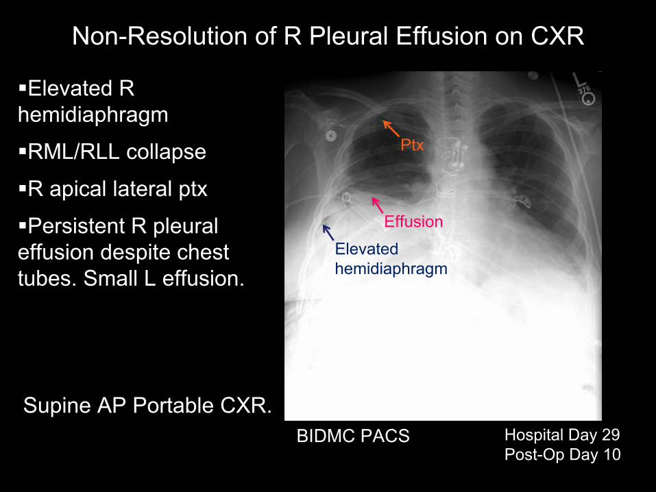
Elevated R hemidiaphragm
RML/RLL collapse
R apical lateral ptx
Persistent R pleural effusion despite chest tubes. Small L effusion.
BIDMC PACS Hospital Day 29Post-Op Day 10
Non-Resolution of R Pleural Effusion on CXR
Ptx
Elevated hemidiaphragm
Effusion
Supine AP Portable CXR.
Surgical team performed bedside pleurodesis
with doxycycline
x 2, then
ordered CXR to evaluate results.
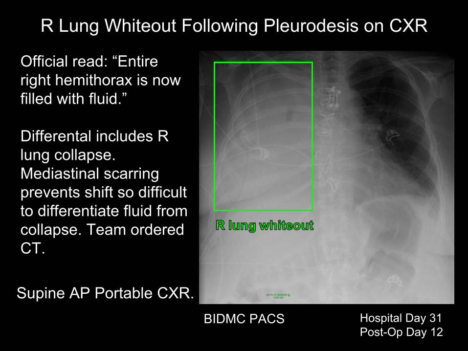
Official read: “Entire right hemithorax
is now
filled with fluid.”
Differental
includes R lung collapse. Mediastinal
scarring
prevents shift so difficult to differentiate fluid from collapse. Team ordered CT.
BIDMC PACS Hospital Day 31Post-Op Day 12
R Lung Whiteout Following Pleurodesis
on CXR
Supine AP Portable CXR.
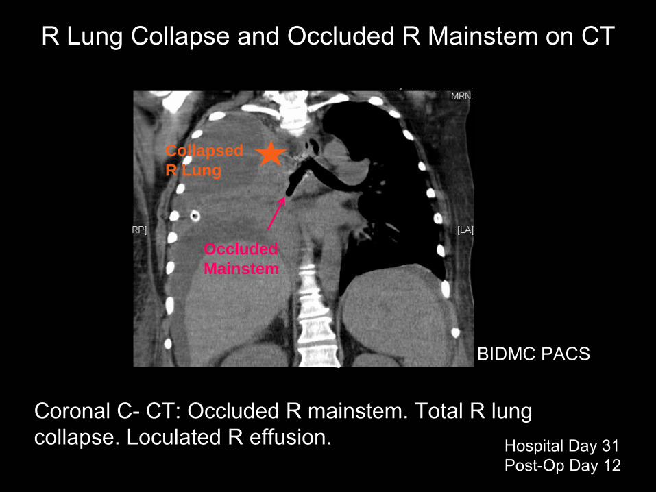
Coronal C-
CT: Occluded R mainstem. Total R lung collapse. Loculated
R effusion.
BIDMC PACS
R Lung Collapse and Occluded R Mainstem
on CT
Hospital Day 31Post-Op Day 12
Collapsed R Lung
Occluded Mainstem
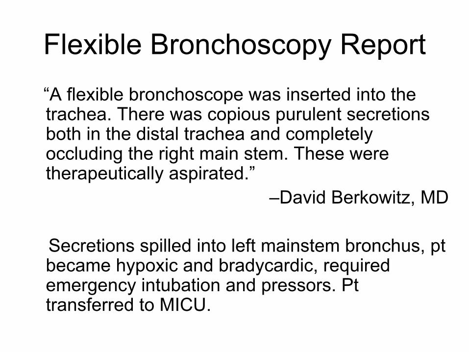
Flexible Bronchoscopy
Report“A flexible bronchoscope was inserted into the trachea. There was copious purulent secretions both in the distal trachea and completely occluding the right main stem. These were therapeutically aspirated.”
–David Berkowitz, MD
Secretions spilled into left mainstem
bronchus, pt became hypoxic and bradycardic, required emergency intubation and pressors. Pt transferred to MICU.
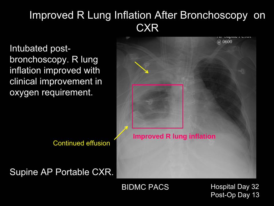
Intubated
post- bronchoscopy. R lung
inflation improved with clinical improvement in oxygen requirement.
BIDMC PACS Hospital Day 32Post-Op Day 13
Improved R Lung Inflation After Bronchoscopy
on CXR
Continued effusionImproved R lung inflation
Supine AP Portable CXR.
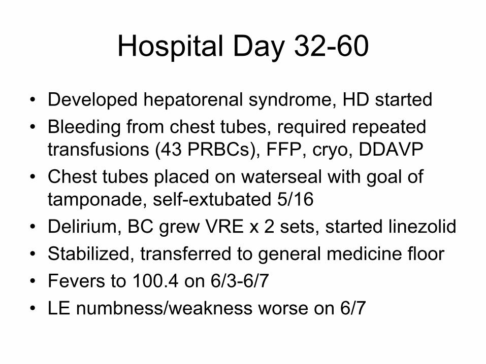
Hospital Day 32-60
•
Developed hepatorenal
syndrome, HD started•
Bleeding from chest tubes, required repeated transfusions (43 PRBCs), FFP, cryo, DDAVP
•
Chest tubes placed on waterseal
with goal of tamponade, self-extubated
5/16
•
Delirium, BC grew VRE x 2 sets, started linezolid•
Stabilized, transferred to general medicine floor
•
Fevers to 100.4 on 6/3-6/7•
LE numbness/weakness worse on 6/7
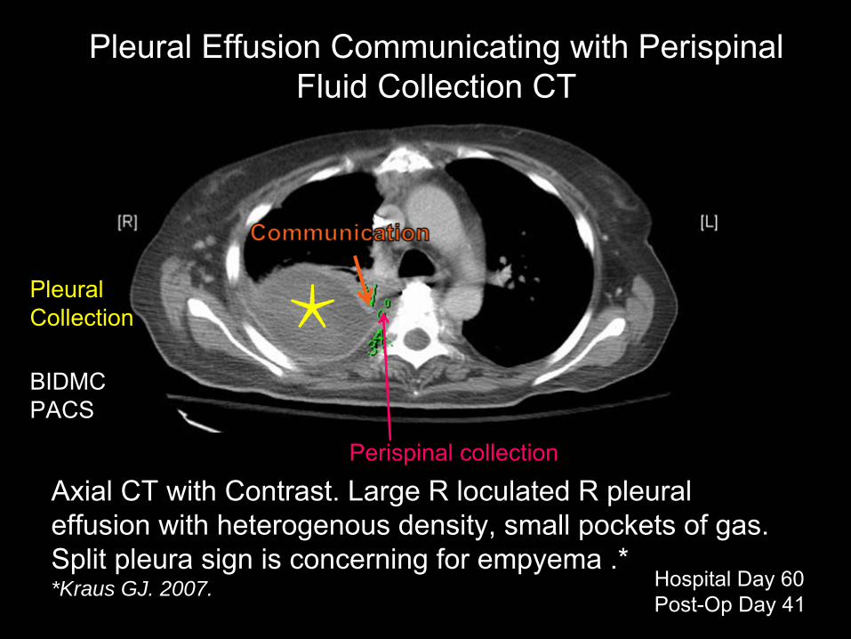
Axial CT with Contrast. Large R loculated
R pleural effusion with heterogenous
density, small pockets of gas.
Split pleura sign is concerning for empyema
.**Kraus GJ. 2007.
BIDMC PACS
Pleural Effusion Communicating with Perispinal Fluid Collection CT
Hospital Day 60Post-Op Day 41
Perispinal
collection
PleuralCollection
Sagittal
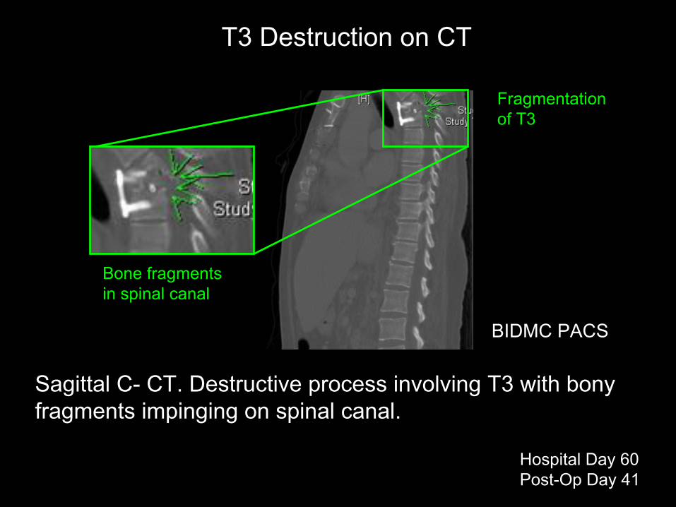
C-
CT. Destructive process involving T3 with bony fragments impinging on spinal canal.
BIDMC PACS
T3 Destruction on CT
Hospital Day 60Post-Op Day 41
Fragmentation of T3
Bone fragments in spinal canal
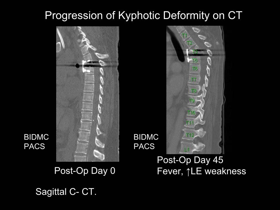
Post-Op Day 0Post-Op Day 45Fever, ↑LE weakness
BIDMC PACS
BIDMC PACS
Progression of Kyphotic
Deformity on CT
Sagittal
C-
CT.
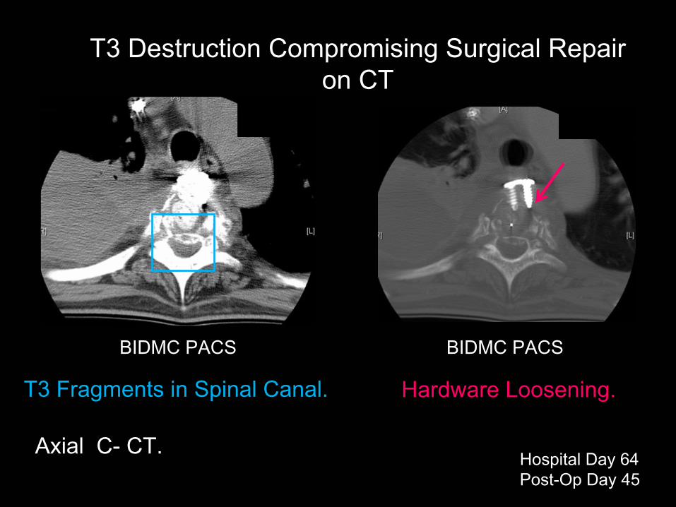
T3 Fragments in Spinal Canal.
BIDMC PACSBIDMC PACS
Hospital Day 64Post-Op Day 45
Hardware Loosening.
T3 Destruction Compromising Surgical Repair on CT
Axial C-
CT.
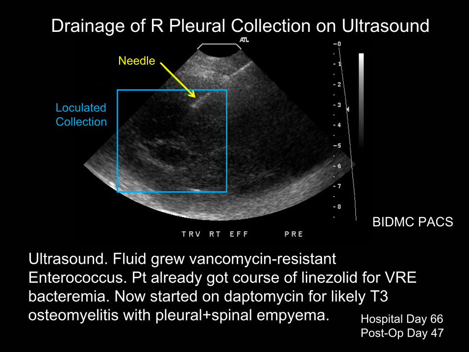
Ultrasound. Fluid grew vancomycin-resistant Enterococcus. Pt already got course of linezolid
for VRE
bacteremia. Now started on daptomycin
for likely T3 osteomyelitis
with pleural+spinal
empyema.
BIDMC PACS
Hospital Day 66Post-Op Day 47
Drainage of R Pleural Collection on Ultrasound
Needle
Loculated
Collection
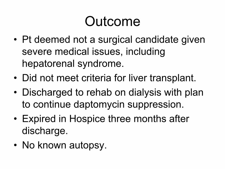
Outcome•
Pt deemed not a surgical candidate given severe medical issues, including hepatorenal
syndrome.
•
Did not meet criteria for liver transplant.•
Discharged to rehab on dialysis with plan to continue daptomycin
suppression.
•
Expired in Hospice three months after discharge.
•
No known autopsy.
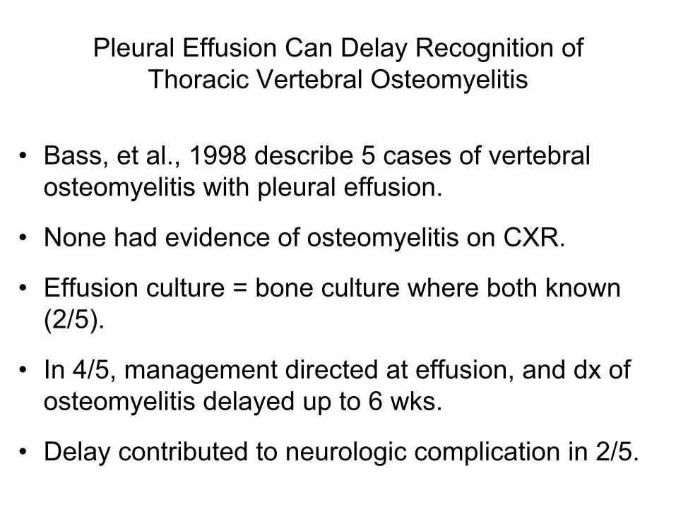
Pleural Effusion Can Delay Recognition of Thoracic Vertebral Osteomyelitis
•
Bass, et al., 1998 describe 5 cases of vertebral osteomyelitis
with pleural effusion.
•
None had evidence of osteomyelitis
on CXR.
•
Effusion culture = bone culture where both known (2/5).
•
In 4/5, management directed at effusion, and dx
of osteomyelitis
delayed up to 6 wks.
•
Delay contributed to neurologic complication in 2/5.
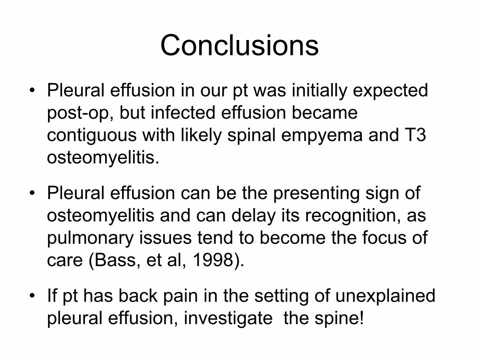
Conclusions•
Pleural effusion in our pt was initially expected post-op, but infected effusion became contiguous with likely spinal empyema
and T3
osteomyelitis.
•
Pleural effusion can be the presenting sign of osteomyelitis
and can delay its recognition, as
pulmonary issues tend to become the focus of care (Bass, et al, 1998).
•
If pt has back pain in the setting of unexplained pleural effusion, investigate the spine!
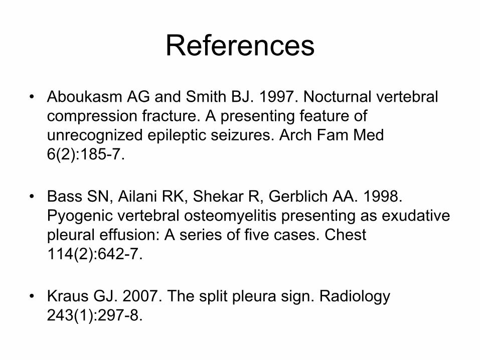
References•
Aboukasm
AG and Smith BJ. 1997. Nocturnal vertebral
compression fracture. A presenting feature of unrecognized epileptic seizures. Arch Fam
Med
6(2):185-7.
•
Bass SN, Ailani
RK, Shekar
R, Gerblich
AA. 1998. Pyogenic
vertebral osteomyelitis
presenting as exudative
pleural effusion: A series of five cases. Chest 114(2):642-7.
•
Kraus GJ. 2007. The split pleura sign. Radiology 243(1):297-8.
Acknowledgements
•
James Knutson, MD•
Gillian Leiberman
•
Maria Levantakis