ventricles and meninges bhatnagar, s. c. (2008); chapter 18
TRANSCRIPT

Ventricles and Meninges
Bhatnagar, S. C. (2008); Chapter 18

Meninges
Three concentric, fibrous tissue layers covering the CNS – Duramater, arachnoid membrane and piamater.
The outer, gray duramater is the toughest among the meninges.
In the brain the dura mater has two spaces: Epidural – space between the skull and duramater Subdural – Space between the dura and arachnoid
The duramater has two layers – External periosteal and internal meningeal

Meninges

Meninges
The duramater forms three cavities as it covers the brain – dural extensions. Falx cerebri – is the largest of the dural extensions
and it projects into the longitudinal fissure that separates the two cerebral hemispheres.
Tentorium Cerebelli - Separates the cerebellum from the inferior portion of the occipital lobes.
Falx Cerebelli - is a small triangular extension of the tentorium cerebelli that separates the two cerebellar hemispheres.

Meninges

Meninges

Meninges
Pathology to the dura of the brain Subdural hematoma: Abnormal collection of
blood between the dura and the arachnoid, usually as a result of torn veins secondary to head trauma.
Epidural hematoma: A collection of blood between the dura and the inner surface of the skull, and is usually due to arterial bleeding.

Meninges
Arachnoid membrane of the brain is a delicate, non-vascular membrane.
The subarachnoid space lies between the arachnoid membrane and the pia mater. Covers the whole CNS Is filled with CSF produced by the ventricular system which
enters through the foramina of the fourth ventricle. Arachnoid membrane is separated from the dura
mater by the subdural space. Arachnoid membrane finger-like projections within this
layer are known as the arachnoid villi. The CSF drains back to the ventricular system through
these villi.

Meninges
Pia mater is the thin, transparent, collagenous, innermost membrane. It is closely attached to the brain tissue and
follows its contours. The brains blood vessels penetrate the pia mater.
Togather the pia mater and arachnoid membrane are known as Leptomeninges while the dura mater is known as the Pachymeninx.

Meninges of the spinal chord
All 3 meninges cover the spinal chord. The spinal dura is rostrally attached to
the foramen magnum (an opening in the occipital bone through which the medulla emerges out of the skull).
Some differences: Spinal duramater is single layered (does not
have the periosteal layer) Spinal dura is a relative loose layer,
punctured with the exiting spinal nerves. Anesthetic agents are injected to the
epidural space for anesthetizing the lower body.
Meninges of the spinal chord
The spinal arachnoid membrane extends all the way to the cauda equina and the subarachnoid space, similar to that of the brain, is filled with CSF.
The spinal pia mater continues with the filum terminale after the conus medullaris until the sacral level of the vertebrae.
Spinal taps (lumbar puncture; extraction of CSF for testing) are performed at the level of L1 or L2 (lumbar cistern) by puncturing the dura and arachnoid space to enter the subarachnoid space.

Meninges of the spinal chord

Ventricles
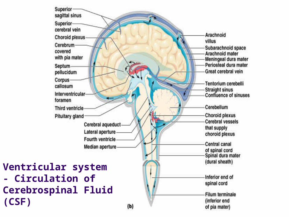
The ventricular system is a set of structures in the brain continuous with the central canal of the spinal cord.
There are four cerebral ventricles: the paired lateral ventricles and the third and fourth ventricle.
Primary function: Circulate Cerebrospinal fluid (CSF). CSF is produced in the choroid plexus of each ventricle by
modified ependymal cells that line the inner membranes. CSF bathes and cushions the brain and spinal chord within
their bony confines during rapid body movements.

Ventricles

Ventricular system
Lateral view Dorsal view

Ventricular system

Ventricular system
The lateral ventricles both communicate via the intraventricular foramen (or foramina of Monro) with the third ventricle, found centrally within the midbrain.
The third ventricle communicates via the cerebral aqueduct with the fourth ventricle.
The two lateral ventricles are relatively large and C-shaped. Roughly wrapping around the dorsal aspects of the
basal ganglia. The roof of each lateral ventricle is formed by the
fibers of the corpus callosum while the floor is formed by the superior surface of the thalamus.

Ventricular system
Each lateral ventricle has a central body and extends into the frontal, temporal and occipital lobes via the frontal (anterior), temporal (inferior), and occipital (posterior) horns, respectively.
The third ventricle is located between the thalamic nucleus and is connected to the fourth ventricle through the cerebral aqueduct. Cerebral aqueduct is ventral to the corpora
quadrigemina of the midbrain.

Ventricular system
The fourth ventricle is located in the lower brainstem. Floor – tegmentum of the pons and medulla Roof - cerebellum
From the fourth ventricle, CSF can pass into the central canal of the spinal cord or into the subarachnoid space of CNS via three small foramina: The central foramen of Magendie and the two lateral foramina of Luschka.

Ventricular system The CSF (of the subarachnoid
space) flows around the superior sagittal sinus to be reabsorbed via the arachnoid villi into the venous system. The superior sagittal sinus
lies within the superior border of the falx cerebri, a two-layered dural structure separating the two cerebral hemispheres.
Arachnoid villi (or granulations) are small protrusions of the arachnoid through the dura layer of the meninges.
Arachnoid villi
Ventricular system - Circulation of Cerebrospinal Fluid (CSF)

CSF
Prevents most blood-borne toxins from entering the brain Impermeable capillaries
Not an absolute barrier Nutrients such as oxygen pass through Allows alcohol, nicotine, and anesthetics through
CSF is normally a clear, amazingly ‘bright’ fluid. If it is cloudy or contains a raised level of protein or traces
of blood - An indication of brain infection, some types of brain or spinal cord tumor, or trauma.

Ventricular system
CSF within the spinal cord can flow all the way down to the end of the cord around the cauda equina where lumbar punctures are performed.
The aqueduct between the third and fourth ventricles is very small, as are the foramina. A blockage will cause high pressure in the lateral
ventricles – Hydrocephalus (otherwise known as water on the brain)
This is an extremely serious condition (although treatable) due to both the damage caused by the pressure (ie., increased intra-cranial pressure) as well as nature of whatever caused the block

Lumbar Puncture or Spinal tap
•Most common purpose – Collect CSF in a case of suspected meningitis. •Commonly performed in children who have a standing fever without a source

CT scan of hydrocephalus and normal Brain

Ventricular system
An increased intracranial pressure can often be recognized by looking into the eye with an ophthalmoscope (also known as Retinography) An instrument which shines a
beam of light on to the retina at the back of the eye to visualize the optic disc, retina, and blood vessels supplying the eye.

Ventricular system
Normally this appears as a clearly-defined, pale concave disc, but if the pressure in the CSF is raised, the disc may bulge forwards into the cavity of the eye.
Increased pressure can also be caused by an expanding tumor or blood clot, or by swelling of a damaged or diseased brain.
Normal Fundus
Abnormal Fundus