vegf receptor promotes hypoxia-induced hematopoietic
TRANSCRIPT

VEGF receptor promotes hypoxia-inducedhematopoietic progenitor proliferation anddifferentiationJonathan Florentin
University of PittsburghScott P. O’Neil
University of PittsburghSathish Babu Vasamsetti
University of PittsburghLee L. Ohayon
University of PittsburghAnagha Arunkumar
University of PittsburghSamit Ghosh
University of PittsburghJennifer C. Boatz
University of PittsburghJustin Sui
University of PittsburghCorrine R. Kliment
University of PittsburghStephen Y. Chan
University of PittsburghPartha Dutta ( [email protected] )
University of Pittsburgh
Research Article
Keywords: obstructive sleep apnea (OSA), chronic obstructive pulmonary disease (COPD), VEGF,metabolic, cardiovascular, neoplastic, neurologic disease
Posted Date: October 27th, 2021
DOI: https://doi.org/10.21203/rs.3.rs-1000955/v1

License: This work is licensed under a Creative Commons Attribution 4.0 International License. Read Full License

VEGF receptor promotes hypoxia-induced hematopoietic progenitor proliferation and
differentiation
Jonathan Florentin1*, Scott P. O’Neil1*, Sathish Babu Vasamsetti1, Lee L. Ohayon1, Anagha
Arunkumar1, Samit Ghosh2, Jennifer C. Boatz2, Justin Sui2, Corrine R. Kliment2, Stephen Y.
Chan1, Partha Dutta1, 3
1 Pittsburgh Heart, Lung, Blood, and Vascular Medicine Institute, Division of Cardiology,
Department of Medicine, University of Pittsburgh School of Medicine, University of Pittsburgh
Medical Center, Pittsburgh, PA 15213
2 Department of Medicine, Division of Pulmonary and Critical Care, University of Pittsburgh,
Pittsburgh, PA 15260, USA
3 Department of Immunology, University of Pittsburgh School of Medicine, Pittsburgh, PA 15213
*These authors contributed equally to this work.
Corresponding Author:
Dr. Partha Dutta, DVM, PhD
Assistant Professor of Medicine and Immunology
Vascular Medicine Institute
Division of Cardiology, Department of Medicine
University of Pittsburgh Medical Center
BST 1720.1
200 Lothrop Street
Pittsburgh, PA 15213, USA
Tel: 412-383-7277
Email: [email protected]
Abstract
Intermittent or chronic hypoxia is associated with morbidity and mortality in many diseases such
as obstructive sleep apnea (OSA) and chronic obstructive pulmonary disease (COPD),
respectively. We found exaggerated inflammation with heightened numbers of inflammatory
leukocytes in these patients compared with age-matched controls. Chronically hypoxic mice
exhibited similar trend and induced proliferation of hematopoietic stem and progenitor cells
(HSPC), precursors of inflammatory leukocytes. Consistently, a transcriptomic analysis of human
HSPC exposed to hypoxic conditions revealed elevated expression of genes involved in progenitor
mobilization. Additionally, bone marrow cells in mice under hypoxic conditions expressed high
amount of vascular endothelial growth factor (VEGF), VEGF receptor (VEGFr) and its target
genes in hypoxic conditions. In silico and ChIP experiments demonstrated that HIF-1⍺ binds to
the promoter region of VEGFr. These results indicate that VEGF signaling in HSPC is an
important mediator of their proliferation and differentiation in hypoxia-induced inflammation and
represents a potential therapeutic target to prevent aberrant inflammation in hypoxia-associated
diseases.

Introduction
Diseases characterized by alveolar hypoxia, such as chronic obstructive pulmonary disease
(COPD) and obstructive sleep apnea (OSA), are highly prevalent and contribute to significant
morbidity and mortality around the world 1-5. OSA is defined by intermittent nocturnal apneic or
hypopneic events that result in impaired alveolar ventilation and subsequent hypoxemia6. COPD
is a highly heterogenous condition characterized by airway inflammation and progressive
obstruction due to noxious environmental exposures (e.g., cigarette smoke), which result in
chronic subclinical and acute severe hypoxia 1. There is a notable overlap in COPD and OSA
comorbidities including metabolic, cardiovascular, neoplastic and neurologic disease, which a
growing body of literature attributes to systemic inflammation 1,7-9. To date, studies of alveolar
hypoxia induced inflammation have focused primarily on the role of resident macrophages, and
their interaction with alveolar endothelial cells mediating production of inflammatory cytokines
10-13. Monocyte chemoattractant protein 1 (MCP-1), c-reactive protein, tumor necrosis factor alpha
(TNF⍺), IL-1, IL-6 and IL-8 are increased in hypoxic lung diseases and generally correlate with
severity of disease 12,14,15. In both OSA and COPD, correction of alveolar hypoxia, with nocturnal
continuous positive airway pressure and supplemental oxygen, respectively, remain the mainstay
therapies 6,16-18. While there is strong evidence that local and systemic inflammation are key
components of hypoxic diseases, a mechanistic role of hypoxia in exaggerated inflammation has
yet to be defined.
Mounting evidence has demonstrated key inflammatory cells involved in the pathogenesis of
various diseases including insulin resistance and atherosclerosis4,19-21. These cells have been
proposed to originate from hematopoietic stem and progenitor cells (HSPC) in the bone marrow
and spleen22-25. Patients with hypoxic lung disease exhibit elevated inflammation and leukocytosis.

The leukocytosis observed in chronic inflammation likely represents production of inflammatory
leukocytes by their progenitors. However, a direct link between hypoxia induced systemic
inflammation and proliferation of HSPC has not been established. Furthermore, the mechanisms
of HSPC proliferation and differentiation in hypoxia have not been studied.
Vascular endothelial growth factor (VEGF) has been identified as an important regulator of
neovascularization by encouraging endothelial cell proliferation in various diseases such as age-
related macular degeneration and neoplastic diseases 26-29. A number of recent studies have
demonstrated elevated VEGF concentrations in peripheral blood of patients with hypoxic lung
disease, which closely correlated with severity of disease 30-35. However, the role of VEGF in HSC
proliferation and inflammatory leukocyte production is not known.
Patients with OSA and COPD, characterized by alveolar hypoxia, displayed increased circulating
myeloid cell numbers. We used two independent mouse models of hypoxia to understand the effect
of hypoxia in HSPC proliferation: C57BL/6 mice exposed to A) 10% O2 for three weeks, an
established mouse model of chronic hypoxia 36-38 and B) cigarette smoke for six months, a COPD
mouse model 39,40. Mice under chronic hypoxic conditions and exposed to cigarette smoke had
elevated numbers of neutrophils, monocytes and macrophages in the blood and lungs suggesting
augmented hematopoiesis. In line with this, a whole genome RNA sequencing analysis of human
HSPC cultured under hypoxic conditions showed increased expression of the genes involved in
hematopoiesis. The number and proliferation of HSPC were increased when mice were exposed
to hypoxia or cigarette smoke. Concomitantly, we measured increased expression of VEGFr in
these activated progenitors indicating a possible role of VEGFr in hematopoiesis. Indeed, VEGFr

blockade in vitro and in vivo diminished HSPC proliferation and their differentiation into myeloid
cells, resulting in attenuated inflammation in hypoxic conditions. Mechanistically, we observed
that HIF-1⍺ binds to the VEGFr promoter and increases its expression in HSPC under hypoxic
conditions. HIF1a silencing reduced HSPC proliferation and their differentiation into
inflammatory leukocytes.
As a whole, the present study describes a role of the VEGFr in the activation of hematopoietic
progenitors and the production of inflammatory myeloid cells in hypoxia. Additionally, this work
proposes the VEGFr as a possible therapeutic target to attenuate inflammatory burden in patients
with hypoxia.

Results
Patients and animals with hypoxia have elevated numbers of inflammatory leukocytes in the blood.
Various diseases associated with intermittent or chronic hypoxia are characterized by exaggerated
systemic inflammation 5-7,20,21,41-43. Leukocytes, primarily myeloid cells, play a major role in
inflammatory diseases including atherosclerosis and diabetes6,19. To ascertain systemic
inflammation in patients with hypoxic lung diseases, we performed a retrospective chart review of
patients with OSA characterized by intermittent hypoxia and COPD marked by chronic hypoxia,
and age-matched control patients to enumerate inflammatory cells. The total numbers of
neutrophils, lymphocytes, and monocytes were significantly increased in the patients with OSA
and COPD compared to the healthy controls (Figure 1 & Suppl. Figure 1A). The level of
intermittent hypoxia has been shown to be linked with oxygen saturation or SpO2. Multiple studies
evaluated SpO2 in patients with COPD and OSA. The average lung SpO2 during the daytime is
89% in COPD patients 44,45. An OSA patient is diagnosed to have SpO2 ≤90%, and a number of
desaturation events and patterns (≥3 similar desaturation/saturation cycles, 15 min time scale) are
≥4 and 12, respectively 46. Of note, the oxygen saturation levels of OSA and COPD patients
included in this study were significantly reduced compared to healthy controls (Suppl. Figure
1B), indicating hypoxia in these patients. These results were confirmed in hypoxic mice which had
a significantly lower oxygen saturation than the normoxic controls (Suppl. Figure 1C).
Additionally, to understand if COPD elevates VEGFR expression in hematopoietic progenitors
and increases lineage output, we employed C57BL/6 mice exposed to cigarette smoke for six
months, an established mouse model of COPD40,47. These mice contained significantly heightened
numbers of lymphocytes, such as B and T cells, in the bone marrow while myeloid cell content
decreased (Suppl. Figure 1D). In contrast, splenic lymphocyte numbers were unaltered (Suppl.

Figure 1E), and myeloid cells were more numerous in smoke-exposed mice compared to the
control group (Suppl. Figure 1E). These data indicate that smoke exposure differentiates bone
marrow progenitors into hematopoietic cells. Furthermore, the data suggest a preferential egress
of myeloid cells from the bone marrow after smoke exposure.
To further delineate the contributions of hypoxia in systemic inflammation, we used a mouse
model of chronic hypoxia. We quantified inflammatory cytokines and leukocytes in mice exposed
to hypoxic conditions compared to normoxic controls. We observed increased expression of Il-
1b, Il-6, Il-18 and Tnfa in the lungs of hypoxic mice (Figure 2A). Consistent with this heightened
expression of the inflammatory cytokine genes, we found elevated concentrations of Il-6 in
peripheral blood and lung parenchyma (Figure 2B). Furthermore, we found that mice in chronic
hypoxic conditions had increased numbers of monocytes, neutrophils and lymphocytes in the
blood compared to normoxic control mice (Figure 2C and Suppl. Figure 2A and B). Similarly,
the lungs of hypoxic mice harbored augmented numbers of inflammatory cells, including
interstitial macrophages (Figure 2D and Suppl. Figure 2C), which are reported to have
pathologic roles in pulmonary diseases characterized by hypoxia48-50. Altogether, these data
demonstrate leukocytosis in patients and mice with hypoxia.
Hypoxia increases proliferation of hematopoietic stem and progenitor cells.
Increased leukocyte numbers in the blood of hypoxic mice may indicate two underlying
mechanisms: A) accelerated egress of bone marrow leukocyte into the blood and B) escalated
production of leukocytes by hematopoietic stem and progenitor cells (HSPC) in the bone marrow.
We found that although hypoxia increased bone marrow leukocyte numbers, there was no

significant difference in the ratios of blood and bone marrow leukocyte numbers between
normoxic and hypoxic mice (Suppl. Figure 2D), suggesting that egress of leukocytes from the
bone marrow is unaltered in hypoxic conditions. To determine whether the observed hypoxia-
induced leukocytosis represents increased production of mature leukocytes, we assessed the
number and proliferation of bone marrow HSPC, which consist of hematopoietic stem cells (HSC),
lineage- Sca-1+ c-Kit+ (LSK or HSPC) cells and granulocyte macrophage progenitors (GMP)
(Suppl. Figure 3). Flow cytometry analysis revealed that hypoxia expanded the numbers and
frequencies of these cells in the femur (Figure 3A). Confocal microscopy confirmed a higher
density of HSPC in the bone marrow of hypoxic mice compared to the normoxic control (Figure
3B). In line with augmented leukocyte and HSPC numbers, mice in hypoxic conditions had higher
proportions of proliferating HSPC (Figure 3C-E). Additionally, we isolated HSPC and GMPs
from mice kept under either normoxic or hypoxic conditions for three weeks. qPCR for the cell
cycle genes revealed that the progenitors expressed higher levels of these genes in hypoxic
conditions (Suppl. Figure 4A). HSPC proliferation is prerequisite for their differentiation into
leukocytes. To understand if HSPC expansion in hypoxic conditions could result in their higher
differentiation, we analyzed a whole genome transcriptome data comparing human CD34+ HSPC
cultured under hypoxic vs. normoxic conditions 51. A pathway analysis revealed that the genes
involved in differentiation of stem cell, cell cycle and stimulation of cells were enriched in HSPC
in hypoxic conditions (Suppl. Figures 4B-7). To discern if hypoxia accelerates HSPC
differentiation, we adoptively transferred GFP+ HSPC into mice housed under normoxic and
hypoxic conditions, and enumerated progenies by flow cytometry. The bone marrow, blood and
lungs of hypoxic mice contained augmented percentages of donor-derived differentiated
leukocytes, lymphocytes, monocytes and neutrophils (Figure 3F and Suppl. Figure 8A). This

difference of unleashed leukocyte production could not be attributed to higher engraftment of
GFP+ HSPC under hypoxia (Suppl. Figure 8B). In summary, these data indicate that hypoxia
drives HSPC into the cell cycle and increases their differentiation into inflammatory cells.
VEGFr expression is increased in progenitor cells following hypoxia exposure and in a COPD
mouse model.
The role of VEGF/ VEGFr in hypoxia-induced angiogenesis is well-documented 33,52,53. To
investigate the possible role of VEGF-A/VEGFr in hypoxia-mediated HSPC proliferation, we first
assessed RNA expression of these genes in the whole bone marrow of hypoxic mice. The
expression of Vegfa, but not Vegfr, was increased in bone marrow cells of hypoxic mice compared
to their normoxic counterparts (Figure 4A). Interestingly, the expression of Vegfr mRNA and
protein in bone marrow HSC and GMP increased in hypoxia (Figure 4B & 4C). In line with these
results, in a mouse model of COPD, we observed that mice exposed to cigarette smoke harbored
higher frequency of Vegfr+ HSPC and GMP (Suppl. Figure 9A). VEGFR expression in the
progenitors also increased (Suppl. Figure 9B). Next, we identified the genes downstream to the
VEGFr signaling using Ingenuity Pathway Analysis (Suppl. Figure 9C). The expression of these
genes was elevated in HSPC cultured under hypoxic conditions (Figure 4D). These data indicate
that hypoxia increases VEGFr signaling in HSPC. Hypoxia-inducible factor 1⍺ (HIF-1⍺), a key
transcription factor increased in hypoxia, binds to several genes, and increases their expression 54-
57. We observed increased Hif1a expression (Suppl. Figure 9D) in BM HSPC and GMP of
hypoxic mice. Flow cytometry (Suppl. Figure 9E) revealed increased numbers of Hif-1⍺+ HSPC
and GMP in hypoxic mice. Additionally, we confirmed decreased expression of prolyl-2
hydroxylase, which accelerates ubiquitination and lysosomal degradation of Hif-1⍺, in HSPC of

hypoxic mice (Suppl. Figure 9F). Corresponding to the fact that Hif-1⍺ increases glycolysis, we
observed elevated expression of glycolytic genes in bone marrow HSPC of hypoxic mice (Suppl.
Figure 9G). To decipher the mechanisms of increased Vegfr expression in hypoxia, we ascertained
if the VEGFR promoter region has HIF-1⍺ binding sites. Our in silico analysis revealed that HIF-
1⍺ has several binding sites on the VEGFr promoters in both humans and mice (Figure 4E).
Chromatin immunoprecipitation experiments confirmed that HIF-1⍺ binds to the Vegfr promoter
in mouse HSPC (Figure 4F). HIF-1⍺ inhibition significantly diminished hypoxia-mediated
expression of VEGFr in human (Figure 4G) and mouse (Figure 4H) HSPC. To delineate the role
of HIF-1⍺ in hypoxia-induced HSPC proliferation, we silenced this transcription factor in mouse
HSPC and observed significant downregulation of the genes involved in cell cycle progression
(Figure 4I) as well as the genes downstream to the VEGFr signaling (Suppl. Figure 9H). In
aggregate, these data indicate that HIF-1⍺ directly binds to the VEGFr promoter in HSPC,
increases VEGFr expression and augments hematopoietic progenitor proliferation under hypoxic
conditions.
VEGFr inhibition decreases HSPC proliferation.
The role of the VEGF signaling is well documented in angiogenesis in different diseases such as
cancer and age-related macular degeneration 27-29,53,58. However, the contribution of VEGFr in
HSPC proliferation has not been studied. To understand the role of VEGFr in hypoxia-mediated
HSPC proliferation, we cultured HSPC sorted from mouse bone marrow with Sugen 5416, a
VEGFr inhibitor, under hypoxic and normoxic conditions. VEGFr inhibition significantly
decreased the expression of cell cycle genes responsible for HSPC proliferation (Figure 5A and
Suppl. Figure 10). To further examine the role of VEGFr signaling in proliferation of HSPC, we
assessed proliferation of GMP in hypoxic mice injected with the VEGFr inhibitor. Compared to

untreated controls, VEGFr inhibitor-injected mice had lower proportions of GMPs in the S-G2-M
cell cycles (Figure 5B & 5C). These mice had increased numbers of quiescent cells in the G0
phase. Interestingly, VEGFr inhibition also increased the frequency of GMP in the G1 phase,
indicating a G1 to S-G2M arrest in the absence of VEGFr signaling. Additionally, there was a
significant reduction in GMP numbers in the bone marrow of hypoxic mice after VEGFr inhibition
(Figure 5D).
VEGFr inhibition decreased inflammation and inflammatory cell numbers.
We wanted to evaluate whether the decreased proliferation of HSPC after VEGFr inhibition would
have an impact on inflammatory leukocyte generation. To this end, we differentiated HSPC
isolated from mouse bone marrow in presence of the VEGFr inhibitor in hypoxic and normoxic
conditions. VEGFr inhibition in hypoxic and normoxic HSPC significantly suppressed their
differentiation into lymphoid cells, myeloid cells, B cells, monocytes and neutrophils (Figure 6A
and Suppl. Figure 11A). Next, we investigated if VEGFr inhibition decreases hypoxia-induced
leukocytosis in vivo. Compared to untreated control mice, VEGFr inhibitor-treated mice had
decreased percentages and numbers of monocytes and neutrophils in peripheral blood and bone
marrow (Suppl. Figure 11B and C and Figure 6B & 6C). Additionally, we utilize an additional
loss of function approach since Sugen is selective rather than specific for VEGFR2 and also
inhibits PDGFR, KIT, RET, FLT-3, ABL and ALK. We silenced Vegfr2 in sorted murine HSPC
placed in hypoxia for 24 hours. We found that the ability of HSPC to differentiate into mature
leukocytes, especially B cells, myeloid cells, monocytes and neutrophils, was reduced in HSPC
treated with siVegfr compared to siCtl (Suppl. Figure 12A). Additionally, we found decreased
expression of cell cycle check point genes such as Cdk3, Cdk4, and Mki67 after Vegfr knock down

(Suppl. Figure 12B). Overall, these data indicate the importance of VEGFr in the proliferation
and differentiation of HSPC into leukocytes under hypoxic conditions.

Discussion
Patients with hypoxic lung disease, including COPD and OSA, demonstrate chronic inflammation
evidenced by elevated inflammatory cytokine levels and inflammatory leukocyte numbers. The
degree of systemic inflammation portends clinical outcomes of hypoxic lung disease as well as
severity of comorbid conditions7,12,42,59,60. There is growing evidence that correction of underlying
hypoxia–for example with CPAP in OSA–can reduce systemic inflammation highlighting the
importance of hypoxia in inflammation61.
Consistent with serum cytokine levels observed in COPD and OSA, we observed increased levels
of IL-1b, IL-6, IL-18, and TNF-a in peripheral blood of mice exposed to hypoxia. Both TNF-a
and IL-6 have been shown to be independent risk factors of increased morbidity and mortality in
hypoxic lung disease, particularly in COPD62. IL-6 has been shown to promote proliferation of
HSC 63, but interestingly, TNF-⍺ has exhibited a contrasting function regulating hematopoiesis 64.
Systemic inflammation has long been postulated to drive the pathogenesis of diseases such as
atherosclerosis. The CANTOS trial, assessing monoclonal antibody inhibition of IL-1b, has
clearly demonstrated a benefit of attenuating inflammation independent of cholesterol lowering
therapies in atherosclerosis 65. Together, these findings illustrate an important role of inflammation
in hypoxic lung disease-associated comorbidities.
Elevated numbers of leukocytes in peripheral blood has received recent attention as a marker of
inflammation and independent risk factor for diseases such as atherosclerosis66,67 and diabetes68.
Lodge et al. illustrated the role of neutrophil-mediated lung damage in patients with COPD 69.

Similarly, neutrophilia has been demonstrated as a key feature of obstructive sleep apnea and
postulated to contribute to the pathogenesis of OSA70. The present study demonstrates a
mechanism of hypoxia-mediated expansion of inflammatory leukocytes, which likely contribute
to the pathogenesis and comorbid conditions of hypoxic lung diseases.
In response to tissue injury, mature leukocytes may be mobilized from sequestered sites or
generated de novo from hematopoietic tissues 71,72. It is reasonable to postulate acute increase in
recruitment of mature leukocytes from hematopoietic sites contributes to a relative paucity of
leukocytes in the bone marrow and abundance in peripheral circulation. In contrast, under chronic
inflammation, expansion of HSPC maintains reservoirs of leukocytes in hematopoietic tissues 73,74.
In line with this, we observed a symmetric expansion of leukocytes in the peripheral blood and
bone marrow in hypoxic conditions.
VEGFr signaling in the proliferation and differentiation of endothelial cells in angiogenesis,
particularly in the development of cancer 75,76, has been well documented. However, the role of
VEGF signaling in the proliferation of hematopoietic stem cells and hematopoiesis has not been
studied. Our study demonstrates that the proliferation of HSPC and leukocytosis under hypoxic
conditions is mediated by VEGFr. Thus, the current study identifies VEGFr as a novel target to
dampen inflammation in diseases characterized by hypoxia. Randomized clinical trials will be
required to validate the therapeutic efficacy of this target.
HIF-1⍺ is an important regulator of VEGFA expression in local hypoxia, such as tumor
microenvironment 77. However, VEGFr regulation by HIF-1⍺ is not known. Our in silico and

molecular experiments demonstrated that HIF-1⍺ directly interacts with the VEGFr promoters in
human and mouse HSPC under hypoxic conditions contributing to increased expression of this
receptor. The importance of this finding was further supported by suppression of HSPC
proliferation and leukocyte differentiation after Hif1a silencing in HSPC. However, we
acknowledge that we do not know if higher HSPC differentiation is due to the direct effect of
hypoxia/ HIF-1⍺ or the resulting inflammation. Although our data show that Hif1a silencing
decreased HSPC proliferation (Figure 4I), HIF-1⍺also promotes inflammation. Congruently, our
and other groups have shown that hypoxia results in systemic inflammation78-80. Proinflammatory
cytokines can drive HSPC into the cell cycle and increase their proliferation. Thus, exaggerated
inflammation may increase lineage output from HSPC in hypoxic mice. Future studies are
warranted to understand these mechanisms to reveal potential molecular targets to reduce
inflammation in hypoxic diseases.

Methods
Animals
All animal experiments were conducted following NIH and ARRIVE guidelines under protocols
approved by the Institutional Animal Care and Use Committee of the University of Pittsburgh.
Ten to twelve-week-old C57BL/6J male mice were exposed to normobaric hypoxic–10% oxygen,
which represents 50% of the normal amount of oxygen, or normoxic conditions for 3 weeks. This
oxygen concentration resulted in increased right ventricular systolic pressure, Fulton index and
pulmonary vascular remodeling 80,81. Of note, we have observed a very high mortality when we
further decreased O2 concentration. Aftyer 21 days in hypoxic chamber, mice underwent right
ventricular catheterization, followed by tissue and blood collection. The mice were anesthetized
with ketamine/xylazine and ventilated through a transtracheal catheter.
Flow Cytometry
C57BL/6 mice were anesthetized, and a small volume of peripheral blood was collected by cardiac
puncture, followed by transcardial perfusion with 15mL of ice-cold PBS. One lobe of the lung
was harvested, minced and digested with collagenase I, collagenase XI, and hyaluronidase for one
hour at 37C. After incubation, a single cell suspension was prepared by passing the digested lung
tissue through a 70 µm nylon strainer, followed by washing with 0.5% bovine serum albumin in
PBS (FACS buffer). The filtrate was washed with 10 mL of FACS buffer and centrifuged at 4o C
for 7 minutes at 350 g. The supernatant was discarded, and the samples were re-suspended and
labeled with 600-fold diluted antibody mixture with the following antibodies: anti-CD45.2 (BD
Biosciences clone 104, cat#560693), CD11b (BD Biosciences, clone M1/70, cat#557657), CD115
(BD Biosciences, clone T38-320, cat# 565249), Ly6G (BD Biosciences, clone 1A8, cat#564979)

and samples were incubated on ice for 1 hour. A Fortessa Flow Cytometer (BD) was used to
acquire data. Data were analyzed with FlowJo software (Tree Star).
Biochemical Assays
Concentrations of cytokines in plasma and parenchymal tissues were quantified by enzyme-linked
immunosorbent assay (ELISA). IL-6 levels were quantified in lung tissue from hypoxic and
normoxic conditions using a mouse IL-6 capture ELISA kit following the manufacturer
instructions (Invitrogen, Cat # BMS603-2). Comparisons between conditions was performed
using Graphpad Prism, and absolute concentration was calculated based on a lyophilized standard
IL-6 included in the kit.
RT PCR
Lung and peripheral blood samples were harvested and immediately snap frozen in liquid nitrogen
until they could be processed. RNA extraction was performed using the PicoPure RNA isolation
kit (Applied BioSystems, Cat # KIT0204) without modification to the manufacturer protocol.
Total RNA was quantified using a NanoDrop spectrophotometer. Complementary DNA was
generated from 100 ng of mRNA per sample using the high capacity cDNA Reverse Transcription
kit (Applied BioSystems, Cat # 4368814). Relative gene expression was determined by qPCR
with the PowerUp SYBR Green reporter (Applied BioSystems, Cat # A25742) and primers
supplied by IDT; gene expression was represented as ΔCt normalized to beta-actin expression.
Whole Mount Imaging
Freshly dissected sternums from C57BL/6 mice exposed to normoxic or hypoxic conditions were
fixed in cold 4% paraformaldehyde (PFA, ThermoFisher Scientific, Cat # 28908) in PBS for 25
minutes without agitation. The bones were washed in PBS three times with 15-minute incubations
before incubating with common lineage biotinylated antibodies, including anti-TER119

(Biolegend, clone TER-119, cat#116204), CD11b (BD Biosciences, clone M1/70, cat#553309),
CD11c (Biolegend, clone N418, cat#117304), CD45R/B220 (Biolegend, clone RA3-6B2,
cat#103204), NK1.1 (Biolegend, clone PK136, cat#108704), GR-1 (Biolegend, clone RB6-8C5,
cat#108404), CD4 (Biolegend, clone GK1.5, cat#100404), CD8 (Biolegend, clone 53-6.7,
cat#100704) and CD127 (Biolegend, clone A7R34, cat#135006). Samples were incubated for 24
hours at 4C, then washed three times with 1% FBS in PBS with 15-minute incubations, then
labeled with Streptavidin FITC antibody, followed by overnight incubation at 4o C. The samples
were sequentially labeled with anti-VE Cadherin (BD Biosciences, clone 11D4.1, cat# 562242)
and anti-CD31 APC antibodies (BD Biosciences, clone MEC 13.3, cat# 551262), followed by
rabbit anti-mouse CD150 then rat anti-rabbit Cy3 (Invitrogen) with three wash steps between
primary and secondary antibody overnight incubations. Lastly, the samples were washed three
times and imaged on a Nikon A1 confocal microscope; images were analyzed using ImageJ
software.
Human Whole Blood data
All human data were collected in accordance with a University of Pittsburgh Independent Review
Board approved protocol. For each sample collected, an informed consent was obtained from all
subjects and/or their legal guardian(s). Patients with intermittent hypoxia, defined by an Apnea
hypoxia index (AHI) > 5, were eligible for inclusion in the obstructive sleep apnea group, and
matched controls with AHI less than 5 were eligible for inclusion in the control group. Patients
with pulmonary function testing (PFT) confirmed chronic obstructive lung disease were eligible
for inclusion in the COPD group. Patients with concurrent conditions known to cause leukocytosis
were excluded, including known history of CVD, dyslipidemia, diabetes mellitus, allergic,
pulmonary or hematological disease, malignancy, recent injury or surgery, recent systemic steroid

use, current infection or any systemic medication use (e.g. hypolipidemic or anti-platelet agents).
Complete blood cell count laboratory data were collected prior to medical interventions.
HSPC Cell Culture
Hematopoietic stem and progenitor cells were isolated by negative magnetic-activated cell sorting
from vertebral, femoral and tibial bone marrow (StemCell Technologies, Cat # 17665). Cells were
cultured in the presence of G-CSF on 96 well round-bottom cell culture plates with 10, 50, or 100
nM concentrations of VEGFr inhibitor, Sugen (SU5416, MedChem Express), or with siRNA
control (siCtl, IDT) or against Vegfr (siVegfr, IDT), for 72 hours prior to hypoxia challenge.
Samples were exposed to normobaric hypoxic conditions (10% O2) for 24 hours prior to RNA
extraction and flow cytometry analysis.
RNA Sequencing
Publicly available differentially expressed genes from HSPC exposed to hypoxic conditions were
assessed for genes of interest including VEFGA and VEGFr and heatmap representations of the
data were prepared. Enriched pathways were discerned using Ingenuity Pathway Analysis.
ChIP Sequencing
To assess the interaction between HIF-1a and VEGFr, we first located the promoter region of
VEGFr using the UCSC genome browser (University of California Santa Cruz). Then, we looked
for potential HIF-1-alpha binding sites onto the promoter region of VEGFr using Transfac software
(GeneXplain). The primers for each HIF-1⍺ binding site were designed using NCBI Primer-
BLAST. Murine HSPC were harvested from the bone marrow of C57BL/6 mice. Cells were
resuspended in complete SFEM media and plated onto 10 cm dishes. Cells were incubated for 24
hours in normoxia or hypoxia. Cells were then harvested and resuspended in PBS at a
concentration 106 cells/ mL. Cells were then fixed, and DNA was extracted and
immunoprecipitated by a ChIP grade HIF-1⍺antibody (Rb polyclonal, Novusbio, Cat # NB100-
479SS) as previously described 82. Finally, real time PCR was run to quantify the amount of HIF1A
bound to VEGFr promoter region in each condition.
Adoptive transfer experiment
C57BL/6 mice were placed under either normoxic or hypoxic conditions (10% O2) for 10 days. At
day 7 of hypoxia, bone marrow HSPC from KIkGR+ mice were negatively enriched using a
magnetic separation method and retro-orbitally injected into either normoxic or hypoxic C57BL/6
mice at a concentration of 20 million cell/mL. Mice were sacrificed, and lungs, bone marrow,
spleen and blood were harvested at day 10 of hypoxia. The percentages of progenies derived from
the adoptively transferred cells were measured by flow cytometry.

Acknowledgments:
This work was supported by National Institute of Health grants R00HL121076-03, R01HL143967,
and R01HL142629, AHA Transformational Project Award (19TPA34910142), AHA Innovative
Project Award (19IPLOI34760566) and ALA Innovation Project Award (IA-629694) (to PD);
NIH grants R01 HL124021, HL 122596, HL 138437, and UH2/UH3 TR002073 as well as the
American Heart Association Established Investigator Award 18EIA33900027 (to S.Y.C.); and the
VMI Postdoctoral Training Program in Translational Research and Entrepreneurship in Pulmonary
and Vascular Biology T32 funded by the National, Heart, Lung and Blood Institute (NHLBI) (to
J. F.). We thank the NIH supported microscopy resources in the Center for Biologic Imaging (NIH
grant 1S10OD019973-01). We thank The Center for Organ Recovery & Education (CORE) as
well as organ donors and their families for the generous donation of tissues used in this study.
Disclosure:
S.Y.C. has served as a consultant for Zogenix, Aerpio, and United Therapeutics; S.Y.C. holds
research grants from Actelion and Pfizer. S.Y.C. has filed patent applications regarding the
targeting of metabolism in pulmonary hypertension. The authors declare no other conflicts of
interest.
Author Contributions
J.F and S.P.O conducted experiments, data analysis, and wrote the manuscript. S.B.., L.L.O and
A.A conducted experiments. S.G. and S.F.O helped with the SpO2 measurement experiment.
C.R.K, S.G, J.C.B and J.S helped with the experiments involving the mouse model of COPD.
S.Y.C reviewed the manuscript and provided us with hypoxic chambers for culturing HSCPC. P.D
designed the research study and composed the manuscript.
References:
Data Availability Statement: For data sharing and information please contact Dr. Partha Dutta, the
corresponding author at [email protected].
1 Maselli, D. J. et al. Clinical Epidemiology of COPD: Insights From 10 Years of the COPDGene
Study. Chest 156, 228-238, doi:https://doi.org/10.1016/j.chest.2019.04.135 (2019).
2 Brat, K. et al. Respiratory parameters predict poor outcome in COPD patients, category
GOLD 2017 B. Int J Chron Obstruct Pulmon Dis 13, 1037-1052, doi:10.2147/COPD.S147262
(2018).
3 Abuzaid, A. S. et al. Meta-Analysis of Cardiovascular Outcomes With Continuous Positive
Airway Pressure Therapy in Patients With Obstructive Sleep Apnea. Am J Cardiol 120, 693-
699, doi:10.1016/j.amjcard.2017.05.042 (2017).
4 Geovanini, G. R. et al. Association between Obstructive Sleep Apnea and Cardiovascular
Risk Factors: Variation by Age, Sex, and Race. The Multi-Ethnic Study of Atherosclerosis.
Ann Am Thorac Soc 15, 970-977, doi:10.1513/AnnalsATS.201802-121OC (2018).
5 Song, S. O. et al. Metabolic Consequences of Obstructive Sleep Apnea Especially
Pertaining to Diabetes Mellitus and Insulin Sensitivity. Diabetes Metab J 43, 144-155,
doi:10.4093/dmj.2018.0256 (2019).
6 Yaggi, H. K. et al. Reducing cardiovascular risk through treatment of obstructive sleep
apnea: 2 methodological approaches. Am Heart J 172, 135-143,
doi:10.1016/j.ahj.2015.07.033 (2016).
7 Vujic, T., Nagorni, O., Maric, G., Popovic, L. & Jankovic, J. Metabolic syndrome in patients
with chronic obstructive pulmonary disease: frequency and relationship with systemic
inflammation. Hippokratia 20, 110-114 (2016).
8 Chen, W., Thomas, J., Sadatsafavi, M. & FitzGerald, J. M. Risk of cardiovascular
comorbidity in patients with chronic obstructive pulmonary disease: a systematic review
and meta-analysis. Lancet Respir Med 3, 631-639, doi:10.1016/S2213-2600(15)00241-6
(2015).
9 Cubillos-Zapata, C. et al. Obstructive Sleep Apnea Monocytes Exhibit High Levels of
Vascular Endothelial Growth Factor Secretion, Augmenting Tumor Progression. Mediators
Inflamm 2018, 7373921, doi:10.1155/2018/7373921 (2018).
10 Chao, J., Wood, J. G. & Gonzalez, N. C. Alveolar macrophages initiate the systemic
microvascular inflammatory response to alveolar hypoxia. Respir Physiol Neurobiol 178,
439-448, doi:10.1016/j.resp.2011.03.008 (2011).
11 Chao, J., Wood, J. G., Blanco, V. G. & Gonzalez, N. C. The systemic inflammation of alveolar
hypoxia is initiated by alveolar macrophage-borne mediator(s). Am J Respir Cell Mol Biol
41, 573-582, doi:10.1165/rcmb.2008-0417OC (2009).
12 Kheirandish-Gozal, L. & Gozal, D. Obstructive Sleep Apnea and Inflammation: Proof of
Concept Based on Two Illustrative Cytokines. Int J Mol Sci 20, doi:10.3390/ijms20030459
(2019).

13 Gonzalez, N. C. & Wood, J. G. Alveolar hypoxia-induced systemic inflammation: what low
PO(2) does and does not do. Adv Exp Med Biol 662, 27-32, doi:10.1007/978-1-4419-1241-
1_3 (2010).
14 Lopez-Campos, J. L. et al. Implications of the inflammatory response for the identification
of biomarkers of chronic obstructive pulmonary disease. Biomark Med 10, 109-122,
doi:10.2217/bmm.15.87 (2016).
15 MacNee, W. Systemic inflammatory biomarkers and co-morbidities of chronic obstructive
pulmonary disease. Ann Med 45, 291-300, doi:10.3109/07853890.2012.732703 (2013).
16 Yokoe, T. et al. Elevated levels of C-reactive protein and interleukin-6 in patients with
obstructive sleep apnea syndrome are decreased by nasal continuous positive airway
pressure. Circulation 107, 1129-1134, doi:10.1161/01.cir.0000052627.99976.18 (2003).
17 Schulz, R. et al. Enhanced release of superoxide from polymorphonuclear neutrophils in
obstructive sleep apnea. Impact of continuous positive airway pressure therapy. Am J
Respir Crit Care Med 162, 566-570, doi:10.1164/ajrccm.162.2.9908091 (2000).
18 Andrade, D. R. et al. Oxygen supplementation increases the total work and muscle
damage markers but reduces the inflammatory response in COPD patients. Respiratory
Physiology & Neurobiology 280, 103475, doi:https://doi.org/10.1016/j.resp.2020.103475
(2020).
19 de Winther, M. P. J. & Lutgens, E. The Link between Hematopoiesis and Atherosclerosis.
N Engl J Med 380, 1869-1871, doi:10.1056/NEJMcibr1901397 (2019).
20 Mo, L. et al. Severe obstructive sleep apnea is associated with significant coronary artery
plaque burden independent of traditional cardiovascular risk factors. Int J Cardiovasc
Imaging 36, 347-355, doi:10.1007/s10554-019-01710-w (2020).
21 Murphy, A. M. et al. Intermittent hypoxia in obstructive sleep apnoea mediates insulin
resistance through adipose tissue inflammation. Eur Respir J 49,
doi:10.1183/13993003.01731-2016 (2017).
22 Baldridge, M. T., King, K. Y., Boles, N. C., Weksberg, D. C. & Goodell, M. A. Quiescent
haematopoietic stem cells are activated by IFN-gamma in response to chronic infection.
Nature 465, 793-797, doi:10.1038/nature09135 (2010).
23 Shi, C. et al. Bone marrow mesenchymal stem and progenitor cells induce monocyte
emigration in response to circulating toll-like receptor ligands. Immunity 34, 590-601,
doi:10.1016/j.immuni.2011.02.016 (2011).
24 Heidt, T. et al. Differential contribution of monocytes to heart macrophages in steady-
state and after myocardial infarction. Circ Res 115, 284-295,
doi:10.1161/CIRCRESAHA.115.303567 (2014).
25 Zigmond, E. et al. Ly6C hi monocytes in the inflamed colon give rise to proinflammatory
effector cells and migratory antigen-presenting cells. Immunity 37, 1076-1090,
doi:10.1016/j.immuni.2012.08.026 (2012).
26 Valipour, A. et al. Circulating vascular endothelial growth factor and systemic
inflammatory markers in patients with stable and exacerbated chronic obstructive
pulmonary disease. Clin Sci (Lond) 115, 225-232, doi:10.1042/CS20070382 (2008).
27 Yang, F. et al. Association between VEGF Gene Polymorphisms and the Susceptibility to
Lung Cancer: An Updated Meta-Analysis. Biomed Res Int 2018, 9271215,
doi:10.1155/2018/9271215 (2018).

28 Zong, S. et al. Prognostic significance of VEGF-C immunohistochemical expression in
colorectal cancer: A meta-analysis. Clin Chim Acta 458, 106-114,
doi:10.1016/j.cca.2016.04.037 (2016).
29 Liu, W., Dong, Z., Hu, R. & Wang, C. Association of Vascular Endothelial Growth Factor (
VEGF) Gene Polymorphisms With Gastric Cancer and Its Development, Prognosis, and
Survival. Technol Cancer Res Treat 17, 1533034617753810,
doi:10.1177/1533034617753810 (2018).
30 Archontogeorgis, K. et al. Serum Levels of Vascular Endothelial Growth Factor and Insulin-
like Growth Factor Binding Protein-3 in Obstructive Sleep Apnea Patients: Effect of
Continuous Positive Airway Pressure Treatment. Open Cardiovasc Med J 9, 133-138,
doi:10.2174/1874192401509010133 (2015).
31 Baz-Davila, R. et al. Role of HIF1A, VEGFA and VEGFR2 SNPs in the Susceptibility and
Progression of COPD in a Spanish Population. PLoS One 11, e0154998,
doi:10.1371/journal.pone.0154998 (2016).
32 Hwangbo, C. et al. Modulation of Endothelial Bone Morphogenetic Protein Receptor Type
2 Activity by Vascular Endothelial Growth Factor Receptor 3 in Pulmonary Arterial
Hypertension. Circulation 135, 2288-2298, doi:10.1161/CIRCULATIONAHA.116.025390
(2017).
33 Ramakrishnan, S., Anand, V. & Roy, S. Vascular endothelial growth factor signaling in
hypoxia and inflammation. J Neuroimmune Pharmacol 9, 142-160, doi:10.1007/s11481-
014-9531-7 (2014).
34 Fu, X. & Zhang, F. Role of the HIF-1 signaling pathway in chronic obstructive pulmonary
disease. Exp Ther Med 16, 4553-4561, doi:10.3892/etm.2018.6785 (2018).
35 Farid Hosseini, R. et al. Serum levels of vascular endothelial growth factor in chronic
obstructive pulmonary disease. Med J Islam Repub Iran 28, 85 (2014).
36 Gomez-Arroyo, J. et al. A brief overview of mouse models of pulmonary arterial
hypertension: problems and prospects. Am J Physiol Lung Cell Mol Physiol 302, L977-991,
doi:10.1152/ajplung.00362.2011 (2012).
37 Vitali, S. H. et al. The Sugen 5416/hypoxia mouse model of pulmonary hypertension
revisited: long-term follow-up. Pulmonary circulation 4, 619-629, doi:10.1086/678508
(2014).
38 Stenmark, K. R., Meyrick, B., Galie, N., Mooi, W. J. & McMurtry, I. F. Animal models of
pulmonary arterial hypertension: the hope for etiological discovery and pharmacological
cure. American Journal of Physiology-Lung Cellular and Molecular Physiology 297, L1013-
L1032, doi:10.1152/ajplung.00217.2009 (2009).
39 Kliment, C. R. et al. Adenine nucleotide translocase regulates airway epithelial
metabolism, surface hydration and ciliary function. Journal of Cell Science 134,
doi:10.1242/jcs.257162 (2021).
40 Hautamaki, R. D., Kobayashi, D. K., Senior, R. M. & Shapiro, S. D. Requirement for
macrophage elastase for cigarette smoke-induced emphysema in mice. Science (New
York, N.Y.) 277, 2002-2004, doi:10.1126/science.277.5334.2002 (1997).
41 Andrijevic, L., Milutinov, S., Andrijevic, I., Jokic, D. & Vukoja, M. Association Between the
Inflammatory Biomarkers and Left Ventricular Systolic Dysfunction in Patients with
Exacerbations of Chronic Obstructive Pulmonary Disease. Balkan Med J 34, 226-231,
doi:10.4274/balkanmedj.2016.1114 (2017).
42 Thomsen, M., Dahl, M., Lange, P., Vestbo, J. & Nordestgaard, B. G. Inflammatory
biomarkers and comorbidities in chronic obstructive pulmonary disease. Am J Respir Crit
Care Med 186, 982-988, doi:10.1164/rccm.201206-1113OC (2012).
43 Yona, S. et al. Fate mapping reveals origins and dynamics of monocytes and tissue
macrophages under homeostasis. Immunity 38, 79-91,
doi:10.1016/j.immuni.2012.12.001 (2013).
44 Soguel Schenkel, N., Burdet, L., de Muralt, B. & Fitting, J. W. Oxygen saturation during
daily activities in chronic obstructive pulmonary disease. Eur Respir J 9, 2584-2589,
doi:10.1183/09031936.96.09122584 (1996).
45 Sliwinski, P., Lagosz, M., Gorecka, D. & Zielinski, J. The adequacy of oxygenation in COPD
patients undergoing long-term oxygen therapy assessed by pulse oximetry at home.
European Respiratory Journal 7, 274-278 (1994).
46 Scott, A. S., Baltzan, M. A. & Wolkove, N. Examination of pulse oximetry tracings to detect
obstructive sleep apnea in patients with advanced chronic obstructive pulmonary disease.
Canadian respiratory journal 21, 171-175, doi:10.1155/2014/948717 (2014).
47 Kliment, C. R. et al. Adenine nucleotide translocase regulates airway epithelial
metabolism, surface hydration and ciliary function. Journal of cell science 134,
doi:10.1242/jcs.257162 (2021).
48 Pugliese, S. C. et al. A Time- and Compartment-Specific Activation of Lung Macrophages
in Hypoxic Pulmonary Hypertension. J Immunol 198, 4802-4812,
doi:10.4049/jimmunol.1601692 (2017).
49 Tuleta, I. et al. Intermittent Hypoxia Contributes to the Lung Damage by Increased
Oxidative Stress, Inflammation, and Disbalance in Protease/Antiprotease System. Lung
194, 1015-1020, doi:10.1007/s00408-016-9946-4 (2016).
50 Kojima, H. et al. Hypoxia-inducible factor-1 alpha deletion in myeloid lineage attenuates
hypoxia-induced pulmonary hypertension. Physiol Rep 7, e14025,
doi:10.14814/phy2.14025 (2019).
51 Wierenga, A. T., Vellenga, E. & Schuringa, J. J. Convergence of hypoxia and TGFβ pathways
on cell cycle regulation in human hematopoietic stem/progenitor cells. PLoS One 9,
e93494, doi:10.1371/journal.pone.0093494 (2014).
52 Semenza, G. L. Regulation of hypoxia-induced angiogenesis: a chaperone escorts VEGF to
the dance. The Journal of Clinical Investigation 108, 39-40, doi:10.1172/JCI13374 (2001).
53 Zhu, H. & Zhang, S. Hypoxia inducible factor-1α/vascular endothelial growth factor
signaling activation correlates with response to radiotherapy and its inhibition reduces
hypoxia-induced angiogenesis in lung cancer. Journal of Cellular Biochemistry 119, 7707-
7718, doi:10.1002/jcb.27120 (2018).
54 Benita, Y. et al. An integrative genomics approach identifies Hypoxia Inducible Factor-1
(HIF-1)-target genes that form the core response to hypoxia. Nucleic acids research 37,
4587-4602, doi:10.1093/nar/gkp425 (2009).
55 Semenza, G. L. Hypoxia-inducible factors: mediators of cancer progression and targets for
cancer therapy. Trends in pharmacological sciences 33, 207-214,
doi:10.1016/j.tips.2012.01.005 (2012).

56 Takeda, N. et al. Endothelial PAS domain protein 1 gene promotes angiogenesis through
the transactivation of both vascular endothelial growth factor and its receptor, Flt-1. Circ
Res 95, 146-153, doi:10.1161/01.RES.0000134920.10128.b4 (2004).
57 Mole, D. R. et al. Genome-wide association of hypoxia-inducible factor (HIF)-1alpha and
HIF-2alpha DNA binding with expression profiling of hypoxia-inducible transcripts. The
Journal of biological chemistry 284, 16767-16775, doi:10.1074/jbc.M901790200 (2009).
58 Gao, S., Ma, J. J. & Lu, C. Prognostic significance of VEGF-C immunohistochemical
expression in breast cancer: a meta-analysis. Tumour Biol 35, 1523-1529,
doi:10.1007/s13277-013-1211-3 (2014).
59 Celli, B. R. et al. Inflammatory biomarkers improve clinical prediction of mortality in
chronic obstructive pulmonary disease. Am J Respir Crit Care Med 185, 1065-1072,
doi:10.1164/rccm.201110-1792OC (2012).
60 Kelly, E., Owen, C. A., Pinto-Plata, V. & Celli, B. R. The role of systemic inflammatory
biomarkers to predict mortality in chronic obstructive pulmonary disease. Expert Rev
Respir Med 7, 57-64, doi:10.1586/ers.12.82 (2013).
61 Perrini, S. et al. Correction of intermittent hypoxia reduces inflammation in obese subjects
with obstructive sleep apnea. JCI Insight 2, doi:10.1172/jci.insight.94379 (2017).
62 Bradford, E. et al. The value of blood cytokines and chemokines in assessing COPD. Respir
Res 18, 180, doi:10.1186/s12931-017-0662-2 (2017).
63 Tie, R. et al. Interleukin-6 signaling regulates hematopoietic stem cell emergence. Exp Mol
Med 51, 1-12, doi:10.1038/s12276-019-0320-5 (2019).
64 Pronk, C. J., Veiby, O. P., Bryder, D. & Jacobsen, S. E. Tumor necrosis factor restricts
hematopoietic stem cell activity in mice: involvement of two distinct receptors. J Exp Med
208, 1563-1570, doi:10.1084/jem.20110752 (2011).
65 Ridker, P. M. Inflammation, cardiovascular disease and cancer: moving toward predictive
medicine. CMAJ 189, E382-E383, doi:10.1503/cmaj.161033 (2017).
66 Kim, J. H., Lim, S., Park, K. S., Jang, H. C. & Choi, S. H. Total and differential WBC counts
are related with coronary artery atherosclerosis and increase the risk for cardiovascular
disease in Koreans. PLoS One 12, e0180332, doi:10.1371/journal.pone.0180332 (2017).
67 Welsh, C. et al. Association of Total and Differential Leukocyte Counts With Cardiovascular
Disease and Mortality in the UK Biobank. Arterioscler Thromb Vasc Biol 38, 1415-1423,
doi:10.1161/ATVBAHA.118.310945 (2018).
68 Koo, H. K. et al. Systemic White Blood Cell Count as a Biomarker Associated with Severity
of Chronic Obstructive Lung Disease. Tuberc Respir Dis (Seoul) 80, 304-310,
doi:10.4046/trd.2017.80.3.304 (2017).
69 Lodge, K. M. et al. S114 Hypoxia drives neutrophil-mediated endothelial damage in copd.
Thorax 72, A69, doi:10.1136/thoraxjnl-2017-210983.120 (2017).
70 Geovanini, G. R. et al. Elevations in neutrophils with obstructive sleep apnea: The Multi-
Ethnic Study of Atherosclerosis (MESA). Int J Cardiol 257, 318-323,
doi:10.1016/j.ijcard.2017.10.121 (2018).
71 Vasamsetti, S. B. et al. Sympathetic Neuronal Activation Triggers Myeloid Progenitor
Proliferation and Differentiation. Immunity 49, 93-106.e107,
doi:10.1016/j.immuni.2018.05.004 (2018).

72 Li, B. et al. Mobilized human hematopoietic stem/progenitor cells promote kidney repair
after ischemia/reperfusion injury. Circulation 121, 2211-2220,
doi:10.1161/circulationaha.109.928796 (2010).
73 Boettcher, S. & Manz, M. G. Regulation of Inflammation- and Infection-Driven
Hematopoiesis. Trends in immunology 38, 345-357, doi:10.1016/j.it.2017.01.004 (2017).
74 Fuchs, A. et al. Trauma Induces Emergency Hematopoiesis through IL-1/MyD88–
Dependent Production of G-CSF. The Journal of Immunology 202, 3020-3032,
doi:10.4049/jimmunol.1801456 (2019).
75 Sui, H. et al. Tanshinone IIA inhibits beta-catenin/VEGF-mediated angiogenesis by
targeting TGF-beta1 in normoxic and HIF-1alpha in hypoxic microenvironments in human
colorectal cancer. Cancer Lett 403, 86-97, doi:10.1016/j.canlet.2017.05.013 (2017).
76 Bao, B. et al. Hypoxia-induced aggressiveness of pancreatic cancer cells is due to increased
expression of VEGF, IL-6 and miR-21, which can be attenuated by CDF treatment. PLoS
One 7, e50165, doi:10.1371/journal.pone.0050165 (2012).
77 Chen, X. et al. Vascular endothelial growth factor (VEGF) regulation by hypoxia inducible
factor-1 alpha (HIF1A) starts and peaks during endometrial breakdown, not repair, in a
mouse menstrual-like model. Human Reproduction 30, 2160-2170,
doi:10.1093/humrep/dev156 (2015).
78 Amsellem, V. et al. Roles for the CX3CL1/CX3CR1 and CCL2/CCR2 Chemokine Systems in
Hypoxic Pulmonary Hypertension. American journal of respiratory cell and molecular
biology 56, 597-608, doi:10.1165/rcmb.2016-0201OC (2017).
79 Florentin, J. et al. Inflammatory Macrophage Expansion in Pulmonary Hypertension
Depends upon Mobilization of Blood-Borne Monocytes. J Immunol 200, 3612-3625,
doi:10.4049/jimmunol.1701287 (2018).
80 Florentin, J. et al. Interleukin-6 mediates neutrophil mobilization from bone marrow in
pulmonary hypertension. Cellular & molecular immunology 18, 374-384,
doi:10.1038/s41423-020-00608-1 (2021).
81 Florentin, J. et al. Inflammatory Macrophage Expansion in Pulmonary Hypertension
Depends upon Mobilization of Blood-Borne Monocytes. J Immunol,
doi:10.4049/jimmunol.1701287 (2018).
82 Dahl, J. A. & Collas, P. A rapid micro chromatin immunoprecipitation assay (ChIP). Nature
Protocols 3, 1032-1045, doi:10.1038/nprot.2008.68 (2008).
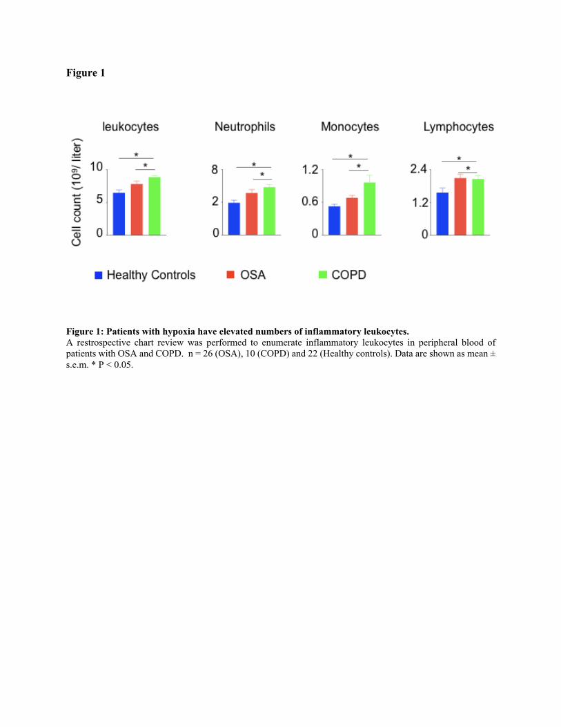
Figure 1
Figure 1: Patients with hypoxia have elevated numbers of inflammatory leukocytes.
A restrospective chart review was performed to enumerate inflammatory leukocytes in peripheral blood of
patients with OSA and COPD. n = 26 (OSA), 10 (COPD) and 22 (Healthy controls). Data are shown as mean ±
s.e.m. * P < 0.05.

Figure 2
Figure 2: Hypoxia induces inflammation
C57BL/6 mice were placed under either normoxic or hypoxic conditions for 21 days. Lungs, blood and bone
marrow were collected. A) Il1b, Il6, Il18 and Tnfa expression was assessed by RT-qPCR. B) IL-6 protein
expression was evaluated in whole lung and serum by ELISA. C) The numbers of blood monocytes and
lymphocytes in hypoxic versus normoxic mice were determined by flow cytometry. D) The numbers of lung
infiltrating neutrophils and interstitial macrophages in these mice were quantified by flow cytometry. n = 5 mice
per condition. Data are shown as mean ± s.e.m. * P < 0.05, ** P<0.01, *** P<0.005, **** P<0.001.

Figure 3
Figure 3: Hypoxia drives HSPC into the cell cycle
C57BL/6 mice were placed under either normoxic or hypoxic conditions for 21 days. Bone marrow was
collected. The frequencies and numbers of the hematopoietic progenitors in the bone marrow were determined
by flow cytometry (A) and whole mount confocal microscopy (B). The frequencies of HSC (C), LSK (D) and
GMP (E) in the cell cycle stages were assessed using intracellular staining of Ki-67 and PI. (F) KikGR-GFP+
HSPC were adoptively transferred into mice housed in either normoxic or hypoxic conditions for seven days.
GFP+ progenies were quantified four days later. n = 5 mice per condition. Data are shown as mean ± s.e.m. * P
< 0.05, ** P<0.01, *** P<0.005, **** P<0.001.

Figure 4
Figure 4: VEGFr expression is increased in HSPC in hypoxia
A-C) C57BL/6 mice were placed under either normoxic or hypoxic conditions for 21 days. Bone marrow was
collected. A) Vegfa and Vegfr expression in the whole bone marrow was assessed by RT-qPCR. B) Vegfr
expression was measured in bone marrow LSK and GMP by flow cytometry. The results are represented as mean
fluorescent intensity (MFI). C) Vegfr gene expression was measured in sorted HSC and GMP by RT-qPCR. D-
F) HSPC were sorted from bone marrow of C57BL/6 mice and cultured in complete SFEM media in either
normoxic or hypoxic conditions. D) Heatmap representing expression of genes downstream to Vegf/Vegfr in
HSPC cultured under normoxic and hypoxic conditions. E) Schematic depicting HIF-1⍺ binding sites on both
murine and human VEGFr promoter regions. F) Confirmation of the binding of HIF-1⍺ to the murine Vegfr
promoter region assessed by ChIP qPCR. G-H) VEGFr expression assessed in human (G) and murine (H) HSPC
transfected with either siCTL or siHIF1A in hypoxic conditions. I) Heatmap showing the expression of the cell
cycle genes in HSPC transfected with either siCtl or siHif1a in hypoxic conditions. n = 5 mice per condition
(A-C), 5 replicates per condition (D-I). Data are shown as mean ± s.e.m. * P < 0.05, ** P<0.01, **** P<0.001.

Figure 5
Figure 5: Vegfr inhibition decreased hematopoietic progenitor proliferation in hypoxia
A) Heatmap depicting expression of the cell cycle genes in mouse HSPC treated with increasing amounts of a
Vegfr inhibitor (sugen, 10-100nM) or vehicle. B-D) C57BL/6 mice were treated with either sugen or vehicle
diluent and placed in either normoxia or hypoxia for 21 days. Bone marrow cells were analyzed using flow
cytometry to assess GMP proliferation (B&C) and number (D). n = 5 replicates or mice per condition. Data are
shown as mean ± s.e.m. * P < 0.05, *** P<0.001, **** P<0.001.
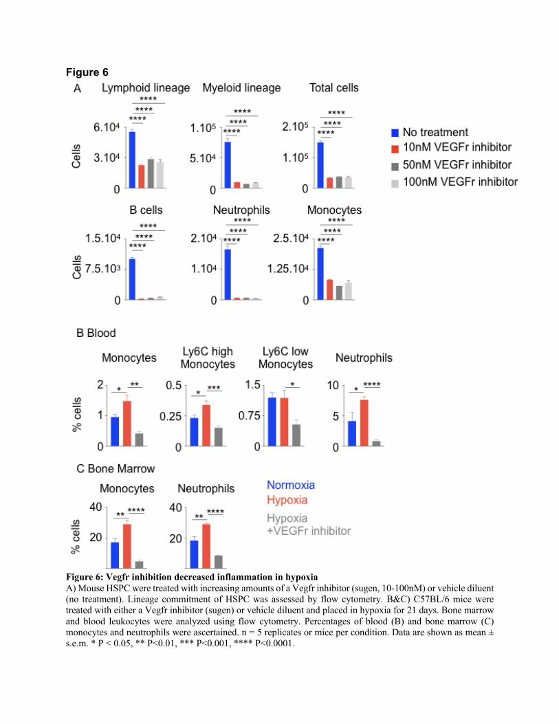
Figure 6
Figure 6: Vegfr inhibition decreased inflammation in hypoxia
A) Mouse HSPC were treated with increasing amounts of a Vegfr inhibitor (sugen, 10-100nM) or vehicle diluent
(no treatment). Lineage commitment of HSPC was assessed by flow cytometry. B&C) C57BL/6 mice were
treated with either a Vegfr inhibitor (sugen) or vehicle diluent and placed in hypoxia for 21 days. Bone marrow
and blood leukocytes were analyzed using flow cytometry. Percentages of blood (B) and bone marrow (C)
monocytes and neutrophils were ascertained. n = 5 replicates or mice per condition. Data are shown as mean ±
s.e.m. * P < 0.05, ** P<0.01, *** P<0.001, **** P<0.0001.


Supplementary Files
This is a list of supplementary �les associated with this preprint. Click to download.
20211020Supplementalmaterial.pdf