vegetable hairs associated with a sphenoid sinus ... · literature review in pubmed. ... oliveira...
TRANSCRIPT
Central Annals of Otolaryngology and Rhinology
Cite this article: Oddó D, Méndez GP, Villanueva P, Huete I, Callejas C, et al. (2017) Vegetable Hairs Associated with a Sphenoid Sinus Aspergilloma. A Case Report. Ann Otolaryngol Rhinol 4(1): 1159.
*Corresponding author
David Oddó, Department of Anatomic Pathology, Faculty of Medicine, Pontificia Universidad Católica de Chile; Chile, Email:
Submitted: 30 December 2016
Accepted: 25 January 2017
Published: 27 January 2017
ISSN: 2379-948X
Copyright© 2017 Oddó et al.
OPEN ACCESS
Keywords• Aspergilloma• Paranasal sinuses• Sphenoidal sinus• Vegetable foreign body• Vegetable hairs
Case Report
Vegetable Hairs Associated with a Sphenoid Sinus Aspergilloma. A Case ReportDavid Oddó1*, Gonzalo P. Méndez1, Pablo Villanueva2, Isidro Huete3, Claudio Callejas4, and José Lorenzoni2
1Department of Anatomic Pathology, Pontificia Universidad Católica de Chile, Chile2Department of Neurosurgery, Pontificia Universidad Católica de Chile, Chile3Department of Radiology, Pontificia Universidad Católica de Chile, Chile4Department of Otorhinolaryngology, Pontificia Universidad Católica de Chile, Chile
Abstract
Foreign vegetable material is an extremely rare finding in biopsies, especially from the upper respiratory tract. The frequency of paranasal fungal balls has shown an increase during last years, with the consequent higher number of biopsies performed and available for analysis. We present the case of a 50 year-old patient with an aspergilloma of the sphenoidal sinus that also revealed foreign material consistent with vegetable hairs. Their particular structure observed by histological analysis allowed the correct recognition of this very unusual foreign body of the upper respiratory tract.
INTRODUCTIONThe finding of vegetable foreign material in biopsies from
upper respiratory tract is very infrequent, with most of the cases been described in other organs. The vegetal material can be present in the thickness of the tissue itself or in natural lumina. It can trigger the primary inflammatory process of the tissue or to be the secondary orsuperimposed pathological event. Although the morphology of the vegetable material is rich and varied, the related publications have only described plant epidermis and hyaline rings in the biopsies, but not vegetal hairs as the primary foreign material.
We present a case of vegetable hairs associated with a fungal ball, which its particular morphology has not been described before in biopsies from upper respiratory tract.
CASE PRESENTATIONThis is a 50 year old female patient with no significant
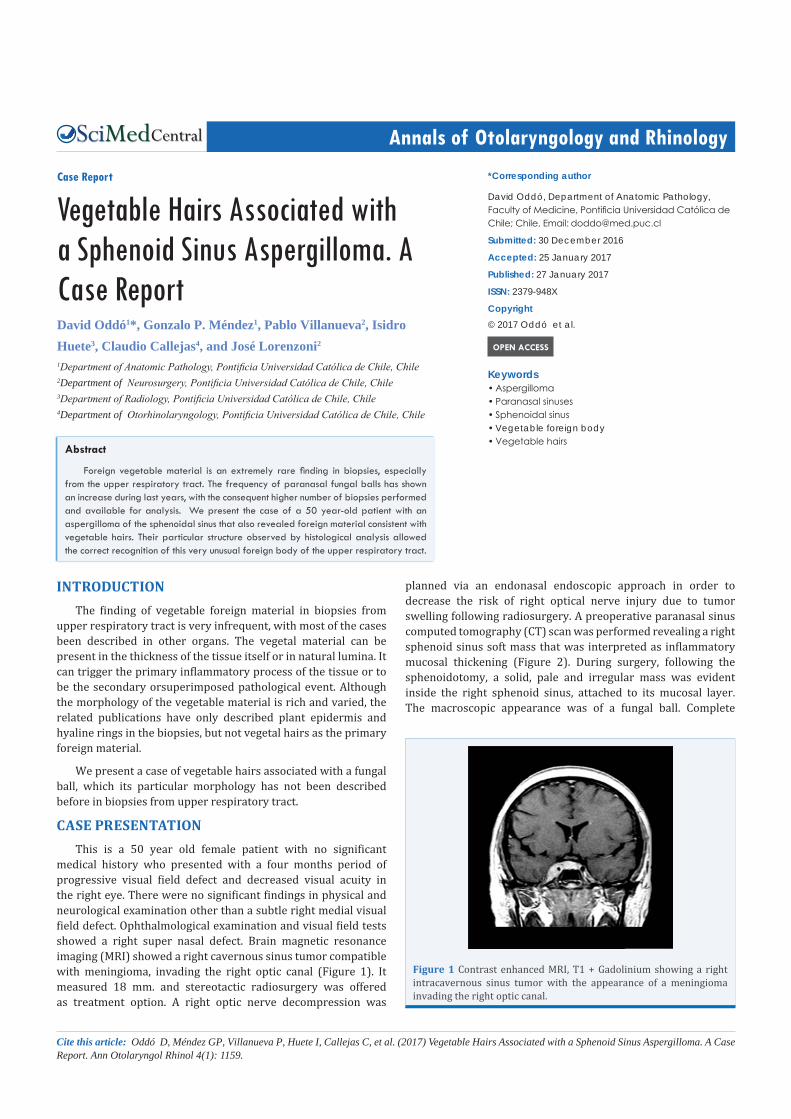
medical history who presented with a four months period of progressive visual field defect and decreased visual acuity in the right eye. There were no significant findings in physical and neurological examination other than a subtle right medial visual field defect. Ophthalmological examination and visual field tests showed a right super nasal defect. Brain magnetic resonance imaging (MRI) showed a right cavernous sinus tumor compatible with meningioma, invading the right optic canal (Figure 1). It measured 18 mm. and stereotactic radiosurgery was offered as treatment option. A right optic nerve decompression was
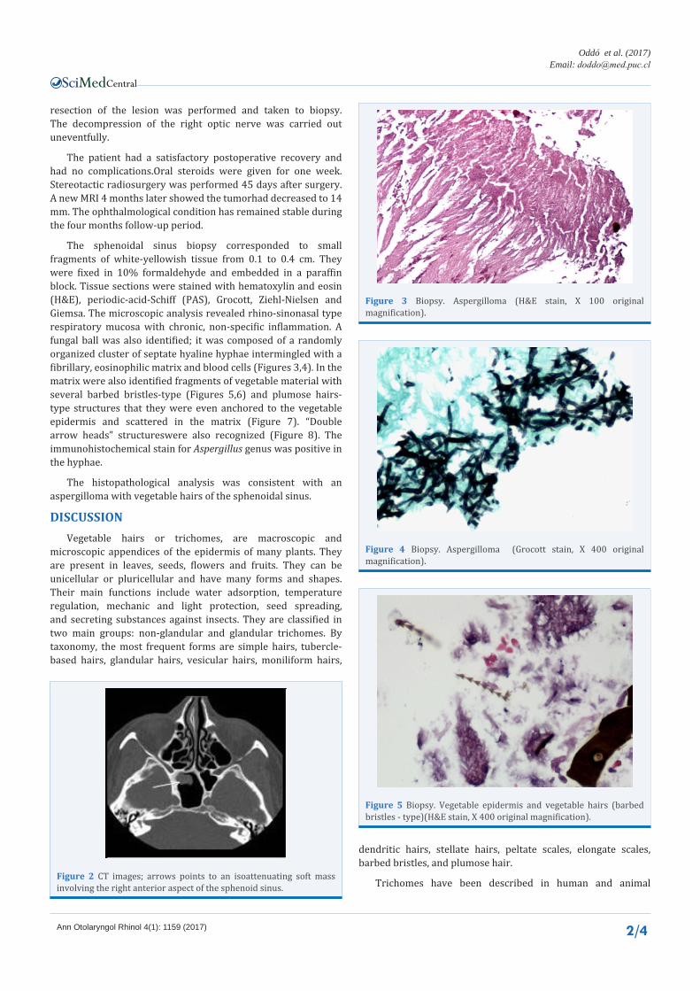
planned via an endonasal endoscopic approach in order to decrease the risk of right optical nerve injury due to tumor swelling following radiosurgery. A preoperative paranasal sinus computed tomography (CT) scan was performed revealing a right sphenoid sinus soft mass that was interpreted as inflammatory mucosal thickening (Figure 2). During surgery, following the sphenoidotomy, a solid, pale and irregular mass was evident inside the right sphenoid sinus, attached to its mucosal layer. The macroscopic appearance was of a fungal ball. Complete
Figure 1 Contrast enhanced MRI, T1 + Gadolinium showing a right intracavernous sinus tumor with the appearance of a meningioma invading the right optic canal.
Central
Oddó et al. (2017)Email:
Ann Otolaryngol Rhinol 4(1): 1159 (2017) 2/4
resection of the lesion was performed and taken to biopsy. The decompression of the right optic nerve was carried out uneventfully.
The patient had a satisfactory postoperative recovery and had no complications.Oral steroids were given for one week. Stereotactic radiosurgery was performed 45 days after surgery. A new MRI 4 months later showed the tumorhad decreased to 14 mm. The ophthalmological condition has remained stable during the four months follow-up period.
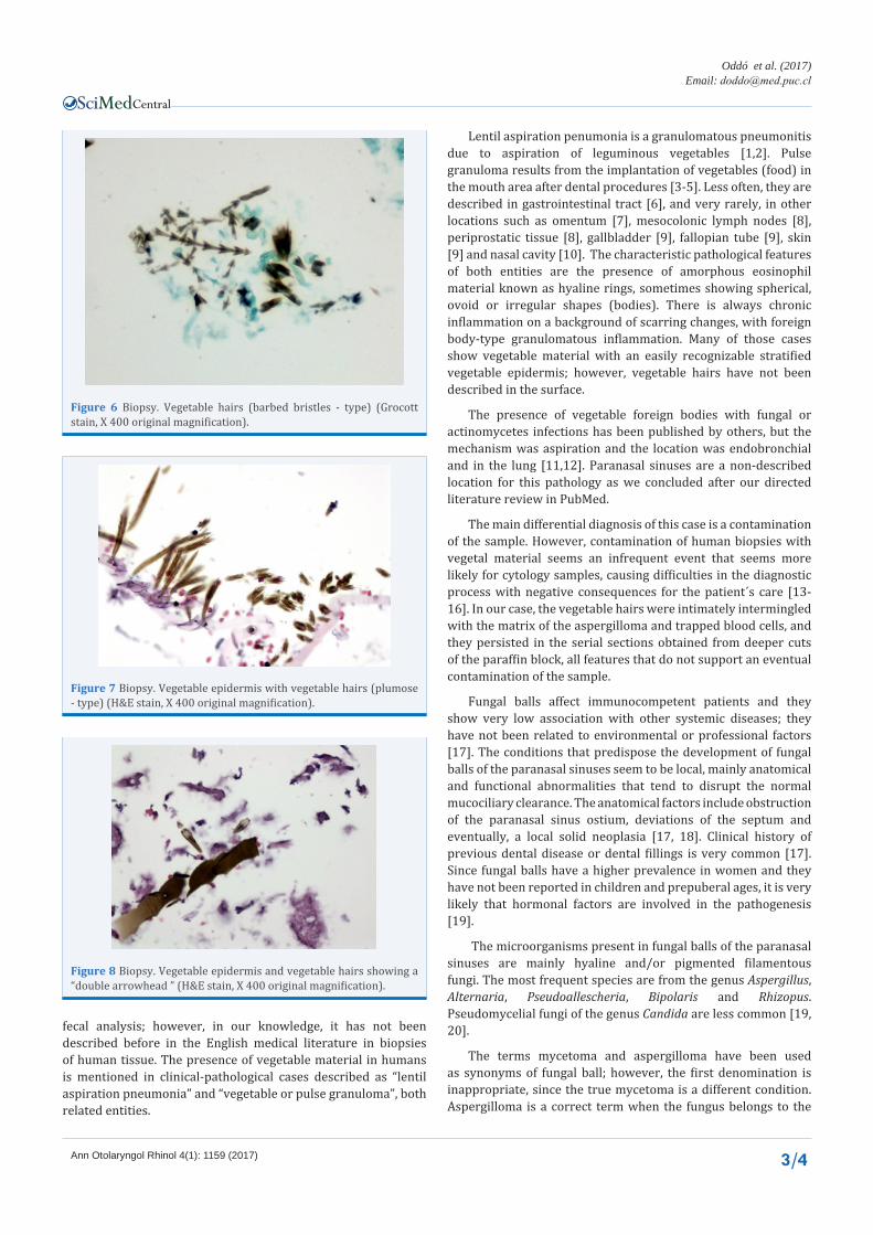
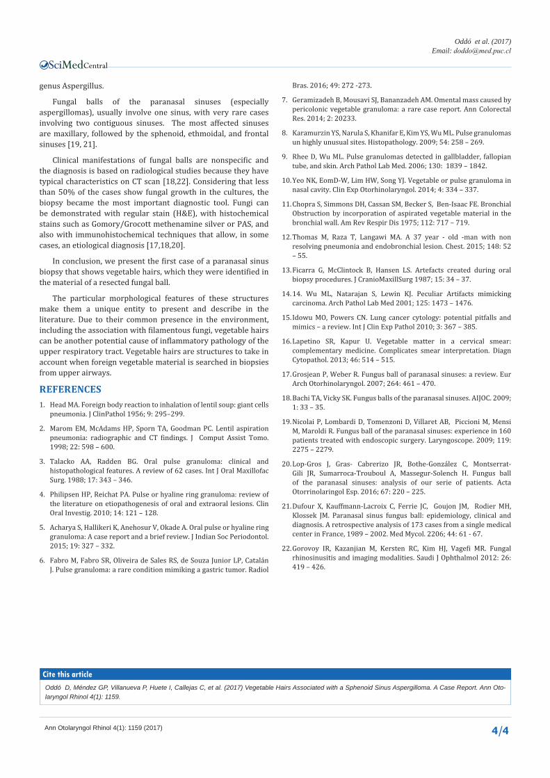
The sphenoidal sinus biopsy corresponded to small fragments of white-yellowish tissue from 0.1 to 0.4 cm. They were fixed in 10% formaldehyde and embedded in a paraffin block. Tissue sections were stained with hematoxylin and eosin (H&E), periodic-acid-Schiff (PAS), Grocott, Ziehl-Nielsen and Giemsa. The microscopic analysis revealed rhino-sinonasal type respiratory mucosa with chronic, non-specific inflammation. A fungal ball was also identified; it was composed of a randomly organized cluster of septate hyaline hyphae intermingled with a fibrillary, eosinophilic matrix and blood cells (Figures 3,4). In the matrix were also identified fragments of vegetable material with several barbed bristles-type (Figures 5,6) and plumose hairs-type structures that they were even anchored to the vegetable epidermis and scattered in the matrix (Figure 7). “Double arrow heads” structureswere also recognized (Figure 8). The immunohistochemical stain for Aspergillus genus was positive in the hyphae.
The histopathological analysis was consistent with an aspergilloma with vegetable hairs of the sphenoidal sinus.
DISCUSSIONVegetable hairs or trichomes, are macroscopic and
microscopic appendices of the epidermis of many plants. They are present in leaves, seeds, flowers and fruits. They can be unicellular or pluricellular and have many forms and shapes. Their main functions include water adsorption, temperature regulation, mechanic and light protection, seed spreading, and secreting substances against insects. They are classified in two main groups: non-glandular and glandular trichomes. By taxonomy, the most frequent forms are simple hairs, tubercle-based hairs, glandular hairs, vesicular hairs, moniliform hairs,
Figure 2 CT images; arrows points to an isoattenuating soft mass involving the right anterior aspect of the sphenoid sinus.
Figure 3 Biopsy. Aspergilloma (H&E stain, X 100 original magnification).
Figure 4 Biopsy. Aspergilloma (Grocott stain, X 400 original magnification).
Figure 5 Biopsy. Vegetable epidermis and vegetable hairs (barbed bristles - type)(H&E stain, X 400 original magnification).
dendritic hairs, stellate hairs, peltate scales, elongate scales, barbed bristles, and plumose hair.
Trichomes have been described in human and animal
Central
Oddó et al. (2017)Email:
Ann Otolaryngol Rhinol 4(1): 1159 (2017) 3/4
fecal analysis; however, in our knowledge, it has not been described before in the English medical literature in biopsies of human tissue. The presence of vegetable material in humans is mentioned in clinical-pathological cases described as “lentil aspiration pneumonia” and “vegetable or pulse granuloma”, both related entities.
Lentil aspiration penumonia is a granulomatous pneumonitis due to aspiration of leguminous vegetables [1,2]. Pulse granuloma results from the implantation of vegetables (food) in the mouth area after dental procedures [3-5]. Less often, they are described in gastrointestinal tract [6], and very rarely, in other locations such as omentum [7], mesocolonic lymph nodes [8], periprostatic tissue [8], gallbladder [9], fallopian tube [9], skin [9] and nasal cavity [10]. The characteristic pathological features of both entities are the presence of amorphous eosinophil material known as hyaline rings, sometimes showing spherical, ovoid or irregular shapes (bodies). There is always chronic inflammation on a background of scarring changes, with foreign body-type granulomatous inflammation. Many of those cases show vegetable material with an easily recognizable stratified vegetable epidermis; however, vegetable hairs have not been described in the surface.
The presence of vegetable foreign bodies with fungal or actinomycetes infections has been published by others, but the mechanism was aspiration and the location was endobronchial and in the lung [11,12]. Paranasal sinuses are a non-described location for this pathology as we concluded after our directed literature review in PubMed.
The main differential diagnosis of this case is a contamination of the sample. However, contamination of human biopsies with vegetal material seems an infrequent event that seems more likely for cytology samples, causing difficulties in the diagnostic process with negative consequences for the patient´s care [13-16]. In our case, the vegetable hairs were intimately intermingled with the matrix of the aspergilloma and trapped blood cells, and they persisted in the serial sections obtained from deeper cuts of the paraffin block, all features that do not support an eventual contamination of the sample.
Fungal balls affect immunocompetent patients and they show very low association with other systemic diseases; they have not been related to environmental or professional factors [17]. The conditions that predispose the development of fungal balls of the paranasal sinuses seem to be local, mainly anatomical and functional abnormalities that tend to disrupt the normal mucociliary clearance. The anatomical factors include obstruction of the paranasal sinus ostium, deviations of the septum and eventually, a local solid neoplasia [17, 18]. Clinical history of previous dental disease or dental fillings is very common [17]. Since fungal balls have a higher prevalence in women and they have not been reported in children and prepuberal ages, it is very likely that hormonal factors are involved in the pathogenesis [19].
The microorganisms present in fungal balls of the paranasal sinuses are mainly hyaline and/or pigmented filamentous fungi. The most frequent species are from the genus Aspergillus, Alternaria, Pseudoallescheria, Bipolaris and Rhizopus. Pseudomycelial fungi of the genus Candida are less common [19, 20].
The terms mycetoma and aspergilloma have been used as synonyms of fungal ball; however, the first denomination is inappropriate, since the true mycetoma is a different condition. Aspergilloma is a correct term when the fungus belongs to the
Figure 6 Biopsy. Vegetable hairs (barbed bristles - type) (Grocott stain, X 400 original magnification).
Figure 7 Biopsy. Vegetable epidermis with vegetable hairs (plumose - type) (H&E stain, X 400 original magnification).
Figure 8 Biopsy. Vegetable epidermis and vegetable hairs showing a “double arrowhead ” (H&E stain, X 400 original magnification).
Central
Oddó et al. (2017)Email:
Ann Otolaryngol Rhinol 4(1): 1159 (2017) 4/4
Oddó D, Méndez GP, Villanueva P, Huete I, Callejas C, et al. (2017) Vegetable Hairs Associated with a Sphenoid Sinus Aspergilloma. A Case Report. Ann Oto-laryngol Rhinol 4(1): 1159.
Cite this article
genus Aspergillus.
Fungal balls of the paranasal sinuses (especially aspergillomas), usually involve one sinus, with very rare cases involving two contiguous sinuses. The most affected sinuses are maxillary, followed by the sphenoid, ethmoidal, and frontal sinuses [19, 21].
Clinical manifestations of fungal balls are nonspecific and the diagnosis is based on radiological studies because they have typical characteristics on CT scan [18,22]. Considering that less than 50% of the cases show fungal growth in the cultures, the biopsy became the most important diagnostic tool. Fungi can be demonstrated with regular stain (H&E), with histochemical stains such as Gomory/Grocott methenamine silver or PAS, and also with immunohistochemical techniques that allow, in some cases, an etiological diagnosis [17,18,20].
In conclusion, we present the first case of a paranasal sinus biopsy that shows vegetable hairs, which they were identified in the material of a resected fungal ball.
The particular morphological features of these structures make them a unique entity to present and describe in the literature. Due to their common presence in the environment, including the association with filamentous fungi, vegetable hairs can be another potential cause of inflammatory pathology of the upper respiratory tract. Vegetable hairs are structures to take in account when foreign vegetable material is searched in biopsies from upper airways.
REFERENCES1. Head MA. Foreign body reaction to inhalation of lentil soup: giant cells
pneumonia. J ClinPathol 1956; 9: 295–299.
2. Marom EM, McAdams HP, Sporn TA, Goodman PC. Lentil aspiration pneumonia: radiographic and CT findings. J Comput Assist Tomo. 1998; 22: 598 – 600.
3. Talacko AA, Radden BG. Oral pulse granuloma: clinical and histopathological features. A review of 62 cases. Int J Oral Maxillofac Surg. 1988; 17: 343 – 346.
4. Philipsen HP, Reichat PA. Pulse or hyaline ring granuloma: review of the literature on etiopathogenesis of oral and extraoral lesions. Clin Oral Investig. 2010; 14: 121 – 128.
5. Acharya S, Hallikeri K, Anehosur V, Okade A. Oral pulse or hyaline ring granuloma: A case report and a brief review. J Indian Soc Periodontol. 2015; 19: 327 – 332.
6. Fabro M, Fabro SR, Oliveira de Sales RS, de Souza Junior LP, Catalán J. Pulse granuloma: a rare condition mimiking a gastric tumor. Radiol
Bras. 2016; 49: 272 -273.
7. Geramizadeh B, Mousavi SJ, Bananzadeh AM. Omental mass caused by pericolonic vegetable granuloma: a rare case report. Ann Colorectal Res. 2014; 2: 20233.
8. Karamurzin YS, Narula S, Khanifar E, Kim YS, Wu ML. Pulse granulomas un highly unusual sites. Histopathology. 2009; 54: 258 – 269.
9. Rhee D, Wu ML. Pulse granulomas detected in gallbladder, fallopian tube, and skin. Arch Pathol Lab Med. 2006; 130: 1839 – 1842.
10. Yeo NK, EomD-W, Lim HW, Song YJ. Vegetable or pulse granuloma in nasal cavity. Clin Exp Otorhinolaryngol. 2014; 4: 334 – 337.
11. Chopra S, Simmons DH, Cassan SM, Becker S, Ben-Isaac FE. Bronchial Obstruction by incorporation of aspirated vegetable material in the bronchial wall. Am Rev Respir Dis 1975; 112: 717 – 719.
12. Thomas M, Raza T, Langawi MA. A 37 year - old -man with non resolving pneumonia and endobronchial lesion. Chest. 2015; 148: 52 – 55.
13. Ficarra G, McClintock B, Hansen LS. Artefacts created during oral biopsy procedures. J CranioMaxillSurg 1987; 15: 34 – 37.
14. 14. Wu ML, Natarajan S, Lewin KJ. Peculiar Artifacts mimicking carcinoma. Arch Pathol Lab Med 2001; 125: 1473 – 1476.
15. Idowu MO, Powers CN. Lung cancer cytology: potential pitfalls and mimics – a review. Int J Clin Exp Pathol 2010; 3: 367 – 385.
16. Lapetino SR, Kapur U. Vegetable matter in a cervical smear: complementary medicine. Complicates smear interpretation. Diagn Cytopathol. 2013; 46: 514 – 515.
17. Grosjean P, Weber R. Fungus ball of paranasal sinuses: a review. Eur Arch Otorhinolaryngol. 2007; 264: 461 – 470.
18. Bachi TA, Vicky SK. Fungus balls of the paranasal sinuses. AIJOC. 2009; 1: 33 – 35.
19. Nicolai P, Lombardi D, Tomenzoni D, Villaret AB, Piccioni M, Mensi M, Maroldi R. Fungus ball of the paranasal sinuses: experience in 160 patients treated with endoscopic surgery. Laryngoscope. 2009; 119: 2275 – 2279.
20. Lop-Gros J, Gras- Cabrerizo JR, Bothe-González C, Montserrat-Gili JR, Sumarroca-Trouboul A, Massegur-Solench H. Fungus ball of the paranasal sinuses: analysis of our serie of patients. Acta Otorrinolaringol Esp. 2016; 67: 220 – 225.
21. Dufour X, Kauffmann-Lacroix C, Ferrie JC, Goujon JM, Rodier MH, Klossek JM. Paranasal sinus fungus ball: epidemiology, clinical and diagnosis. A retrospective analysis of 173 cases from a single medical center in France, 1989 – 2002. Med Mycol. 2206; 44: 61 - 67.
22. Gorovoy IR, Kazanjian M, Kersten RC, Kim HJ, Vagefi MR. Fungal rhinosinusitis and imaging modalities. Saudi J Ophthalmol 2012: 26: 419 – 426.